Embed Size (px)
Citation preview

Feline Neoplasia
Carol Naranjo, LV, DACVP, DECVP, PhDIDEXX Laboratories

POST‐TRAUMATIC OCULAR SARCOMA
Feline post-traumatic ocular sarcoma
• 2nd most common primary intraocular tumor
• Chronically inflamed eyes
• Traumatic event (50% of cases)
– Hx: abnormal eye since adopted
– Latency period 7 y (2 months-10 years)
• Possible male predilection?

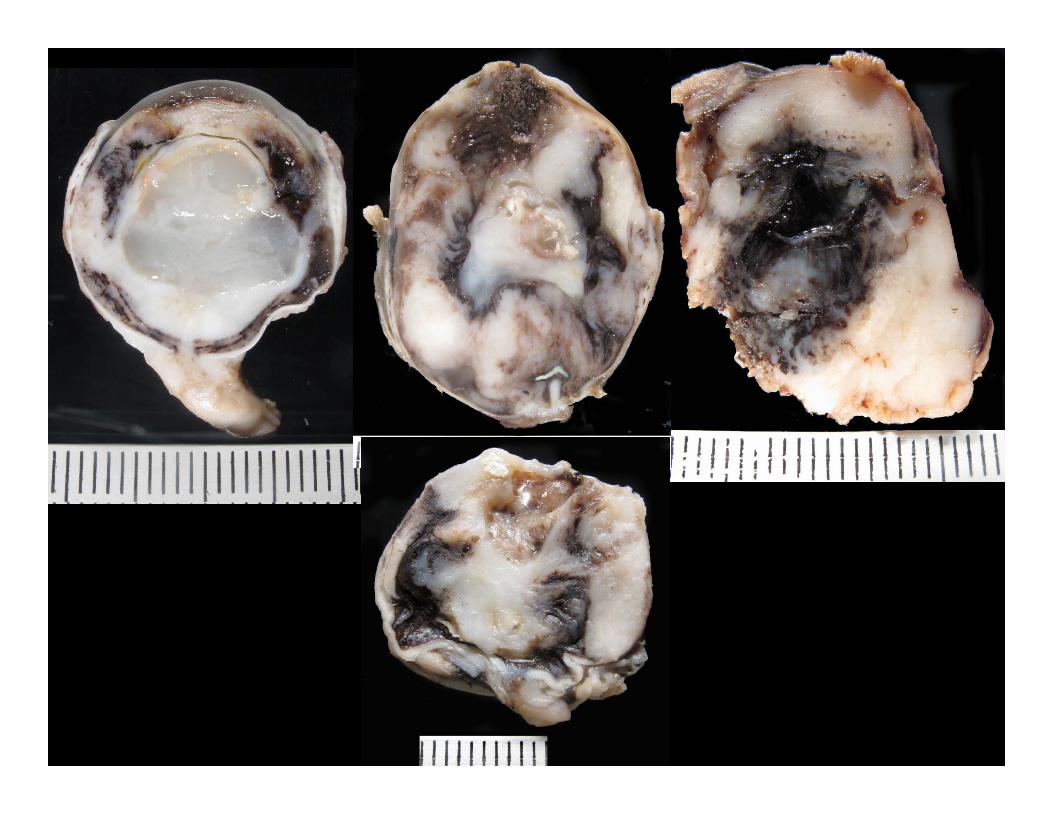
FPTOS – Spindle cell variant
• Most common variant, 70% of cases
• Thought to originate from LEC
• Gross:
– Cells lining the inner surface of the globe
– Progressively filling the globe
– Solid, firm, light tan mass
FPTOS – SCV – Histo
• Neoplastic cells line the inner aspect of globe
– Starting at lens equator/anterior vitreous
• Evolve to destroy most intraocular structures
• Fibrosarcoma to anaplastic sarcoma
• Lens capsule rupture
• PAS-positive membranes


FPTOS – SCV – IHC
• Vimentin +
• SMA + (20%)
• Cytokeratin (15%)
• Alpha A crystallin (30%)
• Collagen type IV in PAS-positive mb

Collagen IV
Vimentin A Crystallin
Zeiss et al., 2003

FPTOS – SCV – Prognosis
• Poor
– Extension beyond sclera (orbit): recurrences
– Invasion of ON or peripheral nerves (brain)
• Mets to distant sites

FPTOS – SCV
• LEC origin?
– Early tumors around lens equator
– Lens capsule rupture
– PAS +, collagen type IV basement mb • Lens capsule
– Alpha A crystallin +


Lens capsule rupture

Lens capsule rupture


FPTOS – Round cell variant
• Post-traumatic lymphoma
• Second-most common variant (24%)
• Grossly:
– Tendency to fill the globe (> lining the globe)
– Light tan and soft tumor


FPTOS – RCV – Histo
• Round cells, solid sheets
• Prominent survival around vessels
– Extensive coagulation necrosis
• High N:C ratio, prominent mitoses
• Frequent lens capsule rupture, not always
• LP infiltrate


FPTOS – RCV – IHC
• Variable (CD79+, CD3+ or both) • Clonality: B cell origin
Courtesy of Dr. Dubielzig (COPLOW)

FPTOS – RCV – DDx
• Different from “classic” lymphoma:
– Tendency to line the globe
– Lens capsule rupture
– Extensive necrosis
– History

FPTOS – RCV – Prognosis
• Hard to asses, but guarded to poor
• No clear association b/w histo and prognosis
• Local recurrence and systemic spread

FPTOS – OSA/Chondrosarcoma
• Least common variant (6%)
• Origin?
• Gross:
– Filling the globe solid

Courtesy of Dr. Dubielzig (COPLOW)
FPTOS – OSA/Chondrosarcoma – Gross

FPTOS – OSA/Chondrosarcoma – Histo
• Osteoid and/or chondroid matrix • Lens capsule rupture
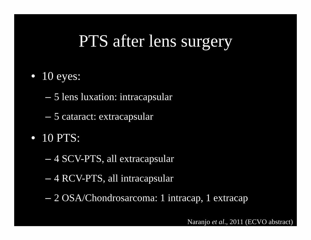
PTS after lens surgery
• 10 eyes:
– 5 lens luxation: intracapsular
– 5 cataract: extracapsular
• 10 PTS:
– 4 SCV-PTS, all extracapsular
– 4 RCV-PTS, all intracapsular
– 2 OSA/Chondrosarcoma: 1 intracap, 1 extracap
Naranjo et al., 2011 (ECVO abstract)

PTS after lens surgery
Courtesy of Dr. Dubielzig (COPLOW)

IRIDOCILIARY EPITHELIAL TUMORS

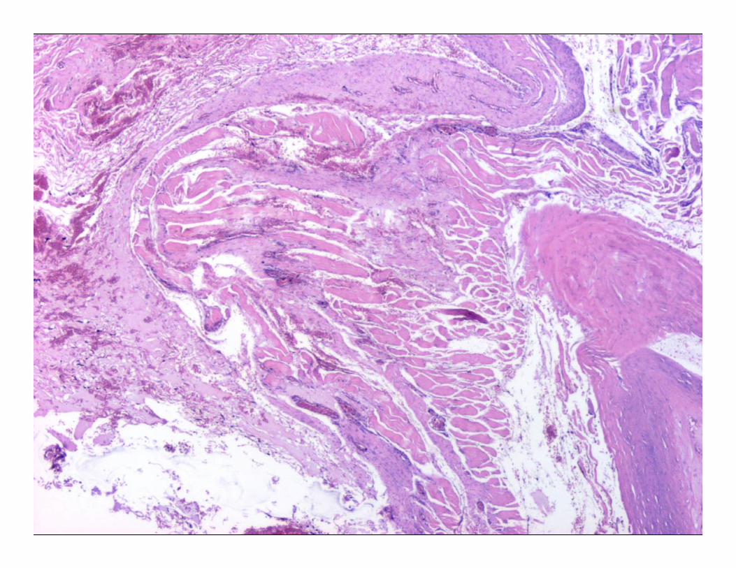
Feline iridociliary epithelial neoplasia
• 3rd most common primary intraocular neoplasm
• Gross:
– Discrete light tan mass in posterior chamber
– Sclera is rarely involved (adenoma > carcinoma)
– There may be cavitated areas within the tumor

Feline iridociliary epithelial neoplasia
• Solid sheets of monomorphic polygonal cells
• Delicate PAS-positive membranes
• Osteoid matrix or metaplastic bone


Courtesy of Dr. Dubielzig (COPLOW)
PAS

Feline iridociliary epithelial neoplasia
• NSE +
• Vimentin + (most of them)
• S100 and GFAP variably +
• Few are cytokeratin +
NSE
VimentinCourtesy of Dr. Dubielzig (COPLOW)

METASTATIC NEOPLASIA

Uveal lymphoma
• Most common metastatic tumor
• 48% cats w/ lymphoma may have ocular signs
(Nerschbach et al., 2013, no cytology or biopsy)
• Less commonly bilateral
• Not particularly associated w/ FIV/FeLV

Uveal lymphoma – Gross
• Anterior > posterior uvea
• Diffuse or discrete mass
– DDx. uveitis or FDIM (amelanotic)
• Light tan


FDIM vs lymphoma
• Round cell amelanotic melanoma
• Lymphoma:
– T cells: CD3
– B cells: CD20, CD79a, PAX5, etc.
• Melanoma: melan A, PNL2, TRP2, etc.
– Sensitivity/specificity… cocktail?

Courtesy of Dr. Dubielzig, COPLOW

Courtesy of Dr. Dubielzig, COPLOW

Courtesy of Dr. Dubielzig, COPLOW
LYMPHOMA!!!

• Lymphoma
• Pulmonary carcinomas
– “Lung-eye syndrome”
• Fibrosarcomas
• SCC
Metastatic tumors to the globe

• Uni- o bilat
• Choroid > anterior uvea
• Anterior uvea: neoplastic cells lining inner aspect
• Invasion of blood vessels: choroidal infarcts
• Orbit can be affected along with globe
Metastatic tumors to the globe


FELINE RESTRICTIVE ORBITAL MYOFIBROBLASTIC SARCOMA

FROMS
• Previously:
– Orbital pseudotumor
– Idiopathic sclerosing orbital pseudotumor
• BUT… features of a true neoplastic process!
– Progressive
– Poorly responsive to treatment
– Frequently leads to euthanasia

FROMS – Clinical
• Restriction of eye and eyelid movement
– Reduced retropulsion
– Lagophthalmos
– Ulcerative keratitis
– Thickened eyelids
• Exophthalmos, enophthalmos or normal position

FROMS – Gross
• Thickened orbital tissues, no mass
• Light tan to white tissue
• Thickening of:
– Eyelids
– Interorbital dermis
– Oral: palate, lips, maxillary gingiva
Bell et al., 2011

FROMS – Histo
• Poorly delineated
• Bland spindle cells w/ collagenous matrix
• Low mitotic activity
• Mild lymphoplasmacytic infiltrates
• Ulcerative keratitits (or sequestrum)

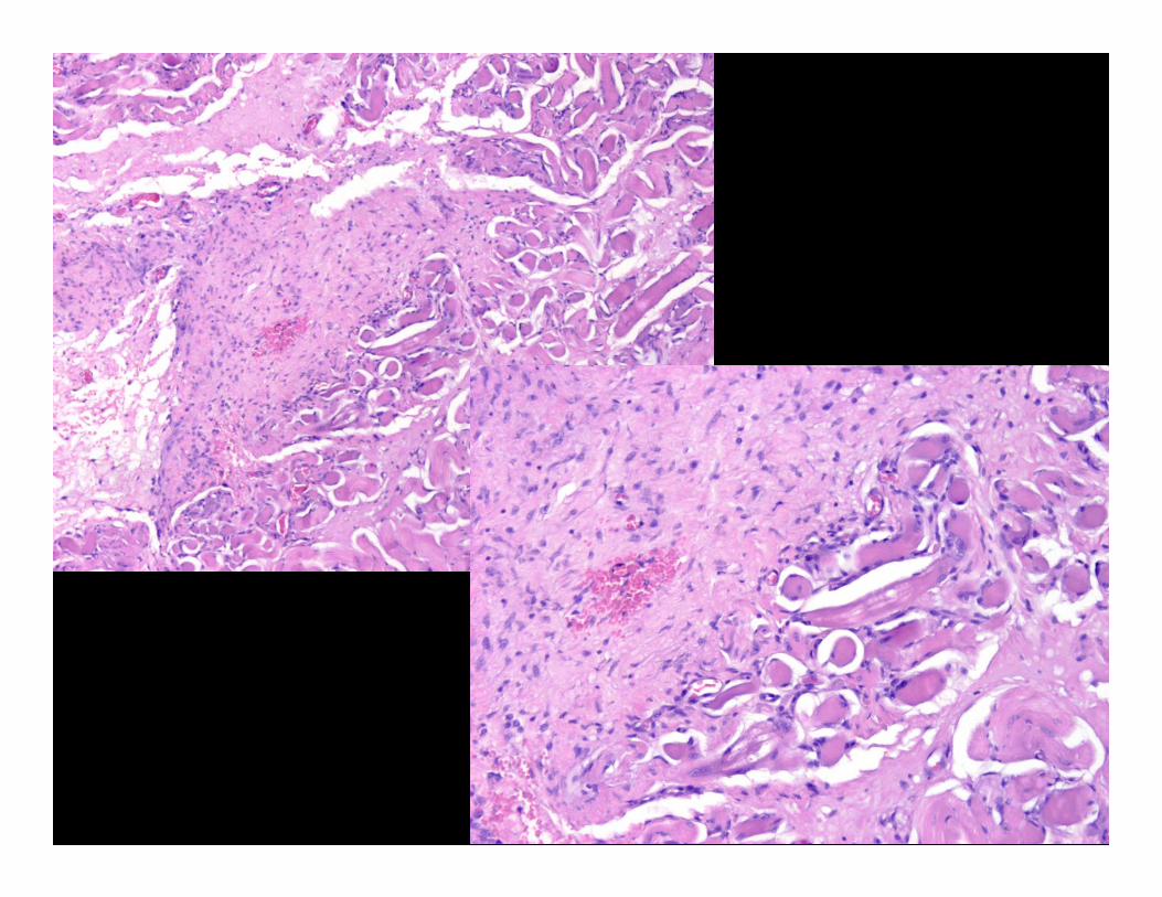


Masson’s trichrome

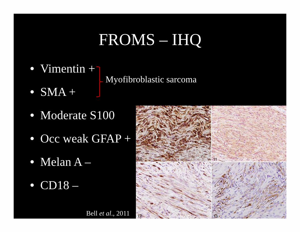
FROMS – IHQ
• Vimentin +
• SMA +
• Moderate S100
• Occ weak GFAP +
• Melan A –
• CD18 –
Myofibroblastic sarcoma
Bell et al., 2011
FROMS - DDx
• Reactive fibroplasia / Granulation tissue:
– History
– Do NOT trim the orbital tissues away from the globe
– Incisional Bx:
• Anterior and superior aspect of episclera
• Eyelid: thickened areas of palpebral conj near fornix
• Skin: thickened areas (deep dermis/SQ)

FROMS – Prognosis
• Poor
• Can spread to contralateral orbit bilateral – 0-8 months b/w eyes
– 0-14 months b/w presentation and oral involvement
• No mets reported
• Survival 3-15 months
Bell et al., 2011