Embed Size (px)
Citation preview
Ni Made HustriniDivisi Ginjal Hipertensi, Departemen Ilmu Penyakit Dalam
FKUI/RSCM - Jakarta
PENENTUAN REGIMEN PD
Contoh Kasus
Laki-laki, 28 tahun
CKD stage 5
Etiologi PGTA: MPGN
eGFR 6 ml/mnt/1,73m2
Diuresis 800 ml/24 jam. BB 60 kg
Dirujuk ke Poli PD untuk TPG
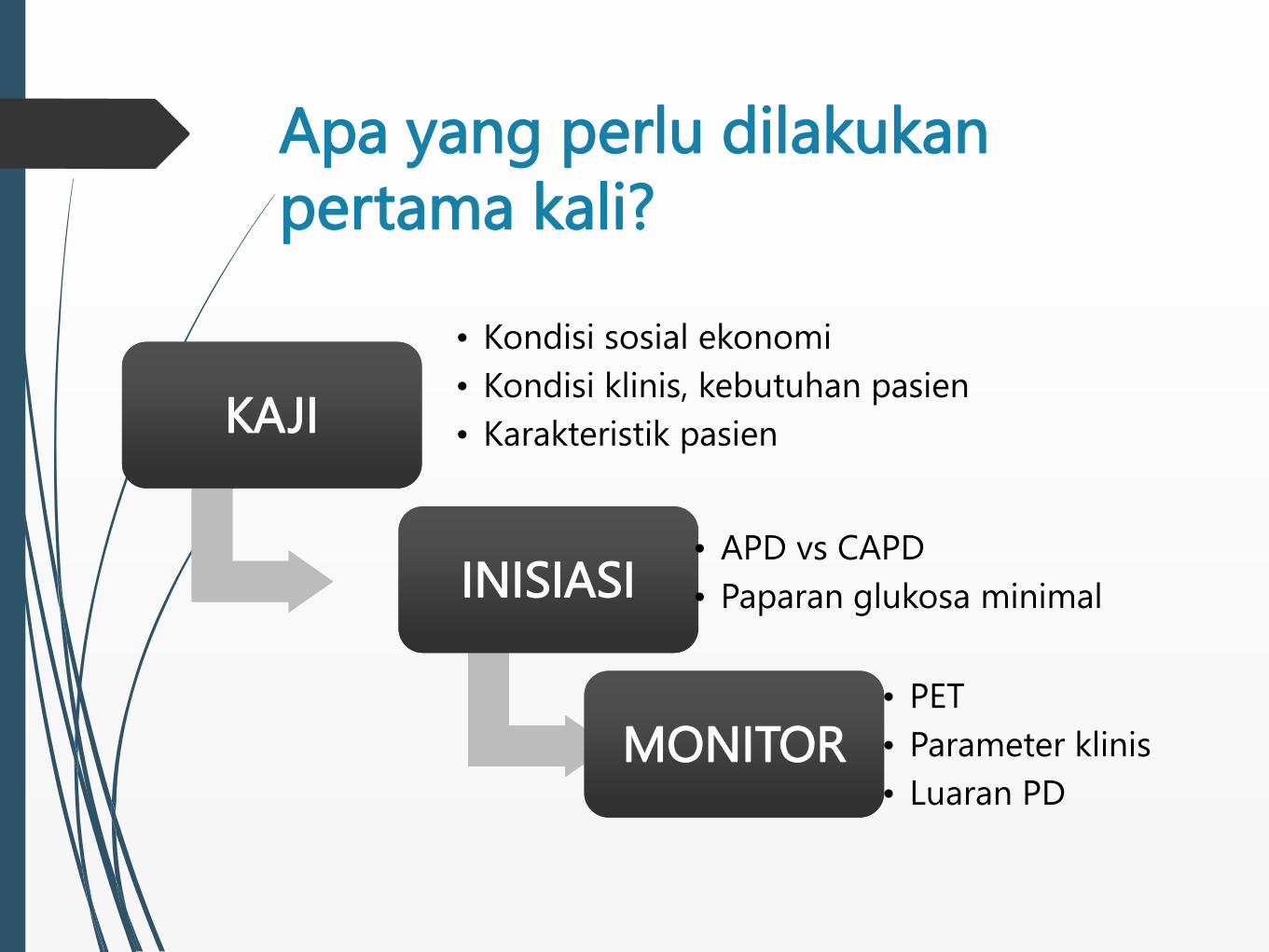
Apa yang perlu dilakukan
pertama kali?
KAJIKAJI
• Kondisi sosial ekonomi
• Kondisi klinis, kebutuhan pasien
• Karakteristik pasien
INISIASIINISIASI• APD vs CAPD
• Paparan glukosa minimal
MONITORMONITOR• PET
• Parameter klinis
• Luaran PD
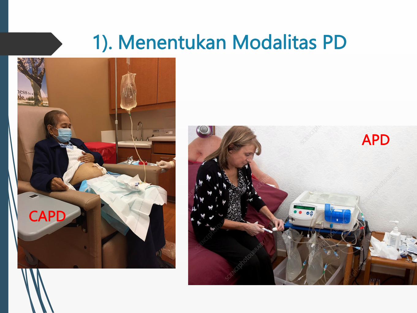
1). Menentukan Modalitas PD
CAPD
APD
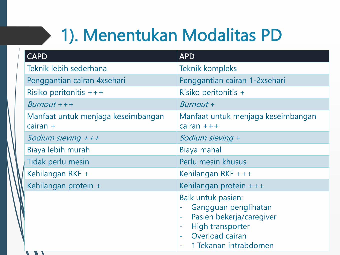
1). Menentukan Modalitas PDCAPD APD
Teknik lebih sederhana Teknik kompleks
Penggantian cairan 4xsehari Penggantian cairan 1-2xsehari
Risiko peritonitis +++ Risiko peritonitis +
Burnout +++ Burnout +
Manfaat untuk menjaga keseimbangan
cairan +
Manfaat untuk menjaga keseimbangan
cairan +++
Sodium sieving +++ Sodium sieving +
Biaya lebih murah Biaya mahal
Tidak perlu mesin Perlu mesin khusus
Kehilangan RKF + Kehilangan RKF +++
Kehilangan protein + Kehilangan protein +++
Baik untuk pasien:
- Gangguan penglihatan
- Pasien bekerja/caregiver
- High transporter
- Overload cairan
- ↑ Tekanan intrabdomen
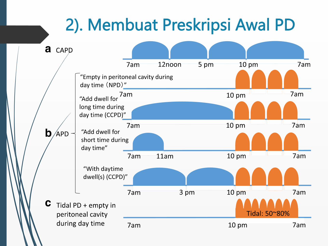
2). Membuat Preskripsi Awal PD
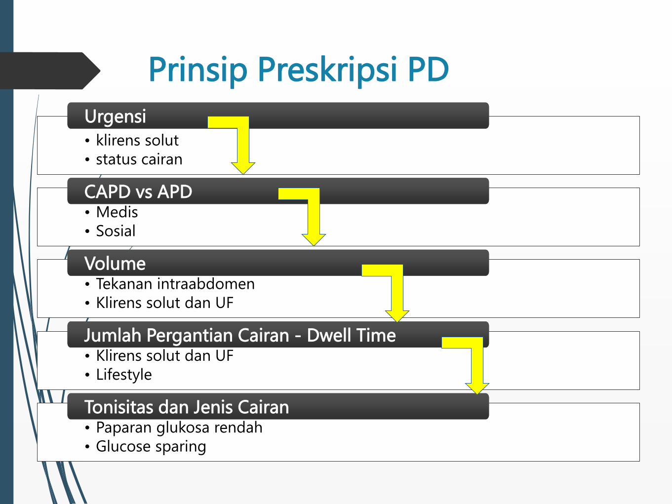
Prinsip Preskripsi PD
• klirens solut
• status cairan
UrgensiUrgensi
• Medis
• Sosial
CAPD vs APDCAPD vs APD
• Tekanan intraabdomen
• Klirens solut dan UF
VolumeVolume
• Klirens solut dan UF
• Lifestyle
Jumlah Pergantian Cairan - Dwell TimeJumlah Pergantian Cairan - Dwell Time
• Paparan glukosa rendah
• Glucose sparing
Tonisitas dan Jenis CairanTonisitas dan Jenis Cairan
3). Incremental PD (iPD)
Merupakan metode PD yang dilakukan pada pasien
yang masih memiliki fungsi ginjal sisa (RKF) untuk
mencapai target klirens solut diawali dengan
melakukan preskripsi dosis PD terendah.
eGFR 4-5 ml/menit/1,73 m2
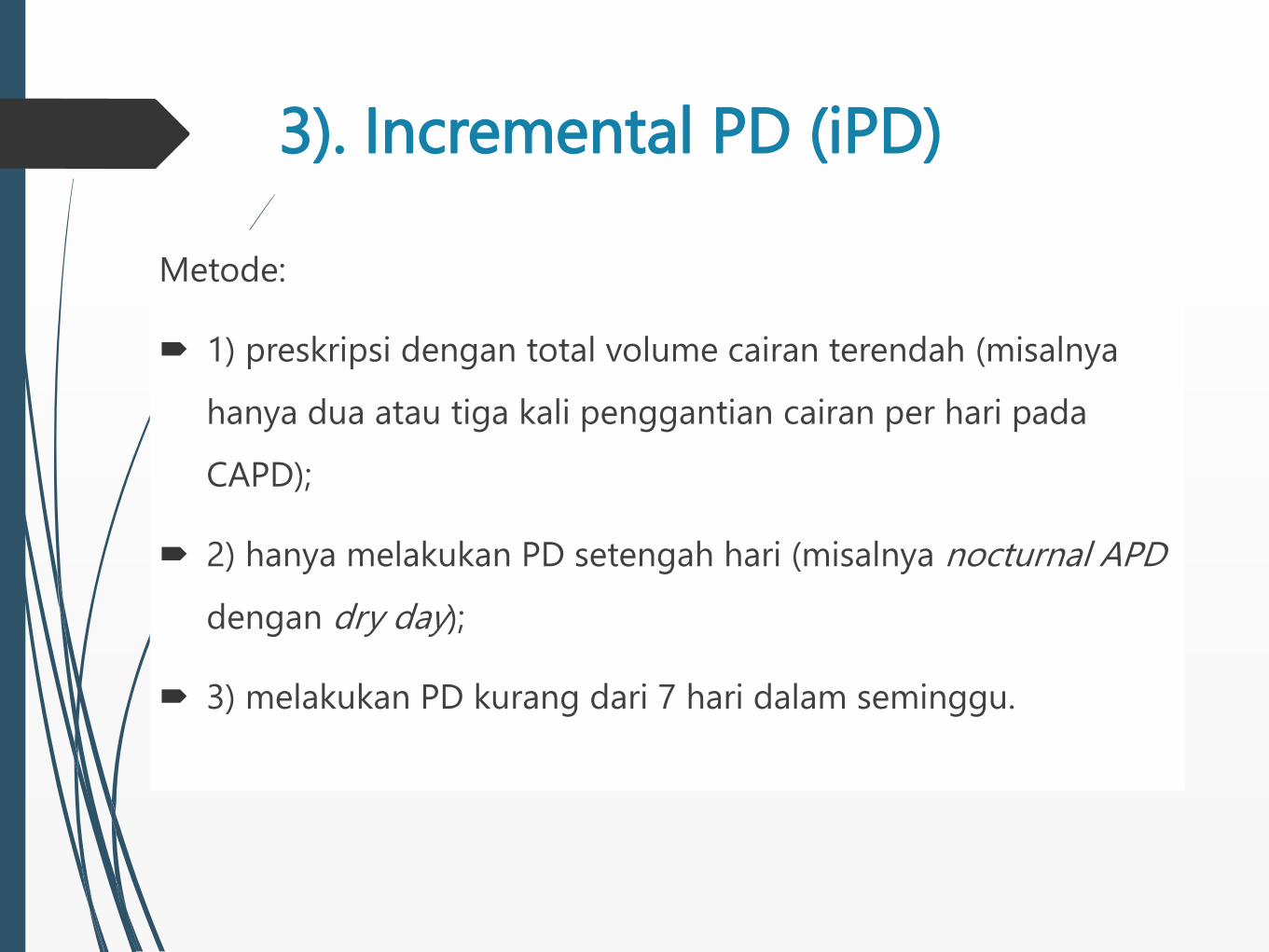
3). Incremental PD (iPD)
Metode:
1) preskripsi dengan total volume cairan terendah (misalnya
hanya dua atau tiga kali penggantian cairan per hari pada
CAPD);
2) hanya melakukan PD setengah hari (misalnya nocturnal APD
dengan dry day);
3) melakukan PD kurang dari 7 hari dalam seminggu.
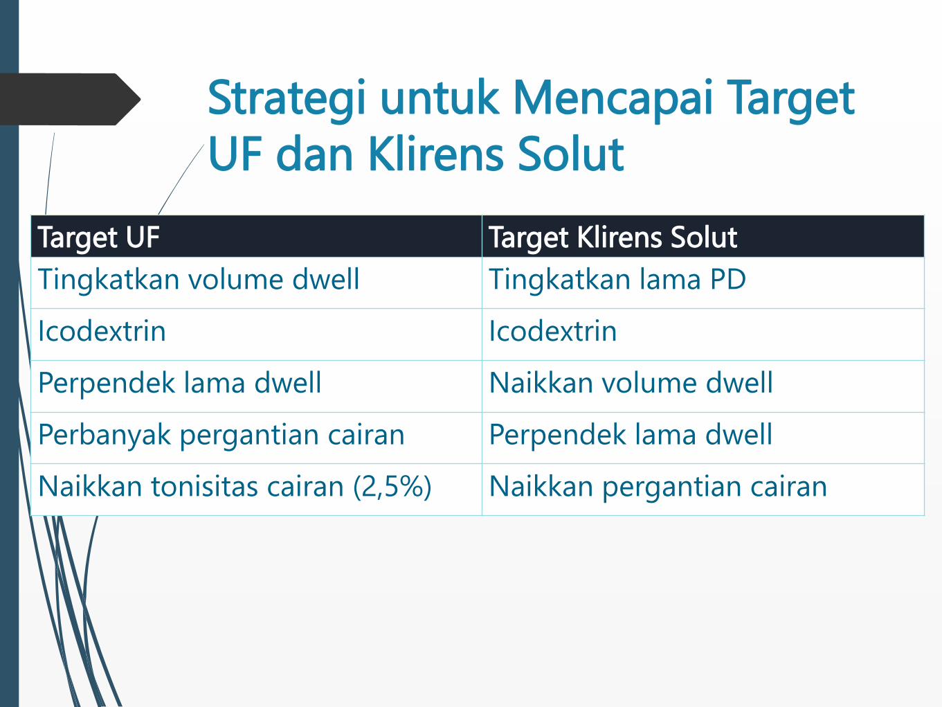
Strategi untuk Mencapai Target
UF dan Klirens Solut
Target UF Target Klirens Solut
Tingkatkan volume dwell Tingkatkan lama PD
Icodextrin Icodextrin
Perpendek lama dwell Naikkan volume dwell
Perbanyak pergantian cairan Perpendek lama dwell
Naikkan tonisitas cairan (2,5%) Naikkan pergantian cairan
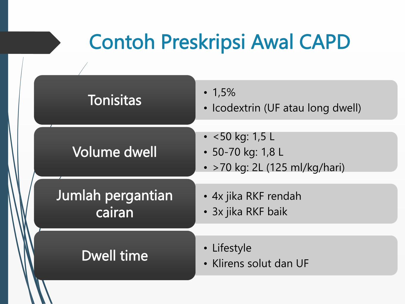
Contoh Preskripsi Awal CAPD
• 1,5%
• Icodextrin (UF atau long dwell)TonisitasTonisitas
• <50 kg: 1,5 L
• 50-70 kg: 1,8 L
• >70 kg: 2L (125 ml/kg/hari)
Volume dwellVolume dwell
• 4x jika RKF rendah
• 3x jika RKF baik
Jumlah pergantian
cairan
Jumlah pergantian
cairan
• Lifestyle
• Klirens solut dan UFDwell timeDwell time
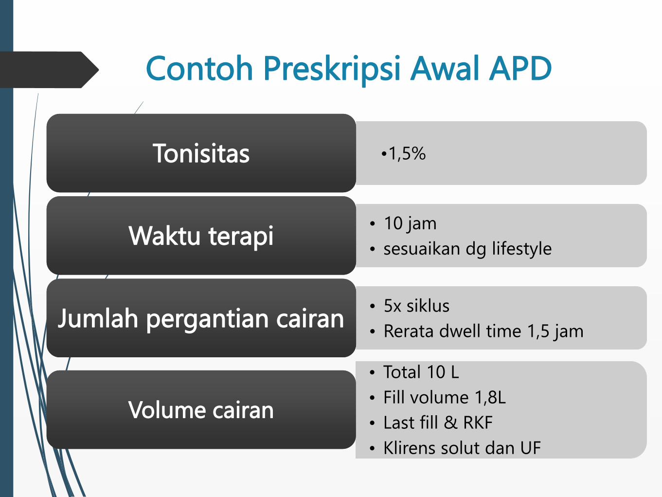
Contoh Preskripsi Awal APD
•1,5%TonisitasTonisitas
• 10 jam
• sesuaikan dg lifestyleWaktu terapiWaktu terapi
• 5x siklus
• Rerata dwell time 1,5 jamJumlah pergantian cairanJumlah pergantian cairan
• Total 10 L
• Fill volume 1,8L
• Last fill & RKF
• Klirens solut dan UF
Volume cairanVolume cairan
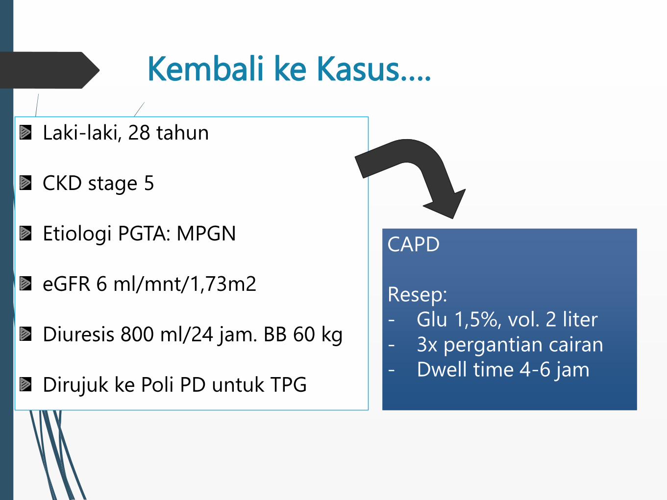
Kembali ke Kasus….
Laki-laki, 28 tahun
CKD stage 5
Etiologi PGTA: MPGN
eGFR 6 ml/mnt/1,73m2
Diuresis 800 ml/24 jam. BB 60 kg
Dirujuk ke Poli PD untuk TPG
CAPD
Resep:
- Glu 1,5%, vol. 2 liter
- 3x pergantian cairan
- Dwell time 4-6 jam
CAPD
Resep:
- Glu 1,5%, vol. 2 liter
- 3x pergantian cairan
- Dwell time 4-6 jam
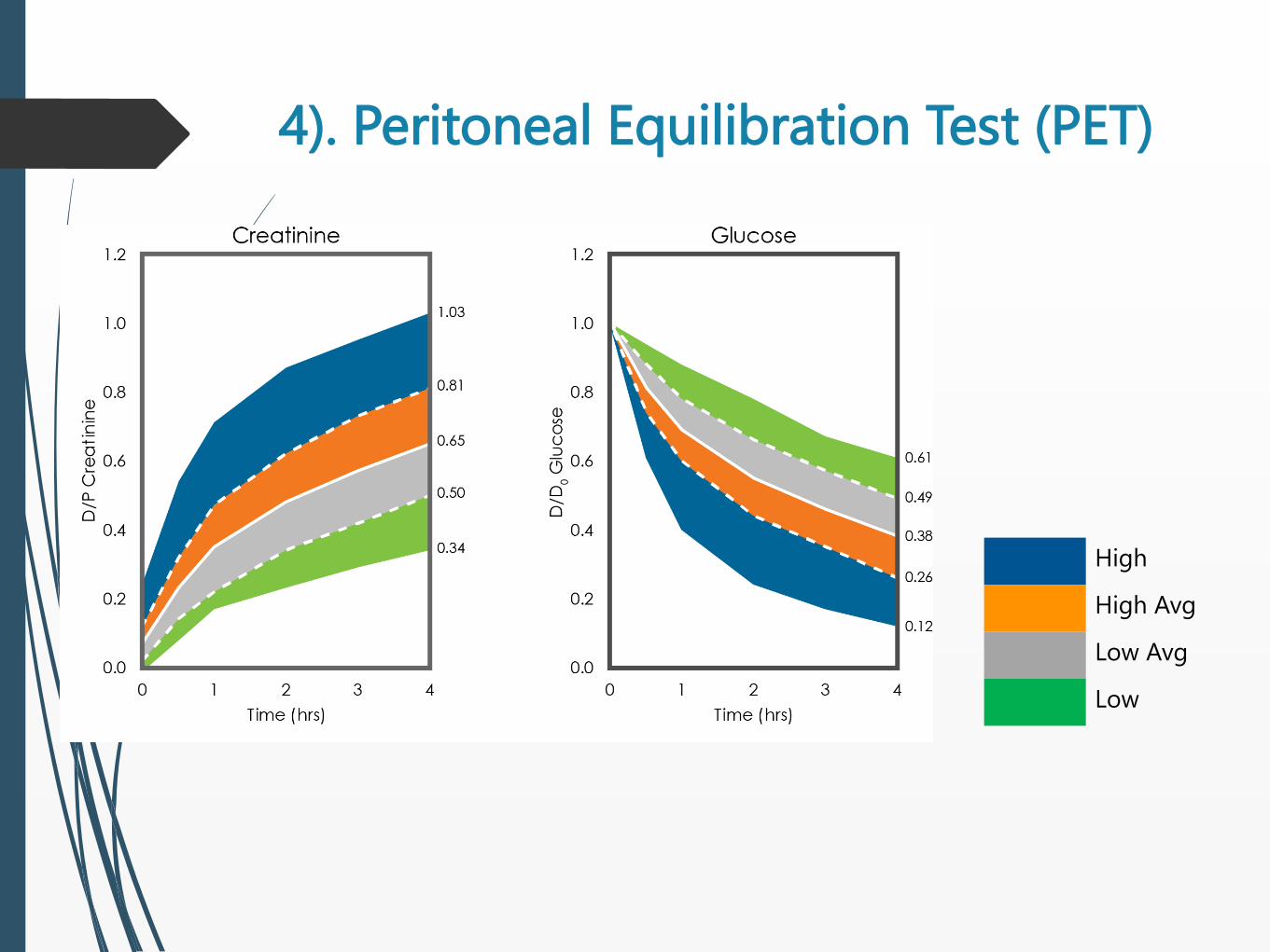
4). Peritoneal Equilibration Test (PET)
High
High Avg
Low Avg
Low
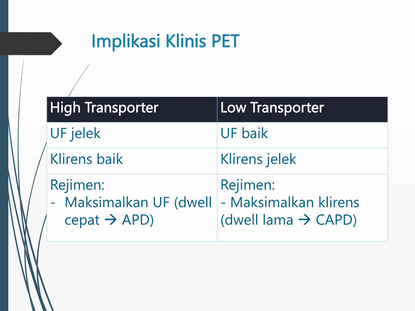
Implikasi Klinis PET
High Transporter Low Transporter
UF jelek UF baik
Klirens baik Klirens jelek
Rejimen:
- Maksimalkan UF (dwell
cepat APD)
Rejimen:
- Maksimalkan klirens
(dwell lama CAPD)
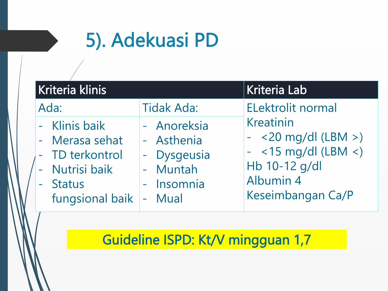
5). Adekuasi PD
Kriteria klinis Kriteria Lab
Ada: Tidak Ada: ELektrolit normal
Kreatinin
- <20 mg/dl (LBM >)
- <15 mg/dl (LBM <)
Hb 10-12 g/dl
Albumin 4
Keseimbangan Ca/P
- Klinis baik
- Merasa sehat
- TD terkontrol
- Nutrisi baik
- Status
fungsional baik
- Anoreksia
- Asthenia
- Dysgeusia
- Muntah
- Insomnia
- Mual
Guideline ISPD: Kt/V mingguan 1,7
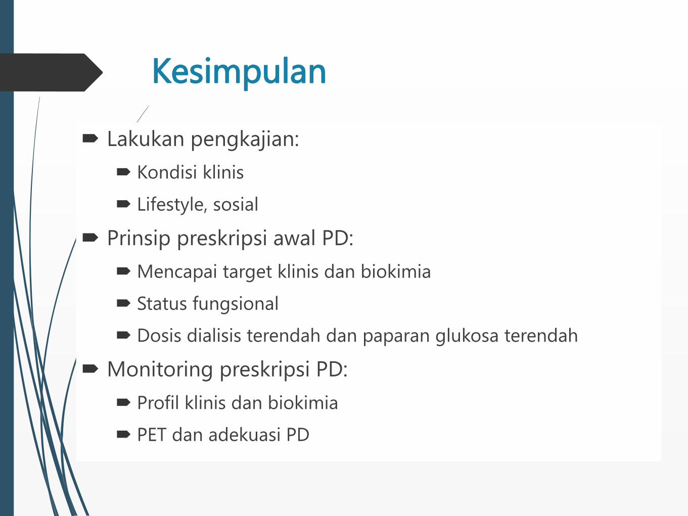
Kesimpulan
Lakukan pengkajian:
Kondisi klinis
Lifestyle, sosial
Prinsip preskripsi awal PD:
Mencapai target klinis dan biokimia
Status fungsional
Dosis dialisis terendah dan paparan glukosa terendah
Monitoring preskripsi PD:
Profil klinis dan biokimia
PET dan adekuasi PD
KOMPLIKASI PD
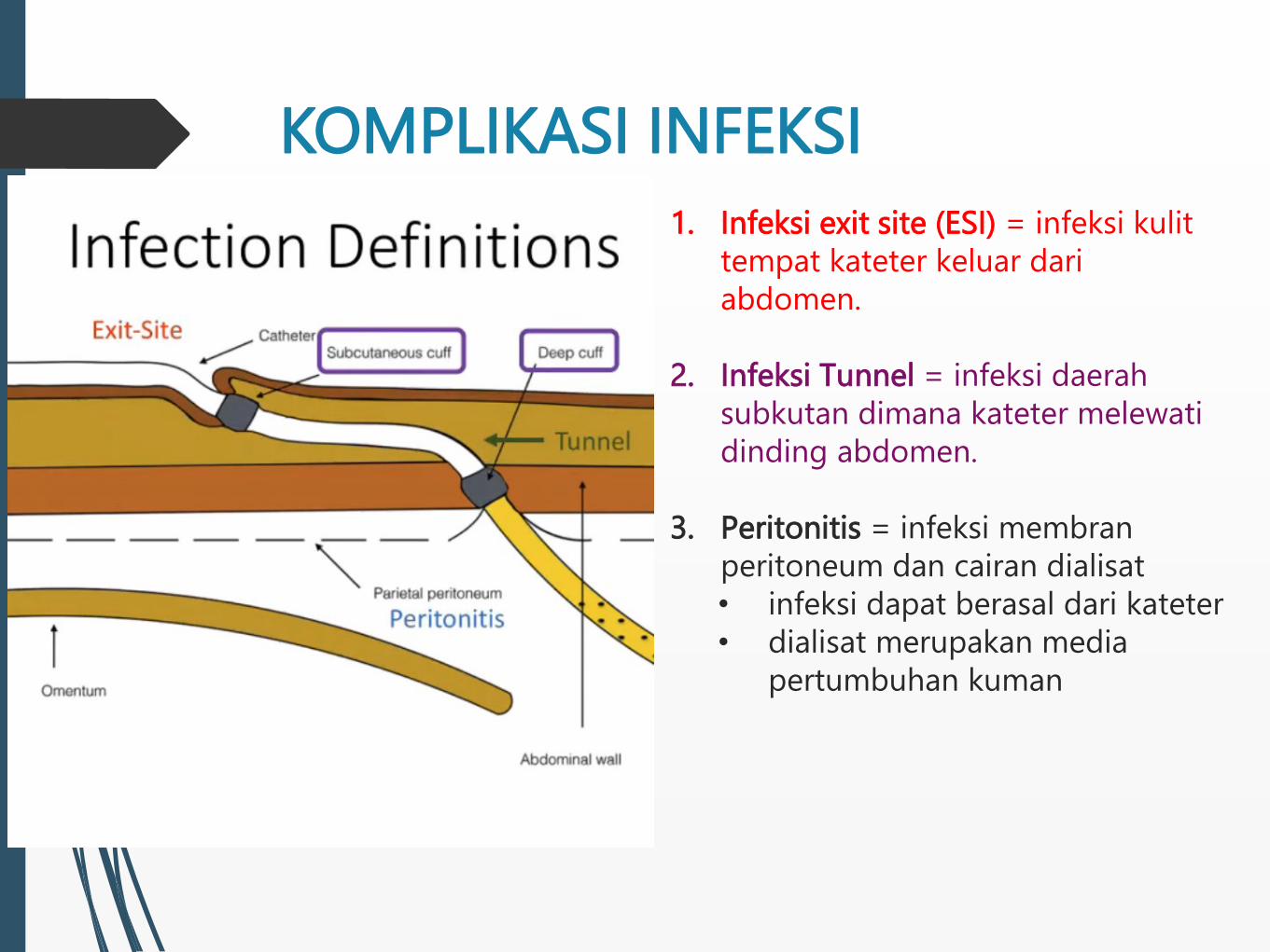
1. Infeksi exit site (ESI) = infeksi kulit
tempat kateter keluar dari
abdomen.
2. Infeksi Tunnel = infeksi daerah
subkutan dimana kateter melewati
dinding abdomen.
3. Peritonitis = infeksi membran
peritoneum dan cairan dialisat
• infeksi dapat berasal dari kateter
• dialisat merupakan media
pertumbuhan kuman
KOMPLIKASI INFEKSI
Contoh Kasus
Laki-laki, 31 tahun
CKD stage 5
Etiologi PGTA: IgAN
CAPD 1,5 tahun
Keluhan: nyeri perut sejak tadi malam
Demam disangkal
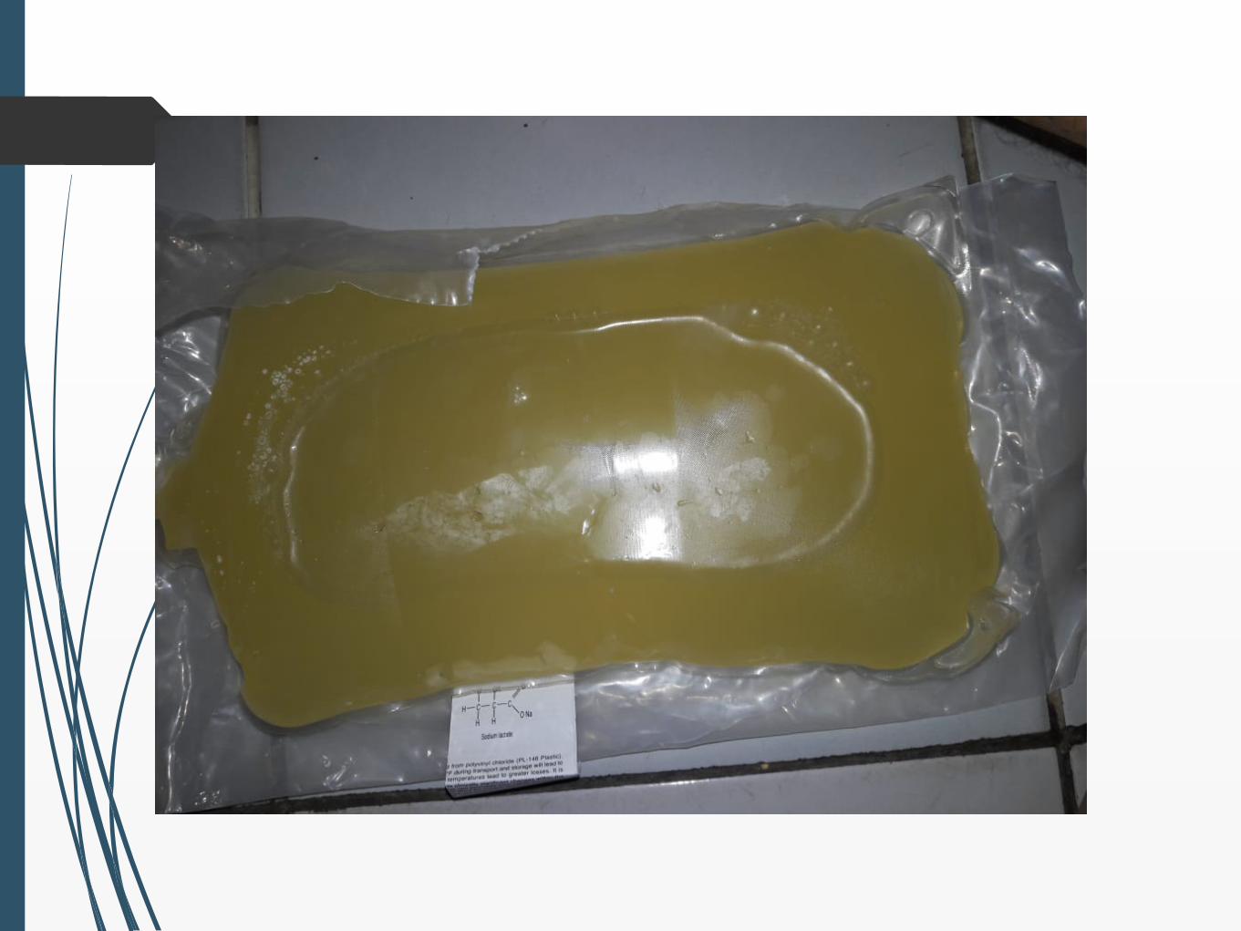
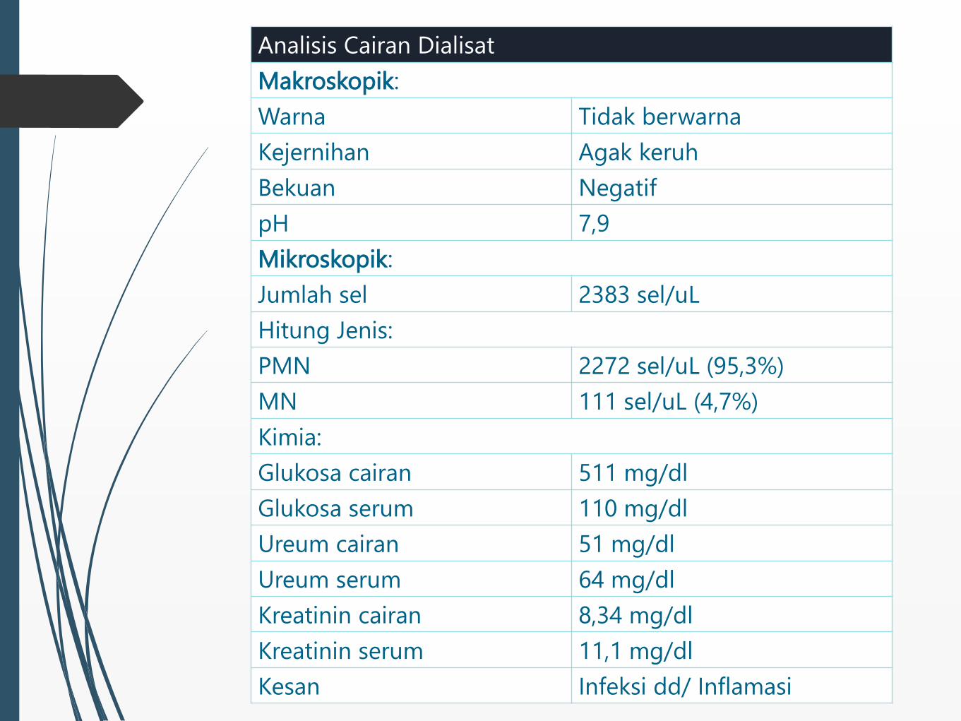
Analisis Cairan Dialisat
Makroskopik:
Warna Tidak berwarna
Kejernihan Agak keruh
Bekuan Negatif
pH 7,9
Mikroskopik:
Jumlah sel 2383 sel/uL
Hitung Jenis:
PMN 2272 sel/uL (95,3%)
MN 111 sel/uL (4,7%)
Kimia:
Glukosa cairan 511 mg/dl
Glukosa serum 110 mg/dl
Ureum cairan 51 mg/dl
Ureum serum 64 mg/dl
Kreatinin cairan 8,34 mg/dl
Kreatinin serum 11,1 mg/dl
Kesan Infeksi dd/ Inflamasi

Diagnosis Peritonitis
Minimal dua (2) dari kriteria dibawah ini:
1. Gejala klinis peritonitis: nyeri perut dan/atau
cairan dialisat keruh
2. Jumlah hitung sel darah putih cairan dialisat
>100/uL atau >0,1x10⁹/L (setelah dwell time
minimal 2 jam), dengan PMN >50%
3. Kultur cairan dialisat positif
Perit Dial Int. 2016;36:481-508

Diagnosis banding cairan
dialisat yang keruh
487
PDI SEPTEMBER 2016 - VOL. 36, NO. 5 ISPD PERITONITIS RECOMMENDATIONS: 2016 UPDATE
diarrhea. In addition, the patient should be questioned about
past history of peritonitis and ESI.
On physical examination, abdominal tenderness is typi-
cally generalized and is occasionally associated with rebound.
Localized pain or tenderness should raise the suspicion of an
underlying surgical pathology. Physical examination should
also include a careful inspection of the catheter tunnel and
exit site. Any discharge from the exit site should be cultured.
The degree of abdominal pain and tenderness are important
factors in deciding whether a patient requires hospital admis-
sion. In general, patients with minimal pain could be treated
on an outpatient basis with intraperitoneal (IP) antibiotic
therapy if this can be arranged. Follow-up within 3 days is
advisable to confirm resolution and appropriateness of the
antibiotic choice.
When peritonitis is suspected, dialysis effluent should
be drained, carefully inspected, and sent for cell count with
differential, Gram stain, and culture (207). An effluent cell
count with white blood cells (WBC) > 100/μL (after a dwell
time of at least 2 hours), with > 50% PMN, is highly sugges-
tive of peritonitis (208). Abdominal X ray is generally not
necessary. Peripheral blood culture is usually not necessary
but should be obtained if the patient is clinically septic. To
prevent delay in treatment, antibiotic therapy (see below)
should be initiated once the appropriate dialysis effluent speci-
mens have been collected, without waiting for the results of
laboratory testing.
The WBC count in the effluent depends in part on the length
of the dwell. For patients on APD with rapid cycle treatment,
the clinician should use the percentage of PMN rather than the
absolute WBC count to diagnose peritonitis, and a proportion
above 50% PMN is strong evidence of peritonitis, even if the
absolute WBC count is less than 100/μL (208). On the other
hand, APD patients without a daytime exchange who present
with abdominal pain during the daytime may have no effluent
to drain. In this case, 1 L of dialysis solution should be infused,
dwelled for 1 to 2 hours, and then drained for inspection and
laboratory testing.
Some PD patients live far away from medical facilities and
cannot be seen expeditiously after the onset of symptoms.
Since prompt initiation of therapy for peritonitis is critical,
this necessitates reliance on immediate patient reporting of
symptoms to the center, and then initiating IP antibiotics in
the home setting. Such an approach requires that the patients
be trained in this technique and that antibiotics be kept at
home. Whenever possible, cultures should be obtained either
at a local facility or by having blood culture bottles kept at
home for use. However, it is important that no one accesses
the PD catheter without the appropriate training or equipment,
which is often the case in smaller emergency departments. In
this case the patient can drain his/ her abdomen and provide
the cloudy effluent for culture. Alternatively, the patient may
place the cloudy effluent bag in the refrigerator until they
can bring the sample to their PD center. The benefit of self-
initiated treatment, however, should be carefully balanced
against the potential problems of over-diagnosis and habitual
misuse of antibiotics.
Identification of Causative Organism
•Werecommendthattheblood-culturebottlebethepre-
ferred technique for bacterial culture of PD effluent (1C) .
•Wesuggestthatsamplingandculturemethodsbereviewed
and improved if more than 15% of peritonitis episodes are
culture-negative (2C) .
Gram stain of the PD effluent should be performed even
though the result is often negative (209). The yield on the Gram
stain is increased if it is performed on centrifuged specimens.
An appropriate method of culturing PD effluent is the most
important step in establishing the causative organism. In some
specialized centers, one could achieve less than 10% rate of
culture negative peritonitis. Identification of the organism and
subsequent antibiotic sensitivities help to guide the choice
of antibiotic, and the type of organism often indicates the
possible source of infection. Bedside inoculation of 5 – 10 mL
effluent in 2 (aerobic and anaerobic) blood-culture bottles
has a reasonable sensitivity, and the culture-negative rate is
typically around 10 – 20%. (210,211). The yield of peritoneal
fluid culture is enhanced by inoculating the fluid directly into
rapid blood-culture bottle kits (e.g. BACTEC, Kent, UK; Septi-
Chek, Roche Diagnostics, Basel, Switzerland; BacT/ Alert,
Biomerieux, Inc., Basingstoke, UK), centrifuging PD fluid
and culturing the pellet, or the lysis centrifugation technique
compared to inoculation into standard blood-culture bottles.
Specifically, centrifugation of 50 mL PD effluent at 3,000 g
for 15 minutes, followed by resuspension of the sediment in
3 – 5 mL supernatant and inoculation on solid culture media
or standard blood-culture media, increases the yield by 5 to
10 times but is more cumbersome (212,213). The combination
of water lysis, Tween-80 blood agar and Triton-X treatment of
the PD effluent is also a sensitive culture method (214). The
specimens should arrive at the laboratory within 6 hours. If
immediate delivery to the laboratory is not possible, the inocu-
lated culture bottles should ideally be incubated at 37°C. The
solid media should be incubated in aerobic, microaerophilic,
and anaerobic environments.
The speed with which bacteriological diagnosis can be
established is very important. Concentration methods do not
TABLE 4
Differential Diagnosis of Cloudy Effluent
•Culture-positiveinfectiousperitonitis
•Infectiousperitonitiswithsterilecultures
•Chemicalperitonitis
•Calciumchannelblockers
•Eosinophiliaoftheeffluent
•Hemoperitoneum
•Malignancy(rare)
•Chylouseffluent(rare)
•Specimentakenfrom“dry”abdomen
This single copy is for your personal, non-commercial use only.
For permission to reprint multiple copies or to order presentation-ready
copies for distribution, contact Multimed Inc. at [email protected]
by g
uest o
n A
pril 5
, 2018
http
://ww
w.p
dico
nnect.co
m/
Do
wnlo
aded
from
Perit Dial Int. 2016;36:481-508
Penyebab Infeksi Tersering pada
PD
1. Komplikasi kateter
kontaminasi saat pergantian cairan (penyebab
peritonitis terbanyak)
penyembuhan luka yang jelek pasca insersi
kateter
trauma
perawatan exit site yang tidak tepat
Penyebab Infeksi Tersering pada
PD
2. Konstipasi
3. Infeksi abdomen atau saluran genitouri atau
tindakan invasif
perpindahan bakteri kedalam kavum
abdomen/dialisat
4. Operasi gigi
bakteremia
5. Hipokalemia
konstipasi
perpindahan bakteri dari saluran cerna dialisat

PERITONITIS
Peritonitis sering terjadi dan penyebab kesakitan
yang bermakna
penyebab kematian pasien PD hingga 15%
penyebab utama membrane failure
Peritonitis dapat dicegah
1. training pasien
2. pencegahan kontaminasi
3. antibiotik profilaksis
4. menghindari konstipasi
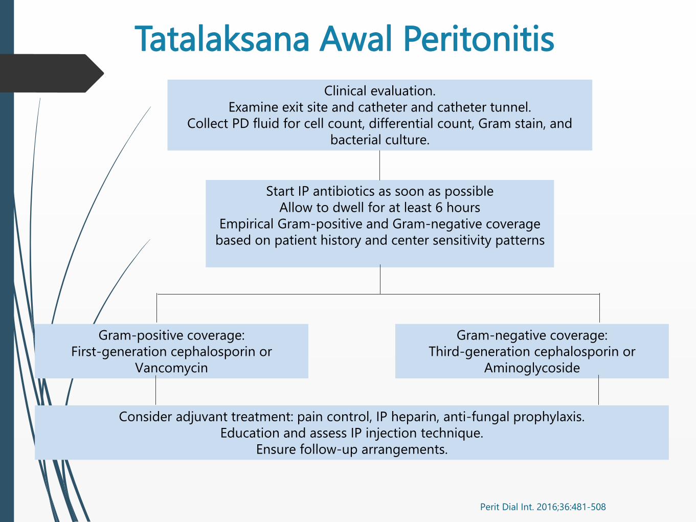
Tatalaksana Awal Peritonitis
Perit Dial Int. 2016;36:481-508
Clinical evaluation.
Examine exit site and catheter and catheter tunnel.
Collect PD fluid for cell count, differential count, Gram stain, and
bacterial culture.
Start IP antibiotics as soon as possible
Allow to dwell for at least 6 hours
Empirical Gram-positive and Gram-negative coverage
based on patient history and center sensitivity patterns
Gram-positive coverage:
First-generation cephalosporin or
Vancomycin
Gram-negative coverage:
Third-generation cephalosporin or
Aminoglycoside
Consider adjuvant treatment: pain control, IP heparin, anti-fungal prophylaxis.
Education and assess IP injection technique.
Ensure follow-up arrangements.
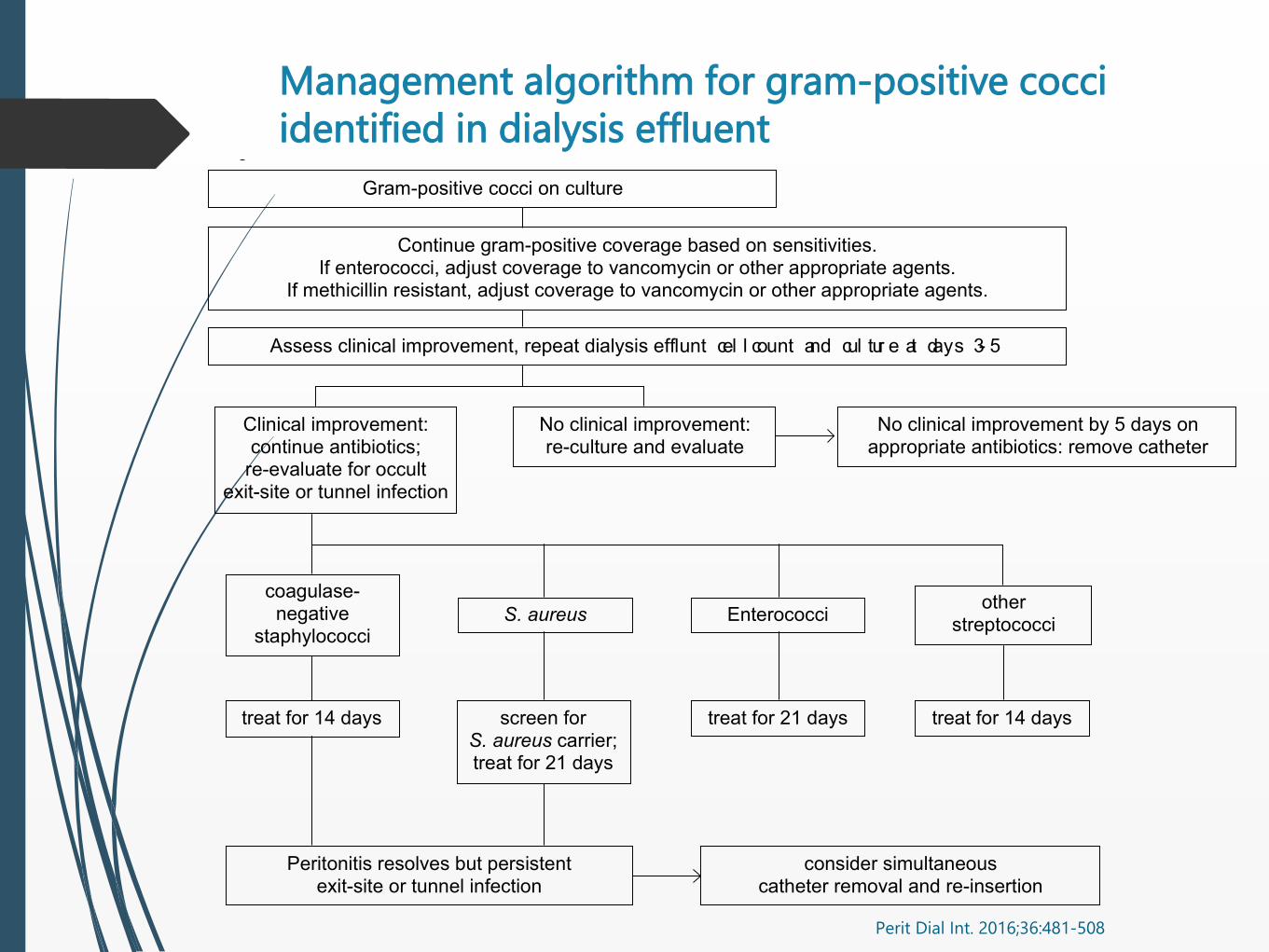
Management algorithm for gram-positive cocci
identified in dialysis effluent
Perit Dial Int. 2016;36:481-508
492
LI et al. SEPTEMBER 2016 - VOL. 36, NO. 5 PDI
removal. Temporary use of icodextrin solution may prevent
fluid overload in PD patients with acute peritonitis (342).
Because of rapid glucose absorption, glycemic control may
worsen in diabetic patients. Blood glucose monitoring with
appropriate adjustments of insulin dosage may be needed.
Protein loss during peritonitis is also increased. Screening for
malnutrition should be undertaken in patients with prolonged
peritoneal inflammation.
SUBSEQUENT MANAGEMENT OF PERITONITIS
•Werecommendthatantibiotictherapybeadjustedto
narrow-spectrum agents, as appropriate, once culture
results and sensitivities are known. (1C) .
The management algorithms for gram-positive cocci and
gram-negative bacilli identified in dialysis effluent are sum-
marized in Figures 2 and 3, respectively. Within 48 hours of
initiating therapy, most patients with PD-related peritonitis
will show considerable clinical improvement. The effluent
should be visually inspected regularly to determine whether
clearing is occurring. If there is no improvement after 48
hours, cell counts and repeat cultures should be performed. In
addition, monitoring of WBC count in PD effluent may predict
treatment response. A retrospective study showed that dialysis
effluentWBCcount1,090/mm3 on day 3 was an independent
prognostic marker for treatment failure (343).
Refractory Peritonitis
•WerecommendthatthePDcatheterberemovedpromptlyin
refractory peritonitis episodes, defined as failure of the PD
effluent to clear up after 5 days of appropriate antibiotics
(1C) .
After initiation of antibiotic treatment, there is usually clini-
cal improvement in 72 hours. Refractory peritonitis is defined
as failure of the PD effluent to clear up after 5 days of appro-
priate antibiotics (Table 7). Catheter removal is indicated in
case of refractory peritonitis, or earlier if the patient ’s clinical
condition is deteriorating, in order to preserve the peritoneum
for future PD as well as preventing morbidity and mortality.
Prolonged attempts to treat refractory peritonitis by antibi-
otics without catheter removal are associated with extended
hospital stay, peritoneal membrane damage, increased risk of
fungal peritonitis, and excessive mortality (344).
Figure 2 — Management algorithm for gram-positive cocci identif ied in dialysis effluent.
Gram-positive cocci on culture
Assess clinical improvement, repeat dialysis efflu
e
nt cel l count and cul tur e at days 3- 5
Clinical improvement:continue antibiotics;
re-evaluate for occult exit-site or tunnel infection
coagulase-negative
staphylococci
treat for 14 days
Peritonitis resolves but persistentexit-site or tunnel infection
consider simultaneouscatheter removal and re-insertion
screen forS. aureus carrier;treat for 21 days
S. aureus Enterococci
treat for 21 days treat for 14 days
otherstreptococci
No clinical improvement:re-culture and evaluate
No clinical improvement by 5 days onappropriate antibiotics: remove catheter
Continue gram-positive coverage based on sensitivities.If enterococci, adjust coverage to vancomycin or other appropriate agents.
If methicillin resistant, adjust coverage to vancomycin or other appropriate agents.
This single copy is for your personal, non-commercial use only.
For permission to reprint multiple copies or to order presentation-ready
copies for distribution, contact Multimed Inc. at [email protected]
by g
uest o
n A
pril 5
, 20
18
http
://ww
w.p
dico
nn
ect.com
/D
ow
nlo
aded
from
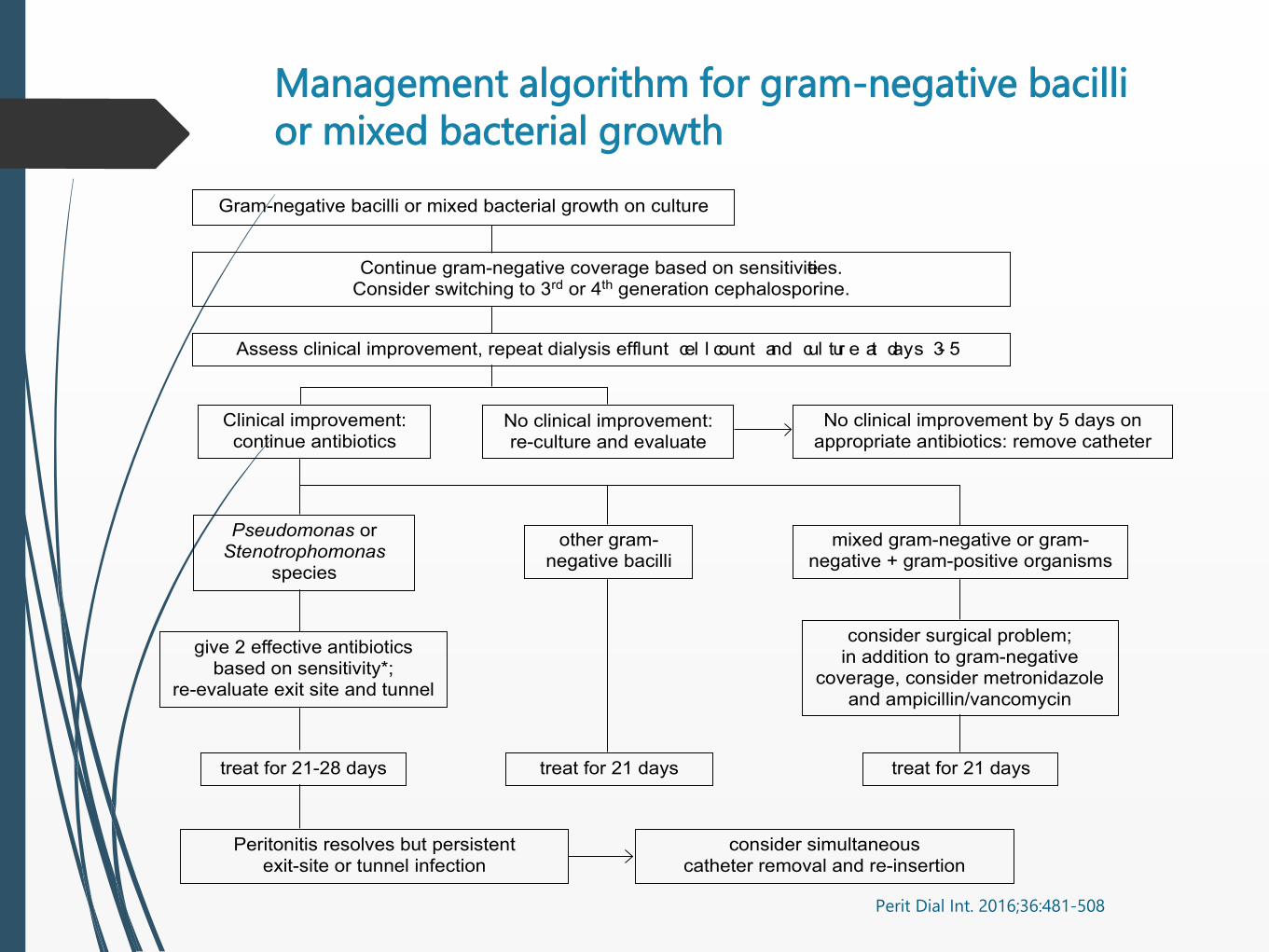
Management algorithm for gram-negative bacilli
or mixed bacterial growth
Perit Dial Int. 2016;36:481-508
493
PDI SEPTEMBER 2016 - VOL. 36, NO. 5 ISPD PERITONITIS RECOMMENDATIONS: 2016 UPDATE
Relapsing, Recurrent, and Repeat Peritonitis
•Werecommendthattimelycatheterremovalbeconsidered
for relapsing, recurrent, or repeat peritonitis episodes (1C) .
The definitions of relapsing, recurrent, and repeat perito-
nitis are summarized in Table 7. Retrospective studies showed
that relapsing, recurrent, and repeat peritonitis episodes are
caused by different species of bacteria and probably represent
distinct clinical entities (166,345–347). When compared to
non-relapsing episodes, relapsing ones are associated with a
lower rate of cure, more ultrafiltration problems, and higher
rate of technique failure (166,348). Recurrent peritonitis epi-
sodes had a worse prognosis than relapsing ones (166,345).
A recent study suggested that bacterial DNA fragment levels
in PD effluent are significantly higher 5 days before and on
the date of completion of antibiotics amongst patients who
subsequently develop relapsing or recurrent peritonitis (349).
Another study suggests that effluent white cell count and leu-
kocyte strip test at the time of stopping antibiotics may also
predict relapse (350). However, further studies are needed to
validate these results and confirm their clinical utility.
Coagulase-Negative Staphylococcus
•Wesuggestthatcoagulase-negativestaphylococcigenerally
be treated with IP cephalosporins or vancomycin, accord-
ing to antimicrobial susceptibility, for a period of 2 weeks.
(2C) .
Coagulase-negative Staphylococcus peritonitis episodes,
especially those caused by S. epidermidis, are mostly due
to touch contamination. Many patients with S. epidermidis
peritonitis have mild clinical symptoms and respond well to
treatment as outpat ients (351–353). In some centers,
the prevalence of methicillin resistance is now very high
(354,355), and vancomycin may have to be considered as
empirical therapy. Even for methicillin-sensit ive strains,
it is important to avoid inadequate IP antibiotic levels,
which may lead to relapsing peritonitis. For this reason,
continuous dosing of IP f irst-generation cephalosporins is
preferable to intermittent dosing. Effective antibiotic treat-
ment for 2 weeks is generally suf f icient (351–354). The
patient ’s exchange technique should be reviewed to prevent
another episode.
Figure 3 — Management algorithm for gram-negative bacilli or mixed bacterial growth identif ied in dialysis effluent.
* Trimethoprim/ sulfamethoxazole is preferred for Stenotrophomonas species.
Gram-negative bacilli or mixed bacterial growth on culture
Continue gram-negative coverage based on sensitivities.Consider switching to 3rd or 4th generation cephalosporine.
Assess clinical improvement, repeat dialysis efflu
e
nt cel l count and cul tur e at days 3- 5
Clinical improvement:continue antibiotics
Pseudomonas orStenotrophomonas
species
give 2 effective antibioticsbased on sensitivity*;
re-evaluate exit site and tunnel
treat for 21-28 days
Peritonitis resolves but persistentexit-site or tunnel infection
consider simultaneouscatheter removal and re-insertion
treat for 21 days treat for 21 days
other gram-negative bacilli
No clinical improvement:re-culture and evaluate
No clinical improvement by 5 days onappropriate antibiotics: remove catheter
mixed gram-negative or gram-negative + gram-positive organisms
consider surgical problem;in addition to gram-negative
coverage, consider metronidazoleand ampicillin/vancomycin
This single copy is for your personal, non-commercial use only.
For permission to reprint multiple copies or to order presentation-ready
copies for distribution, contact Multimed Inc. at [email protected]
by g
uest o
n A
pril 5
, 201
8http
://ww
w.p
dico
nnect.co
m/
Dow
nlo
aded
from

Indikasi Pencabutan Kateter
497
PDI SEPTEMBER 2016 - VOL. 36, NO. 5 ISPD PERITONITIS RECOMMENDATIONS: 2016 UPDATE
The treatment protocol should be based on general proto-
cols for treatment of tuberculosis but is often started with 4
drugs: rifampicin, isoniazid, pyrazinamide, and ofloxacin. A
previous study showed that rifampicin levels in PD effluent are
often low (407). Intraperitoneal rifampicin treatment has been
advocated but is not available in many countries. In general,
pyrazinamide and ofloxacin could be stopped after 2 months,
while rifampicin and isoniazid should be continued for a total
of 12 to 18 months (407–413). Pyridoxine (50 to 100 mg/ day)
should be given to avoid isoniazid-induced neurotoxicity. On
the other hand, long-term use of pyridoxine at a higher dose
(e.g. 200 mg daily) is in itself associated with neuropathy and
should be avoided. Streptomycin, even in reduced doses, may
cause ototoxicity after prolonged use and should be avoided.
Ethambutol is associated with a high risk of optic neuritis
in dialysis patients and must be used with appropriate dos-
age reduction. Previous reports suggest a dose of 15 mg/ kg
every 48 hours or thrice weekly for up to 2 months (414). The
optimal treatment for drug-resistant tuberculous peritonitis
remainsunknown.
Many patients respond to anti-tuberculous therapy without
catheter removal (407–413,415). However, it is important
to dif ferentiate patients with miliary tuberculosis, whose
peritonitis is part of the disseminated disease, from those
with isolated tuberculous peritonitis without extraperitoneal
infection, because the duration of anti-tuberculous therapy
is different.
Non-Tuberculous Mycobacterial Peritonitis
Data on peritonitis caused by non-tuberculous mycobacte-
ria are limited but may be increasing (21,416–422). It is not
uncommon for non-tuberculous mycobacteria to be misidenti-
fied as gram-positive diphtheroids. Over half of the isolates are
rapidly growing species, such as M. fortuitum and M. chelonae
(420), and often become positive on routine bacteriologic
cultures in 3 to 5 days. It has been postulated that extensive
use of topical gentamicin ointment for exit-site infection
may predispose patients to non-tuberculous mycobacterial
infection of the exit site (144). The treatment regimen for non-
tuberculous mycobacterial peritonitis is not well established
and requires individualized protocols based on susceptibility
testing. Catheter removal is usually necessary, and experience
with non-removal is limited (420–422). The type and duration
of antibiotic therapy are variable, and the optimal treatment
regimen is poorly defined and depends on species and drug
susceptibilities (416–422).
Catheter Removal and Re-Insertion
•WerecommendthatPDcathetersberemovedforrefractory,
relapsing, or fungal peritonitis unless there are clinical
contraindications (1C) .
•WesuggestthatitisappropriatetoconsiderreturntoPD
for many patients who have had their catheter removed for
refractory, relapsing, or fungal peritonitis (2C).
•Wesuggestthatifre-insertionofanewcatheterisattempt-
ed after a PD catheter is removed for refractory, relapsing,
or fungal peritonitis, it be performed at least 2 weeks after
catheter removal and complete resolution of peritoneal
symptoms (2D) .
Indications for catheter removal are summarized in Table 8.
For refractory peritonitis and fungal peritonitis, simultaneous
re-insertion of a new PD catheter is not recommended, and
patients should be put on temporary hemodialysis. Observa-
tional studies suggest that effective antibiotics should be
continued for at least 2 weeks after catheter removal for
refractory peritonitis (423,424).
After severe episodes of peritonitis, around 50% of patients
could potentially return to PD (423–425). An ANZDATA Registry
study demonstrated that return to PD after catheter removal
and temporary hemodialysis for peritonitis was not associated
with inferior patient-level clinical outcomes when compared
with other patients who either never required hemodialysis
transfer for peritonitis or who had permanent hemodialysis
transfer for peritonitis (426). Furthermore, subsequent peri-
tonitis-free, technique and patient survival following return
to PD were not associated with organism type or duration of
time from hemodialysis transfer to PD restart (426). There are
few data on the optimal duration between catheter removal for
peritonitis and re-insertion of a new catheter. Observational
studies suggest a minimum period of 2 to 3 weeks (423–425),
although some would recommend later re-insertion in cases
of fungal peritonitis (397,398). Re-insertion of a new catheter
should be done by laparoscopic or mini-laparotomy approach
so that adhesion can be directly visualized. Ultrafiltration
problems are common after return to PD (423,424). A small
proportion of patients with PD-related peritonitis develop
recurrent intra-abdominal collection that requires percuta-
neous drainage after catheter removal (427). The chance of
a successful return to PD is very low in this group of patients,
and direct conversion to long-term hemodialysis should be
considered (427).
FUTURE RESEARCH
There are some new antibiotics that, to the best of our
knowledge, have not been tried for the treatment of PD-related
peritonitis. For example, ceftaroline has good coverage of
TABLE 8
Indications for Catheter Removal
•Refractoryperitonitis
•Relapsingperitonitis
•Refractoryexit-siteandtunnelinfection
•Fungalperitonitis
•Catheterremovalmayalsobeconsideredfor
– repeat peritonitis
– mycobacterial peritonitis
– multiple enteric organisms
This single copy is for your personal, non-commercial use only.
For permission to reprint multiple copies or to order presentation-ready
copies for distribution, contact Multimed Inc. at [email protected]
by
gu
est on
April 5
, 20
18
http
://ww
w.p
dico
nnect.co
m/
Do
wnlo
aded
from
Perit Dial Int. 2016;36:481-508
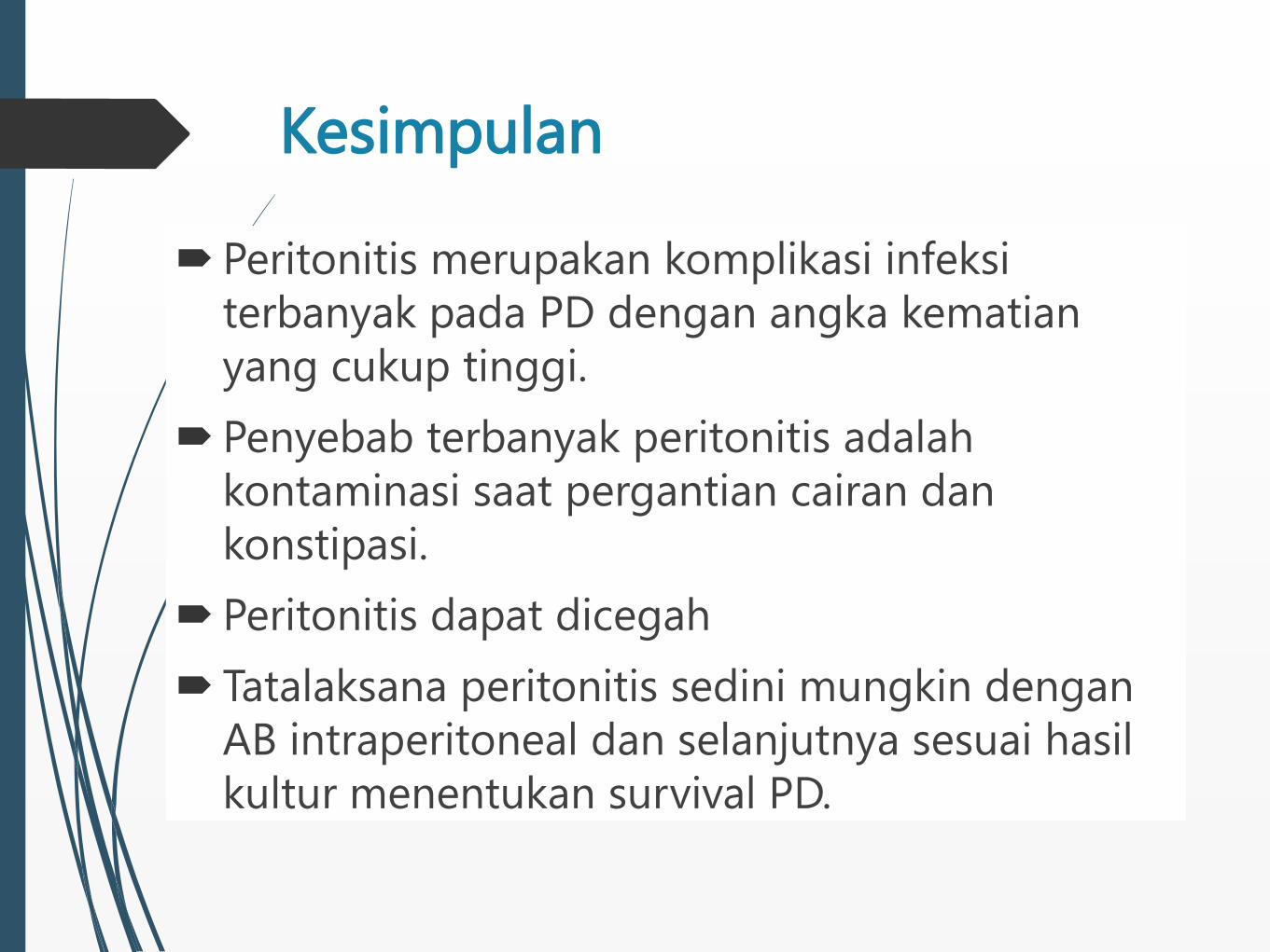
Kesimpulan
Peritonitis merupakan komplikasi infeksi
terbanyak pada PD dengan angka kematian
yang cukup tinggi.
Penyebab terbanyak peritonitis adalah
kontaminasi saat pergantian cairan dan
konstipasi.
Peritonitis dapat dicegah
Tatalaksana peritonitis sedini mungkin dengan
AB intraperitoneal dan selanjutnya sesuai hasil
kultur menentukan survival PD.

KOMPLIKASI NON INFEKSI
KASUS – GANGGUAN OUTFLOW
Laki-laki 54 tahun, dalam selama CAPD 3 tahun.
Keluhan: BB dan lingkar perut bertambah sjk 3 hari
yl.
Pasien memakai dianeal 2,5% 3x dan 1,5% 1x,
namun UF masih jelek dan waktu drain sangat
lama.
Cairan jernih.
Inflow lancar, namun outflow sangat lama.
500 ml pertama sekitar 30 menit, kemudian aliran
berhenti.
Apa yang akan Anda Lakukan?
A. Memiringkan pasien ke satu sisi dan
sebaliknya untuk melihat apakah outflow
membaik.
B. Bilas kateter dengan heparin
C. Transfer ke HD – PD dianggap gagal.
D. Berikan laksatif
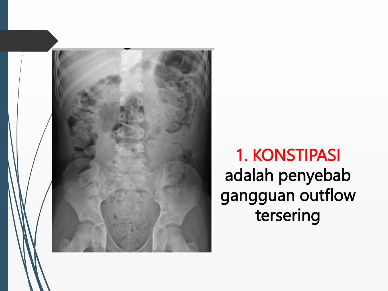
1. KONSTIPASI
adalah penyebab
gangguan outflow
tersering
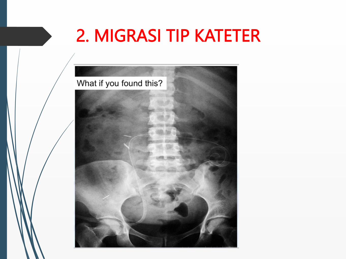
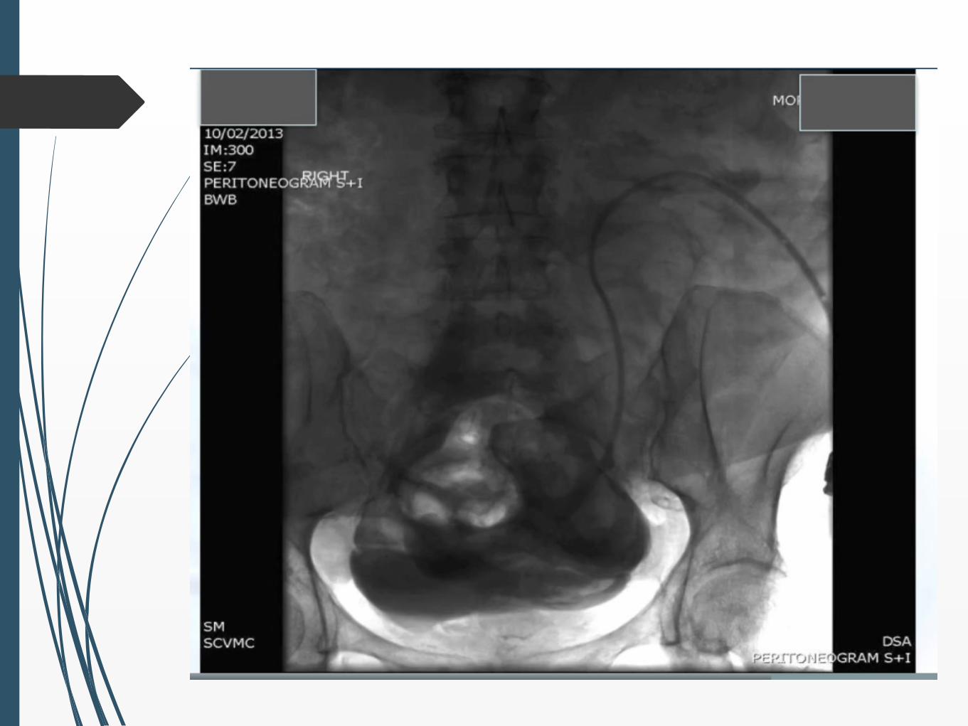
2. MIGRASI TIP KATETER
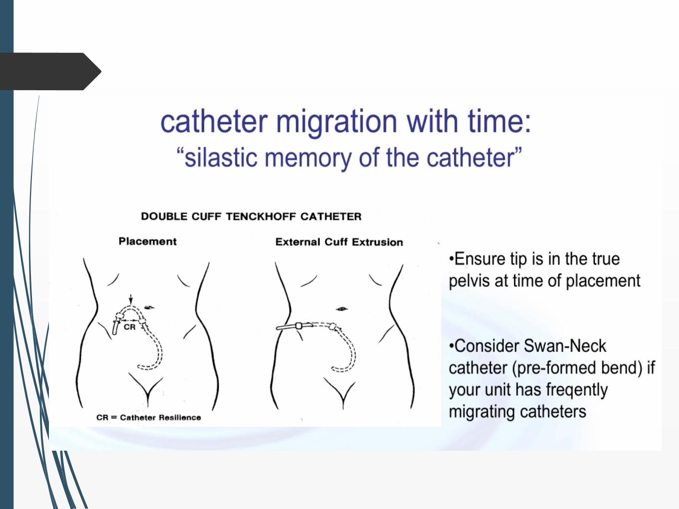

Migrasi Kateter
Diagnosis awal dengan foto polos
abdomen.
Jika kateter keluar dari pelvis:
trochar radiologi atau laparoskopi untuk
menarik kateter ke dalam pelvis.
reposisi laparoskopi.
Kembali ke kasus….
inflow tidak ada masalah outflow
lambat
dialisat jernih
BNO: tidak ada konstipasi dan kateter tidak
bermigrasi keatas.
langkah selanjutnya?
Kasus…...
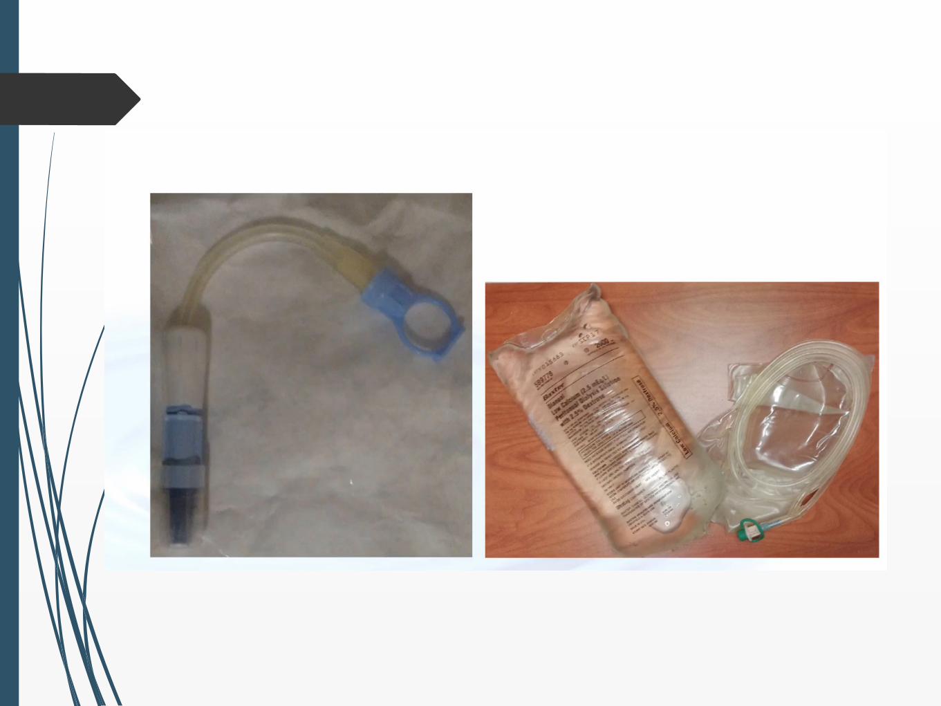
Perawat mencoba flush kateter dengan
spuit.
Flush lancar namun sulit diaspirasi
Apa yang akan Anda lakukan
selanjutnya?
A. memberikan lagi urus-urus
B. flush dengan larutan NS-heparin
C. memberikan 1-2 L dialisat
D. injeksi tPA IP
E. transfer ke HD
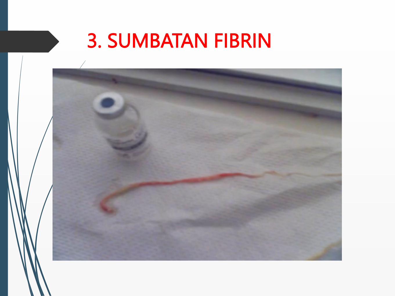
3. SUMBATAN FIBRIN
Kembali lagi ke kasus…...
inflow tidak ada masalah, outflow lambat,
cairan jernih
BNO: tidak ada konstipasi, tidak ada migrasi
kateter
Ns sudah flush kateter dengan spuit, flush lancar
namun aspirasi susah
Fibrin negatif
langkah selanjutnya?

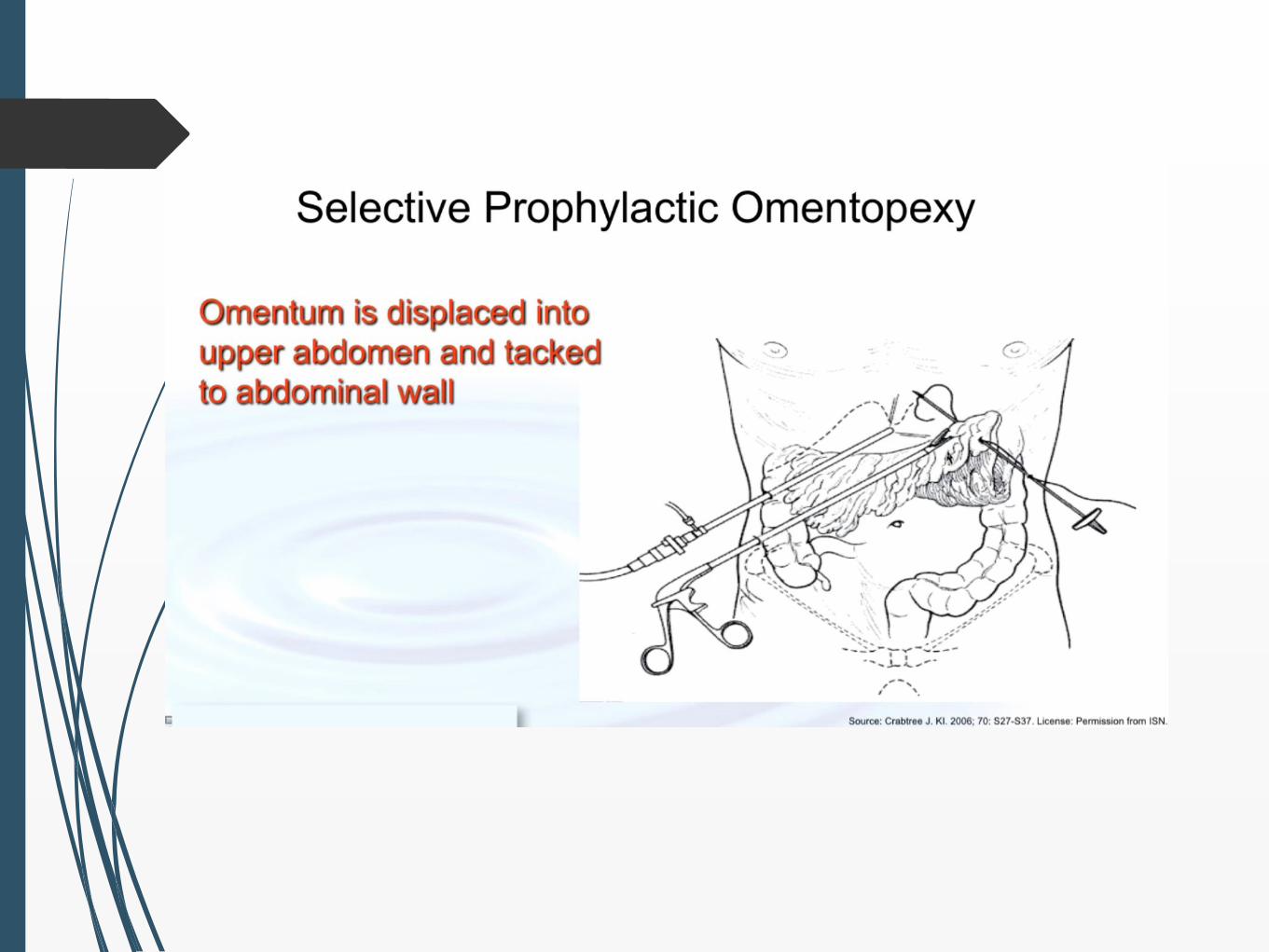
4. OMENTAL WRAP
Lebih banyak terjadi pada:
anak-anak
jika posisi tip kateter diluar pelvis
biasanya inflow baik namun outflow jelek
dapat dicegah dengan omentopeksi pada anak
dan orang dengan omentum yang ‘berlebih’
Perlu tindakan bedah:
omentektomi dengan/atau tanpa penggantian
kateter PD
Diagnosis Banding Gangguan
Outflow
konstipasi
migrasi tip kateter
sumbatan fibrin
adhesi/perlengketan
omental wrap
Lainnya….....???
Diagnosis Banding Gangguan
Outflow
1. konstipasi
2. migrasi tip kateter
3. adhesi/perlengketan
4. omental wrap
5. fibrin, klot, obstruksi eksternal (frangible,
transfer set)
6. bocor
PENINGKATAN TEKANAN INTRA-
ABDOMINAL
Hernia
Kebocoran perikateter, abdomen dan
genital
Hidrothoraks
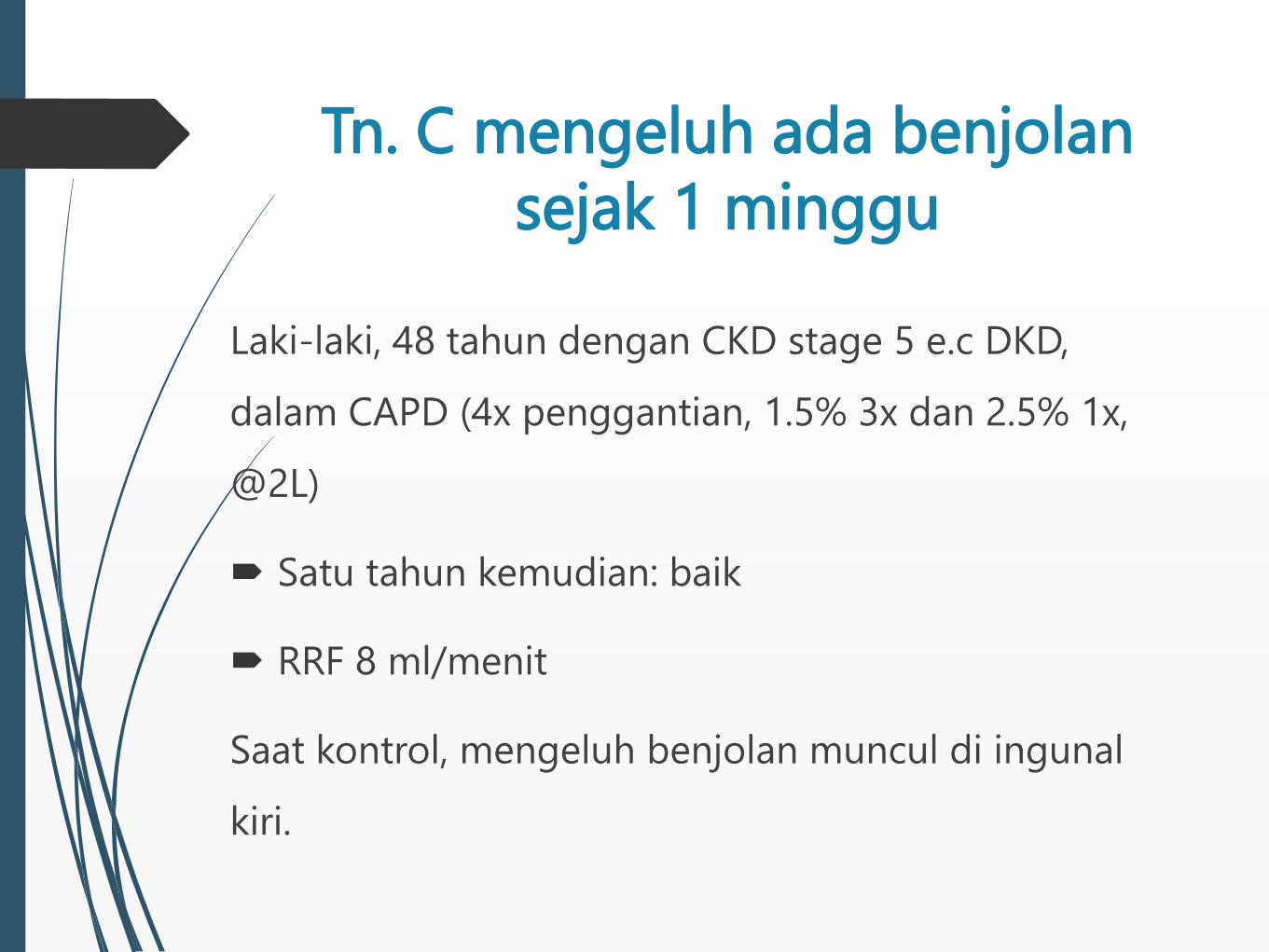
Tn. C mengeluh ada benjolan
sejak 1 minggu
Laki-laki, 48 tahun dengan CKD stage 5 e.c DKD,
dalam CAPD (4x penggantian, 1.5% 3x dan 2.5% 1x,
@2L)
Satu tahun kemudian: baik
RRF 8 ml/menit
Saat kontrol, mengeluh benjolan muncul di ingunal
kiri.
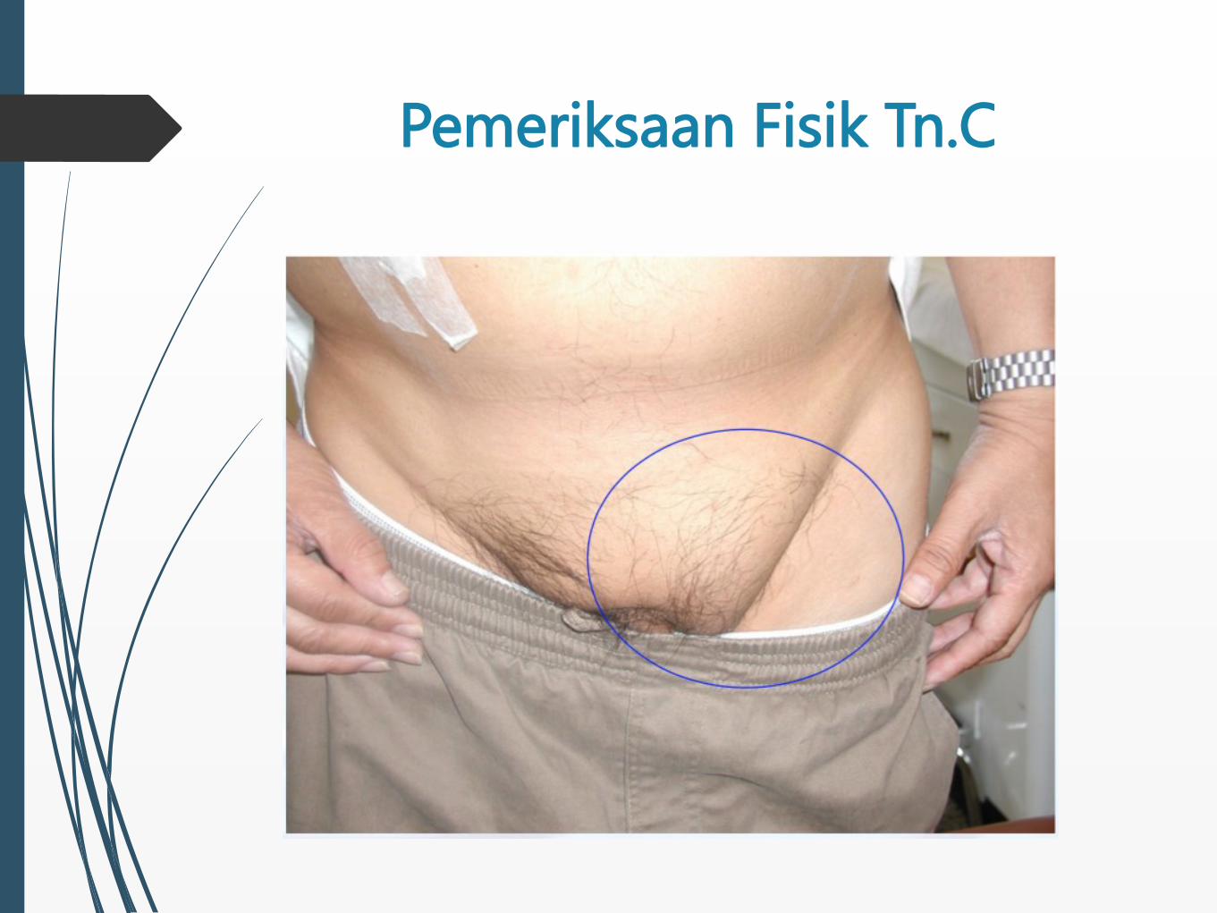
Pemeriksaan Fisik Tn.C
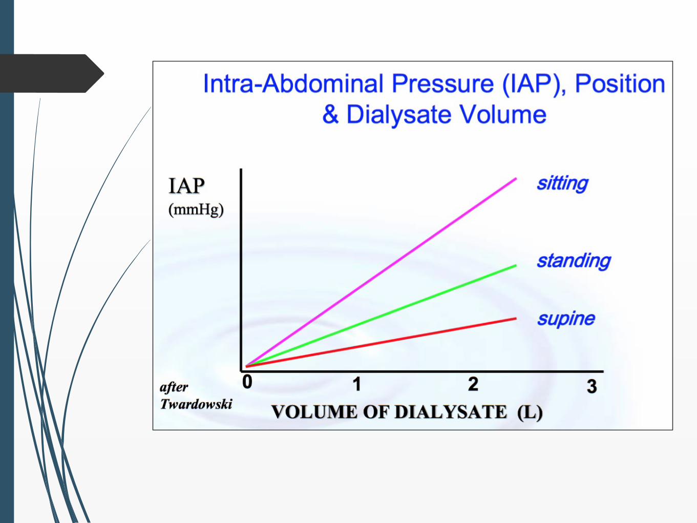
Peningkatan Tekanan Intra-
Abdomen (TIAb)
Adanya dialisat didalam kavum peritoneum
akan meningkatkan TIAb
Peningkatan ini tergantung dari:
Volume dialisat yang masuk
Posisi pasien (duduk > berdiri > tidur)
Usia, indeks massa tubuh
Batuk, mengangkat beban, mengedan
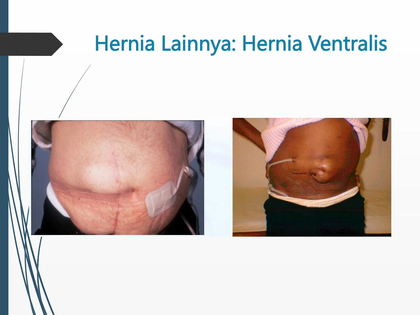
Hernia Lainnya: Hernia Ventralis
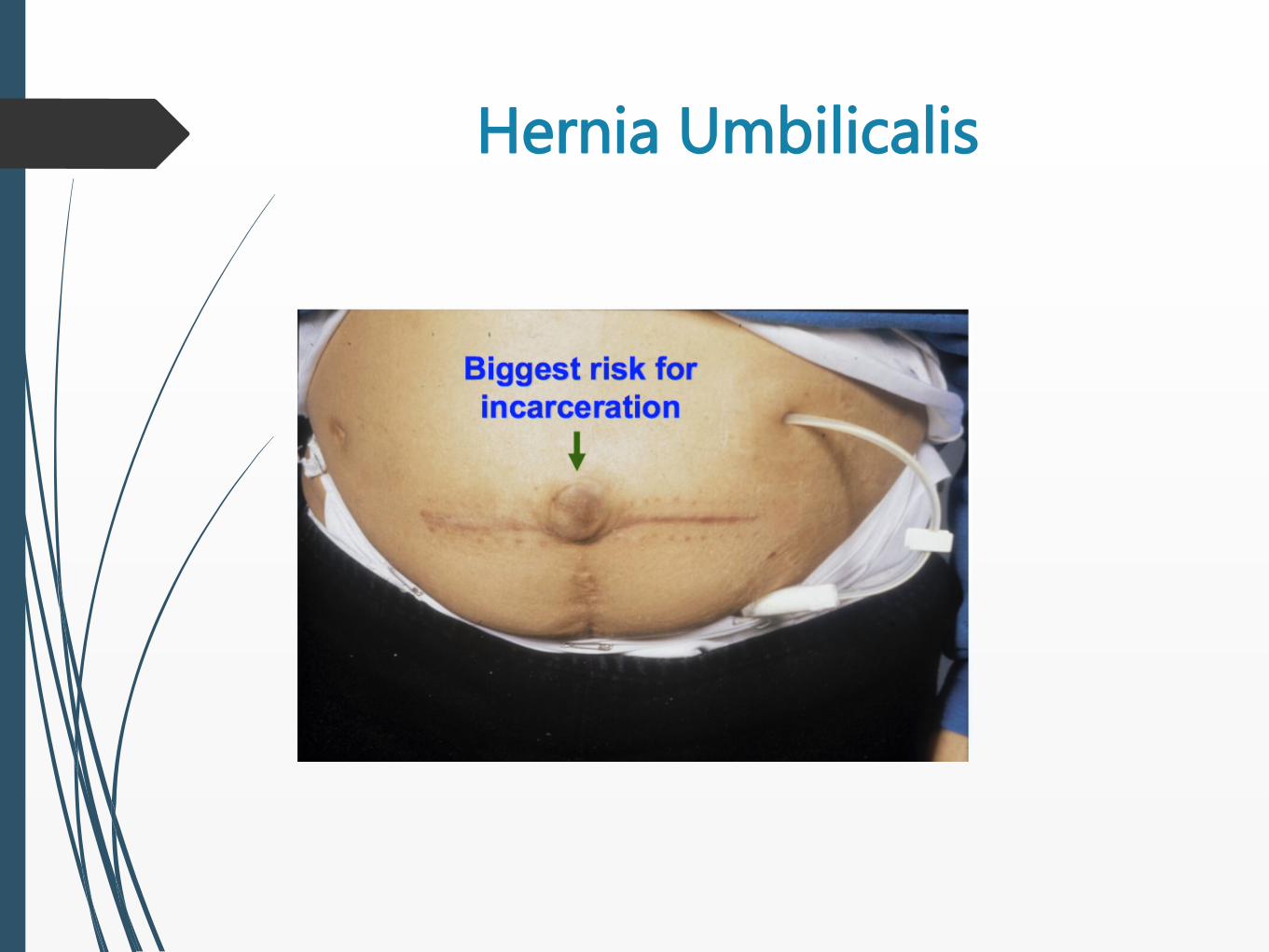
Hernia Umbilicalis
Tatalaksana:
Inspeksi dan evaluasi perkiraan lokasi hernia
Konsultasi bedah:
Tidak selalu harus di-repair, namun cenderung
membesar, menyebabkan kebocoran dialisat
Risiko inkarserasi usus, peritonitis
Evaluasi kaitan dengan postur, aktivitas dan
cairan dialisat
Bisa dilakukan modifikasi PD (NIPD atau PD volume
kecil)
HERNIA

HERNIA
Tatalaksana:
Edukasi pasien tentang tanda-tanda inkarserasi
Repair bedah:
Dialisis perioperatif sangat tergantung dari fungsi
ginjal dan kondisi pasien
Tidak selalu harus transfer ke HD
Mulai kembali PD dengan volume kecil, posisi tidur,
tingkatkan volume setelah 2 minggu

Sesak Nafas yang Memberat
pada Pasien PD
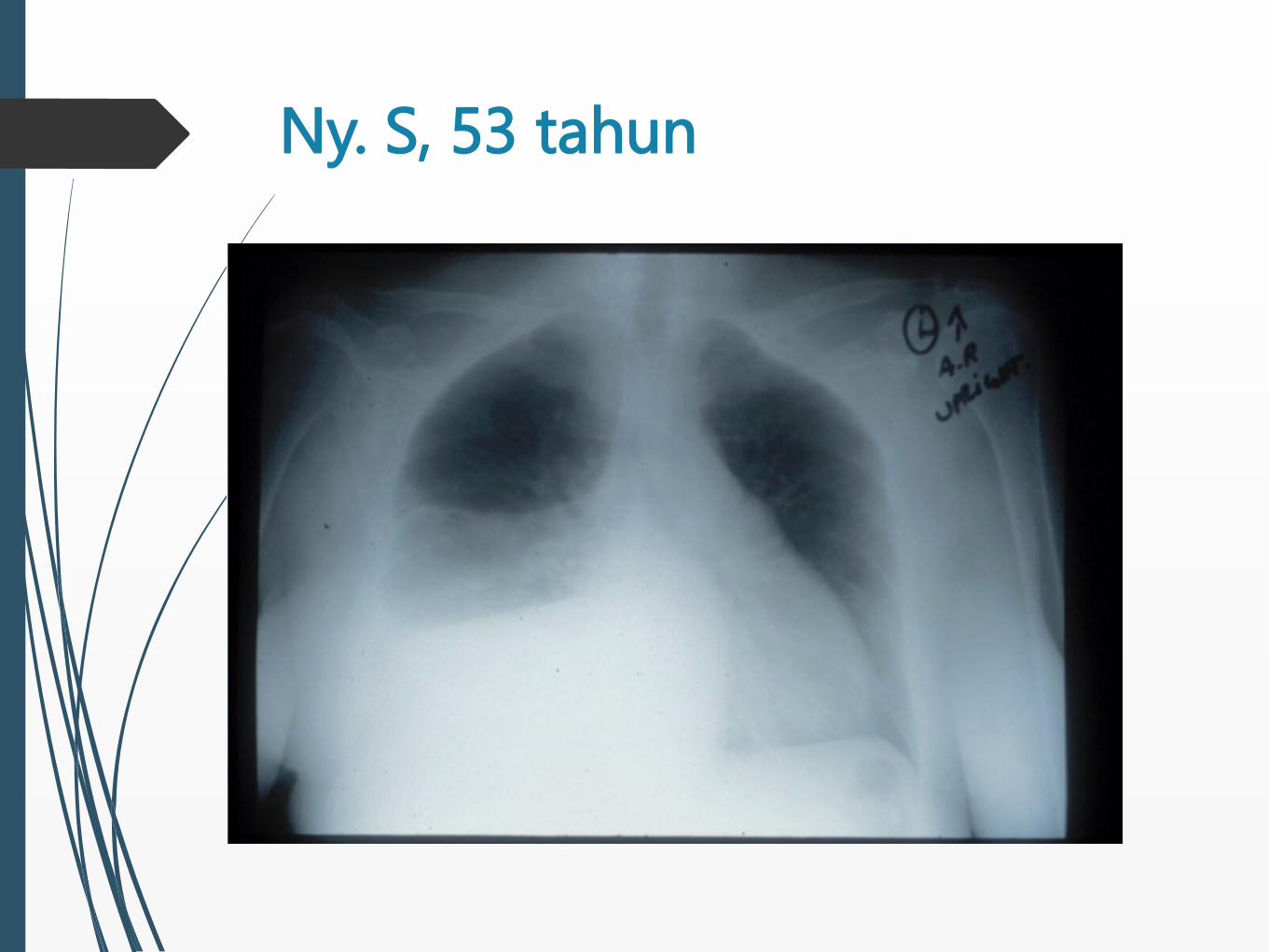
Ny. S, 53 tahun dengan CKD stage 5 e.c DKD,
telah dilakukan insersi kateter PD.
1 minggu setelah inisiasi PD, pasien
menghubungi suster PD karena keluhan sesak
nafas yang memberat sejak 3 hari.
Tidak ada batuk/mengi/dahak.
Ada penambahan BB 1 kg dari target BBK.
Ny. S, 53 tahun
HIDROTHORAKS
Definisi: ditemukannya cairan dialisat didalam
kavum pleura.
Insiden: sekitar <5%
Patogenesis: perpindahan dialisat, akibat
peningkatan TIAb, dari peritoneum ke dalam
kavum pleura melalui defek diafragma yang
kongenital ataupun didapat
HIDROTHORAKS
Gejala/Tanda:
Dapat asimptomatik
Sesak nafas
Tidak membaik dengan dialisat konsentrasi
tinggi
UF <<
Efusi pleura kanan pada rontgen thoraks
PKD
HiDROTHORAKS
Diagnosis:
Thorakosentesis untuk mengurangi
simtom dan/atau untuk diagnostik
Analisis cairan pleura:
Transudat
Kadar glukosa tinggi (umumnya, namun
tidak selalu – icodextrin iodine: blue-
black)
Hitung sel bervariasi
HIDROTHORAKS
Diagnosis
Pemeriksaan radionuclide atau radiocontrast
Melihat pergerakan cairan dari peritoneum kedalam
rongga thoraks
Technetium-99m tagged macro-aggregated
albumin atau Tc-99 sulfur colloid
Methylene blue dye kedalam dialisat diikuti
thorakosentesis tidak dianjurkan!
Visualisasi langsung dengan thorakoskopi
(VATS)
HIDROTHORAKS
Tatalaksana:
Thorakosentesis jika diperlukan (pasien
mengeluh sangat sesak)
Stop PD
Hemodialisis sementara
Mesothelium dapat memperbaiki dirinya sendiri
dan menutup defek yang ada hanya dengan
mengistirahatkan PD (2-6 minggu)
HIDROTHORAKS
Tatalaksana:
Coba mulai PD dengan volume kecil (glukosa dialisat
dalam kavum pleura dapat berfungsi sebagai agen
sklerosis)
Monitor ketat tanda uremia!
Pleurodesis (talc, tetrasiklin, bleomisin, darah
autologus)
Repair operasi atau pleuroskopik (defek diafragma
diidentifikasi dan diberikan patch atau dijahit)
50% pasien berhasil direpair dan melanjutkan PD
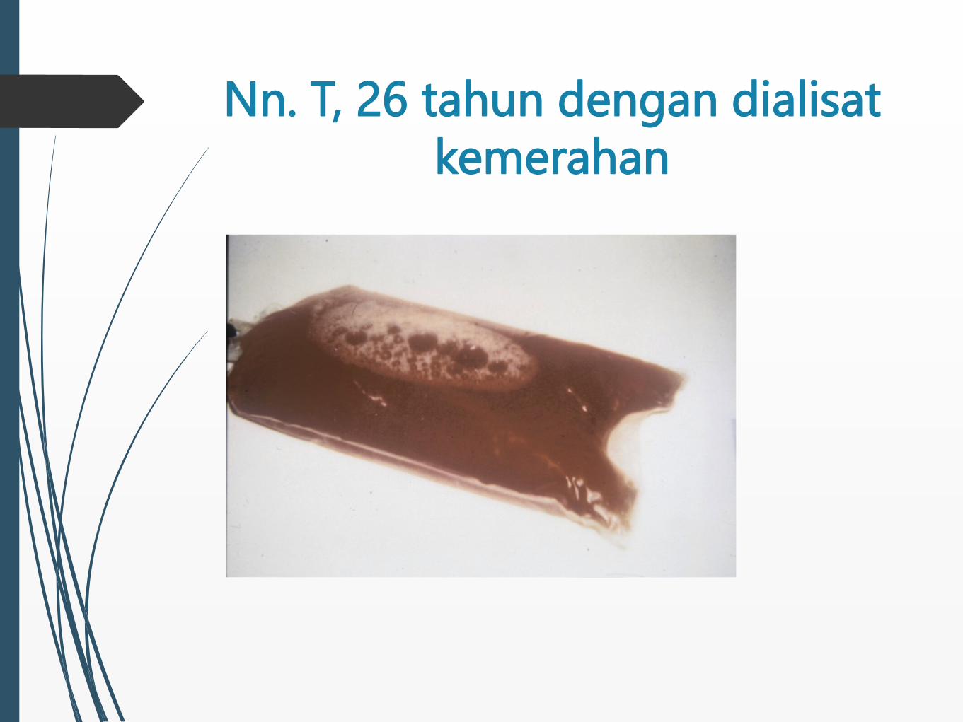
Nn. T, 26 tahun dengan dialisat
kemerahan

HEMOPERITONEUM
Definisi:
Darah pada cairan dialisat
Gejala/Tanda:
Menakutkan!
Pikirkan penyebab “jinak” dan “serius”
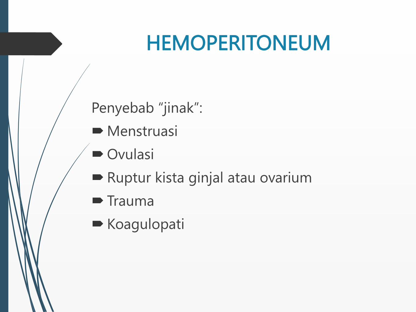
HEMOPERITONEUM
Penyebab “jinak”:
Menstruasi
Ovulasi
Ruptur kista ginjal atau ovarium
Trauma
Koagulopati
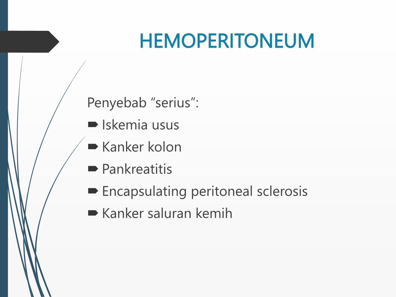
HEMOPERITONEUM
Penyebab “serius”:
Iskemia usus
Kanker kolon
Pankreatitis
Encapsulating peritoneal sclerosis
Kanker saluran kemih
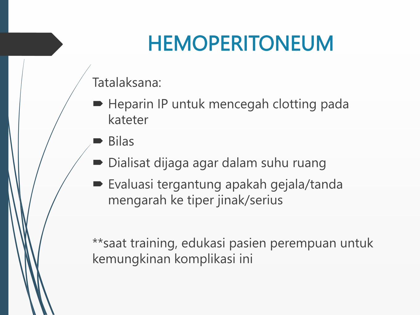
HEMOPERITONEUM
Tatalaksana:
Heparin IP untuk mencegah clotting pada
kateter
Bilas
Dialisat dijaga agar dalam suhu ruang
Evaluasi tergantung apakah gejala/tanda
mengarah ke tiper jinak/serius
**saat training, edukasi pasien perempuan untuk
kemungkinan komplikasi ini
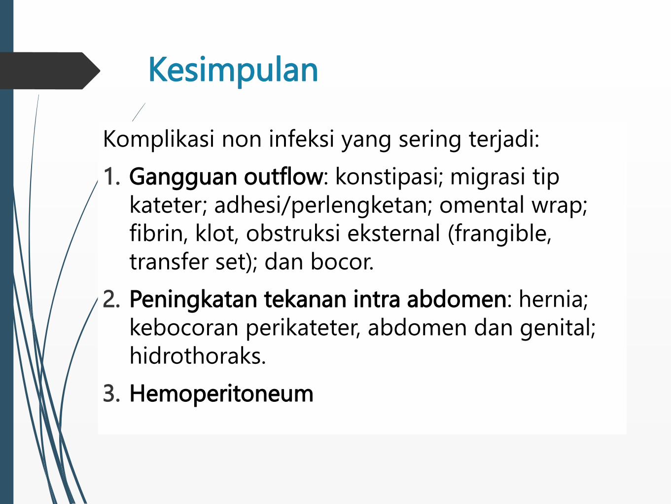
Kesimpulan
Komplikasi non infeksi yang sering terjadi:
1. Gangguan outflow: konstipasi; migrasi tip
kateter; adhesi/perlengketan; omental wrap;
fibrin, klot, obstruksi eksternal (frangible,
transfer set); dan bocor.
2. Peningkatan tekanan intra abdomen: hernia;
kebocoran perikateter, abdomen dan genital;
hidrothoraks.
3. Hemoperitoneum