Embed Size (px)
Citation preview

抗がん剤と天然物-古典的薬剤から分子標的薬まで-
青木 俊二兵庫医療大学薬学部
古典的抗がん剤としての天然物
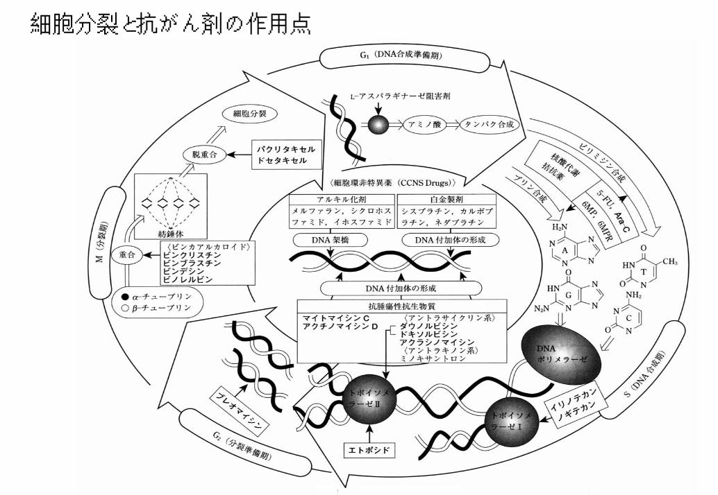
細胞分裂と抗がん剤の作用点
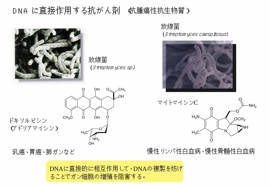
DNA に直接作用する抗がん剤 (抗腫瘍性抗生物質)
放線菌(Streptomyces caespitosus)
放線菌(Streptomyces sp.)
CH3O O
O OH
OH O
OH
O CH3
O
OHNH2
H3C
マイトマイシンCO
O
NNH
H3C
H2N OCH3
O
ONH2
ドキソルビシン(アドリアマイシン)
慢性リンパ性白血病、慢性骨髄性白血病乳癌、胃癌、肺ガンなど
DNAに直接的に相互作用して、DNAの複製を妨げることでガン細胞の増殖を阻害する。
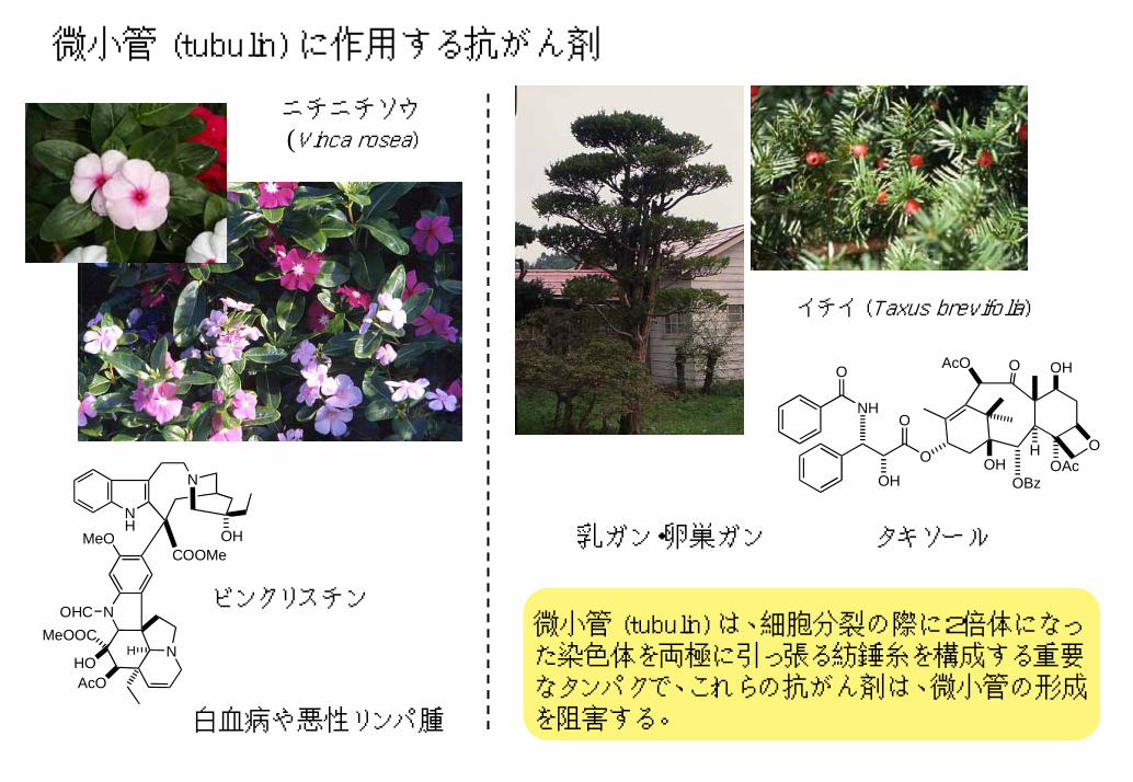
微小管 (tubulin) に作用する抗がん剤
NH
N
COOMeOH
N
NMeOOC
HOAcO
OHC
MeO
H
OOAc
H
OBz
OAcO OHO
OHOOH
ONHイチイ
ニチニチソウ(Vinca rosea)
イチイ (Taxus brevifolia)
乳ガン・卵巣ガン タキソール
ビンクリスチン
白血病や悪性リンパ腫
微小管 (tubulin) は、細胞分裂の際に2倍体になった染色体を両極に引っ張る紡錘糸を構成する重要なタンパクで、これらの抗がん剤は、微小管の形成を阻害する。
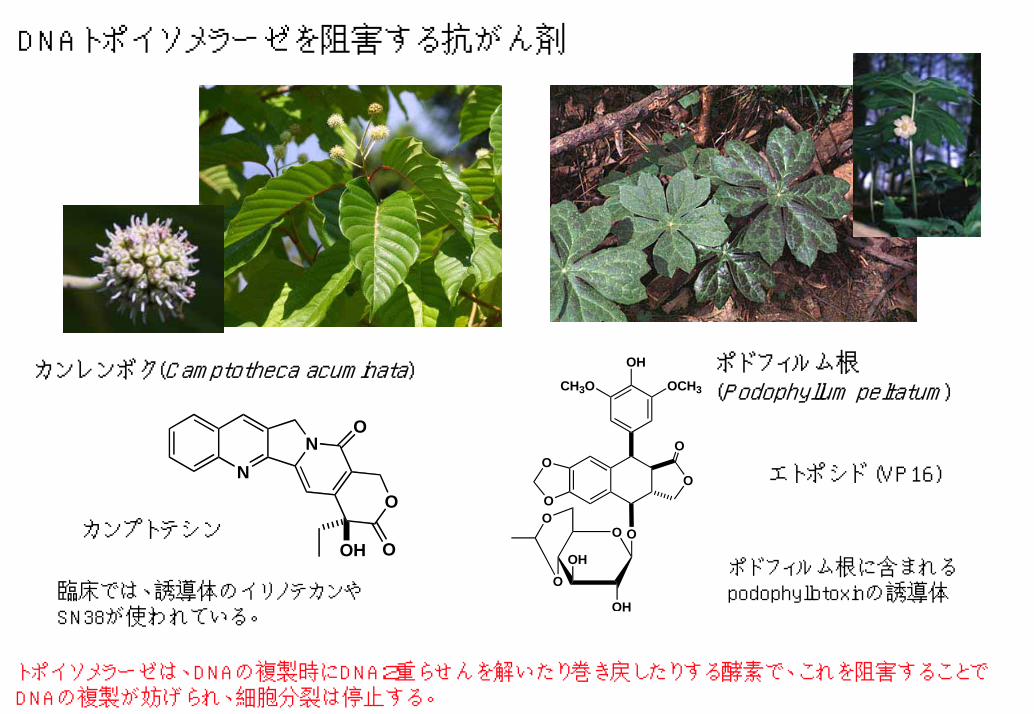
DNAトポイソメラーゼを阻害する抗がん剤
ポドフィルム根(Podophyllum peltatum)CH3O OCH3
OH
OO
O
O
OH
OH
OO
O
O
カンレンボク(Camptotheca acuminata)
カンプトテシン
NN
O
O
OOH
エトポシド (VP16)
ポドフィルム根に含まれるpodophyllotoxinの誘導体臨床では、誘導体のイリノテカンや
SN38が使われている。
トポイソメラーゼは、DNAの複製時にDNA2重らせんを解いたり巻き戻したりする酵素で、これを阻害することでDNAの複製が妨げられ、細胞分裂は停止する。
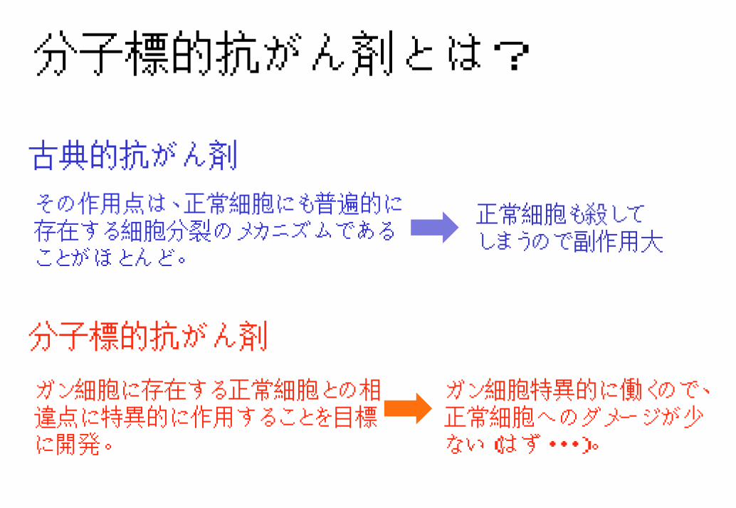
分子標的抗がん剤とは?
古典的抗がん剤
その作用点は、正常細胞にも普遍的に存在する細胞分裂のメカニズムであることがほとんど。
正常細胞も殺してしまうので副作用大
分子標的抗がん剤
ガン細胞特異的に働くので、正常細胞へのダメージが少ない(はず・・・)。
ガン細胞に存在する正常細胞との相違点に特異的に作用することを目標に開発。
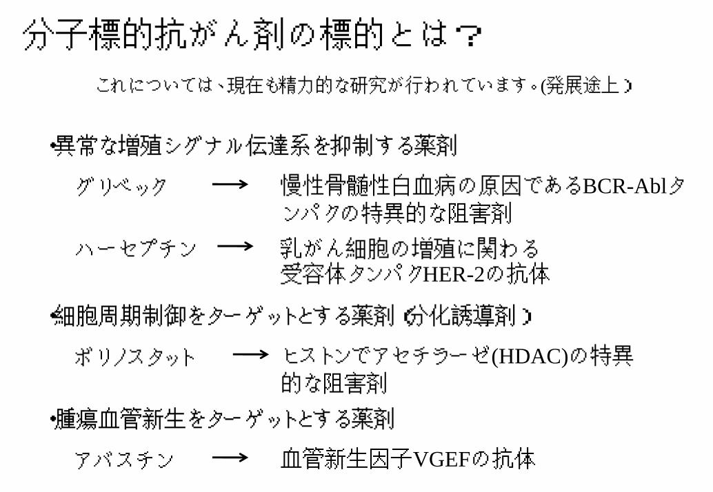
分子標的抗がん剤の標的とは?
これについては、現在も精力的な研究が行われています。(発展途上)
・異常な増殖シグナル伝達系を抑制する薬剤
慢性骨髄性白血病の原因であるBCR-Ablタンパクの特異的な阻害剤
グリベック
ハーセプチン 乳がん細胞の増殖に関わる受容体タンパクHER-2の抗体
・細胞周期制御をターゲットとする薬剤(分化誘導剤)
ヒストンでアセチラーゼ(HDAC)の特異的な阻害剤
ボリノスタット
・腫瘍血管新生をターゲットとする薬剤
血管新生因子VGEFの抗体アバスチン
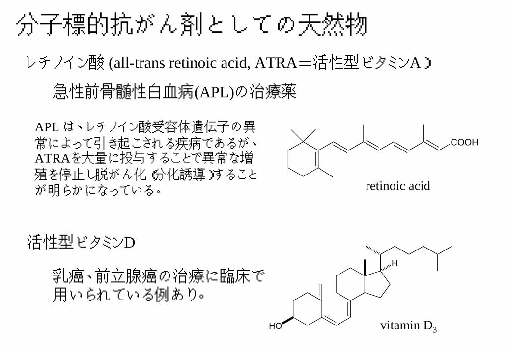
分子標的抗がん剤としての天然物
レチノイン酸 (all-trans retinoic acid, ATRA=活性型ビタミンA)
急性前骨髄性白血病(APL)の治療薬
APL は、レチノイン酸受容体遺伝子の異常によって引き起こされる疾病であるが、ATRAを大量に投与することで異常な増殖を停止し脱がん化(分化誘導)することが明らかになっている。
COOH
retinoic acid
活性型ビタミンD
HO
H
乳癌、前立腺癌の治療に臨床で用いられている例あり。
vitamin D3
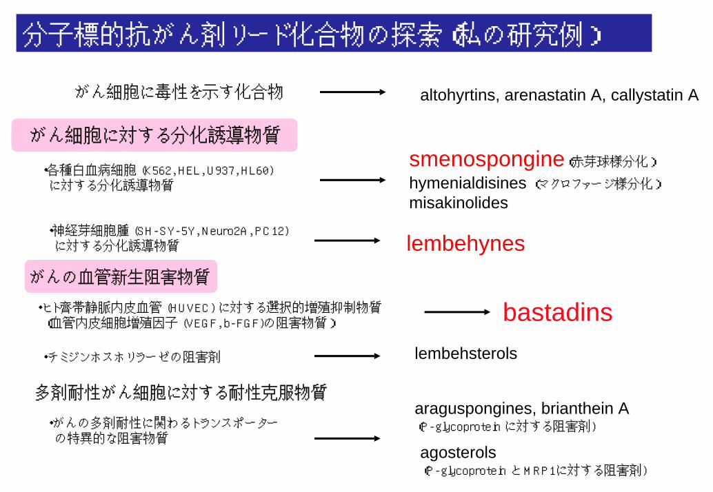
分子標的抗がん剤リード化合物の探索(私の研究例)
がん細胞に毒性を示す化合物 altohyrtins, arenastatin A, callystatin A
がん細胞に対する分化誘導物質
・各種白血病細胞 (K562, HEL, U937, HL60) に対する分化誘導物質
・神経芽細胞腫 (SH-SY-5Y, Neuro2A, PC12) に対する分化誘導物質
がんの血管新生阻害物質
・ヒト齊帯静脈内皮血管 (HUVEC) に対する選択的増殖抑制物質(血管内皮細胞増殖因子 (VEGF, b-FGF)の阻害物質)
・チミジンホスホリラーゼの阻害剤
smenospongine(赤芽球様分化)hymenialdisines (マクロファージ様分化)misakinolides
lembehynes
bastadinslembehsterols
多剤耐性がん細胞に対する耐性克服物質araguspongines, brianthein A (P-glycoprotein に対する阻害剤)
agosterols(P-glycoprotein と MRP1に対する阻害剤)
・がんの多剤耐性に関わるトランスポーターの特異的な阻害物質
ガン細胞に対する分化誘導物質の探索
1)慢性骨髄性白血病細胞に対する分化誘導物質の例
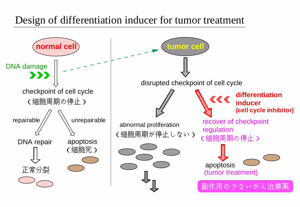
Design of differentiation inducer for tumor treatment
normal cell tumor cell
DNA damage
checkpoint of cell cycle(細胞周期の停止)
repairable unrepairable
DNA repair apoptosis(細胞死)
正常分裂
disrupted checkpoint of cell cycle
differentiation inducer (cell cycle inhibitor)
abnormal proliferation recover of checkpoint regulation(細胞周期の停止)
apoptosis(tumor treatment)
(細胞周期が停止しない)
副作用の少ないがん治療薬
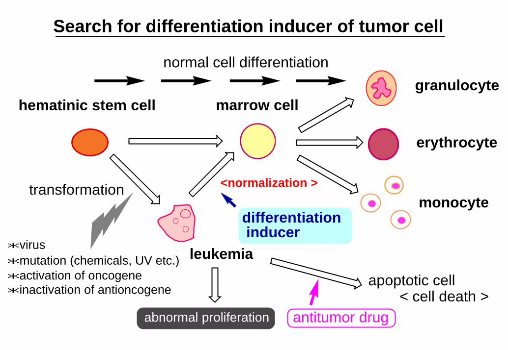
Search for differentiation inducer of tumor cell
granulocyte
erythrocyte
monocyte
hematinic stem cell marrow cell
transformation
leukemia
abnormal proliferation antitumor drug
apoptotic cell
normal cell differentiation
< cell death >
<normalization >
differentiation inducer
*activation of oncogene*inactivation of antioncogene
*mutation (chemicals, UV etc.)*virus
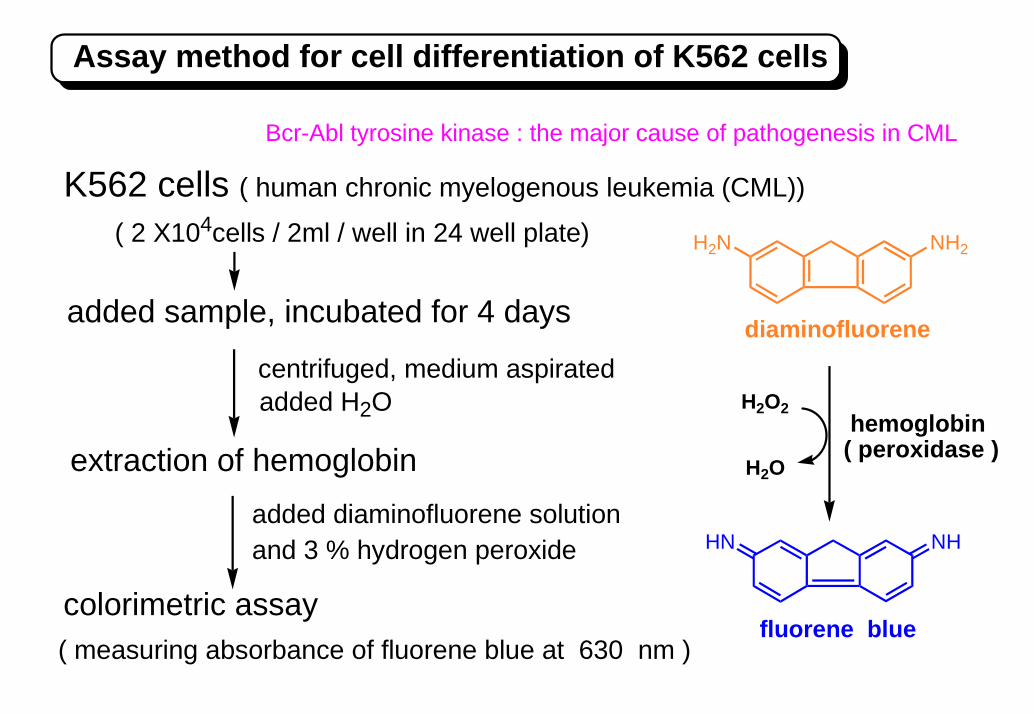
NH2H2N
H2O2
H2O
HN NH
Assay method for cell differentiation of K562 cells
added sample, incubated for 4 days
extraction of hemoglobin
( 2 X10⁴cells / 2ml / well in 24 well plate)
fluorene blue
hemoglobin( peroxidase )
( measuring absorbance of fluorene blue at 630 nm )
diaminofluorene
added diaminofluorene solution
and 3 % hydrogen peroxide
added H2Ocentrifuged, medium aspirated
K562 cells ( human chronic myelogenous leukemia (CML))
colorimetric assay
Bcr-Abl tyrosine kinase : the major cause of pathogenesis in CML
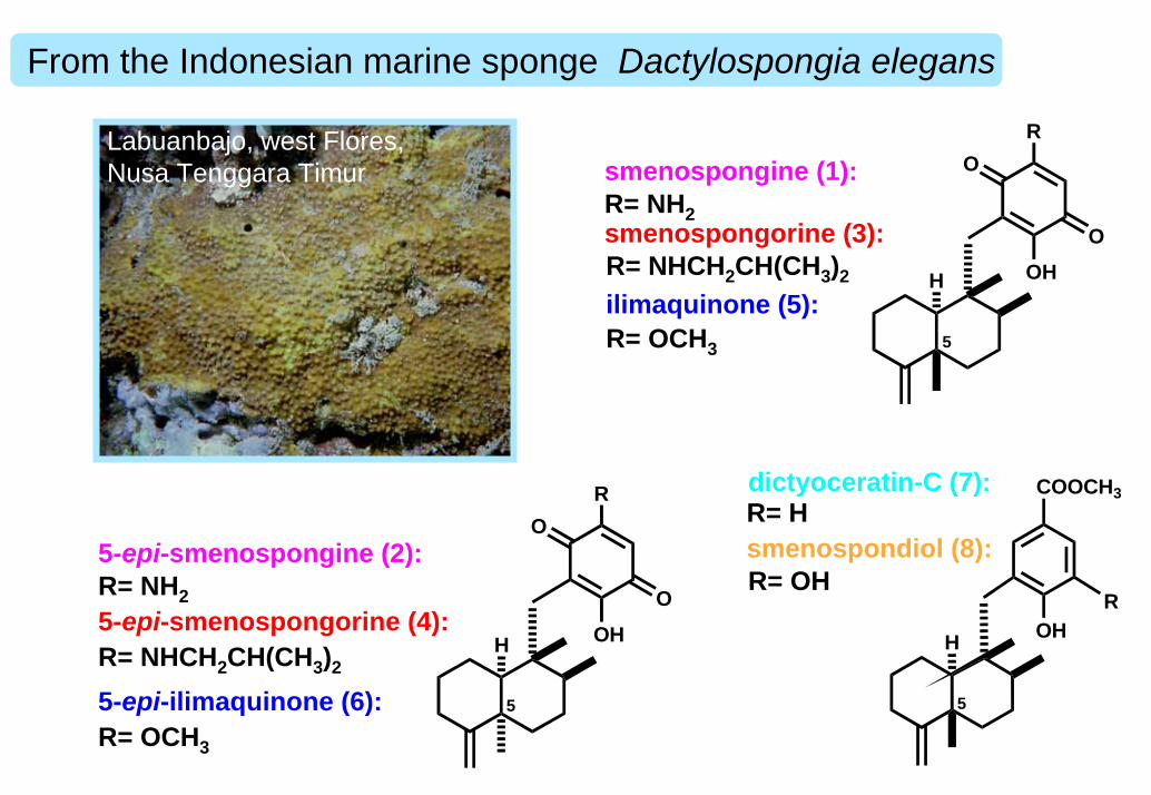
From the Indonesian marine sponge Dactylospongia elegans
Labuanbajo, west Flores, Nusa Tenggara Timur
OOH
R
H
O
5
smenospongine (1):R= NH2smenospongorine (3):R= NHCH2CH(CH3)2
ilimaquinone (5):R= OCH3
OHH
COOCH3
5
R
dictyoceratin-C (7):R= Hsmenospondiol (8):R= OH
OOH
R
H
O
5
5-epi-smenospongine (2):R= NH25-epi-smenospongorine (4):R= NHCH2CH(CH3)2
5-epi-ilimaquinone (6):R= OCH3
control
aphidicolin
smenospongine
(15 µM)
(15 µM)
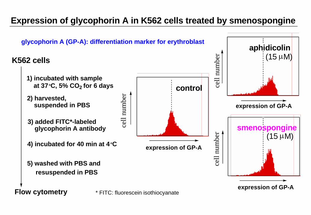
Expression of glycophorin A in K562 cells treated by smenospongine
expression of GP-A
K562 cells
1) incubated with sampleat 37°C, 5% CO2 for 6 days
2) harvested,suspended in PBS
3) added FITC*-labeledglycophorin A antibody
4) incubated for 40 min at 4°C
5) washed with PBS and
Flow cytometry * FITC: fluorescein isothiocyanate
resuspended in PBS
glycophorin A (GP-A): differentiation marker for erythroblast
expression of GP-A
expression of GP-A
cell
num
ber
cell
num
ber
cell
num
ber
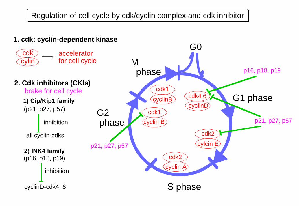
Regulation of cell cycle by cdk/cyclin complex and cdk inhibitor
G0
cyclinB
cdk1
cyclin Bcdk1
cyclin Acdk2
cylcin E
cdk2
cyclinDcdk4,6 G1 phase
p16, p18, p19
p21, p27, p57
p21, p27, p57
S phase
G2 phase
M phase
2. Cdk inhibitors (CKIs)
1) Cip/Kip1 family
2) INK4 family
all cyclin-cdks
inhibition
inhibition
cyclinD-cdk4, 6
(p21, p27, p57)
(p16, p18, p19)
1. cdk: cyclin-dependent kinase
cdkcylin
accelerator for cell cycle
brake for cell cycle
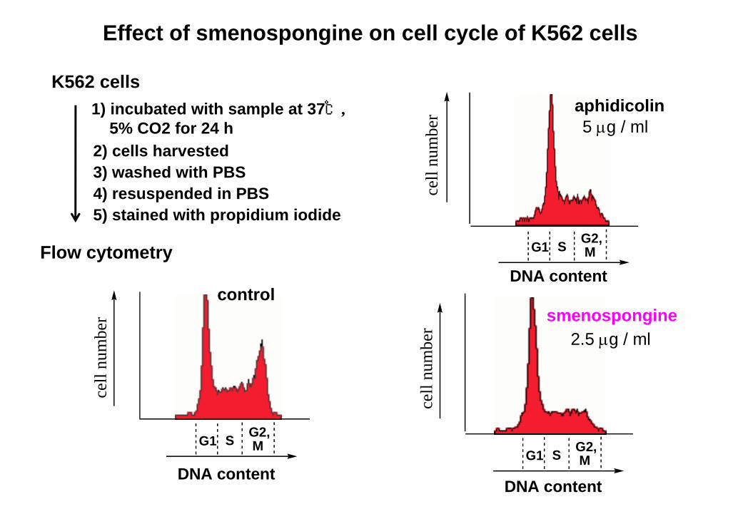
Effect of smenospongine on cell cycle of K562 cells
K562 cells1) incubated with sample at 37℃,
5% CO2 for 24 h2) cells harvested3) washed with PBS4) resuspended in PBS5) stained with propidium iodide
aphidicolin5 µg / ml
G1 G2, MS
DNA content
cell
num
ber
Flow cytometry
control
G1 G2, MS
DNA content
cell
num
ber smenospongine
2.5 µg / ml
G1 G2, MS
DNA content
cell
num
ber
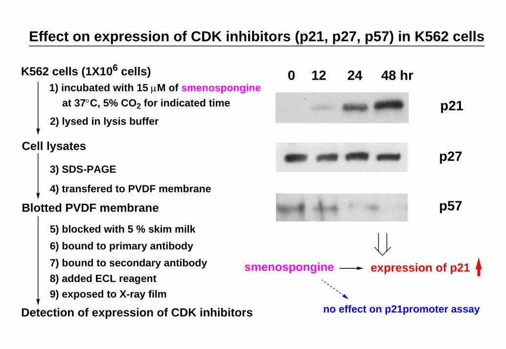
Effect on expression of CDK inhibitors (p21, p27, p57) in K562 cells
K562 cells (1X10⁶ cells)1) incubated with 15 µM of smenospongine
2) lysed in lysis buffer
0 12 24 48 hr
p21
p27
p57
3) SDS-PAGE
4) transfered to PVDF membrane
5) blocked with 5 % skim milk6) bound to primary antibody7) bound to secondary antibody8) added ECL reagent9) exposed to X-ray film
Cell lysates
Blotted PVDF membrane
Detection of expression of CDK inhibitors
at 37°C, 5% CO2 for indicated time
expression of p21
no effect on p21promoter assay
smenospongine
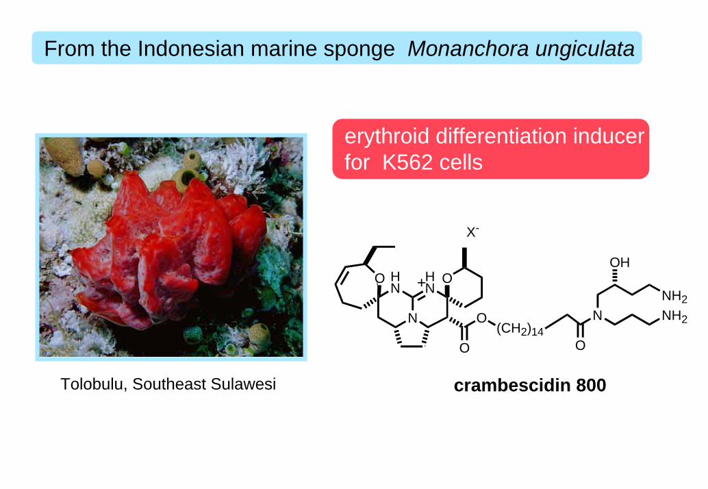
From the Indonesian marine sponge Monanchora ungiculata
erythroid differentiation inducer for K562 cells
Tolobulu, Southeast Sulawesi
N
HN
HN
OO
O
O(CH2)14
O
NNH2NH2
OH+
X-
crambescidin 800
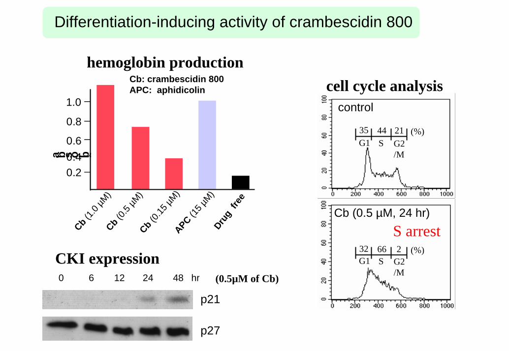
Differentiation-inducing activity of crambescidin 800
Cb (0.15
µM)
APC(15
µM)
Cb (1.0 µ
M)Cb (0.
5 µM)
Drug
free
0.20.40.6
0.8
1.0
a b s o r b aCb: crambescidin 800 APC: aphidicolin
hemoglobin production
G1 S G2/M
35 (%)44 21
control
G1 S G2/M
32 (%)66 2
S arrestCb (0.5 µM, 24 hr)
cell cycle analysis
0 6 12 24 48 hr
p21
p27
CKI expression(0.5µM of Cb)
ガン細胞に対する分化誘導物質の探索
2)神経芽細胞腫に対する分化誘導物質の例
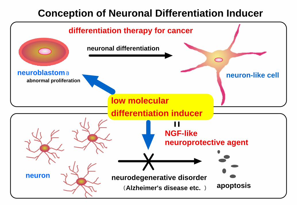
Conception of Neuronal Differentiation Inducer
neuroblastoma
apoptosisneuron
(Alzheimer's disease etc. )neurodegenerative disorder
NGF-like neuroprotective agent
abnormal proliferation
low molecular differentiation inducer
neuron-like cell
differentiation therapy for cancer
neuronal differentiation
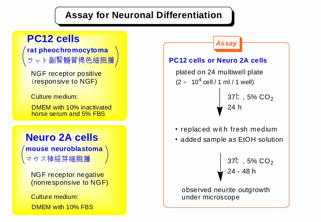
Assay for Neuronal Differentiation
PC12 cellsrat pheochromocytomaラット副腎髄質褐色細胞腫
・ replac ed w it h fresh medium・ added sample as EtOH solution
observed neurite outgrowth under microscope
37℃, 5% CO224 - 48 h
37℃, 5% CO224 h
plated on 24 multiwell plate(2 × 10⁴ cell / 1 ml / 1 well)
DMEM with 10% inactivated horse serum and 5% FBS
Neuro 2A cellsmouse neuroblastomaマウス神経芽細胞腫
DMEM with 10% FBS
Culture medium:
PC12 cells or Neuro 2A cells
NGF receptor positive( responsive to NGF)
NGF receptor negative(nonresponsive to NGF)
Culture medium:
Assay
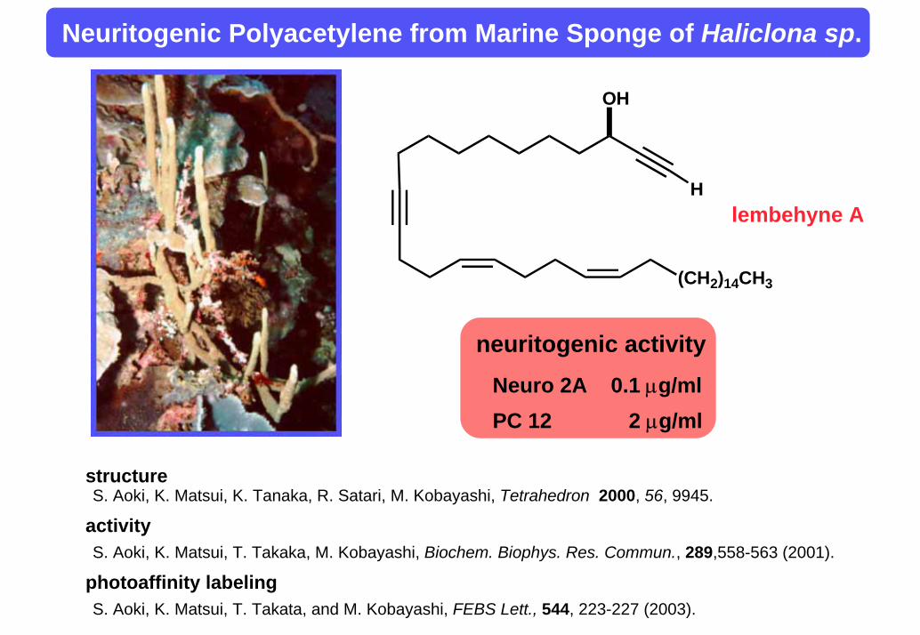
Neuritogenic Polyacetylene from Marine Sponge of Haliclona sp.
structureS. Aoki, K. Matsui, K. Tanaka, R. Satari, M. Kobayashi, Tetrahedron 2000, 56, 9945.
S. Aoki, K. Matsui, T. Takata, and M. Kobayashi, FEBS Lett., 544, 223-227 (2003).
activityS. Aoki, K. Matsui, T. Takaka, M. Kobayashi, Biochem. Biophys. Res. Commun., 289,558-563 (2001).
photoaffinity labeling
(CH2)14CH3
H
OH
lembehyne A
neuritogenic activityNeuro 2A 0.1 µg/mlPC 12 2 µg/ml
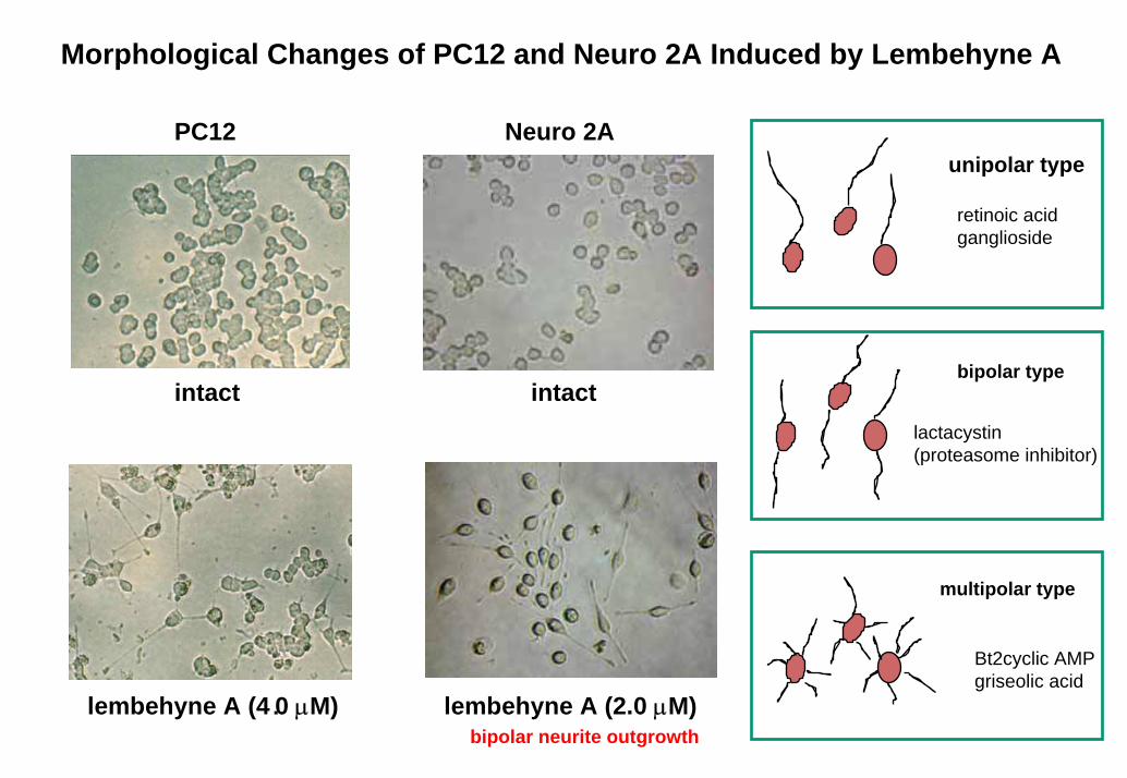
Morphological Changes of PC12 and Neuro 2A Induced by Lembehyne A
PC12
intact
Neuro 2A
intact
unipolar type
retinoic acidganglioside
bipolar type
lactacystin(proteasome inhibitor)
multipolar type
Bt2cyclic AMPgriseolic acid
lembehyne A (4.0 µM) lembehyne A (2.0 µM)bipolar neurite outgrowth
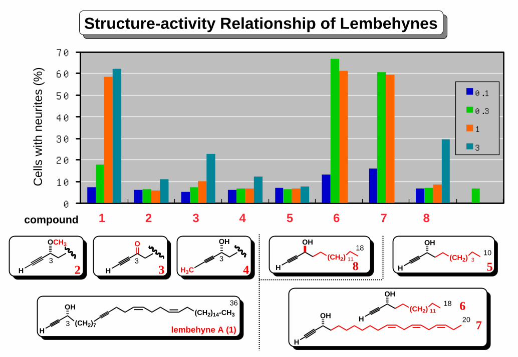
Structure-activity Relationship of Lembehynes
0 . 1
0 . 3
1
3
compound 1 2 3 4 5 6 7 8
Cel
ls w
ith n
eurit
es (%
)
0
10
20
30
40
50
60
70
OCH3
H3
2
O
H3
3
OH
H3C3
4(CH2) 3
H
OH10
5(CH2) 11
H
OH18
8
(CH2)14-CH3(CH2)7
H
OH
3
36
lembehyne A (1)
(CH2) 11H
OH
H
OH 20
18 67
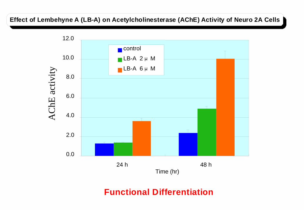
0.0
2.0
4.0
6.0
8.0
10.0
12.0
24 h 48 hTime (hr)
control
LB-A 2 μM
LB-A 6 μM
Effect of Lembehyne A (LB-A) on Acetylcholinesterase (AChE) Activity of Neuro 2A Cells
Functional Differentiation
AC
hE a
ctiv
ity
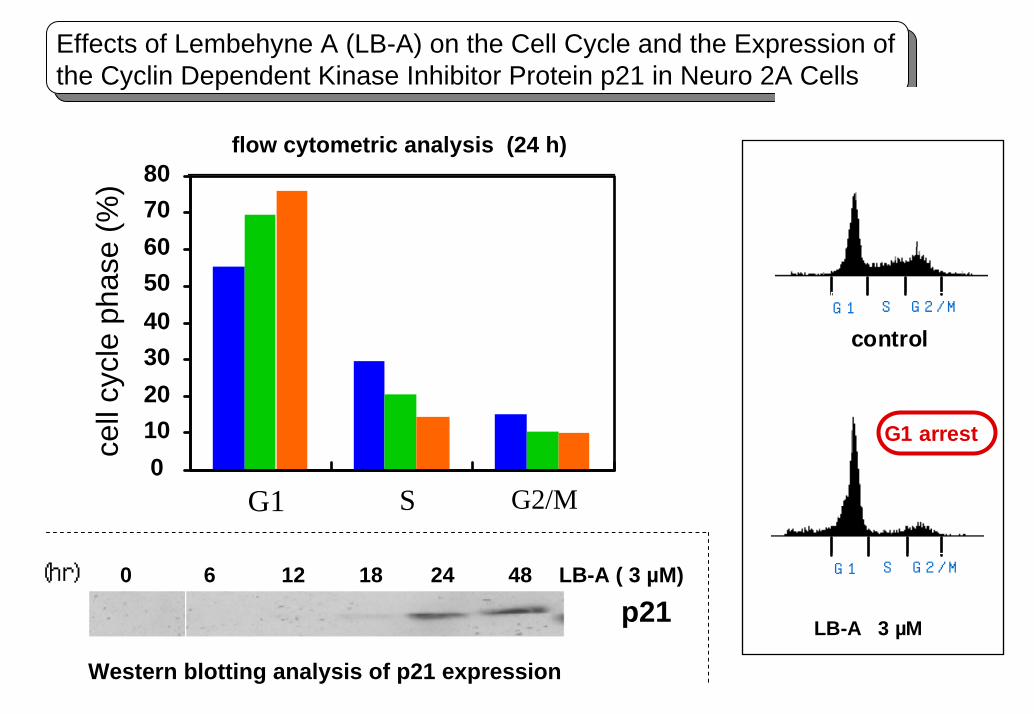
01020304050607080
Effects of Lembehyne A (LB-A) on the Cell Cycle and the Expression of the Cyclin Dependent Kinase Inhibitor Protein p21 in Neuro 2A Cells
G1 S G2/M
Western blotting analysis of p21 expression
(hr) LB-A ( 3 µM)
p210 6 12 18 24 48
flow cytometric analysis (24 h)ce
ll cy
cle
phas
e (%
)
G1 S G2 /M
G1 S G2 /M
control
G1 arrest
LB-A 3 µM
既存の活性物質O
S
HOOCHN CH3
O
NHO
H3COH CH3
CH3
OH
O
O N
N
N
N
NH2
OHHOOCHOOC
OH
NHOH
O
N
O
(CH2)13
H
OH
ONN
HN
NHCH3
MeO
H3C
O
CH3 CH3
COOHH3C CH3
CH3
O
HO
OH
O
OCH3
O
HO
H
OH
lactacystin
staurosporin
griseolic acid
trichostatin A
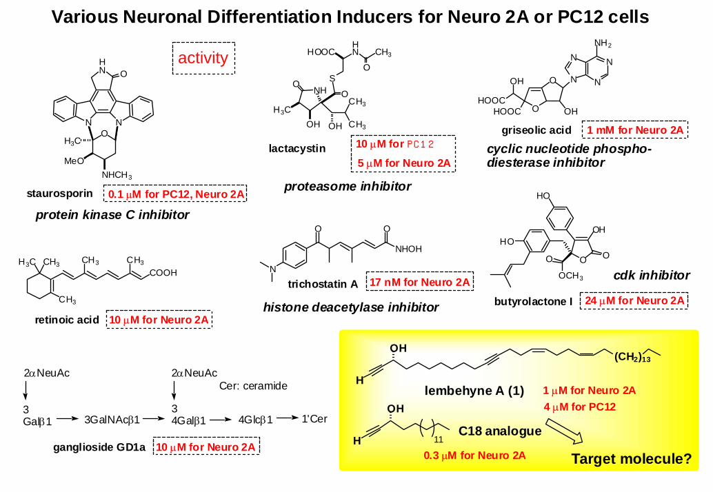
Various Neuronal Differentiation Inducers for Neuro 2A or PC12 cells
protein kinase C inhibitor
proteasome inhibitor
histone deacetylase inhibitor
cyclic nucleotide phospho-diesterase inhibitor
ganglioside GD1a
2αNeuAc
3Galβ1 3GalNAcβ1
34Galβ1 4Glcβ1 1'Cer
2αNeuAcCer: ceramide lembehyne A (1)
Target molecule?
retinoic acidbutyrolactone I
cdk inhibitor17 nM for Neuro 2A
5 µM for Neuro 2A
10 µM for Neuro 2A
0.1 µM for PC12, Neuro 2A
24 µM for Neuro 2A
1 mM for Neuro 2A
10 µM for Neuro 2A
activity
10 µM for PC1 2
11C18 analogue
1 µM for Neuro 2A4 µM for PC12
0.3 µM for Neuro 2A
血管新生阻害活性を有する
海綿由来成分bastadin類のin vivo抗腫瘍効果
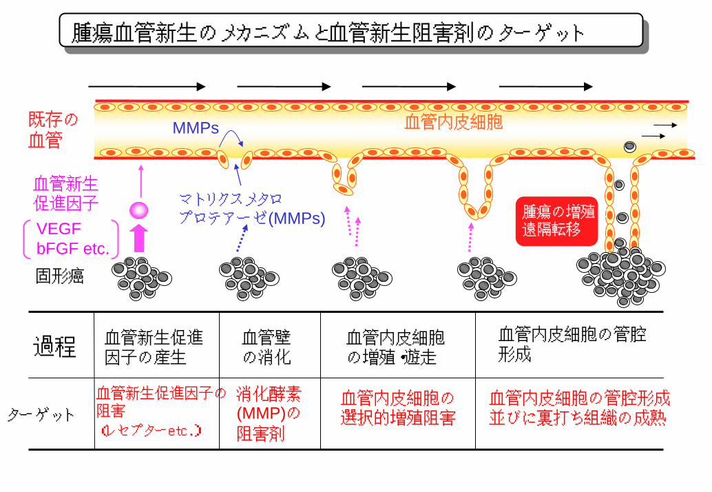
腫瘍血管新生のメカニズムと血管新生阻害剤のターゲット
既存の血管
血管新生促進因子 マトリクスメタロ
プロテアーゼ(MMPs)
MMPs 血管内皮細胞
固形癌
VEGFbFGF etc.
腫瘍の増殖と遠隔転移
血管新生促進因子の産生
血管壁の消化
血管内皮細胞の増殖・遊走
血管内皮細胞の管腔形成
血管新生促進因子の阻害
(レセプターetc.)
消化酵素(MMP)の阻害剤
血管内皮細胞の選択的増殖阻害
血管内皮細胞の管腔形成並びに裏打ち組織の成熟
過程
ターゲット
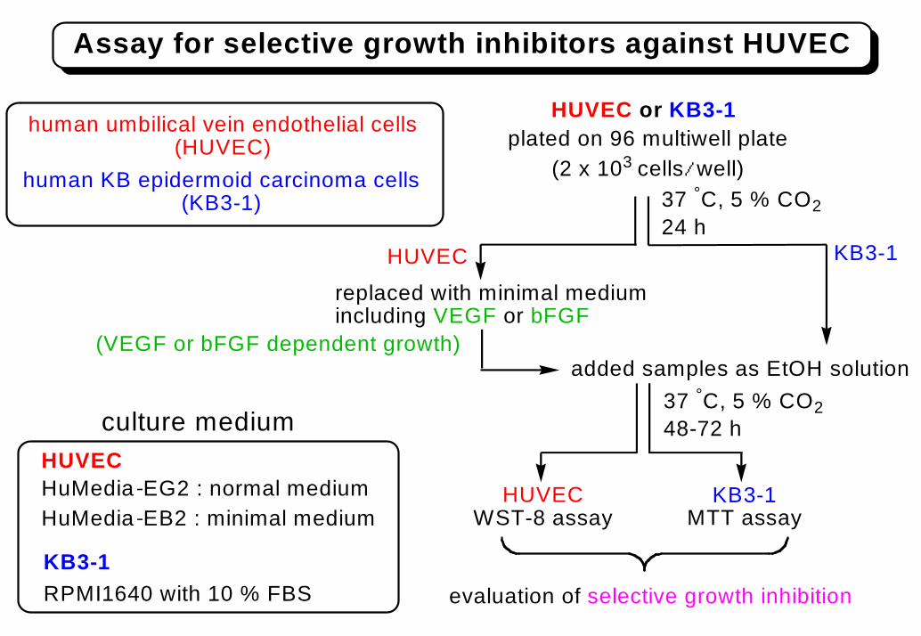
HUVEC or KB3-1plated on 96 multiwell plate
(2 x 10³ cells/well)
replaced with minimal mediumincluding VEGF or bFGF
HUVECWST-8 assay
KB3-1MTT assay
evaluation of selective growth inhibition
Assay for selective growth inhibitors against HUVEC
37 °C, 5 % CO224 h
37 °C, 5 % CO248-72 hculture medium
human umbilical vein endothelial cells(HUVEC)
HuMedia-EG2 : normal mediumHuMedia-EB2 : minimal medium
human KB epidermoid carcinoma cells(KB3-1)
RPMI1640 with 10 % FBS
added samples as EtOH solution(VEGF or bFGF dependent growth)
HUVEC
KB3-1
HUVEC KB3-1
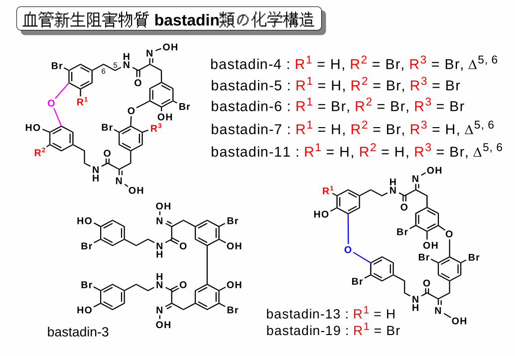
血管新生阻害物質 bastadin類の化学構造
HN
N
O
OHO
NNH
O
O
OH
OH
R3Br
Br
HO
Br
R2
56
R1bastadin-5 : R1 = H, R2 = Br, R3 = Brbastadin-6 : R1 = Br, R2 = Br, R3 = Brbastadin-7 : R1 = H, R2 = Br, R3 = H, ∆5, 6
bastadin-11 : R1 = H, R2 = H, R3 = Br, ∆5, 6
bastadin-4 : R1 = H, R2 = Br, R3 = Br, ∆5, 6
HN
N
O
OHBr O
NNH
O
O
Br
HO
OH
OH
BrBr
R1
bastadin-13 : R1 = Hbastadin-19 : R1 = Br
NH
NOH
O
HN
NOH
O
HO
HO
Br
Br OH
OH
Br
Br
bastadin-3
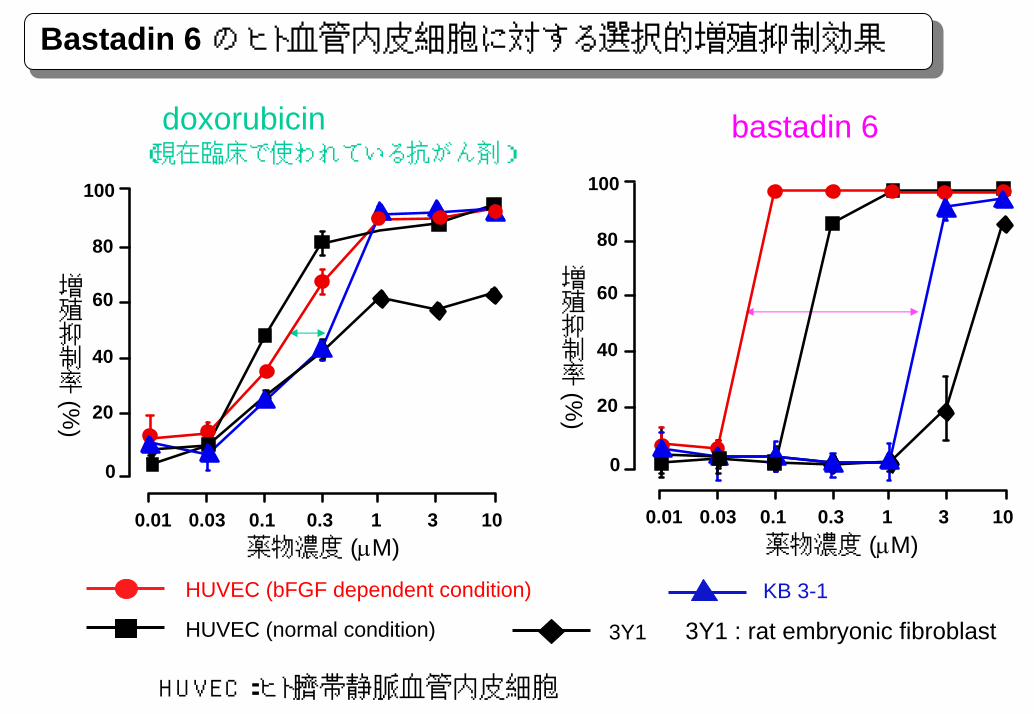
Bastadin 6 のヒト血管内皮細胞に対する選択的増殖抑制効果
doxorubicin(現在臨床で使われている抗がん剤)
bastadin 6
0
100
20
40
60
80
0.01 0.03 0.1 0.3 1 3 10薬物濃度 (µM)
増殖抑制率(%
)0
100
20
40
60
80
0.01 0.03 0.1 0.3 1 3 10
増殖抑制率(%
)
薬物濃度 (µM)
HUVEC (bFGF dependent condition) KB 3-1
3Y1 : rat embryonic fibroblastHUVEC (normal condition) 3Y1
HUVEC:ヒト臍帯静脈血管内皮細胞

Selective growth inhibition of cortistatins
Compounds 1 2 3 4Cell line IC50 S.I. IC50 S.I. IC50 S.I. IC50 S.I.
HUVECs 0.0018 1.1 0.019 0.12KB3-1 7 3900 120 110 150 7900 55 460Neuro2A 6 3300 160 150 180 9500 >300 -K562 7 3900 200 180 >300 - >300 -NHDF 6 3300 >300 - >300 - >300 -- : not determine IC50 = µM
cortistatin A (1) : R = Hcortistatin B (2) : R = OH
cortistatin C (3) : R = Hcortistatin D (4) : R = OH
HOR
N
OH
H
N
OHO
O
N
OH
H
N
O
R
Selective growth inhibition against HUVEC
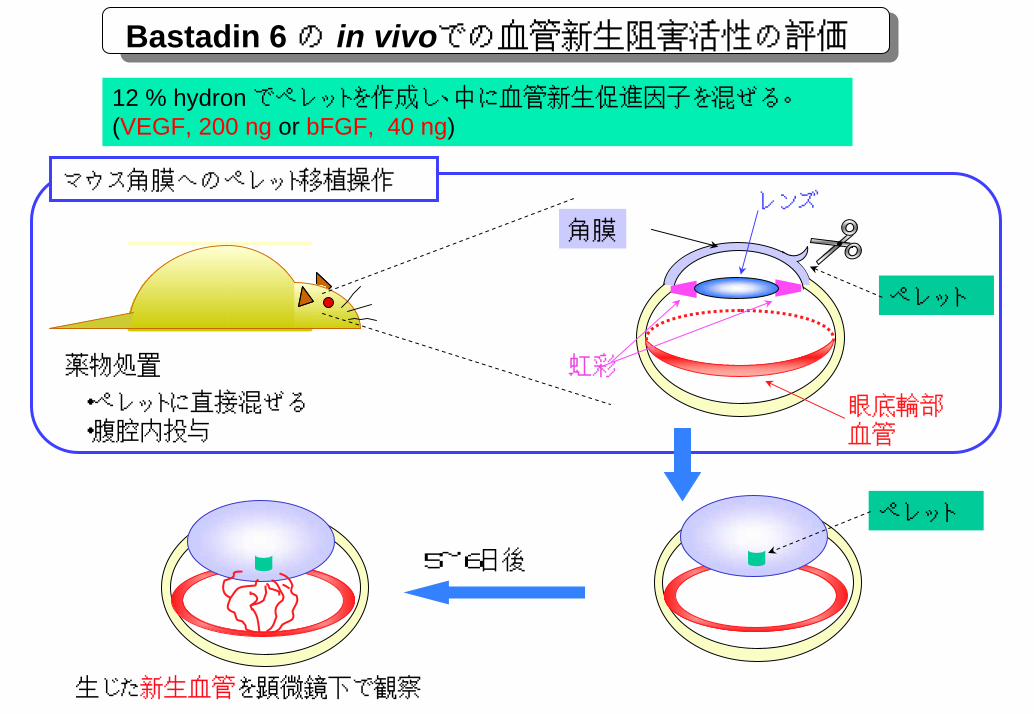
Bastadin 6の in vivoでの血管新生阻害活性の評価
12 % hydron でペレットを作成し、中に血管新生促進因子を混ぜる。(VEGF, 200 ng or bFGF, 40 ng)
マウス角膜へのペレット移植操作
5~6日後
角膜レンズ
虹彩
眼底輪部血管
ペレット
ペレット
薬物処置
・ペレットに直接混ぜる・腹腔内投与
生じた新生血管を顕微鏡下で観察
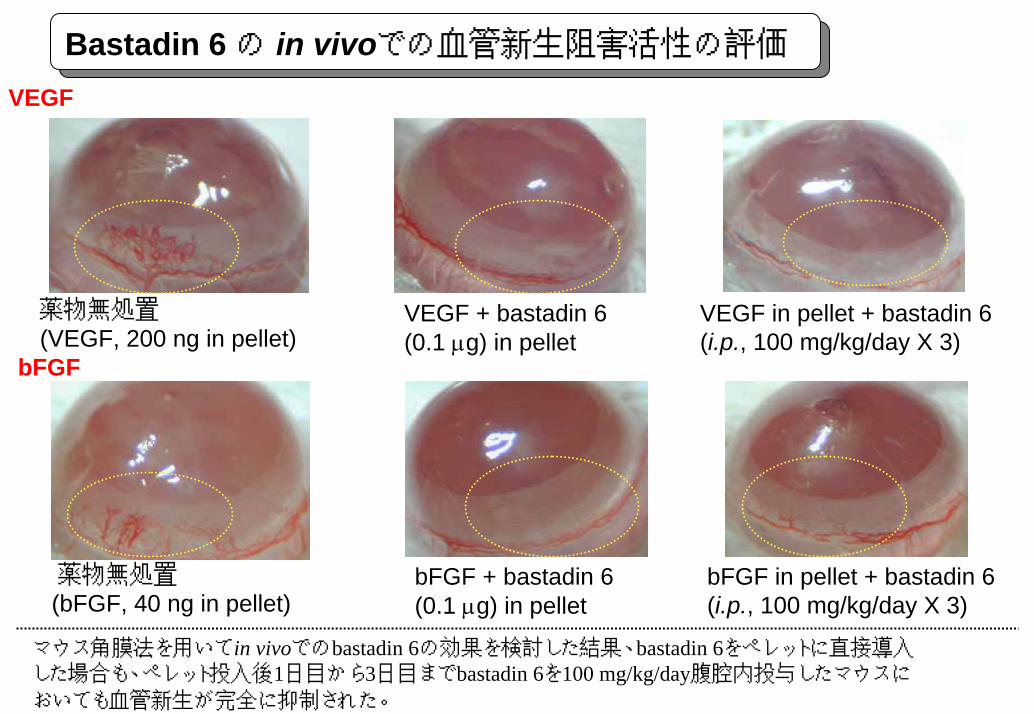
Bastadin 6の in vivoでの血管新生阻害活性の評価VEGF
bFGF
薬物無処置(VEGF, 200 ng in pellet)
薬物無処置(bFGF, 40 ng in pellet)
bFGF + bastadin 6(0.1 µg) in pellet
VEGF + bastadin 6 (0.1 µg) in pellet
VEGF in pellet + bastadin 6 (i.p., 100 mg/kg/day X 3)
bFGF in pellet + bastadin 6 (i.p., 100 mg/kg/day X 3)
マウス角膜法を用いてin vivoでのbastadin 6の効果を検討した結果、bastadin 6をペレットに直接導入した場合も、ペレット投入後1日目から3日目までbastadin 6を100 mg/kg/day腹腔内投与したマウスにおいても血管新生が完全に抑制された。
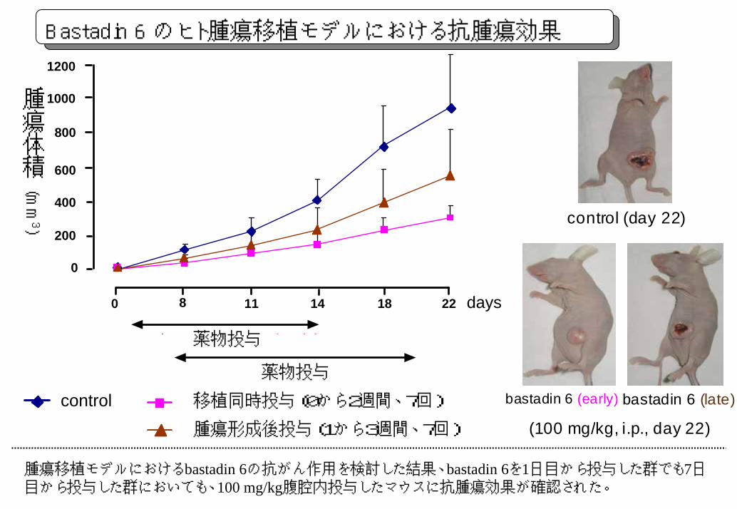
control (day 22)
bastadin 6 (early) bastadin 6 (late)
(100 mg/kg, i.p., day 22)
0
200
400
600
800
1000
1200
0 8 11 14 18 22 days
drug treatment (early)
drug treatment (late)
control early treatment (7 times in 0-2 week)late treatment (7 times in 1-3 week)
腫瘍体積
(mm3)
薬物投与
薬物投与
移植同時投与(0から2週間、7回)
腫瘍形成後投与(1から3週間、7回)
Bastadin 6 のヒト腫瘍移植モデルにおける抗腫瘍効果
腫瘍移植モデルにおけるbastadin 6の抗がん作用を検討した結果、bastadin 6を1日目から投与した群でも7日目から投与した群においても、100 mg/kg腹腔内投与したマウスに抗腫瘍効果が確認された。