Embed Size (px)
Citation preview

1
改正医療法・感染症法を考慮した院内感染防止ガイドライン
平成18年度 厚生労働科学研究費補助金 (新興・再興感染症研究事業)
「薬剤耐性菌等に関する研究」(H18-新興-11)
分担研究「医療機関における院内感染対策マニュアル作成のためのガイドライン」
作成の研究班
武澤 純 名古屋大学大学院医学系研究科救急・集中治療医学/教授(分担研究者)
土井まつ子 愛知医科大学看護学部/教授
仲井美由紀 愛知医科大学看護学部/講師
脇本寛子 愛知医科大学看護学部/講師
森澤雄司 自治医科大学医学部感染制御学/助教授
朝野和典 大阪大学医学部附属病院感染制御部/教授
井上善文 医療法人川崎病院外科/外科総括部長
鳥居啓三 名古屋大学医学部附属病院中央感染制御部/助教授
杉浦伸一 名古屋大学医学部附属病院医療経営管理部/講師
鈴木里和 国立感染症研究所細菌第二部/研究員
山根一和 国立感染症研究所細菌第二部/主任研究官
土手健太郎 愛媛大学医学部附属病院集中治療部/助教授
西村匡司 徳島大学病態情報医学講座救急・集中治療医学/教授
平潟洋一 長崎大学医学部・歯学部附属病院第二内科/講師
金光敬二 東北大学医学部附属病院検査部/講師
宮里明子 東北大学大学院感染制御・検査診断学/助手
洪 愛子 (社)日本看護協会認定部/認定部長
工藤友子 静岡県立静岡がんセンター/副看護師長
印田宏子 HAICS研究会/感染管理認定看護師
福岡敏雄 名古屋大学大学院医学系研究科救急・集中治療医学/助手
小野寺睦雄 名古屋大学大学院医学系研究科救急・集中治療医学/助手
2
ガイドライン作成の手順
本ガイドラインは院内感染防止のために必要とされている多数の項目の中から、以下のように
Evidence-based Clinical Practice Guideline作成の方法に従って、エビデンスのレベルや推奨
度等を考慮しつつ、医療施設において励行されるべき「骨子」について整理し記述した。
a) 論文の調査方法
論文の調査は、我が国および欧米の院内感染対策に関して出版された主要な著書と
Medline/PubMed、Cochrane Library、Best Evidence、日本医学中央雑誌などのコンピュータ
化されたデータベース、およびEvidence Based Medicine、ACP Journal Clubなどの 2次情報
雑誌を対象とした。さらに、必要に応じて、ハンドサーチも行った。
今回の集大成に当たっては、主に 2000 年以降に発表された研究や総論、ガイドラインを検討し
た。検索したデータベースはMedline と Cochrane Control Trial Registryである。
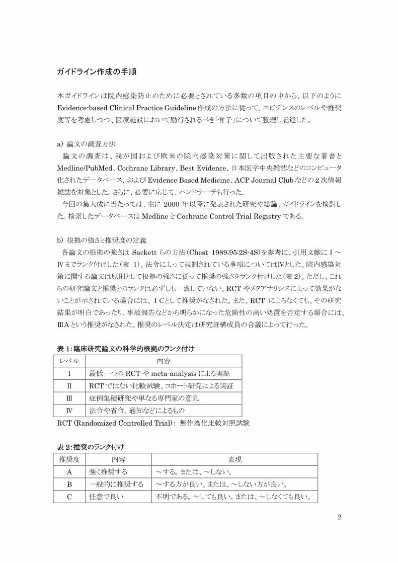
b) 根拠の強さと推奨度の定義
各論文の根拠の強さは Sackett らの方法(Chest 1989;95:2S-4S)を参考に、引用文献にⅠ~
Ⅳまでランク付けした(表 1)。法令によって規制されている事項についてはⅣとした。院内感染対
策に関する論文は原則として根拠の強さに従って推奨の強さをランク付けした(表 2)。ただし、これ
らの研究論文と推奨とのランクは必ずしも一致していない。RCT やメタアナリシスによって効果がな
いことが示されている場合には、ⅠCとして推奨がなされた。また、RCT によらなくても、その研究
結果が明白であったり、事故報告などから明らかになった危険性の高い処置を否定する場合には、
ⅢA という推奨がなされた。推奨のレベル決定は研究班構成員の合議によって行った。
表 1:臨床研究論文の科学的根拠のランク付け
レベル 内容
Ⅰ 最低一つの RCTやmeta-analysisによる実証
Ⅱ RCTではない比較試験、コホート研究による実証
Ⅲ 症例集積研究や単なる専門家の意見
Ⅳ 法令や省令、通知などによるもの
RCT (Randomized Controlled Trial): 無作為化比較対照試験
表 2:推奨のランク付け
推奨度 内容 表現
A 強く推奨する ~する。または、~しない。
B 一般的に推奨する ~する方が良い。または、~しない方が良い。
C 任意で良い 不明である。~しても良い。または、~しなくても良い。
3
c) このガイドラインは今後、院内感染対策中央会議、感染症関連学会、職能団体、病院団体など
の専門職組織に意見を招請し、その後広く社会から意見をいただいた後に確定する予定である。
d) 定期的見直しの必要性
このガイドラインは現時点での推奨に根拠を与える文献と、一部 bench study の結果や院内感
染事例報告を参考に作成されている。今後、本ガイドラインには 2~3 年ごとの定期的な見直しが
必要である。なお、このガイドラインでは院内感染対策を標準化できるように作成しているが、乳幼
児・小児や易感染性患者などでは特別な対策が必要であるため、できれば、これらの患者を対象と
したガイドラインが別途策定されることが望ましい。
4
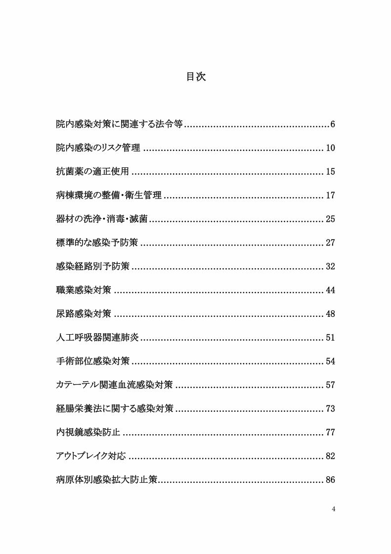
目次
院内感染対策に関連する法令等 .................................................. 6
院内感染のリスク管理 .............................................................. 10
抗菌薬の適正使用 .................................................................. 15
病棟環境の整備・衛生管理 ....................................................... 17
器材の洗浄・消毒・滅菌 ............................................................ 25
標準的な感染予防策 ............................................................... 27
感染経路別予防策 .................................................................. 32
職業感染対策 ........................................................................ 44
尿路感染対策 ........................................................................ 48
人工呼吸器関連肺炎 ............................................................... 51
手術部位感染対策 .................................................................. 54
カテーテル関連血流感染対策 ................................................... 57
経腸栄養法に関する感染対策 ................................................... 73
内視鏡感染防止 ..................................................................... 77
アウトブレイク対応 ................................................................... 82
病原体別感染拡大防止策 ......................................................... 86

5
6
院内感染対策に関連する法令等
武澤 純
1 届出
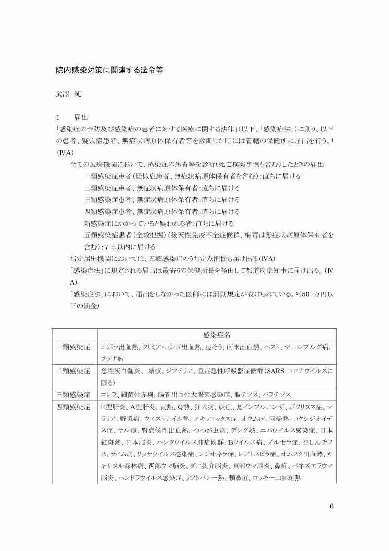
「感染症の予防及び感染症の患者に対する医療に関する法律」(以下、「感染症法」)に則り、以下
の患者、疑似症患者、無症状病原体保有者等を診断した時には管轄の保健所に届出を行う。1
(ⅣA)
全ての医療機関において、感染症の患者等を診断(死亡検案事例も含む)したときの届出
一類感染症患者(疑似症患者、無症状病原体保有者を含む):直ちに届ける
二類感染症患者、無症状病原体保有者:直ちに届ける
三類感染症患者、無症状病原体保有者:直ちに届ける
四類感染症患者、無症状病原体保有者:直ちに届ける
新感染症にかかっていると疑われる者:直ちに届ける
五類感染症患者(全数把握)(後天性免疫不全症候群、梅毒は無症状病原体保有者を
含む):7日以内に届ける
指定届出機関においては、五類感染症のうち定点把握も届け出る(ⅣA)
「感染症法」に規定される届出は最寄りの保健所長を経由して都道府県知事に届け出る。(Ⅳ
A)
「感染症法」において、届出をしなかった医師には罰則規定が設けられている。2(50 万円以
下の罰金)
感染症名
一類感染症 エボラ出血熱、クリミア・コンゴ出血熱、痘そう、南米出血熱、ペスト、マールブルグ病、
ラッサ熱
二類感染症 急性灰白髄炎、 結核、ジフテリア、重症急性呼吸器症候群(SARS コロナウイルスに
限る)
三類感染症 コレラ、細菌性赤痢、腸管出血性大腸菌感染症、腸チフス、パラチフス
四類感染症 E型肝炎、A型肝炎、黄熱、Q熱、狂犬病、炭疽、鳥インフルエンザ、ボツリヌス症、マ
ラリア、野兎病、ウエストナイル熱、エキノコックス症、オウム病、回帰熱、コクシジオイデ
ス症、サル痘、腎症候性出血熱、つつが虫病、デング熱、ニパウイルス感染症、日本
紅斑熱、日本脳炎、ハンタウイルス肺症候群、Bウイルス病、ブルセラ症、発しんチフ
ス、ライム病、リッサウイルス感染症、レジオネラ症、レプトスピラ症、オムスク出血熱、キ
ャサヌル森林病、西部ウマ脳炎、ダニ媒介脳炎、東部ウマ脳炎、鼻疽、ベネズエラウマ
脳炎、ヘンドラウイルス感染症、リフトバレー熱、類鼻疽、ロッキー山紅斑熱
7
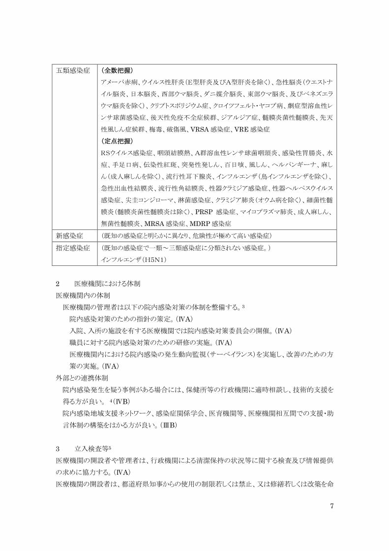
2 医療機関における体制
医療機関内の体制
医療機関の管理者は以下の院内感染対策の体制を整備する。3
院内感染対策のための指針の策定。(ⅣA)
入院、入所の施設を有する医療機関では院内感染対策委員会の開催。(ⅣA)
職員に対する院内感染対策のための研修の実施。(ⅣA)
医療機関内における院内感染の発生動向監視(サーベイランス)を実施し、改善のための方
策の実施。(ⅣA)
外部との連携体制
院内感染発生を疑う事例がある場合には、保健所等の行政機関に適時相談し、技術的支援を
得る方が良い。 4(ⅣB)
院内感染地域支援ネットワーク、感染症関係学会、医育機関等、医療機関相互間での支援・助
言体制の構築をはかる方が良い。(ⅢB)
3 立入検査等5
医療機関の開設者や管理者は、行政機関による清潔保持の状況等に関する検査及び情報提供
の求めに協力する。(ⅣA)
医療機関の開設者は、都道府県知事からの使用の制限若しくは禁止、又は修繕若しくは改築を命
五類感染症 (全数把握)
アメーバ赤痢、ウイルス性肝炎(E型肝炎及びA型肝炎を除く)、急性脳炎(ウエストナ
イル脳炎、日本脳炎、西部ウマ脳炎、ダニ媒介脳炎、東部ウマ脳炎、及びベネズエラ
ウマ脳炎を除く)、クリプトスポリジウム症、クロイツフェルト・ヤコブ病、劇症型溶血性レ
ンサ球菌感染症、後天性免疫不全症候群、ジアルジア症、髄膜炎菌性髄膜炎、先天
性風しん症候群、梅毒、破傷風、VRSA感染症、VRE感染症
(定点把握)
RSウイルス感染症、咽頭結膜熱、A群溶血性レンサ球菌咽頭炎、感染性胃腸炎、水
痘、手足口病、伝染性紅斑、突発性発しん、百日咳、風しん、ヘルパンギーナ、麻し
ん(成人麻しんを除く)、流行性耳下腺炎、インフルエンザ(鳥インフルエンザを除く)、
急性出血性結膜炎、流行性角結膜炎、性器クラミジア感染症、性器ヘルペスウイルス
感染症、尖圭コンジローマ、淋菌感染症、クラミジア肺炎(オウム病を除く)、細菌性髄
膜炎(髄膜炎菌性髄膜炎は除く)、PRSP 感染症、マイコプラズマ肺炎、成人麻しん、
無菌性髄膜炎、MRSA感染症、MDRP感染症
新感染症 (既知の感染症と明らかに異なり、危険性が極めて高い感染症)
指定感染症 (既知の感染症で一類~三類感染症に分類されない感染症。)
インフルエンザ(H5N1)
8
じられる事がある。(ⅣA)
医療機関の開設者は、都道府県知事からの開設の許可の取り消し、閉鎖を命じられる事がある。
(ⅣA)
4 業務委託6
施設管理者は微生物学的検査、医療機器等の滅菌又は消毒、医療施設の清掃等の業務を委
託することが出来る。(ⅣC)
医療機関の管理者は、医療法施行令に定める業務を委託する場合は、その業務を適正に行う
能力のある者として、医療法施行規則に定める基準を満たす者に委託する。(ⅣA)
委託する業務に関する最終的責任は医療機関にある。(ⅣA)
5 診療報酬(平成 18年度診療報酬改定)7
以下の算定要件全てを満たさない場合、入院基本料の算定は認められない。(ⅣA)
院内感染防止対策を実施している。
「院内感染防止対策委員会(院内感染対策委員会)」が設置され、月 1 回程度、定期的に開催
されている。
「感染情報レポート」が医療機関により週 1回程度作成され、活用される体制が取られている。
「感染情報レポート」は、入院中の患者からの各種細菌の検出状況や薬剤感受性成績のパター
ン等が医療機関の疫学情報として把握、活用されることを目的として作成される。
「感染情報レポート」は、各病棟からの拭き取り等による各種細菌の検出状況を記すものでな
い。
職員等に手指衛生管理の励行を徹底させるとともに、各病室に水道又は擦式手指消毒薬が設
置されている。
医療安全対策加算の施設基準に係る届出には、専任の院内感染管理者が配置されている。(Ⅳ
A)
6 労働安全衛生法関連(ここでは、事業者を医療機関の管理者と同義として考える)(ⅣA)
事業者は、病原体等による健康障害を防止するため必要な措置を講じなければならない。 8
事業者は、労働者を就業させる建設物その他の作業場について、清潔等に必要な措置及び労
働者の健康、風紀及び生命の保持のため必要な措置を講じなければならない。9
事業者は、労働者を雇い入れ、又は労働者の作業内容を変更したときは、業務に関して発生す
るおそれのある疾病の原因及び予防に関する内容等の安全又は衛生のため必要な事項につ
いて、教育を行なわなければならない。10
事業者は、病毒伝ぱのおそれのある伝染性の疾病にかかった者については、その就業を禁止
しなければならない。11
事業者は、病原体により汚染された排気、排液又は廃棄物については、消毒、殺菌等適切な処

9
理をした後に、排出し、又は廃棄しなければならない。12
事業者は、病原体による汚染のおそれの著しい業務に従事する労働者に使用させるために、保
護手袋、保護衣、保護眼鏡、呼吸用保護具、履物等適切な保護具を備えなければならない。13
事業者は、保護具又は器具の使用によって、労働者に疾病感染のおそれがあるときは、各人専
用のものを備え、又は疾病感染を予防する措置を講じなければならない。14
事業者は、病原体によって汚染のおそれの著しい作業場においては、作業場外に休憩の設備
を設けなければならない。15
事業者は、身体又は被服を汚染するおそれのある業務に労働者を従事させるときは、洗眼、洗
身若しくはうがいの設備、更衣設備又は洗濯のための設備を設けなければならない。16
1 感染症法第 12条第 1項 2 感染症法第 69条第 1項第 1号 3 医療法第 6条の 10、医療法施行規則第 11条第 2項 4 「医療施設における院内感染の防止について」(平成 17年 2月 1日医政指発第 0201004号)の(別
記) 5 医療法第 24条第 1項、医療法第 25条第 1項、医療法第 29条第 1項第 3号 6 医療法第 15条の 2、医療法施行令第 4条の 7、医療法施行規則第 9条の 7~15、「病院、診療所等
の業務委託について」(平成 5年 2月 15日指第 14号) 7 基本診療料の施設基準等」(平成 18年厚生労働省告示第 93号)、「基本診療料の施設基準等及び
その届出に関する手続きの取扱いについて」(平成 18年 3月 6日保医発第 0306002号) 8 労働安全衛生法第 22条第 1項第 1号 9 労働安全衛生法第 23条 10 労働安全衛生規則第 35条第 1項第 5号 11 労働安全衛生規則第 61条第 1項 1号 12 労働安全衛生規則第 581条 13 労働安全衛生規則第 593、594条 14 労働安全衛生規則第 598条 15 労働安全衛生規則第 614条 16 労働安全衛生規則第 625条 1項

10
院内感染のリスク管理
朝野和典
1 院内感染対策に関する責任と権限および組織
1.1 病院、有床診療所の管理者(以下、施設管理者)は院内感染対策など医療安全の確
保に関して責任をもつ。17(ⅣA)
1.2 施設管理者は、院内感染対策委員会(infection control committee ;ICC)を設置
する。18(ⅣA)
1.3 施設管理者は、院内感染対策委員会の構成員として、施設管理者、看護部、薬剤部
門、検査部門、事務部門の責任者感染症対策が専門の医師等の職員を配置する。
18(ⅣA)
1.4 施設管理者は院内感染対策委員会を月に 1回程度開催する。18(ⅣA)
1.5 施設管理者は、感染対策の実務的責任者(専任の院内感染管理者)を任命する方
が良い。18(ⅣB)
1.6 施設管理者は、感染対策チーム(ICT)を組織し、院内感染対策に関する日常活動
を行う方が良い。18(ⅣB)
2 感染対策担当者(ICTなど)の機能と業務
2.1 施設管理者は感染対策担当者に院内感染対策の実施に関する権限を委譲する。19
(ⅢA)
2.2 施設管理者は院内感染対策の実施に関する財政的措置を行なう。19(ⅢA)
2.3 感染対策担当者あるいは ICTの構成員は、感染制御医師(ICD)、感染管理看護師
(ICN)および感染制御担当者(ICP;臨床検査技師、薬剤師など)などとする方が良
い。19(ⅢB)
2.4 ICTの中に、専任の院内感染管理者を配置する方が良い。19(ⅣB)
2.5 感染対策担当者は ICD、ICN、ICP(薬剤師、臨床検査技師)などの専門認定を取
得する方が良い。20, 21, 22, 23(ⅢB)
2.6 感染対策担当者は、院内感染対策として職員の健康管理、教育、感染対策相談(コ
ンサルテーション)、発生動向監視(サーベイランス)、対策実施の適正化(レギュレー
ション)、および介入(インターベンション)を行なう。24(ⅢA)
3 管理システムの構築
3.1 施設管理者は、各部署において、業務を行ないながら感染対策担当者と協力して感
染対策や情報の収集を行なう、看護師(リンクナース)を配備する方が良い。25, 26(Ⅱ
B)
11
4 教育、研修
4.1 感染対策担当者は、職員を対象として、施設全体あるいは部署や職種を限定して、
定期的に院内感染対策に関する教育と実習を行なう。27, 28, 29(ⅡA)
4.2 感染対策担当者は、院内感染の増加が疑われた場合、あるいは確認された場合は、
介入のひとつの手段として職員を対象として、施設全体あるいは部署や職種を限定
して、院内感染対策に関する教育と実習を行なう。30(ⅡA)
4.3 院内感染管理に関する情報を関連部署に提供する。31(ⅣA)
5 感染対策相談(コンサルテーション)
5.1 各部署からの院内感染対策に関する質問に対し、施設の疫学的情報を考慮し、根拠
に基づく改善指導を行なう方が良い。(ⅢB)
5.2 病院内で発生した感染症の診断、治療に関する質問に対し、施設の疫学的情報を
考慮し、根拠に基づく診療指導を行なう方が良い。32(ⅡB)
6 発生動向監視(サーベイランス)
6.1 感染対策担当者は、1週間に一度程度各部署における院内感染事例を把握する。18
(ⅣA)
6.2 感染対策担当者は、病院感染の発生率に関するサーベイランスを部署とターゲットを
絞って実施する。(ⅡA)
6.3 感染対策担当者は、院内あるいは外注の検査会社からの情報をもとに、1週間に1回
程度、微生物の分離状況を把握する。18(ⅣA)
6.4 感染対策担当者は、院内感染に関する情報を分析、評価し、効率的な感染対策に
役立てる。18(ⅣA)
6.5 感染対策担当者は、院内感染の発生状況を 1 ヶ月に1度程度、院内感染対策委員
会に報告し、対策に活用する。18(ⅣA)
6.6 感染対策担当者は地域や全国のサーベイランスへの参加を促し、自施設の感染防
止機能を相対的に評価する方が良い。33, 34(ⅢB)
7 対策実施の適正化(レギュレーション)
7.1 感染対策担当者は、最新のエビデンスに基づいたガイドラインを参考に、自施設の
実情に合わせたマニュアル(手順書)を作成し、それを各部署に配布する。35(ⅣA)
7.2 マニュアルには、「標準予防策」、「感染経路別予防策」、「職業感染予防策」、「疾患
別感染対策」、「洗浄・消毒」、「抗菌薬適正使用」などに関する施設の実情や各部署
の特有の対策を盛り込んだ項目を含んだ方が良い。(ⅣB)
7.3 感染対策担当者はマニュアルに、定期的に新しい情報を取り入れ、改訂を行なう。19
(ⅣA)

12
7.4 感染対策担当者は、職員が病院内のマニュアルを遵守していることを定期的に調査
して確認する。19(ⅢA)
7.5 感染対策担当者は、耐性菌の分離率を減尐させるため、抗菌薬の適正使用をマニュ
アル化し、職員に周知する。35, 36, 37(ⅠA)
7.6 感染対策担当者は、特定抗菌薬(広域スペクトラムを有する抗菌薬、抗 MRSA 薬な
ど)の使用に際しては許可制もしくは届出制をとり、抗菌薬の適正使用を監視する。19,
38, 39, 40, 41, 42, 43, 44, 45(ⅢA)
8 改善への介入(インターベンション)
8.1 感染対策担当者はサーベイランスデータなどから院内感染の増加が疑われ、あるい
は確認された場合には、疫学的手法を用いて要因分析を行う。46, 47, 48, 49, 50, 51, 52
(ⅡA)
8.2 感染対策担当者は院内感染の増加が確認された場合には、要因分析から得られた
データを基に改善策を講じる。(ⅣA)
8.3 感染対策担当者はサーベイランスデータ、巡回による所見、要因分析の結果などの
情報を迅速に関係部署に知らせ、情報を共有する。(ⅢA)
9 職員健康管理
9.1 施設管理者は、定期的に行われる職員の健康診断を実施する。19(ⅣA)
9.2 施設管理者は血液や体液に暴露する可能性のある職員には、B型肝炎ワクチンを接
種する。19, 53(ⅡA)
9.3 施設管理者は風疹、流行性耳下腺炎、麻疹、水痘に対する抗体陰性の職員にそれ
ぞれのワクチン接種、および毎年インフルエンザワクチンの接種を実施する方が良い。
54(ⅢB)
9.4 施設管理者は、結核を疑われる職員を他者への感染の可能性がある期間は休業さ
せる。(ⅣA)
9.5 施設管理者は、急性胃腸炎(ノロ、ロタウイルス感染症を含む)、流行性角結膜炎など
の伝染性疾患に職員が罹患した場合、二次感染の可能性がなくなるまで休業を含め
て病原微生物に応じた対策を実施する。(ⅢA)
17 医療法(http://law.e-gov.go.jp/htmldata/S23/S23HO205.html) 18 診療報酬(http://www.mhlw.go.jp/topics/2006/03/dl/tp0314-1b01.pdf) 19 日本医療機能評価機構(http://jcqhc.or.jp/html/index.htm) 20 ICD制度協議会(http://www.icd.umin.jp/) 21 日本看護協会(http://www.nurse.or.jp/senmon/kansen/index.html) 22 日本病院薬剤師会(http://www.jshp.or.jp/index.htm) 23 日本臨床微生物学会(http://www.jscm.org/icmt/index.html) 24 国立大学医学部附属病院感染対策協議会;病院感染対策ガイドライン 25 Dawson SJ. The role of the infection control link nurse. J Hosp Infect. 2003;54:251-257.

13
26 Tsuchida T, Makimoto K, Toki M, Sakai K, Onaka E, Otani Y. The effectiveness of a
nurse-initiated intervention to reduce catheter-associated bloodstream infections in an
urban acute hospital: An intervention study with before and after comparison. Int J Nurs
Stud. 2006 (in press) 27 増田道明、藤澤隆一、山本勝彦、他.医師の卒後臨床研修開始時における感染制御教育の試み.
環境感染 2005, 20:193-199. 28 医療施設における新規採用看護職に対する感染管理教育とその評価.環境感染 2004,
19:409-414. 29 Wisniewski MF, Kim S, Trick WE, et al. Effect of education on hand hygiene beliefs and
practices: a 5-year program. Infect Control Hosp Epidemiol. 2007;28:88-91. 30 Haley RW, Cushion NB, Tenover FC, et al Eradication of endemic methicillin-resistant
Staphylococcus aureus infections from a neonatal intensive care unit. J Infect Dis. 1995
Mar;171(3):614-624. 31 Oie S, Kamiya A. Assessment of and intervention for the misuse of aldehyde disinfectants
in Japan. Infect Control Hosp Epidemiol. 2002 Feb;23(2):98-99. 32 Takakura S, Fujihara N, Saito T, et al. Improved clinical outcome of patients with
Candida bloodstream infections through direct consultation by infectious diseases
physicians in a Japanese university hospital. Infect Control Hosp Epidemiol. 2006 Sep;27
(9):964-968. 33 Yoshida J, Shinohara M, Ishikawa M, et al. Surgical site infection in general and thoracic
surgery: surveillance of 2 663 cases in a Japanese teaching hospital. Surg Today.
2006;36:114-118. 34 Suka M, Yoshida K, Takezawa J. A practical tool to assess the incidence of nosocomial
infection in Japanese intensive care units: the Japanese Nosocomial Infection Surveillance
System. J Hosp Infect. 2006;63:179-184. 35 Dellit TH, Owens RC, McGowan JE Jr, et al. Infectious Diseases Society of America and
the Society for Healthcare Epidemiology of America guidelines for developing an
institutional program to enhance antimicrobial stewardship. Clin Infect Dis. 2007
15;44:159-177. 36 Solomon DH, Van Houten L, Glynn RJ, Academic detailing to improve use of
broad-spectrum antibiotics at an academic medical center. Arch Intern Med. 2001,
27;161:1897-1902. 37 Fraser GL, Stogsdill P, Dickens JD Jr,et al. Antibiotic optimization. An evaluation of
patient safety and economic outcomes.Arch Intern Med. 1997, 25;157. 38 Seligman SJ. Reduction in antibiotic costs by restricting use of an oral cephalosporin. Am
J Med 1981; 71:941–944. 39 Britton HL, Schwinghammer TL, Romano MJ. Cost containment through restriction of
cephalosporins. Am J Hosp Pharm 1981; 38:1897–1900. 40 Hayman JN, Sbravati EC. Controlling cephalosporin and aminoglycoside costs through
pharmacy and therapeutics committee restrictions. Am J Hosp Pharm 1985; 42:1343–1347. 41 Woodward RS, Medoff G, Smith MD, Gray JL. Antibiotic cost savings from formulary
restrictions and physician monitoring in a medicalschool-affiliated hospital. Am J Med 1987;
83:817–823. 42 Coleman RW, Rodondi LC, Kaubisch S, Granzella NB, O’Hanley PD. Cost-effectiveness of
prospective and continuous parenteral antibiotic control: Experience at the Palo Alto
Veterans Affairs Medical Center from 1987 to 1989. Am J Med 1991; 90:439–444. 43 Maswoswe JJ, Okpara AU. Enforcing a policy for restricting antimicrobial drug use. Am J
Health Syst Pharm 1995; 52:1433–1435. 44 White AC, Atmar RL, Wilson J, Cate TR, Stager CE, Greenberg SB. Effects of requiring
prior authorization for selected antimicrobials: expenditures, susceptibilities, and clinical
outcomes. Clin Infect Dis 1997; 25:230–239. 45 Pear SM, Williamson TH, Bettin KM, Gerding DN, Galgiani JN. Decrease in nosocomial
Clostridium difficile-associated diarrhea by restricting clindamycin use. Ann Intern Med

14
1994; 120:272–277. 46 Morimoto Y, Sugiura T, Tatebayashi S, et al. Reduction in incidence of
methicillin-resistant Staphylococcus aureus (MRSA) after radical surgery for head and
neck cancer. Spec Care Dentist. 2006 ;26:209-213. 47 Nagashima G, Kikuchi T, Tsuyuzaki H, et al. To reduce catheter-related bloodstream
infections: is the subclavian route better than the jugular route for central venous
catheterization? J Infect Chemother. 2006;12:363-365. 48 Kikuchi T, Nagashima G, Taguchi K, et al. Contaminated oral intubation equipment
associated with an outbreak of carbapenem-resistant Pseudomonas in an intensive care unit.
J Hosp Infect. 2007;65:54-57. 49 Konishi T, Watanabe T, Morikane K, et al. Prospective surveillance effectively reduced
rates of surgical site infection associated with elective colorectal surgery at a university
hospital in Japan.Infect Control Hosp Epidemiol. 2006;27:526-8. Epub 2006 Apr 20. 50 Yanai M, Uehara Y, Takahashi S. Surveillance of infection control procedures in dialysis
units in Japan: a preliminary study. Ther Apher Dial. 2006;10:78-86. 51 Takahashi H, Kramer MH, Yasui Y, et al. Nosocomial Serratia marcescens outbreak in
Osaka, Japan, from 1999 to 2000. Infect Control Hosp Epidemiol. 2004 Feb;25(2):156-61. 52 Morikane K, Nishioka M, Tanimura H, et al. Using surveillance data to direct infection
control efforts to reduce surgical-site infections following clean abdominal operations in
Japan. Infect Control Hosp Epidemiol. 2002 Jul;23(7):404-406. 53 Lahaye D, Strauss P, Baleux C, et al. Cost-benefit analysis of hepatitis-B vaccination.
Lancet. 1987 22:441-3. 54 Asari S, Deguchi M, Tahara K, et al. Seroprevalence survey of measles, rubella, varicella,
and mumps antibodies in health care workers and evaluation of a vaccination program in a
tertiary care hospital in Japan. Am J Infect Control. 2003;31:157-162.
15
抗菌薬の適正使用
平潟洋一
1 抗菌薬の適正使用の原則
1.1 抗菌薬の使用制限だけではなく、抗菌薬の適正使用と他の感染対策との組み合わ
せにより耐性菌の出現を抑制する。55, 56(ⅡA)
1.2 1.2 2002 年に発表された CDC の「薬剤耐性の予防のためのキャンペーン
(Campaign to Prevent Antimicrobial Resistance in Healthcare Settings)」
(www.cdc.gov/drugresistance/healthcare)57は計 12のステップからなる 4つの戦
略で構成されている。そのうちのひとつである「抗菌薬の適正使用」は、下記に示すよ
うに12のステップのうちその半数に当たる6ステップを占めており、これを参考に適正
使用を推進する(表)。(ⅢA)
表:入院中の成人における耐性菌を防止するための 12のステップより一部引用
戦略:抗菌薬の適正使用
Step 5. 抗菌薬使用の標準化
Step 6. 病院全体および疾病ごとの薬剤感受性データの活用
Step 7. 血液培養の偽陽性に対して抗菌薬を使用しない
Step 8. 除菌を目的として抗菌薬を投与しない
Step 9. バンコマイシンの適正使用
Step 10. 治療終了あるいは感染が否定された場合は速やかに投与を中止する
2 周術期予防投与
2.1 手術部位感染の防止に抗菌薬の予防的投与を行う。(ⅠA)
2.2 執刀開始1~2時間前に抗菌薬の投与を開始する。(ⅠA)
2.3 セファゾリンを使用し、手術時間が 3時間を越える場合は、術中の追加投与を 2-5時
間毎に行なう。(ⅠA)
2.4 清潔手術における手術後の抗菌薬投与は 24時間以内とする。(ⅠA)
2.5 準清潔手術における手術後の抗菌薬投与は 4日以内とする方が良い。(ⅡB)
3 微生物検査の結果と抗菌薬の選択
3.1 抗菌薬投与を開始する前に、感染が疑われる部位から採取した検体や血液の培養
を行なう方が良い。(ⅢB)
3.2 感受性検査結果を得るまではグラム染色結果や院内における主要な細菌の感受性
パターンを参考に抗菌薬を選択する方が良い。(ⅢB)
16
3.3 感受性試験の結果に基づいて抗菌薬の続行または変更を行う。(ⅢA)
4 広域、狭域の選択
4.1 重症感染症、重篤な基礎疾患を有する患者の感染症、複数菌感染症が疑われる場
合は広域抗菌薬を初期治療薬として選択する。(ⅡA)
4.2 感受性試験の結果が判明すれば、狭域抗菌薬への変更を行う。(ⅢA)
5 適正投与回数
5.1 βラクタム薬は投与回数を増やして使用する方が良い。(ⅢB)
5.2 アミノ配糖体は 1日投与量を分割せずに単回投与する方が良い。(ⅠB)
5.3 ニューキノロン薬は投与回数を減らして 1回投与量を増加させる方が良い。(ⅢB)
55 Dellit TH, Owens RC, McGowan JE, et al. Infectious Diseases Society of America and the
Society for Healthcare Epidemiology of America Guidelines for developing an institutional
program to enhance antimicrobial stewardship. Clin Infect Dis 2007: 44; 159-177. 56 CDC: Management of Multidrug-Resistant Organisms in Healthcare Settings, 2006
(http://www.cdc.gov/ncidod/dhqp/pdf/ar/mdroGuideline2006.pdf) 57 Brinsley K, Srinivasan A, Sinkowitz-Cochran R, et al. Implementation of the Campaign to
Prevent Antimicrobial Resistance in Healthcare Settings: 12 Steps to Prevent Antimicrobial
Resistance Among Hospitalized Adults--experiences from 3 institutions. Am J Infect
Control. 2005; 33: 53-54.
17
病棟環境の整備・衛生管理
土井まつ子
1 病棟衛生管理の責任と権限
1.1 看護師長は病棟環境の整備・衛生管理を実施する責任者としての役割を果たす。
(ⅢA)
1.2 看護師長は、院内感染対策委員会の指導の下に、病棟環境の整備・衛生管理を行う。
(ⅢA)
1.3 看護師長は職員に病棟環境の整備・衛生管理に関する教育の機会を与える。(ⅢA)
1.4 看護師長は委託業者との契約内容の履行状況を確認し改善を図る。(ⅢA)
1.5 看護師長は入院患者及び病棟への訪問者に感染防止に関する実践的な教育の機
会を与える。(ⅢA)
2 清掃
2.1 基本原則
2.1.1 清掃方法についてはマニュアル(委託業務を含む)を作成し、定期的に見直す。
(ⅢA)
2.1.2 病棟は汚染区域(トイレ、汚物処理室等)と清潔区域(薬剤調製区域等)、およ
び生活区域(病室、食堂、面会室等)等に分けることにより環境整備を効率的
に実施する。(ⅢA)
2.1.3 最初に目に見える汚れを除去する。(ⅢA)
2.1.4 清掃は次の 3つに分類して実施する。(ⅢA)
2.1.4.1.1日常清掃:毎日行う清掃であり、原則として消毒薬を用いる必要はな
い。
2.1.4.1.2 手指が高頻度に接触する表面(ベッド柵、オーバーテーブル、
ナースコール、スイッチ、医療機器など:高頻度接触表面)は 1
回/日以上の日常清掃または中水準以下の消毒薬を用いて消
毒を行う方が良い。58(ⅢB)
2.1.4.1.3 接触の尐ない床面は日常清掃を行う。(ⅢA)
2.1.4.1.3.1 床の清掃は洗剤を用いた湿式清掃を行う。(ⅢA)
2.1.4.1.3.2 床がカーペットで覆われている場合は掃除機で
清掃を行う。(ⅢA)
2.1.4.1.3.3 床の清掃で使用するモップはモップヘッドを交換
できるものを使用した方が良い。(ⅢB)
2.1.4.1.3.4 使用後のモップヘッドはリネン類の洗浄方法に準
18
じて処理した方が良い。(ⅢB)
2.1.4.1.3.5 モップヘッドは乾燥した状態で保管する。(ⅢA)
2.1.4.1.3.6 床表面はワックスで覆われている方が良い。(Ⅲ
B)
2.1.4.2 定期清掃:一定期間ごとに行う清掃であり、消毒薬を用いる必要はな
い。(ⅢA)
2.1.4.2.1 換気口や窓の格子、壁面、カーテンは目に見える汚染がない
限り定期清掃をする。(ⅢA)
2.1.4.3 緊急清掃:血液・体液による環境の汚染時には、除染と消毒を行う。
59(ⅢA)
2.1.4.3.1 血液・体液による汚染を清掃する際には、防御用具(手袋、エ
プロンなど)を装着する。(ⅣA)
2.2 生花や鉢植えの植物は易感染患者(白血球数 1,000/mm3以下など)の病室や病棟
には置かない。60, 61, 62, 63(ⅢA)
3 リネン
3.1 業者に委託する場合はその委託内容を十分検討する。委託しない場合は以下の項
目で行う。(ⅢA)
3.1.1 クリーニングを行う場所は、細菌の汚染程度により、①汚染作業区域(受取、選
別、消毒を行う場所)、②準汚染作業区域(洗い、乾燥を行う場所)、③清潔作
業区域(仕上、引渡しを行う場所)に分け、従業員が各区域を認識できるように
する方が良い。64(ⅣB)
3.1.2 使用済のリネン・寝具類は、熱水(80℃で10分間)で消毒する方が良い。64(Ⅲ
B)
3.1.3 低温洗濯機を使用する場合は、以下の手順で行う。64(ⅣA)
3.1.3.1 適量の洗剤を使用し、60~70℃の適量の温湯中で 10 分間以上洗
う。
3.1.3.2 0.025%次亜塩素酸ナトリウムで、上と同様の方法で、再度洗う。
3.1.3.3 すすぎは清浄な水を用いて、初回は約 60%の温湯中で約 5分間行
い、2回目以降常温水中で約 3分間 4回以上繰り返して行うこと。こ
の場合、各回ごとに換水する。
3.1.4 感染性リネン(血液・体液に汚染されたリネン)は専用の容器または袋に密封し、
搬送する。(ⅢA)
3.1.5 感染性リネンは熱水(80℃で 10 分間)で消毒するか、0.025%次亜塩素酸ナト
リウム液で 30℃で 5分間以上浸する。64(ⅣA)
3.2 リネンは使用後のリネンとは区別して保管する。64(ⅣA)
19
3.3 リネンは目に見える汚染のある場合直ちに交換する(ⅢA)
3.4 使用前の患者の身体清拭用タオルは使用直前に加湿・加温する方が良い。(ⅢB)
3.5 使用後の身体清拭用タオルはその日のうちに洗濯し乾燥させる方が良い。65(ⅢB)
4 建築物基準
4.1 手洗い設備を各病室の出入り口付近に設置する。(ⅣA)
4.2 病室の床面積は患者 1人につき 6.4m2以上とする。66(ⅣA)
4.3 ベッド間隔は尐なくとも 1m以上とする。(ⅢA)
4.4 病棟には複数の個室を設ける方が良い。(ⅢB)
4.5 病院内には尐なくとも 1 室は隔離個室として、排気を独立させ、陰圧制御を可能とす
る方が良い。(ⅢB)
4.5.1 隔離病室内は居室部分と前室部分及びトイレ・シャワーを区分する方が良い。
(ⅢB)
4.5.2 隔離病室の前室には、手洗い設備を設ける。(ⅢA)
5 病棟内設備(水回り、汚物処理室、処置室、尿量計)
5.1 流し
5.1.1 手洗い用の流しでは汚染物を取り扱わない方が良い。(ⅢB)
5.1.2 流しは、水が手に跳ね返らないように、深さのあるシンクを採用する方が良い。
(ⅢB)
5.1.3 流しは、水をためて使用しない方が良い。(ⅢB)
5.1.4 流しには、オーバーフローや栓は、つけない方が良い。67(ⅢB)
5.1.5 水道の蛇口はシンクの底との距離を保ち、吐水管が弓なりに湾曲しているグー
スネックタイプの方が良い。(ⅢB)
5.1.6 水道の水栓は、自動水栓もしくはワンタッチレバー式の方が良い。(ⅢB)
5.1.7 流しは中を 1日 1回は洗剤を用いて清掃し、周囲は水分を拭き取る方が良い。
(ⅢB)
5.2 浴室、シャワー室
5.2.1 特定の病原体を保有する患者は、最後に入浴するか専用の浴室を使用する。
68(ⅢA)
5.2.2 浴室は使用後に1日1回中性洗剤で湯垢が残らないように洗浄し、乾燥させる。
(ⅢA)
5.2.3 シャワーヘッドは、定期的に清掃する。69(ⅢA)
5.2.4 易感染患者(白血球数 1,000/mm3 以下)が使用するシャワーヘッドは,フィル
ターを装着する方が良い。70(ⅢB)
5.3 トイレ
20
5.3.1 便器は 1日 1回以上、中性洗剤を使用して洗浄を行う。71(ⅢA)
5.3.2 便座、水洗レバー、ドアノブなどの高頻度接触部位は、1日 1回以上低水準消
毒薬もしくはアルコールベースの消毒薬で清拭する方が良い。72(ⅢB)
5.3.3 腸管感染症患者は、共用のトイレを使用しない。やむを得ず共用トイレを使用
する場合は、腸管感染症患者使用後に 0.1%次亜塩素酸ナトリウム液などを用
いて消毒する。73(ⅣA)
5.4 尿量計、便器・尿器の管理
5.4.1 不必要な尿量測定は行わない。(ⅢA)
5.4.2 自動尿量測定装置(以下尿量計)を操作した後は、手洗いや手指消毒を行う。
(ⅢA)
5.4.3 尿量計の操作パネルを 1 日 1 回以上、低水準消毒薬もしくはアルコールベー
スの消毒薬で清拭する。(ⅢA)
5.4.4 便器や尿器の洗浄には、ベッドパンウオッツシャー(便器洗浄機)を使用する方
が良い。(ⅢB)
5.4.5 便器や尿器を用手で洗浄する場合は使用毎に洗剤を用いて洗浄を行い、
0.1%塩化ベンザルコニウム液、0.1%塩化ベンゼトニウム液、0.05%次亜塩素
酸ナトリウム液などを用いて消毒し、十分乾燥させる。74(ⅢA)
5.4.6 便器や尿器の用手洗浄を行う場合は、肘までの手袋、エプロン、フェイスシー
ルド・マスクを着用する。(ⅢA)
5.4.7 尿器は個人使用とし、共用しない方が良い。(ⅢB)
5.4.8 1日 1回は洗浄と消毒を行う方が良い。(ⅢB)
5.5 汚物処理室
5.5.1 汚物処理室での作業の前後には手洗いを行う。(ⅢA)
5.5.2 汚物の処理は、汚物処理室で手袋、撥水性のガウン、フェイスシールド、マスク
を着用して行う。(ⅢA)
5.5.3 汚物処理室は 1日 1回以上、清掃を行う。(ⅢA)
5.5.4 血液や体液による汚染がある場合には、まずペーパータオルと洗剤で拭き取り
(除染)、中水準消毒を行う。75, 76(ⅢA)
5.6 処置室
5.6.1 処置室の衛生管理の責任者を決める。(ⅢA)
5.6.2 処置室は、以下のように清潔区域と不潔区域を区別して使用する。(ⅢA)
5.6.2.1 清潔区域:患者の処置(傷の手当て、簡単な縫合手術、投薬・注射、
採血、身体計測、侵襲の高い処置)を行う場所
5.6.2.2 不潔区域:処置に伴う感染性廃棄物の後始末をする場所
5.6.3 1人の患者の処置毎に片付ける。(ⅢA)
5.6.4 処置用ベッドをシーツで覆う場合、目に見える汚染のある場合は交換する。(Ⅲ
21
A)
5.6.5 清潔操作を行う作業台の表面を使用前に消毒用アルコールなどで清拭する。
(ⅢA)
6 病棟における薬剤混合の仕方と保存方法
6.1 病棟での混合薬剤数は極力尐なくする。77, 78, 79(ⅡA)
6.2 やむをえず病棟で薬剤混合を行う場合は、専用スペースで行う。80(ⅢA)
6.3 注射薬の混合は、クリーンベンチなど無菌的な環境下で行う方が良い。80, 81, 82, 83
(ⅢB)
6.4 作業面は消毒用エタノールなどを使用して消毒する方が良い。84, 85(ⅢB)
6.5 薬剤師は薬剤混合、調製場所の選択・薬剤の管理に関して指導・助言をする。(Ⅲ
A)
6.6 薬剤の混合にあたっては、その作業に専念できるように係を決める方が良い。 86(Ⅲ
B)
6.7 薬剤混合作業では、マスクと専用のガウンを着用し、手洗いを行った後に清潔の手袋
(未滅菌で良い)を使用する。87, 88(ⅡA)
6.8 輸液製剤は、混合後 28時間以内に投与を終了する。(ⅢA)
6.9 混合を必要とする薬剤は、要時調製とする。混合薬剤の保管が必要な場合には、冷
蔵庫を用いる。89, 90(ⅢA)
6.9.1 静脈内注射薬の混合、ライン接続・交換・サイトケアなどの輸液管理に関する
教育を行う。91, 92, 93(ⅡA)
7 医療廃棄物
7.1 廃棄物が発生した場所(病棟)で、感染性医療廃棄物と非感染性廃棄物とを区別す
る。94, 95(ⅣA)
7.2 感染性医療廃棄物を安全に移動ができるように、破損や漏出しない保管容器を使用
する。95(ⅣA)
7.3 感染性医療廃棄物の容器には、形状や材質、汚染状況によって、バイオハザードマ
ークなどを添付する。95(ⅣA)
7.3.1 血液などの液状又は泥状の廃棄物は赤色のマークまたは「液状・泥状」と表示
する。95(ⅣA)
7.3.2 固形状(血液などが付着したガーゼなど)は橙色のマークまたは「固形状」と表
示する。95(ⅣA)
7.3.3 鋭利な廃棄物には黄色のマークまたは「鋭利なもの」と表示する。95(ⅣA)
7.4 一旦容器に入れた廃棄物は、素手で触れたり、取り出さない。95(ⅣA)
7.5 感染性医療廃棄物は、他の廃棄物と区別して病棟内に一時保管する。 一時保管は、
22
極力短期間とし、関係者以外が立ち入れないようにする。95(ⅣA)
7.6 保管した感染性医療廃棄物は、委託した特別管理産業廃棄物収集運搬業者が収集
し、処理現場まで搬送する。95(ⅣA)
7.7 耐貫通性容器内の廃棄物、液状の廃棄物、感染性廃棄物は、容器の変形や内容物
の圧縮・移し変えを行わない方が良い。95(ⅣB)
7.8 医療廃棄物の発生や処理の状況を定期的に確認する。95, 96(ⅣA)
7.9 標準的な感染予防策の実施、防護用具の使用、リキャップ禁止などの作業管理を行
うとともに、安全器材の導入など安全な作業環境を整える。95, 97(ⅣA)
7.10 病棟関係者(医師、看護師、清掃作業員、患者など)に対して、廃棄物の取り扱い・職
業暴露の予防について周知する。95, 97(ⅣA)
58 厚生労働省:医療施設における院内感染の防止について(医政指発第 0201004号、平成 17年 2
月 1日) 59 Suzuki A, Namba Y, Matsuura M, Horisawa A. (1984). Bacterial contamination of floors
and other surfaces in operating rooms: a five-year survey. J Hyg (Lond). 93(3):559-566. 60 Hedayati MT, Mohseni-Bandpi A, & Moradi S. A survey on the pathogenic fungi in soil
samples of potted plants from Sari hospitals. Iran. Journal of Hospital Infection. 2004;
58(1):59-62. 61 Staib, F. Ecological and epidemiological aspects of aspergilli pathogenic for man and
animal in Berlin (West). Zentralblatt für Bakteriologie, Mikrobiologie, und Hygiene. Series
A, Medical microbiology, infectious diseases, virology, parasitology. 1984:257(2):240-245. 62 Staib F, Folkens U, Tompak B, Abel T, Thiel D. (1978): A comparative study of antigens
of Aspergillus fumigatus isolates from patients and soil of ornamental plants in the
immunodiffusion test. Zentralbl Bakteriol [Orig A]. 242(1): 93-99. 63 Bartzokas CA, Holley MP, Sharp CA. (1975). Bacteria in flower vase water: incidence
and significance in general ward practice. Br J Surg. 62(4):295-297. 64 厚生省健康政策局指導課長通知: 病院、診療所等の業務委託について、別添 1病院寝具の受託
洗濯施設に関する衛生基準(平成 5年 2月 15日、指第 4号) 65 Barrie D, Hoffman PN, Wilson JA, Kramer JM(1994). Contamination of hospital linen by
Bacillus cereus Epidemiol Infect. 113(2):297-306. 66 厚生労働省医政局長通知: 医療法等の一部を改正する法律等の施行について(医政発第 125号、
平成 13年 2月 22日) 67 Chrysebacterium(Flavobacterium) meningosepticum outbreak associated with
colonization of water taps in a neonatal intensive care unit. The Journal of Hospital
Infection. 47:188-92,2001. 68 Noble M.A.,Lsaac―Renton J.L.et al: The toilet as a transmission vector of
vancomycin-resistant enterococci,The Hospital Infection Society. 40:237-241,1998. 69 Lester G. et al: Isolation of Legionella pneumophila from Hosipital shower heads. Annales
of Internal Medicine. 94(2):195-97,1981. 70 Engelhart S.,Krizek L.et al: Pseudomonas aeruginosa outbreak in a
haematology-oncology unit associated with contaminated surface cleaning equipment.
Journal of Hospital infection. 52:93-8,2002. 71 Hambraeus A.et al: Disinfection or cleaning of hospital toitets-an evalution of different
routines. Journal of Hospital Infection. 1:159-63,1980. 72 Bhalla A. et al: Acquisition of nosocomial pathogens on hand after contact with
environmental surfaces near hospitalized patients. Infect Control Hosp Epidemiol.
23
25:164-7,2004. 73 Kaatz GW.et al: Acqusition of Clostridium dificile from the hospital environment. Am J
Epidemiol. 127:1289-94,1988. 74 Knowles S., Herra C.et al: An outbreak of multiply resistant Serratia marcescens:the
importance of persistent carriage. Bone Marrow Transplantation. 25:873-7,2000. 75 Druce JD. et al: Susceptibility of HIV to inactivation by disinfection and ultravioler light.
J Hosp Infect. 30:167-80,1995. 76 Van Bueren J:Inactivation of HIV-1 by chemical disinfectants: sodium hypochlorite.
Epidemiol Infect. 115:567-79,1995. 77 Denyer SP, Blackburn JE, Worral AK, et al: In-use microbiologicial contamination of IV
infusion fluids. The Pharmaceutical Journal. 1981;227:419-423. 78 Kundsin RB: Microbial hazards in the assembly of intravenous infusion. “Adovances in
Parental Nutrition” Press, Lancaster, 1983; 319-324. 79 橋本守、長谷川博康、木村緑、他:混合輸液療法における微生物汚染. 静岡県立総合病院医学雑
誌. 1987; 3: 57-58. 80 American Society of Hospital Pharmatics: ASHP technical assistance bulletin on quality
assurance for pharmacy-prepared sterile products. Am J Hosp Pharm. 1993; 50: 2386-2398. 81 Davies WL, Lamy PP, Kilter ME et al: Environmental control with laminar flow. Hosp
Pharm. 1969; 4: 8-16. 82 Santell JP, Kamalich RF: National Survey of quality assureance activities for
pharmacy-prepared sterile products in hospitals and home infusion facilities. 1995; Am J
Health Syst Pharm 1996; 53: 2591-2605. 83 Langford SA: Microbial survival in infusion fluids: the relevance to the management of
aseptic facilities. Hosp Pharm 2000; 7: 228-236. 84 Engelhart S, Krizek L., Glasmacher A. et.al.: Pseudomonas aeruginosa outbreak in a
hematology-oncology unit associated with contaminated surface cleaning equipment, J.
Hosp.Infect. 52; 93-98, 2002. 85 坂本真紀,中西正典,菅 紀子 他:注射薬セット用ワゴンの汚染調査. 日病薬誌.
1996;32(7,8):799-802. 86 Alonso-Echanove J, Edwards JR, Richards MJ et al:Effect of nurse staffing and
antimicrobial-impregnated central venous catheters on the risk for bloodstream infections in
intensive care units. Infect Control Hosp Epidemiol. 2003 Dec; 24(12): 916-25. 87 Casewell M, Phillips I: Hands as route of transmission for Krebsiella species. Brit Med J.
1997; 2:1315-1317. 88 Johnson S. et al: Prospective, controlled study of vinyl glove use to interrupt Clostridium
difficile nosocomial transmission. Am J Med. 1990; 88:137-140. 89 Langford SA: Microbial survival in infusion fluids: the relevance to the management of
asptic facilities. Hosp Pharm. 2000;7:228-236. 90 Jarvis WR, Highsmith AK, Allen JR et al: Polymicrobial bacteremia associated with lipid
emulsion in a neonatal intensive care unit. Pediatr Infect Dis. 1983;2:203-208. 91 Warren DK, Zack JE, Cox MJ et al: An educational intervention to prevent
catheter-associated bloodstream infections in a nonteaching, Community medical center.
Crit Care Med. 2003;31(7):1959-1963. 92 Coopersmith CM, Rebmann TL, Zack JE, Ward MR, et al: Effect of an education program
on decreasing catheter-related bloodstream infections in the surgical intensive care unit.
Crit Care Med. 2002;30(1):59-64. 93 Warren DK, Zack JE, Mayfield JL, Chen A et al: The Effect of an educational program on
the incidence of central venous catheter-associated bloodstream infection in a medical ICU.
Chest. 2004;126(5):1612-1618. 94 環境省:廃棄物の処理及び清掃に関する法律、第 137号(改正:平成 18年 6月 2日) 95 環境省大臣官房廃棄物・リサイクル対策部産業廃棄物課適正処理推進室:廃棄物処理法に基づく
感染性廃棄物マニュアル,平成 16年 3月 16日,1-53.

24
96 Hagen DL AI-Humaid F, Blake MA: Infectious waste surveys in a Saudi Arabian hospital:
An important quality improvement tool. AJIC. 2001;29(3):198-202. 97 Paul Becker and John Morawetz: Impacts of Health and Safety Education: Comparison of
Worker Activities Before and After Training. Am J Ind Med. 2004;46(1):63-70.

25
器材の洗浄・消毒・滅菌
洪愛子
1 器材の洗浄
1.1 滅菌や高水準消毒の必要な器材の処理は各部署で行わず、中央部門で一括処理し
た方が良い。98(ⅡB)
1.2 再利用可能な器材は、消毒と滅菌の前に有機物の汚染を除去するために洗浄を行う。
99(ⅢA)
2 滅菌の適応及び確認方法
2.1 無菌の組織または血管内などに使用される医療器具や手術器具などの器材は滅菌
する。100(ⅢA)
2.2 滅菌されている器材を使用する場合は、以下の方法で滅菌されていることを確認し、
使用する。(ⅢA)
2.2.1 化学的インジゲータの色の変化で滅菌状態を確認する。101(ⅢA)
2.2.2 施設内で滅菌物の有効期限が設定されている場合は、有効期限内であるか確
認する。101(ⅢA)
2.2.3 滅菌バックに破れ、水などによる濡れや汚染がないことを確認する。101(ⅢA)
3 高水準消毒の適応及び確認方法
3.1 内視鏡や気管チューブなど粘膜に触れる器材または、傷のある皮膚に触れる器材は、
高水準消毒薬(0.55w/v%オルトフタルアルデヒド、2w/v%グルタルアルデヒドや 0.3
w/v%過酢酸)を用いて高水準消毒する。99(ⅢA)
3.2 高水準消毒した器材は、高水準消毒済みであることを確認できるように、タグを付け
ておく方が良い。(ⅢB)
4 低水準消毒または洗浄の適応
4.1 傷のない皮膚に触れる器材は、洗浄もしくは低水準消毒する。102(ⅢA)
5 医療用単回使用製品の再利用
5.1 単回使用製品は、再利用しない。103(ⅣA)
98 Weber DJ, Rutala WA. Environmental issues and nosocomial infections. In : Wenzel RP.
ed. : Prevention and control of nosocomial infections. 2nd ed. Baltimore:Williams and
Wilkins. 1993:420‐49. 99 Spach DH, Silverstein FE, Stamm VE. Transmission of Infection by Gastrointestinal
26
Endoscopy and Bronchoscopy. Ann Intern Med. 1993:15:118(2):117-128. 100 Singh J, Bhatia R, Gandhi JC, et al:Outbreak of viral hepatitis B in a rural community
in India linked to inadequately sterilized needles and syringes. Bull. World Health Organ.
1998;76:93-8. 101 小林寛伊, 大久保憲, 永井勲, 松本謙一編集:医療現場における滅菌保障のガイドライン 2005.
医器学. 2005;75(9):7‐89. 102 Mourouga SD, Copin P, Bessmert G, et al:Routine disinfection of environmental
surfaces. Myth or reality? J Hosp Infect. 1999 Jun;42(2):113-7. 103 厚生労働省医政局長. 単回使用医療用具に関する取り扱いについて(医政発第 0209003号)
2004年 2月 9日
27
標準的な感染予防策
洪愛子
1 標準的な感染予防策
感染源の有無にかかわらず、血液・すべての体液、分泌物、排泄物・傷のある皮膚・粘膜を介する、
微生物の伝播リスクを減らすために、すべての患者に対して下記の具体策を行うことが標準的な感
染予防策である。その主な内容は手洗い(手指衛生)、手袋やマスクなど個人防護具の使用、鋭
利器材の取り扱いである。ここでは1996年に発表されたCDCの医療施設における隔離予防策に
示された内容に限定せず、最新の研究文献から具体的な手技について示す。
1.1 医療環境では、すべての患者との接触に対して下記の手洗い、手袋、ガウン、マス
ク・ゴーグル、鋭利器材の取り扱いを標準的な感染予防策として適用する。(ⅣA)
1.2 すべての医療従事者に対して標準的な感染予防策について教育訓練を実施する。
また、その遵守状況を継続的にモニタリングし、その結果を職員教育に活用する。
(ⅣA)
2 手洗い(手指衛生)
手洗い(手指衛生)の定義
手指衛生: 手洗い、手指消毒のいずれも含み総称
手洗い: 普通石けん(非抗菌性)と流水による手洗い
手指消毒: 手指洗浄消毒薬と流水で手指を洗浄消毒することまたは、擦式手指消毒薬で
手指を消毒すること
2.1 手袋使用の有無にかかわらず、患者に直接接触する前には手指消毒をする。104(Ⅱ
A)
2.2 手が目に見えて汚染しているとき、あるいは蛋白性生体物質で汚染しているか、血液
やその他の体液で汚染しているときは、石鹸あるいは手指洗浄消毒薬と流水で手洗
いをする。105(ⅡA)
2.3 目に見える汚れがない場合は、アルコールを主成分とする擦式手指消毒薬を用いて
手指消毒をする。106(ⅡA)
2.4 血液、体液あるいは分泌物、粘膜、傷のある皮膚や創傷被覆材に接触した後はたと
え目に見えて汚染がなくとも、流水で手洗いをする。(ⅢA)
2.5 傷のない皮膚に触れた後は手指消毒をする。107, 108, 109, 110(ⅢA)
2.6 手袋を外した後は手指消毒をする。111(ⅡA)
2.7 同じ患者であっても業務や処置の合間には異なる局所部位への交差感染を防ぐた
28
めに手指消毒をする。112(ⅡA)
2.8 芽胞菌(C. difficileなど)に接触した疑いがある場合はアルコールを主成分とする擦
式手指消毒製剤ではなく、石鹸と流水による手洗いあるいは手指洗浄消毒製剤と流
水で手指を洗浄消毒する。113(ⅡA)
2.9 手洗いの遵守率の向上には恒常的な教育・研修や、様々な介入(手洗いに関するキ
ャンペーンの実施、手洗い状況のモニター)を組み合わせて繰り返し行なう。114, 115
(ⅢA)
2.10 手洗いによる刺激性接触皮膚炎の発症を抑えるためハンドローションやクリームで手
の皮膚をケアする。116, 117(ⅢA)
3 手袋
3.1 血液、体液あるいは分泌物、粘膜、傷のある皮膚に接触する可能性がある時、あるい
は血液、体液で汚染された物品(医療器材)に接触する時は手袋を着用する。118(Ⅲ
A)
3.2 手袋を外す動作で手指が汚染される可能があるため、手袋を外した後は、手指消毒
をする。119, 120(ⅡA)
3.3 粘膜や創傷皮膚(無菌組織を含まない)への接触の際には、清潔な(未滅菌で良い)
手袋を使用する。121(ⅢA)
3.4 ガーゼ交換時には清潔な(未滅菌で良い)手袋を着用する。(ⅢA)
3.5 内視鏡検査処置でも内視鏡を操作する際には、粘膜や体液との接触するため、清潔
な(未滅菌で良い)手袋を使用する。122(ⅢA)
3.6 患者の健全な皮膚に接触する場合であっても、医療従事者が手に切り傷、病変部、
あるいは皮膚炎があるときには、清潔な(未滅菌で良い)手袋を使用する。(ⅢA)
3.7 単回使用の手袋の再処理使用はしない。123, 124(ⅣA)
3.8 同じ患者であっても、処置毎に清潔な(未滅菌で良い)手袋を交換する。125(ⅢA)
4 ガウン
4.1 処置や患者ケアの過程で皮膚や着衣の汚染が予測される場合は撥水性のガウンを
着用する。126, 127, 128(ⅢA)
4.2 着用していたガウンは使用後直ちに外し、廃棄する。その後手指消毒を行なう。(Ⅲ
A)
5 マスク・ゴーグル
5.1 処置や患者ケアの過程で目・鼻・口の粘膜に体液などによる汚染が予測される場合
(血液やその他体液、分泌物の飛散)はマスク・ゴーグル・フェイスシールドを使用す
る。129(ⅢA)
29
5.2 使用していたマスク・ゴーグルは使用後直ちに外す。その際に汚染した表面に触れな
いようにし、直ちに手洗いをする。(ⅢA)
6 鋭利器材
6.1 処置に際しては、安全装置付き器材を使用する。また、安全装置付き器具は教育・研
修の実施後に導入する。130(ⅡA)
6.2 手術時の鋭利器材の受け渡しにはハンズフリーテクニックを用い、手での直接の受け
渡しを避けた方が良い。(ⅢB)
6.3 注射針はリキャップを行わない。131(ⅢA)
6.4 耐貫通性専用廃棄缶(廃棄容器)は密閉可能で、容易に手が届く場所に設置する。
132(ⅢA)
6.5 使用後の鋭利器材は直ちに専用廃棄容器に廃棄する。133(ⅢA)
6.6 廃棄容器をあふれるほどいっぱいにしてはならない。八分目に達した際に容器を交
換廃棄する方が良い。133(ⅢB)
6.7 廃棄容器を移動させる時や交換する時には蓋をする方が良い。(ⅢB)
6.8 職業感染予防策の教育・研修を提供する。134(ⅡA)
6.9 針刺し・切創事故発生後の対応をマニュアル化する。(ⅣA)
104 Mortimer EA Jr, Lipsitz PJ,Wolinsky E,Gonzaga AJ,Rammelkamp CH Jr.Transmission
of staphylococci between newborns. Importance of the hands to personne. Am J Dis Child.
1962;104:289-95. 105 Larson E. A causal link between handwashing and risk of infection? Examination of the
evidence. Infect Control. 1988;9:28-36. 106 Pittet D, Hugonnet S, Harbarth S, et al. Effectiveness of a hospital-wide programme to
improve compliance with hand hygiene.Infection Control Programme. Lancet.
2000;356:1307-1312. 107 Ehrenkranz NJ,Alfonso BC. Failure of bland soap handwash to prevent hand transfer of
patient bacteria to urethral catheters. Infect Control Hosp Epidemiol. 1991 Nov;12:654-62. 108 McFarland LV,Mulligan ME,Kwok RY,Stamm WE. Nosocomial acquisition of
Clostridium difficile infection. N Engl J Med. 1989 Jan 26;320:204-10. 109 Casewell M,Phillips I. Hands as route of transmission for Klebsiella species. Br Med J.
1977 Nov 19;2(6098):1315-7. 110 Mortimer EA Jr, Lipsitz PJ, Wolinsky E, Gonzaga AJ, Rammelkamp CH Jr.
Transmission of staphylococci between newborns. Importance of the hands to personnel. Am
J Dis Child. 1962 Sep;104:289-95. 111 Patarakul K, Tan-Khum A, Kanha S, Padungpean D, Jaichaiyapum OO. Cross-sectional
survey of hand-hygiene compliance and attitudes of health care workers and visitors in the
intensive care units at King Chulalongkorn Memorial Hospital. J Med Assoc Thai. 2005
Sep;88 Suppl 4:S287-93. 112 Ojajarvi J. Effectiveness of hand washing and disinfection methods in removing
transient bacteria after patient nursing. J Hyg (Camb) 1980; 85: 193-203. 113 McFarland LV,Mulligan ME,Kwok RY,Stamm WE. Nosocomial acquisition of
Clostridium difficile infection. N Engl J Med. 1989 Jan 26;320:204-10. 114 Won SP, Chou HC, Hsieh WS, et al.Handwashing program for the prevention of
30
nosocomial infections in a neonatal intensive care unit. Infect Control Hosp Epidemiol
2004;25:742-746. 115 Patarakul K, Tan-Khum A, Kanha S, Padungpean D, Jaichaiyapum OO. Cross-sectional
survey of hand-hygiene compliance and attitudes of health care workers and visitors in the
intensive care units at King Chulalongkorn Memorial Hospital. J Med Assoc Thai. 2005.
Sep;88 Suppl 4:S287-93. 116 Berndt U, Wigger-Alberti W, Gabard B, and Elsner P. Efficacy of a barrier cream and its
vehicle as protective measures against occupational irritant dermatitis. Contact Dermatitis
2000; 42: 77-80. 117 McCormick RD, Buchman TL, and Maki D. Double-blind, randomized trial of scheduled
use of a novel barrier cream and an oil-containing lotion for protecting the hands of health
care workers. Am J Infect Control 2000; 28: 302-10. 118 Lynch P, Jackson MM, Cummings MJ, Stamm WE. Rethinking the role of isolation
practices in the prevention of nosocomial infections. Ann Intern Med 1987; 107:243-6. 119 Tenorio AR, Badri SM, Sahgal NB, et al.Effectiveness of gloves in theprevention of hand
carriage of vancomycin-resistant Enterococcus species by health care workers after patient
care.Clin Infect Dis 2001;32:826-829. 120 Doebbeling BN, Pfaller MA, Houston AK, Wenzel RP.Removal of nosocomial pathogens
from the contaminated glove.Implications for glove reuse and handwashing. Ann Intern Med.
1988 Sep 1;109:394-8. 121 Lynch P, Jackson MM, Cummings MJ, Stamm WE. Rethinking the role of isolation
practices in the prevention of nosocomial infections. Ann Intern Med 1987; 107:243-6. 122 Mitchell HM,Lee A,Carrick J. Increased incidence of Campylobacter pylori infection in
gastroenterologists: further evidence to support person-to-person transmission of C. pylori.
Scand J Gastroenterol. 1989 May;24:396-400. 123 Doebbeling BN, Pfaller MA, Houston AK, Wenzel RP.Removal of nosocomial pathogens
from the contaminated glove. Implications for glove reuse and handwashing. Ann Intern
Med. 1988 Sep 1;109(5):394-398. 124 Korniewicz DM, Laughon BE, Butz A, Larson E.Integrity of vinyl and latex procedures
gloves.Nurs Res 1989;38:144-146. 125 Patterson JE,Vecchio J,Pantelick EL,Farrel P,Mazon D,Zervos MJ,Hierholzer WJ Jr.
Association of contaminated gloves with transmission of Acinetobacter calcoaceticus var.
anitratus in an intensive care unit. Am J Med. 1991 Nov;91:479-83. 126 Lynch P,Jackson MM,Cummings MJ,Stamm WE.Rethinking the role of isolation
practices in the prevention of nosocomial infections. Ann Intern Med. 1987;107:243-246. 127 Boyce JM, Opal SM, Chow JW, Zervos MJ, Potter-Bynoe G, Sherman CB, Romulo RL,
Fortna S, Medeiros AA. Outbreak of multidrug-resistant Enterococcus faecium with
transferable vanB class vancomycin resistance. J Clin Microbiol. 1994 May;32:1148-53. 128 Slaughter S, Hayden MK, Nathan C, Hu TC, Rice T, Van Voorhis J, Matushek M,
Franklin C, Weinstein RA. A comparison of the effect of universal use of gloves and gowns
with that of glove use alone on acquisition of vancomycin-resistant enterococci in a medical
intensive care unit. Ann Intern Med. 1997 Jun 15;126:1000-1. 129 Department of Labor, Occupational Safety and Health Administration. Occupational
exposure to bloodborne pathogens; final rule. Federal Register 1991; 56:64004-182. 130 Adams D,Elliott TS. Impact of safety needle devices on occupationally acquired
needlestick injuries: a four-year prospective study. J Hosp Infect. 2006 Sep;64:50-5. 131 Aiken LH, Sloane DM,Klocinski JL.Hospital nurses' occupational exposure to blood:
prospective, retrospective, and institutional reports. Am J Public Health. 1997 Jan;87:103-7. 132 Bilski B. Needlestick injuries in nurses the Poznan study. Int J Occup Med Environ
Health. 2005;18:251-4. 133 Krasinski K,LaCouture R,Holzman RS.Effect of changing needle disposal systems on
needle puncture injuries. Infect Control. 1987 Feb;8:59-62. 134 Ihrig M,Cookson ST,Campbell K,Hartstein AI,Jarvis WR.Evaluation of the acceptability

31
of a needleless vascular-access system by nurses. Am J Infect Control. 1997 Oct;25:434-8.
32
感染経路別予防策
洪愛子
一般的な感染予防策だけでは予防することができない、感染性の強い、または疫学的に重要な病
原体による感染を防止するために、感染経路別予防策(空気感染隔離予防策、飛沫感染予防策、
接触感染予防策)を実施する。
1 空気感染隔離予防策
1.1 結核、麻疹、水痘が診断されたか、または疑いのある患者には、空気感染隔離予防
策を実施する。135(ⅡA)
1.2 患者配置
1.2.1 患者は、以下の条件を備えた個室管理とする。136(ⅡA)
1.2.1.1 部屋は陰圧室とする。陰圧室は、扉を閉めて毎日陰圧室の視覚的な
モニタリング(スモークテストまたはペーパーテストなど)を実施する。
136(ⅢA)
1.2.1.2 1時間に尐なくとも 12回の換気を行う。137(ⅢA)
1.2.1.3 空気は独立換気とする。空気を再循環させる場合は、排気側に
HEPA フィルターを設置する。138(ⅢA)
1.2.1.4 入退室時以外は、部屋の扉を閉めておく。136(ⅢA)
1.2.1.5 空気感染隔離予防策の必要な患者が多数発生し、陰圧室が不足し
た場合は、感染対策チームに相談する。(ⅢA)
1.3 医療従事者の感染防止対策
1.3.1 肺結核、喉頭結核、漏出する結核皮膚病変を有している患者の部屋に入室す
る時には、N95マスクを装着する。139(ⅢA)
1.4 病院内における患者移送
1.4.1 治療上必要な時以外は患者移送を制限する。(ⅢA)
1.4.2 患者が病室外に出る場合は、サージカルマスクを装着させる。(ⅢA)
1.4.3 患者移送を行う医療従事者は、サージカルマスクを着用する。(ⅢA)
2 飛沫感染予防策
2.1 乳幼児のアデノウィルス感染症、インフルエンザ、咽頭ジフテリア、インフエンザ菌性
髄膜炎、髄膜菌炎性髄膜炎、アデノウィルス性肺炎、マイコプラズマ肺炎、乳幼児の
A群溶連菌感染症、百日咳が診断されるか、または疑われる場合は、飛沫感染予防
策を実施する。(ⅢA)
2.2 患者配置
33
2.2.1 個室管理とする。140(ⅢA)
2.2.2 個室が不足する場合は、病原体ごとにコホート隔離する。138(ⅢA)
2.2.3 コホート隔離をする場合は、患者間は 1m以上開け、伝播を最小限にするため
にカーテンで仕切る。(ⅢA)
2.3 医療従事者の感染防止対策
2.3.1 患者と 1m以内で接する時には、サージカルマスクを着用する。141(ⅢA)
2.4 病院内における患者移送
2.4.1 必要時以外患者移送を制限する。(ⅢA)
2.4.2 患者が病室外に出るときには、サージカルマスクを装着させる。(ⅢA)
2.4.3 患者移送を行う医療従事者は、マスク着用の必要はない。(ⅢA)
3 接触感染予防策
3.1 多剤耐性菌の保菌または感染の患者には、接触感染予防策を適応する。
3.2 患者配置
3.2.1 個室管理とする。142(ⅡA)
3.2.2 個室が不足する場合は、病原体ごとにコホート隔離する。141(ⅢA)
3.2.3 コホート隔離を行う時は、ベッド間は1m以上空け、伝播を最小限にするために
カーテンで仕切り、患者間の移動の際は、手指衛生を徹底する。(ⅢA)
3.3 手指衛生と手袋
3.3.1 病室入室時には手指消毒後に手袋を装着し、退室時には手袋を外し手指消
毒する。143(ⅠA)
3.4 ガウン
3.4.1 着衣が患者と直接接触するか、環境表面に触れることにより着衣の汚染が予
測される時には、ガウンを着用した方が良い。144(ⅢB)
3.4.2 退室時にはガウンを脱いで手指消毒を行う。(ⅢA)
3.5 病院内における患者移送
3.5.1 医療上必要時以外患者移送を制限する。(ⅢA)
3.5.2 患者を移送する場合は、患者の感染または保菌している場所を覆う。(ⅢA)
3.5.3 患者移送を行う医療従事者は、移送の前に接触感染予防策で使用した手袋と
ガウンを外し、手指消毒を実施する。(ⅢA)
3.5.4 患者移送を行う医療従事者は新しい手袋とガウンを着用する。(ⅢA)
3.6 環境表面
3.6.1 病室内の日常清掃では、モップヘッドを病室ごとに交換する。(ⅢA)
3.6.2 病室内のカーテンは、患者ごとに交換する。(ⅢA)
表:感染症及び病態別予防対策のタイプと期間
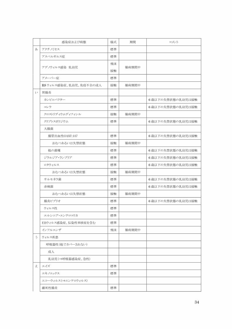
34
感染症および病態 様式 期間 コメント
あ アクチノミセス 標準
アスペルギルス症 標準
アデノウィルス感染 乳幼児 飛沫
接触 罹病期間中
アメーバー症 標準
RS ウィルス感染症、乳幼児、免疫不全の成人 接触 罹病期間中
い 胃腸炎
カンピロバクター 標準
6歳以下の失禁状態の乳幼児は接触
コレラ 標準
6歳以下の失禁状態の乳幼児は接触
クロストリディウムディフィシル 接触 罹病期間中
クリプトスポリジウム 標準
6歳以下の失禁状態の乳幼児は接触
大腸菌
腸管出血性O157:H7 標準
6歳以下の失禁状態の乳幼児は接触
おむつあるいは失禁状態 接触 罹病期間中
他の菌種 標準
6歳以下の失禁状態の乳幼児は接触
ジラルジア・ランブリア 標準
6歳以下の失禁状態の乳幼児は接触
ロタウィルス 標準
6歳以下の失禁状態の乳幼児は接触
おむつあるいは失禁状態 接触 罹病期間中
サルモネラ菌 標準
6歳以下の失禁状態の乳幼児は接触
赤痢菌 標準
6歳以下の失禁状態の乳幼児は接触
おむつあるいは失禁状態 接触 罹病期間中
腸炎ビブリオ 標準
6歳以下の失禁状態の乳幼児は接触
ウィルス性 標準
エルシニア・エンテロコリカ 標準
EBウィルス感染症、伝染性単核症を含む 標準
インフルエンザ 飛沫 罹病期間中
う ウィルス疾患
呼吸器性(他でカバーされない)
成人
乳幼児(⇒呼吸器感染症、急性)
え エイズ 標準
エキノコックス 標準
エコーウィルス(⇒エンテロウィルス)
壊死性腸炎 標準

35
HIV感染症 標準
エボラ出血熱 接触 罹病期間中
エルシニア・エンテロコリティカ胃腸炎⇒胃腸炎
エンテロウィルス感染症
成人 標準
乳幼児 接触 罹病期間中
お オウム病 標準
か 回帰熱 標準
疥癬 接触 有効な治療開始
後24時間まで
回虫症 標準
ガス壊疽 標準
川崎病 標準
肝炎、ウィルス性
A型 標準
おむつあるいは失禁患者 接触 発症後1週間 3歳以下の乳幼児は入院期間中
3~14歳の小児は発症後2週間
B型、HBs陽性 標準
C型およびその他特定されない非A非B 標準
E型 標準
カンジダ症(粘膜皮膚型を含む) 標準
カンピロバクター胃腸炎(⇒胃腸炎)
き Q熱 標準
狂犬病 標準
蟯虫症 標準
蟯虫 標準
ギランバレー症候群 標準
く クラミジア・トラコマティス
結膜炎 標準
性器 標準
呼吸器 標準
クリプトコッカス症 標準
クリプトスポリジオーシス(⇒胃腸炎)
クループ(⇒乳幼児の呼吸器感染症)
クロイツフェルトヤコブ病 標準
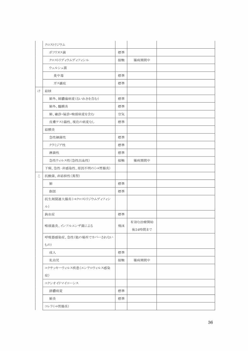
36
クロストリジウム
ボツリヌス菌 標準
クロストリディウムディフィシル 接触 罹病期間中
ウェルシュ菌
食中毒 標準
ガス壊疽 標準
け 結核
肺外、肺膿瘍病変(るいれきを含む) 標準
肺外、髄膜炎 標準
肺、確診・疑診・喉頭病変を含む 空気
皮膚テスト陽性、現在の病変なし 標準
結膜炎
急性細菌性 標準
クラミジア性 標準
淋菌性 標準
急性ウィルス性(急性出血性) 接触 罹病期間中
下痢、急性‐非感染性、原因不明の(⇒胃腸炎)
こ 抗酸菌、非結核性(異型)
肺 標準
創部 標準
抗生剤関連大腸炎(⇒クロストリジウムディフィシ
ル)
鉤虫症 標準
喉頭蓋炎、インフルエンザ菌による 飛沫 有効な治療開始
後24時間まで
呼吸器感染症、急性(他の場所でカバーされない
もの)
成人 標準
乳幼児 接触 罹病期間中
コクサッキーウィルス疾患(エンテロウィルス感染
症)
コクシオイドマイコーシス
排膿病変 標準
肺炎 標準
コレラ(⇒胃腸炎)
37
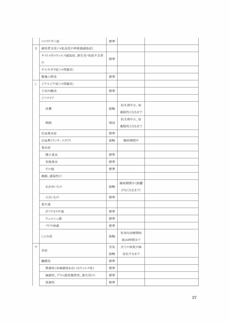
コロラドダニ症 標準
さ 細気管支炎(⇒乳幼児の呼吸器感染症)
サイトメガロウィルス感染症、新生児・免疫不全者
の 標準
サルモネラ症(⇒胃腸炎)
塹壕口腔炎 標準
し ジアルジア症(⇒胃腸炎)
子宮内膜炎 標準
ジフテリア
皮膚 接触 抗生剤中止、培
養陰性となるまで
咽頭 飛沫 抗生剤中止、培
養陰性となるまで
住血吸虫症 標準
出血熱(ラッサ、エボラ) 接触 罹病期間中
条虫症
矮小条虫 標準
有鉤条虫 標準
その他 標準
褥創、感染性の
おおきいもの 接触 罹病期間中(排膿
がなくなるまで)
小さいもの 標準
食中毒
ボツリヌス中毒 標準
ウェルシュ菌 標準
ブドウ球菌 標準
しらみ症 接触 有効な治療開始
後24時間まで
す 水痘
空気
接触
全ての病変が痂
皮化するまで
髄膜炎 標準
無菌性(非細菌性あるいはウィルス性) 標準
細菌性、グラム陰性腸管性、新生児の) 標準
真菌性 標準
38
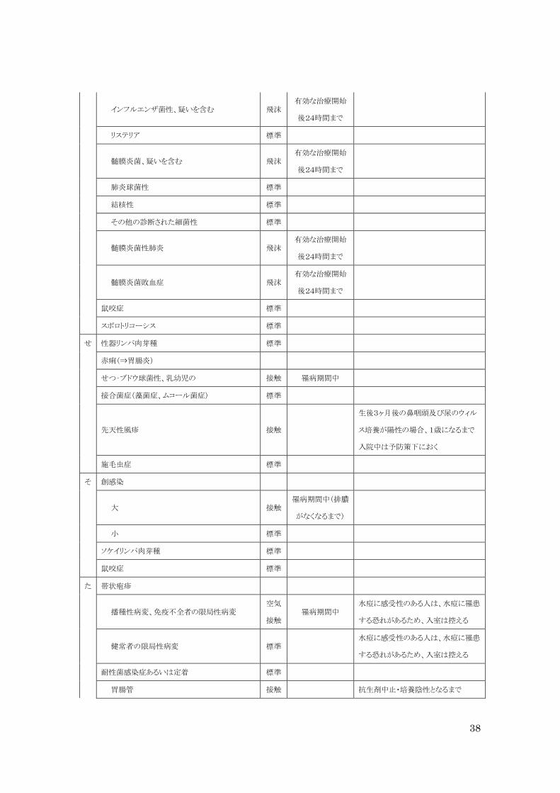
インフルエンザ菌性、疑いを含む 飛沫 有効な治療開始
後24時間まで
リステリア 標準
髄膜炎菌、疑いを含む 飛沫 有効な治療開始
後24時間まで
肺炎球菌性 標準
結核性 標準
その他の診断された細菌性 標準
髄膜炎菌性肺炎 飛沫 有効な治療開始
後24時間まで
髄膜炎菌敗血症 飛沫 有効な治療開始
後24時間まで
鼠咬症 標準
スポロトリコーシス 標準
せ 性器リンパ肉芽種 標準
赤痢(⇒胃腸炎)
せつ‐ブドウ球菌性、乳幼児の 接触 罹病期間中
接合菌症(藻菌症、ムコール菌症) 標準
先天性風疹 接触
生後3ヶ月後の鼻咽頭及び尿のウィル
ス培養が陽性の場合、1歳になるまで
入院中は予防策下におく
施毛虫症 標準
そ 創感染
大 接触 罹病期間中(排膿
がなくなるまで)
小 標準
ソケイリンパ肉芽種 標準
鼠咬症 標準
た 帯状疱疹
播種性病変、免疫不全者の限局性病変 空気
接触 罹病期間中
水痘に感受性のある人は、水痘に罹患
する恐れがあるため、入室は控える
健常者の限局性病変 標準
水痘に感受性のある人は、水痘に罹患
する恐れがあるため、入室は控える
耐性菌感染症あるいは定着 標準
胃腸管 接触
抗生剤中止・培養陰性となるまで
39
呼吸器 接触
抗生剤中止・培養陰性となるまで
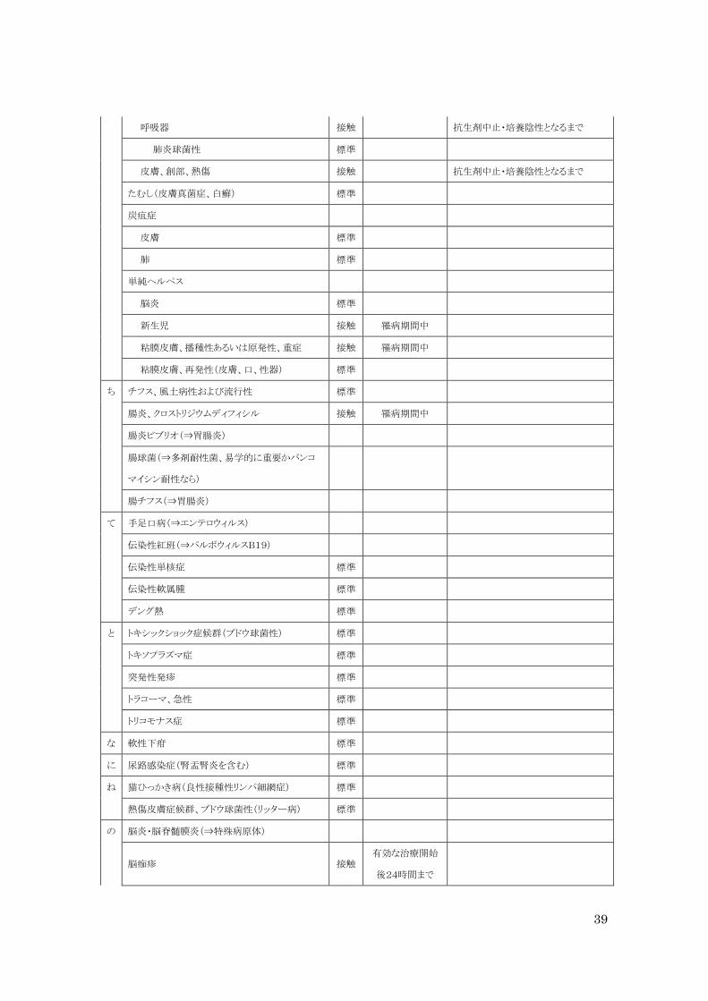
肺炎球菌性 標準
皮膚、創部、熱傷 接触
抗生剤中止・培養陰性となるまで
たむし(皮膚真菌症、白癬) 標準
炭疽症
皮膚 標準
肺 標準
単純ヘルペス
脳炎 標準
新生児 接触 罹病期間中
粘膜皮膚、播種性あるいは原発性、重症 接触 罹病期間中
粘膜皮膚、再発性(皮膚、口、性器) 標準
ち チフス、風土病性および流行性 標準
腸炎、クロストリジウムディフィシル 接触 罹病期間中
腸炎ビブリオ(⇒胃腸炎)
腸球菌(⇒多剤耐性菌、易学的に重要かバンコ
マイシン耐性なら)
腸チフス(⇒胃腸炎)
て 手足口病(⇒エンテロウィルス)
伝染性紅班(⇒パルボウィルスB19)
伝染性単核症 標準
伝染性軟属腫 標準
デング熱 標準
と トキシックショック症候群(ブドウ球菌性) 標準
トキソプラズマ症 標準
突発性発疹 標準
トラコーマ、急性 標準
トリコモナス症 標準
な 軟性下疳 標準
に 尿路感染症(腎盂腎炎を含む) 標準
ね 猫ひっかき病(良性接種性リンパ細網症) 標準
熱傷皮膚症候群、ブドウ球菌性(リッター病) 標準
の 脳炎・脳脊髄膜炎(⇒特殊病原体)
脳痂疹 接触 有効な治療開始
後24時間まで

40
膿瘍
ドレッシングで覆っていないあるいはド
レッシングで排膿が封じ込めないもの
排膿、多量 接触 罹病期間中 ドレッシングで覆ってあるもの
排膿、尐量あるいは微量 標準
ノカルジア症、排膿病変あるいは他の症状 標準
野兎病
排膿病変 標準
肺 標準
Norwalk Agent胃腸炎(⇒ウィルス性胃腸炎)
は 肺炎
アデノウィルス 飛沫
接触 罹病期間中
細菌性、他の列挙されていないグラム陰性菌
を含む 標準
Burlholdria Cepacia、襄胞繊維症におけ
る、気道定着を含む 標準
クラミジア性 標準
真菌 標準
インフルエンザ菌
成人 標準
乳幼児 飛沫 有効な治療開始
後24時間まで
レジオネラ 標準
髄膜炎菌(⇒多剤耐性菌)
マイコプラズマ(原発性異型肺炎) 飛沫 罹病期間中
肺炎球菌 飛沫
多剤耐性(⇒多剤耐性)
カリニ原虫 標準
免疫不全者との同室を避ける
黄色ブドウ球菌 標準
A群溶連菌
成人 標準
乳幼児(⇒呼吸器感染症、急性) 飛沫 有効な治療開始
後24時間まで
梅毒
皮膚・粘膜の、先天性・原発性・二次性を含む 標準

41
潜在的・無症候性梅毒反応陽性者 標準
白癖症 標準
破傷風 標準
バビシア症 標準
パラインフルエンザウィルス感染症、乳幼児の呼
吸器性 接触 罹病期間中
パルボウィルスB19 標準
ハンタウィルス呼吸器症候群 標準
ハンセン病 標準
ひ ヒトブラズマ症 標準
羊鷲口瘡 標準
ヒト由来ウィルス脳炎(東、西ベネゼエラ馬脳炎:
セントルイス、カリフォルニア脳炎) 標準
ヒト由来ウィルス熱(デング熱、黄熱、コロラドダニ
熱) 標準
百日咳 飛沫 有効な治療開始
後 5日まで
ふ 風疹
ブドウ球菌(黄色ブドウ球菌)
皮膚、創傷・熱傷の
大 接触 罹病期間中
小 標準
腸炎 標準
熱傷皮膚症候群 接触 罹病期間中
トキシックショック症候群 標準
ブルセラ症(波状熱、マルタ熱、地中海熱) 標準
分芽菌症 北アメリカ、皮膚・肺 標準
糞中症 標準
へ 閉鎖腔感染症
排膿、尐量 標準
排膿なし 標準
ペスト
腺ペスト 標準
肺ペスト 飛沫 有効な治療開始
後72時間まで
42
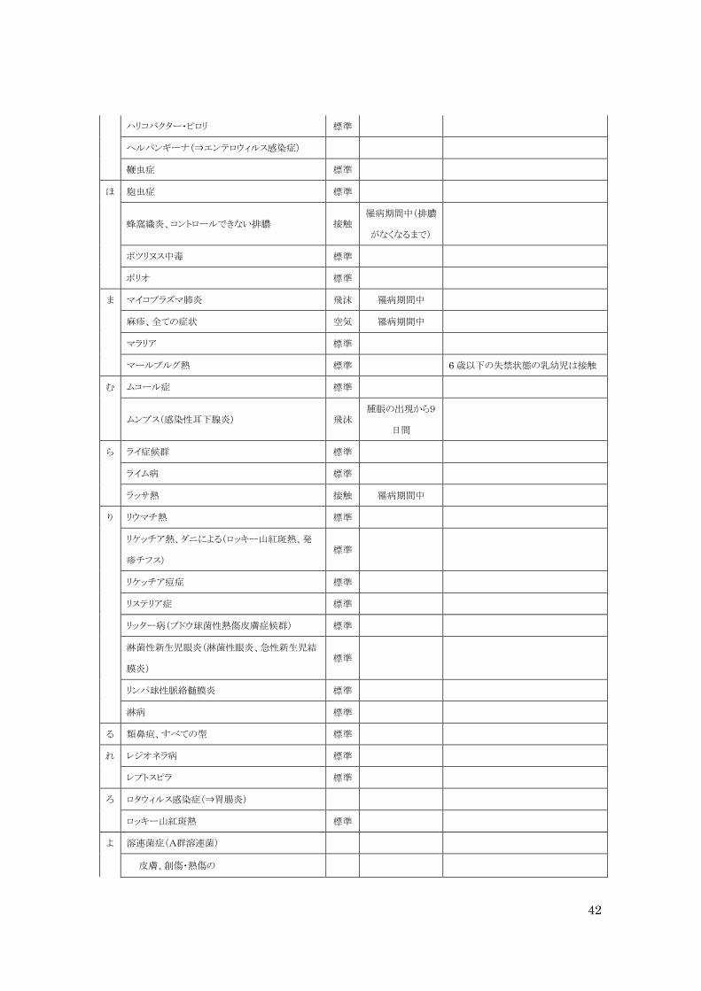
ハリコバクター・ピロリ 標準
ヘルパンギーナ(⇒エンテロウィルス感染症)
鞭虫症 標準
ほ 胞虫症 標準
蜂窩織炎、コントロールできない排膿 接触 罹病期間中(排膿
がなくなるまで)
ボツリヌス中毒 標準
ポリオ 標準
ま マイコプラズマ肺炎 飛沫 罹病期間中
麻疹、全ての症状 空気 罹病期間中
マラリア 標準
マールブルグ熱 標準
6歳以下の失禁状態の乳幼児は接触
む ムコール症 標準
ムンプス(感染性耳下腺炎) 飛沫 腫脹の出現から9
日間
ら ライ症候群 標準
ライム病 標準
ラッサ熱 接触 罹病期間中
り リウマチ熱 標準
リケッチア熱、ダニによる(ロッキー山紅斑熱、発
疹チフス) 標準
リケッチア痘症 標準
リステリア症 標準
リッター病(ブドウ球菌性熱傷皮膚症候群) 標準
淋菌性新生児眼炎(淋菌性眼炎、急性新生児結
膜炎) 標準
リンパ球性脈絡髄膜炎 標準
淋病 標準
る 類鼻疸、すべての型 標準
れ レジオネラ病 標準
レプトスピラ 標準
ろ ロタウィルス感染症(⇒胃腸炎)
ロッキー山紅斑熱 標準
よ 溶連菌症(A群溶連菌)
皮膚、創傷・熱傷の

43
大 接触 有効な治療開始
後24時間まで
小 標準
子宮内膜炎(産褥敗血症) 標準
乳幼児の咽頭炎 飛沫 有効な治療開始
後24時間まで
乳幼児の肺炎 飛沫 有効な治療開始
後24時間まで
乳幼児の猩紅熱 飛沫 有効な治療開始
後24時間まで
新生児の溶連菌症(B群溶連菌) 標準
溶連菌症(非A非B群)、他の箇所でカバーされ
ない 標準
多剤耐性(⇒多剤耐性菌)
わ Vincents angina 標準
標準:標準的な感染予防策、 空気:空気感染隔離予防策、 飛沫:飛沫感染予防策、 接触:接触感染予防策
135 Kenyon A,Ridzon R, Luskin-HawkR, et al. A Nosocomial outbreak of
Multidrug-Resistant Tuberculosis.Ann Intern Med. 1997;127(1):32-36. 136 Pavelchak N, DePersis RP, London M, et al:Identification of factors that disrupt
negative air pressurization of respiratory isolation rooms. Infect Control Hosp Epidemiol.
2000;21:191-5. 137 大久保憲, 筧淳夫他. 病院空調設備の設計・管理指針 HEAS‐02‐2004. 日本医療福祉設備協
会. 138 Haley CE, McDonald RC, Rossi L, Jones WD, Jr., Haley RW, Luby JP: Tuberculosis
epidemic among hospital personnel. Infect Control Hosp Epidemiol. 1989;10:204-10. 139 Ryan MG:Developing a respiratory protection program, Understanding the written
elements. AAOHN J. 2001;49:293–307. 140 Drinka PJ, Krause P, Nest L, Goodman BM, Gravenstein S: Risk of acquiring influenza
A in a nursing home from a culture-positive roommate. Infect Control Hosp Epidemiol.
2003;24:872-4. 141 Seto WH, Tsang D, Yung RWH, et al: Effectiveness of precautions against droplets and
contact in prevention of nosocomial transmission of severe acute respiratory syndrome
(SARS). Lancet. 2003;361:1591-1620. 142 Chang VT, Nelson K. The role of physical proximity in nosocomial diarrhea. Clin Infect
Dis. 2000;31:717-22. 143 Trick WA,Weinstein RA,et al: Comparison of routine glove use and contact-isolation
precautions to prevent transmission of multidrug-resistant bacteria in a long-term care
facility.J Am Geriatr Soc. 2004 Dec;52(12):2003-9. 144 Sally W, et al: Effectiveness of barrier precautions and surveillance cultures to control
transmission of multidrug‐resistant organisms. A systematic review of the literature.AJIC.
2006;34:484‐494.
44
職業感染対策
鳥居啓三
1 基本原則
1.1 職業感染予防策として標準的な感染予防策の徹底、安全器材の導入など業務中に
血液・体液に直接的に曝露されないようにする。(ⅡA)
1.2 施設管理者は医療従事者が曝露事故にあった場合にそなえて、緊急報告、緊急処
置、治療、予防、経過観察などのマニュアルを整備する。(ⅣA)
1.3 患者由来の感染源に曝露した場合はHBV、HCV、HIVの感染リスクの評価をする。
145(ⅢA)
1.4 患者由来の血液や体液などに曝露した皮膚は石鹸と水で、粘膜は水で洗う。145(Ⅱ
A)
1.5 血液や体液に曝露した事故者は、速やかに院内感染対策担当者、あるいは施設管
理責任者に報告する。(ⅢA)
1.6 施設管理責任者は事故報告を受けたら、緊急処置がとれる体制を整備する。(ⅢA)
1.7 感染対策担当者は曝露事故の全数とその後の経過を把握する。(ⅢA)
1.8 EPINet 日本語版を用いた事故サーベイランスを実施し、事故防止に必要な対策を
講じる方が良い。(ⅢB)
2 B型肝炎
2.1 血液や体液に曝露する可能性のある医療従事者は B 型肝炎ワクチン接種をうける。
145, 146, 147(ⅡA)
2.2 汚染源の HBs 抗原および曝露者のワクチン接種歴や HBs 抗体が不明な場合は検
査により確認する。145, 148(ⅢA)
2.3 曝露者がHBワクチン(3回接種)未実施でHBs抗原、HBs抗体の両方が陰性の場
合は、事故後速やかに抗 HBs ヒト免疫グロブリン製剤を投与し、初回の HB ワクチン
(3回接種)を開始する。149, 150(ⅢA)
2.4 曝露者がHBワクチン(3回接種)接種者でHBs抗体が陰性の場合は、事故後速や
かに抗 HBs ヒト免疫グロブリン製剤を投与し、HB ワクチン(3 回接種)の追加が必要
であれば開始する。149, 150(ⅢA)
2.5 曝露者が 2 度の HB ワクチンでも HBs 抗体陰性の場合は、事故直後と一カ月後に
抗 HBs ヒト免疫グロブリン製剤の接種を受ける。151, 152, 153(ⅢA)
2.6 曝露者の HBs 抗原、HBs 抗体、AST(GOT)、ALT(GPT)を、事故直後、1 カ月後、
3カ月後、6カ月後および 1年後に検査する方が良い。(ⅢB)
2.7 曝露者がHBVキャリアの場合は、肝臓の専門医を受診した方が良い。(ⅢB)

45
3 C型肝炎
3.1 曝露者の HCV 抗体および AST(GOT)、ALT(GPT)を、事故直後、1 カ月後、3 カ
月後、6カ月後および 1年後に検査する方が良い。145, 154(ⅢB)
3.2 曝露者に有効性が証明されている予防法はないため、免疫グロブリン製剤やインタ
ーフェロンなどの投与は行わない方が良い。155(ⅢB)
3.3 HCV 抗体の陽転、あるいは ALT の上昇を認めた時は HCV-RNA 検査を行う。156
(ⅢA)
3.4 HCV-RNAが陽転化した場合はインターフェロンによる治療を行う。157(ⅢA)
4 HIV
4.1 HIV抗体陽性の血液や体液による汚染事故発生に備えて、HIV抗体の緊急検査や
専門医への相談のための連絡網などを予め決めておく。(ⅢA)
4.2 HIV 抗体陽性の血液や体液による汚染事故が起きた場合は、曝露者は直ちに HIV
専門医に予防内服について相談する。158(ⅢA)
4.3 事故直後、HIV 専門医と連絡がとれない場合は、一刻も早く1回目の抗 HIV 薬を服
用し、専門医と連絡がとれ次第その後の服用について相談する。158(ⅢA)
4.3.1 72時間以降の服用は効果が減弱するので、それ以前に行う。159, 160(ⅢA)
4.4 曝露者は予防内服の実施の如何にかかわらず、事故直後、1 カ月後、3 カ月後、6カ
月後および 1年後に検査する方が良い。158(ⅢB)
5 ワクチン接種
5.1 水痘、麻疹、風疹、流行性耳下腺炎に関して、これらの患者に接する機会の多い部
署の医療従事者で各々のウイルスに対する抗体陰性者はワクチンを接種する。161
(ⅠA)
5.2 患者に接する医療従事者はインフルエンザワクチンを接種する。162, 163(ⅡA)
5.3 血液や体液に曝露する可能性のある医療従事者は B 型肝炎ワクチン接種をうける。
145, 146, 147(ⅡA)
6 医療廃棄物
6.1 施設管理者は医療行為等によって生じた廃棄物は自らの責任において処理する。
(ⅣA)
6.2 施設管理者は、施設内で生じる感染性廃棄物を処理するために、特別管理産業廃
棄物管理責任者を置き、管理体制の充実を図る。(ⅣA)
6.3 施設管理者は、施設内で生じる感染性廃棄物の取扱いについて管理規定を作製し、
感染性廃棄物の処理が適正に行われているか監視する。(ⅣA)
6.4 感染性廃棄物と非感染性廃棄物の分別を行い、それぞれの廃棄容器には感染性
46
(バイオハザードマーク)や非感染性であることを明記したラベルなどの表示を行う。
(ⅣA)
6.5 感染性廃棄物の施設内における移動は、移動の途中で内容物が飛散・流出するお
それのないように蓋付きの容器などを使用する。(ⅣA)
7 保険
7.1 労働契約を結んだ医療従事者を雇用する医療機関は労働者災害補償保険法に従
い、労災保険加入のために必要な手続きを行なう。(ⅣA)
7.2 雇用関係に無い者(臨床実習の学生など)が診療に関与する場合は、施設管理者は
事前に保険に加入するよう勧告する(ⅢA)
8 健康診断
8.1 施設管理者は、業務に従事する者に対して結核に係る定期の健康診断を実施する。
(ⅣA)
145 Updated U.S. Public Health Service Guidelines for the Management of Occupational
Exposures to HBV, HCV, and HIV and Recommendations for Postexposure Prophylaxis.
MMWR Recomm Rep. 2001;50(RR-11):1-52. 146 Occupational exposure to bloodborne pathogens--OSHA. Final rule. Fed Regist
1991;56(235):64004-182.
.147 Poland GA, Jacobson RM. Clinical practice: prevention of hepatitis B with the hepatitis
B vaccine. N Engl J Med. 2004;351(27):2832-8. 148 Puro V, Cicalini S, De Carli G, Soldani F, Ippolito G. Towards a standard HIV post
exposure prophylaxis for healthcare workers in Europe. Euro Surveill. 2004;9(6):40-3. 149 Beasley RP, Hwang LY, Lee GC, et al. Prevention of perinatally transmitted hepatitis B
virus infections with hepatitis B virus infections with hepatitis B immune globulin and
hepatitis B vaccine. Lancet. 1983;2(8359):1099-102. 150 Stevens CE, Toy PT, Tong MJ, et al. Perinatal hepatitis B virus transmission in the
United States. Prevention by passive-active immunization. JAMA. 1985;253(12):1740-5. 151 Grady GF, Lee VA, Prince AM, et al. Hepatitis B immune globulin for accidental
exposures among medical personnel: final report of a multicenter controlled trial. J Infect
Dis. 1978;138(5):625-38. 152 Seeff LB, Zimmerman HJ, Wright EC, et al. A randomized, double blind controlled trial
of the efficacy of immune serum globulin for the prevention of post-transfusion hepatitis. A
Veterans Administration cooperative study. Gastroenterology. 1977;72(1):111-21. 153 Prince AM, Szmuness W, Mann MK, et al. Hepatitis B "immune" globulin: effectiveness
in prevention of dialysis-associated hepatitis. N Engl J Med. 1975;293(21):1063-7. 154 Puro V, De Carli G, Cicalini S, et al. European recommendations for the management of
healthcare workers occupationally exposed to hepatitis B virus and hepatitis C virus. Euro
Surveill. 2005;10(10):260-4. 155 Alter MJ. Occupational exposure to hepatitis C virus: a dilemma. Infect Control Hosp
Epidemiol. 1994;15(12):742-4. 156 Recommendations for prevention and control of hepatitis C virus (HCV) infection and
HCV-related chronic disease. Centers for Disease Control and Prevention. MMWR Recomm。
Rep 1998;47(RR-19):1-39.
47
157 Jaeckel E, Cornberg M, Wedemeyer H, et al. Treatment of acute hepatitis C with
interferon alfa-2b. N Engl J Med。 2001;345(20):1452-7. 158 Panlilio AL, Cardo DM, Grohskopf LA, Heneine W, Ross CS. Updated U.S. Public Health
Service guidelines for the management of occupational exposures to HIV and
recommendations for postexposure prophylaxis. MMWR Recomm Rep. 2005;54(RR-9):1-17. 159 Tsai CC, Emau P, Follis KE, et al. Effectiveness of postinoculation
(R)-9-(2-phosphonylmethoxypropy) adenine treatment for prevention of persistent simian
immunodeficiency virus SIVmne infection depends critically on timing of initiation and
duration of treatment. J Virol. 1998;72(5):4265-73 160 Tsai CC, Follis KE, Sabo A, et al. Prevention of SIV infection in macaques by
(R)-9-(2-phosphonylmethoxypropyl) adenine. Science. 1995;270(5239):1197-9. 161 Bolyard EA, Tablan OC, Williams WW, Pearson ML, Shapiro CN, Deitchmann SD.
Guideline for infection control in healthcare personnel, 1998. Hospital Infection Control
Practices Advisory Committee. Infect Control Hosp Epidemiol. 1998;19(6):407-63. 162 Poland GA, Tosh P, Jacobson RM. Requiring influenza vaccination for health care
workers: seven truths we must accept. Vaccine. 2005;23(17-18):2251-5. 163 Harper SA, Fukuda K, Uyeki TM, Cox NJ, Bridges CB. Prevention and control of
influenza. Recommendations of the Advisory Committee on Immunization Practices (ACIP).
MMWR Recomm Rep. 2005;54(RR-8):1-40.
48
尿路感染対策
土手健太郎
1 膀胱留置カテーテルの取り扱いの原則
1.1 個人・教育
1.1.1 膀胱留置カテーテルの無菌的な挿入と維持に関する知識と技術を持った医療
従事者が膀胱留置カテーテルを取り扱う。 164, 165(ⅢA)
1.1.2 膀胱留置カテーテルを取り扱う医療従事者は膀胱留置カテーテルに留置に伴
う合併症に関する教育を定期的にうける。166, 167(ⅢA)
2 膀胱留置カテーテルの取り扱い
2.1 使用原則
2.1.1 膀胱留置カテーテルは必要時にのみ留置し、医療従事者の便宜のために使
用しない。166, 167(ⅢA)
2.2 カテーテル挿入
2.2.1 膀胱留置カテーテルを操作する直前及び直後には手指消毒をする。168(ⅢA)
2.2.2 膀胱留置カテーテルは清潔器具を用いて無菌的操作で挿入する。169(ⅢA)
2.2.3 膀胱留置カテーテル挿入前に陰部洗浄を行う。(ⅢA)
2.2.4 膀胱留置カテーテル挿入には滅菌済みの単回使いきりの粘滑剤を使用する
方が良い。166, 167(ⅢB)
2.2.5 膀胱留置カテーテル挿入後はカテーテルの移動と尿道の牽引を避けるため、
下腹部に固定する方が良い。170(ⅢB)
2.3 カテーテルの選択
2.3.1 尿道損傷を最小限にするため、漏れない範囲で、できる限り細い外径の膀胱
留置カテーテルを用いる方が良い。171(ⅢB)
2.3.2 銀合金で被覆した膀胱留置カテーテルを使用する。172, 173, 174(ⅠA)
2.3.3 閉鎖式採尿システム(膀胱留置カテーテルと採尿バックが一体化したもの)を
使用する。166, 167(ⅢA)
2.4 カテーテルの交換
2.4.1 定期的な膀胱留置カテーテルの交換はしない方が良い。175(ⅢB)
3 採尿システムの取り扱い
3.1 閉鎖式採尿システムの接続部は外さない。165(ⅢA)
3.2 カテーテルと採尿システムは屈曲しないようにする。166, 167(ⅢA)
3.3 採尿バッグは定期的に空にする(一杯になってから捨てるようなことは避ける)。176(Ⅲ
A)

49
3.4 尿の回収時、排液口を回収容器に接触させない。(ⅢA)
3.5 尿の回収容器は患者ごとに使用し、ベットパンウォッシャーで一回ごとに洗浄する。
(ⅢA)
3.6 閉塞したカテーテルは入れ替える。166, 167(ⅢA)
3.7 採尿バッグは常に膀胱より下の高さに置く。166, 167(ⅢA)
3.8 採尿バッグは床に直接接触しないようにする。(ⅢA)
3.9 尿の検体採取の為に尐量の新鮮尿を必要とするときにはサンプリングポートを消毒し
た後、採取する。165(ⅢA)
3.10 大量の尿を必要とするときは、採尿バッグの排液口から採取する方が良い。166, 167
(ⅢB)
4 外尿道口の衛生管理
4.1 外尿道口周囲を清潔に保つには洗浄のみ行い、消毒はしない。166(ⅢA)
5 膀胱洗浄の適応と方法
5.1 治療上必要な場合以外は膀胱洗浄を避ける。166, 167(ⅢA)
5.2 洗浄が必要な場合は大容量の滅菌シリンジと滅菌生理食塩水を用い、無菌操作で
行う。166, 167(ⅢA)
5.3 抗菌薬を用いた膀胱洗浄は行わない。177, 178(ⅠA)
6 定期的細菌培養
6.1 定期的な尿の培養検査はしない方が良い。179(ⅡB)
164 Garibaldi RA, Burke JP, Dickman ML, Smith CB. Factors predisposing to bacteria
during indwelling urethral catheterization. N Engl J Med 1974;291:215-8. 165 Kunin CM, McCormack RC. Prevention of catheter-induced urinary tract infections by
sterile closed drainage. N Engl J Med 1966;274:1155-62. 166 CDC Guideline for prevention of catheter-associated urinary tract infections;
(http//www.cdc.gov/nciod/hip/Guide/uritract.htm): 1982. 167 国立大学病院集中治療部協議会 ICU感染制御CPG策定委員会編:尿路感染対策. ICU感染防
止ガイドライン. じほう社. 東京. 168 Steere AC, Mallison GF. Handwashing practices for the prevention of nosocomial
infections. Ann Intern Med 1975;83:683-90. 169 Kass EH, Schneiderman LJ. Entry of bacteria into the urinary tract of patients with
inlying catheters. N Engl J Med 1957;256:556-7. 170 Desautels RF, Walter CW, Graves RC. et al. Technical advances in the prevention of
urinary tract infection. J Urol 1962; 87: 487-90. 171 Kunin CM. Detection, prevention and management of urinary tract infections. 3rd ed.
Philadelphia: Lea and Febiger, 1979. 172 Saint S, Veenstra DL, Sullivn SD, Chenoweth C, Fendrick M. The potential clinical and
economic benefits of silver alloy urinary catheters in preventing urinary tract infection. Arch
Intern Med 2000; 160: 2670-5.

50
173 Karchmer TB, Eve T, Giannetta RN. A randomized crossover study of silver-coated
urinary catheters in hospitalized patients. Arch Intern Med 2000; 160: 3294-98. 174 Rupp ME, Fitzgerald t, Marion N. Effect of silver-coated urinary catheters: Efficacy,
cost-effectiveness, and antimicrobial resistance. Am J Infect Control 2004; 32: 445-50. 175 Stamm WE. Guidelines for the prevention of catheter-associated urinary tract infections.
Ann Intern med 1975;82:386-90. 176 Marrie TJ, Major H, Gurwith M, et al. Prolonged outbreak of nosocomial urinary tract
infection with a single strain of Pseudomonas aeruginosa. Can Med J 1978; 119: 593-6. 177 Warren JW, Platt R, Thomas KJ, Rosner B, Kass EH. Antibiotic irrigation and
catheter-associated urinary tract infections. N Engl J Med 1978; 299:570-73. 178 Britt MR, Garibaldi RA, Miller WA, Hebertson RM, Burke JP. Antimicrobial prophylaxis
for catheter-associated bacteriuria. Antimicrob Agents Chemother 1977; 11: 240-43. 179 Mooney BS, Garbaldi RA, Britt MR. Natural history of catheter-associated bacteriuria:
implication for protection. In: Proceedings of the 11th International Congress of
Chemotherapy and the 19th Interscience Conference on Antimicrobial Agents and
Chemotherapy. Boston. October 8-12, 1979. Washington, D.C.; American Society of
Microbiology 1980: 1083-5.

51
人工呼吸器関連肺炎
西村匡司
1 教育・サーベイランス
1.1 感染教育およびサーベイランスの役割
1.1.1 人工呼吸器関連肺炎防止に関する標準化された教育・研修を実施する方が良
い。180(ⅡB)
1.1.2 全国的なサーベイランスを参考にし、自施設の人工呼吸器関連肺炎防止能力
を客観的に評価する方が良い。(ⅡB)
2 器具の消毒
2.1 特別の汚染がない限り、人工呼吸器の本体表面は除染・消毒をする必要はない。
(ⅢA)
2.2 汚染があった場合は使用説明書にしたがって除染、消毒をする。(ⅢA)
2.3 人工呼吸器関連肺炎(VAP)の原因が人工呼吸器内部の汚染であることが疑われる
時は、人工呼吸器の内部回路の、除染・消毒を行なう。(ⅠA)
2.4 人工呼吸器に関連した単回使用部品の再利用は行なわない。(ⅢA)
2.5 再使用可能な人工呼吸器回路を、新規患者に使用する時は滅菌する。(ⅢA)
2.6 回路内への結露は患者側へ流入しないように除去する。(ⅡA)
3 人工呼吸器回路の交換
3.1 人工呼吸器回路を同一患者に使用する際は 1週間以内に定期的交換をする必要は
ない。(ⅠA)
3.2 バクテリアフィルター付き人工鼻を使用している時には、汚染や閉塞が明らかでない
限り、回路の交換はしない。181(ⅠA)
4 バクテリアフィルター付きの人工鼻
4.1 成人症例で喀痰による閉塞の危険のない患者では人工鼻を使用する。182(ⅠA)
4.2 小児症例では人工鼻を使用しない。(ⅢA)
4.3 結核、新型インフルエンザ、SARS など空気感染を起こす可能性のある肺炎患者に
人工呼吸管理を行う場合は、呼気の室内排出側に、バクテリアフィルター付きの人工
鼻を装着する方が良い(ⅢB)
5 周辺機器や手技・操作の管理
5.1 ネブライザーの薬液注入部は高レベル(グルタルアルデヒドなど)消毒後に滅菌水で
52
洗浄し空気乾燥を行うか、滅菌を行う。(ⅠA)
5.2 吸入薬剤の調製は無菌的に行う。(ⅠA)
5.3 加温加湿器には滅菌水を使う。(ⅠA)
5.4 加温加湿器の給水は閉鎖式を用いる方が良い。(ⅢB)
5.5 回路に結露した水をぬく場合は一方弁付きのトラップを使用する方が良い。(ⅢB)
6 吸引操作、気管内吸引カテーテル(閉鎖/開放)の管理
6.1 気管内吸引前後には手指消毒を行う。(ⅢA)
6.2 気管内吸引操作には清潔な手袋(未滅菌のもので良い)を着用して行う方が良い。
(ⅢB)
6.3 吸引チューブは一回ごとの使い捨てにする。(ⅡA)
6.4 閉鎖式吸引システムを使用しても良い。183(ⅠC)
6.5 開放式気管内吸引操作は清潔操作とする。(ⅢA)
6.6 気管内吸引操作は必要最小限に留める。(ⅢA)
6.7 吸引チューブの洗浄には滅菌水を使用する。(ⅢA)
6.8 気管内吸引と口腔内吸引が終わった吸引チューブは廃棄し、薬液に浸して再利用し
たりしない。(ⅢA)
6.9 吸引回路および吸引瓶は当該患者専用とする。(ⅢA)
6.10 アンビューバックやジャクソンリースは汚染がなければ患者ごとに交換する。(ⅢA)
7 気管切開
7.1 気管切開を行なう場合は高度バリアプリコーション(滅菌手袋、長い袖の滅菌ガウン、
マスク、帽子と大きな滅菌覆布)で行なう。(ⅢA)
7.2 気管切開チューブを交換するときは手指消毒を行い、清潔な未滅菌手袋を用いる。
(ⅢB)
8 気管チューブの選択と経路
8.1 VAPを防ぐ観点からは経口挿管と経鼻挿管のどちらを選択しても良い。(ⅠC)
8.2 カフ上部の貯留物を吸引するための側孔付きの気管チューブを使用する。184(ⅠA)
8.3 気管内チューブの抜管時または気管チューブを動かす前にはカフ上の分泌物を吸
引・除去した方が良い。(ⅢB)
9 ストレス潰瘍予防薬
9.1 ストレス潰瘍の危険性が尐ない患者に対してH2-blockerを投与しない。(ⅠA)
9.2 ストレス潰瘍の危険性の高い患者には sucralfate など、胃の pH を上げない薬剤を
使う方が良い。(ⅡB)

53
9.3 明らかな上部消化管出血が存在する患者やストレス潰瘍の危険が高い患者では
H2-blockerや PPIを投与する。(ⅠA)
10 体位
10.1 経管栄養を行なう患者では上体を 30~45°挙上して人工呼吸管理を行う。185(Ⅰ
A)
10.2 経管栄養を行なっていない患者でも上体を挙上した方が良い。(ⅡB)
11 口腔内清拭
11.1 定期的に口腔内清拭を行なう。(ⅡA)
12 予防的抗菌薬の投与
12.1 人工呼吸器関肺炎予防の目的で抗菌薬の全身投与を行なわない。(ⅢA)
180 Tolentino-Delosreyes AF, Ruppert SD, Shiao SY. Evidence-based practice: use of the
ventilator bundle to prevent ventilator-associated pneumonia. Am J Crit Care 2007;
16:2027. 181 Lorente L, Lecuona M, Galván R, Ramos MJ, Mora ML, Sierra A. Periodically changing
ventilator circuits is not necessary to prevent ventilator associated pneumonia when a heat
and moisture exchanger is used. Infect Control Hosp Epidemiol 2004; 25:1077–1082. 182 Boots RJ, George N, Faoagali JL, Druery J, Dean K, Heller RF. Double-heater-wire
circuits and heat-and-moisture exchangers and the risk of ventilator-associated pneumonia.
Crit Care Med 2006; 34:687–693. 183 Topelia A, Harmancia A, Cetinkayab Y, Akdenizb S, Unalb S. Comparison of the effect of
closed versus open endotracheal suction systems on the development of ventilator-associated
pneumonia. J Hosp Infect 2004; 58, 14–19. 184 Shorr AF, O’Malley PG. Continuous subglottic suctioning for the prevention of
ventilator-associated pneumonia. Potential economic implications. Chest 2001;
119:228235. 185 Drakulovic MB, torres A, Bauer TT, Nicolas JM, Nogué S, Ferrer M. Supine body
position as a risk factor for nosocomial pneumonia in mechanically ventilated patients: a
randomised trial. Lancet 1999; 354:1851-58.

54
手術部位感染対策
小野寺睦雄
1 術前の患者管理
1.1 待機手術で手術部位から離れた部位に感染がある場合は、感染治療後に手術を行
う。186, 187(ⅡA)
1.2 血糖値を管理し、周術期の高血糖状態を避ける方が良い。188, 189, 190(ⅡB)
1.3 待機手術では尐なくとも 30日前に禁煙を行わせる方が良い。191, 192(ⅡB)
1.4 術前の入院期間を短くする方が良い。191, 193(ⅢB)
2 術前処置
2.1 除毛および剃毛
2.1.1 剃毛は行わない。194(ⅡA)
2.1.2 除毛が必要な場合にはサージカル・クリッパーや除毛クリームを使用し、手術
直前に行う。195, 196(ⅡA)
2.2 手術の前夜または当日朝に消毒薬入りの洗浄剤を用いてシャワー浴または入浴を行
っても良い。197(ⅢC)
3 皮膚の消毒
3.1 消毒を行う前に切開部位とその周囲を洗浄し、汚染を取り除く。(ⅢA)
3.2 アルコール含有クロルヘキシジンまたはポピドンヨードを用いる。(ⅢA)
3.3 消毒は切開部位から外側に向かって同心円状に行う。消毒の範囲は追加切開や切
開の延長に対応できるような範囲とする。(ⅢA)
4 術者の手指衛生
4.1 爪は短く切り、清潔にする。(ⅢA)
4.2 手や腕に装身具を付けない。(ⅢA)
4.3 手から肘の上まで石鹸と流水で手洗いを行い、その後手首まで擦式手指消毒薬を用
いて手指消毒を行う。198, 199(ⅡA)
5 手術室医療従事者の管理
5.1 排膿のある手術室の医療従事者は感染が治癒するまで就業を制限する。(ⅢA)
5.2 黄色ブドウ球菌や A 群連鎖球菌を保菌している手術室の医療従事者であっても、伝播
に関与していない限り、業務から外す必要はない。200(ⅢA)
55
6 手術室の換気
6.1 手術室内は廊下その他の区域に対して陽圧を維持する。201(ⅢA)
6.2 1 時間あたり 15 回以上の換気を行う。そのうち 3 回以上は外気で換気する。201, 202
(ⅢA)
6.3 再循環した空気であっても外気であっても、空気はフィルターを通過させる。(ⅢA)
6.4 空気は天井から床の方向に流れるようにする。(ⅢA)
6.5 SSIを防止する目的で紫外線照射を用いない。(ⅢA)
6.6 必要時以外は手術室の扉を閉めておく。(ⅢA)
6.7 整形外科的な人工物の植え込み術を行う場合、ウルトラクリーン・エアーを供給できる
手術室で行う方が良い。203(ⅢB)
6.8 手術室に入るスタッフは最小限に制限する。(ⅢA)
7 手術時の服装と覆布
7.1 手術中の手術室もしくは滅菌器械が展開されている部屋に入室する全ての医療従事
者は、口と鼻を完全に覆うサージカルマスクと頭髪を完全に覆う帽子を着用する。(Ⅲ
A)
7.2 手術用ガウンや覆布は撥水性のあるものを使用する。(ⅢA)
8 ドレーン
8.1 ドレーンは手術創とは異なる切開部位から、個別に留置する。(ⅢA)
8.2 ドレーンは早期に抜去する。204(ⅢA)
8.3 閉鎖吸引式のドレーンを使用しても良い。205(ⅡC)
9 手術創管理
9.1 一次閉鎖された手術創はガーゼで被覆するよりも、適切な保温、湿潤環境が維持で
きるドレッシング材を用いる。206(ⅢA)
9.2 ドレッシング材の交換を行う場合や手術部位に接触する場合には、処置の前後に手
指消毒をおこない清潔な(未滅菌で良い)手袋を使用する。(ⅢA)
9.3 閉鎖されていない切開創のドレッシング材を交換する場合には、無菌操作で行う。
(ⅢA)
10 SSIサーベイランス
10.1 全国的なサーベイランスに参加して手術部位感染の発生率の施設間比較を行うこと
により、自施設における手術部位感染対策の有効性を客観的に評価する方が良い。
207(ⅢB)
56
186 Edwards LD. The epidemiology of 2056 remote site infections and 1966 surgical wound
infections occurring in 1865 patients: a four year study of 40,923 operations at
Rush-Presbyterian-St. Luke’s Hospital, Chicago. Ann Surg 1976;184:758-66. 187 Simchen E, Rozin R, Wax Y. The Israeli Study of Surgical Infection of drains and the risk
of wound infection in operations for hernia. Surg Gynecol Obstet 1990;170:331-7. 188 Zerr KJ, Furnary AP, Grunkemeier GL, Bookin S, Kanhere V, Starr A. Glucose control
lowers the risk of wound infection in diabetics after open heart operations. Ann Thorac Surg
1997;63:356-61. 189 Furnary AP, Zerr KJ, Grunkemeier GL, Starr A. Continuous intravenous insulin
infusion reduces the incidence of deep sternal wound infection in diabetic patients after
cardiac surgical procedures. Ann Thorac Surg. 1999;67:352-60. 190 Trick WE, Scheckler WE, Tokars JI, Jones KC, Reppen ML, Smith EM, Jarvis WR.
Modifiable risk factors associated with deep sternal site infection after coronary artery
bypass grafting. J Thorac Cardiovasc Surg. 2000;119:108-14. 191 Nagachinta T, Stephens M, Reitz B, Polk BF. Risk factors for surgical wound infection
following cardiac surgery. J Infect Dis 1987;156:967-73. 192 Beitsch P, Balch C. Operative morbidity and risk factor assessment in melanoma
patients undergoing inguinal lymph node dissection. Am J Surg 1992;164:462-6. 193 Mishriki SF, Law DJ, Jeffery PJ. Factors affecting the incidence of postoperative wound
infection. J Hosp Infect 1990;16:223-30. 194 Winston KR. Hair and neurosurgery. Neurosurgery. 1992;31:320-9. 195 Niel-Weise BS, Wille JC, van den Broek PJ. Hair removal policies in clean surgery:
systematic review of randomized, controlled trials. Infect Control Hosp Epidemiol.
2005;26:923-8. 196 Tanner J, Woodings D, Moncaster K. Preoperative hair removal to reduce surgical site
infection. Cochrane Database Syst Rev. 2006 Jul 19;3:CD004122. 197 Webster J, Osborne S. Preoperative bathing or showering with skin antiseptics to
prevent surgical site infection. Cochrane Database Syst Rev. 2006 Apr 19;2:CD004985. 198 Hobson DW, Woller W, Anderson L, Guthery E. Development and evaluation of a new
alcohol-based surgical hand scrub formulation with persistent antimicrobial characteristics
and brushless application. Am J Infect Control 1998;26:507-12. 199 Mulberry G, Snyder AT, Heilman J, Pyrek J, Stahl J. Evaluation of a waterless, scrubless
chlorhexidine gluconate/ethanol surgical scrub for antimicrobial efficacy. Am J Infect
Control 2001;29:377-82. 200 Bolyard EA, Tablan OC, Williams WW, Pearson ML, Shapiro CN, Deitchman SD, et al.
Guideline for infection control in healthcare personnel, 1998. Hospital Infection Control
Practices Advisory Committee. Am J Infect Control 1998;26:289-354. 201日本医療福祉設備協会. 日本医療福祉設備協会規格「病院空調設備の設計・管理指針」. 2004. 202 American Institute of Architects. Guidelines for design and construction of hospital and
health care facilities. Washington (DC): American Institiute of Architects Press; 1996. 203 Lidwell OM, Elson RA, Lowbury EJ, Whyte W, Blowers R, Stanley SJ, et al. Ultraclean
air and antibiotics for prevention of postoperative infection. A multicenter study of 8,052
joint replacement operations. Acta Orthop Scand 1987;58:4-13. 204 Drinkwater CJ, Neil MJ. Optimal timing of wound drain removal following total joint
arthroplasty. J Arthroplasty 1995;10:185-9. 205 Parker MJ, Roberts CP, Hay D. Closed Suction Drainage for Hip and Knee Arthroplasty.
AMeta-Analysis. J Bone Joint Surg Am 2004;86:1146-52. 206 Linsky CB et al: The effect of dressing on wound inflammation and scar tissue. In Dineen
P. and Hidrick-Smith D.eds., The Surgical Wound. Lea & Febiger,
Philadelphia.pp191-205,1981. 207 Haley RW, Culver DH, White JW, Morgan WM, Emori TG, Munn VP. The efficacy of
infection surveillance and control programs in preventing nosocomial infections in US
hospitals. Am J Epidemiol 1985;121:182-205.
57
カテーテル関連血流感染対策
井上善文
1 中心静脈カテーテルの衛生管理
1.1 中心静脈栄養法(total parenteral nutrition:TPN)の適応208, 209, 210, 211, 212, 213,
214, 215, 216, 217, 218, 219, 220, 221, 222, 223, 224, 225, 226, 227, 228
1.1.1 栄養療法が必要な場合は可能な限り経腸栄養を用いる。(ⅡA)
1.1.2 静脈栄養は経腸栄養または経口摂取が不可能または不十分な場合に用いる。
(ⅢA)
1.1.3 中心静脈栄養法は静脈栄養の長期化が予測される場合に用いる。(ⅢA)
1.2 中心静脈カテーテル選択の基準
1.2.1 必要最小限の内腔数のカテーテルを選択する。229, 230, 231, 232, 233, 234, 235, 236,
237, 238(ⅠA)
1.2.2 長期使用が予想される患者では、長期留置用のカテーテルを選択する。239,
240, 241, 242, 243, 244, 245, 246, 247(ⅡA)
1.3 カテーテル挿入部位
1.3.1 感染防止のためにはカテーテル挿入は鎖骨下静脈穿刺を第一選択とする。248,
249, 250, 251, 252, 253, 254, 255(ⅡA)
1.4 皮下トンネルの作成
1.4.1 短期間の留置では、皮下トンネルを作成する必要はない。256, 257, 258, 259, 260,
261(ⅠA)
1.5 定期的な入れ換え
1.5.1 定期的にカテーテルを入れ換える必要はない。262, 263, 264(ⅡA)
1.6 高度バリアプレコーション
1.6.1 中心静脈カテーテル挿入時は高度バリアプレコーション(清潔手袋、長い袖の
滅菌ガウン、マスク、帽子と大きな清潔覆布)を行う。265, 266, 267, 268(ⅠA)
1.7 抗菌薬の予防投与
1.7.1 中心静脈カテーテル挿入に伴う抗菌薬の予防投与は行わない。269, 270, 271,
272, 273, 274(ⅡA)
1.8 カテーテル挿入時の皮膚の消毒剤
1.8.1 カテーテル挿入時の消毒は、0.5%クロルヘキシジンアルコールまたは 10%ポ
ビドンヨードを用いる。275, 276, 277, 278, 279, 280(ⅠA)
1.9 カテーテル留置期間中の皮膚の消毒剤
1.9.1 カテーテル挿入部皮膚の処置で用いる消毒薬は、以下の 3 つから選択する:
0.5%クロルヘキシジンアルコール、10%ポビドンヨード、ヨードチンキ。281, 282,
58
283, 284, 285, 286, 287, 288(ⅡA)
1.10 挿入部位の剃毛
1.10.1 穿刺に先立って局所の剃毛はしない。除毛が必要であれば、医療用
電気バリカンなどを用いる。289, 290, 291, 292(ⅠA)
1.11 カテーテル挿入部の抗菌薬含有軟膏やポビドンヨードゲルの塗布
1.11.1 抗菌薬含有軟膏を使用しない。 293, 294, 295, 296, 297(ⅡA)
1.11.2 ポビドンヨードゲルを使用しない。298, 299(ⅡA)
1.12 カテーテル挿入部の観察
1.12.1 カテーテル挿入部の発赤、圧痛、汚染、ドレッシングの剥がれなどを
毎日観察する。(ⅢC)
1.13 ドレッシング
1.13.1 滅菌されたガーゼ型ドレッシングまたはフィルム型ドレッシングを使用
する。300, 301, 302, 303, 304, 305, 306, 307, 308(ⅠA)
1.14 ドレッシング交換の頻度
1.14.1 ドレッシング交換は週 1-2 回、曜日を決めて定期的に行う。309, 310,
311, 312, 313, 314, 315, 316, 317, 318(ⅢB)
1.15 一体型輸液ラインの使用
1.15.1 一体型輸液ラインを用いる方が良い。319, 320, 321, 322, 323(ⅢB)
1.16 ニードルレスシステム
1.16.1 ニードルレスシステムの感染防止効果は明らかでないことを理解して
使用を決める。324, 325, 326, 327, 328, 329, 330, 331, 332, 333, 334, 335, 336, 337, 338, 339,
340, 341, 342, 343, 344(ⅡC)
1.17 三方活栓
1.17.1 三方活栓は手術室や ICU 以外では、輸液ラインに組み込まない。
345, 346, 347, 348, 349, 350, 351, 352, 353, 354, 355(ⅡA)
1.17.2 三方活栓から側注する場合は、消毒用エタノールを使用する。(Ⅱ
A)
1.18 輸液ラインの管理
1.18.1 輸液ラインとカテーテルの接続部の消毒には消毒用エタノールを用
いる。356, 357(ⅡA)
1.18.2 輸液ラインは曜日を決めて週 1-2 回定期的に交換する。358, 359, 360,
361, 362, 363, 364, 365(ⅡB)
1.19 脂肪乳剤の投与に使用する輸液ラインの交換頻度
1.19.1 脂肪乳剤の投与に使用する輸液ラインは、24時間以内に交換する。
366, 367, 368, 369(ⅢA)
1.20 インラインフィルター

59
1.20.1 インラインフィルターを使用する。370, 371, 372, 373, 374, 375(ⅢA)
1.21 カテーテルロック
1.21.1 作り置きしたヘパリン生食によるカテーテルロックは行わない。376, 377,
378, 379, 380(ⅣA)
1.22 輸液・薬剤とその調製法
1.22.1 高カロリー輸液製剤の調製に関する基本的考え方
1.22.1.1 高カロリー輸液製剤への薬剤の混合は、可能な限り薬剤師の管理下
で無菌環境下で行う。381, 382, 383(ⅢA)
1.22.1.2 高カロリー輸液を投与するにあたっては、薬剤の数量および回路の
接続数を最尐化する。384, 385(ⅢA)
1.23 病棟での薬剤調製
1.23.1 薬剤師は薬液混合法、調製場所の選択・清潔管理に関して指導・助
言する。386, 387, 388, 389, 390(ⅢA)
1.23.2 病棟での混合薬剤数は可能な限り尐なくする。391(ⅢA)
1.23.3 混合場所は専用スペースで行う。392(ⅢA)
1.23.4 無菌設備を設置する方がよい。393, 394, 395, 396, 397, 398(ⅢB)
1.23.5 作業面の消毒は消毒用アルコールを使用する方がよい。399(ⅢB)
1.23.6 紫外線殺菌灯や空気清浄機は使わない方が良い。400(ⅢB)
1.23.7 薬剤の混合にあたっては、その作業に専念できるように係を決める方
が良い。(ⅢB)
1.23.8 混合操作時は専用ガウンを着用し、手洗いの後に非滅菌手袋を着
用して作業を行う方が良い。401, 402, 403, 404, 405, 406(ⅢB)
1.24 高カロリー輸液基本薬・輸液剤の選択および使用
1.24.1 基本原則
1.24.1.1 糖電解質液とアミノ酸製剤を混合する場合は、高カロリー輸液用キッ
ト製剤を使用する方が良い。(ⅢB)
1.24.1.2 スリーインワンバッグ製剤(アミノ酸、糖質、脂肪が一つのバッグに入
っているもの)では細菌が混入すると急速に増殖する。また、フィルタ
ーが使用できないため、微量元素製剤と高カロリー輸液用総合ビタミ
ン剤以外は混注しない。(ⅢA)
1.24.1.3 スリーインワンバッグ製剤では完全閉鎖ルートとし、その製剤の輸液
ルートからの側注は禁止する。(ⅢA)
1.24.1.4 脂肪乳剤を含んだ製剤は、三方活栓にひび割れを生じさせることが
あるので、接続部での液漏れや汚染を監視する。(ⅡA)
1.24.2 高カロリー輸液基本液への薬剤の添加
1.24.2.1 高カロリー輸液にアルブミン製剤を加えない。407(ⅡA)
60
1.24.2.2 高カロリー輸液には、脂肪乳剤を加えない。408, 409, 410, 411(ⅢB)
1.24.3 調製後の保存方法
1.24.3.1 高カロリー輸液製剤は、混合後 28時間以内に投与が完了するように
計画する。(ⅢA)
1.24.3.2 高カロリー輸液製剤を保存する必要がある場合には無菌環境下で調
製し、冷蔵庫保存をする。(ⅢA)
1.25 CR-BSI
1.25.1 CRBSIが疑われる場合の対処
1.25.1.1 CR-BSIが疑われる場合は血液培養を行う。(ⅢA)
1.25.1.2 他に感染源が考えられない場合にはカテーテルを抜去する。(ⅢA)
1.25.1.3 カテーテル抜去時には、血液培養とともにカテーテルの先端培養を
行う。(ⅢA)
1.25.1.4 真菌が原因による場合には、眼科的診察を行う。412, 413, 414(ⅢA)
1.25.2 ガイドワイヤーでの入れ換え
1.25.2.1 感染源が不明で、カテーテル敗血症を除外するためには、ガイドワイ
ヤーを用いて入れ換える方が良い。415, 416, 417, 418, 419, 420, 421, 422,
423, 424, 425(ⅡB)
1.26 システムとしてのカテーテル管理
1.26.1 中心静脈カテーテルのチームによる管理
1.26.1.1 専門チームによるカテーテル管理を行う方が良い。426, 427, 428, 429,
430, 431 432, 433, 434(ⅡB)
1.26.1.2 ICUでは看護師-患者比を適正に保つ方が良い。435, 436(ⅡB)
2 末梢静脈カテーテルの衛生管理
2.1 留置部位
2.1.1 上肢の静脈を使用する方が良い。437(ⅢB)
2.2 カテーテルの選択
2.2.1 カテーテルは、静脈炎予防のためには、可能な限り、細径のものを使用する方
が良い。438, 439, 440(ⅢB)
2.3 末梢静脈カテーテルの留置期間
2.3.1 静脈炎のリスクを減らすため、末梢静脈カテーテルは 96 時間以上留置しない
方が良い。441, 442, 443, 444, 445, 446, 447(ⅢB)
2.4 交換頻度
2.4.1 末梢静脈カテーテルの輸液ラインは、カテーテル入れ替え時に交換する方が
良い。(ⅢB)
2.5 カテーテルロック

61
2.5.1 カテーテルロックを実施する場合は、作り置きしたヘパリン生食は使用しない。
448, 449(ⅢA)
2.6 静脈炎発生時の対応
2.6.1 静脈炎の徴候(発赤、腫脹、疼痛)がある場合は、カテーテルを抜去する。450
(ⅢA)
2.7 静脈炎予防薬剤の使用
2.7.1 静脈炎予防のためのステロイド剤、ヘパリン、血管拡張剤は、使用しない方が
良い。451, 452(ⅡB)
2.8 刺入部のドレッシング管理法
2.8.1 カテーテル刺入部は滅菌のドレッシングで被覆し、カテーテル入れ換え時に交
換した方がよい。453, 454, 455(ⅢB)
2.9 末梢のアミノ酸製剤
2.9.1 アミノ酸加糖電解質製剤を投与する場合は側注を避けるなどの厳密な衛生管
理下においてのみ使用する。456(ⅢA)
208 Gallagher-Allred CR, Voss AC, Finn SC, et al: Malnutrition and clinical outcomes: the
case for medical nutritional therapy. J Am Diet Assoc 1996;96:361-366. 209 Bistrian BR, Blackburn GL, Vitale J, et al: Prevalence of malnutrition in general medical
patients. JAMA 1976;23:1567-1570. 210 Kaminski MV, Jr: The case for nutrition support: Eliminating hospital-acquired
malnutrition improves outcome and reduces costs. Health Progress 73:69-78,1992. 211 Messner RL, Stephens N, Wheeler WE, et al: Effect of admission nutritional status on
length of hospital stay. Gastroenterol Nurs 13:202-205:1991. 212 Reilly JJ Jr, Hull SF, Albert N, et al: Economic impact of malnutrition: a model system
for hospitalized patients. JPEN 12:371-376,1988. 213 Martin CM, Doig GS, Heyland DK, et al: Multicentre, cluster-randomized clinical trial of
algorithms for critical-care enteral and parenteral therapy(ACCEPT). CMAJ
2004;170:197-20. 214 Moore EE, Jones TN: Benefits of immediate jejunostomy feeding after major abdominal
trauma-a prospective, randomized study. J Trauma 1986;26:784-881. 215 Windsor AC, Kanwar S, Li AG, et al: Compared with parenteral nutrition, enteral
feeding attenuates the acute phase response and improves disease severity in acute
pancreatitis. Gut 1998;42:431-435. 216 Kudsk KA, Croce MA, Fabian TC, et al: Enteral versus parenteral feeding. Effects on
septic morbidity after blunt and penetrating abdominal trauma. Ann Surg
1992;215:503-511. 217 Lipman TO: Bacterial translocation and enteral nutrition in humans: an outsider looks
in. JPEN 1995;19:156-165. 218 Reynolds JV, Kanwar S, Welsh FK, et al: Does the route of feeding modify gut barrier
function and clinical outcome in patients after major upper gastrointestinal surgery? JPEN
1997;21:196-201. 219 Braunschweig CL, Levy P, Sheean PM, et al: Enteral compared with parenteral
nutrition: a meta-analysis. Am J Clin Nutr 2001;74:534-542. 220 Lim ST, Choa RG, Lam KH, et al: Total parenteral nutrition versus gastrostomy in the
preoperative preparation of patients with carcinoma of the oesophagus. Br J Surg
1981;68:69-72.

62
221 Sako K, Lore JM, Kauman S, et al: Parenteral hyperalimentation in surgical patients
with head and neck cancer: a randomized study. J Surg Oncol 1981;16:391-402. 222 Campos AC, Meguid MM: A critical appraisal of the usefulness of perioperative
nutritional support. Am J Clin Nutr 1992;55:117-130. 223 Lipman TO: Grains or veins: is enteral nutrition really better than parenteral nutrition?
A look at the evidence. JPEN 1998;22:167-182. 224 Hernandez G, Velasco N, Wainstein C, et al: Gut mucosal atrophy after a short enteral
fasting period in critically ill patients. J Crit Care 1999;14:73-77. 225 Pacelli F, Bossola M, Papa V, et al: Enteral vs parenteral nutrition after major abdominal
surgery: an even match. Acta Surg 2001;136:933-936. 226 ASPEN Board of Directors and The Clinical Guidelines Task Force: Guidelines for the
use of parenteral and enteral nutrition in adult and pediatric patients. JPEN 2002;26(1
Suppl). 227 The Veteran Affairs Total Parenteral Nutrition Cooperative Study Group. Perioperative
total parenteral nutrition in surgical patients. N Engl J Med 1991;325:525-532. 228 van den Berghe G, Wouters P, Weekers F, et al: Intensive insulin therapy in the critically
ill patients. N Engl J Med 2001;345:1359-1367. 229 awar M, Mehta Y, Kapoor P, et al: Central venous catheter-related blood stream
infections: incidence, risk factors, outcome, and associated pathogens. J Cardiothorac Vasc
Anesth 2004;18:304-308. 230 Zurcher M, Tramer MR, Walder B: Colonization and bloodstream infection with single-
versus multi-lumen central venous catheters: a quantitative systematic review. Anesth
Analg 2004;99:177-182. 231 Pemberton LB, Lyman B, Lander V et al: Sepsis from triple- vs single-lumen catheters
during total parenteral nutrition in surgical or critically ill patients. Arch Surg
1986;121:591-594. 232 Yeung C, May J, Hughes R: Infection rate for single-lumen vs triple-lumen subclavian
catheters. Infect Control Hosp Epidemiol 1988;9:154-158. 233 Hilton E, Haslett TM, Borenstein MT et al: Central catheter infections: single- versus
triple-lumen catheters. Influence of guidewires on infection rates when used for replacement
of catheters. Am J Med 1988;84:667-672. 234 McCarthy MC, Shives JK, Robinson RJ et al: Prospective evaluation of single and triple
lumen catheters in total parenteral nutrition. JPEN 1987;11:259-262. 235 Clark-Christoff N, Watters VA, Sparks W et al: Use of triple-lumen subclavian catheters
for administration of total parenteral nutrition. JPEN 1992;16:403-407. 236 Dezfulian C, Lavelle J, Nallamothu BK, et al: Rates of infection for single-lumen versus
multilumen central venous catheters: a meta-analysis. Crit Care Med 2003;31:2385-2390. 237 Ma TY, Yoshinaka R, Banaag A et al: Total parenteral nutrition via multilumen catheters
does not increase the risk of catheter-related sepsis: a randomized, prospective study. Clin
Infect Dis 1998;27:500-50. 238 Lee RB, Buckner M, Sharp KW: Do multi-lumen catheters increase central venous
catheter sepsis compared to single-lumen catheters? J Trauma 1988;28:1472-1475. 239 Schuman ES, Winters V, Gross GF et al: Management of Hickman catheter sepsis. Am J
Surg. 1985;149:627-628. 240 Shapiro ED, Wald ER, Nelson KA et al: Broviac catheter-related bacteremia in oncology
patients. Am J Dis Child 1982;136:679-681. 241 Gyves J, Ensminger W, Niederhuber J et al: Totally-implanted system for intravenous
chemotherapy in patients with cancer. Am J Med 1982;73:841-845. 242 Wurzel CL, Halom K, Feldman JG: Infection rates of Broviac-Hickman catheters and
implantable venous devices. Am J Dis Child 1988;142:536-540. 243 Andrivet P, Bacquer A, Ngoc CV et al: Lack of clinical benefit from subcutaneous tunnel
insertion of central venous catheters in immunocompromised patients. Clin Infect Dis
1994;18:199-206.

63
244 Coyle VM, McMullan R, Morris TC, et al: Catheter-related bloodstream infection in adult
haematology patients: catheter removal practice and outcome. J Hosp Infect
2004;57:325-331. 245 井上善文、小西綾子、庄野史代、ほか: Groshong Peripherally inserted central venous
catheter(PICC):管理の実際と問題点. JJPEN 1999;21:137-145. 246 Harter C, Ostendorf T, Bach A, et al: Peripherally inserted central venous catheters for
autologous blood progenitor cell plantation in patients with haematological malignancies.
Support Care Cancer 2003;11:790-794. 247 Maki DG, Kluger DM, Crnich CJ: The risk of bloodstream infection in adults with
different intravascular devices: a systematic review of 200 published prospective studies.
Mayo Clin Proc 2006;81:1159-1171. 248 Mermel LA, McCormick RD, Springman SR et al: The pathogenesis and epidemiology of
catheter-related infection with pulmonary artery Swan-Ganz catheters: a prospective study
utilizing molecular subtyping. Am J Med 1991;91:197S-205S. 249 Lorente L, Henry C, Martin MM, et al: Central venous catheter-related infection in a
prospective and observational study of 2,595 catheters. Crit Care 2005;9:R631-635. 250 Eisen LA, Narasimhan M, Berger JS, et al: Mechanical complications of central venous
catheters. J Intensive Care Med 2006;21:40-46. 251 Trottier SJ, Veremakis C, O’Brien J et al: Femoral deep vein thrombosis associated with
central venous catheterization: results from a prospective, randomized trial. Crit Care Med
1995;23:52-59. 252 Kemp L, Burger J, Choban P, et al: The effect of catheter type and site on infection rates
in total parenteral nutrition patients. JPEN 1994;18:71-74. 253 Merrer J, De Jonghe B, Golliot F, et al: Complications of femoral and subclavian venous
catheterization in critically ill patients: a randomized controlled trial. JAMA
2001;286:700-707. 254 Duerksen DR, Papineau N, Siemens J, et al: Peripherally inserted central catheters for
parenteral nutrition: a comparison with centrally inserted catheters. JPEN 1999;23:85-89. 255 Ng PK, Ault MJ, Ellrodt AG, et al: Peripherally inserted central catheters in general
medicine. Mayo Clin Proc 1997;72:225-233. 256 Timsit JF, Sebille V, Farkas JC et al: Effect of subcutaneous tunneling on internal
jugular catheter-related sepsis in critically ill patients: a prospective randomized
multicenter study. JAMA 1996;276:1416-1420. 257 Timsit JF, Bruneel F, Cheval C et al: Use of tunneled femoral catheters to prevent
catheter-related infection. A randomized, controlled trial. Ann Intern Med 1999;130:729-735. 258 Nahum E, Levy I, Katz J, et al: Efficacy of subcutaneous tunneling for prevention of
bacterial colonization of femoral central venous catheters in critically ill children. Pediar
Infect Dis J 2002;21:1000-1004. 259 von Meyenfeldt MM, Stapert J, deJong PC et al: TPN catheter sepsis: Lack of effect of
subcutaneous tunneling of PVC catheters on sepsis rate. JPEN 1980;4:514-517. 260 Andrivet P, Bacquer A, Ngoc CV et al: Lack of clinical benefit from subcutaneous tunnel
insertion of central venous catheters in immunocompromised patients. Clin Infect Dis
1994;18:199-206. 261 Randolph AG, Cook DJ, Gonzales CA et al: Tunneling short-term central venous
catheters to prevent catheter-related infections. A meta-analysis of randomized, controlled
trials. Crit Care Med 1998;26:1452-1457. 262 Cobb DK, High KP, Sawyer RG et al: A controlled trial of scheduled replacement of
central venous and pulmonary artery catheters. N Engl J Med 1992;327:1062-1068. 263 Eyer S, Brummitt C, Crossley K et al: Catheter-related sepsis: prospective, randomized
study of three different methods of long-term catheter maintenance. Crit Care Med
1990;18:1073-1079. 264 Askew AA, Tuggle DW, Judd T et al: Improvement in catheter sepsis rate in burned
children. J Pediatr Surg 1990;25:117-119.
64
265 Raad II, Hohn DC, Gilbreath J et al: Prevention of central venous catheter-related
infections by using maximal sterile barrier precautions during insertion. Infect Control
Hosp Epidemiol 1994;15:231-238. 266 Hu KK, Veenstra DL, Lipsky BA, et al: Use of maximal sterile barriers during central
venous catheter insertion: clinical and economic outcomes. Clin Infect Dis
2004;39:1441-1445. 267 Hu KK, Lipsky BA, Veenstra DL, et al: Using maximal barriers to prevent central
venous catheter-related infection: a systematic evidence-based review. Am J Infect Control
2004;32:142-146. 268 Maki DG: Yes, Virginia, aseptic technique is very important: Maximal barrier
precautions during insertion reduce the risk of central venous catheter-related bacteremia.
Infect Control Hosp Epidemiol 1994;15:227-230. 269 Bock SN, Lee RE, Fisher B, et al: A prospective randomized trial evaluating prophylactic
antibiotics to prevent triple-lumen catheter-related sepsis in patients treated with
immunotherapy. J Clin Oncol 1990;8:161-169. 270 Ljungman P, Hagglund H, Bjorkstrand B, et al: Preoperative teicoplanin for prevention
of gram-positive infections in neutropenic patients with indwelling central venous catheters:
a randomized, controlled study. Support Care Cancer 1997;5:485-488. 271 Lim SH, Smith MP, Machin SJ, et al: A prospective randomized study of prophylactic
teicoplanin to prevent early Hickman catheter-related sepsis in patients receiving intensive
chemotherapy for haematological malignancies. Eur J Haematol Suppl 1993;54:10-13. 272 Ranson MR, Oppenheim BA, Jackson A, et al: Double-blind placebo controlled study of
vancomycin prophylaxis for central venous catheter insertion in cancer patients. J Hosp
Infect 1990;15:95-102. 273 van de Wetering MD, van Woensel JB: Prophylactic antibiotics for preventing early
central venous catheter Gram positive infections in oncology patients. Cochrane Database
Syst Rev 2003;(2):CD003295. 274 McKee R, Dunsmuir R, Whitby M, et al: Does antibiotic prophylaxis at the time of
catheter insertion reduce the incidence of catheter-related sepsis in intravenous nutrition? J
Hosp Infect 1985;6:419-425. 275 Kinirons B, Mimoz O, Lafendi L, et al: Chlorhexidine versus povidone iodine in
preventing colonization of continuous epidural catheters in children: a randomized,
controlled trial. Anesthesiology 2001;94:239-244. 276 Kasuda H, Fukuda H, Togashi H, et al: Skin disinfection before epidural catheterization:
comparative study of povidone-iodine versus chlorhexidine ethanol. Dermatology
2002;204:42-46. 277 Maki DG, Ringer M, Alvarado CJ: Prospective randomized trial of povidone-iodine,
alcohol, and chlorhexidine for prevention of infection associated with central venous and
arterial catheters. Lancet 1991;338(8763):339-343. 278 Mimoz O, Pieroni L, Lawrence C, et al: Prospective, randomized trial of two antiseptic
solutions for prevention of central venous or arterial catheter colonization and infection in
intensive care unit patients. Crit Care Med 1996;24:1818-1823. 279 Humar A, Ostromecki A, Direnfeld J, et al: Prospective randomized trial of 10%
povidone-iodine versus 0.5% tincture of chlorhexidine as cutaneous antisepsis for prevention
of central venous catheter infection. Clin Infect Dis 2000;31:1001-1007. 280 O’Grady NP, Alexander M, Dellinger EP, et al: Guidelines for the prevention of
intravascular catheter-related infections. Infect Control Hosp Epidemiol 2002;23:759-769. 281 Clemence MA, Walker D, Farr BM: Central venous catheter practices: results of a survey.
Am J Infect Control 1995;23:5-12. 282 Chaiyakunapruk N, Veenstra DL, Lipsky BA, et al: Chlorhexidine compared with
povidone-iodine solution for vascular catheter-site care: a meta-analysis. Ann Intern Med
2002;136:792-801. 283 Maki DG, Ringer M, Alvarado CJ: Prospective randomized trial of povidone-iodine,
65
alcohol, and chlorhexidine for prevention of infection associated with central venous and
arterial catheters. Lancet 1991;338:339-343. 284 Mimoz O, Pieroni L, Lawrence C et al: Prospective, randomized trial of two antiseptic
solutions for prevention of central venous or arterial catheter colonization and infection in
intensive care unit patients. Crit Care Med 1996;24:1818-1823. 285 Humar A, Ostromecki A, Direnfeld J et al: Prospective randomized trial of 10%
povidone-iodine versus 0.5% tincture of chlorhexidine as cutaneous antisepsis for prevention
of central venous catheter infection. Clin Infect Dis 2000; 31: 1001-1007. 286 Strand CL, Wajsbort RR, Sturmann K: Effect of iodophor vs iodine tincture skin
preparation on blood culture contamination rate. JAMA 1993;269:1004-1006. 287 Little JR, Murray PR, Traynor PS et al: A randomized trial of povidone-iodine compared
with iodine tincure for venipuncture site disinfection: effect on rates of blood culture
contamination. Am J Med 1999;107:119-125. 288 井上善文、阪尾 淳、廣田昌紀、ほか: 中心静脈カテーテル挿入部管理における希ヨードチンキの
意義. 外科治療 2006;94:857-862. 289 Tanner J, Woodings D, Moncaster K: Preoperative hair removal to reduce surgical site
infection. Cochrane Database Syst Rev 2006;2:CD004122. 290 Kjonniksen I, Andersen BM, Sondenaa VG, et al: Preoperative hair removal—a
systematic literature review. AORN J 2002;75:928-938. 291 Alexander JW, Fischer JE, Boyajian M, et al: The influence of hair-removal methods on
wound infections. Arch Surg 1983;118:347-352. 292 Balthazar ER, Colt JD, Nicholas RL: Preoperative hair removal: a random prospective
study of shaving versus clipping. South Med J 1982;75:799-801. 293 Maki DG, Band JD: A comparative study of polyantibiotic and iodophor ointments in
prevention of vascular catheter-related infection. Am J Med 1981;70:739-744. 294 Norden CW: Application of antibiotic ointment to the site of venous catheterization:A
controlled trial. J Infect Dis 1969;120:611-615. 295 Thodis E, Bhaskaran S, Pasadakis P, et al: Decrease in Staphylococcus aureus exit-site
infections and peritonitis in CAPD patients by local application of mupirocin ointment at the
catheter exit site. Perit Dial Int 1998;18:261-270. 296 Sesso R, Barbosa D, Leme IL, et al: Saphylococcus aureus prophylaxis in hemodialysis
patients using central venous catheter:effect of mupirocin ointment. Am Soc Nephrol
1998;9:1085-1092. 297 Zakrzewska-Bode A, Muytjens HL, Liem KD, et al:Mupirocin resistance in
coagulase-negative staphylococci, after topical prophylaxis for the reduction of colonization
of central venous catheters. J Hosp Infect 1995;31:189-193. 298 Levin A, Mason AJ, Jindal KK et al: Prevention of hemodialysis subclavian vein catheter
infections by topical povidone-iodine. Kidney Int 1991;40:934-938. 299井上善文, 吉川正人, 阪尾淳, ほか: 中心静脈カテーテル挿入部管理におけるポビドンヨードおよ
びポビドンヨードゲル使用の意義に関する検討―クロルヘキシジンアルコールとの比較. 外科と代謝・
栄養 2004;38;21-27. 300 Conly JM, Grieves K, Peters B: A prospective, randomized study comparing transparent
and dry gauze dressings for central venous catheters. J Infect Dis 1989;159:310-319. 301 Meylan PR: Increased risk of bacterial colonization of intravenous catheters covered with
transparent adhesive polyurethane bandages, compared to classical gauze bandages.
Schweiz Med Wochenschr 1987;117:2013-2016. 302 Vazquez RM, Jarrard MM: Care of the central venous catheterization site: the use of a
transparent polyurethane film. JPEN 1984;8:181-186. 303 Hoffmann KK, Western SA, Kaiser DL et al: Bacterial colonization and
phlebitis-associated risk with transparent polyurethane film for peripheral intravenous site
dressings. Am J Infect Control 1988;16:101-106. 304 Ricard P, Martin R, Marcoux JA: Protection of indwelling vascular catheters: incidence of
bacterial contamination and catheter-related sepsis. Crit Care Med 1985;13:541-543.

66
305 Maki DG, Stolz SS, Wheeler S et al: A prospective, randomized trial of gauze and two
polyurethane dressings for site care of pulmonary artery catheters: implications for catheter
management. Crit Care Med 1994;22:1729-1737. 306 Hoffmann KK, Weber DJ, Samsa GP et al: Transparent polyurethane film as an
intravenous catheter dressing. A meta-analysis of the infection risks. JAMA
1992;267:2072-2076. 307 Gillies D, O’Riordan E, Carr D, et al: Central venous catheter dressings: a systematic
review. J Adv Nurs 2003;44:623-632. 308 Palidar PJ, Siminowitz DA, Oreskovich MR: Use of Op site as an occlusive dressing for
total parenteral nutrition catheters. JPEN 1982;6:150-151. 309 Jarrard MM, Olson CM, Freeman JB: Daily dressing change effects on skin flora beneath
subclavian catheter dressings during total parenteral nutrition. JPEN 1980;4:391-392. 310 Gantz NM, Presswood GM, Goldberg R, et al: Effects of dressing type and change
interval on intravenous therapy complication rates. Diagn Microbiol Infect Dis
1984;2:325-332. 311 Powell C, Regan C, Fabri PJ et al: Evaluation of Opsite catheter dressings for parenteral
nutrition: A prospective, randomized study. JPEN 1982;6:43-46. 312 Nehme AE, Trigger JA: Catheter dressings in central venous parenteral nutrition: A
prospective randomized comparative study. Nutr Support Serv 1984;4:42-50. 313 Laura R, Degl’lnnocenti M, Mocali M et al: Comparison of two different time interval
protocols for central venous catheter dressing in bone marrow transplant patients: results of
a randomized,multicenter study. Haematologica 2000;85:275-279. 314 Engervall P, Ringertz S, Hagman E et al: Change of central venous catheter dressings
twice a week is superior to once a week in patients with haematological malignancies. J
Hosp Infect 1995;29:275-286. 315 Brandt B, DePalma J, Irwin M, et al: Comparison of central venous catheter dressings in
bone marrow transplant recipients. Oncol Nurs Forum 1996;23:829-836. 316 Young GP, Alexeyeff M, Russell DM et al: Catheter sepsis during parenteral nutrition:
the safety of long-term Opsite dressings. JPEN 1988;12:365-370. 317 Rasero L, DeglI’nnocenti M, Mocali M, et al: Comparison of two different protocols on
change of medication in central venous catheterization in patients with bone marrow
transplantation: results of a randomized multicenter study. Assist Inferm Ric
2000;19:112-119. 318 Benhamou E, Fessard E, Com-Nougue C, et al: Less frequent catheter dressing changes
decrease local cutaneous toxicity of high-dose chemotherapy in children, without increasing
the rate of catheter-related infections: results of a randomized trial. Bone Marrow
Transplant 2002;29:653-658. 319 Sitges-Serra A, Linares J, Garau J: Catheter sepsis: the clue is the hub. Surgery
1985;97:355-357. 320 Sitges-Serra A, Hernandez R, Maestro S et al: Prevention of catheter sepsis: the hub.
Nutrition 1997;13(4 suppl):30s-35s. 321 Inoue Y, Nezu R, Matsuda H, et al: Experimental Study of Hub Contamination : Effect of
a New Connection Device : The I System. JPEN 1992;16:178-180. 322 Mahieu LM, De Dooy JJ, Lenaerts AE, et al: Catheter manipulations and the risk of
catheter-associated bloodstream infection in neonatal intensive care unit patients. J Hosp
Infect 2001;48:20-26. 323 Stotter AT, Ward H, Waterfield AH, et al: Junctional caqre: the key to prevention of
catheter sepsis in intravenous feeding. JPEN 1987;11:159-162. 324 Casey AL, Worthington T, Lambert PA, et al: A randomized, prospective clinical trial to
assess the potential infection risk associated with the PosiFlow needleless connector. J Hosp
Infect 2003;54:288-293. 325 Yebenes JC, Vidaur L, Serra-Prat M, et al: Prevention of catheter-related bloodstream
infection in critically ill patients using a disinfectable, needle-free connector: a randomized

67
controlled trial. Am J Infect Control 2004;32:291-295. 326 Yebenes JC, Martnez R, Serra-Prat M, et al: Resistance to the migration of
microorganisms of a needle-free disinfectable connector. Am J Infect Control
2003;31:462-464. 327 Brown JD, Moss HA, Elliott TS: The potential for catheter microbial contamination from
a needleless connector. J Hosp Infect 1997;36:181-189. 328 Brouza E, Munoz P, Lopez-Rodringues J, et al: A needleless closed system device
(CLAVE) protects from intravascular catheter tip and hub colonization: a prospective
randomized study. J Hosp Infect 2003;54:279-287. 329 Seymour VM, Dhallu TS, Moss HA, et al: A prospective clinical study to investigate the
microbial contamination of a needleless connector. J Hosp Infect 2000;45:165-168. 330 Mendelson MH, short LJ, Schechter CB, et al: Study of a needleless intermittent
intravenous-access system for peripheral infusions: analysis of staff, patient, and
institutional outcomes. Infect Control Hosp Epidemiol 1998;19:401-406. 331 Danzig LE, short LJ, Collins K, et al: Bloodstream infections associated with a needleless
intravenous infusion system in patients receiving home infusion therapy. JAMA
1995;273:1862-1864. 332 McDonald LC, Banerjee SN, Jarvis WR: Line-associated bloodstream infections in
pediatric intensive-care-unit patients associated with a needleless device and intermittent
intravenous therapy. Infect Control Hosp E;idemiol 1998;19:772-777. 333 Kellerman S, Shay DK, Howard J, et al: Bloodstream infections in home infusion
patients: the influence of race and needleless intravascular access devices. J Pediatr
1996;129:711-717. 334 Maragakis LL, Bradley KL, Song X, et al: Increased catheter-related bloodstream
infection rates after the introduction of a new mechanical valve intravenous access port.
Infect Control Hosp Epidemiol 2006;27:67-70. 335 Cookson ST, Ihrig M, O’Mara EM, et al: Increased bloodstream infection rates in surgical
patients associated with variation from recommended use and care following
implementation of a needleless device. Infect Control Hosp Epidemiol 1998;19:23-27. 336 Do AN, Ray BJ, Banerjee SN et al: Bloodstream infection associated with needleless
device use and the importance of infection-control practices in the home health care setting.
J Infect Dis 1999;179:442-448. 337 McDonald LC, Banerjee SN, Jarvis WR: Line-associated bloodstream infections in
pediatric intensive-care-unit patients associated with a needleless device and intermittent
intravenous therapy. Infect Control Hosp Epidemiol 1998;19:772-777. 338 Arduino MJ, Bland LA, Danzig LE et al: Microbiologic evaluation of needleless and
needle-access devices. Am J Infect Control 1997;26:377-380. 339 Brown JD, Moss HA, Elliott TS: The potential for catheter microbial contamination from
a needleless connector. J Hosp Infect 1997;36:181-189. 340 Mendelson MH, Short LJ, Schechter CB et al: Study of a needleless intermittent
intravenous-access system for peripheral infusions: analysis of staff, patient, and
institutional outcomes. Infect Control Hosp Epidemiol 1998;19:401-406. 341 Menyhay SZ, Maki DG: Disinfection of needleless catheter connectors and access ports
with alcohol may not prevent microbial entry: the promise of a novel antiseptic-barrier cap.
Infect Control Hosp Epidemiol 2006;27:23-27. 342 Luebke MA, Arduino MJ, Duda DL, et al: Comparison of the microbial barrier properties
of a needleless and a conventional needle-based intravenous access system. Am J Infect
Control 1998;26:437-441. 343 Inoue Y, Nezu R, Matsuda H, et al: Experimental study of hub contamination: effect of a
new connection device: the I-system. JPEN 1992;16:178-180. 344 Inoue Y, Nezu R, Matsuda H, et al: Prevention of catheter-related sepsis during
parenteral nutrition: effect of a new connection device. JPEN 1992;16:581-585. 345 Sitges-Serra A: Strategies for prevention of catheter-related bloodstream infections.

68
Support Care Cancer 1999;7:391-395. 346 Maki DG, Goldman DA, Rhame FS: Infection control in intravenous therapy. Ann Intern
Med 1973;79:867-887. 347 Ryan JA, Abel RM, Abbott WM et al: Catheter complications in total parenteral
nutrition: A prospective study of 200 consecutive patients. 1974;290:757-761. 348 Brosnan KM, Parham AM, Rutledge B, et al: Stopcock contamination. Am J Nurs
1988;88:320-324. 349 McArthur BJ, Hargiss C, Schoenknecht FD: Stopcock contamination in an ICU. Am J
Nurs 1975;75:96-97. 350 Walrath JM, Abbott NK, Caplan E et al: Stopcock bacterial contamination in invasive
monitoring systems. Heart Lung 1979;8:100-104. 351 Mueller-Premru M, Gubina M, Kaufmann ME et al: Use of semi-quantitative and
quantitative culture methods and typing for studying the epidemiology of central venous
catheter-related infections in neonates on parenteral nutrition. J Neo Microbiol
1999;48:451-460. 352 Gibilisco PA, Lopez GA, Appleman MD et al: In vitro contamination of
“Piggyback/Heparin lock” assemblies: Prevention of contamination with a closed, positive
locking device (Click-Lock). JPEN 1986;10:431-434. 353 Ruschman KL, Fulton JS: Effectiveness of disinfectant techniques on intravenous
tubing latex injection ports. Intraven Nurs 1993;16:304-308. 354 井上善文, 根津理一郎, 李鐘甲、ほか: カテーテル敗血症予防のための新しい輸液ライン接続シ
ステムの開発−実験的・臨床的検討−. 外科と代謝・栄養 1989;23:292-302. 355 内山昭則, 妙中信之, 井上善文, ほか: 薬剤静注時の細菌侵入防止対策; インジェクションプラグ
の応用. ICU と CCU 1992;16:157-160. 356 Salzman MB, Isenberg HD, Rubin LG: Use of disinfectants to reduce microbial
contamination of hubs of vascular catheters. J Clin Microbiol 1993;31:475-479. 357 Ruschman KL, Fulton JS: Effectiveness of disinfectant techniques on intravenous tubing
latex injection ports. J Intraven Nurs 1993;16:304-308. 358 Snydman DR, Donnelly-Reidy M, Perry LK et al: Intravenous tubing containing burettes
can be safely changed at 72-hour intervals. Infect Control 1987;8:113-116. 359 Josephson A, Gombert ME, Sierra MF et al: The relationship between intravenous fluid
contamination and the frequency of tubing replacement. Infect Control 1985;6:367-370. 360 Maki DG, Botticelli JT, LeRoy ML et al: Prospective study of replacing administration
sets for intravenous therapy at 48- vs 72-hour intervals. 72 hours is safe and cost-effective.
JAMA 1987;258:1777-1781. 361 Sitges-Serra A, Linares J, Perez JL et al: A randomized trial on the effect of tubing
changes on hub contamination and catheter sepsis during parenteral nutrition. JPEN
1985;9:322-325. 362 Raad I, Hanna HA, Awad A, et al: Optimal frequency of changing intravenous
administration sets: is it safe to prolong use beyond 72 hours? Infect Control Hosp Epidemiol
2001;22:136-139. 363 Gillies D, O’Riordan L, Wallen M, et al: Timing of intravenous administration set
changes: a systematic review. Infect Control Hosp Epidemiol 2004;25:240-250. 364 Simon A, Fleischhack G, Wiszniewsky G, et al: Influence of prolonged use of intravenous
administration sets in paediatric cancer patients on CVAD-related bloodstream infection
rates and hospital resources. Infection 2006;34:258-263. 365 Inoue Y, Nezu R, Matsuda H, et al: Prevention of catheter-related sepsis during
parenteral nutrition: effect of a new connection device. JPEN 1992;16:581-585. 366 Pearson ML, the Hospital Infection Control Practices Advisory Committee. Guidelines
for prevention of intravascular device-related infections. Infect Control Hosp Epidemiol
1996;17:435-473. 367 Matlow AG. Kitai I. Kirpalani H. et al: A randomized trial of 72- versus 24-hour
intravenous tubing set changes in newborns receiving lipid therapy. Infect Control Hosp

69
Epidemiology 1999;20:487-93. 368 Didier ME, Fischer S, Maki DG: Total nutrient admixtures appear safer than lipid
emulsion alone as regards microbial contamination: growth properties of microbial
pathogens at room temperature. JPEN 1998;22:291-296. 369 Josephson A, Gombert ME, Sierra MF, et al: The relationship between intravenous fluid
contamination and the frequency of tubing replacement. Infect Control 1985;6:367-370. 370 Falchuck KH, Peterson L, McNeil BJ: Microparticulate induced phlebitis:Its prevention
by in-line filtration. N Engl J Med 1985;312:78-82. 371 Allcutt DA, Lort D, McCollum CN: Final inline filtration for intravenous infusions:
Prospective hospital study. Br J Surg 1983;70:111-113. 372 井上善文, 吉田祥吾, 田平洋一, ほか: 栄養療法の実施状況に関するアンケート調査結果報告
(7). 静脈経腸栄養 2005;20:33-45. 373 遠藤善裕, 谷徹, 岡藤太郎, ほか: 高カロリー輸液時使用の 0.22μm フィルターの真菌通過性の
検討. 外科と代謝・栄養 17:466-468,1983. 374井上善文, 石井一成: 0.2μm輸液フィルターの Candida albicans除去能に関する実験的検討.
外科と代謝・栄養 2006;40:229-237. 375 井上善文: 0.2μmグレードの輸液フィルターは Candida albicansの通過を完全に阻止できる
か? 外科と代謝・栄養 2006;40:95. 376 Hamilton RA, Plis JM, Clay C, et al: Heparin sodium versus 0.9% sodium chloride
injection for maintaining patency of indwelling intermittent infusion devices. Clin Pharm
1988;7:439-443. 377 矢野邦夫: ヘパリンロックと生食ロックに関するエビデンス. 看護技術 2002;48:84-87. 378 井上善文, 西田仁, 前田一葉: 静脈カテーテルに対するヘパリンロックと生食ロックの比較―家兎
を用いた実験的検討―. 外科と代謝・栄養 2004;38:83-91. 379 Randolph AG, Cook DJ, Gonzales CA, et al: Benefit of heparin in peripheral venous and
arterial catheters: systematic review and meta-analysis of randomized controlled trials.
BMJ 1998;316:969-975. 380 郡司聖子, 古川裕之, 宮下知冶, ほか: 入院患者における末梢静脈カテーテルの閉塞に対する
ヘパリンロックの効果. 医療薬学 2006;32:87-95. 381 Santell JP, Kamalich RF: National survey of quality assurance activities for
pharmacy-prepared sterile products in hospitals and home infusion facilities, 1995. Am J
Health-Syst Pharm 1996; 53: 2591-2605. 382 Langford SA: Microbial survival in infusion fluids: the relevance to the management of
aseptic facilities. Hosp Pharm 2000;7: 228-236. 383 井上善文, 吉田祥吾, 田平洋一, ほか: 栄養療法の実施状況に関するアンケート調査結果報告
(6). 静脈経腸栄養 2004;19:37-48. 384 滝口進, 他: Contamination に係わるカテーテル感染症の検討. 外科と代謝・栄養
1983;17:116-119. 385 Denyer SP, Blackburn JE, Worral AV et al: In-use microbial contamination of
intravenous infusion fluids. J Pharmacol 1981;227:419-425. 386 Hayazaki T, Sanada S, Kurono S: A comparison of microbial contamination of
intravenous hyperalimentaion fluids prepared in clean booth and in the nurse station, Jpn J
Hosp Pharm 1992;18:111-119. 387 Davies WL, Lamy PP, Kilter EE et al: Environmental control with laminar flow. Hosp
Pharm 1969;4:8-16. 388 望月淳代, 池谷延房, 前田小百合, 他: 中心静脈栄養調製時の細菌汚染-ハイカリック液をベース
として-. 医薬ジャーナル 1986;22:1171-1173. 389 橋本守, 長谷川博康, 木村緑, 他: 混合輸液療法における微生物汚染. 静岡県立総合病院医学
雑誌 1987;3:57-58. 390 影向範昭, 川合千尋, 松本久, 他: 高カロリー輸液の細菌汚染とその対策. 菌学 1990;78:
678-683.
70
391 Kundsin RB: Microbial hazards in the assembly of intravenous infusion. In ―Advances in
Parenteral Nutrition‖ Press, Lancaster, 1983;319. 392 Engelhart S, Krizek L, Glasmacher A et al:Pseudomonas aeruginosa outbreak in a
haematology-oncology unit associated with contaminated surface cleaning equipment. J
Hosp Infect. 2002;52:93-98. 393 鳥越克巳, 沼田修, 須藤正二, 他: NICUにおけるMRSA定着(Colonization)の予防に関する
研究. 小児保健 1994;55:60-66. 394 向野賢治, 松尾信恵, 奥園夏美, 他: 電子式空気清浄機クリアベールの院内感染防止に関する
有用性の検討. 日環感 1996;11:189-192. 395 渡邉好文, 名和肇, 小池直人: 空中浮遊菌に対する紫外線空気殺菌器の効果の追跡調査. 日環
感 1997;12:174-176. 396 高島征助: 紫外線照射の理論と実際. 医器学 1994;64:245-250 397 古橋正吉: 紫外線照射殺菌法の現況. 医器学 1990;60:315-326. 398 新太喜治: 消毒法-紫外線殺菌. 感染制御学 1996;164-169. 399 坂本真紀, 中西正典, 菅紀子, 他: 注射薬セット用ワゴンの汚染調査. 日病薬誌
1996;32:799-802. 400 向野賢治, 松尾信恵, 奥園夏美, 他: 電子式空気清浄機クリアベールの院内感染防止に関する
有用性の検討. 日環感 1996;11:189-192. 401 矢野久子, 小林實伊: 看護婦の衛生学的手洗い行動. 日環感 1995;10:40-43. 402 垣花シゲ, 植村恵美子, 岩永正明: 病棟看護婦の鼻腔内細菌叢について. 日環感
1998;13:234-237. 403 高橋夕子, 岡部忠志, 沖村幸枝, 他: 看護業務における手の細菌汚染と消毒効果. 日環感
1999;14:270-274. 404 重松聡, 前田康典, 前田貴美子, 他: 医療従事者のMRSA保菌に関する検討‐職業別の保菌状
況とムピロシン軟膏による除菌効果-. 日環感 1998;13:238-244. 405 Casewell M, Phillips I: Hands as route of transmission for Klebsiella species. Brit Med J
1997;2:1315-1317. 406 Steere A C, Mallison G F: Hand-washing practices for the prevention of nosocomial
infection. Ann Intern Med 1975;83:683-690. 407 Mirtallo JM, Caryer K, Schneider PJ et al: Growth of bacteria and fungi in parenteral
nutrition solutions containing albumin. Am J Hosp Pharm 1981;38:1907-1910. 408 Kim CH, Lewis DE, Kumar A: Bacterial and fungal growth in intravenous fat emulsions.
Am J Hosp Pharm 1983;40:2159-2161. 409 McKee KT, Melly MA, Greene HL et al: Gram-negative bacillary sepsis associated with
use of lipid emulsion in parenteral nutrition. Am J Dis Child 1979;133:649-650. 410 Jarvis WR, Highsmith AK, Allen JR et al: Polymicrobial bacteremia associated with lipid
emulsion in a neonetal intensive care unit. Pediatr Infect Dis 1983;2:203-208. 411 Goldman DA, Martin WT, Worthington JW: Growth of bacterial and fungi in total
parenteral nutrition solutions. Am J Surg 1973;126:314-318. 412 Stratov I, Gottlieb T, Bradbury R, et al: Candidaemia in an Australian teaching hospital:
relationship to central line and TPN use. J Infect 1998;36:203-207. 413 Henderson DK, Edwards JE Jr, Montgomerie JZ: Hematogenous candida
endophthalmitis in patients receiving parenteral hyperalimentation fluids. J Infect Dis
1981;143:655-661. 414 矢野啓子:カンジダ眼内炎. 臨床と微生物 2001;28:201-206. 415 Cook D, Randolph A, Kernerman P et al: Central venous catheter replacement
strategies: a systemic review of the literature. Crit Care Med 1997;25:1417-1424. 416 Tabone E, Latour JF, Mignot A et al: Alteration of the inner surface of venous catheters
by antineoplastic drugs. Biomaterials 1991;12:741-746. 417 Lederle FA, Parenti CM, Berskow LC et al:The ideal intravenous catheter. Ann Intern
Med 1992;116:737-738. 418 Parenti CM, Lederle FA, Impola CL et al: Reduction of unnecessary intravenous catheter
71
use. Internal medicine house staffs participate in a successful quality improvement project.
Arch Intern Med 1994;154:1829-1832. 419 Bjorson HS, Colley R, Bower RH et al: Association between microorganism growth at the
catheter insertion site and colonization of the catheter in patients receiving total parenteral
nutrition. Surgery 1982;92:720-727. 420 Pettigrew RA, Lang SDR, Haydock DA et al: Catheter related sepsis in patients on
intravenous nutrition: A prospective study of quantitative cultures and guidewire changes
for suspective sepsis. Br J Surg 1985;72:52-55 421 Newsome HH Jr, Armstrong CW, Mayhall CG et al: Mechanical complications from
insertion of subclavian venous feeding catheters: comparison of de novo percutaneous
venipuncture to change of catheter over guidewire. JPEN 1984;8:560-562. 422 Cobb DK, High KP, Sawyer RG et al: A controlled trial of scheduled replacement of
central venous and pulmonary artery catheters. N Engl J Med 1992;327:1062-1068. 423 Eyer S, Brummitt C, Crossley K et al: Catheter-related sepsis: prospective, randomized
study of three different methods of long-term catheter maintenance. Crit Care Med
1990;18:1073-1079. 424 Michel LA, Bradpiece HA, Randour P et al: Safety of central venous catheter change over
a guidewire for suspected catheter-related sepsis: a prospective randomized trial. Int Surg
1988;73:180-186. 425 Armstrong CW, Mayhall CG, Miller KB et al: Prospective study of catheter replacement
and other risk factors for infection of hyperalimentation catheters. J Infect Dis
1986;154:808-816. 426 Sanders RA, Sheldon GF: Septic complications of total parenteral nutrition: A five year
experience. Am J Surg 1976;132:214-22. 427 Gianino MS, Brunt LM, Eisenberg PG: The impact of nutritional support team on the
cost and management of multilumen central venous catheters. J Intraven Nurs
1992;15:327-332. 428 Nehme AE: Nutritional support of the hospitalized patient: The team concept. JAMA
1980;243:1906-1908. 429 Nelson DB, Kien CL, Mohr B et al: Dressing changes by specialized personnel reduce
infection rates in patients receiving central venous parenteral nutrition. JPEN
1986;10:220-222. 430 Faubin WC, Wesley JR, Khaldi N et al: Total parenteral nutrition catheter sepsis: impact
of the team approach. JPEN 1986;10:642-645. 431 Maas A, Flament P, Pardou A, et al: Central venous catheter-related bacteraemia in
critically ill neonates: risk factors and impact of a prevention program. J Hosp Infect
1998;40:211-224. 432 Skoutaskis VA, Martinez DR, Miller WA, et al: Team approach to total parenteral
nutrition. Am J Hosp Pharm 1975;32:693-697. 433 Soifer NE, Borzak S, Edlin BR et al: Prevention of peripheral venous catheter
complications with an intravenous therapy team: a randomized controlled study. Arch
Intern Med 1998;158:473-477. 434 Jansen D: The impact of a clinical nurse’s role on CVC infections and bacteremia: a two
year comparative, retrospective study. Aust Nurs J 1994;1:22-25. 435 Fridkin SK, Pear SM, Williamson TH et al: The role of understaffing in central venous
catheter-associated bloodstream infections. Infect Control Hosp Epidemiol 1996;17:150-158. 436 Dorsey G, Borneo HT, Sun SJ et al: A heterogenous outbreak of Enterobacter cloacae and
Serratia marcesens infections in a surgical intensive care unit. Infect Control Hosp
Epidemiol 2000;21:465-469. 437 井上善文, 徳永秋子, 森澤美穂, 他: 末梢静脈輸液路における静脈炎発生に影響する因子につ
いての検討. 外科治療 2000;82:627-634. 438 Tayssen P: Postinfusion phlebitis and the caliber of the catheter. Ugeskr Laeg
1973;135:1238-1241.

72
439 Kohlhardt SR, Smith RC: Fine-bore silicone catheters for peripheral intravenous
nutrition in adults. Br Med J 1989;229:1380. 440 Madan M, Alexander DJ, McMahon MJ: Influence of catheter type on the occurrence of
thrombophlebitis. Lancet 1992;339:101-103. 441 Grune F, Schrappe M, Basten J, et al: Phlebitis rate and time kinetics of short peripheral
intravenous catheters. Infection 2004;32:30-32. 442 White SA: Peripheral intravenous therapy-related phlebitis rates in an adult population.
J Intraven Nurs 2001;24:19-24. 443 Couzigou C, Lamory J, Salmon-Ceron D, et al: Short peripheral venous catheters: effect
of evidence-based guidelines on insertion, maintenance and outcomes in a university
hospital. J Hosp Infect 2005;59:197-204. 444 Lai KK: Safety of prolonging peripheral cannula and i.v.tubing use from 72 hours to 96
hours. Am J Infect Control 1998;26:66-70, 445 Homer LD, Holmes KR: Risks associated with 72- and 96-hour peripheral intravenous
catheter dwell times. J Intraven Nurs 1998;21:301-305. 446 Catney MR, Hillis S, Wakefield B, et al: Relationship between peripheral intravenous
catheter dwell time and the development of phlebitis and infiltration. J Infus Nurs
2001;24:332-341. 447 Tager IB, Ginsberg MB, Ellis SE et al: An epidemiologic study of the risks associated
with peripheral intravenous catheters. Am J Epidemiol 1983;118:839-851. 448 Randolph AG, Cook DJ, Gonzales CA, et al: Benefit of heparin in peripheral venous and
arterial catheters: systematic review and meta-analysis of randomized controlled trials.
BMJ 1998;316:969-975. 449 郡司聖子, 古川裕之, 宮下知冶, ほか: 入院患者における末梢静脈カテーテルの閉塞に対する
ヘパリンロックの効果. 医療薬学 2006;32:87-95. 450 Tagalakis V, Kahn SR, Libman M, et al: The epidemiology of peripheral vein infusion
thrombophlebitis: a critical review. Am J Med 2002;113:146-151. 451 Payne-James JJ, Khawaja HT: First choice for total parenteral nutrition:the peripheral
route. JPEN 1993;17:468-478. 452 Tighe MJ, Wong C, Pharm B et al: Do heparin, hydrocortisone, and glyceryl trinitrate
influence thrombophlebitis during full intravenous nutrition via a peripheral vein? JPEN
1995;19:507-509. 453 Hoffmann KK, Western SA, Kaiser DL, et al: Bacterial colonization and
phlebitis-associated risk with transparent polyurethane film for peripheral intravenous site
dressings. Am J Infect Control 1988;16:101-106. 454 Tripepi-Bova KA, Woods KD, Loach MC: A comparison of transparent polyurethane and
dry gauze dressings for peripheral i.v. catheter sites: rates of phlebitis, infiltration, and
dislodgment by patients. Am J Crit Care 1997;6:377-381. 455 Craven DE, Lichtenberg DA, Kunches LM, et al: A randomized study of comparing a
transparent polyurethane dressing to a dry gauze dressing for peripheral intravenous
catheter sites. Infect Control 1985;6:361-366. 456 岩谷昭, 中川沙織, 種池郁恵, ほか: セラチア感染と輸液投与時の衛生管理. 新潟医学会雑誌
2003;117:469-478.
73
経腸栄養法に関する感染対策
井上善文
1 経腸栄養チューブ留置に関連した感染対策
1.1 経鼻チューブ留置時の副鼻腔炎や中耳炎の予防
1.1.1 5~12Fの口径の経腸栄養専用チューブを用いる方が良い。457, 458, 459(ⅢB)
1.1.2 長期留置の場合は胃瘻・腸瘻へ変更する方が良い。(ⅢC)
1.2 経鼻チューブ挿入時の気管内誤挿入の予防
1.2.1 経腸栄養用経鼻チューブ挿入後には先端位置を X 線撮影で確認する。460,
461, 462, 463, 464(ⅢA)
1.3 胃瘻造設に関連した瘻孔部感染予防対策
1.3.1 咽頭部の細菌を胃瘻造設部に定着させない工夫、ストッパーによる胃壁に対
する過度の圧迫を避ける工夫を実施する方が良い。465(ⅢB)
1.3.2 胃瘻周囲の状態を毎日観察し、清潔な状態を保つ。466(ⅢB)
2 細菌性腸炎予防対策
2.1 経腸栄養剤調製時の注意点
2.1.1 感染予防のためには、バッグ型:RTH(Ready-To-Hang)製剤を用いる方が良
い。467, 468, 469, 470, 471(ⅡB)
2.1.2 調製する必要がある経腸栄養剤は、投与直前に調製する方が良い。472(ⅡB)
2.1.3 経腸栄養剤を調製後、投与までに時間がある場合は冷蔵庫内に保存する方
が良い。(ⅢB)
2.1.4 開封した後、冷蔵していない状態で 8 時間以上経過したものは廃棄する方が
良い。473(ⅢB)
2.2 経腸栄養剤投与時の注意点
2.2.1 溶解・希釈を行う製剤では 8時間以内に、RTH製剤では 24時間以内に投与
を完了する。474, 475, 476, 477(ⅡA)
2.2.2 経腸栄養剤投与容器は使用のたびに洗浄・消毒し、経腸栄養剤の注ぎ足しを
しない方が良い。478, 479(ⅢB)
2.2.3 経腸栄養投与ラインは、使用するたびに洗浄・消毒を行う方が良い。480, 481,
482, 483, 484(ⅢB)
2.2.4 空腸瘻から経腸栄養剤を投与する場合は、胃瘻を介した場合よりも厳重な清
潔操作を行う方が良い。485, 486(ⅢB)
3 誤嚥性肺炎防止対策
74
3.1 胃瘻からの経腸栄養で誤嚥性肺炎が発生する場合は、腸瘻から投与する方が良い。
487, 488, 489, 490, 491, 492, 493, 494, 495, 496, 497, 498, 499, 500, 501(ⅢB)
3.2 経腸栄養剤投与時には、誤嚥性肺炎防止のために上半身を挙上する方が良い。502,
503, 504(ⅢB)
3.3 過度に急速に胃内に経腸栄養剤を注入することにより胃食道逆流に伴う誤嚥性肺炎
を起こす危険があるので、安全な投与速度を決定するために徐々に投与速度をあげ
る方が良い。505, 506, 507, 508(ⅢB)
3.4 経腸栄養剤の固形化・半固形化も考慮しても良い。509, 510(ⅢC)
457 Baskin WN: Acute complications associated with bedside placement of feeding tubes.
Nutr Clin Pract 2006;21:40-55. 458 Lai PB, Pang PC, Chan SK, et al: Necrosis of the nasal ala after improper taping of a
nasogastric tube. Int J Clin Pract 2001;55:145. 459 井上善文: 経腸栄養法におけるPEGの位置付け. PEG(胃瘻)栄養: 曽和融生監修、フジメディカ
ル出版 2006;pp13-19. 460 Benya R, Langer S, Mobarhan S: Flexible nasogastric feeding tube tip malposition
immediately after placement. JPEN 14:108-109,1990. 461 Bankier AA, Wiesmayr MN, Henk C, et al: Radiographic detection of intrabronchial
malpositions of nasogastric tubes and subsequent complications in intensive care unit
patients. Intensive Care Med 23:406-410,1990. 462 National Patient Safety Agency (2005):NPSA issues new safety advice to NHS on
reducing the harm caused by misplaced nasogastric feeding tubes. (http://www.npsa.nhs.uk/) 463 Woodal BH, Winfield DF, Bisset GS 3rd: Inzdvertent trachobronchial placement of
feeding tubes. Radiology 1987;165:727-729. 464 Araujo-Preza CE, Melhado ME, Gutierrez FJ, et al: Use of capnometry to verify feeding
tube placdement. Crit Care Med 2002;30:2255-2259. 465 西口幸雄, 平川弘聖: PEG の手技手技の選択と施行手順. PEG実践マニュアル:曽和融生監修、
フジメディカル出版 2001;pp20-23. 466 清水加世子, 山崎芳朗: チューブ周囲のスキンケアとドレッシング. PEG(胃瘻)栄養:曽和融生監
修、フジメディカル出版 2006;pp97-100. 467 Hsu TC, Chen NR, Sullivan MM, et al: Effect of high ambient temperature on
contamination and physical stability of one-liter ready-to-hand enteral delivery systems.
Nutrition 2000;16:165-167. 468 Anderson KR, Norris DJ, Godfrey LB, et al: Bacterial contamination of tube-feeding
formulas. JPEN 1984;8:673-678. 469 永井鑑, 五関謹秀, 長浜雄志, ほか: 経腸栄養に起因すると推測される E.cloacae敗血症の 2例.
日本静脈・経腸栄養研究会誌 1997;12:164-167. 470 Okuma T, Nakamura M, Totake H, et al: Microbial contamination of enteral feeding
formulas and diarrhea. Nutrition 2000;16:719-722. 471 Anderson KR, Norris DJ, Godfrey LB, et al: Bacterial contamination of tube-feeding
formulas. JPEN 1984;8:673-678. 472 尾家重治, 弘長恭三, 神代昭, ほか: 医薬品の微生物汚染と院内感染:経腸栄養剤. 薬局
1989;40:1139-1142. 473 尾家重治, 神谷晃: 経腸栄養剤の細菌汚染例. CHEMOTHERAPY 1992;40:743-746. 474 大熊利忠: 経腸栄養法の器材とその取り扱い、管理、合併症と対策. コメディカルのための静脈・
経腸栄養ガイドライン: 日本静脈経腸栄養学会編. 南江堂 2000,pp27-34. 475 Baldwin BA, Zagoren AJ, Rose N: Bacterial contamination of continuously infused
enteral alimentation with needle catheter jejunostomy-clinical implications. JPEN
75
1984;8:30-33. 476 Van Enk RA, Furtado D: Bacterial contamination of enteral nutrient solutions: intestinal
colonization and sepsis in mice after ingestion. JPEN 1986;10:503-507. 477 Vaughan LA, Manore M, Winston DH: Bacterial safety of a closed-administration system
for enteral nutrition solutions. J Am Diet Assoc 1988;88:35-37. 478 疋田茂樹, 溝手博義, 平川信子, ほか: 経腸栄養剤の細菌増殖の予防対策. JJPEN
1998;20:73-76. 479 宇佐美真, 大柳治正, 斎藤洋一: 投与栄養剤の調製法. 日本臨床 1991;49:213-217. 480 Lucia Rocha Carvalho M, Beninga Morais T, Ferraz Amaral D, et al: Hazard analysis
and critical control point system approach in the evaluation of environmental and
procedural sources of contamination of enteral feedings in three hospitals. JPEN
2000;24:296-303. 481 Roy S, Rigal M, Doit C, et al: Bacterial contamination of enteral nutrition in a paediatric
hospital. J Hosp Infect 2005;59:311-316. 482 Oie S, Kamiya A: Comparison of microbial contamination of enteral feeding solution
between repeated use of administration sets after washing with water and after washing
followed by disinfection. J Hosp Infect 2001;48:304-307. 483 Oie S, Kamiya A, Hironaga K, et al: Microbial contamination of enteral feeding solution
and its prevention. Am J Infect Control 1993;21:34-38. 484 Matlow A, Wray R, Goldman C, et al: Microbial contamination of enteral feed
administration sets in a pediatric institution. Am J Infect Control 2003;31:49-53. 485 Patchell CJ, Anderton A, Holden C, et al: Reducing bacterial contamination of enteral
feeds. Arch Dis Child 1998;78:166-168. 486 Lee CH, Hodgkiss IJ: The effect of poor handling procedures on enteral feeding systems
in Hong Kong. J Hosp Infect 1999;42:119-123. 487 Ott L, Annis K, Hatton J, et al: Postpyloric enteral feeding costs for patients with severe
head injury: blind placement, endoscopy, and PEG/J versus TPN. J Neurotrauma
1999;16:233-242. 488 Strong RM, Condon SC, Solinger MR, et al: Equal aspiration rates from postpylorus and
intragastric-placed small-bore nasoenteric feeding tubes: a randomized, prospective study.
JPEN 1992;16:59-63. 489 Heyland DK, Drover JW, MacDonald S, et al: Effect of postpyloric feeding on
gastroesophageal regurgitation and pulmonary microaspiration: results of a randomized
controlled trial. Crit Care Med 2001;29:1495-1501. 490 Cole MJ, Smith JT, Molnar C, et al: Aspiration after percutaneous gastrostomy.
Assessment by Tc-99m labeling of the enteral feed. J Clin Gastroenterol 1987;9:90-95. 491 Gomes GF, Pisani JC, Macedo ED, et al The nasogastric feeding tube as a risk factor for
aspiration and aspieration pneumonia. Curr Opin Clin Nutr Metab Care 2003;6:327-333. 492 Saxe JM, Ledgeermwood MD, Lucas CE, et al: Lower esophageal sphincter dysfunction
precludes safe gastric feeding after head injury. J trauma 37:581-584,1994. 493 Yavagal DR, Karnad DR, Oak JL: Metoclopramide for preventing pneumonia in critically
ill patients receiving enteral tube feeding: A randomized controlled trial. Crit Care Med
28:1408-1411,2000. 494 Lararus BA, Murphy JB, Culpepper L: Aspiration associated with long-term gastric
versus jejunal feeding: A critical analysis of the literature. Arch Phys Med Rehab
71:46-53,1990. 495 Montecalvo MA, Steger KA, Farber HW, et al: Nutritional outcome and pneumonia in
critical care patients randomized to gastric versus jejunal tube feedings. Crit Care Med
20:1377-1387,1992. 496 Strong RM, Condon SC, Solinger MR, et al: Equal aspiration rates for postpylorus and
intragastric-placed small bore nasoenteric feeding tubes: A randomized, prospective study.
JPEN 16:59-63,1922. 497 Fox KA, Mularski KA, Sarfati MR: Aspiration pneumonia in patients fed through

76
nasoenteric tubes. Am J Surg 170:564-567,1995. 498 Methany N, Eisenberg P, Spies M: Aspiration pneumonia in patients fed through
nasoenteric tubes. Heart Lung 15:256-261,1986. 499 Mullen H, Roubenoff RA, Roubenoff R: Risk of pulmonary aspiration among patients
receiving enteral nutrition support. JPEN 16:160-164,1992. 500 Weltz CR, Morris JB, Mullen JL: Surgical jejunostomy in aspiration risk patients. Ann
Surg 1992;215:140-145. 501 Cech AC, Morris JB, Mullen JL, et al: Long-term enteral access in aspiration-prone
patients. J Intensive Care Med 1995;10:179-186. 502 Orozco-Levi M, Torres A, Ferrer M, et al: Semirecumbent position protects from
pulmonary aspiration but not completely from gastroesophageal refluc in mechanically
ventilated patients. Am J Respir Crit Care Med 1995;152:1387-1390. 503 Castel H, Tiengou LE, Besancon I, et al: What is the risk of nocturnal supine enteral
nutrition? Clin Nutr 2005;24:1014-1018. 504 Drakukovic MB, Torres A, Bauer TT, et al: Supine body position as a risk factor for
Nosocomial pneumonia in mechanically ventilated patients: a randomized trial. Lancet
1999;354:1851-1858. 505 Torres A, Serra-Batlles J, Ros E, et al: Pulmonary aspiration of gastric contents in
patients receiving mechanical ventilation: the effect of body position. Ann Intern Med
1992;116:540-543. 506 McClave SA, Snider HL, Lowen CC, et al: Use of residual volumes as a marker for
enteral feeding intolerance: Prospective blinded comparison with physical examination and
radiographic findings. JPEN 16:99-105,1992. 507 Heyland D, Cook DJ, Winder B, et al: Enteral nutrition in the critically ill patients: A
prospective survey. Crit Care Med 23:1055-1060,1995. 508 Lin HC, VanCritters GW: Stopping enteral feeding for arbitrary gastric residual volumes
may not be physiologically sound: results of a computed simulated model. JPEN
21:280-289,1997. 509 Kanie J, Suzuki Y, Akatsu H, et al: Prevention of gastro-esophageal reflux by an
application of half-solid nutrients in patients with percutaneous endoscopic gastrostomy
feeding. J Am Geriatr Soc 2004;52:466-467. 510 合田文則: 胃瘻からの半固形短時間摂取法ガイドブック-胃瘻患者の QOL向上をめざして. 医歯
薬出版. 東京. 2006.
77
内視鏡感染防止
金光敬二
1 内視鏡室
1.1 施設で内視鏡検査を施行する部門を一カ所に集約する方が良い(内視鏡センターな
ど)。511(ⅢB)
1.1.1 内視鏡の衛生管理と保守点検の責任者をおく。(ⅣA)
1.1.2 内視鏡の洗浄・消毒、個人防護具の使い方、内視鏡の保管、検査室の清掃な
どに関する手順を標準化する。(ⅢA)
1.2 施設に適合した内視鏡の運用方法をマニュアル化する。(ⅣA)
1.2.1 使用するたびに製造業者の取扱説明書に従って、圧力/リーク・テストを実行す
る。511, 512, 513, 514(ⅠA)
1.2.2 使用後の内視鏡はコンテナに入れ周囲に汚染のないように洗浄室まで搬送す
る。(ⅢA)
1.3 内視鏡室は、医療従事者や患者の安全を考慮して設計され、換気設備により有害な
消毒剤の曝露を最小限とする。511, 514,515, 516, 517, 518, 519(ⅠA)
1.4 術者、介助者、洗浄する者が、個人防護具をいつでも使用できるようにして、化学物
質、血液、他の感染性物質に曝露されないようにする。520, 521, 522(ⅢA)
1.4.1 術者は、検査中に手袋、ガウン、マスク、ゴーグル(またはフェースシールド)を
着用する。
1.4.2 検査終了後、個人防護具をすべて外し、手指消毒をおこなう。
1.4.3 個人防護具をしたままカルテなどの記載はおこなわない。
1.4.4 介助者は、必要に応じて個人防護具を使用するが、患者毎に個人防護具を変
える。
1.4.5 洗浄する者は、手袋、ガウン、マスク、ゴーグル(またはフェースシールド)を着
用する。
1.4.6 洗浄終了後、個人防護具をすべて外し、手指消毒をおこなう。
1.4.7 洗浄中であっても個人防護具をしたまま検査室から出ない。
1.5 内視鏡室に勤務するすべての職員は、標準的な感染管理上の推奨事項(例えば標
準的な感染予防策)について訓練を受け、それを厳守する。523(ⅢA)
1.5.1 内視鏡の衛生管理に関する院内研修会を開催する。(ⅣA)
1.5.2 独自に院内研修会を持ちにくい施設では、地域で連携して内視鏡の衛生管理
に関する院外研修会に参加する。(尐なくとも年1回以上)(ⅣA)
1.6 内視鏡が使用前のものか、使用後のものか判別できるよう、医療機関で取り決めをし
ておく。(ⅢA)
78
1.6.1 内視鏡の保管、検査室、洗浄室の順で動線を設定し、使用前の内視鏡と使用
後の内視鏡が交差しないようにする。
1.6.2 使用後の内視鏡は直ちに専用の搬送用トレイに入れ、洗浄室に搬送する。
1.7 患者名、診療録番号、手技名、術者、内視鏡シリアルナンバー、洗浄者、内視鏡洗
浄消毒装置についての検査記録簿を作成し記録する。511, 515(ⅣA)
2 内視鏡の一次洗浄
2.1 内視鏡の部品(送気・送水と吸引バルブなど)を取扱説明書に従って取り外し、完全
に酵素系洗剤に浸漬する。524, 525(ⅢA)
2.2 酵素系洗剤は、使用毎に廃棄する。512, 514, 525(ⅢA)
2.3 自動洗浄消毒器を使用する場合でも必ず一次洗浄を行う。511, 512, 513, 514, 515, 526, 527,
528, 529, 530, 531, 532, 533, 534, 535, 536(ⅢA)
2.4 内視鏡のチャンネル、部品、コネクタや開口部の大きさに合ったブラシを用いて洗浄
する。514, 531, 536(ⅢA)
2.4.1 洗浄用品は、ディスポーザブル製品にするか、使用毎に洗浄後、消毒する。514,
531, 536(ⅢA)
3 内視鏡の再処理(消毒・滅菌)
3.1 内視鏡は、使用毎に高水準消毒か滅菌する。511, 512, 514, 515, 523, 526, 531, 536, 537(ⅢA)
3.1.1 高水準消毒薬として、グルタールアルデヒド、フタラール、過酢酸を使用する。
(ⅢA)
3.1.2 内視鏡に適合した消毒薬を取扱説明書に従って選択する。512, 514, 515, 531, 538,
539, , 540, 541, 542(ⅢA)
3.2 再利用可能な生検鉗子は滅菌する。511, 512, 513, 514, 515, 523, 526, 529, 531, 543, 544(ⅢA)
3.3 浸漬法を行う場合、内視鏡や部品を高水準消毒薬に完全に浸漬する。高水準消毒
薬が全てのチャンネルを満たしていることを確認する。511, 513, 514, 515, 529, 531, 538, 539, 540
(ⅠB)
3.4 取扱説明書により内視鏡と自動洗浄消毒器の適合性について確認する。512, 514, 515,
531, 538, 539, 540, 541, 542(ⅠB)
3.5 用手法で消毒する場合、高水準消毒の後に、滅菌水、濾過水のいずれかで内視鏡
をすすぎ、チャンネルを洗い流して、消毒薬を除去する。511, 512, 513, 515, 529, 533, 545,
546, 547, 548(ⅢA)
3.5.1 内視鏡をすすいだ水は一回毎に排水する。511, 512, 513, 515, 529, 533, 543, 546, 547, 548
3.5.2 チャンネルに 70~90%のエタノールまたはイソプロパノールを通した後、送気
して乾燥させる。511, 512, 513, 515, 529, 533, 545, 546, 547, 548
3.6 高水準消毒薬については最小有効濃度を日常的に確認する。511, 512, 514, 515, 529, 537,

79
549(ⅢA)
3.6.1 使用開始前に消毒薬をチェックして結果を記録する。
3.6.2 化学的インジケータで有効濃度を下回っていた場合、その消毒薬は廃棄する。
511, 512, 514, 525, 529, 537, 549
3.6.3 自動洗浄消毒器に消毒薬をつぎ足す場合は、最初に入れた溶液を基準として
期限を設定する(用手洗浄の場合も同様)。消毒薬を後からつぎ足しても、使
用期限は延長しない。514, 529, 550
3.7 送水ボトルとその連結チューブは、最低一日一回高水準消毒または滅菌を行う。送
水ボトルには滅菌水を入れる。511, 551, 552, 553, 554
4 内視鏡の保管
4.1 内視鏡は汚染しないように保管する。(ⅡA)
4.1.1 洗浄、消毒した内視鏡は、専用の保管庫で保管する。(ケースに保管しない)
4.1.2 保管庫のドアは閉めておく。
4.2 内視鏡は乾燥しやすいように垂直に立てて保管する。(製造元の指示に従ってキャッ
プ、弁、他の取り外し可能な器具は外しておく)511, 512, 514, 529, 531, 555(ⅡA)
511 Alvarado CJ, Reichelderfer M. APIC guidelines for infection prevention and control in
flexible endoscopy. Am J Infect Control 2000; 28: 138-55. 512 American Society for Testing and Materials. Standard practice for cleaning and
disinfection of flexible fiberoptic and video endoscopes used in the examination of the hollow
viscera. West Conshohocken 2000; F1518-00. 513 Recommended practices for use and care of endoscopes. 2002 Standards, recommended
practices, and guidelines. Denver: AORN, 2002: 229-32. 514 SGNA. Standards of infection control in reprocessing of flexible gastrointestinal
endoscopes. Gastroenterol Nurs 2000; 23: 172-87. 515 DiMarino AJ Jr, Leung J, Ravich W, Wolf D, Zuckerman G, Walters V, et al. Reprocessing
of flexible gastrointestinal endoscopes. Gastrointest Endosc 1996; 43: 540-6. 516 Rutala WA, Hamory BH. Expanding role of hospital epidemiology: employee
health-chemical exposure in the health care setting. Infect Control Hosp Epidemiol 1989; 10:
261-6. 517 OSHA. Air contaminants final rule. Federal Register 1993; 58: 35338-51. 518 Weber DJ, Rutala WA. Occupational risks associated with the use of selected
disinfectants and sterilants. In: Rutala WA, editor. Disinfection, sterilization, and antisepsis
in healthcare. Champlain: Polyscience Publications; 1998. p. 211-26. 519 American Conference of Governmental Industrial Hygienists (ACGIH). Threshold limit
values for chemical substances and physical agents and biologic exposure indices.
Cincinnati; 2001. 520 OSHA. Hazard Communication Standard.29 CFR 1910.1200. Washington, D.C. 521 OSHA. Occupational exposure to bloodborne pathogens: final rule. Federal Register
1991; 56: 64003-182. 522 Carr-Locke DL, Conn MI, Faigel DO, Laing K, Leung JW, Mills MR, et al. Per msonal
protective equipment. Gastrointest Endosc 1999; 49: 854-7. 523 Garner JS, Favero MS. CDC guideline for handwashing and hospital environmental
control, 1985. Infect Control Hosp Epidemiol 1986; 7: 231-43.

80
524 Merritt K, Hitchins VM, Brown SA. Safety and cleaning of medical materials and devices.
J Biomed Mater Res 2000; 53: 131-6. 525 Babb JR, Bradley CR. Endoscope reprocessing: where do we go from here? J Hosp Infect
1995; 30: 543-51. 526 Spach DH, Silverstein FE, Stamm WE. Transmission of infection by gastrointestinal
endoscopy and bronchoscopy. Ann Intern Med 1993; 118:117-28. 527 Hanson PJV, Gor D, Clarke JR, Chadwick MV, Nicholson G, Shah N, et al.
Contamination of endoscopes used in AIDS patients. Lancet 1989; 2: 86-8. 528 Vesley D, Norlien KG, Nelson B, Ott B, Streifel AJ. Signficant factors in the disinfection
and sterilization of flexible endoscopes. Am J Infect Control 1992; 20: 291-300. 529 Rutala WA. APIC guideline for selection and use of disinfectants. Am J Infect Control
1996; 24: 313-42. 530 Urayama S, Kozarek RA, Sumida S, Raltz S, Merriam L, Pethigal P. Mycobacteria and
glutaraldehye: is high-level disinfection of endoscopes possible? Gastrointest Endosc 1996;
43: 451-6. 531 BSG. Cleaning and disinfection of equipment for gastrointestinal endoscopy. Report of a
Working party of the British Society of Gastroenterology Endoscopy Committee. Gut 1998;
42: 585-93. 532 Chu NS, McAlister D, Antonoplos PA. Natural bioburden levels detected on flexible
gastrointestinal endoscopes after clinical use and manual cleaning. Gastrointest Endosc
1998; 48: 137-42. 533 Cronmiller JR, Nelson DK, Salman G, Jackson DK, Dean RS, Hsu JJ, et al.
Antimicrobial efficacy of endoscopic disinfection procedures: a controlled, multifactorial
investigation. Gastrointest Endosc 1999; 50: 152-8. 534 Kovacs BJ, Chen YK, Kettering JD, Aprecio RM, Roy I. Highlevel disinfection of
gastrointestinal endoscopes: are current guidelines adequate? Am J Gastroenterol 1999; 94:
1546-50. 535 Vesley D, Melson J, Patricia S. Microbial bioburden in endoscope reprocessing and an
in-use evaluation of the high-level disinfection capabilities of Cidex PA. Gastroenterology
Nursing 1999; 22: 63-8. 536 ESGE. Guidelines on cleaning and disinfection in GI endoscopy. Endoscopy 2000; 32:
77-83. 537 Recommended practices for high-level disinfection. 2002 Standards, recommended
practices, and guideline. Denver: AORN; 2002. p. 211-6. 538 Rutala WA, Weber DJ. Importance of lumen flow in liquid chemical sterilization. Am J
Infect Control 1999; 20: 458-9. 539 Sorin M, Segal-Maurer S, Urban C, Combest A, Rahal JJ. Nosocomial tranmission of
imipenem-resistant Pseudomonas aeruginosa following bronchoscopy associated with
improper connection to the STERIS System 1 processor. Infect Control Hosp Epidemiol
2001;20:514-6. 540 CDC. Bronchoscopy-related infections and pseudoinfections-New York, 1996 and 1998.
MMWR 1999; 48: 557-60. 541 Allen JI. Pseudomonas aeruginosa infection during endoscopy: reply [letter].
Gastroenterology 1987; 93: 1451. 542 Streulens MJ, Rost F, Deplano A, Maas A, Schwam V, Serruys E, et al. Pseudomonas
aeruginosa and Enterobacteriaceae bacteremia after biliary endoscopy: an outbreak
investigation using DNA macrorestriction analysis. Am J Med 1993; 95: 489-98. 543 Bronowicki J-P, Venard V, Botté C, Monhoven N, Gastin I,Choné L, et al.
Patient-to-patient transmission of hepatitis C virus during colonoscopy. N Engl J Med 1997;
337: 237-40. 544 Graham DY, Osato MS. Disinfection of biopsy forceps and culture of Helicobacter pylori
from gastric mucosal biopsies [letter]. Am J Gastroenterol 1999; 94: 1422-3. 545 Alvarado CJ, Stolz SM, Maki DG. Nosocomial infections from contaminated endoscopes:

81
a flawed automated endoscope washer. An investigation using molecular epidemiology. Am J
Med 1991; 91 (suppl.3B): 272-80S. 546 Gerding DN, Peterson LR, Vennes JA. Cleaning and disinfection of fiberoptic endoscopes:
evaluation of glutaraldehyde exposure time and forced-air drying. Gastroenterology 1982;
83: 613-8. 547 Collins FM. Kinetics of tuberculocidal response by alkaline glutaraldehye in solution and
on an inert surface. J Appl Bacteriol 1986; 61: 87-93. 548 Collins FM. Bactericidal activity of alkaline glutaraldehyde solution against a number of
atypical mycobacterial species. J Appl Bacteriol 1986; 61: 247-51. 549 Cole EC, Rutala WA, Nessen L, Wannamaker NS, Weber DJ. Effect of methodology,
dilution, and exposure time on the tuberculocidal activity of glutaraldehyde-based
disinfectants. Appl Environ Microbiol 1990; 56: 1813-7. 550 Nelson DB, Barkun AN, Block KP, Burdick JS, Ginsberg GG, Greenwald DA, et al.
Transmission of infection by gastrointestinal endoscopy. Gastrointest Endosc 2001; 54:
824-8. 551 Meenhorst PL, Reingold AL, Groothuis D.Water-related nosocomial pneumonia caused
by Legionella pneumophila serogroups 1 and 10. J Infect Dis 1985; 152: 356-64. 552 Wright EP, Collins CH, Yates MD. Mycobacterium xenopi and Mycobacterium kansasii in
a hospital water supply. J Hosp Infect 1985; 6: 175-8. 553 Rutala WA, Weber DJ. Water as a reservoir of nosocomial pathogens. Infect Control Hosp
Epidemiol 1997; 18: 609-16. 554 Wallace Jr RJ, Brown BA, Driffith DE. Nosocomial outbreaks/pseudo-outbreaks caused
by nontuberculous mycobacteria. Annu Rev Microbiol 1998; 52: 453-90. 555 Noy MF, Harrison L, Holmes GKT, Cockel R. The significance of bacterial contamination
of fiberoptic endoscopes. J Hosp Infect 1980; 1: 53-61.
82
アウトブレイク対応
鈴木里和
1 対応組織
1.1 院内感染対策委員会
1.1.1 院内感染対策委員会はアウトブレイクが疑われる場合に報告を受ける体制
を整備しておく。(ⅣA)
1.1.2 施設管理者または院内感染対策担当責任者はアウトブレイクが疑われる場
合、緊急に臨時院内感染対策委員会を開催する。(ⅣA)
1.2 外部調査委員会
1.2.1 院内感染対策委員会では感染源・感染経路の特定が困難な場合は保健所な
どの行政機関、関連学会などの協力のもと外部調査委員会を設置する。(Ⅲ
A)
1.2.2 外部調査委員会は院内感染対策委員会の感染源・感染経路に関する調査結
果と対策の妥当性を評価し改善策を提言した後に、公開する。(ⅢA)
2 対応の基本手順
2.1 アウトブレイクの発生を疑った場合、以の手順で対応を進める。556(ⅢA)
1. アウトブレイクの確認
2. 範囲(病棟・期間)の確認とアウトブレイク症例の確定
3. 対応
4. 感染源・感染経路に関する調査
5. アウトブレイク収束の確認
3 アウトブレイクの確認
3.1 アウトブレイクを疑う情報を収集できる報告システムを構築する。(ⅣA)
3.2 院内感染対策委員会はアウトブレイクを疑った場合、過去の発生状況及び原因病原
体の分離や患者の迅速診断、血清診断結果に基づきアウトブレイクの発生を確認す
る。(ⅢA)
4 アウトブレイクの範囲とアウトブレイク症例の確定
4.1 院内感染対策委員会が未把握の感染症/無症状病原体保有患者の有無を以下の方
法で確認し、対応および調査の対象とする病棟及び期間を定める。(ⅢA)
4.1.1 薬剤耐性菌による事例の場合には保菌検査を行う。(ⅢA)
4.1.2 薬剤耐性菌の保菌検査は症例と同一の看護単位の全入院患者を対象とした

83
方が良い。(ⅢB)
4.1.3 アウトブレイク対応の症例を明確にするため、アウトブレイク症例の定義を作成
する(ⅢA)
4.1.4 アウトブレイク症例の定義には①アウトブレイクの対象となる感染症/無症状病
原体保有患者の定義、②アウトブレイクの開始期間、③アウトブレイクの対象と
なる病棟、の 3項目を含めた方が良い。(ⅢB)
5 対応
5.1 初期対応
5.1.1 アウトブレイクの原因病原体の伝播経路に応じて症例の隔離を実施する。
5.1.1.1 処理病原体に応じた設備が整備された病室で個室またはコホート管
理を実施する。(ⅢA)
5.1.2 対象となる病棟のすべての入院患者の湿性体液物質に触れる処置をする際に
は手袋・マスク・ガウン等の使用と手指の衛生管理を徹底・強化する。(ⅢA)
5.1.3 対象となる病棟における手指衛生管理の実施を徹底・強化する。(ⅢA)
5.1.4 症例の隔離のため職員に対する感染対策に関する情報提供を行った方が良
い。(ⅢB)
5.1.5 医療器材の滅菌と消毒・使用薬剤の衛生管理および処置時の衛生管理を再
度見直す。(ⅢA)
5.1.6 予防薬やワクチンがある場合は、その適応を考慮した方が良い。557, 558(ⅢB)
5.2 初期対応後の対応
5.2.1 初期対応の有効性を評価するため、新規のアウトブレイク症例の有無を継続的
に監視する。(ⅢA)
5.2.2 新規のアウトブレイク症例が発生した場合、初期対応を講じた後も病原体の伝
播が阻止されていない可能性があるので、以下の対応をする。
5.2.2.1 感染源、感染経路に関する調査を実施し随時対策を追加する。(Ⅲ
A)
5.2.2.2 感染源、感染経路の特定が困難な場合は以下のような外部専門家
の支援を要請する。(ⅢA)
1. 保健所や地方衛生研究所
2. 各都道府県を通じて要請する国立感染症研究所実地疫学専門
家養成プログラム
3. 院内感染地域支援ネットワーク事業
4. 大学等の医育機関
5. 感染症関連学会等
5.2.2.3 当該病棟への新規入院の中止等、診療を制限した方が良い。559(Ⅲ
84
B)
5.3 事例後対応
5.3.1 継続的な監視を行っても新規の症例発症が一定期間*認められなかった場合
にアウトブレイクの収束と判断してよい。(一定期間:一般的には潜伏期間の
2−3倍の期間。)(ⅢC)
5.3.2 アウトブレイクの収束が確認された後、感染源、感染経路に関しての調査結果
を参照に、一時的に強化していた種々の対策を継続可能な対策に切り替えて
いく。(ⅢA)
6 調査
6.1 事例の早期収束および再発防止のため、アウトブレイク症例に共通する感染源・感染
経路に関する調査をおこなう。(ⅢA)
6.2 病原体・患者検体の確保および検査
6.2.1 分離された病原体や採取された血清などの検体は保管する。560, 561(ⅢA)
6.2.2 環境消毒の前に環境調査を行なう。(ⅢA)
7 情報の公開
7.1 事例の概要、感染源・感染経路に関する調査内容、改善策をまとめ、患者の了解を
得た後で、公開する。(ⅢA)
8 患者・家族・医療従事者への情報提供
8.1 入院患者およびその家族に対しては、初期対応時に診療担当者と院内感染対策の
担当者等から十分な情報を提供し対策に必要な協力を要請する。(ⅢA)
8.2 すべての職員に対して、院内感染対策委員会よりアウトブレイク事例ならびにその対
策に関する情報を提供する。(ⅢA)
556 Goodman RA, Buehler JW, Koplan JP. The epidemiologic field investigation: science and
judgment in public health practice.Am J Epidemiol. 1990 Jul;132(1):9-16. 557 Centers for Disease Control and Prevention. Prevention and control of influenza:
recommendations of the advisory committee on immunization practices (ACIP). MMWR
Morb Mortal Wkly Rep 2002;51(RR03):1-31. 558 Biellik RJ, Clements CJ. Strategies for minimizing nosocomial measles transmission.
Bull WHO 1997;75(4):367-375. 559 William RJ. Investigation of Outbreaks. In: Mayhall CG ed. Hospital epidemiology and
Infection Control 3rd edition. Lippincott Williams & Wilikins, 2004. 560 Beck-Sague CM, Jarvis WR, Brook JH, Culver DH, Potts A, Gay E, Shotts BW, Hill B,
Anderson RL, Weinstein MP. Epidemic bacteremia due to Acinetobacter baumannii in five
intensive care units.Am J Epidemiol. 1990 Oct;132(4):723-33. 561 Jereb JA, Burwen DR, Dooley SW, Haas WH, Crawford JT, Geiter LJ, Edmond MB,
Dowling JN, Shapiro R, Pasculle AW, et al. Nosocomial outbreak of tuberculosis in a renal
transplant unit: application of a new technique for restriction fragment length
polymorphism analysis of Mycobacterium tuberculosis isolates. J Infect Dis. 1993

85
Nov;168(5):1219-24.

86
病原体別感染拡大防止策
荒川宜親
1 多剤耐性菌
原 則:多剤耐性菌による感染症を防止、低減化させる為には、①耐性菌の早期検出、②感染源
や感染経路の特定と予防策による伝播・感染拡大の防止、③抗菌薬の使用法に関する点検と見
直し、の三点が重要であるが、②と③については、別章で詳しく論じられるため、本章では、骨子の
みを記述する。
1.1 バンコマイシン耐性腸球菌:VRE
1.1.1 VREによる感染症患者を減尐または患者予後を改善するためは、保菌率を下
げる方が良い。562, 563, 564, 565, 566(ⅡB)
1.1.2 ハイリスク患者を収容、治療する骨髄移植病棟などではVRE保菌者のスクリー
ニングと汚染・感染防止策(標準的な感染予防策、接触感染予防策)を実施す
る方が良い。567, 568, 569, 570(ⅡB)
1.1.3 長期抗菌薬使用患者では定期的便培養を行う方が良い。571(ⅡB)
1.1.4 VRE の保菌者の多い MICU では、汚染・感染防止策をとる。572, 573, 574(Ⅱ
A)
1.1.5 第三世代セファロスポリンやバンコマイシンの投与は、術後の VRE 感染症のリ
スク因子になるため、予防投与は避ける方が良い。575, 576, 577(ⅡB)
1.2 メチシリン耐性黄色ブドウ球菌:MRSA
1.2.1 MRSA感染症を低下させるには、MRSAの保菌を減尐させるため、スクリーニ
ングをした方が良い。578(ⅡB)
1.2.2 MRSA 感染症を低下させるには、保菌者、感染症患者の個室収容、汚染・感
染防止策(標準的な感染予防策、接触感染予防策)を行う。579, 580(ⅡA)
1.3 多剤耐性緑膿菌:MDRP
1.3.1 多剤耐性緑膿菌の感染症は、ICU患者の予後を悪化させるため、その発生や
拡散を予防する。581, 582, 583, 584, 585(ⅡA)
1.3.2 抗菌薬の長期投与は多剤耐性緑膿菌の選択や定着を促進するため、行わな
い。586(ⅡA)
1.4 薬剤耐性菌全般
1.4.1 薬剤耐性菌による感染症を減らす為には、看護師の配置を十分に行う方が良
い。587, 588(ⅡB)
2 結核菌

87
2.1 救急外来、剖検室で、結核が疑われる患者に接する職員は、N95 マスクを着用する。
(ⅢA)
2.2 外来の予診で結核が疑われる患者については、トリアージ(患者の予診に基づく疑似
患者の選り分けと個室待機、優先診療)を行う。(ⅢA)
2.3 活動性の結核症およびそれが疑われる患者を確認した場合は、医師は、直ちに、最
寄りの保健所長に報告し、「結核症指定医療機関」に紹介し、転送する。(ⅣA)
2.4 活動性の結核患者に接した職員には、健康診断と予防投薬を行う。(ⅣA)
2.5 転送が困難な重症の排菌患者は、陰圧空調の個室に収容し、患者に接する際は、
手指衛生の励行とN95マスクを着用する。(ⅣA)
2.6 多剤耐性結核菌(MDR-TB、XDR-TB)と判定された場合は、直ちに、「結核症指定
医療機関」の専門家に相談し対策を講じる。(ⅣA)
2.7 結核菌またはそれを含む臨床材料を扱う場合は、必要な防護具を付け安全キャビネ
ットの中で操作する。(ⅣA)
3 バチルス属菌等非侵襲性環境細菌
3.1 バチルス属菌等の環境菌が血液培養で分離された場合は、輸液(ルート)、静脈注射
薬などの汚染による可能性を第一に考慮し、調査と対策を行う。589, 590(ⅡA)
3.2 バチルス属菌等の環境菌の、同時多発事例や急激な分離件数の増加を確認した場
合は、環境汚染調査を実施した方が良い。(ⅢB)
4 飛沫感染、飛沫核感染(=空気感染)で伝播するウイルス等
4.1 麻疹、水痘、インフルエンザ、SARS 等、飛沫感染や飛沫核感染(=空気感染)で伝
播拡散する病原体による院内感染には、伝播様式に応じた感染予防策を実施する。
(ⅣA)
4.2 外来では、疑似患者については、トリアージを行う。(ⅢA)
4.3 空気感染の感染源となりうる患者を入院させる場合は、陰圧空調の個室に収容する。
(ⅣA)
4.4 SARS については、疑似患者の段階で、「患者」とみなして必要な法的手続きと感染
拡大防止策を講じる。(ⅣA)
5 ヒト−ヒト感染が極めて低いとされている病原体
5.1 レジオネラ、アスペルギルス、非結核性抗酸菌など、ヒトからヒトへ感染する頻度が極
めて低いとされている病原体による感染症については、一般的な感染予防策は無用
である。(ⅢA)
5.2 給水(湯)設備やシャワーの蓮口などでレジオネラが繁殖しないよう、定期的な消毒と
ともに、水温を 20℃以下、遊離残留塩素濃度を 0.2~0.4mg/L 程度に常時保つか、
88
貯湯槽で 60℃以上、吹き出し口で 55℃以上の熱湯を給湯する。591, 592(ⅣA)
5.3 レジオネラ感染症が施設内で発生した場合は、浴槽、リハビリ用プール、シャワーの
蓮口などの湿潤箇所の培養検査と検出箇所の使用禁止、衛生管理を徹底する。(Ⅲ
A)
5.4 レジオネラの院内感染防止には、銅-銀イオン化システムを利用する方が良い。593
(ⅡB)
6 Clostridium difficile、ノロウイルスなど糞便、吐物を介して感染する病原体
6.1 C. difficile:患者は個室収容かコホーティングを行い、汚染・伝播防止策(標準的な
感染予防策、接触感染予防策)の徹底、排便介助、オムツ交換、糞便処理の際の手
指衛生の徹底、通常洗剤により、手が触れる箇所の定期的拭き取りによる芽胞の物
理的除去、高濃度汚染が疑われる場合は、次亜塩素酸 Na などを用いて消毒する。
(ⅡA)
6.2 ガチフロキサシン、モキシフロキサシンの投与後に分離された株は、北米で流行して
いる強毒型の BⅠ/NAP1/027株か否か検査する方が良い。(ⅡB)
6.3 重篤な腸管感染症状を呈する患者では強毒型の BⅠ/NAP1/027 株を想定し、培養
検査を行方が良い。(ⅡB)
6.4 ノロウイルスでは患者は個室収容かコホーティングし、汚染・感染防止策(標準的な感
染予防策、接触感染予防策)を徹底する。(ⅢA)
6.5 下痢、嘔吐物の処理時には、次亜塩素酸 Na などの消毒剤(塩素濃度 200ppm)を
使用する。594(ⅡA)
7 消毒薬に抵抗性を示す細菌
7.1 C. difficile、バチルス属菌など芽胞を形成する菌種に対しては、一般の消毒処置が
無効であるため、手が触れやすく、汚染されやすい箇所を通常洗剤を用いて定期的
に物理的な拭き取りによる除染を行う。(ⅡA)
7.2 芽胞の汚染が想定される場合には、次亜塩素酸Naを含む消毒剤を用いた消毒を行
う。(ⅡA)
7.3 1.1 クロルヘキシジンに抵抗性を示すBurkholderia cepaciaやブドウ糖非発酵菌
群によるアウトブレークが発生した場合には、消毒薬の使用が使用説明書通りに行わ
れているかの点検を行い、湿潤箇所の拭き取り検査、さらに消毒薬抵抗性株の出現
を考慮して対策を行う方が良い。(ⅢB)
8 食品を介して感染する可能性のある病原体
8.1 ノロウイルス、サルモネラ、腸管出血性大腸菌(O157 など)カンピロバクターなど、汚
染された食品を介して感染する可能性のある病原体による感染症が同時多発した場
89
合は、食中毒とともに院内感染の両面からの調査と対策を実施する。(ⅣA)
8.2 ノロウイルス:前述を参照。
8.3 サルモネラ、腸管出血性大腸菌(O157 など)など:標準的な感染予防策の励行、可
能な場合は個室収容、下利便、吐物の処理の際の汚染・感染拡大防止策を徹底す
る。(ⅣA)
562 McNeil SA, Malani PN, Chenoweth CE, Fontana RJ, Magee JC, Punch JD, Mackin ML,
Kauffman CA. Vancomycin-resistant enterococcal colonization and infection in liver
transplant candidates and recipients: a prospective surveillance study. Clin Infect Dis. 2006
Jan 15;42(2):195-203. 563 Lodise TP, McKinnon PS, Tam VH, Rybak MJ. Clinical outcomes for patients with
bacteremia caused by vancomycin-resistant enterococcus in a level 1 trauma center. Clin
Infect Dis. 2002 Apr 1;34(7):922-9. 564 DiazGranados CA, Jernigan JA. Impact of vancomycin resistance on mortality among
patients with neutropenia and enterococcal bloodstream infection. J Infect Dis. 2005 Feb
15;191(4):588-95. 565 Joels CS, Matthews BD, Sigmon LB, Hasan R, Lohr CE, Kercher KW, Norton J, Sing RF,
Heniford BT. Clinical characteristics and outcomes of surgical patients with
vancomycin-resistant enterococcal infections. Am Surg. 2003 Jun;69(6):514-9. 566 Dantas SR, Moretti-Branchini ML. Impact of antibiotic-resistant pathogens colonizing
the respiratory secretions of patients in an extended-care area of the emergency department.
Infect Control Hosp Epidemiol. 2003 May;24(5):351-5. 567 Kapur D, Dorsky D, Feingold JM, Bona RD, Edwards RL, Aslanzadeh J, Tutschka PJ,
Bilgrami S. Incidence and outcome of vancomycin-resistant enterococcal bacteremia
following autologous peripheral blood stem cell transplantation. Bone Marrow Transplant.
2000 Jan;25(2):147-52. 568 Grayson ML, Grabsch EA, Johnson PD, Olden D, Aberline M, Li HY, Hogg G, Abbott M,
Kerr PG. Outcome of a screening program for vancomycin-resistant enterococci in a hospital
in Victoria. Med J Aust. 1999 Aug 2;171(3):133-6. 569 Tsiatis AC, Manes B, Calder C, Billheimer D, Wilkerson KS, Frangoul H. Incidence and
clinical complications of vancomycin-resistant enterococcus in pediatric stem cell transplant
patients.Bone Marrow Transplant. 2004 May;33(9):937-41. 570 Knoll M, Daeschlein G, Okpara-Hofmann J, Klare I, Wilhelms D, Wolf HH, Borneff-Lipp
M. Outbreak of vancomycin-resistant enterococci (VRE) in a hematological oncology ward
and hygienic preventive measures. A long-term study. Onkologie. 2005 Apr;28(4):187-92. 571 Shadel BN, Puzniak LA, Gillespie KN, Lawrence SJ, Kollef M, Mundy LM. Surveillance
for vancomycin-resistant enterococci: type, rates, costs, and implications. Infect Control
Hosp Epidemiol. 2006 Oct;27(10):1068-75. 572 D'Agata EM, Gautam S, Green WK, Tang YW. High rate of false-negative results of the
rectal swab culture method in detection of gastrointestinal colonization with
vancomycin-resistant enterococci. Clin Infect Dis. 2002 Jan 15;34(2):167-72. 573 Puzniak LA, Leet T, Mayfield J, Kollef M, Mundy LM. To gown or not to gown: the effect
on acquisition of vancomycin-resistant enterococci. Clin Infect Dis. 2002 Jul 1;35(1):18-25. 574 Srinivasan A, Song X, Ross T, Merz W, Brower R, Perl TM. A prospective study to
determine whether cover gowns in addition to gloves decrease nosocomial transmission of
vancomycin-resistant enterococci in an intensive care unit. Infect Control Hosp Epidemiol.
2002 Aug;23(8):424-8. 575 Dahms RA, Johnson EM, Statz CL, Lee JT, Dunn DL, Beilman GJ. Third-generation
cephalosporins and vancomycin as risk factors for postoperative vancomycin-resistant
enterococcus infection. Arch Surg. 1998 Dec;133(12):1343-6.

90
576 Ostrowsky BE, Venkataraman L, D'Agata EM, Gold HS, DeGirolami PC, Samore MH.
Vancomycin-resistant enterococci in intensive care units: high frequency of stool carriage
during a non-outbreak period. Arch Intern Med. 1999 Jul 12;159(13):1467-72. 577 Padiglione AA, Wolfe R, Grabsch EA, Olden D, Pearson S, Franklin C, Spelman D,
Mayall B, Johnson PD, Grayson ML. Risk factors for new detection of vancomycin-resistant
enterococci in acute-care hospitals that employ strict infection control procedures.
Antimicrob Agents Chemother. 2003 Aug;47(8):2492-8. 578 Desai D, Desai N, Nightingale P, Elliott T, Neuberger J. Carriage of methicillin-resistant
Staphylococcus aureus is associated with an increased risk of infection after liver
transplantation. Liver Transpl. 2003 Jul;9(7):754-9. 579 Bissett L. Controlling the risk of MRSA infection: screening and isolating patients. Br J
Nurs. 2005 Apr 14-27;14(7):386-90 580 West TE, Guerry C, Hiott M, Morrow N, Ward K, Salgado CD. Effect of targeted
surveillance for control of methicillin-resistant Staphylococcus aureus in a community
hospital system. Infect Control Hosp Epidemiol. 2006 Mar;27(3):233-8. 581 Bukholm G, Tannaes T, Kjelsberg AB, Smith-Erichsen N. An outbreak of
multidrug-resistant Pseudomonas aeruginosa associated with increased risk of patient
death in an intensive care unit. Infect Control Hosp Epidemiol. 2002 Aug;23(8):441-6. 582 Aloush V, Navon-Venezia S, Seigman-Igra Y, Cabili S, Carmeli Y. Multidrug-resistant
Pseudomonas aeruginosa: risk factors and clinical impact. Antimicrob Agents Chemother.
2006 Jan;50(1):43-8. 583 Giamarellos-Bourboulis EJ, Papadimitriou E, Galanakis N, Antonopoulou A, Tsaganos T,
Kanellakopoulou K, Giamarellou H. Multidrug resistance to antimicrobials as a
predominant factor influencing patient survival. Int J Antimicrob Agents. 2006
Jun;27(6):476-81. 584 Zavascki AP, Barth AL, Fernandes JF, Moro AL, Goncalves AL, Goldani LZ. Reappraisal
of Pseudomonas aeruginosa hospital-acquired pneumonia mortality in the era of
metallo-beta-lactamase-mediated multidrug resistance: a prospective observational study.
Crit Care. 2006;10(4):R114. 585 Osmon S, Ward S, Fraser VJ, Kollef MH. Hospital mortality for patients with bacteremia
due to Staphylococcus aureus or Pseudomonas aeruginosa. Chest. 2004 Feb;125(2):607-16. 586 Ortega B, Groeneveld AB, Schultsz C. Endemic multidrug-resistant Pseudomonas
aeruginosa in critically ill patients. Infect Control Hosp Epidemiol. 2004 Oct;25(10):825-31. 587 Cho SH, Ketefian S, Barkauskas VH, Smith DG. The effects of nurse staffing on adverse
events, morbidity, mortality, and medical costs. Nurs Res. 2003 Mar-Apr;52(2):71-9. 588 Blatnik J, Lesnicar G. Propagation of methicillin-resistant Staphylococcus aureus due to
the overloading of medical nurses in intensive care units.
J Hosp Infect. 2006 Jun;63(2):162-6. 589 Carretto E, Barbarini D, Poletti F, Marzani FC, Emmi V, Marone P. Bacillus cereus fatal
bacteremia and apparent association with nosocomial transmission in an intensive care unit.
Scand J Infect Dis. 2000;32(1):98-100. 590 Hernaiz C, Picardo A, Alos JI, Gomez-Garces JL. Nosocomial bacteremia and catheter
infection by Bacillus cereus in an immunocompetent patient. Clin Microbiol Infect. 2003
Sep;9(9):973-5. 591 O'Neill E, Humphreys H. Surveillance of hospital water and primary prevention of
nosocomial legionellosis: what is the evidence? J Hosp Infect. 2005 Apr;59(4):273-9. 592 平成11年11月26日付生衛発第 1679号厚生省生活衛生局長通知「レジオネラ症防止対策につ
いて」 593 Modol J, Sabria M, Reynaga E, Pedro-Botet ML, Sopena N, Tudela P, Casas I, Rey-Joly C.
Hospital-acquired legionnaires disease in a university hospital: impact of the copper-silver
ionization system. Clin Infect Dis. 2007 Jan 15;44(2):263-5. 594 平成 17年 2月 22日付厚生労働省健康局長、医薬食品局長、雇用均等・児童家庭局長、社会・
援護局長、老健局長連名通知.