Embed Size (px)
Citation preview

112
Polymer(Korea), Vol. 42, No. 1, pp. 112-118 (2018)
https://doi.org/10.7317/pk.2018.42.1.112
ISSN 0379-153X(Print)
ISSN 2234-8077(Online)
알지네이트 스폰지 및 하이드로젤의 습윤성 창상치유 특성
박가영 · 염정현 · 양동준* · 박근오* · 김윤희** · 전세화** · 김태정*** · 오은정*** · 정호윤*** · 최진현†
경북대학교 바이오섬유소재학과, *(주)메가젠임플란트, **(주)테고사이언스, ***경북대학교병원 성형외과
(2017년 7월 28일 접수, 2017년 8월 16일 수정, 2017년 8월 22일 채택)
Moisture Wound Healing Characteristics of Alginate Sponge and Hydrogel
Ga Young Park, Jeong Hyun Yeum, Dong Joon Yang*, Guen Oh Park*, Yun Hee Kim**, Saewha Jeon**,
Tae Jung Kim***, Eun Jung Oh***, Ho Yun Chung***, and Jin Hyun Choi†
Department of Bio-fibers and Materials Science, Kyungpook National University, Daegu 41566, Korea
*Megagen Implant, Gyeongsangbuk-do 38552, Korea
**Tego Science, Seoul 08505, Korea
***Department of Plastic Surgery, Kyungpook National University Hospital, Daegu 41944, Korea
(Received July 28, 2017; Revised August 16, 2017; Accepted August 22, 2017)
초록: 건조/가교법으로 제조한 알지네이트 스폰지 및 하이드로젤의 물리적, 생물학적 특성 및 창상치유 특성을 고찰
하였다. 하이드로젤은 스폰지 대비 높은 평형 함수율을 보유하였고, 자체적으로 수분을 함유하고 있기 때문에 상대
적으로 우수한 습윤 창상치유 환경을 제공할 수 있었다. 알지네이트 스폰지 및 하이드로젤의 사이토카인 결속효과
에 기인하여 대식세포로부터 분비되는 전염증성 사이토카인의 함량이 감소됨을 확인하였으며, 특히 하이드로젤의
사이토카인 억제효과가 더욱 두드러지게 나타났는데, 이는 보다 팽윤된 상태에서 알지네이트 분자의 사이토카인에
대한 결속력이 증가함을 의미한다. 창상형성 초기 하이드로젤에 의한 창상치유 및 수축 효과가 스폰지에 비해 우수
한 것으로 나타났으나, 상피화는 스폰지를 적용했을 때 보다 우세하게 진행되었다. 조직학적 평가와 RNA 발현 분
석으로부터 알지네이트 스폰지 및 하이드로젤은 혈관 및 콜라겐 섬유의 형성, 상피조직의 재생 및 단백질의 생성
등을 촉진함을 확인하였다.
Abstract: Alginate sponge and hydrogel were prepared by a drying/crosslinking method and their wound healing char-
acteristics were investigated comparatively. The alginate hydrogel had a higher equilibrium water content than the sponge,
providing a moist wound healing condition without absorbing exudate from a wound. The amounts of proinflammatory
cytokines released by macrophages were lowered due to the cytokine-binding effects of the alginate sponge and hydrogel.
The hydrogel lowered the cytokine level more dominantly than the sponge, suggesting that the affinity of alginate mol-
ecules to cytokines increases at a more swollen state. The hydrogel allowed superior wound healing and contraction at
the early stage of application. However, epithelialization was conspicuous when the sponge was applied. It was confirmed
through histological examination and RNA expression analysis that angiogenesis, formation of collagen fibers, regen-
eration of epithelium, and production of protein were promoted using the alginate sponge and hydrogel as wound dressing
materials.
Keywords: alginate, sponge, hydrogel, cytokine, wound healing.
Introduction
A large number of dressings are currently used in the man-
agement of burns, split graft donor sites, chronic ulcers, and so
on.1,2 There are two kinds of dressings; dry type and wet type.
It has been reported that wounds reepithelialize more rapidly
under moist conditions than under dry ones and the rate of der-
mal repair increases under moist conditions.3,4
Sodium alginate, a linear copolymer of 1,4-linked β-D-man-
nuronate (M) and α-D-guluronate (G) residues, is isolated from
marine algae and well dissolved in water due to negatively
charged carbonyl group. Alginate is widely used in industry
and medicine for many applications such as scaffolds and
wound dressings due to low toxicity, favorable mechanical
†To whom correspondence should be addressed.E-mail: [email protected]
©2018 The Polymer Society of Korea. All rights reserved.

Moisture Wound Healing Characteristics of Alginate Sponge and Hydrogel 113
Polymer(Korea), Vol. 42, No. 1, 2018
properties, and capacity for bioresorption of the constituent
materials.5-8 Alginate dressings are widely used in the treat-
ment of exuding wounds. Alginate maintains a physiologically
moist micro environment that promotes healing and the for-
mation of granulation tissue. Alginate can be rinsed away by
saline irrigation, thus the removal of the dressing does not
interfere with healing granulation tissue. This makes dressing
changes virtually painless. Alginate non-woven fabrics are
clinically applied for moisture wound healing.
Several types of moist wound dressings are developed for
application to variety of wounds. Films, foams, hydrocolloids,
hydrogels, and hydrofibers are typical moist wound dressings.
Each has different physical property and biological contri-
bution for wound healing. Highly water-soluble sodium algi-
nate is crosslinked with multivalent metal cations, mostly Ca2+
ions to produce insoluble calcium alginate in the forms of
hydrogels, sponges, sheets, beads, and non-woven fabrics.
Though alginate non-woven fabrics were widely and clinically
commercialized, they have the current concerns with cyto-
toxicity and the foreign-body reactions caused by dressing
debris.9
In this study, the alginate sponge and hydrogel were pre-
pared by a drying/crosslinking method and their physical and
biological characteristics including equilibrium water content,
cytotoxicity, and proinflammatory cytokine level were eval-
uated. Finally, comparative study on the wound healing effects
of each dressing was carried out with an animal model.
Experimental
Preparation of Alginate Sponge and Hydrogel. One gram
of sodium alginate (Mw~500000, FMC Biopolymer, Norway)
was dissolved completely in 100 mL of deionized water for an
hour. The aqueous sodium alginate solution was poured in a
petri dish and freeze-dried for 3 days. The freeze-dried matrix
was crosslinked by dipping in 0.2 M CaCl2 solution, washed
thoroughly with deionized water, and freeze-dried again to pre-
pare an insoluble alginate sponge. For hydrogel preparation,
the aqueous sodium alginate solution was vacuum-dried for a
day. The dried matrix was crosslinked by dipping in 0.2 M
CaCl2 solution, and washed thoroughly with deionized water.
Equilibrium Water Content. To estimate the free swell
absorptive capacity of each sample, the equilibrium water con-
tent was measured. The alginate sponge and hydrogel were
swollen in excess of deionized water for 72 h at room tem-
perature. The swollen sponge and hydrogel were hung for 30
min to remove free water on the sample surface by gravity.
The equilibrium water content was calculated by the following
equation: Equilibrium water content (%)=(M0−Md)/ M0×100,
where M0 and Md are the weights of swollen and dried sample,
respectively.
Cytotoxicity. Half a gram of the UV-sterilized sample was
incubated in 50 mL of dulbecco’s modified eagle’s medium
(DMEM) (HyClone, USA) at 37 oC for 24 h under shaking.
Afterwards, the extract was filtered to remove insoluble mate-
rial residues and sterilized by passage through a 0.2 mm filter.
Primary-cultured human fibroblast cells (passage number: 5),
which had been previously isolated from human skin provided
by the department of plastics and reconstructive surgery,
Kyungpook National University Hospital, Korea, were cul-
tured in DMEM supplemented with 1% penicillin and 10%
fetal bovine serum. The cells were cultured at 37 oC in 5% CO2
atmosphere for 3-5 days and 1.0×10⁶ cells were seeded into
each well of 96-well culture plates. After 24 h, the culture
medium was replaced by either fresh DMEM or the extract.
Cells were then further incubated for 24 h. After replacing the
old medium, 3-(4,5-dimethylthiazol-2-yl)-2,5-diphenyltetra-
zolium bromide (MTT) (Sigma, USA) solution (5.0 mg/mL)
was added to each well, and the cells were incubated for 4 h.
The cell viability was obtained from the degree of mito-
chondrial reduction of MTT to formazan by succinic dehy-
drogenase. The absorbance at 570 nm was measured using a
microplate reader (Molecular devices, USA). Cell viability (%)
was expressed as the relative absorbance of the sample to that
of the control (means±SD). Differences were considered sta-
tistically significant at a level of p<0.05.
Proinflammatory Cytokine Level. RAW 264.7 cells
(murine macrophage cell line, Korean Cell Line Bank) were
cultured in the growth medium at 37 oC. When the cells
reached 80% confluence, they were trypsinized with 0.25%
trypsin containing 1 μΜ ethylenediaminetetraacetic acid (Gibco,
USA) and counted by a hemacytometer (Hausser Scientific,
USA) prior to further use. Then the cells (1.0×10⁵) were cul-
tured and stimulated in 1.5 mL DMEM supplemented with 1%
penicillin, 10% fetal bovine serum and 1.0 μg/mL of lipo-
polysaccharide with the sample (diameter: 0.5 cm and thick-
ness: 0.1 cm) for 24 h. The pro-inflammatory cytokine level of
each culture supernatant was quantified using an enzyme-
linked immunosorbent assay kit (R&D system, USA) accord-
ing to the manufacturer’s protocol. Cytokine (%) was expressed
as the relative absorbance of the sample to that of the control
(means±SD). Differences were considered statistically sig-

114 G. Y. Park et al.
폴리머, 제42권 제1호, 2018년
nificant at a level of p<0.05.
In vivo Wound Healing Test. All animal experiments were
reviewed and approved by the Institutional Animal Care and
Use Committee (IACUC) of Kyungpook National University
(Permit Number: 2013-0042). All animal experiments were
performed in accordance with the National Institute of Health
Guidelines for the Care and Use of Laboratory Animals and
the guidelines of IACUC. Male Sprague-Dawley (SD) rats (6
weeks old, 200~250 g) were used to evaluate the wound heal-
ing characteristics of sponge and hydrogel. Full thickness skin
wound of 1.5 cm ×1.5 cm area was made on the back of rats.
In order to induce anesthesia during the incision, 10 mg/kg of
xylazine hydrogen chloride (Rompun, Bayer Korea, Korea)
and 100 mg/kg of ketamine hydrochloric acid (Ketamine,
Huons, Korea) were mixed and injected into the abdominal
cavity of rats. After the rats were anesthetized, they were prone
positioned. Then the hair on the back of rats was shaved and
sterilized using povidone-iodine and 70% alcohol. After mak-
ing incisions with a No. 15 surgical blade, the muscular and
subcutaneous layers were separated using a Metzenbaum scis-
sors, and whole-layer skin wounds were made. After forming
wounds, gauze (control) and the samples were applied on the
wound sites. 12 rats were used for each group. Finally, the
wounds were covered with gauze and fixed lightly using a 2-
inch elastic support bandage.
Evaluation of Wound Healing. For gross examination of
wound, the Visitrak Grid films and the Visitrak Digital wound
analysis system (Smith & Nephew, UK) were used. The border
of whole-layer wound which was made right after the oper-
ation was traced on a sterile, transparent film, and the cor-
responding area was labeled W0. After a few days, granulomatous
tissue was formed in the central area of the wound. The outer
area of wound that was healed due to epithelialization was
traced and labeled Wi, and the actual area of granulomatous tis-
sue was labeled Ui (Figure 1). After a digital photo was taken,
each wound was drawn on the Visitrak Grid film and the
whole wound and epithelialized areas were measured. The per-
centages of wound contraction, wound healing, and wound
epithelialization were calculated according to the following
equations: Wound healed (%)=(W0−Ui)/W0×100, Wound con-
traction (%)=(W0−Wi)/W0×100, Wound epithelialization (%)=
(Wi−Ui)/W0×100.
Histology. The wound tissue was extracted from the section
of wound margin including normal skin, and fixed with 3.7%
formaldehyde for 1 h. Then, the fixed tissue was embedded
with paraffin. For Hematoxylin and Eosin (H&E) staining, the
paraffin-embedded tissues were sectioned and stained with
Mayer’s hematoxylin for 15 min and washed in running tap
water. Counterstaining with eosin was performed for 15 s~2
min depending on the age of the eosin and the depth of the
counterstain desired. And then the wound tissues were dehy-
drated with 95% alcohol until excess eosin was removed.
Lastly, the tissues were washed with xylene and mounted in
slides. For Masson’s trichrome staining, the fixed tissues were
stained in Weigert’s iron hematoxylin working solution for 10
min. After rinsed in running warm tap water, the wound tissues
were stained in Biebrich scarlet-acid fuchsin solution for 10~
15 min and differentiated in phosphomolybdic-phosphotung-
stic acid solution for 10~15 min. Afterward, the tissues were
transferred directly to aniline blue solution, stained for 5~10
min, and differentiated in 1% acetic acid solution for 2~5 min.
Finally, the stained wound tissues were dehydrated very
quickly with 95% ethyl alcohol, absolute ethyl alcohol, and
cleared in xylene. Under a light microscope, the inflammatory
reaction of the wound site, vascularization, collagen fiber for-
mation and arrangement, and the regeneration of epithelia
were observed.
Reverse Transcriptase-Polymerase Chain Reaction
(RT-PCR). RT-PCR was performed for analysis of mRNA
expression of skin wounds of rats. RNA was separated using
RNA isolation kit (Invitrogen, USA). One milligram of
extracted RNA was reversely transcribed to complete DNA
(cDNA) using reverse transcriptase. Polymerase chain reaction
of reversely transcribed cDNA was performed using Gene-
Amp RNA PCR kit (Takara, Japan). In our experiment, the
genes which was amplified using RT-PCR were glyceralde-
hydes-3-phosphate dehydrogenase (GAPDH), vascular endo-
thelial growth factor (VEGF), and transforming growth factor
β (TGF-β). GAPDH was a housekeeping gene which was used
Figure 1. W0, Wi, and Ui measured from the whole wound and epi-
thelialized areas in a rat.

Moisture Wound Healing Characteristics of Alginate Sponge and Hydrogel 115
Polymer(Korea), Vol. 42, No. 1, 2018
as a control for normalization of the loading volumes to pro-
duce equal levels of expression. The RT-PCR reactions were
performed through 28 cycles of denaturation (94 oC, 30 s),
annealing (60 oC, 30 s), and extension (72 oC, 30 s) for gene
amplification. After stabilization at 72 oC for 5 min, the ampli-
fied cDNA was separated on a 1.5% agarose gel containing 1.0
mg/mL ethidium bromide (Sigma, USA). The gel was scanned
and its image was captured by LAS4000 (Fujifilm, Japan).
Results and Discussion
Sodium alginate, a sodium salt of alginic acid, is soluble in
water and has the unique property of easily forming a gel in the
presence of multivalent metal cations such as Ca2+, Cu2+, and
Zn2+. Therefore, alginate can be easily processed into various
shapes through its gelation property. However, simple addition
of aqueous CaCl2 solution to sodium alginate solution results
in a heterogeneous sol/gel mixture including insoluble pre-
cipitates, which is hardly fabricated to a construct with a
desired shape. For this reason, we prepared the sheet type algi-
nate sponge and hydrogel by the drying/crosslinking method.
Through the drying step, a sodium alginate sheet was pro-
duced. Afterward, the dried sodium alginate sheet was dipped
in an aqueous CaCl2 solution to prepare a calcium alginate
sheet. The appearance of a calcium alginate construct depended
on the drying condition and shape of a mold. For the sponge
preparation, two step freeze-drying (freeze drying/crosslinking/
freeze-drying) was applied. The resulting calcium alginate
sponge was highly porous and able to absorb a large amount
of water. As shown in Table 1, the alginate sponge had a high
free swell absorptive capacity, suggesting that it is applicable
to the wound with a lot of exudate. In this case, the moist
wound healing can be conducted by absorbing exudate. On the
other hand, the alginate hydrogel sheet was produced from a
vacuum-dried sodium alginate sheet film. In its crosslinking
process, calcium alginate formation took place first on the sur-
face of film and the migration of calcium ions was limited. On
the other hand, a freeze-dried sodium alginate sheet had a lot
of pores to provide a better condition for Ca2+ migration. It is
considered that the alginate hydrogel had more uncrosslinked
sodium alginate moieties inside than the sponge, which is
responsible for a higher equilibrium water content. In spite of
this, the hydrogel is not able to absorb exudate as much as the
sponge because it is a water-containing wound dressing mate-
rial. However, the hydrogel can provide a moist wound healing
condition without absorbing exudate from a wound.
It is well known that alginate is a biocompatible polymer
and the treatment of cells with the extracts of alginate dress-
ings had little negative influence on cell viability and cell pro-
liferation.10 Figure 2 shows the extract of alginate sponge and
hydrogel did not affect the viability of human fibroblasts. The
extraction was carried out using a cell culture medium. The
alginate sponge and hydrogel could be dissolved in part during
the extraction process because some crosslinking calcium ions
were possibly exchanged by sodium ions in the media. Although
free alginate molecules are considered to have no cytotoxicity
at all, it should be noted that cytotoxic NaCl can be formed
while crosslinking sodium alginate with CaCl2. In order to
minimize the cytotoxicity of a calcium alginate construct, com-
plete washing is essential right after crosslinking procedure.
The wound healing process is separated into three over-
lapping phases: (1) inflammation, (2) re-epithelialization and
granulation tissue formation, and (3) matrix formation and
remodeling.11,12 It has long been speculated that proinflam-
matory cytokines play an important role in wound repair. The
invasion of monocytes into the wound tissue and their dif-
ferentiation into macrophages are essential for normal repair.13
Activated macrophages can produce several proinflammatory
cytokines, including interleukins l alpha and beta (IL-1α and
IL-1β),14 interleukin 6 (IL-6),15 and tumor necrosis factor alpha
(TNF-α).16,17 These cytokines exert a series of biological activ-
ities which might be important for wound healing. IL-1 is a
pleiotropic proinflammatory cytokine responsible for funda-
Table 1. Equilibrium Water Contents of the Alginate Sponge
and Hydrogel
Water content (%)
Alginate sponge 92.5
Alginate hydrogel 95.5
Figure 2. Relative viability of human fibroblasts treated with the
extracts of alginate sponge and hydrogel (n=3, p>0.6).
116 G. Y. Park et al.
폴리머, 제42권 제1호, 2018년
mental functions in wound healing, inflammation, and host
antitumor responses.18 A small amount of IL-1 is necessary for
host defense and wound healing, whereas overproduction of
IL-1 can hinder the early phase of wound healing.19,20 TNF is
another central mediator of inflammatory responses, playing
important roles in antimicrobial defense, wound healing, and
defense against malignant disorders.21 Although small amounts
of TNF are necessary for host defense against infection, over-
production of TNF can be detrimental. TNF-α, associated with
chronic inflammation, is secreted by macrophages and mast
cells. The levels of these cytokines are profoundly elevated in
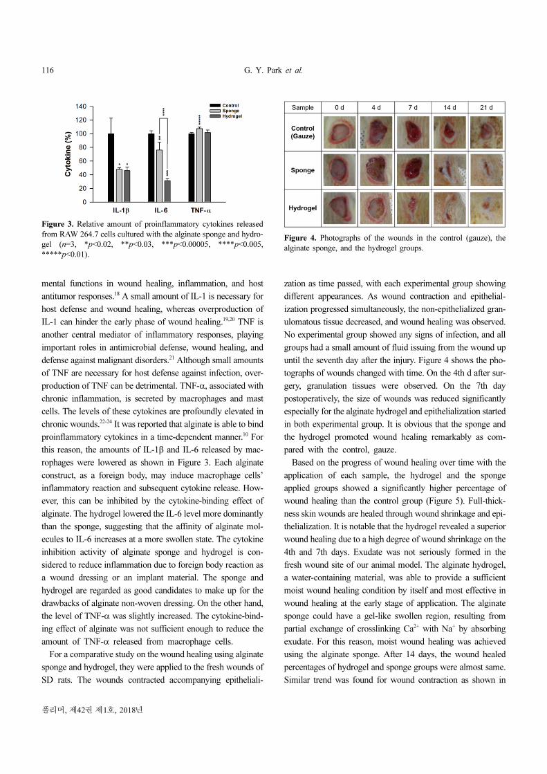
chronic wounds.22-24 It was reported that alginate is able to bind
proinflammatory cytokines in a time-dependent manner.10 For
this reason, the amounts of IL-1β and IL-6 released by mac-
rophages were lowered as shown in Figure 3. Each alginate
construct, as a foreign body, may induce macrophage cells’
inflammatory reaction and subsequent cytokine release. How-
ever, this can be inhibited by the cytokine-binding effect of
alginate. The hydrogel lowered the IL-6 level more dominantly
than the sponge, suggesting that the affinity of alginate mol-
ecules to IL-6 increases at a more swollen state. The cytokine
inhibition activity of alginate sponge and hydrogel is con-
sidered to reduce inflammation due to foreign body reaction as
a wound dressing or an implant material. The sponge and
hydrogel are regarded as good candidates to make up for the
drawbacks of alginate non-woven dressing. On the other hand,
the level of TNF-α was slightly increased. The cytokine-bind-
ing effect of alginate was not sufficient enough to reduce the
amount of TNF-α released from macrophage cells.
For a comparative study on the wound healing using alginate
sponge and hydrogel, they were applied to the fresh wounds of
SD rats. The wounds contracted accompanying epitheliali-
zation as time passed, with each experimental group showing
different appearances. As wound contraction and epithelial-
ization progressed simultaneously, the non-epithelialized gran-
ulomatous tissue decreased, and wound healing was observed.
No experimental group showed any signs of infection, and all
groups had a small amount of fluid issuing from the wound up
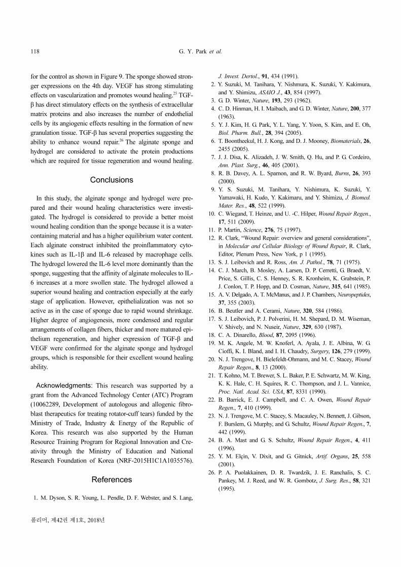
until the seventh day after the injury. Figure 4 shows the pho-
tographs of wounds changed with time. On the 4th d after sur-
gery, granulation tissues were observed. On the 7th day
postoperatively, the size of wounds was reduced significantly
especially for the alginate hydrogel and epithelialization started
in both experimental group. It is obvious that the sponge and
the hydrogel promoted wound healing remarkably as com-
pared with the control, gauze.
Based on the progress of wound healing over time with the
application of each sample, the hydrogel and the sponge
applied groups showed a significantly higher percentage of
wound healing than the control group (Figure 5). Full-thick-
ness skin wounds are healed through wound shrinkage and epi-
thelialization. It is notable that the hydrogel revealed a superior
wound healing due to a high degree of wound shrinkage on the
4th and 7th days. Exudate was not seriously formed in the
fresh wound site of our animal model. The alginate hydrogel,
a water-containing material, was able to provide a sufficient
moist wound healing condition by itself and most effective in
wound healing at the early stage of application. The alginate
sponge could have a gel-like swollen region, resulting from
partial exchange of crosslinking Ca2+ with Na+ by absorbing
exudate. For this reason, moist wound healing was achieved
using the alginate sponge. After 14 days, the wound healed
percentages of hydrogel and sponge groups were almost same.
Similar trend was found for wound contraction as shown in
Figure 3. Relative amount of proinflammatory cytokines released
from RAW 264.7 cells cultured with the alginate sponge and hydro-
gel (n=3, *p<0.02, **p<0.03, ***p<0.00005, ****p<0.005,
*****p<0.01).
Figure 4. Photographs of the wounds in the control (gauze), the
alginate sponge, and the hydrogel groups.

Moisture Wound Healing Characteristics of Alginate Sponge and Hydrogel 117
Polymer(Korea), Vol. 42, No. 1, 2018
Figure 6. On the other hand, epithelialization was conspicuous
when the alginate sponge was applied especially on the 7th and
14th days as shown in Figure 7. The sponge promoted wound
shrinkage and contraction less actively than hydrogel, resulting
in the higher values of Wi, Wi−Ui, and would epithelialization
(%). On the contrary, the Wi for the hydrogel group decreased
rapidly to lower the Wi−Ui. In this case, granulation prevailed
over epithelialization with rapid wound shrinkage and con-
traction.
Up until the 7th day after the injury, polymorphonuclear
cells, neutrocytes and lymphocytes, indications of inflamma-
tion, were seen in every experimental group (Figure 8(A)).
Also, higher degree of angiogenesis and more condensed, reg-
ular arrangements of collagen fibers were seen in the alginate
sponge and hydrogel groups on 14th day (Figure 8(B)). When
the wounds were completely healed, thicker, more matured,
and more regenerated epithelia were found in the alginate
sponge and hydrogel groups. In the RT-PCR experiment, the
expressions of protein such as TGF-β and VEGF for the algi-
nate sponge and hydrogel groups were much higher than those
Figure 8. Histology for wound tissues of the control (gauze), the
sponge, and the hydrogel groups (A: H&E staining, B: Masson’s tri-
chrome staining).
Figure 9. RT-PCR analysis of TGF-β and VEGF for the control
(gauze) (a); the sponge (b); the hydrogel (c) groups.
Figure 5. Wound healed (%) of the control (gauze), the sponge, and
the hydrogel groups (n=3, *p<0.15, **p<0.25, ***p<0.25,
****p<0.15, *****p<0.05).
Figure 6. Wound contraction (%) of the control (gauze), the sponge,
and the hydrogel groups (n=3, *p<0.15, **p<0.4, ***p<0.5,
****p<0.4, *****p<0.2, ******p<0.05).
Figure 7. Wound epithelialization (%) of the control (gauze), the
sponge, and the hydrogel groups (n=3, *p<0.25).
118 G. Y. Park et al.
폴리머, 제42권 제1호, 2018년
for the control as shown in Figure 9. The sponge showed stron-
ger expressions on the 4th day. VEGF has strong stimulating
effects on vascularization and promotes wound healing.25 TGF-
β has direct stimulatory effects on the synthesis of extracellular
matrix proteins and also increases the number of endothelial
cells by its angiogenic effects resulting in the formation of new
granulation tissue. TGF-β has several properties suggesting the
ability to enhance wound repair.26 The alginate sponge and
hydrogel are considered to activate the protein productions
which are required for tissue regeneration and wound healing.
Conclusions
In this study, the alginate sponge and hydrogel were pre-
pared and their wound healing characteristics were investi-
gated. The hydrogel is considered to provide a better moist
wound healing condition than the sponge because it is a water-
containing material and has a higher equilibrium water content.
Each alginate construct inhibited the proinflammatory cyto-
kines such as IL-1β and IL-6 released by macrophage cells.
The hydrogel lowered the IL-6 level more dominantly than the
sponge, suggesting that the affinity of alginate molecules to IL-
6 increases at a more swollen state. The hydrogel allowed a
superior wound healing and contraction especially at the early
stage of application. However, epithelialization was not so
active as in the case of sponge due to rapid wound shrinkage.
Higher degree of angiogenesis, more condensed and regular
arrangements of collagen fibers, thicker and more matured epi-
thelium regeneration, and higher expression of TGF-β and
VEGF were confirmed for the alginate sponge and hydrogel
groups, which is responsible for their excellent wound healing
ability.
Acknowledgments: This research was supported by a
grant from the Advanced Technology Center (ATC) Program
(10062289, Development of autologous and allogenic fibro-
blast therapeutics for treating rotator-cuff tears) funded by the
Ministry of Trade, Industry & Energy of the Republic of
Korea. This research was also supported by the Human
Resource Training Program for Regional Innovation and Cre-
ativity through the Ministry of Education and National
Research Foundation of Korea (NRF-2015H1C1A1035576).
References
1. M. Dyson, S. R. Young, L. Pendle, D. F. Webster, and S. Lang,
J. Invest. Dertol., 91, 434 (1991).
2. Y. Suzuki, M. Tanihara, Y. Nishmura, K. Suzuki, Y. Kakimura,
and Y. Shimizu, ASAIO J., 43, 854 (1997).
3. G. D. Winter, Nature, 193, 293 (1962).
4. C. D. Hinman, H. I. Maibach, and G. D. Winter, Nature, 200, 377
(1963).
5. Y. J. Kim, H. G. Park, Y. L. Yang, Y. Yoon, S. Kim, and E. Oh,
Biol. Pharm. Bull., 28, 394 (2005).
6. T. Boontheekul, H. J. Kong, and D. J. Mooney, Biomaterials, 26,
2455 (2005).
7. J. J. Disa, K. Alizadeh, J. W. Smith, Q. Hu, and P. G. Cordeiro,
Ann. Plast. Surg., 46, 405 (2001).
8. R. B. Davey, A. L. Sparnon, and R. W. Byard, Burns, 26, 393
(2000).
9. Y. S. Suzuki, M. Tanihara, Y. Nishimura, K. Suzuki, Y.
Yamawaki, H. Kudo, Y. Kakimaru, and Y. Shimizu, J. Biomed.
Mater. Res., 48, 522 (1999).
10. C. Wiegand, T. Heinze, and U. -C. Hilper, Wound Repair Regen.,
17, 511 (2009).
11. P. Martin, Science, 276, 75 (1997).
12. R. Clark, “Wound Repair: overview and general considerations”,
in Molecular and Cellular Biiology of Wound Repair, R. Clark,
Editor, Plenum Press, New York, p 1 (1995).
13. S. J. Leibovich and R. Ross, Am. J. Pathol., 78, 71 (1975).
14. C. J. March, B. Mosley, A. Larsen, D. P. Cerretti, G. Braedt, V.
Price, S. Gillis, C. S. Henney, S. R. Kronheim, K. Grabstein, P.
J. Conlon, T. P. Hopp, and D. Cosman, Nature, 315, 641 (1985).
15. A. V. Delgado, A. T. McManus, and J. P. Chambers, Neuropeptides,
37, 355 (2003).
16. B. Beutler and A. Cerami, Nature, 320, 584 (1986).
17. S. J. Leibovich, P. J. Polverini, H. M. Shepard, D. M. Wiseman,
V. Shively, and N. Nuseir, Nature, 329, 630 (1987).
18. C. A. Dinarello, Blood, 87, 2095 (1996).
19. M. K. Angele, M. W. Knoferl, A. Ayala, J. E. Albina, W. G.
Cioffi, K. I. Bland, and I. H. Chaudry, Surgery, 126, 279 (1999).
20. N. J. Trengove, H. Bielefeldt-Ohmann, and M. C. Stacey, Wound
Repair Regen., 8, 13 (2000).
21. T. Kohno, M. T. Brewer, S. L. Baker, P. E. Schwartz, M. W. King,
K. K. Hale, C. H. Squires, R. C. Thompson, and J. L. Vannice,
Proc. Natl. Acad. Sci. USA, 87, 8331 (1990).
22. B. Barrick, E. J. Campbell, and C. A. Owen, Wound Repair
Regen., 7, 410 (1999).
23. N. J. Trengove, M. C. Stacey, S. Macauley, N. Bennett, J. Gibson,
F. Burslem, G. Murphy, and G. Schultz, Wound Repair Regen., 7,
442 (1999).
24. B. A. Mast and G. S. Schultz, Wound Repair Regen., 4, 411
(1996).
25. Y. M. Elçin, V. Dixit, and G. Gitnick, Artif. Organs, 25, 558
(2001).
26. P. A. Puolakkainen, D. R. Twardzik, J. E. Ranchalis, S. C.
Pankey, M. J. Reed, and W. R. Gombotz, J. Surg. Res., 58, 321
(1995).


![제2주 인상재 알지네이트 작업시간과 경화시간 측정elearning.kocw.net/KOCW/document/2015/shinhan/ryujaekyung/2.pdf · [제2주] 인상재 - 알지네이트 작업시간과](https://img.pdfslide.tips/doc/110x75/5e0ed7ebe4c303358e2c502e/oe2-f-oee-oeee-eoee-oe2.jpg)


![[ 주 ] 넷클립스](https://img.pdfslide.tips/doc/110x75/56815409550346895dc20721/-56815409550346895dc20721.jpg)


