Embed Size (px)
Citation preview
One-Step Preparation of Glycopeptide Microspheres Basedon a-Amino Acid-N-carboxyanhydride Polymerization UsingInterfacial Protocols
Jing Wang, Changsheng Liu, Ping Chi
Key Laboratory for Ultrafine Materials of Ministry of Education, Engineering Research Center for Biomedical Materialsof Ministry of Education, East China University of Science and Technology, Shanghai 200237, People’s Republic of China
Received 14 May 2007; revised 3 February 2008; accepted 17 March 2008Published online 20 August 2008 in Wiley InterScience (www.interscience.wiley.com). DOI: 10.1002/jbm.b.31186
Abstract: A type of polysaccharide-polypeptide hybrid material, chitin derivative with
polypeptide side chains was prepared by the graft copolymerization of L-leucine N-
carboxyanhydride triggered by water-soluble chitin (WSC). The studies on surface tension
and aggregation demonstrated surface activity of WSC. Using this extraordinary property, the
microspheres were synchronously obtained via interfacial polymerization. The method
employed here to form the microspheres was in direct contrast to previous syntheses that used
either templating method or oil-in-water emulsion. The study provided a facile approach for
synthesizing microspheres with a variety of distinct polypeptide and regulated graft length,
which had mimetic structure of glycoconjugation in extracellular matrix. Furthermore, the
swelling capability of the microsphere in both acidic aqueous and organic solvents would give
promising application in drug delivery. ' 2008 Wiley Periodicals, Inc. J Biomed Mater Res Part B: Appl
Biomater 89B: 45–54, 2009
Keywords: bioactive material; biomimetic; extracellular matrix; graft/copolymer
INTRODUCTION
Many strategies have attempted to develop temporary scaf-
folds for tissue engineering using biodegradable and bio-
compatible synthetic or natural polymers over the past
decade.1–4 Aliphatic polyesters such as polylactide (PLA),
polyglycolide (PGA), and their copolymer poly(lactide-co-glycolide) (PLGA) are among the few synthetic polymers
that have been used widely in the tissue engineering. How-
ever, cell affinity toward synthetic polymers is generally
poor as a consequence of their low hydrophilicity and lack
of surface recognition sites, which affect cell suspension
penetrating into the scaffolds.5–8 Principally, the scaffold’s
design should mimic the structure and biological function
of native extracellular matrix (ECM), which provide me-
chanical support and regulate cell activities.9–12
ECM, a stable structural element surrounding the
cells,13–16 may provide environments for proper growth,
differentiation, and migration of cells, and serve as reser-
voir of regulatory molecules in tissues such as growth fac-
tors.17–20 The components of ECM can be subdivided into
four classes: glycopeptides, proteoglycans, elastins, and col-
lagens. Because of the complexity of the ECM, studying
cell behavior in ECM derivatives or even single component
of the ECM would make it difficult to decipher the impor-
tance of each factor involved.21–24
Since numerous cellular activation processes are initiated
by ECM ligands, novel methods for mimicking the natural
ECM are of great interest for improving the biocompatibility
or creating new biomaterials. However, the utility of collagen
and proteoglycans, such as condroitin sulfates and hyaluronic
acid, has been limited by extremely high price and poor me-
chanical properties. To establish an ideal scaffold mimicking
the natural ECM, it is desirable to develop a simple approach
to combine the advantages of both natural and synthetic poly-
mers, which could enhance affinities with cells and speed up
tissue regeneration, as well as substitutes of ECMs.
Polysaccharides are interesting materials as their carbo-
hydrate moieties interact with or are integral components
of many cell adhesion molecules and matrix glycopro-
teins.25,26 Chitin, a natural polysaccharide consisting of b-(1?4)-linked 2-acetamide-2-deoxy-D-glucopyranose, is one
of the most abundant and easily obtained biopolymers next
to cellulose. It is commonly found in the shells of crusta-
cean such as crab and shrimp, exoskeletons or cuticles of
many invertebrates, and in the cell walls of most fungi and
Correspondence to: C. Liu (e-mail: [email protected])Contract grant sponsor: National Science Fund for Distinguished Young Scholars
of China; Contract grant number: 20425621Contract grant sponsor: Major State Basic Research Program of China; Contract
grant number: 2005CCA01000Contract grant sponsor: Basic Research Foundation of Shanghai Science and
Technique Committee; Contract grant number: 05DJ14005Contract grant sponsor: Shanghai Leading Academic Discipline Project; Contract
grant number: B502
' 2008 Wiley Periodicals, Inc.
45
some algae. This polymer is known to be nontoxic, bio-
compatible, and enzymatically biodegradable.27 Further-
more, chitin has structural characteristics similar to
glycosaminoglycans (GAGs, main component of proteogly-
cans such as condroitin sulfates and hyaluronic acid), which
occurs ubiquitously within the ECM.28–30 So, it is an excel-
lent starting template for the design of ECM-like materials.
Kurita et al.31,32 has showed the possibility of preparing chi-
tin-protein complexes by the graft copolymerization of
amino acid N-carboxyanhydride (NCA) onto chitin. Ma and
coworkers33 also reported the synthesis of chitosan-g-poly(L-leucine). Nevertheless, the previous study focused on
the synthesis of the copolymer. The graft production had
comparatively poor solubility in most of the common sol-
vents, which hampered its further process and applications.
In this study, we have developed a simplified polysaccha-
ride-polypeptide system mimicking the glycoconjugations in
ECM, which allows us to modulate the components and
properties expediently. Water-soluble chitin (WSC) with
degree of acetylation close to 0.50 was selected as the poly-
saccharide backbone moiety and was found possessing par-
ticular surfactant activities. In the light of these findings, it
was interesting to investigate the applicability of preparing
the glycoconjugate polymeric microsphere in situ without
additional emulsion. To our knowledge, such microsphere
based upon the polysaccharide-polypeptide hybrid materials
has not yet been reported in literature. This work provided a
novel drug carrier and a unique method for preparing the
glycoconjugate microsphere.
MATERIALS AND METHODS
Materials
All reagents used were available from commercial sources.
Chitosan with average molecular weight (Mw) of 1.5 3 105
and 90% degree of deacetylation (DD) was from Shanghai
Kabo and was used for the preparation of graft copolymer.
L-leucine was purchased from Huamei Biochemical. Tetra-
hydrofuran (THF) and n-hexane were distilled from so-
dium/benzophenone in an inert atmosphere to remove
water before using. Ethyl acetate was distilled from P2O5.
All other solvents were of analytical grade and used with-
out further purification. Pyrene was purchased from Sigma
and used as received.
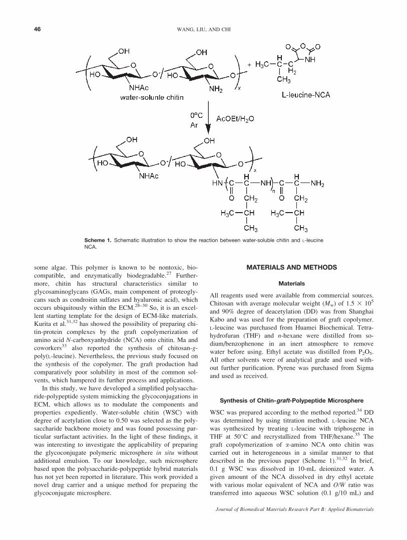
Synthesis of Chitin-graft-Polypeptide Microsphere
WSC was prepared according to the method reported.34 DD
was determined by using titration method. L-leucine NCA
was synthesized by treating L-leucine with triphosgene in
THF at 508C and recrystallized from THF/hexane.35 The
graft copolymerization of a-amino NCA onto chitin was
carried out in heterogeneous in a similar manner to that
described in the previous paper (Scheme 1).31,32 In brief,
0.1 g WSC was dissolved in 10-mL deionized water. A
given amount of the NCA dissolved in dry ethyl acetate
with various molar equivalent of NCA and O/W ratio was
transferred into aqueous WSC solution (0.1 g/10 mL) and
Scheme 1. Schematic illustration to show the reaction between water-soluble chitin and L-leucine
NCA.
46 WANG, LIU, AND CHI
Journal of Biomedical Materials Research Part B: Applied Biomaterials
subjected to the reaction at 08C for 2 h under Argon. After
that, the mixture was poured into 120-mL N,N-dimethylfor-
mamide (DMF). The resulting grafting copolymer was fil-
tered and washed with DMF/deionized water (10:1) for
purification. The resultant microparticles were then washed
with acetone in grades and collected after lyophilizing.
Cryobreaking of Hollow Microspheres
Cryobreaking technique was carried out under ultrasonifica-
tion in liquid nitrogen medium36 to obtain the broken vesi-
cle and details as follows. Microspheres dispersed in DMF
were frozen by immersing into liquid nitrogen medium and
then partially melted by naturally exposing to the air. At
this point, the microspheres were placed into an ultrasonifi-
cation chamber, and let the tip of the ultrasonicator touch
the solid/liquid interface. The process lasted for about
45–50 s. After that, the sample was ready for subsequent
lyophilization.
Surface Tension Measurement
A RADIAN 300 Processor Tensionmeter (Thermo Cahn.)
was used to determine the surface tension of chitosan and
WSC solutions at various concentrations with plate method
at 258C 6 0.18C. All of the solutions were kept at room
temperature for 1 day before surface tension measure-
ment.37,38
Fluorescence Spectroscopy
Pyrene was selected as a fluorescent probe to demonstrate
the presence of hydrophobic domains. A known volume of
pyrene in methanol was put into a series of flasks and
evaporated under an argon purge to remove the solvent.
Then, an aqueous WSC solution was added into the test
tube. The final concentration of pyrene was l 3 1026 mol/
L. The solutions were sonicated for 15 min in an ultrasonic
bath and then kept at room temperature for 2 h before
spectroscopic analysis. The steady-state fluorescence spec-
tra were carried out on a Cary Eclipse fluorescence spectro-
photometer (Varian, USA) at 258C. The excitation
wavelength of pyrene was set to 335 nm. The hydrophobic
index, I1/I3, was calculated as the ratio of the intensities at
the first (374 nm) and the third (385 nm) vibrational peak
in the pyrene emission spectra.39
Characterization
1H NMR spectra of graft copolymers were obtained with
an AM-400 spectrometer (Bruker) at room temperature,
using CF3COOD as solvent. The Fourier transform infrared
transmission spectra were recorded using an attenuated
total reflection (ATR) method in AVATAR360 spectrome-
ter (Nicolet). Microspheres were observed through TE2000-
U optical microscope with a CCD camera (Nicon). Mor-
phological investigations of microsphere were performed
with a JEOL-JSM-6360LV scanning electron microscope
(Japan), operating at an acceleration voltage of 15 kV. The
microparticles were lightly sputter-coated with a thin layer
of Au/Pd prior to the observation.
Swelling Behavior Studies
In the swelling experiment, the dried microparticles of
about 20 mg were placed in a cylindrical tube. One millili-
ter of deionized water, hydrochloric acid (pH 5 5, 3), and
DMF were added to soak the microparticles, respectively.
Then seal the tube with a lid to avoid volatilizing. After
sufficient time for achieving swelling equilibrium, the tube
was centrifuged and the supernatant was removed. The
swollen microparticles were collected and weighed quickly.
The swelling ratio (Q) was calculated with the following
formula:
Q ¼ W2 �W1
W1
3100%
where W1 is the weight of the dried microparticles, W2 is
the weight of the swollen microparticles. Each sample was
measured in triplicate.
RESULTS
Surface Tension and Aggregation Behaviorof Water-Soluble Chitin
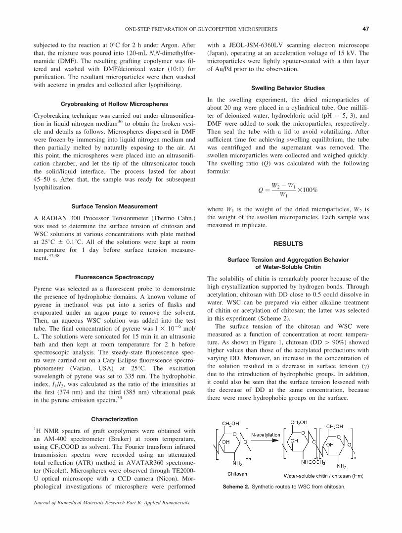
The solubility of chitin is remarkably poorer because of the
high crystallization supported by hydrogen bonds. Through
acetylation, chitosan with DD close to 0.5 could dissolve in
water. WSC can be prepared via either alkaline treatment
of chitin or acetylation of chitosan; the latter was selected
in this experiment (Scheme 2).
The surface tension of the chitosan and WSC were
measured as a function of concentration at room tempera-
ture. As shown in Figure 1, chitosan (DD [ 90%) showed
higher values than those of the acetylated productions with
varying DD. Moreover, an increase in the concentration of
the solution resulted in a decrease in surface tension (c)due to the introduction of hydrophobic groups. In addition,
it could also be seen that the surface tension lessened with
the decrease of DD at the same concentration, because
there were more hydrophobic groups on the surface.
Scheme 2. Synthetic routes to WSC from chitosan.
47ONE-STEP PREPARATION OF GLYCOPEPTIDE MICROSPHERES
Journal of Biomedical Materials Research Part B: Applied Biomaterials
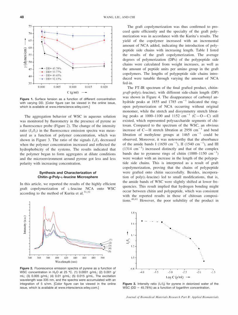
The aggregation behavior of WSC in aqueous solution
was monitored by fluorometry in the presence of pyrene as
a fluorescence probe (Figure 2). The change of the intensity
ratio (I1/I3) in the fluorescence emission spectra was meas-
ured as a function of polymer concentration, which was
shown in Figure 3. The ratio of the signals I1/I3 decreased
when the polymer concentration increased and reflected the
hydrophobicity of the systems. The results indicated that
the polymer began to form aggregates at dilute conditions
and the microenvironment around pyrene got less and less
polarity with increasing concentration.
Synthesis and Characterization ofChitin-g-Poly-L-leucine Microsphere
In this article, we reported the results of the highly efficient
graft copolymerization of L-leucine NCA onto WSC
according to the method of Kurita et al.31,32
The graft copolymerization was thus confirmed to pro-
ceed quite efficiently and the specialty of the graft poly-
merization was in accordance with the Kurita’s results. The
yield of the copolymer increased with an incremental
amount of NCA added, indicating the introduction of poly-
peptide side chains with increasing length. Table I listed
the results of the graft copolymerization. The average
degrees of polymerization (DPs) of the polypeptide side
chains were calculated from weight increases, as well as
the amount of peptide units per amino group in the graft
copolymers. The lengths of polypeptide side chains intro-
duced were tunable through varying the amount of NCA
fed-in.
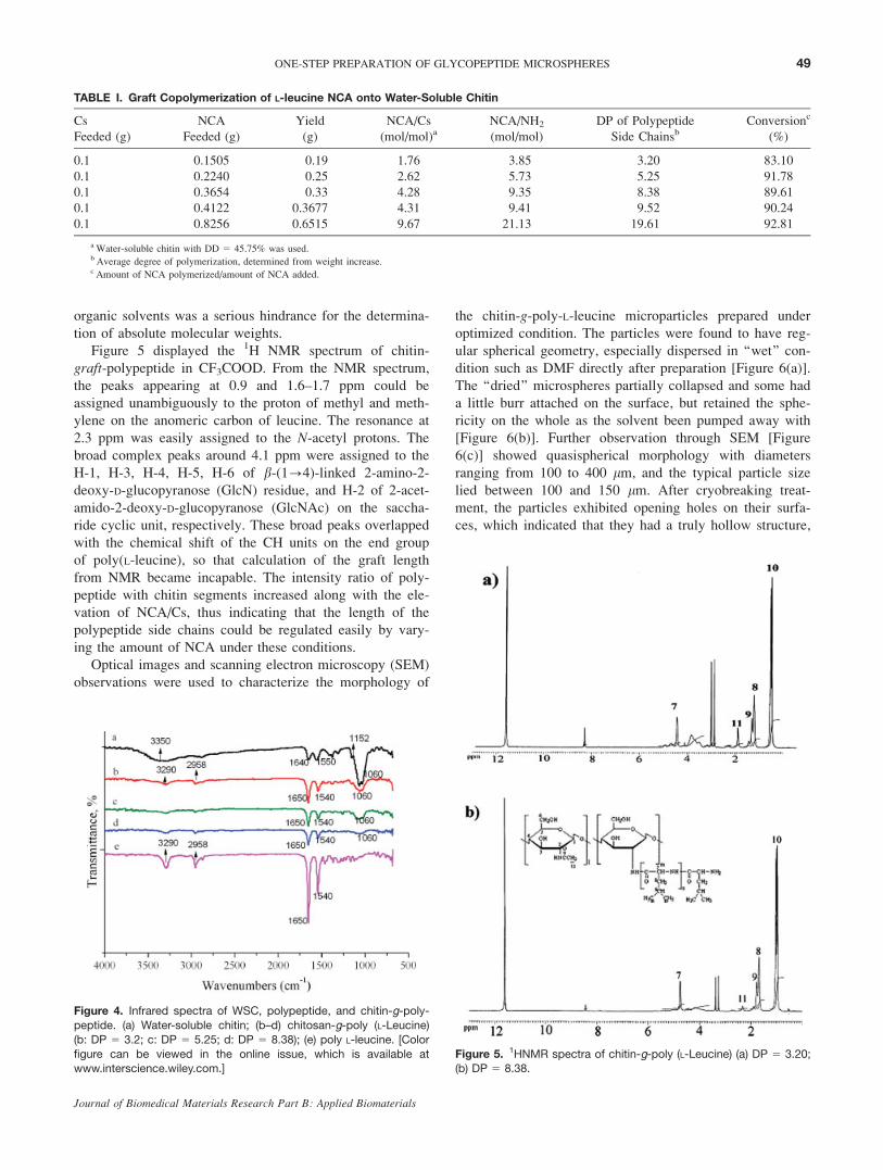
The FT-IR spectrum of the final grafted product, chitin-
graft-poly(L-leucine), with different side-chain length (DP)
was shown in Figure 4. The disappearance of carbonyl an-
hydride peaks at 1855 and 1785 cm21 indicated the ring-
open polymerization of NCA occurring without original
monomer, while the stretch and dissymmetry stretch librat-
ing peaks at 1000–1100 and 1152 cm21 (C��O��C) still
existed, which represented polysaccharide segments of chi-
tosan. Compared to the spectrum of the WSC, an obvious
increase of C��H stretch libration at 2958 cm21 and bend
libration of methylene groups at 1465 cm21 could be
observed. Moreover, it was noteworthy that the absorbance
of the amide bands I (1650 cm21), II (1540 cm21), and III
(1314 cm21) increased distinctly and that of the complex
bands due to pyranose rings of chitin (1000–1150 cm21)
were weaker with an increase in the length of the polypep-
tide side chains. This is interpreted as a result of graft
copolymerization, proving that the chains of polypeptide
were grafted onto chitin successfully. Besides, incorpora-
tion of poly(L-leucine) led to small modifications, that is,
the amide bands of WSC were slightly shifted at lower fre-
quencies. This result implied that hydrogen bonding might
occur between chitin and polypeptide, which was consistent
with the reported results in those of chitosan composi-
tions.40,41 However, the poor solubility of the product in
Figure 1. Surface tension as a function of different concentrationwith varying DD. [Color figure can be viewed in the online issue,
which is available at www.interscience.wiley.com.]
Figure 2. Fluorescence emission spectra of pyrene as a function ofWSC concentration in H2O at 25 8C. (1) 0.0001 g/mL; (2) 0.001 g/
mL; (3) 0.005 g/mL; (4) 0.01 g/mL; (5) 0.015 g/mL. The excitation
wavelength was 335 nm, and the spectra were accumulated with an
integration of 5 s/nm. [Color figure can be viewed in the onlineissue, which is available at www.interscience.wiley.com.]
Figure 3. Intensity ratio (I1/I3) for pyrene in deionized water of theWSC (DD 5 45.78%) as a function of logarithm concentration.
48 WANG, LIU, AND CHI
Journal of Biomedical Materials Research Part B: Applied Biomaterials
organic solvents was a serious hindrance for the determina-
tion of absolute molecular weights.
Figure 5 displayed the 1H NMR spectrum of chitin-
graft-polypeptide in CF3COOD. From the NMR spectrum,
the peaks appearing at 0.9 and 1.6–1.7 ppm could be
assigned unambiguously to the proton of methyl and meth-
ylene on the anomeric carbon of leucine. The resonance at
2.3 ppm was easily assigned to the N-acetyl protons. Thebroad complex peaks around 4.1 ppm were assigned to the
H-1, H-3, H-4, H-5, H-6 of b-(1?4)-linked 2-amino-2-
deoxy-D-glucopyranose (GlcN) residue, and H-2 of 2-acet-
amido-2-deoxy-D-glucopyranose (GlcNAc) on the saccha-
ride cyclic unit, respectively. These broad peaks overlapped
with the chemical shift of the CH units on the end group
of poly(L-leucine), so that calculation of the graft length
from NMR became incapable. The intensity ratio of poly-
peptide with chitin segments increased along with the ele-
vation of NCA/Cs, thus indicating that the length of the
polypeptide side chains could be regulated easily by vary-
ing the amount of NCA under these conditions.
Optical images and scanning electron microscopy (SEM)
observations were used to characterize the morphology of
the chitin-g-poly-L-leucine microparticles prepared under
optimized condition. The particles were found to have reg-
ular spherical geometry, especially dispersed in ‘‘wet’’ con-
dition such as DMF directly after preparation [Figure 6(a)].
The ‘‘dried’’ microspheres partially collapsed and some had
a little burr attached on the surface, but retained the sphe-
ricity on the whole as the solvent been pumped away with
[Figure 6(b)]. Further observation through SEM [Figure
6(c)] showed quasispherical morphology with diameters
ranging from 100 to 400 lm, and the typical particle size
lied between 100 and 150 lm. After cryobreaking treat-
ment, the particles exhibited opening holes on their surfa-
ces, which indicated that they had a truly hollow structure,
TABLE I. Graft Copolymerization of L-leucine NCA onto Water-Soluble Chitin
Cs
Feeded (g)
NCA
Feeded (g)
Yield
(g)
NCA/Cs
(mol/mol)aNCA/NH2
(mol/mol)
DP of Polypeptide
Side ChainsbConversionc
(%)
0.1 0.1505 0.19 1.76 3.85 3.20 83.10
0.1 0.2240 0.25 2.62 5.73 5.25 91.78
0.1 0.3654 0.33 4.28 9.35 8.38 89.61
0.1 0.4122 0.3677 4.31 9.41 9.52 90.24
0.1 0.8256 0.6515 9.67 21.13 19.61 92.81
aWater-soluble chitin with DD 5 45.75% was used.b Average degree of polymerization, determined from weight increase.c Amount of NCA polymerized/amount of NCA added.
Figure 4. Infrared spectra of WSC, polypeptide, and chitin-g-poly-
peptide. (a) Water-soluble chitin; (b–d) chitosan-g-poly (L-Leucine)
(b: DP 5 3.2; c: DP 5 5.25; d: DP 5 8.38); (e) poly L-leucine. [Color
figure can be viewed in the online issue, which is available atwww.interscience.wiley.com.]
Figure 5. 1HNMR spectra of chitin-g-poly (L-Leucine) (a) DP 5 3.20;(b) DP 5 8.38.
49ONE-STEP PREPARATION OF GLYCOPEPTIDE MICROSPHERES
Journal of Biomedical Materials Research Part B: Applied Biomaterials
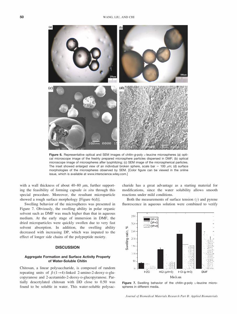
with a wall thickness of about 40–80 lm, further support-
ing the feasibility of forming capsule in situ through this
special procedure. Moreover, the resultant microparticle
showed a rough surface morphology [Figure 6(d)].
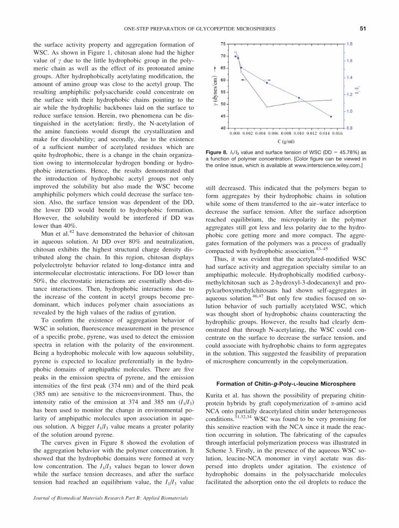
Swelling behavior of the microspheres was presented in
Figure 7. Obviously, the swelling ability in polar organic
solvent such as DMF was much higher than that in aqueous
medium. At the early stage of immersion in DMF, the
dried microparticles were quickly swollen due to very fast
solvent absorption. In addition, the swelling ability
decreased with increasing DP, which was imputed to the
effect of longer side chains of the polypeptide moiety.
DISCUSSION
Aggregate Formation and Surface Activity Propertyof Water-Soluble Chitin
Chitosan, a linear polysaccharide, is composed of random
repeating units of b-(1?4)-linked 2-amino-2-deoxy-D-glu-
copyranose and 2-acetamido-2-deoxy-D-glucopyranose. Par-
tially deacetylated chitosan with DD close to 0.50 was
found to be soluble in water. This water-soluble polysac-
charide has a great advantage as a starting material for
modifications, since the water solubility allows smooth
reactions under mild conditions.
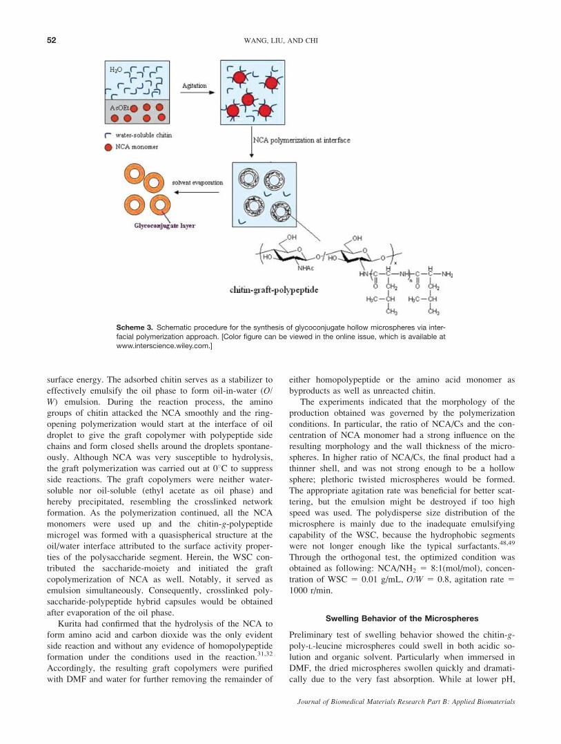
Both the measurements of surface tension (c) and pyrene
fluorescence in aqueous solution were combined to verify
Figure 6. Representative optical and SEM images of chitin-g-poly L-leucine microspheres (a) opti-
cal microscope image of the freshly prepared microsphere particles dispersed in DMF; (b) opticalmicroscope image of microspheres after lyophilizing; (c) SEM image of the microspherical particles.
The inset showed enlarged view of an individual broken sphere, scale bar 5 100 lm; (d) surface
morphologies of the microspheres observed by SEM. [Color figure can be viewed in the online
issue, which is available at www.interscience.wiley.com.]
Figure 7. Swelling behavior of the chitin-g-poly L-leucine micro-spheres in different media.
50 WANG, LIU, AND CHI
Journal of Biomedical Materials Research Part B: Applied Biomaterials
the surface activity property and aggregation formation of
WSC. As shown in Figure 1, chitosan alone had the higher
value of c due to the little hydrophobic group in the poly-
meric chain as well as the effect of its protonated amine
groups. After hydrophobically acetylating modification, the
amount of amino group was close to the acetyl group. The
resulting amphiphilic polysaccharide could concentrate on
the surface with their hydrophobic chains pointing to the
air while the hydrophilic backbones laid on the surface to
reduce surface tension. Herein, two phenomena can be dis-
tinguished in the acetylation: firstly, the N-acetylation of
the amine functions would disrupt the crystallization and
make for dissolubility; and secondly, due to the existence
of a sufficient number of acetylated residues which are
quite hydrophobic, there is a change in the chain organiza-
tion owing to intermolecular hydrogen bonding or hydro-
phobic interactions. Hence, the results demonstrated that
the introduction of hydrophobic acetyl groups not only
improved the solubility but also made the WSC become
amphiphilic polymers which could decrease the surface ten-
sion. Also, the surface tension was dependent of the DD,
the lower DD would benefit to hydrophobic formation.
However, the solubility would be interfered if DD was
lower than 40%.
Mun et al.42 have demonstrated the behavior of chitosan
in aqueous solution. At DD over 80% and neutralization,
chitosan exhibits the highest structural charge density dis-
tributed along the chain. In this region, chitosan displays
polyelectrolyte behavior related to long-distance intra and
intermolecular electrostatic interactions. For DD lower than
50%, the electrostatic interactions are essentially short-dis-
tance interactions. Then, hydrophobic interactions due to
the increase of the content in acetyl groups become pre-
dominant, which induces polymer chain associations as
revealed by the high values of the radius of gyration.
To confirm the existence of aggregation behavior of
WSC in solution, fluorescence measurement in the presence
of a specific probe, pyrene, was used to detect the emission
spectra in relation with the polarity of the environment.
Being a hydrophobic molecule with low aqueous solubility,
pyrene is expected to localize preferentially in the hydro-
phobic domains of amphipathic molecules. There are five
peaks in the emission spectra of pyrene, and the emission
intensities of the first peak (374 nm) and of the third peak
(385 nm) are sensitive to the microenvironment. Thus, the
intensity ratio of the emission at 374 and 385 nm (I1/I3)
has been used to monitor the change in environmental po-
larity of amphipathic molecules upon association in aque-
ous solution. A bigger I1/I3 value means a greater polarity
of the solution around pyrene.
The curves given in Figure 8 showed the evolution of
the aggregation behavior with the polymer concentration. It
showed that the hydrophobic domains were formed at very
low concentration. The I1/I3 values began to lower down
while the surface tension decreases, and after the surface
tension had reached an equilibrium value, the I1/I3 value
still decreased. This indicated that the polymers began to
form aggregates by their hydrophobic chains in solution
while some of them transferred to the air–water interface to
decrease the surface tension. After the surface adsorption
reached equilibrium, the micropolarity in the polymer
aggregates still got less and less polarity due to the hydro-
phobic core getting more and more compact. The aggre-
gates formation of the polymers was a process of gradually
compacted with hydrophobic association.43–45
Thus, it was evident that the acetylated-modified WSC
had surface activity and aggregation specialty similar to an
amphipathic molecule. Hydrophobically modified carboxy-
methylchitosan such as 2-hydroxyl-3-dodecanoxyl and pro-
pylcarboxymethylchitosans had shown self-aggregates in
aqueous solution.46,47 But only few studies focused on so-
lution behavior of such partially acetylated WSC, which
was thought short of hydrophobic chains counteracting the
hydrophilic groups. However, the results had clearly dem-
onstrated that through N-acetylating, the WSC could con-
centrate on the surface to decrease the surface tension, and
could associate with hydrophobic chains to form aggregates
in the solution. This suggested the feasibility of preparation
of microsphere concurrently in the copolymerization.
Formation of Chitin-g-Poly-L-leucine Microsphere
Kurita et al. has shown the possibility of preparing chitin-
protein hybrids by graft copolymerization of a-amino acid
NCA onto partially deacetylated chitin under heterogeneous
conditions.31,32,34 WSC was found to be very promising for
this sensitive reaction with the NCA since it made the reac-
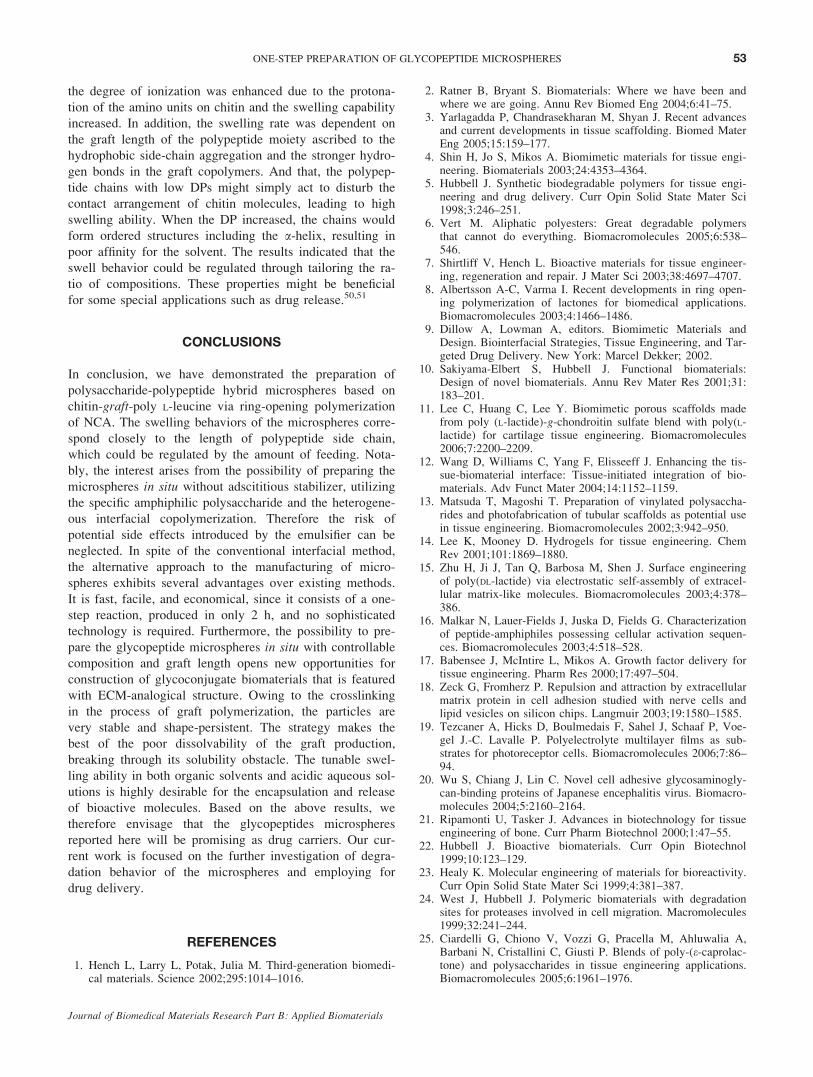
tion occurring in solution. The fabricating of the capsules
through interfacial polymerization process was illustrated in
Scheme 3. Firstly, in the presence of the aqueous WSC so-
lution, leucine-NCA monomer in vinyl acetate was dis-
persed into droplets under agitation. The existence of
hydrophobic domains in the polysaccharide molecules
facilitated the adsorption onto the oil droplets to reduce the
Figure 8. I1/I3 value and surface tension of WSC (DD 5 45.78%) asa function of polymer concentration. [Color figure can be viewed in
the online issue, which is available at www.interscience.wiley.com.]
51ONE-STEP PREPARATION OF GLYCOPEPTIDE MICROSPHERES
Journal of Biomedical Materials Research Part B: Applied Biomaterials
surface energy. The adsorbed chitin serves as a stabilizer to
effectively emulsify the oil phase to form oil-in-water (O/W) emulsion. During the reaction process, the amino
groups of chitin attacked the NCA smoothly and the ring-
opening polymerization would start at the interface of oil
droplet to give the graft copolymer with polypeptide side
chains and form closed shells around the droplets spontane-
ously. Although NCA was very susceptible to hydrolysis,
the graft polymerization was carried out at 08C to suppress
side reactions. The graft copolymers were neither water-
soluble nor oil-soluble (ethyl acetate as oil phase) and
hereby precipitated, resembling the crosslinked network
formation. As the polymerization continued, all the NCA
monomers were used up and the chitin-g-polypeptidemicrogel was formed with a quasispherical structure at the
oil/water interface attributed to the surface activity proper-
ties of the polysaccharide segment. Herein, the WSC con-
tributed the saccharide-moiety and initiated the graft
copolymerization of NCA as well. Notably, it served as
emulsion simultaneously. Consequently, crosslinked poly-
saccharide-polypeptide hybrid capsules would be obtained
after evaporation of the oil phase.
Kurita had confirmed that the hydrolysis of the NCA to
form amino acid and carbon dioxide was the only evident
side reaction and without any evidence of homopolypeptide
formation under the conditions used in the reaction.31,32
Accordingly, the resulting graft copolymers were purified
with DMF and water for further removing the remainder of
either homopolypeptide or the amino acid monomer as
byproducts as well as unreacted chitin.
The experiments indicated that the morphology of the
production obtained was governed by the polymerization
conditions. In particular, the ratio of NCA/Cs and the con-
centration of NCA monomer had a strong influence on the
resulting morphology and the wall thickness of the micro-
spheres. In higher ratio of NCA/Cs, the final product had a
thinner shell, and was not strong enough to be a hollow
sphere; plethoric twisted microspheres would be formed.
The appropriate agitation rate was beneficial for better scat-
tering, but the emulsion might be destroyed if too high
speed was used. The polydisperse size distribution of the
microsphere is mainly due to the inadequate emulsifying
capability of the WSC, because the hydrophobic segments
were not longer enough like the typical surfactants.48,49
Through the orthogonal test, the optimized condition was
obtained as following: NCA/NH2 5 8:1(mol/mol), concen-
tration of WSC 5 0.01 g/mL, O/W 5 0.8, agitation rate 51000 r/min.
Swelling Behavior of the Microspheres
Preliminary test of swelling behavior showed the chitin-g-poly-L-leucine microspheres could swell in both acidic so-
lution and organic solvent. Particularly when immersed in
DMF, the dried microspheres swollen quickly and dramati-
cally due to the very fast absorption. While at lower pH,
Scheme 3. Schematic procedure for the synthesis of glycoconjugate hollow microspheres via inter-
facial polymerization approach. [Color figure can be viewed in the online issue, which is available at
www.interscience.wiley.com.]
52 WANG, LIU, AND CHI
Journal of Biomedical Materials Research Part B: Applied Biomaterials
the degree of ionization was enhanced due to the protona-
tion of the amino units on chitin and the swelling capability
increased. In addition, the swelling rate was dependent on
the graft length of the polypeptide moiety ascribed to the
hydrophobic side-chain aggregation and the stronger hydro-
gen bonds in the graft copolymers. And that, the polypep-
tide chains with low DPs might simply act to disturb the
contact arrangement of chitin molecules, leading to high
swelling ability. When the DP increased, the chains would
form ordered structures including the a-helix, resulting in
poor affinity for the solvent. The results indicated that the
swell behavior could be regulated through tailoring the ra-
tio of compositions. These properties might be beneficial
for some special applications such as drug release.50,51
CONCLUSIONS
In conclusion, we have demonstrated the preparation of
polysaccharide-polypeptide hybrid microspheres based on
chitin-graft-poly L-leucine via ring-opening polymerization
of NCA. The swelling behaviors of the microspheres corre-
spond closely to the length of polypeptide side chain,
which could be regulated by the amount of feeding. Nota-
bly, the interest arises from the possibility of preparing the
microspheres in situ without adscititious stabilizer, utilizing
the specific amphiphilic polysaccharide and the heterogene-
ous interfacial copolymerization. Therefore the risk of
potential side effects introduced by the emulsifier can be
neglected. In spite of the conventional interfacial method,
the alternative approach to the manufacturing of micro-
spheres exhibits several advantages over existing methods.
It is fast, facile, and economical, since it consists of a one-
step reaction, produced in only 2 h, and no sophisticated
technology is required. Furthermore, the possibility to pre-
pare the glycopeptide microspheres in situ with controllable
composition and graft length opens new opportunities for
construction of glycoconjugate biomaterials that is featured
with ECM-analogical structure. Owing to the crosslinking
in the process of graft polymerization, the particles are
very stable and shape-persistent. The strategy makes the
best of the poor dissolvability of the graft production,
breaking through its solubility obstacle. The tunable swel-
ling ability in both organic solvents and acidic aqueous sol-
utions is highly desirable for the encapsulation and release
of bioactive molecules. Based on the above results, we
therefore envisage that the glycopeptides microspheres
reported here will be promising as drug carriers. Our cur-
rent work is focused on the further investigation of degra-
dation behavior of the microspheres and employing for
drug delivery.
REFERENCES
1. Hench L, Larry L, Potak, Julia M. Third-generation biomedi-cal materials. Science 2002;295:1014–1016.
2. Ratner B, Bryant S. Biomaterials: Where we have been andwhere we are going. Annu Rev Biomed Eng 2004;6:41–75.
3. Yarlagadda P, Chandrasekharan M, Shyan J. Recent advancesand current developments in tissue scaffolding. Biomed MaterEng 2005;15:159–177.
4. Shin H, Jo S, Mikos A. Biomimetic materials for tissue engi-neering. Biomaterials 2003;24:4353–4364.
5. Hubbell J. Synthetic biodegradable polymers for tissue engi-neering and drug delivery. Curr Opin Solid State Mater Sci1998;3:246–251.
6. Vert M. Aliphatic polyesters: Great degradable polymersthat cannot do everything. Biomacromolecules 2005;6:538–546.
7. Shirtliff V, Hench L. Bioactive materials for tissue engineer-ing, regeneration and repair. J Mater Sci 2003;38:4697–4707.
8. Albertsson A-C, Varma I. Recent developments in ring open-ing polymerization of lactones for biomedical applications.Biomacromolecules 2003;4:1466–1486.
9. Dillow A, Lowman A, editors. Biomimetic Materials andDesign. Biointerfacial Strategies, Tissue Engineering, and Tar-geted Drug Delivery. New York: Marcel Dekker; 2002.
10. Sakiyama-Elbert S, Hubbell J. Functional biomaterials:Design of novel biomaterials. Annu Rev Mater Res 2001;31:183–201.
11. Lee C, Huang C, Lee Y. Biomimetic porous scaffolds madefrom poly (L-lactide)-g-chondroitin sulfate blend with poly(L-lactide) for cartilage tissue engineering. Biomacromolecules2006;7:2200–2209.
12. Wang D, Williams C, Yang F, Elisseeff J. Enhancing the tis-sue-biomaterial interface: Tissue-initiated integration of bio-materials. Adv Funct Mater 2004;14:1152–1159.
13. Matsuda T, Magoshi T. Preparation of vinylated polysaccha-rides and photofabrication of tubular scaffolds as potential usein tissue engineering. Biomacromolecules 2002;3:942–950.
14. Lee K, Mooney D. Hydrogels for tissue engineering. ChemRev 2001;101:1869–1880.
15. Zhu H, Ji J, Tan Q, Barbosa M, Shen J. Surface engineeringof poly(DL-lactide) via electrostatic self-assembly of extracel-lular matrix-like molecules. Biomacromolecules 2003;4:378–386.
16. Malkar N, Lauer-Fields J, Juska D, Fields G. Characterizationof peptide-amphiphiles possessing cellular activation sequen-ces. Biomacromolecules 2003;4:518–528.
17. Babensee J, McIntire L, Mikos A. Growth factor delivery fortissue engineering. Pharm Res 2000;17:497–504.
18. Zeck G, Fromherz P. Repulsion and attraction by extracellularmatrix protein in cell adhesion studied with nerve cells andlipid vesicles on silicon chips. Langmuir 2003;19:1580–1585.
19. Tezcaner A, Hicks D, Boulmedais F, Sahel J, Schaaf P, Voe-gel J.-C. Lavalle P. Polyelectrolyte multilayer films as sub-strates for photoreceptor cells. Biomacromolecules 2006;7:86–94.
20. Wu S, Chiang J, Lin C. Novel cell adhesive glycosaminogly-can-binding proteins of Japanese encephalitis virus. Biomacro-molecules 2004;5:2160–2164.
21. Ripamonti U, Tasker J. Advances in biotechnology for tissueengineering of bone. Curr Pharm Biotechnol 2000;1:47–55.
22. Hubbell J. Bioactive biomaterials. Curr Opin Biotechnol1999;10:123–129.
23. Healy K. Molecular engineering of materials for bioreactivity.Curr Opin Solid State Mater Sci 1999;4:381–387.
24. West J, Hubbell J. Polymeric biomaterials with degradationsites for proteases involved in cell migration. Macromolecules1999;32:241–244.
25. Ciardelli G, Chiono V, Vozzi G, Pracella M, Ahluwalia A,Barbani N, Cristallini C, Giusti P. Blends of poly-(e-caprolac-tone) and polysaccharides in tissue engineering applications.Biomacromolecules 2005;6:1961–1976.
53ONE-STEP PREPARATION OF GLYCOPEPTIDE MICROSPHERES
Journal of Biomedical Materials Research Part B: Applied Biomaterials
26. Lin C, Lin J. Characterization and blood coagulation evalua-tion of the water-soluble chitooligosaccharides prepared by afacile fractionation method. Biomacromolecules 2003;4:1691–1697.
27. Shepherd R, Reader S, Falshaw A. Chitosan functional prop-erties. Glycoconj J 1997;14:535–542.
28. Morimoto M, Saimoto H, Usui H, Okamoto Y, Minami S,Shigemasa Y. Biological activities of carbohydrate-branchedchitosan derivatives. Biomacromolecules 2001;2:1133–1136.
29. Ravi Kumar MNV, Muzzarelli RAA, Muzzarelli C, SashiwaH, Domb AJ. Chitosan chemistry and pharmaceutical perspec-tives. Chem Rev 2004;104:6017–6084.
30. Liu W, Zhang X, Sun S, Sun G, Yao K. N-Alkylated chitosanas a potential nonviral vector for gene transfection. BioconjugChem 2003;14:782–789.
31. Kurita K, Yoshida A, Koyama Y. Studies on Chitin, Part 13:New polysaccharide/polypeptide hybrid materials based onchitin and poly (c-methyl-L-glutamate). Macromolecules 1988;21:1579–1583.
32. Kurita K, Iwawaki S, Ishii S. Introduction of poly (L-alanine)side chain as versatile spacer arms having a terminal freeamino group and immobilization of NADH active sites.J Polym Sci Part A: Polym Chem 1992;30:685–688.
33. Xu F, Ma J, Li Y, Wang Y. Synthesis and characterization ofchitin-g-poly (L-leucine) copolymer. J Funct Polym 2003;16:138–141.
34. Sato H, Tsuge S. Characterization of chitin-based polymerhybrids by temperature-programmed analytical pyrolysis tech-niques, Part 1: Chitin-graft-poly (2-methyl-2-oxazoline)/poly(vinyl chloride). Macromolecules 1997;30:4030–4037.
35. Kricheldorf HR. a-Amino Acid-N-Carboxyanhydrides andRelated Heterocycles. New York: Springer-Verlag; 1987.
36. Sun Q, Deng Y. In situ synthesis of temperature-sensitive hol-low microspheres via interfacial polymerization. J Am ChemSoc 2005;127:8274–8275.
37. Lee D-W, Powersb K, Baneya R. Physicochemical propertiesand blood compatibility of acylated chitosan nanoparticles.Carbohydr Polym 2004;58:371–377.
38. Zhu A, Chan-Park M, Dai S, Li L. The aggregation behaviorof O-carboxymethylchitosan in dilute aqueous solution. Col-loids Surf B: Biointerfaces 2005;43:143–149.
39. Amiji M. Pyrene fluorescence study of chitosan self-associa-tion in aqueous solution. Carbohydr Polym 1995;26:211–213.
40. Fuentes S, Retuert J, Ubilla A, Fernandez J, Gonzalez G.Relationship between composition and structure in chitosan-based hybrid films. Biomacromolecules 2000;1:239–243.
41. Chang S, Kong L, Hwang F, Chang B. Chitosan-catalyzedaggregation during the biomimetic synthesis of silica nanopar-ticles. Chem Mater 2006;18:702–707.
42. Mun S, Decker E, McClements D. Effect of molecular weightand degree of deacetylation of chitosan on the formation ofoil-in-water emulsions stabilized by surfactant-chitosan mem-branes. J Colloid Interface Sci 2006;296:581–590.
43. Grant J, Cho J, Allen C. Self-assembly and physicochemicaland rheological properties of a polysaccharide-surfactant sys-tem formed from the cationic biopolymer chitosan and non-ionic sorbitan esters. Langmuir 2006;22:4327–4335.
44. Rinaudo M, Auzely R, Vallin C, Mullagaliev I. Specific inter-actions in modified chitosan systems. Biomacromolecules2005;6:2396–2407.
45. Liu C, Kashappa D, Chen X, Park H. Linolenic acid-modifiedchitosan for formation of self-assembled nanoparticles.J Agric Food Chem 2005;53:437–441.
46. Sui W, Song G, Chen G, Xu G. Aggregate formation and sur-face activity property of an amphiphilic derivative of chitosan.Colloids Surf A: Physicochem Eng Aspects 2005;256:29–33.
47. Sui W, Yin C, Chen Y, Zhang Z, Kong X. Self-assembly ofan amphiphilic derivative of chitosan and micellar solubiliza-tion of puerarin. Colloids Surf B: Biointerfaces 2006;48:13–16.
48. Kuo S, Niu G, Chang S, Kuo C, Bair M. A one-step methodfor fabricating chitosan microspheres. J Appl Polym Sci2004;94:2150–2157.
49. Wang Q, Dordick J, Linhardt R. Synthesis and application ofcarbohydrate-containing polymers. Chem Mater 2002;14:3232–3244.
50. Shantha K, Harding D. Synthesis and characterization ofchemically modified chitosan microspheres. Carbohydr Polym2002;48:247–253.
51. Yao F, Chen W, Wang H, Liu H, Yao K, Sun P, Lin H. Astudy on cytocompatible poly(chitosan-g-L-lactic acid). Poly-mer 2003;44:6435–6441.
54 WANG, LIU, AND CHI
Journal of Biomedical Materials Research Part B: Applied Biomaterials


![Bioresorbable microspheres as devices for the controlled ... · efficacious drug delivery technique is the use of nanoparticles [32] or microspheres [33]. Since the release of PTX](https://img.pdfslide.tips/doc/110x75/5f74acc2250dba119220991c/bioresorbable-microspheres-as-devices-for-the-controlled-efficacious-drug-delivery.jpg)