Embed Size (px)
Citation preview

วารสารโลหิตวิทยาและเวชศาสตรบริการโลหิต ป ที่ 24 ฉบับ ที่ 2 เมษายน-มิถุนายน 2557
119
Introduction
Peripheral T-cell lymphoma, not otherwise
specified (PTCL, NOS) is a heterogeneous category of
nodal and extranodal mature T-cell lymphomas. This
lymphoid neoplasmis the most common subtype of
mature NK/T-cell lymphoma and classified according
to the World Health Organization (WHO) classification
of the hematopoietic and lymphoid neoplasms 2008.1,2
Diagnosis is made when other specific entities derived
from mature T lymphocytes have been excluded.
Most of patients present with peripheral lymph node
involvement and partly with generalized disease such
Received 10 February 2014 Accepted 2 April 2014
Requests for reprints should be addressed to Jakrawadee Julamanee,
Division of Clinical Hematology, Department of Internal Medicine,
Faculty of Medicine, Prince of Songkla University, Hat Yai, Songkhla,
Thailand 90110. Email: [email protected]
Original Article
Prognostic Impact of p53, Bcl-2, and p-glycoprotein Expressions in Peripheral T-cell Lymphoma, Not Otherwise Specified (PTCL, NOS) in Thai Patients
Jakrawadee Julamanee1, Kanita Kayasut2, Arnuparp Lekhakula1, Pongtep Viboonjuntra1 and
Daolada Kongkabpan1
Department of 1Internal Medicine; 2Pathology, Faculty of Medicine, Prince of Songkla University, Hat Yai, Songkhla, Thailand
Abstract
Objective: To define the expression of p53, Bcl-2, and p-glycoprotein and to correlate the findings with disease
parameters, response to therapy, and clinical outcomes of patients with PTCL, NOS. Materials and Methods:
Adult patients with diagnosis of PTCL, NOS were reviewed from 2001 to 2012. Clinical parameters and treatment
outcomes data were extracted. The specimens were stained for p53, Bcl-2, and p-glycoprotein. The results
were analyzed for association with disease stage, IPI, PIT score, response rate (RR), and overall survival (OS).
Results: Forty-nine patients (38 males, 11 females) were enrolled. The median age was 58 years old. Of those,
B symptoms were presented in 55% and elevated LDH in 54%. Eighty-two percent had good ECOG scores,
61% in stages III-IV, 80% with extranodal lesions, and 40% with marrow involvement. Sixty-three percent were
classified as low to low-intermediate risk according to IPI and 46% had PIT score of 0-1. Most patients (74%) were
treated by CHOP chemotherapy. Of 35 patients evaluated, the ORR overall response rate was 63% with 40%
complete remission. P53, Bcl-2, and p-glycoprotein were positive in 84%, 47%, and 29%, respectively. Expression
of these biomarkers was not significantly correlated with survivals nor any prognostic factors. Median survival
was 16.2 months. With univariate analysis, OS was significantly associated with clinical stage, IPI, and PIT
score but not with the biomarker expressions. All of them remained significance with multivariate analysis.
Conclusions: PTCL, NOS presented more in males with extranodal lesions and advanced stage. Less than half
of patients achieved CR with CHOP regimen. The expressions of p53, Bcl-2, and p-glycoprotein did not show
any significant influence on prognostic predictors and OS.
Keywords : l P53 l Bcl-2 l P-glycoprotein l Peripheral T-cell lymphoma, not otherwise specified
l Survival
J Hematol Transfus Med 2014;24:119-27.
Jakrawadee Julamanee, et al.
J Hematol Transfus Med Vol. 24 No. 2 April-June 2014
120
as bone marrow (BM), liver, spleen, and extranodal
tissues.3 PTCL, NOS is more predominant in elderly
male with advanced stage and high prognostic scores.2
PTCL, NOS accounts for 25.9% of all mature
NK/T-cell lymphoma from the recent report by the
International T-cell lymphoma project which more
prevalent in North America and Europe.2 The
incidence in Thailand reported by the Thai Lymphoma
Study Group was 31% of all PTCL.4 Nowadays, there
are various modalities of treatment but they produce
unsatisfactory outcomes. The standard treatment in
PTCL, NOS remains uncertain.
There are several investigators studied the
apoptotic pathways and found their involvement in
pathogenesis of lymphoid neoplasms including PTCL.5-8
The expression of p53 and Bcl-2 have been shown
to be significantly associated with PTCL progression
and clinical outcomes.9-10 In addition, p-glycoprotein
which is involved in the resistance to several cytotoxic
agents was also demonstrated in PTCL.9,11 The purpose
of this study was to define the expression of p53, Bcl-
2, and p-glycoprotein and to correlate the findings
with the disease parameters, response to therapy, and
survival of Thai patients with PTCL, NOS.
Materials and Methods
The patients were enrolled from January 2001
to December 2012 at Songklanagarind Hospital.
The eligibility criteria were patients who were 18
years oldor older and newly diagnosed as PTCL,
NOS according to WHO classification 2008.1 The
diagnosis of PTCL, NOS was based on the histologic
features which are described in WHO classification
2008.1 Immunohistochemical staining had been
performed using antibodies against T-, B-, and NK-
cell differentiation antigens including CD3, CD4, CD5,
CD8, CD20, CD30, CD56, CD79a, and TIA-1. In case of
inconclusive diagnosis, T-cell receptor (TCR) gamma
gene was performed for confirmation of monoclonality
of the disease. The corresponding paraffin-embedded
specimens were stained immunohistochemically for
p53, Bcl-2, and p-glycoprotein.
The clinical parameters and treatment outcomes
wereretrospectively reviewed from medical records.
Extracted data included age, sex, clinical presentation,
B symptoms, performance status (PS), clinical stage,
lactate dehydrogenase (LDH) level, prognostic scores,
treatment options, response, salvage therapy, and
death. Clinical stage in this study was assessed
by using Ann Arbor staging system. International
Prognostic Index (IPI) including Ann Arbor stage,
extranodal involvement, age, LDH level, and PS
was also used.12,13 Regarding IPI, the patients were
classified into two groups: low to low-intermediate IPI
and high-intermediate to high IPI. Moreover, this study
also applied the Prognostic Index for PTCL, NOS (PIT
score) based on age, PS, LDH level, and bone marrow
involvement. According to PIT scoring system, the
patients were subdivided into two groups: score 0-1
and score 2-4.14
This study was approved by the Ethics Committee
of the Faculty of Medicine, Prince of Songkla University
(EC: 55-113-14-3-3). All deaths were registered by the
Department of Provincial Administration, Ministry of
Interior, using certificates issued by a physician stating
the cause of death. All living patients were confirmed
directly by phone calling, mailing or checking the
census records from the Hat Yai City Municipality.
Forty-nine patients were fulfilled the eligibility
criteria and were recruited in this study.
Immunohistochemical study
Tumor samples were obtained by tissue biopsy at
the time of the initial diagnosis. Immunohistochemistry
was performed on formalin-fixed paraffin-embedded
tissue samples. The 5-μm-thick sections were cut on
aminopropyltriethoxysilane-coated slides. The sections
were stained for monoclonal antibodies of p53 (DO-7,
DAKO, Glostrup, Denmark, 1:300), Bcl-2 (Novocastra
Laboratories, UK, 1:450) and p-glycoprotein (Novocastra
Laboratories, UK, 1:50) by using the automated BOND-
Prognostic impact of p53, Bcl-2, and p-glycoprotein expressions in peripheral T-cell lymphoma,
not otherwise specified (PTCL, NOS) in Thai patients
วารสารโลหิตวิทยาและเวชศาสตรบริการโลหิต ป ที่ 24 ฉบับ ที่ 2 เมษายน-มิถุนายน 2557
121
MAX system (Leica Biosystems).
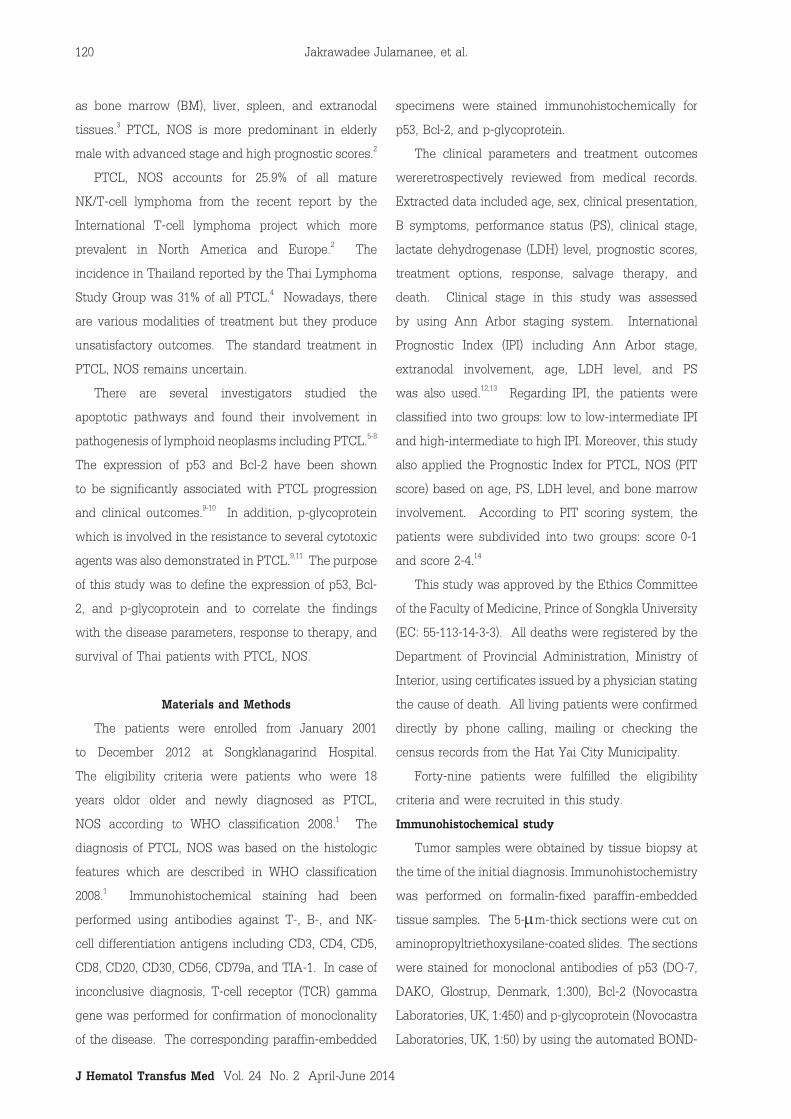
Scoring was analyzed in the area of highest protein
expression. The results were semiquantitatively
scored as follows; score of 0 positivity when completely
negative reactions were found inside the tumor cells
and positive scores of 1+, 2+, 3+, and 4+ when < 10%,
10-50%, 51-90%, and > 90% of the tumor exhibited
positive reactions, respectively. The cases exhibiting
a majority of positive tumor cells (> 10% or ≥ 2+
positivity) were considered as positive expression. The
tumor cells were differentiated from normal reactive
T-cell lymphocytes by using morphology criteria and
compared with hematoxylin and eosin (H&E) staining.
The H&E and immunohistochemical staining results
are shown in Figure 1.
Statistical analysis
Statistical analysis was done using Stata Software
Packages, version 13.1. The clinical parameters and
treatment outcomes were compared among patients
with or without expressions of p53, Bcl-2, p-glycoprotein
or combined Bcl-2/p53 using a Chi-square test.
Survival analysis was performed with the Kaplan-Meier
method. Overall survival (OS) was calculated as the
time interval from the date of diagnosis to death or last
follow-up. Survival analyses between subgroups were
compared using log-rank test. Multivariate analyses
for OS were performed using a Cox regression model.
A cut-off P value of 0.05 was considered statistically
significant for all statistical analyses.
Figure 1. Immunohistochemical detection showing (x400), a. H&E staining in PTCL, NOS; b. strongly positive
nuclear staining for p53 (4+); c. strongly positive cytoplasmic staining for Bcl-2 (4+); d. strongly positive
cytoplasmic staining for p-glycoprotein (3+)
a. b.
c. d.
Jakrawadee Julamanee, et al.
J Hematol Transfus Med Vol. 24 No. 2 April-June 2014
122
Results
Patient characteristics and treatment outcomes
Forty-nine patients were analysed in this study.
They were 38 males and 11 females, giving male to
female ratio of 3.5:1. The median age was 58 years
old (range 18-89). B symptoms were presented in 55%
and elevated LDH in 54% of the patients. Eighty-
two percent had good ECOG scores, 61% in stages
III-IV, 80% with extranodal lesions, and 40% with bone
marrow involvement. Hemophagocytic activity was
found in 10.5% of those who had marrow involvement.
Sixty-three percent were classified as low to low-
intermediate IPI and 46% had PIT score of 0-1. Most
patients (74%) were treated by CHOP chemotherapy
and only 10% by radiotherapy. Of the 35 patients
evaluated, the overall response rate was 63% with 40%
complete remission. The patient characteristics and
treatment outcomes are summarized in Table 1.
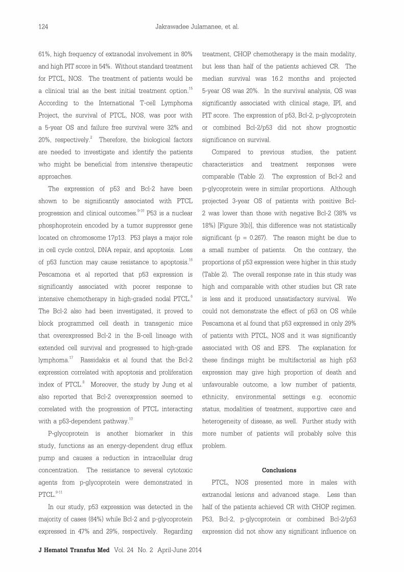
The expression of p53, Bcl-2 and p-glycoprotein
The p53, Bcl-2, and p-glycoprotein positivity were
demonstrated in 41 (84%), 23 (47%), and 14 (29%)
patients, respectively. The expression of p53, Bcl-2,
and p-glycoprotein were not significantly correlated
with advanced stage, higher IPI, higher PIT score, and
response rate. We also analyzed combined Bcl-2/p53
according to their expressions with clinical stage, IPI,
PIT score, response rate, and survivals. The results
did not show any significant association, as well.
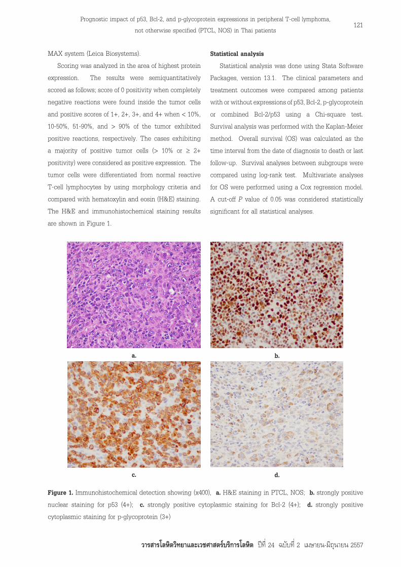
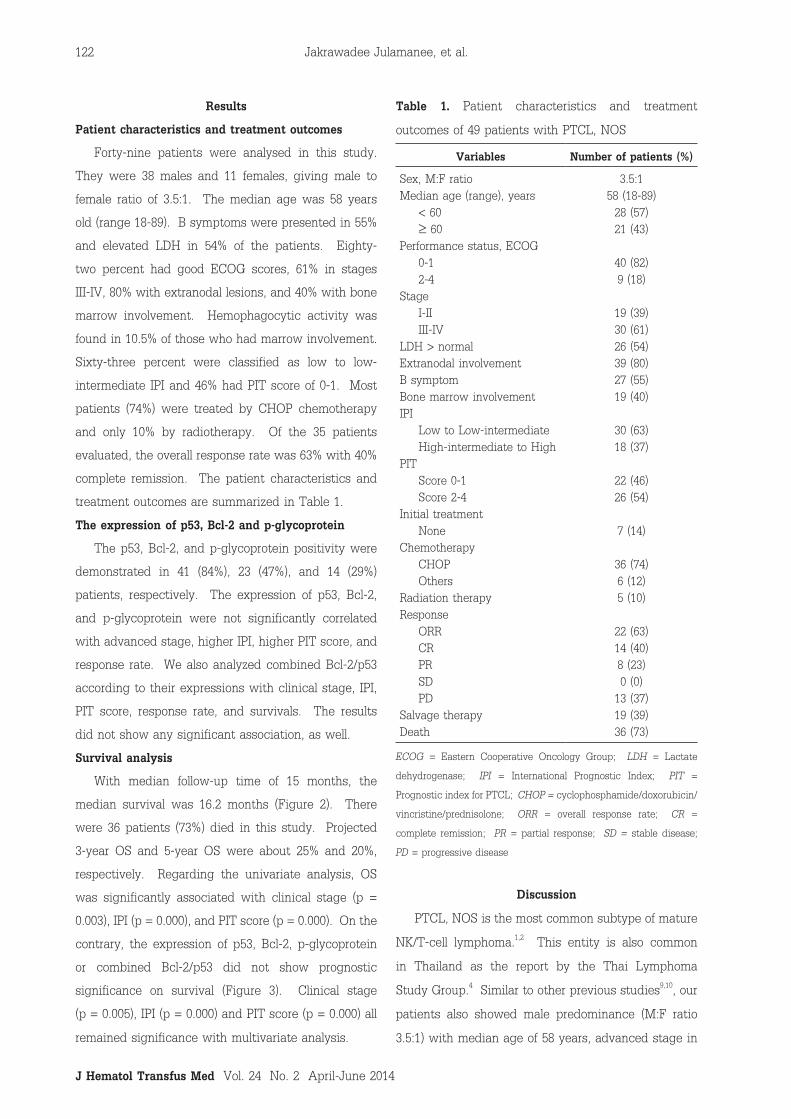
Survival analysis
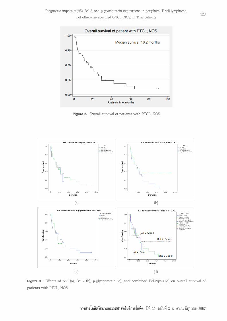
With median follow-up time of 15 months, the
median survival was 16.2 months (Figure 2). There
were 36 patients (73%) died in this study. Projected
3-year OS and 5-year OS were about 25% and 20%,
respectively. Regarding the univariate analysis, OS
was significantly associated with clinical stage (p =
0.003), IPI (p = 0.000), and PIT score (p = 0.000). On the
contrary, the expression of p53, Bcl-2, p-glycoprotein
or combined Bcl-2/p53 did not show prognostic
significance on survival (Figure 3). Clinical stage
(p = 0.005), IPI (p = 0.000) and PIT score (p = 0.000) all
remained significance with multivariate analysis.
Table 1. Patient characteristics and treatment
outcomes of 49 patients with PTCL, NOS
Variables Number of patients (%)
Sex, M:F ratioMedian age (range), years
< 60≥ 60
Performance status, ECOG0-12-4
StageI-IIIII-IV
LDH > normalExtranodal involvementB symptomBone marrow involvementIPI
Low to Low-intermediateHigh-intermediate to High
PIT Score 0-1Score 2-4
Initial treatment None
ChemotherapyCHOPOthers
Radiation therapyResponse
ORRCRPRSDPD
Salvage therapyDeath
3.5:158 (18-89)28 (57)21 (43)
40 (82)9 (18)
19 (39)30 (61)26 (54)39 (80)27 (55)19 (40)
30 (63)18 (37)
22 (46)26 (54)
7 (14)
36 (74)6 (12)5 (10)
22 (63)14 (40)8 (23)0 (0)
13 (37)19 (39)36 (73)
ECOG = Eastern Cooperative Oncology Group; LDH = Lactate
dehydrogenase; IPI = International Prognostic Index; PIT =
Prognostic index for PTCL; CHOP = cyclophosphamide/doxorubicin/
vincristine/prednisolone; ORR = overall response rate; CR =
complete remission; PR = partial response; SD = stable disease;
PD = progressive disease
Discussion
PTCL, NOS is the most common subtype of mature
NK/T-cell lymphoma.1,2 This entity is also common
in Thailand as the report by the Thai Lymphoma
Study Group.4 Similar to other previous studies9,10, our
patients also showed male predominance (M:F ratio
3.5:1) with median age of 58 years, advanced stage in
Prognostic impact of p53, Bcl-2, and p-glycoprotein expressions in peripheral T-cell lymphoma,
not otherwise specified (PTCL, NOS) in Thai patients
วารสารโลหิตวิทยาและเวชศาสตรบริการโลหิต ป ที่ 24 ฉบับ ที่ 2 เมษายน-มิถุนายน 2557
123
Figure 2. Overall survival of patients with PTCL, NOS
Figure 3. Effects of p53 (a), Bcl-2 (b), p-glycoprotein (c), and combined Bcl-2/p53 (d) on overall survival of
patients with PTCL, NOS
(a) (b)
(c) (d)
Jakrawadee Julamanee, et al.
J Hematol Transfus Med Vol. 24 No. 2 April-June 2014
124
61%, high frequency of extranodal involvement in 80%
and high PIT score in 54%. Without standard treatment
for PTCL, NOS. The treatment of patients would be
a clinical trial as the best initial treatment option.15
According to the International T-cell Lymphoma
Project, the survival of PTCL, NOS, was poor with
a 5-year OS and failure free survival were 32% and
20%, respectively.2 Therefore, the biological factors
are needed to investigate and identify the patients
who might be beneficial from intensive therapeutic
approaches.
The expression of p53 and Bcl-2 have been
shown to be significantly associated with PTCL
progression and clinical outcomes.9-10 P53 is a nuclear
phosphoprotein encoded by a tumor suppressor gene
located on chromosome 17p13. P53 plays a major role
in cell cycle control, DNA repair, and apoptosis. Loss
of p53 function may cause resistance to apoptosis.16
Pescamona et al reported that p53 expression is
significantly associated with poorer response to
intensive chemotherapy in high-graded nodal PTCL.6
The Bcl-2 also had been investigated, it proved to
block programmed cell death in transgenic mice
that overexpressed Bcl-2 in the B-cell lineage with
extended cell survival and progressed to high-grade
lymphoma.17 Rassidakis et al found that the Bcl-2
expression correlated with apoptosis and proliferation
index of PTCL.8 Moreover, the study by Jung et al
also reported that Bcl-2 overexpression seemed to
correlated with the progression of PTCL interacting
with a p53-dependent pathway.10
P-glycoprotein is another biomarker in this
study, functions as an energy-dependent drug efflux
pump and causes a reduction in intracellular drug
concentration. The resistance to several cytotoxic
agents from p-glycoprotein were demonstrated in
PTCL.9-11
In our study, p53 expression was detected in the
majority of cases (84%) while Bcl-2 and p-glycoprotein
expressed in 47% and 29%, respectively. Regarding
treatment, CHOP chemotherapy is the main modality,
but less than half of the patients achieved CR. The
median survival was 16.2 months and projected
5-year OS was 20%. In the survival analysis, OS was
significantly associated with clinical stage, IPI, and
PIT score. The expression of p53, Bcl-2, p-glycoprotein
or combined Bcl-2/p53 did not show prognostic
significance on survival.
Compared to previous studies, the patient
characteristics and treatment responses were
comparable (Table 2). The expression of Bcl-2 and
p-glycoprotein were in similar proportions. Although
projected 3-year OS of patients with positive Bcl-
2 was lower than those with negative Bcl-2 (38% vs
18%) [Figure 3(b)], this difference was not statistically
significant (p = 0.267). The reason might be due to
a small number of patients. On the contrary, the
proportions of p53 expression were higher in this study
(Table 2). The overall response rate in this study was
high and comparable with other studies but CR rate
is less and it produced unsatisfactory survival. We
could not demonstrate the effect of p53 on OS while
Pescamona et al found that p53 expressed in only 29%
of patients with PTCL, NOS and it was significantly
associated with OS and EFS. The explanation for
these findings might be multifactorial as high p53
expression may give high proportion of death and
unfavourable outcome, a low number of patients,
ethnicity, environmental settings e.g. economic
status, modalities of treatment, supportive care and
heterogeneity of disease, as well. Further study with
more number of patients will probably solve this
problem.
Conclusions
PTCL, NOS presented more in males with
extranodal lesions and advanced stage. Less than
half of the patients achieved CR with CHOP regimen.
P53, Bcl-2, p-glycoprotein or combined Bcl-2/p53
expression did not show any significant influence on
Prognostic impact of p53, Bcl-2, and p-glycoprotein expressions in peripheral T-cell lymphoma,
not otherwise specified (PTCL, NOS) in Thai patients
วารสารโลหิตวิทยาและเวชศาสตรบริการโลหิต ป ที่ 24 ฉบับ ที่ 2 เมษายน-มิถุนายน 2557
125
Table 2. Clinical comparison with previous studies
Variables This study
(n = 49)
Jung et al, 200610
(n = 74)
Pescarmona et al, 20019
(n = 45)
M:F ratio 3.5:1 2.2:1 2.2:1
Median age (years) 58 46 55
Histology subtypes (%)
PTCL, NOS 100 60 100
LDH > normal (%) 54 38 56
IPI (%)
L-LI
HI-H
63
37
53
47
67
33
PIT (%)
Score 0-1
Score 2-4
46
54
73
27
0
0
Biomarkers expression (%)
p53
Bcl-2
p-glycoprotein
84
47
29
45
45
0
28.9
51.1
17.8
Treatment (%)
None
Chemotherapy
Radiation therapy
14
86
10
8
64
4
0
86
Response (%)
ORR
CR
PR
NR
63
40
23
37
75
64
11
0
0
64.1
0
37
Death (%) 73 32 37
Overall survival
Conclusions
Median 16.2 months
OS was associated with
clinical stage, higher IPI
and PIT score (p < 0.00)
3-year OS 82.5%
Bcl-2 was associated with
stage (p = 0.021), IPI (p =
0.038)
Bcl-2/p53 was associated
with advanced stage (p =
0.008) and higher IPI (p =
0.001)
P53 was significant in OS
(p = 0.0032) and EFS (p =
0.0004)
Bcl-2 was significant in
EFS (p = 0.0491)
M = male; F = female; LDH = lactate dehydrogenase; IPI = International Prognostic Index; L-LI = low to low-intermediate; HI-H = high-
intermediate to high; PIT = Prognostic index for PTCL; ORR = overall response rate; CR = complete remission; PR = partial response; NR
= no-response; OS = overall survival; EFS = event free survival; NA = not available
Jakrawadee Julamanee, et al.
J Hematol Transfus Med Vol. 24 No. 2 April-June 2014
126
clinical outcomes such as RR and OS. The number
of patient in this study was small. Therefore, larger
number of patients with other biomarker expressions
are needed for further investigation.
Acknowledgement
This research was supported by grants from
Faculty of Medicine, Prince of Songkla University and
The Thai Society of Hematology.
References1. Swerdlow SH, Campo E, Harris NL, et al. WHO Classification
of Tumours of Haematopoietic and Lymphoid Tissues. 4th ed.
Lyon, France: IARC Press; 2008.
2. Vose J, Armitage J, Weisenburger D. International T-Cell
Lymphoma Project. International peripheral T-cell and natural
killer/T-cell lymphoma study: pathology findings and clinical
outcomes. J Clin Oncol. 2008;26:4124-30.
3. Rizvi MA, Evens AM, Tallman MS, et al. T-cell non-Hodgkin
lymphoma. Blood 2006;107:1255-64.
4. Lekhakula A, Rujirojjindakul P, Siritanaratkul N, et al.
Multicenter study on peripheral T-cell lymphoma (PTCL)
in Thailand: Clinical features, histopathology, outcome and
prognosis. Presented at the 4th Panpacific Conference on
Lymphoma. June 22-25, 2009.
5. Lowe SW, Lin AW. Apoptosis in cancer. Carcinogenesis.
2000;21:485-495.
6. Pescarmona E, Pignoloni P, Santangelo C, et al. Expression of
p53 and retinoblastoma gene in high-grade nodal peripheral T-
cell lymphomas: immunohistochemical and molecular findings
suggesting different pathogenetic pathways and possible clini-
cal implications. J Pathol 1999;188:400-6.
7. Ten Berge RL, Meijer CJ, Dukers DF, et al. Expression levels of
apoptosis-related proteins predict clinical outcome in anaplastic
large cell lymphoma. Blood 2002;99:4540-6.
8. Rassidakis GZ, Jones D, Lai R, et al. BCL-2 family proteins in
peripheral T-cell lymphomas: correlation with tumour apoptosis
and proliferation. J Pathol 2003;200:240-8.
9. Pescarmona E, Pignoloni P, Puopolo M, et al. p53 over-expres-
sion identifies a subset of nodal peripheral T-cell lymphomas
with a distinctive biological profile and poor clinical outcome. J
Pathol 2001;195:361-6.
10. Jung TJ, Kim DH, Kwak EK, et al. Clinical role of Bcl-2, Bax,
or p53 overexpression in peripheral T-cell lymphomas. Ann
Hematol 2006;85:575-81.
11. Oshimi K. Progress in understanding and managing natural
killer-cell malignancies. Br J Haematol 2007;139:532-44.
12. The International Non-Hodgkin’s Prognostic Factors Project. A
predictive model for aggressive non-Hodgkin’s lymphomas. The
International Non-Hodgkin’s Lymphoma Prognostic Factors Proj-
ect. N Engl J Med 1993;329:987-94.
13. Gisselbrecht C, Gaulard P, Lepage E, et al. Prognostic signifi-
cance of T-cell phenotype in aggressive non-Hodgkin’s lympho-
mas. Groupe d’Etudes des Lymphomes de l’Adulte (GELA).
Blood 1998;92:76-82.
14. Gallamini A, Stelitano C, Calvi R, et al. Peripheral T-cell
lymphoma unspecified (PTCL-U): a new prognostic model from
a retrospective multicentric clinical study. Blood 2004;103:
2474-9.
15. NCCN Clinical Practice Guideline in Oncology for Non-Hodgkins
Lymphomas. (TCEL-1) 2013; Version 2.2013: www.NCCN.org.
16. Amundson SA, Myers TG, Fornace AJ Jr. Roles for p53 in
growth arrest and apoptosis: putting on the breaks after geno-
toxic stress. Oncogene. 1998;17:3287-99.
17. Korsmeyer SJ. BCL-2 gene family and the regulation of
programmed cell death. Cencer Res 1999;59:1693-700.
Prognostic impact of p53, Bcl-2, and p-glycoprotein expressions in peripheral T-cell lymphoma,
not otherwise specified (PTCL, NOS) in Thai patients
วารสารโลหิตวิทยาและเวชศาสตรบริการโลหิต ป ที่ 24 ฉบับ ที่ 2 เมษายน-มิถุนายน 2557
127
การแสดงออกของ p53, Bcl-2 และ p-glycoprotein และความสัมพันธกับการพยากรณโรคในผูปวยมะเร็งตอมน้ำาเหลืองชนิด PTCL, NOS
จักราวดี จุฬามณี1 คณิตา กายะสุต2 อานุภาพ เลขะกุล1 พงษเทพ วิบูลยจันทร1 และ ดาวลดา คงกับพันธ11ภาควิชาอายุรศาสตร 2ภาควิชาพยาธิวิทยา คณะแพทยศาสตร มหาวิทยาลัยสงขลานครินทร จังหวัดสงขลา
บทคัดยอ
วัตถุประสงค เพื่อศึกษาอัตราการแสดงออกของ p53 Bcl-2 และ p-glycoprotein และความสัมพันธระหวางการแสดงออกของสาร
โปรตีนกับพยากรณโรคและอัตราการรอดชีวิตในผูปวยมะเร็งตอมน้ำาเหลืองชนิด PTCL, NOS วัสดุและวิธีการ ศึกษายอนหลังใน
กลุมผูปวยผูใหญที่ไดรับการวินิจฉัยโรคมะเร็งตอมน้ำาเหลืองชนิด PTCL, NOS ตั้งแตป พ.ศ. 2544-2555 โดยเก็บขอมูลพื้นฐานทาง
คลินิกและผลการรักษาของผูปวย รวมกับศึกษาการแสดงออกของ p53 Bcl-2 และ p-glycoprotein ในชิ้นเนื้อที่ยอมเพิ่มเติมดวย
วิธี immunohistochemistry นำาผลการศึกษาทั้งหมดมาวิเคราะหเพื่อหาความสัมพันธกับระยะโรค ปจจัยการพยากรณโรค อัตรา
การตอบสนองตอการรักษา และอัตราการรอดชีวิต ผลการศึกษา มีผูปวยในการศึกษาทั้งหมด 49 ราย (หญิง 38 รายและชาย
11 ราย) คามัธยฐานของอายุเทากับ 58 ป ผูปวยมี B symptom รอยละ 55 และมี LDH สูง รอยละ 54 ผูปวยสวนใหญอยูใน
good ECOG score (รอยละ 82) และโรคอยูในระยะ III-IV (รอยละ 61) รอยละ 80 ของผูปวยทั้งหมดมีอาการแสดงนอกตอม
น้ำาเหลือง และรอยละ 40 มีโรคในไขกระดูก เมื่อจำาแนกตามปจจัยการพยากรณโรคพบวาผูปวยรอยละ 63 จัดอยูในกลุม low-
low-intermediate IPI และรอยละ 46 มี PIT score 0-1 ผูปวยรอยละ 74 ไดรับการรักษาดวยยาสูตร CHOP มีผูปวยทั้งหมด
35 รายจากทั้งหมดที่ประเมินการตอบสนองพบวาอัตราการตอบสนองตอการรักษาคิดเปนรอยละ 63 และอัตราโรคสงบรอยละ 40
อัตราการแสดงออกของ p53 Bcl-2 และ p-glycoprotein คิดเปนรอยละ 84 47 และ 29 ตามลำาดับ การแสดงออกของสารโปรตีน
ดังกลาวไมสัมพันธกับการรอดชีวิตหรือการพยากรณโรคของผูปวย ผูปวยในการศึกษามีอัตราการรอดชีวิตเฉลี่ย 16.2 เดือน จากการ
วิเคราะหปจจัยที่มีความสัมพันธกับอัตราการรอดชีวิต พบวาระยะของโรค IPI และ PIT score มีความสัมพันธอยางมีนัยสำาคัญทาง
สถิติทั้งจากการวิเคราะหแบบ univariate และ multivariate สรุป ผูปวย PTCL, NOS สวนใหญเปนเพศชาย มักจะมีอาการ
แสดงนอกตอมน้ำาเหลืองและมีการดำาเนินโรคอยูในระยะรุนแรง การรักษาดวยยาสูตร CHOP นั้นทำาใหผูปวยเขาสูระยะโรคสงบได
นอยกวารอยละ 50 การแสดงออกของ p53 Bcl-2 และ p-glycoprotein ไมมีความสัมพันธกับปจจัยพยากรณโรคและอัตราการ
รอดชีวิต
คำาสำาคัญ : l P53 l Bcl-2 l P-glycoprotein l Peripheral T-cell lymphoma, not otherwise specified
l Survival
วา รสาร โลหิต วิทยา และ เวชศาสตร บริการ โลหิต 2557;24:119-27.

J Hematol Transfus Med Vol. 24 No. 2 April-June 2014
128