Embed Size (px)
Citation preview
Tetrahydrobiopterin Has a Glucose-Lowering Effectby Suppressing Hepatic Gluconeogenesis inan Endothelial Nitric Oxide Synthase–DependentManner in Diabetic MiceAbulizi Abudukadier,
1Yoshihito Fujita,
1Akio Obara,
1Akiko Ohashi,
2Toru Fukushima,
1Yuichi Sato,
1
Masahito Ogura,1Yasuhiko Nakamura,
1Shimpei Fujimoto,
1Masaya Hosokawa,
1Hiroyuki Hasegawa,
2
and Nobuya Inagaki1
Endothelial nitric oxide synthase (eNOS) dysfunction inducesinsulin resistance and glucose intolerance. Tetrahydrobiopterin(BH4) is an essential cofactor of eNOS that regulates eNOS ac-tivity. In the diabetic state, BH4 is oxidized to 7,8-dihydrobiopterin,which leads to eNOS dysfunction owing to eNOS uncoupling. Thecurrent study investigates the effects of BH4 on glucose metabolismand insulin sensitivity in diabetic mice. Single administration of BH4lowered fasting blood glucose levels in wild-type mice with strepto-zotocin (STZ)-induced diabetes and alleviated eNOS dysfunction byincreasing eNOS dimerization in the liver of these mice. Liver hasa critical role in glucose-lowering effects of BH4 through suppres-sion of hepatic gluconeogenesis. BH4 activated AMP kinase(AMPK), and the suppressing effect of BH4 on gluconeogenesiswas AMPK-dependent. In addition, the glucose-lowering effectand activation of AMPK by BH4 did not appear in mice withSTZ-induced diabetes lacking eNOS. Consecutive administrationof BH4 in ob/ob mice ameliorated glucose intolerance and insulinresistance. Taken together, BH4 suppresses hepatic gluconeo-genesis in an eNOS-dependent manner, and BH4 has a glucose-lowering effect as well as an insulin-sensitizing effect in diabeticmice. BH4 has potential in the treatment of type 2 diabetes.Diabetes 62:3033–3043, 2013
Nitric oxide (NO) is a biological messenger pro-duced by NO synthase (NOS), which includesendothelial (eNOS), inducible (iNOS), and neu-ronal (nNOS) isoforms. eNOS-derived NO is
well-known to have a pivotal role in physiological regula-tion of endothelial function (1,2). eNOS dysfunction occursin conditions of diabetes and is known to induce insulinresistance and glucose intolerance (3–5). Insulin resistancecaused by eNOS dysfunction is thought to be induced byendothelial dysfunction, leading to decreased skeletalmuscle blood flow and glucose uptake (4). On the otherhand, glucose transport in isolated skeletal muscle is lowerin eNOS-deficient (eNOS2/2) mice, indicating that eNOSexpressed in skeletal muscle also regulates its glucose
uptake (4). Moreover, eNOS2/2 mice are insulin resistantat the level of liver (5). These studies suggest that eNOSplays a central role in the regulation of glucose metabolismand insulin sensitivity and represents several therapeutictargets for type 2 diabetes.
The function of eNOS is regulated by multiple factorssuch as mRNA expression of eNOS, L-arginine, influx ofCa2+, and tetrahydrobiopterin (BH4) (2,6,7). BH4 is an es-sential cofactor for eNOS catalysis and functions as anallosteric modulator of arginine binding (7,8). Binding ofBH4 to eNOS elicits a conformational change that increa-ses the affinity for binding of arginine-based ligands. BH4binding also plays a role in dimer formation of the activeand stabilized form of eNOS (8). BH4 is converted to 7,8-dihydrobiopterin (BH2) by exposure to oxidative stresssuch as diabetes (8,9). Increase in BH2 induces dysfunc-tion of eNOS, as BH2 is inactive for NOS cofactor functionand competes with BH4 for BH4 binding (8,9). Further-more, in states of diabetes and high glucose, de novo syn-thesis of BH4, which is rate limited by GTP cyclohydrolase I(GTPCH I), is impaired (10–13). Thus, the availability of BH4is reduced and the function of eNOS is altered so that theenzyme produces superoxide anion (O2
2) rather than NO,a phenomenon called “eNOS uncoupling” (7,8,14). Supple-mentation of BH4 can improve endothelial dysfunction byelevating the BH4-to-BH2 ratio, leading to recoupling ofeNOS, and has been used in clinical trials with patients withatherosclerotic diseases for the expected vasodilatationeffects of BH4 through NO production (15). However, it isunclear whether BH4 improves glucose metabolism andinsulin sensitivity in diabetic conditions.
In the current study, we investigated the effects of BH4on blood glucose levels and insulin sensitivity in diabeticmice. Fasting blood glucose levels are regulated by thelevel of hepatic gluconeogenesis, elevation of which is themajor cause of fasting hyperglycemia in diabetes (16,17).We demonstrate here that BH4 lowers fasting blood glucoselevels and suppresses gluconeogenesis in liver in an eNOS-dependent manner. In addition, BH4 has an amelioratingeffect on glucose intolerance as well as insulin resistance indiabetic mice. Using primary hepatocytes isolated frommouse liver, we have clarified the mechanism by which BH4suppresses hepatic gluconeogenesis. These data suggestthat BH4 has potential as a novel therapeutic approach todiabetes.
RESEARCH DESIGN AND METHODS
Male C57/BL6 (wild-type) mice andmale heterozygous Ins2Akita (diabetic Akita)mice, which exhibit hyperglycemia with reduced b-cell mass caused by a point
From the 1Department of Diabetes and Clinical Nutrition, Graduate School ofMedicine, Kyoto University, Kyoto, Japan; and the 2Department of Func-tional Morphology, Nihon University School of Medicine, Tokyo, Japan.
Corresponding author: Nobuya Inagaki, [email protected] 10 September 2012 and accepted 27 April 2013.DOI: 10.2337/db12-1242This article contains Supplementary Data online at http://diabetes
.diabetesjournals.org/lookup/suppl/doi:10.2337/db12-1242/-/DC1.� 2013 by the American Diabetes Association. Readers may use this article as
long as the work is properly cited, the use is educational and not for profit,and the work is not altered. See http://creativecommons.org/licenses/by-nc-nd/3.0/ for details.
diabetes.diabetesjournals.org DIABETES, VOL. 62, SEPTEMBER 2013 3033
ORIGINAL ARTICLE
mutation in the insulin 2 gene that leads to misfolded insulin and severe en-doplasmic reticulum stress, were obtained from Shimizu (Kyoto, Japan) (18).Male eNOS2/2 mice in the C57/BL6 mice background were obtained from TheJackson Laboratory (Bar Harbor, ME). Male B6.V-Lepob/J (ob/ob) mice wereobtained from Charles River Japan (Yokohama, Japan). Mice with streptozotocin(STZ)-induced diabetes were made by injection of STZ (120 mg/kg i.p.) to 7-week-old wild-type or eNOS2/2 mice. At 3 weeks after injection of STZ, the animalswere confirmed to be diabetic by both high blood glucose levels ($15 mmol/L)and other diabetic features, including polyuria, polydipsia, and hyperglycemia.
The mice were maintained in a temperature-controlled (25 6 2°C) envi-ronment with a 12-h light/dark cycle with free access to standard laboratorychow and water. All experiments were carried out with mice aged 8–10 weeks.The animals were maintained and used in accordance with the Guidelines forAnimal Experiments of Kyoto University. All experiments involving animalswere conducted in accordance with the Guidelines for Animal Experiments ofKyoto University and were approved by the Animal Research Committee,Graduate School of Medicine, Kyoto University.Preparations and cultures of mouse hepatocyte and aortic endothelial
cell. Mouse hepatocytes were isolated by collagenase digestion as previouslydescribed (19). Primary hepatocytes were prepared by seeding in sixwell type1 collagen–coated plates at a density of 1.5 3 106 cells in Dulbecco’s modifiedEagle’s medium (DMEM) (low glucose, 5.6 mmol/L) containing 10% (vol/vol)FBS, 100 nmol/L regular insulin, 50 units/mL penicillin, and 50 mg/mL strepto-mycin. Hepatocytes were then cultured overnight in a humidified atmosphere (5%CO2) at 37°C. As for mouse endothelial cells (ECs), the aorta was dissected andfilled with collagenase type II solution. After incubation for 45 min at 37°C, ECswere removed from the aorta and collected by centrifugation at 1,200 rpm for5 min. The EC was cultured in a sixwell collagen type I–coated dish for 1 week.Glucose production via gluconeogenesis in hepatocytes. Freshly isolatedhepatocytes from mice fasted for 16 h were treated in 24-well plates (7.5 3 105
cells/well) in buffer A, which consisted of 0.5 mL Krebs-Ringer bicarbonatemedium of 119.4 mmol/L NaCl, 3.7 mmol/L KCl, 2.7 mmol/L CaCl2, 1.3 mmol/LKH2PO4, 1.3 mmol/L MgSO4, and 24.8 mmol/L NaHCO3 without glucose; 2%(wt/vol) BSA; 0.24 mmol/L 3-isobutyl-1-methylxanthine; and gluconeogeneticsubstrates (1 mmol/L pyruvate plus 10 mmol/L lactate). Hepatocytes weretreated with BH4 (Schircks Laboratories, Jona, Switzerland), sodium nitro-prusside (SNP), NG-nitro-L-arginine methyl ester (Sigma, St. Louis, MO),sepiapterin (Schircks Laboratories), erythro-9-(2-hydroxy-3-nonyl)adenine(EHNA) (Wako, Osaka, Japan), and compound C (Sigma). Glucose productionwas measured by glucose oxidation method as previously described (19).Immunoblotting analysis of hepatocytes. Western blotting was performedas previously described (19). Primary hepatocytes cultured overnight wereincubated in buffer A treated with BH4, SNP, sepiapterin, and EHNA. Hepa-tocytes were homogenized in lysis buffer. Cell lysates (50–150 mg protein/lane) were heated at 95°C for 5 min and subjected to electrophoresis on 6–10%(vol/vol) sodium dodecyl sulfate–polyacrylamide gels and transferred ontonitrocellulose membranes. For analysis of eNOS dimerization, the sampleswere not heated and the temperature was maintained at ,15°C during elec-trophoresis. Primary antibodies used were anti–phosphorylated (phospho-)AMP kinase (AMPK)a (Thr172), anti-AMPKa, anti–phospho-acetyl-CoA car-boxylase (ACC) (Ser79), anti-ACC, anti–phospho-eNOS (Ser1177), anti–phospho-Akt (Ser473), anti-Akt (all at 1:1,000 dilution; Cell Signaling Technology,Danvers, MA), anti-eNOS polyclonal antibody (1:500 dilution; BD TransductionLaboratories, San Jose, CA), anti-CD31 monoclonal antibody (1:2,000 dilution;Dianova, Hamburg, Germany), anti–GTPCH I (1:3,000; kind gift from Prof.H. Ichinose, Tokyo Institute of Technology), anti–dihydrofolate reductase(DHFR), anti–a1-antitrypsin (1:500; Santa Cruz, Delaware, CA), and anti–b-actin(1:5,000; Sigma). Secondary antibodies used were horseradish peroxidase–conjugated anti-rabbit, -mouse, -rat, or -goat antibody (GE Healthcare, Buck-inghamshire, U.K.). The fluorescent bands were visualized using a detectionsystem (Amersham ECL Plus; GE Healthcare) and quantified by densitometryusing Image J software from National Institutes of Health (Bethesda, MD).Cell transfection and short interfering RNA. Stealth short interfering RNA(siRNA) of AMPKa1 was purchased from Invitrogen (Carlsbad, CA). Thesequences of siRNA for AMPKa1 were 59-UCUCUUUCCUGAGGACCCAUCUUAU-39 and 59-AUAAGAUGGGUCCUCAGGAAAGAGA-39. The sequences ofcontrol siRNAs were 59-ACCAACAACAGUUUGGGAAUAGGGA-39and 59- UCCCUAUUCCCAAACUGUUGUUGGU-39. Isolated hepatocytes in DMEM (lowglucose, 5.6 mmol/L) containing 10% (vol/vol) FBS and 100 nmol/L regularinsulin were mixed with Opti-MEM containing siRNA and Lipofectamine RNAiMAX (Invitrogen) and were plated on wells and then incubated at 37°C ina CO2 incubator. The final amounts of hepatocytes, DMEM, Opti-MEM, siRNA,and Lipofectamine RNAi MAX were 5.0 3 105 cells/mL, 75% (vol/vol), 25%(vol/vol), 50 nmol/L, and 0.2%, respectively. Medium was replaced withDMEM 6 h after transfection. Forty-eight hours after transfection, the mediumwas replaced with buffer A, the cells were incubated for 60 min with orwithout BH4, and the glucose content of the supernatant was measured.
Nitrite/nitrate analysis. Primary hepatocytes and liver tissues were ho-mogenized in buffer A, and the amount of nitrite/nitrate in the supernatant wasdetermined by a fluorescence method.Immunocytochemistry. The hepatocytes were incubated with rabbit poly-clonal anti-nitrotyrosine antibody (1:100 dilution; Millipore, Billerica, MA). Cellswere then incubated with goat anti-rabbit IgG fluorescein-conjugated sec-ondary antibody (1:100 dilution, Alexa Fluor 488; Invitrogen). Fluorescence incells was monitored as previously described (19).Measurement of adenine nucleotide content. After primary isolatedhepatocytes were incubated in buffer A with or without BH4 and SNP for 30 min,treatment was stopped by rapid addition of 0.1 mL of 2 mol/L HClO4, followedby mixing by vortex and sonication in ice-cold water for 3 min. Adenine nu-cleotide contents were measured by a luminometric method as previously de-scribed (19,20).Isolation of total RNA and quantitative RT-PCR. Total RNA was isolatedfrom livers of 10 week-old wild-type mice, wild-type mice with STZ-induceddiabetes, and ob/obmice using Trizol (Invitrogen) as previously described (21).The mouse sequence of forward and reverse primers to detect GTPCH I andDHFR, glucose 6-phosphatase (G6Pase), phosphoenolpyruvate carboxykinase(PEPCK), and glyceraldehyde-3-phosphate dehydrogenase as an inner controlare shown in Supplementary Table 1. SYBR Green PCR Master Mix (AppliedBiosystems, Foster, CA) was prepared for the quantitative RT-PCR run. Thethermal cycling conditions were denaturation at 95°C for 10 min followed by50 cycles at 95°C for 15 s and 60°C for 1 min. mRNA levels were measured byreal-time quantitative RT-PCR using ABI PRISM 7000 Sequence DetectionSystem (Applied Biosystems).Biopterin analysis. Tissues or whole blood of wild-type mice and wild-typemice with STZ-induced diabetes was collected. For measurement of uptake ofBH4 in liver, BH4 (20 mg/kg) dissolved with 0.9% (wt/vol) sterile saline wasadministrated intraperitoneally to wild-type mice. After cervical dislocation,the mice were abdominally dissected and liver tissues were collected at 0, 30,60, 120, and 180 min after injection. The organs were weighed, frozen imme-diately in liquid N2, and then stored at 280°C. Total biopterin, BH4, and BH2
were measured as previously described (22).Effect of BH4 on blood glucose levels of wild-type mice with STZ-
induced diabetes, eNOS2/2
mice with STZ-induced diabetes, and
diabetic Akita mice. Blood glucose levels were measured in wild-type micewith STZ-induced diabetes, eNOS2/2 mice with STZ-induced diabetes, and di-abetic Akita mice fasted for 16 h, and BH4 (20 mg/kg) or metformin (250 mg/kg;Sigma) in 0.9% (wt/vol) sterile saline or 0.9% sterile saline alone was injectedintraperitoneally. Blood glucose levels were measured again 2 h after injection.Effect of BH4 on blood glucose levels of ob/ob mice. Blood glucose levelsand body weight of ob/obmice were measured. The mice were divided into twogroups shown in Supplementary Table 2, and 0.9% (wt/vol) sterile saline withor without BH4 (10 mg/kg) was injected intraperitoneally twice a day for10 days. Fed blood glucose levels were measured. After fasting overnight for16 h, fasting blood glucose levels were measured.Intraperitoneal glucose tolerance test. Wild-type mice were fasted over-night for 16 h, and glucose (2 g/kg) was injected intraperitoneally with BH4 (20mg/kg) in 0.9% (wt/vol) sterile saline or 0.9% sterile saline alone. After 10 days’treatment of saline with or without BH4 (20 mg/kg), ob/ob mice were fastedovernight for 16 h, and glucose (1 g/kg) was injected intraperitoneally. Bloodglucose levels and plasma insulin concentrations were measured at 0, 30, 60,90, and 120 min after injection. Plasma insulin concentrations were determinedby using an ELISA kit (Shibayagi, Gunma, Japan). Homeostasis model assess-ment of insulin resistance (HOMA-IR) was calculated with the following for-mula: [fasting insulin (mU/L) 3 fasting plasma glucose (mmol/L)]/22.5.Pyruvate tolerance test. Pyruvate, BH4, and sepiapterin were dissolved with0.9% (wt/vol) sterile saline. Wild-type, eNOS2/2, and ob/ob mice were fastedovernight for 16 h, and pyruvate (1 g/kg) was injected intraperitoneally with orwithout BH4 (20 mg/kg) and sepiapterin (20 mg/kg). Blood glucose levels weremeasured at 0, 30, 60, 90, and 120 min after injection.Insulin tolerance test. After 10 days’ treatment of saline with or without BH4
(20 mg/kg), ob/ob mice were fasted for 6 h, and regular insulin (1 units/kg i.p.)was injected with 0.9% sterile saline. Blood glucose levels were measured at 0,30, 60, 90, and 120 min after injection.Statistics. Comparison between two groups was performed using unpairedStudent t test (not noted) and paired Student t test. For more than two groups,one-way or two-way ANOVA followed by post hoc Bonferroni testing wasperformed. A value of P , 0.05 was considered statistically significant.
RESULTS
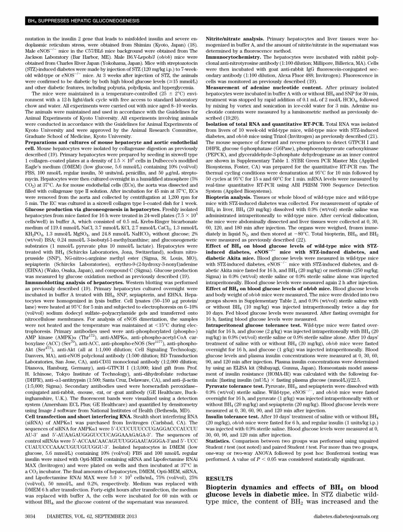
Biopterin dynamics and effects of BH4 on bloodglucose levels in diabetic mice. In STZ diabetic wild-type mice, the content of BH2 was increased and the
BH4 SUPPRESSES HEPATIC GLUCONEOGENESIS
3034 DIABETES, VOL. 62, SEPTEMBER 2013 diabetes.diabetesjournals.org
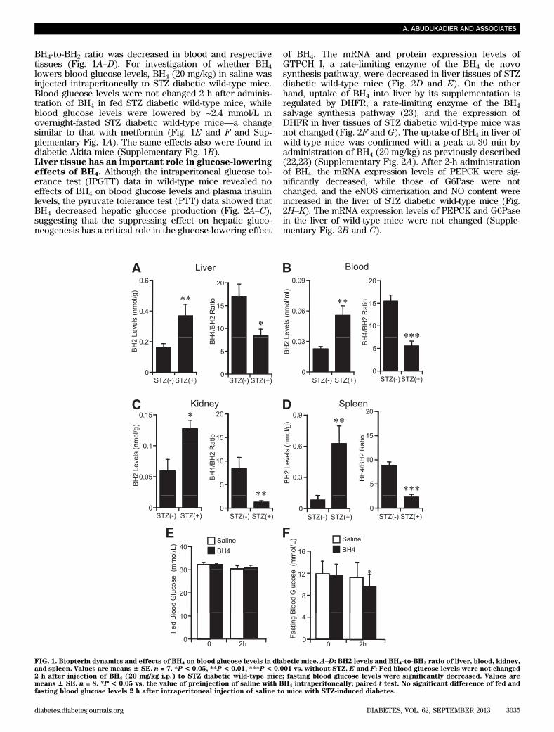
BH4-to-BH2 ratio was decreased in blood and respectivetissues (Fig. 1A–D). For investigation of whether BH4lowers blood glucose levels, BH4 (20 mg/kg) in saline wasinjected intraperitoneally to STZ diabetic wild-type mice.Blood glucose levels were not changed 2 h after adminis-tration of BH4 in fed STZ diabetic wild-type mice, whileblood glucose levels were lowered by ~2.4 mmol/L inovernight-fasted STZ diabetic wild-type mice—a changesimilar to that with metformin (Fig. 1E and F and Sup-plementary Fig. 1A). The same effects also were found indiabetic Akita mice (Supplementary Fig. 1B).Liver tissue has an important role in glucose-loweringeffects of BH4. Although the intraperitoneal glucose tol-erance test (IPGTT) data in wild-type mice revealed noeffects of BH4 on blood glucose levels and plasma insulinlevels, the pyruvate tolerance test (PTT) data showed thatBH4 decreased hepatic glucose production (Fig. 2A–C),suggesting that the suppressing effect on hepatic gluco-neogenesis has a critical role in the glucose-lowering effect
of BH4. The mRNA and protein expression levels ofGTPCH I, a rate-limiting enzyme of the BH4 de novosynthesis pathway, were decreased in liver tissues of STZdiabetic wild-type mice (Fig. 2D and E). On the otherhand, uptake of BH4 into liver by its supplementation isregulated by DHFR, a rate-limiting enzyme of the BH4salvage synthesis pathway (23), and the expression ofDHFR in liver tissues of STZ diabetic wild-type mice wasnot changed (Fig. 2F and G). The uptake of BH4 in liver ofwild-type mice was confirmed with a peak at 30 min byadministration of BH4 (20 mg/kg) as previously described(22,23) (Supplementary Fig. 2A). After 2-h administrationof BH4, the mRNA expression levels of PEPCK were sig-nificantly decreased, while those of G6Pase were notchanged, and the eNOS dimerization and NO content wereincreased in the liver of STZ diabetic wild-type mice (Fig.2H–K). The mRNA expression levels of PEPCK and G6Pasein the liver of wild-type mice were not changed (Supple-mentary Fig. 2B and C).
FIG. 1. Biopterin dynamics and effects of BH4 on blood glucose levels in diabetic mice. A–D: BH2 levels and BH4-to-BH2 ratio of liver, blood, kidney,and spleen. Values are means 6 SE. n = 7. *P < 0.05, **P < 0.01, ***P < 0.001 vs. without STZ. E and F: Fed blood glucose levels were not changed2 h after injection of BH4 (20 mg/kg i.p.) to STZ diabetic wild-type mice; fasting blood glucose levels were significantly decreased. Values aremeans 6 SE. n = 8. *P < 0.05 vs. the value of preinjection of saline with BH4 intraperitoneally; paired t test. No significant difference of fed andfasting blood glucose levels 2 h after intraperitoneal injection of saline to mice with STZ-induced diabetes.
A. ABUDUKADIER AND ASSOCIATES
diabetes.diabetesjournals.org DIABETES, VOL. 62, SEPTEMBER 2013 3035
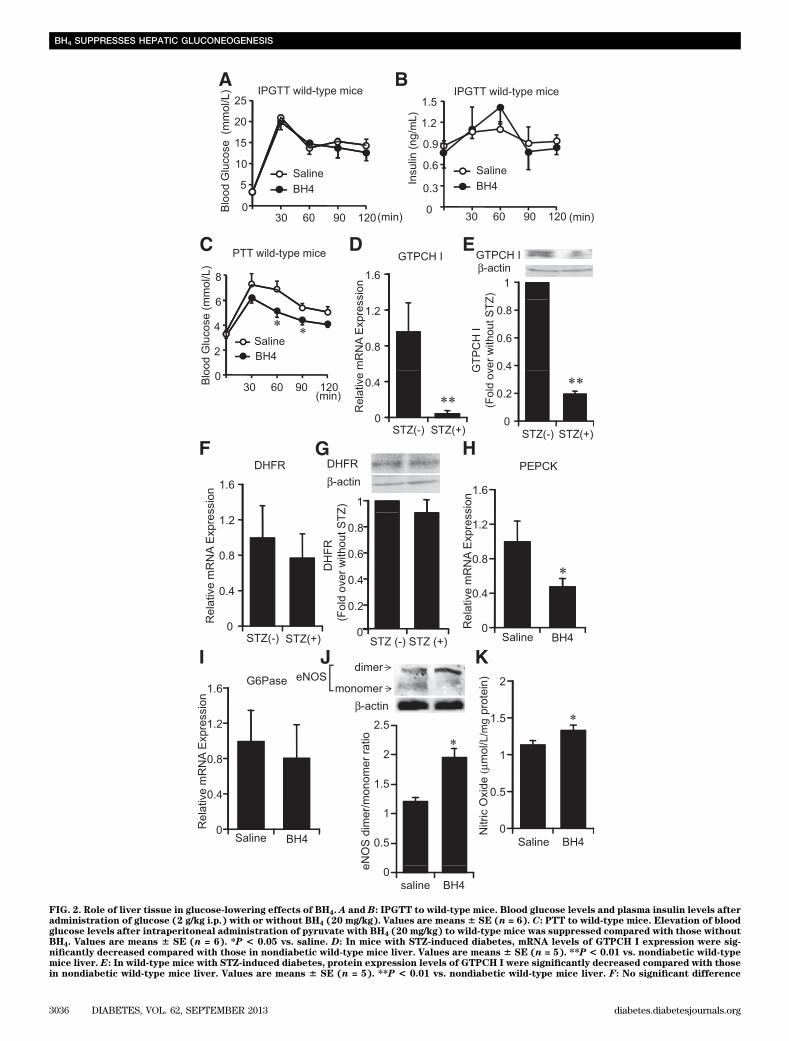
FIG. 2. Role of liver tissue in glucose-lowering effects of BH4. A and B: IPGTT to wild-type mice. Blood glucose levels and plasma insulin levels afteradministration of glucose (2 g/kg i.p.) with or without BH4 (20 mg/kg). Values are means 6 SE (n = 6). C: PTT to wild-type mice. Elevation of bloodglucose levels after intraperitoneal administration of pyruvate with BH4 (20 mg/kg) to wild-type mice was suppressed compared with those withoutBH4. Values are means 6 SE (n = 6). *P < 0.05 vs. saline. D: In mice with STZ-induced diabetes, mRNA levels of GTPCH I expression were sig-nificantly decreased compared with those in nondiabetic wild-type mice liver. Values are means 6 SE (n = 5). **P < 0.01 vs. nondiabetic wild-typemice liver. E: In wild-type mice with STZ-induced diabetes, protein expression levels of GTPCH I were significantly decreased compared with thosein nondiabetic wild-type mice liver. Values are means 6 SE (n = 5). **P < 0.01 vs. nondiabetic wild-type mice liver. F: No significant difference
BH4 SUPPRESSES HEPATIC GLUCONEOGENESIS
3036 DIABETES, VOL. 62, SEPTEMBER 2013 diabetes.diabetesjournals.org
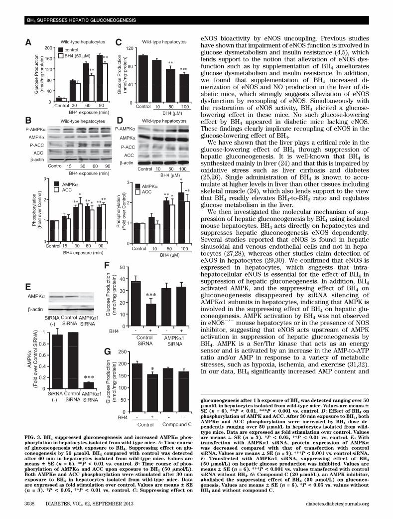
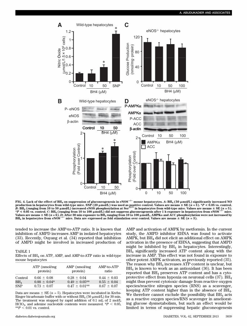
BH4 suppresses gluconeogenesis and increases AMPKaphosphorylation in wild-type mouse hepatocytes. AseNOS expression was confirmed in isolated hepatocytesfrom wild-type mice (Supplementary Fig. 3), we examinedthe direct effect of BH4 in suppression of hepatic gluco-neogenesis using hepatocytes isolated from wild-type micefasted for 16 h. In a time course study of exposure to BH4,the suppressing effect on gluconeogenesis appeared after60 min (P , 0.01 vs. corresponding control) (Fig. 3A). Wethen investigated the increment of AMPKa phosphorylationby time course exposure of BH4 to hepatocytes. AMPK wasactivated after 30 min by BH4 (Fig. 3B). After 60 min ex-posure to BH4, gluconeogenesis was dose-dependentlysuppressed at doses of 50 and 100 mmol/L BH4 (control,101.7 6 3.7 nmol/mg protein; 50 mmol/L BH4, 72.4 6 7.1nmol/mg protein, P , 0.01 vs. control; 100 mmol/L BH4,60.66 4.1 nmol/mg protein, P, 0.001 vs. control) (Fig. 3C).AMPK was activated at doses of 50 and 100 mmol/L BH4 by30 min exposure (Fig. 3D). In accordance with the activa-tion of AMPK, an increase in phosphorylation of ACC byBH4 was confirmed (Fig. 3B and D). For determinationof whether BH4 suppresses gluconeogenesis in an AMPK-dependent manner, the effect of silencing AMPK was ex-amined (Fig. 3E). By transfection of AMPKa1 siRNA, thesuppressing effect of BH4 on gluconeogenesis disappeared(Fig. 3F). The suppressing effect of BH4 on gluconeogenesisalso disappeared in the presence of compound C, an AMPKinhibitor (Fig. 3G).BH4 suppresses gluconeogenesis and increases AMPKaphosphorylation eNOS dependently in hepatocytes.Exposure to BH4 in hepatocytes increased NO productionand eNOS phosphorylation (Fig. 4A and B). To examinewhether BH4 suppresses hepatic gluconeogenesis andactivates AMPK in the absence of eNOS, we performedexperiments using mouse hepatocytes lacking eNOS. Inhepatocytes isolated from eNOS2/2 mice, BH4 did notsuppress gluconeogenesis (control, 103.9 6 10.8 nmol/mgprotein; 50 mmol/L BH4, 98.5 6 11.3 nmol/mg protein; 100mmol/L BH4, 89.1 6 10.9 nmol/mg protein, P = NS vs.control) (Fig. 4C). BH4 did not alter AMPKa and ACCphosphorylation in hepatocytes lacking eNOS (Fig. 4D).The suppressing effect of BH4 on gluconeogenesis andactivation of AMPK also disappeared in the presence ofNG-nitro-L-arginine methyl ester, an NOS inhibitor (Sup-plementary Fig. 4A and B). SNP, an NO donor, has sup-pressing effects on gluconeogenesis and increases the effectson AMPK activation both in wild-type and eNOS2/2 hepa-tocytes (Supplementary Fig. 5A–D). Immunocytochemicalstaining of primary cultured hepatocytes from wild-type micewith anti-nitrotyrosine antibody, which detects ONOO2,showed that ONOO2 production was not increased by ex-posure with BH4 or SNP (Supplementary Fig. 5E).Effect of BH4 on adenine nucleotide content inhepatocytes. For investigation of the mechanism ofAMPK activation by BH4 in hepatocytes, the adenine nucle-otide content with exposure of BH4 to hepatocytes wasmeasured. BH4 and SNP significantly increased AMP contentin wild-type mouse hepatocytes (Table 1). Unexpectedly,
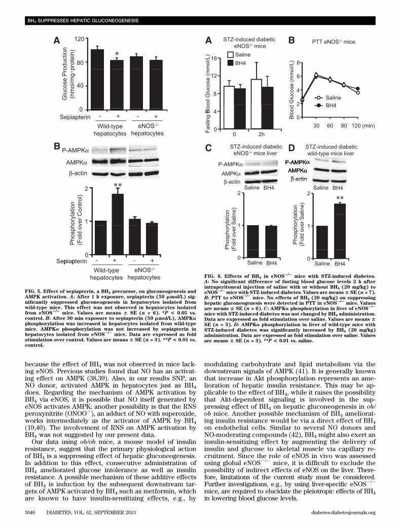
BH4 also significantly increased ATP content. To clarifythe mechanism by which BH4 increases AMP content andactivates AMPK in hepatocytes, we examined the effect ofAMP deaminase (AMPD) on activation of AMPK and sup-pression of gluconeogenesis by BH4. Although EHNA,a known AMPD inhibitor, activated AMPK and suppressedhepatic gluconeogenesis, BH4 did not have an additiveeffect on EHNA (Supplementary Fig. 6A and B). Theseresults indicate that inhibition of AMPD, at least in part,contributes to AMP accumulation by BH4 in hepatocytes.Sepiapterin, a BH4 precursor, suppresses gluconeogenesisand increases AMPK activation. Similarly to BH4,sepiapterin is absorbed in hepatocytes and immediatelyconverted to BH4 via a salvage pathway of BH4 bio-synthesis (23). Sepiapterin was found to suppress gluco-neogenesis and activate AMPK (Fig. 5A and B). However,these effects were abolished in hepatocytes lacking eNOS(Fig. 5A and B).Role of eNOS in in vivo action of BH4 on glucosemetabolism. The lowering effect of BH4 on fastingblood glucose levels disappeared in STZ-induced diabeticeNOS2/2 mice (Fig. 6A). The PTT data showed that BH4did not decrease hepatic glucose production in eNOS2/2
mice (Fig. 6B). Similar results were also obtained insepiapterin administration (Supplementary Fig. 7A and B).We then compared the effects of BH4 on phosphorylationof AMPKa in liver tissues of these diabetic mice. BH4 ac-tivated AMPK in both STZ diabetic wild-type mice liver anddiabetic Akita mice liver but not in STZ diabetic eNOS2/2
mice liver (Fig. 6C and D and Supplementary Fig. 8A).AMPKa phosphorylation was not changed by fasting for 16 hin liver tissues of wild-type mice (Supplementary Fig. 8B).Effects of BH4 on glucose metabolism and insulinsensitivity in ob/ob mice. Our PTT data show that thesuppressing effect on gluconeogenesis is also confirmedby single administration of BH4 in ob/ob mice (Fig. 7A),while the mRNA expression levels of PEPCK and G6Pasein the liver (Supplementary Fig. 9A and B), fasting and fedblood glucose levels, and IPGTT data were not changed(data not shown). By consecutive administration of BH4(20 mg/kg) in saline for 10 days to ob/obmice, fasting bloodglucose levels were significantly lowered by 3.9 mmol/Land fed blood glucose levels tended to be decreasedcompared with those in ob/ob mice treated with salinealone (Fig. 7B and C). Our IPGTT, HOMA-IR, and insulintolerance test data suggest that consecutive administrationof BH4 ameliorates glucose intolerance as well as insulinresistance (Fig. 7D–G). Phosphorylation of AMPKa, ACC,and Akt was increased in liver tissues of BH4-treated ob/obmice compared with those in saline-treated mice (Fig. 7Hand I).
DISCUSSION
The current study shows that BH4, known as a cofactor ofeNOS, has a glucose-lowering effect in diabetic mice. TheBH4-to-BH2 ratio was found to be decreased in various tis-sues of mice in the diabetic state, indicating deterioration of
of mRNA expression levels of DHFR in liver was detected between nondiabetic mice and mice with STZ-induced diabetes. Values are means 6 SE(n = 10). G: No significant difference of protein expression levels of DHFR in liver was detected between nondiabetic mice and mice with STZ-induced diabetes. Values are means 6 SE (n = 5). H and I: In liver tissues of wild-type mice with STZ-induced diabetes treated with BH4, mRNAlevels of PEPCK were significantly decreased compared with those treated without BH4. The mRNA levels of G6Pase were not changed. Values aremeans 6 SE (n = 6), *P < 0.05 vs. saline. J: Liver tissues of eNOS dimer and monomer expression 2 h after intraperitoneal injection of saline withor without BH4 (20 mg/kg) to wild-type mice with STZ-induced diabetes. Densitometric analysis of the ratio of eNOS dimer to monomer. Values aremeans 6 SE (n = 5). *P < 0.05 vs. saline. K: In liver tissues of wild-type mice with STZ-induced diabetes treated with BH4, NO content was sig-nificantly increased compared with those treated without BH4. Values are means 6 SE (n = 5). *P < 0.05 vs. saline.
A. ABUDUKADIER AND ASSOCIATES
diabetes.diabetesjournals.org DIABETES, VOL. 62, SEPTEMBER 2013 3037
eNOS bioactivity by eNOS uncoupling. Previous studieshave shown that impairment of eNOS function is involved inglucose dysmetabolism and insulin resistance (4,5), whichlends support to the notion that alleviation of eNOS dys-function such as by supplementation of BH4 amelioratesglucose dysmetabolism and insulin resistance. In addition,we found that supplementation of BH4 increased di-merization of eNOS and NO production in the liver of di-abetic mice, which strongly suggests alleviation of eNOSdysfunction by recoupling of eNOS. Simultaneously withthe restoration of eNOS activity, BH4 elicited a glucose-lowering effect in these mice. No such glucose-loweringeffect by BH4 appeared in diabetic mice lacking eNOS.These findings clearly implicate recoupling of eNOS in theglucose-lowering effect of BH4.
We have shown that the liver plays a critical role in theglucose-lowering effect of BH4 through suppression ofhepatic gluconeogenesis. It is well-known that BH4 issynthesized mainly in liver (24) and that this is impaired byoxidative stress such as liver cirrhosis and diabetes(25,26). Single administration of BH4 is known to accu-mulate at higher levels in liver than other tissues includingskeletal muscle (24), which also lends support to the viewthat BH4 readily elevates BH4-to-BH2 ratio and regulatesglucose metabolism in the liver.
We then investigated the molecular mechanism of sup-pression of hepatic gluconeogenesis by BH4 using isolatedmouse hepatocytes. BH4 acts directly on hepatocytes andsuppresses hepatic gluconeogenesis eNOS dependently.Several studies reported that eNOS is found in hepaticsinusoidal and venous endothelial cells and not in hepa-tocytes (27,28), whereas other studies claim detection ofeNOS in hepatocytes (29,30). We confirmed that eNOS isexpressed in hepatocytes, which suggests that intra-hepatocellular eNOS is essential for the effect of BH4 insuppression of hepatic gluconeogenesis. In addition, BH4activated AMPK, and the suppressing effect of BH4 ongluconeogenesis disappeared by siRNA silencing ofAMPKa1 subunits in hepatocytes, indicating that AMPK isinvolved in the suppressing effect of BH4 on hepatic glu-coneogenesis. AMPK activation by BH4 was not observedin eNOS2/2 mouse hepatocytes or in the presence of NOSinhibitor, suggesting that eNOS acts upstream of AMPKactivation in suppression of hepatic gluconeogenesis byBH4. AMPK is a Ser/Thr kinase that acts as an energysensor and is activated by an increase in the AMP-to-ATPratio and/or AMP in response to a variety of metabolicstresses, such as hypoxia, ischemia, and exercise (31,32).In our data, BH4 significantly increased AMP content and
FIG. 3. BH4 suppressed gluconeogenesis and increased AMPKa phos-phorylation in hepatocytes isolated from wild-type mice. A: Time courseof gluconeogenesis with exposure to BH4. Suppressing effect on glu-coneogenesis by 50 mmol/L BH4 compared with control was detectedafter 60 min in hepatocytes isolated from wild-type mice. Values aremeans 6 SE (n = 6). **P < 0.01 vs. control. B: Time course of phos-phorylation of AMPKa and ACC upon exposure to BH4 (50 mmol/L).Both AMPKa and ACC phosphorylation were stimulated after 30 minexposure to BH4 in hepatocytes isolated from wild-type mice. Dataare expressed as fold stimulation over control. Values are means 6 SE(n = 3). *P < 0.05, **P < 0.01 vs. control. C: Suppressing effect on
gluconeogenesis after 1 h exposure of BH4 was detected ranging over 50mmol/L in hepatocytes isolated from wild-type mice. Values are means6SE (n = 6). **P < 0.01, ***P < 0.001 vs. control. D: Effect of BH4 onphosphorylation of AMPK and ACC. After 30 min exposure to BH4, bothAMPKa and ACC phosphorylation were increased by BH4 dose de-pendently ranging over 50 mmol/L in hepatocytes isolated from wild-type mice. Data are expressed as fold stimulation over control. Valuesare means 6 SE (n = 3). *P < 0.05, **P < 0.01 vs. control. E: Withtransfection with AMPKa1 siRNA, protein expression of AMPKawas decreased compared with that of transfection with controlsiRNA. Values are means 6 SE (n = 3). ***P < 0.001 vs. control siRNA.F: Transfected with AMPKa1 siRNA, suppressing effect of BH4
(50 mmol/L) on hepatic glucose production was inhibited. Values aremeans 6 SE (n = 6). ***P < 0.001 vs. values transfected with controlsiRNA without BH4. G: Compound C (20 mmol/L), an AMPK inhibitor,abolished the suppressing effect of BH4 (50 mmol/L) on gluconeo-genesis. Values are means 6 SE (n = 6). *P < 0.05 vs. values withoutBH4 and without compound C.
BH4 SUPPRESSES HEPATIC GLUCONEOGENESIS
3038 DIABETES, VOL. 62, SEPTEMBER 2013 diabetes.diabetesjournals.org
tended to increase the AMP-to-ATP ratio. It is known thatinhibition of AMPD increases AMP in isolated hepatocytes(33). Recently, Ouyang et al. (34) reported that inhibitionof AMPD might be involved in increased production of
AMP and activation of AMPK by metformin. In the currentstudy, the AMPD inhibitor EHNA was found to activateAMPK, but BH4 did not elicit an additional effect on AMPKactivation in the presence of EHNA, suggesting that AMPDmight be inhibited by BH4 in hepatocytes. Interestingly,BH4 significantly increased ATP content along with theincrease in AMP. This effect was not found in exposure toother potent AMPK activators, as previously reported (35).The reason why BH4 increases ATP content is unclear, butBH4 is known to work as an antioxidant (36). It has beenreported that BH4 preserves ATP content and has a cyto-protective effect from hypoxia on neuronal cells (37). BH4might thus prevent cytotoxic damage from reactive oxygenspecies/reactive nitrogen species (RNS) as a scavenger,keeping ATP content higher than in the absence of BH4.We therefore cannot exclude the possibility that BH4 actsas a reactive oxygen species/RNS scavenger in ameliorat-ing glucose dysmetabolism, but such an effect would belimited in terms of suppressing hepatic gluconeogenesis
FIG. 4. Lack of the effect of BH4 on suppression of gluconeogenesis in eNOS2/2
mouse hepatocytes. A: BH4 (50 mmol/L) significantly increased NOproduction in hepatocytes from wild-type mice. SNP (20 mmol/L) was used as positive control. Values are means6 SE (n = 5). *P< 0.05 vs. control.B: BH4 (ranging from 10 to 50 mmol/L) increased eNOS phosphorylation at Ser
1177in hepatocytes from wild-type mice. Values are means 6 SE (n = 5).
*P < 0.05 vs. control. C: BH4 (ranging from 10 to 100 mmol/L) did not suppress gluconeogenesis after 1 h exposure in hepatocytes from eNOS2/2
mice.Values are means6 SE (n = 6).D: After 30 min exposure to BH4 ranging from 10 to 100mmol/L, AMPKa and ACC phosphorylation were not increased byBH4 in hepatocytes from eNOS
2/2mice. Data are expressed as fold stimulation over control. Values are means 6 SE (n = 3).
TABLE 1Effects of BH4 on ATP, AMP, and AMP-to-ATP ratio in wild-typemouse hepatocytes
ATP (nmol/mgprotein)
AMP (nmol/mgprotein)
AMP-to-ATPratio
Control 0.66 6 0.08 0.28 6 0.04 0.44 6 0.03BH4 0.88 6 0.04* 0.49 6 0.05** 0.55 6 0.04SNP 0.73 6 0.07 0.47 6 0.01** 0.67 6 0.07
Data are means 6 SE (n = 5). Hepatocytes were incubated in Krebs-Ringer bicarbonate buffer with or without BH4 (50 mmol/L) for 30 min.The treatment was stopped by rapid addition of 0.1 mL of 2 mol/LHClO4, and adenine nucleotide contents were measured. *P , 0.05,**P , 0.01 vs. control.
A. ABUDUKADIER AND ASSOCIATES
diabetes.diabetesjournals.org DIABETES, VOL. 62, SEPTEMBER 2013 3039
because the effect of BH4 was not observed in mice lack-ing eNOS. Previous studies found that NO has an activat-ing effect on AMPK (38,39). Also, in our results SNP, anNO donor, activated AMPK in hepatocytes just as BH4does. Regarding the mechanism of AMPK activation byBH4 via eNOS, it is possible that NO itself generated byeNOS activates AMPK; another possibility is that the RNSperoxynitrite (ONOO2), an adduct of NO with superoxide,works intermediately as the activator of AMPK by BH4(19,40). The involvement of RNS on AMPK activation byBH4 was not suggested by our present data.
Our data using ob/ob mice, a mouse model of insulinresistance, suggest that the primary physiological actionof BH4 is a suppressing effect of hepatic gluconeogenesis.In addition to this effect, consecutive administration ofBH4 ameliorated glucose intolerance as well as insulinresistance. A possible mechanism of these additive effectsof BH4 is induction by the subsequent downstream tar-gets of AMPK activated by BH4 such as metformin, whichare known to have insulin-sensitizing effects, e.g., by
modulating carbohydrate and lipid metabolism via thedownstream signals of AMPK (41). It is generally knownthat increase in Akt phosphorylation represents an ame-lioration of hepatic insulin resistance. This may be ap-plicable to the effect of BH4, while it raises the possibilitythat Akt-dependent signaling is involved in the sup-pressing effect of BH4 on hepatic gluconeogenesis in ob/ob mice. Another possible mechanism of BH4 ameliorat-ing insulin resistance would be via a direct effect of BH4on endothelial cells. Similar to several NO donors andNO-moderating compounds (42), BH4 might also exert aninsulin-sensitizing effect by augmenting the delivery ofinsulin and glucose to skeletal muscle via capillary re-cruitment. Since the role of eNOS in vivo was assessedusing global eNOS2/2 mice, it is difficult to exclude thepossibility of indirect effects of eNOS on the liver. There-fore, limitations of the current study must be considered.Further investigations, e.g., by using liver-specific eNOS2/2
mice, are required to elucidate the pleiotropic effects of BH4in lowering blood glucose levels.
FIG. 5. Effect of sepiapterin, a BH4 precursor, on gluconeogenesis andAMPK activation. A: After 1 h exposure, sepiapterin (50 mmol/L) sig-nificantly suppressed gluconeogenesis in hepatocytes isolated fromwild-type mice. This effect was not observed in hepatocytes isolatedfrom eNOS
2/2mice. Values are means 6 SE (n = 6). *P < 0.05 vs.
control. B: After 30 min exposure to sepiapterin (50 mmol/L), AMPKaphosphorylation was increased in hepatocytes isolated from wild-typemice. AMPKa phosphorylation was not increased by sepiapterin inhepatocytes isolated from eNOS
2/2mice. Data are expressed as fold
stimulation over control. Values are means 6 SE (n = 3). **P < 0.01 vs.control.
FIG. 6. Effects of BH4 in eNOS2/2
mice with STZ-induced diabetes.A: No significant difference of fasting blood glucose levels 2 h afterintraperitoneal injection of saline with or without BH4 (20 mg/kg) toeNOS
2/2mice with STZ-induced diabetes. Values are means6 SE (n = 7).
B: PTT to eNOS2/2
mice. No effects of BH4 (20 mg/kg) on suppressinghepatic gluconeogenesis were detected in PTT in eNOS
2/2mice. Values
are means 6 SE (n = 6). C: AMPKa phosphorylation in liver of eNOS2/2
mice with STZ-induced diabetes was not changed by BH4 administration.Data are expressed as fold stimulation over saline. Values are means 6SE (n = 3). D: AMPKa phosphorylation in liver of wild-type mice withSTZ-induced diabetes was significantly increased by BH4 (20 mg/kg)administration. Data are expressed as fold stimulation over saline. Valuesare means 6 SE (n = 3). **P < 0.01 vs. saline.
BH4 SUPPRESSES HEPATIC GLUCONEOGENESIS
3040 DIABETES, VOL. 62, SEPTEMBER 2013 diabetes.diabetesjournals.org
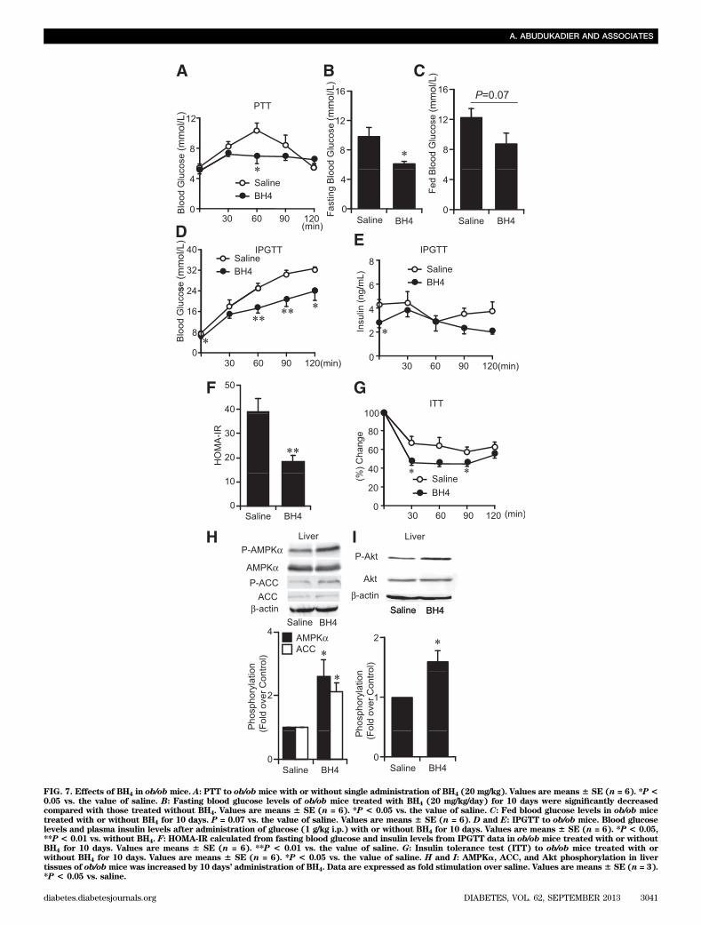
FIG. 7. Effects of BH4 in ob/ob mice. A: PTT to ob/obmice with or without single administration of BH4 (20 mg/kg). Values are means 6 SE (n = 6). *P<0.05 vs. the value of saline. B: Fasting blood glucose levels of ob/ob mice treated with BH4 (20 mg/kg/day) for 10 days were significantly decreasedcompared with those treated without BH4. Values are means 6 SE (n = 6). *P < 0.05 vs. the value of saline. C: Fed blood glucose levels in ob/ob micetreated with or without BH4 for 10 days. P = 0.07 vs. the value of saline. Values are means 6 SE (n = 6). D and E: IPGTT to ob/ob mice. Blood glucoselevels and plasma insulin levels after administration of glucose (1 g/kg i.p.) with or without BH4 for 10 days. Values are means 6 SE (n = 6). *P < 0.05,**P < 0.01 vs. without BH4. F: HOMA-IR calculated from fasting blood glucose and insulin levels from IPGTT data in ob/ob mice treated with or withoutBH4 for 10 days. Values are means 6 SE (n = 6). **P < 0.01 vs. the value of saline. G: Insulin tolerance test (ITT) to ob/ob mice treated with orwithout BH4 for 10 days. Values are means 6 SE (n = 6). *P < 0.05 vs. the value of saline. H and I: AMPKa, ACC, and Akt phosphorylation in livertissues of ob/obmice was increased by 10 days’ administration of BH4. Data are expressed as fold stimulation over saline. Values are means6 SE (n = 3).*P < 0.05 vs. saline.
A. ABUDUKADIER AND ASSOCIATES
diabetes.diabetesjournals.org DIABETES, VOL. 62, SEPTEMBER 2013 3041
The glucose-lowering effect of BH4 by single admin-istration intraperitoneally on fasting blood glucose lev-els in STZ diabetic mice was similar to that of metformin(250 mg/kg). The dose of metformin that we used wasadjusted to previous studies in mice (43) and is morethan fivefold higher than that in clinical use for type 2diabetic patients (44). We demonstrate here the lower-ing effects of BH4 on blood glucose levels using adosage similar to that of BH4 used in patients withphenylketonuria as a cofactor of phenylalanine hy-droxylase (45).
Numerous clinical trials have been performed on theeffect of BH4 as a cofactor of eNOS on endothelial dys-function in a variety of vascular diseases including coro-nary artery disease (15). While many of the results aredisappointing (46), BH4 remains a viable candidate forclinical use if the design of the various trials is reconsid-ered. Several of the studies reported that BH4 levels areplainly decreased and that uncoupled eNOS is found inthe diabetic state and not in nondiabetic states (47).Moreover, nondiabetic patients were included in most ofthe clinical trials (46); those trials should be performedin patients with diabetes. The current study, further-more, clarifies a novel concept of the relationshipbetween BH4 and glucose metabolism and insulin re-sistance that suggests a new approach to the preventionof macrovascular complications of diabetes induced byendothelial dysfunction as well as amelioration of thedisease itself.
In conclusion, BH4 has a glucose-lowering effectby suppressing hepatic gluconeogenesis in an eNOS-dependent manner and ameliorates glucose intoleranceas well as insulin resistance in diabetic mice, suggest-ing that BH4 has potential in the treatment of type 2diabetes.
ACKNOWLEDGMENTS
This study was supported by Scientific Research grants;a grant for Leading Project for Biosimulation from theMinistry of Education, Culture, Sports, Science, and Tech-nology of Japan; a grant from Core Research for EvolutionalScience and Technology (CREST) of Japan Science andTechnology Cooperation; a grant from the Ministry ofHealth, Labor, and Welfare, Japan; and a grant from KyotoUniversity Global Center of Excellence (COE) Program“Center for Frontier Medicine.”
No potential conflicts of interest relevant to this articlewere reported.
A.A. and Y.F. researched data, contributed to discussion,and wrote, reviewed, and edited the manuscript. A.Ob. andA.Oh. researched data and contributed to discussion. T.F.,Y.S., M.O., Y.N., S.F., and M.H. contributed to discussion.H.H. researched data and contributed to discussion. N.I.contributed to discussion and wrote, reviewed, and editedthe manuscript. N.I. is the guarantor of this work and, assuch, had full access to all the data in the study and takesresponsibility for the integrity of the data and the accuracyof the data analysis.
Parts of this study were presented in abstract form at the71st Scientific Sessions of the American Diabetes Associ-ation, San Diego, California, 24–28 June 2011.
The authors thank Ryo Tanaka and Miho Nishimura fortheir efforts in collaboration in the Student ResearchProgram, Department of Biosciences, Teikyo Universityof Science and Technology.
REFERENCES
1. Triggle CR, Ding H. A review of endothelial dysfunction in diabetes: a fo-cus on the contribution of a dysfunctional eNOS. J Am Soc Hypertens2010;4:102–115
2. Huang PL. eNOS, metabolic syndrome and cardiovascular disease. TrendsEndocrinol Metab 2009;20:295–302
3. Kim JA, Montagnani M, Koh KK, Quon MJ. Reciprocal relationships be-tween insulin resistance and endothelial dysfunction: molecular andpathophysiological mechanisms. Circulation 2006;113:1888–1904
4. Duplain H, Burcelin R, Sartori C, et al. Insulin resistance, hyperlipidemia,and hypertension in mice lacking endothelial nitric oxide synthase. Cir-culation 2001;104:342–345
5. Shankar RR, Wu Y, Shen HQ, et al. Mice with gene disruption of bothendothelial and neuronal nitric oxide synthase exhibit insulin resistance.Diabetes 2000;49:684–687
6. Stuehr D, Pou S, Rosen GM. Oxygen reduction by nitric-oxide synthases.J Biol Chem 2001;276:14533–14536
7. Vásquez-Vivar J, Kalyanaraman B, Martásek P, et al. Superoxide generationby endothelial nitric oxide synthase: the influence of cofactors. Proc NatlAcad Sci USA 1998;95:9220–9225
8. Crabtree MJ, Channon KM. Synthesis and recycling of tetrahydrobiopterinin endothelial function and vascular disease. Nitric Oxide 2011;25:81–88
9. Landmesser U, Dikalov S, Price SR, et al. Oxidation of tetrahydrobiopterinleads to uncoupling of endothelial cell nitric oxide synthase in hyperten-sion. J Clin Invest 2003;111:1201–1209
10. Meininger CJ, Marinos RS, Hatakeyama K, et al. Impaired nitric oxideproduction in coronary endothelial cells of the spontaneously diabetic BBrat is due to tetrahydrobiopterin deficiency. Biochem J 2000;349:353–356
11. Xu J, Wu Y, Song P, et al. Proteasome-dependent degradation of guanosine59-triphosphate cyclohydrolase I causes tetrahydrobiopterin deficiency indiabetes mellitus. Circulation 2007;116:944–953
12. Meininger CJ, Cai S, Parker JL, et al. GTP cyclohydrolase I gene transferreverses tetrahydrobiopterin deficiency and increases nitric oxide syn-thesis in endothelial cells and isolated vessels from diabetic rats. FASEB J2004;18:1900–1902
13. Ding QF, Hayashi T, Packiasamy AR, et al. The effect of high glucose onNO and O2- through endothelial GTPCH1 and NADPH oxidase. Life Sci2004;75:3185–3194
14. Kietadisorn R, Juni RP, Moens AL. Tackling endothelial dysfunction bymodulating NOS uncoupling: new insights into its pathogenesis and ther-apeutic possibilities. Am J Physiol Endocrinol Metab 2012;302:E481–E495
15. Katusic ZS, d’Uscio LV, Nath KA. Vascular protection by tetrahy-drobiopterin: progress and therapeutic prospects. Trends Pharmacol Sci2009;30:48–54
16. Wajngot A, Chandramouli V, Schumann WC, et al. Quantitative con-tributions of gluconeogenesis to glucose production during fasting in type2 diabetes mellitus. Metabolism 2001;50:47–52
17. Lin HV, Accili D. Hormonal regulation of hepatic glucose production inhealth and disease. Cell Metab 2011;14:9–19
18. Oyadomari S, Koizumi A, Takeda K, et al. Targeted disruption of the Chopgene delays endoplasmic reticulum stress-mediated diabetes. J Clin Invest2002;109:525–532
19. Fujita Y, Hosokawa M, Fujimoto S, et al. Metformin suppresses hepaticgluconeogenesis and lowers fasting blood glucose levels through reactivenitrogen species in mice. Diabetologia 2010;53:1472–1481
20. Fujimoto S, Mukai E, Hamamoto Y, et al. Prior exposure to high glucoseaugments depolarization-induced insulin release by mitigating the declineof ATP level in rat islets. Endocrinology 2002;143:213–221
21. Ogura M, Nakamura Y, Tanaka D, et al. Overexpression of SIRT5 confirmsits involvement in deacetylation and activation of carbamoyl phosphatesynthetase 1. Biochem Biophys Res Commun 2010;393:73–78
22. Sawabe K, Wakasugi KO, Hasegawa H. Tetrahydrobiopterin uptake in sup-plemental administration: elevation of tissue tetrahydrobiopterin in micefollowing uptake of the exogenously oxidized product 7,8-dihydrobiopterinand subsequent reduction by an anti-folate-sensitive process. J PharmacolSci 2004;96:124–133
23. Sawabe K, Suetake Y, Nakanishi N, et al. Cellular accumulation of tetra-hydrobiopterin following its administration is mediated by two differentprocesses; direct uptake and indirect uptake mediated by a methotrexate-sensitive process. Mol Genet Metab 2005;86(Suppl. 1):S133–S138
24. Hoshiga M, Hatakeyama K, Watanabe M, et al. Autoradiographic distri-bution of [14C]tetrahydrobiopterin and its developmental change in mice.J Pharmacol Exp Ther 1993;267:971–978
25. Matei V, Rodríguez-Vilarrupla A, Deulofeu R, et al. Three-day tetrahy-drobiopterin therapy increases in vivo hepatic NOS activity and reducesportal pressure in CCl4 cirrhotic rats. J Hepatol 2008;49:192–197
BH4 SUPPRESSES HEPATIC GLUCONEOGENESIS
3042 DIABETES, VOL. 62, SEPTEMBER 2013 diabetes.diabetesjournals.org
26. Elrod JW, Duranski MR, Langston W, et al. eNOS gene therapy exacerbateshepatic ischemia-reperfusion injury in diabetes: a role for eNOS un-coupling. Circ Res 2006;99:78–85
27. Shah V, Cao S, Hendrickson H, et al. Regulation of hepatic eNOS by caveolinand calmodulin after bile duct ligation in rats. Am J Physiol GastrointestLiver Physiol 2001;280:G1209–G1216
28. Wei CL, Khoo HE, Lee KH, et al. Differential expression and localization ofnitric oxide synthases in cirrhotic livers of bile duct-ligated rats. NitricOxide 2002;7:91–102
29. McNaughton L, Puttagunta L, Martinez-Cuesta MA, et al. Distribution ofnitric oxide synthase in normal and cirrhotic human liver. Proc Natl AcadSci USA 2002;99:17161–17166
30. Mei Y, Thevananther S. Endothelial nitric oxide synthase is a key mediatorof hepatocyte proliferation in response to partial hepatectomy in mice.Hepatology 2011;54:1777–1789
31. Zhang BB, Zhou G, Li C. AMPK: an emerging drug target for diabetes andthe metabolic syndrome. Cell Metab 2009;9:407–416
32. Hardie DG. The AMP-activated protein kinase pathway—new players up-stream and downstream. J Cell Sci 2004;117:5479–5487
33. Carabaza A, Ricart MD, Mor A, et al. Role of AMP on the activation ofglycogen synthase and phosphorylase by adenosine, fructose, and gluta-mine in rat hepatocytes. J Biol Chem 1990;265:2724–2732
34. Ouyang J, Parakhia RA, Ochs RS. Metformin activates AMP kinase throughinhibition of AMP deaminase. J Biol Chem 2011;286:1–11
35. Fogarty S, Hardie DG. Development of protein kinase activators: AMPK asa target in metabolic disorders and cancer. Biochim Biophys Acta 2010;1804:581–589
36. Kojima S, Ona S, Iizuka I, et al. Antioxidative activity of 5,6,7,8-tetrahydrobiopterin and its inhibitory effect on paraquat-induced celltoxicity in cultured rat hepatocytes. Free Radic Res 1995;23:419–430
37. Delgado-Esteban M, Almeida A, Medina JM. Tetrahydrobiopterin deficiencyincreases neuronal vulnerability to hypoxia. J Neurochem 2002;82:1148–1159
38. Kang KT, Sullivan JC, Spradley FT, et al. Antihypertensive therapy in-creases tetrahydrobiopterin levels and NO/cGMP signaling in small ar-teries of angiotensin II-infused hypertensive rats. Am J Physiol Heart CircPhysiol 2011;300:H718–H724
39. Higaki Y, Hirshman MF, Fujii N, et al. Nitric oxide increases glucose up-take through a mechanism that is distinct from the insulin and contractionpathways in rat skeletal muscle. Diabetes 2001;50:241–247
40. Zhang J, Xie Z, Dong Y, et al. Identification of nitric oxide as an endoge-nous activator of the AMP-activated protein kinase in vascular endothelialcells. J Biol Chem 2008;283:27452–27461
41. Viollet B, Guigas B, Leclerc J, et al. AMP-activated protein kinase in theregulation of hepatic energy metabolism: from physiology to therapeuticperspectives. Acta Physiol (Oxf) 2009;196:81–98
42. Cook S, Scherrer U. Insulin resistance, a new target for nitric oxide-delivery drugs. Fundam Clin Pharmacol 2002;16:441–453
43. Shaw RJ, Lamia KA, Vasquez D, et al. The kinase LKB1 mediates glucosehomeostasis in liver and therapeutic effects of metformin. Science 2005;310:1642–1646
44. Inzucchi SE, Maggs DG, Spollett GR, et al. Efficacy and metabolic effectsof metformin and troglitazone in type II diabetes mellitus. N Engl J Med1998;338:867–872
45. Blau N. Defining tetrahydrobiopterin (BH4)-responsiveness in PKU. J In-herit Metab Dis 2008;31:2–3
46. Moens AL, Kietadisorn R, Lin JY, et al. Targeting endothelial and myocardialdysfunction with tetrahydrobiopterin. J Mol Cell Cardiol 2011;51:559–563
47. Heitzer T, Krohn K, Albers S, et al. Tetrahydrobiopterin improves endothelium-dependent vasodilation by increasing nitric oxide activity in patients withType II diabetes mellitus. Diabetologia 2000;43:1435–1438
A. ABUDUKADIER AND ASSOCIATES
diabetes.diabetesjournals.org DIABETES, VOL. 62, SEPTEMBER 2013 3043