Embed Size (px)
Citation preview
Pacemaker-neuron–dependent disturbance of themolecular clockwork by a Drosophila CLOCK mutanthomologous to the mouse Clock mutationEuna Leea,b, Eunjoo Choc, Doo Hyun Kanga,b, Eun Hee Jeongb, Zheng Chend, Seung-Hee Yood,and Eun Young Kim (김은영)a,b,c,1
aNeuroscience Graduate Program, BK21 Plus Program, Department of Biomedical Sciences, Ajou University School of Medicine, Yeongtong-gu, Suwon,Kyunggi-do 16499, Republic of Korea; bDepartment of Brain Science, Ajou University School of Medicine, Yeongtong-gu, Suwon, Kyunggi-do 16499,Republic of Korea; cChronic Inflammatory Disease Research Center, Ajou University School of Medicine, Yeongtong-gu, Suwon, Kyunggi-do 16499, Republicof Korea; and dDepartment of Biochemistry and Molecular Biology, McGovern Medical School, The University of Texas Health Science Center at Houston,Houston, TX 77030
Edited by Joseph S. Takahashi, Howard Hughes Medical Institute, University of Texas Southwestern Medical Center, Dallas, TX, and approved June 28, 2016(received for review December 1, 2015)
Circadian clocks are composed of transcriptional/translational feed-back loops (TTFLs) at the cellular level. In Drosophila TTFLs, thetranscription factor dCLOCK (dCLK)/CYCLE (CYC) activates clock tar-get gene expression, which is repressed by the physical interactionwith PERIOD (PER). Here, we show that amino acids (AA) 657–707of dCLK, a region that is homologous to the mouse Clock exon19-encoded region, is crucial for PER binding and E-box–dependenttransactivation in S2 cells. Consistently, in transgenic flies expressingdCLK with an AA657–707 deletion in the Clock (Clkout) genetic back-ground (p{dClk-Δ};Clkout), oscillation of core clock genes’ mRNAsdisplayed diminished amplitude compared with control flies, andthe highly abundant dCLKΔ657–707 showed significantly decreasedbinding to PER. Behaviorally, the p{dClk-Δ};Clkout flies exhibited ar-rhythmic locomotor behavior in the photic entrainment conditionbut showed anticipatory activities of temperature transitionand improved free-running rhythms in the temperature entrainmentcondition. Surprisingly, p{dClk-Δ};Clkout flies showed pacemaker-neuron–dependent alterations in molecular rhythms; the abundanceof dCLK target clock proteins was reduced in ventral lateral neurons(LNvs) but not in dorsal neurons (DNs) in both entrainment condi-tions. In p{dClk-Δ};Clkout flies, however, strong but delayedmolecularoscillations in temperature cycle-sensitive pacemaker neurons, suchas DN1s and DN2s, were correlated with delayed anticipatory ac-tivities of temperature transition. Taken together, our study re-veals that the LNv molecular clockwork is more sensitive thanthe clockwork of DNs to dysregulation of dCLK by AA657–707 de-letion. Therefore, we propose that the dCLK/CYC-controlled TTFLoperates differently in subsets of pacemaker neurons, which maycontribute to their specific functions.
circadian rhythm | CLOCK | dorsal neuron | lateral neuron | TTFL
Circadian timing systems are composed of cell-autonomous os-cillators that enable living organisms to anticipate environ-
mental cyclic changes, thereby orchestrating behavior and physiologythroughout the day. The cell-autonomous oscillator contains tran-scriptional/translational feedback loops (TTFLs) composed ofpositive and negative molecular components, driving the rhythmicoscillation of gene expression with 24-h periodicity (1, 2). In Dro-sophila, the positive components are the basic helix–loop–helix-containing and Period–Arnt–Sim (PAS)-containing transcriptionfactor dCLOCK (dCLK) and CYCLE (CYC), which form a het-erodimer and rhythmically bind to E-box sequences (CACGTC) toactivate transcription of clock genes and clock-controlled genes(reviewed in ref. 1). In the core loop of the TTFL, dCLK/CYCtranscribes period (per) and timeless (tim) genes and the translatedPER and TIM proteins form heterodimers and translocate intothe nucleus during mid-evening. The PER/TIM heterodimersthen physically interact with the dCLK/CYC complex to inhibit
dCLK/CYC-activated transcription. Degradation of PER and TIMproteins by timely controlled posttranslational modifications ulti-mately releases repression of dCLK/CYC activity, initiating a newtranscriptional cycle. In the secondary loop of the TTFL, dCLK/CYC activates transcription of vrille (vri) and par domain protein I«(pdp I«), and VRI and PDP Ie function to repress and activatetranscription of dClk, respectively, to increase the robustness of theTTFL (3, 4). Highly conserved molecular mechanisms operate inmammals (reviewed in ref. 5). Mouse CLK (mCLK) and brain andmuscle Arnt-like 1 (BMAL1), which are the dCLK and CYC ortho-logs, activate the transcription of genes in the negative arm of theTTFL, including mPer1, 2, 3 and cryptochrome1, 2 (cry1, 2). Heter-odimeric mPER/mCRY complexes subsequently enter the nucleus toinhibit transcription of their own genes. mCLK/BMAL1 also tran-scribes the genes encoding nuclear receptors Rora, b, c and Rev-erbα,β, which activate and repress, respectively the expression of Bmal1.Drosophila displays bimodal peaks of locomotor activity under
a standard 12-h/12-h light/dark (LD) photic entrainment condi-tion. Morning and evening peaks of activity occur around dawnand dusk, respectively. In Drosophila, ∼150 anatomically distinctpacemaker neurons are located in the lateral and dorsal parts ofthe brain and control respective activity peaks (6–8). The neu-ropeptide pigment dispersing factor (PDF)-positive large ventral
Significance
The circadian clock drives ∼24-hour rhythms in behavior andphysiology of organisms, and is dependent on transcriptional/translational feedback loops (TTFLs) at the cellular level. Pace-maker neurons in the brain control specific circadian behaviors inresponse to environmental timing cues such as light and temper-ature cycles. We show here that flies expressing dCLOCK (dCLK)lacking amino acids 657–707, homologous to the mouse Clockmutation, display pacemaker-neuron–dependent disturbance ofthe molecular clockwork. Specifically, the molecular rhythms inlight-sensitive pacemaker neurons were significantly disrupted,but the molecular rhythms in temperature-sensitive pacemakerneurons were robust. Our results suggest that the dCLK-controlledTTFL operates differently in subsets of pacemaker neurons, whichcontributes to their specific functions, such as differential sensi-tivity to entraining cues.
Author contributions: E.L., S.-H.Y., and E.Y.K. designed research; E.L., E.C., D.H.K., E.H.J.,S.-H.Y., and E.Y.K. performed research; E.L., E.C., S.-H.Y., and E.Y.K. analyzed data; andE.L., Z.C., and E.Y.K. wrote the paper.
The authors declare no conflict of interest.
This article is a PNAS Direct Submission.1To whom correspondence should be addressed. Email: [email protected].
This article contains supporting information online at www.pnas.org/lookup/suppl/doi:10.1073/pnas.1523494113/-/DCSupplemental.
E4904–E4913 | PNAS | Published online August 3, 2016 www.pnas.org/cgi/doi/10.1073/pnas.1523494113
Dow
nloa
ded
by g
uest
on
Apr
il 9,
202
0
lateral neurons (lLNvs) respond to light and play a crucial role inmediating light input from the eyes to regulate arousal, sleep, andthe startle response (9–12). Under constant dark conditions afterentrainment, morning and evening locomotor activity peaks arecontrolled by morning oscillators composed of PDF-positive smallLNvs (sLNvs) and evening oscillators composed of dorsal lateralneurons (LNds), a fifth sLNv, and some dorsal neurons (DN1s)(6, 8, 13, reviewed in ref. 14). Besides photic cycles, 12-h/12-h high/low temperature cycles that differ by as little as 2–3 °C can syn-chronize Drosophila behavioral locomotor rhythms and molecularoscillations (15–17). Different pacemakers might be responsible forcontrolling morning and evening activity peaks under temperaturecycles. When photic and temperature cues are both present, LNvsand LNds are sensitive to photic transition, whereas DN1s, DN2s,DN3s, and lateral posterior neurons are more sensitive to temper-ature transitions (18). In addition, blue-light photopigment CRY-negative DN1 and CRY-null DN2 are known to play a prominentrole in entrainment to temperature cycles (18–21).Physical interaction between positive and negative circadian fac-
tors is crucial for the regulation of circadian transcription (22–28).Here, we provide evidence that a small region of dCLK [amino acids(AA) 657–707], homologous to the peptide sequence encoded byexon 19 of mCLK is crucial for its interaction with PER, although itmight not function as the direct binding domain. We show that amutant of dCLK with this region deleted (dCLK-Δ) exhibited re-duced E-box–dependent transcriptional activity in S2 cells. Expres-sion of dCLK-Δ in the Clkout genetic background (herein namedp{dClk-Δ};Clkout) rendered flies arrhythmic in the photic entrain-ment condition. Surprisingly, the levels of dCLK target clock pro-teins were significantly reduced in LNvs; in comparison, other subsetsof clock neurons, such as DNs, were less affected. In addition,p{dClk-Δ};Clkout flies exhibited anticipatory behavior of temperature
transition, which is correlated with strong yet delayed oscillations ofcore clock proteins in temperature cycle-sensitive clock neurons,including DN1s and DN2s. Our results suggest that LNv molecularclockwork is more sensitive than DN molecular clockwork todysregulation of dCLK by AA657–707 deletion. Taken together, wepropose that the dCLK/CYC-controlled TTFL operates differentlyin subsets of pacemaker neurons despite the same molecular con-stituents, thereby modulating specific functions of pacemaker neu-rons, such as sensitivities to various entraining cues.
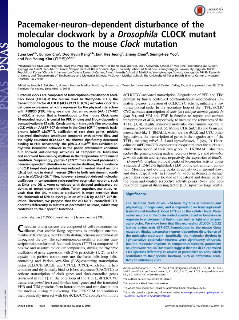
ResultsdCLK Internally Deleted for AA657–707 (dCLK-Δ) Shows ReducedBinding to PER and Transcriptional Activity in S2 Cells. To identify re-gions of dCLK that are crucial for interaction with PER, we gen-erated a series of dCLK internal deletion mutants (Fig. 1A). Weperformed coimmunoprecipitation analyses and quantified thedCLK–PER interaction by calculating the ratio of pulled-downPER to each immunoprecipitated dCLK variant (Fig. 1 B and C).The dCLK variant with deletion of AA579–778 (region 10 in Fig.1A) manifested the most severe impairment in binding PER. Pre-vious reports show that the AA657–707 region of dCLK enclosed inAA579–778 is homologous to the 51-aa segment encoded by exon19 of mClk, which is deleted in Clock/Clock mutant mice (29, 30)(Fig. 1A). Interestingly, the C terminus of apCLK of the silk-moth Antheraea pernyi also has a sequence homologous to the exon19 region in mCLK, and a C-terminal truncation mutant of apCLKcomplexed with apBMAL1 could not be inhibited by apPER,suggesting the presence of an apPER binding domain in this region(31). Thus, we evaluated whether dCLK AA657–707, which isconserved in all three species, is the minimal region required forPER binding. We generated a deletion mutant of dCLK lackingAA657–707 (dCLK-Δ) and performed coimmunoprecipitation
Fig. 1. dCLK subregion encompassing AA657–707 isrequired for binding with PER. (A) Schematic dia-gram illustrates the protein domains of dCLK. In thealignment between dCLK AA657–707 and mCLKAA514–564, black shading indicates amino acid identityand gray shading indicates conserved substitution. bHLH,basic helix–loop–helix. (B) S2 cells were cotransfectedwithpAct-per and pMT-HA-dClk (full length, FL) or vectorsencoding internally deleted dCLK variant indicated bythe numbers 1–12, corresponding to the numbers in theschematic diagram in A. Protein extracts (input) weresubjected to immunoprecipitation (IP) using anti-HAantibody for dCLK or normal IgG. The immune com-plexes were analyzed by immunoblotting. (C) Relativelevels of PER pulled down with each dCLK variant areshown. Values are the mean ± SEM from three inde-pendent experiments. (D) S2 cells were cotransfectedwith pAct-per andpMT-HA-dClk (FL) or pMT-HA-dClkΔ657–707 (Δ), and immunoprecipitation was performed asdescribed in B. (E) S2 cells were cotransfectedwith pMT-cyc-3F and pMT-HA-dClk (FL) or pMT-HA-dClkΔ657–707 (Δ).Immunoprecipitation was performed with anti-FLAGantibody, and immune complexes were analyzed by im-munoblotting. (F) HEK293T cells were transiently cotrans-fected with pcDNA3-mper1-V5, pcDNA3-mper2-V5, orpcDNA3-mper3-V5 in combination with pCMV10-3FLAG-mClk (FL) or pCMV10-3FLAG-mClkΔ19 (Δ). Immunopre-cipitation was performed with an anti-FLAG antibody,and immune complexes were analyzed by immuno-blotting. The immunoblot is representative of threeindependent experiments.
Lee et al. PNAS | Published online August 3, 2016 | E4905
NEU
ROSC
IENCE
PNASPL
US
Dow
nloa
ded
by g
uest
on
Apr
il 9,
202
0
experiments to assess its ability to interact with PER. The dCLK-Δshowed severely attenuated binding to PER (Fig. 1D), suggestingthat this minimal region is required for PER binding. However,dCLK-Δ retained the ability to interact with CYC (Fig. 1E).Because AA657–707 of dCLK displays significant sequence ho-
mology to the 51-aa segment encoded by exon 19 of mClk, we nextexamined whether this domain is also required for interaction be-tween mCLK and the three mPER proteins (mPER1, mPER2, andmPER3). We performed coimmunoprecipitation analyses betweeneither full-length mCLK or mCLK lacking exon 19 (mCLKΔ19)and each mPER homolog in mammalian HEK293 cells (Fig. 1F).Intriguingly, like dCLK-Δ, mCLKΔ19 exhibited impaired interac-tion with mPER1, mPER2, and mPER3, suggesting that the bio-chemical feature of interaction between PER and CLK is conservedin flies and mammals.The exon 19 region of mCLK is crucial for its transcriptional
activity (22, 32). Next, we examined whether dCLK-Δ displayscompromised transcriptional activity using a luciferase reporter-based assay in S2 cells (22, 28). Transfection of full-length dCLKincreased E-box–dependent luciferase expression, whereas dCLK-Δdid not significantly induce luciferase expression (Fig. S1). Giventhat steady-state levels of ectopically expressed full-length dCLK anddCLK-Δ were similar in S2 cells (Fig. 1 D and E), these resultssuggest that dCLK-Δ is impaired in transcriptional activation andthat AA657–707 of dCLK is essential, not only for PER interactionbut also for transcriptional activation in S2 cells.Next, to examine whether the AA657–707 functions as a direct
PER binding domain, we performed an in vitro binding assay usingpurified GST-tagged dCLK fragments and in vitro transcribed/translated PER (Fig. S2). GST-tagged full-length dCLK, butnot dCLK fragments AA80–448, AA579–778, AA657–707, andAA841–956, associated with PER. To our surprise, the AA380–528fragment strongly associated with PER, suggesting that this regionis a novel direct PER binding region (Fig. S2). Therefore, at pre-sent, it is not clear which region of dCLK is a direct binding site forPER, but AA657–707 of dCLK is clearly important for interactingwith PER and for dCLK transcriptional activity; moreover, thisregion is homologous to the exon 19 region of mCLK.
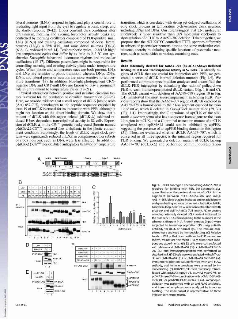
p{dClk-Δ};Clkout Flies Display Arrhythmic Behavior in the PhoticEntrainment Condition. To investigate the in vivo clock functionof AA657–707 of dCLK, we generated transgenic flies harboringthe dClkΔ657–707 transgene. Previous reports have shown that thewild-type version of this transgene fully rescues the arrhythmicity ofClkout flies, which do not express functional dCLK (33, 34) (Fig. 2Aand Table 1). Two independent fly lines expressing dCLKΔ657–707were obtained and switched to the Clkout genetic background[referred to as p{dClk-Δ},2M;Clkout and p{dClk-Δ},4M;Clkout].Both p{dClk-Δ};Clkout fly lines manifested arrhythmic behaviors(Fig. 2 B and C and Table 1). Interestingly, both p{dClk-Δ};Clkoutlines displayed little or no morning startled activities; however,evening startled activities were observed, and the flies were pref-
erentially active during the dark phase (Fig. 2 B and C). These dailyactivity patterns were highly similar to the daily activity patternsof Clkout (Fig. 2D) and ClkJrk flies, neither of which expressesfunctional dCLK protein (33–35). Under constant dark conditions,the p{dClk-Δ},2M;Clkout flies showed dampened morningand evening activity peaks with delayed phases, whereas thep{dClk-Δ},4M;Clkout flies did not manifest evident peaks of ac-tivity (Fig. 2 B and C). Thus, we used the p{dClk-Δ},4M;Clkout flyline, which showed more severe circadian behavioral defects, forfurther analyses unless otherwise stated. Taken together, these dataclearly indicate that AA657–707 of dCLK is essential in generatingcircadian locomotor rhythmicity in the photic entrainment condition.
Fig. 2. p{dClk-Δ};Clkout flies manifest abnormal locomotor behavior underLD and constant dark (DD) conditions. The locomotor activity of p{dClk-WT};Clkout (A), p{dClk-Δ},2M;Clkout (B), p{dClk-Δ},4M;Clkout (C), and Clkout (D) flylines is shown. Each panel represents the average activity of male flies for agiven genotype during the 12-h/12-h LD cycle, followed by the DD condition.The white vertical bars represent locomotor activity during the light phase ofthe LD cycle, and the black vertical bars represent locomotor activity duringthe dark phase of the LD cycle. The gray vertical bars represent locomotoractivity during the subjective light phase of the DD condition. The whitehorizontal bars and black horizontal bars below each panel indicate 12-h pe-riods of lights-on and lights-off, respectively.
Table 1. Behavioral rhythms of p{dClk-Δ};Clkout flies
Genotype Temperature, °C No.* Tau ± SEM, h Rhythmicity,† % Power‡
w;p{dClk-WT},1A;Clkout 25§ 32 23.8 ± 0.13 84.4 115.1w;p{dClk-Δ},2M;Clkout 25 39 AR AR ARw;p{dClk-Δ},4M;Clkout 25 30 AR AR ARClkout 25 28 AR AR ARw;p{dClk-WT},1A;Clkout 29/24# 28 23.1 ± 0.12 67.8 68.8w;p{dClk-Δ},2M;Clkout 29/24 30 24.9 ± 0.44 66.7 74.2
AR, arrhythmic.*Total number of flies that survived until the end of the testing period.†Percentage of flies with activity rhythms having a power value of ≥10 and a width value of ≥2.‡Measure of the strength or amplitude of the rhythm.§Flies were kept at 25 °C and exposed to 4 d of a 12-h/12-h LD cycle, followed by 7 d of a DD condition.#Flies were kept in constant darkness and exposed to 12-h/12-h temperature cycles of 29 °C/24 °C for 6 d, followed by7 d of constant 24 °C.
E4906 | www.pnas.org/cgi/doi/10.1073/pnas.1523494113 Lee et al.
Dow
nloa
ded
by g
uest
on
Apr
il 9,
202
0
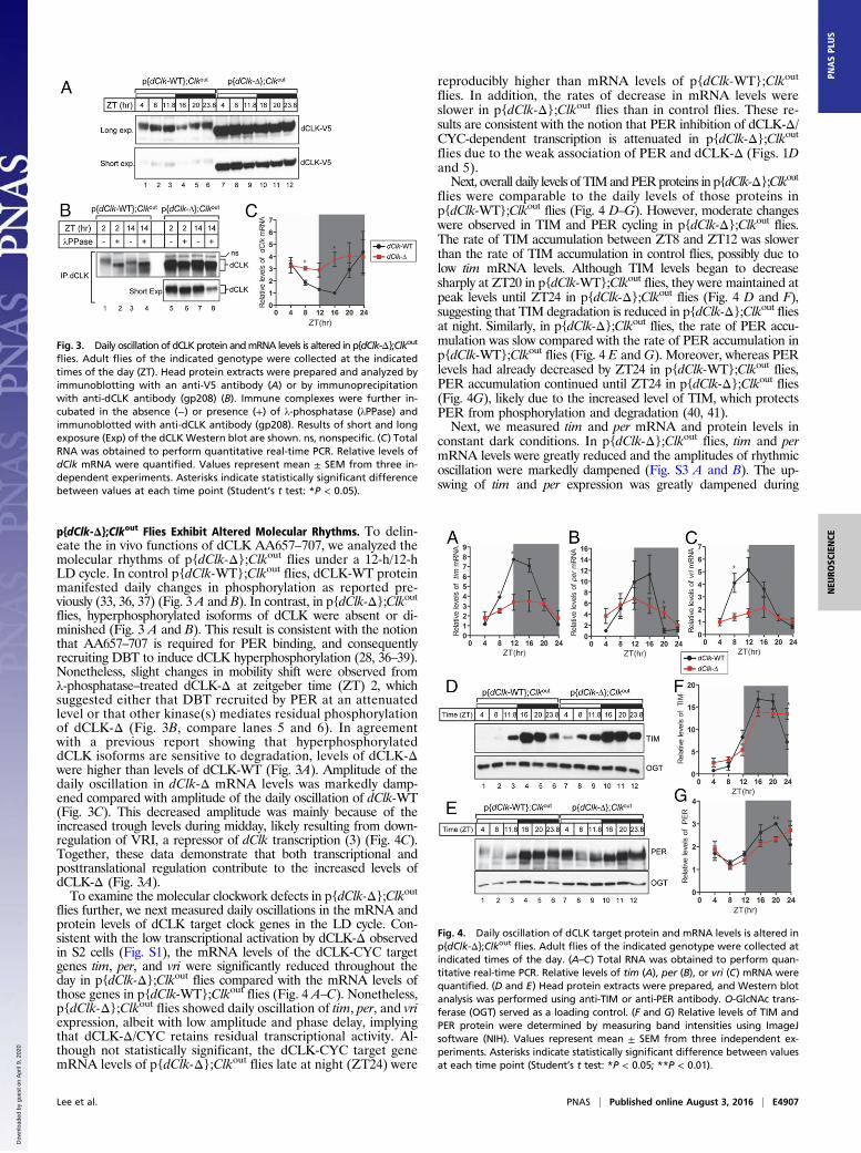
p{dClk-Δ};Clkout Flies Exhibit Altered Molecular Rhythms. To delin-eate the in vivo functions of dCLK AA657–707, we analyzed themolecular rhythms of p{dClk-Δ};Clkout flies under a 12-h/12-hLD cycle. In control p{dClk-WT};Clkout flies, dCLK-WT proteinmanifested daily changes in phosphorylation as reported pre-viously (33, 36, 37) (Fig. 3 A and B). In contrast, in p{dClk-Δ};Clkoutflies, hyperphosphorylated isoforms of dCLK were absent or di-minished (Fig. 3 A and B). This result is consistent with the notionthat AA657–707 is required for PER binding, and consequentlyrecruiting DBT to induce dCLK hyperphosphorylation (28, 36–39).Nonetheless, slight changes in mobility shift were observed fromλ-phosphatase–treated dCLK-Δ at zeitgeber time (ZT) 2, whichsuggested either that DBT recruited by PER at an attenuatedlevel or that other kinase(s) mediates residual phosphorylationof dCLK-Δ (Fig. 3B, compare lanes 5 and 6). In agreementwith a previous report showing that hyperphosphorylateddCLK isoforms are sensitive to degradation, levels of dCLK-Δwere higher than levels of dCLK-WT (Fig. 3A). Amplitude of thedaily oscillation in dClk-Δ mRNA levels was markedly damp-ened compared with amplitude of the daily oscillation of dClk-WT(Fig. 3C). This decreased amplitude was mainly because of theincreased trough levels during midday, likely resulting from down-regulation of VRI, a repressor of dClk transcription (3) (Fig. 4C).Together, these data demonstrate that both transcriptional andposttranslational regulation contribute to the increased levels ofdCLK-Δ (Fig. 3A).To examine the molecular clockwork defects in p{dClk-Δ};Clkout
flies further, we next measured daily oscillations in the mRNA andprotein levels of dCLK target clock genes in the LD cycle. Con-sistent with the low transcriptional activation by dCLK-Δ observedin S2 cells (Fig. S1), the mRNA levels of the dCLK-CYC targetgenes tim, per, and vri were significantly reduced throughout theday in p{dClk-Δ};Clkout flies compared with the mRNA levels ofthose genes in p{dClk-WT};Clkout flies (Fig. 4 A–C). Nonetheless,p{dClk-Δ};Clkout flies showed daily oscillation of tim, per, and vriexpression, albeit with low amplitude and phase delay, implyingthat dCLK-Δ/CYC retains residual transcriptional activity. Al-though not statistically significant, the dCLK-CYC target genemRNA levels of p{dClk-Δ};Clkout flies late at night (ZT24) were
reproducibly higher than mRNA levels of p{dClk-WT};Clkout
flies. In addition, the rates of decrease in mRNA levels wereslower in p{dClk-Δ};Clkout flies than in control flies. These re-sults are consistent with the notion that PER inhibition of dCLK-Δ/CYC-dependent transcription is attenuated in p{dClk-Δ};Clkoutflies due to the weak association of PER and dCLK-Δ (Figs. 1Dand 5).Next, overall daily levels of TIMand PERproteins in p{dClk-Δ};Clkout
flies were comparable to the daily levels of those proteins inp{dClk-WT};Clkout flies (Fig. 4 D–G). However, moderate changeswere observed in TIM and PER cycling in p{dClk-Δ};Clkout flies.The rate of TIM accumulation between ZT8 and ZT12 was slowerthan the rate of TIM accumulation in control flies, possibly due tolow tim mRNA levels. Although TIM levels began to decreasesharply at ZT20 in p{dClk-WT};Clkout flies, they were maintained atpeak levels until ZT24 in p{dClk-Δ};Clkout flies (Fig. 4 D and F),suggesting that TIM degradation is reduced in p{dClk-Δ};Clkout fliesat night. Similarly, in p{dClk-Δ};Clkout flies, the rate of PER accu-mulation was slow compared with the rate of PER accumulation inp{dClk-WT};Clkout flies (Fig. 4 E andG). Moreover, whereas PERlevels had already decreased by ZT24 in p{dClk-WT};Clkout flies,PER accumulation continued until ZT24 in p{dClk-Δ};Clkout flies(Fig. 4G), likely due to the increased level of TIM, which protectsPER from phosphorylation and degradation (40, 41).Next, we measured tim and per mRNA and protein levels in
constant dark conditions. In p{dClk-Δ};Clkout flies, tim and permRNA levels were greatly reduced and the amplitudes of rhythmicoscillation were markedly dampened (Fig. S3 A and B). The up-swing of tim and per expression was greatly dampened during
Fig. 3. Daily oscillation of dCLKprotein andmRNA levels is altered in p{dClk-Δ};Clkout
flies. Adult flies of the indicated genotype were collected at the indicatedtimes of the day (ZT). Head protein extracts were prepared and analyzed byimmunoblotting with an anti-V5 antibody (A) or by immunoprecipitationwith anti-dCLK antibody (gp208) (B). Immune complexes were further in-cubated in the absence (−) or presence (+) of λ-phosphatase (λPPase) andimmunoblotted with anti-dCLK antibody (gp208). Results of short and longexposure (Exp) of the dCLKWestern blot are shown. ns, nonspecific. (C) TotalRNA was obtained to perform quantitative real-time PCR. Relative levels ofdClk mRNA were quantified. Values represent mean ± SEM from three in-dependent experiments. Asterisks indicate statistically significant differencebetween values at each time point (Student’s t test: *P < 0.05).
Fig. 4. Daily oscillation of dCLK target protein and mRNA levels is altered inp{dClk-Δ};Clkout flies. Adult flies of the indicated genotype were collected atindicated times of the day. (A–C) Total RNA was obtained to perform quan-titative real-time PCR. Relative levels of tim (A), per (B), or vri (C) mRNA werequantified. (D and E) Head protein extracts were prepared, and Western blotanalysis was performed using anti-TIM or anti-PER antibody. O-GlcNAc trans-ferase (OGT) served as a loading control. (F and G) Relative levels of TIM andPER protein were determined by measuring band intensities using ImageJsoftware (NIH). Values represent mean ± SEM from three independent ex-periments. Asterisks indicate statistically significant difference between valuesat each time point (Student’s t test: *P < 0.05; **P < 0.01).
Lee et al. PNAS | Published online August 3, 2016 | E4907
NEU
ROSC
IENCE
PNASPL
US
Dow
nloa
ded
by g
uest
on
Apr
il 9,
202
0
the subjective day, presumably due to the persistent repression ofdCLK-Δ/CYC by elevated levels of PER until circadian time (CT)8 (discussed below). In p{dClk-Δ};Clkout flies, TIM levels weresignificantly higher than in p{dClk-WT};Clkout flies during thesubjective early morning (CT4 and CT8) and slowly reachedthe peak at CT20, although at a much reduced level comparedwith p{dClk-WT};Clkout flies (Fig. S3 C and D). Similarly, PERlevels were markedly higher with concomitant increases in hyper-phosphorylated isoforms during the subjective early morning (CT4and CT8), probably due to the high levels of TIM protecting PERfrom phosphorylation and degradation in p{dClk-Δ};Clkout flies(Fig. S3 C and E). Taken together, these results suggest that quasi-normal oscillations of PER and TIM proteins in fly heads in the LDcycle are mainly driven by light-mediated degradation of TIM,
which ultimately affects PER stability (42–45). Accordingly, mo-lecular oscillations of per/tim mRNAs and proteins were rapidlydampened in the absence of light, leading to behavioral arrhyth-micity in constant dark conditions.
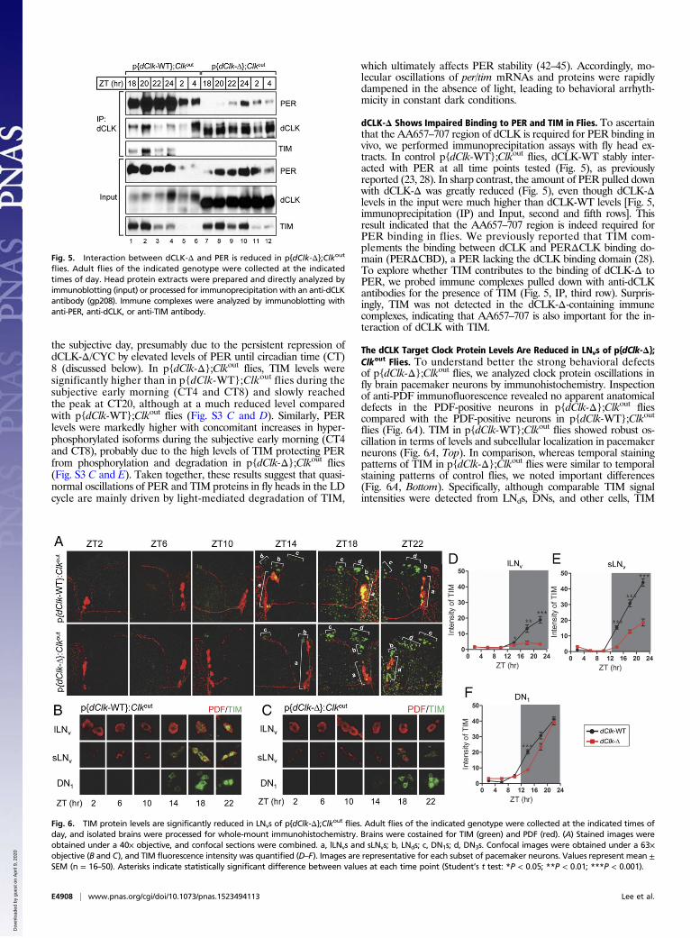
dCLK-Δ Shows Impaired Binding to PER and TIM in Flies. To ascertainthat the AA657–707 region of dCLK is required for PER binding invivo, we performed immunoprecipitation assays with fly head ex-tracts. In control p{dClk-WT};Clkout flies, dCLK-WT stably inter-acted with PER at all time points tested (Fig. 5), as previouslyreported (23, 28). In sharp contrast, the amount of PER pulled downwith dCLK-Δ was greatly reduced (Fig. 5), even though dCLK-Δlevels in the input were much higher than dCLK-WT levels [Fig. 5,immunoprecipitation (IP) and Input, second and fifth rows]. Thisresult indicated that the AA657–707 region is indeed required forPER binding in flies. We previously reported that TIM com-plements the binding between dCLK and PERΔCLK binding do-main (PERΔCBD), a PER lacking the dCLK binding domain (28).To explore whether TIM contributes to the binding of dCLK-Δ toPER, we probed immune complexes pulled down with anti-dCLKantibodies for the presence of TIM (Fig. 5, IP, third row). Surpris-ingly, TIM was not detected in the dCLK-Δ-containing immunecomplexes, indicating that AA657–707 is also important for the in-teraction of dCLK with TIM.
The dCLK Target Clock Protein Levels Are Reduced in LNvs of p{dClk-Δ};Clkout Flies. To understand better the strong behavioral defectsof p{dClk-Δ};Clkout flies, we analyzed clock protein oscillations infly brain pacemaker neurons by immunohistochemistry. Inspectionof anti-PDF immunofluorescence revealed no apparent anatomicaldefects in the PDF-positive neurons in p{dClk-Δ};Clkout fliescompared with the PDF-positive neurons in p{dClk-WT};Clkoutflies (Fig. 6A). TIM in p{dClk-WT};Clkout flies showed robust os-cillation in terms of levels and subcellular localization in pacemakerneurons (Fig. 6A, Top). In comparison, whereas temporal stainingpatterns of TIM in p{dClk-Δ};Clkout flies were similar to temporalstaining patterns of control flies, we noted important differences(Fig. 6A, Bottom). Specifically, although comparable TIM signalintensities were detected from LNds, DNs, and other cells, TIM
Fig. 5. Interaction between dCLK-Δ and PER is reduced in p{dClk-Δ};Clkout
flies. Adult flies of the indicated genotype were collected at the indicatedtimes of day. Head protein extracts were prepared and directly analyzed byimmunoblotting (input) or processed for immunoprecipitation with an anti-dCLKantibody (gp208). Immune complexes were analyzed by immunoblotting withanti-PER, anti-dCLK, or anti-TIM antibody.
Fig. 6. TIM protein levels are significantly reduced in LNvs of p{dClk-Δ};Clkout flies. Adult flies of the indicated genotype were collected at the indicated times ofday, and isolated brains were processed for whole-mount immunohistochemistry. Brains were costained for TIM (green) and PDF (red). (A) Stained images wereobtained under a 40× objective, and confocal sections were combined. a, lLNvs and sLNvs; b, LNds; c, DN1s; d, DN3s. Confocal images were obtained under a 63×objective (B and C), and TIM fluorescence intensity was quantified (D–F). Images are representative for each subset of pacemaker neurons. Values represent mean ±SEM (n = 16–50). Asterisks indicate statistically significant difference between values at each time point (Student’s t test: *P < 0.05; **P < 0.01; ***P < 0.001).
E4908 | www.pnas.org/cgi/doi/10.1073/pnas.1523494113 Lee et al.
Dow
nloa
ded
by g
uest
on
Apr
il 9,
202
0
signal was greatly reduced in large LNvs (lLNvs) and small LNvs(sLNvs) in p{dClk-Δ};Clkout flies (Fig. 6A). To verify that TIMlevels were differentially affected in subsets of pacemaker neuronsin p{dClk-Δ};Clkout flies, we examined TIM staining at a highermagnification in sLNvs, lLNvs, and DN1s at 4-h intervals beginningat ZT2 and quantified TIM signal intensity (Fig. 6 B–F). Asexpected, TIM levels were greatly reduced in LNvs, with more se-vere effects from lLNvs than sLNvs at all time points throughout theday (Fig. 6 D and E). In contrast, TIM signal intensity in DN1s wascomparable between p{dClk-WT};Clkout flies and p{dClk-Δ};Clkoutflies, albeit with slightly attenuated TIM accumulation (Fig.6F). To determine whether the reduced TIM staining is due todelayed accumulation of TIM, we further analyzed TIM stain-ing intensity at ZT24 and obtained similar results showing re-duced TIM intensity in LNvs, but not in LNds or DN1s, of p{dClk-Δ};Clkout flies (Fig. S4 A and B). Finally, to exclude thepossibility that these striking pacemaker-neuron–dependentdifferences in TIM staining intensity were due to differences ingenetic background, we next examined TIM staining in theother p{dClk-Δ};Clkout transgenic fly line (2M) (Fig. S5). Bothlines of p{dClk-Δ};Clkout flies exhibited similarly reduced TIMstaining in LNvs, but not in DN1s, compared with p{dClk-WT};Clkout
flies, indicating that genetic background is not the cause of theobserved phenotype.Next, to examine whether these pacemaker-neuron–dependent
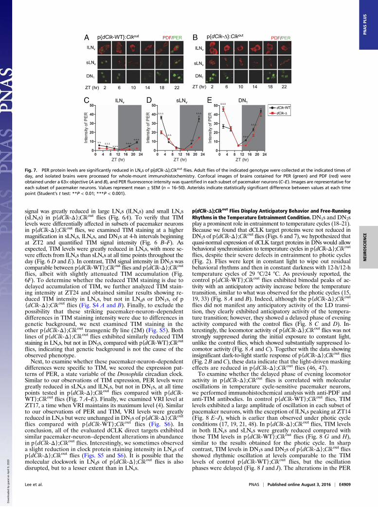
differences were specific to TIM, we scored the expression pat-terns of PER, a state variable of the Drosophila circadian clock.Similar to our observations of TIM expression, PER levels weregreatly reduced in sLNvs and lLNvs, but not in DN1s, at all timepoints tested in p{dClk-Δ};Clkout flies compared with p{dClk-WT};Clkout flies (Fig. 7 A–E). Finally, we examined VRI level atZT17, a time when VRI maintains its maximum level (4). Similarto our observations of PER and TIM, VRI levels were greatlyreduced in LNvs but were unchanged in DN1s of p{dClk-Δ};Clkoutflies compared with p{dClk-WT};Clkout flies (Fig. S6). Inconclusion, all of the evaluated dCLK direct targets exhibitedsimilar pacemaker-neuron–dependent alterations in abundancein p{dClk-Δ};Clkout flies. Interestingly, we sometimes observeda slight reduction in clock protein staining intensity in LNds ofp{dClk-Δ};Clkout flies (Figs. S5 and S6). It is possible that themolecular clockwork in LNds of p{dClk-Δ};Clkout flies is alsodisrupted, but to a lesser extent than in LNvs.
p{dClk-Δ};Clkout Flies Display Anticipatory Behavior and Free-RunningRhythms in the Temperature Entrainment Condition.DN1s and DN2splay a prominent role in entrainment to temperature cycles (18–21).Because we found that dCLK target proteins were not reduced inDN1s of p{dClk-Δ};Clkout flies (Figs. 6 and 7), we hypothesized thatquasi-normal expression of dCLK target proteins in DNs would allowbehavioral synchronization to temperature cycles in p{dClk-Δ};Clkoutflies, despite their severe defects in entrainment to photic cycles(Fig. 2). Flies were kept in constant light to wipe out residualbehavioral rhythms and then in constant darkness with 12-h/12-htemperature cycles of 29 °C/24 °C. As previously reported, thecontrol p{dClk-WT};Clkout flies exhibited bimodal peaks of ac-tivity with an anticipatory activity increase before the temperaturetransition, similar to what was observed for the photic cycles (15,19, 33) (Fig. 8 A and B). Indeed, although the p{dClk-Δ};Clkoutflies did not manifest any anticipatory activity of the LD transi-tion, they clearly exhibited anticipatory activity of the tempera-ture transition; however, they showed a delayed phase of eveningactivity compared with the control flies (Fig. 8 C and D). In-terestingly, the locomotor activity of p{dClk-Δ};Clkout flies was notstrongly suppressed during the initial exposure to constant light,unlike the control flies, which showed substantially suppressed lo-comotor activity (Fig. 8 A and C). Together with the data showinginsignificant dark-to-light startle response of p{dClk-Δ};Clkout flies(Fig. 2 B andC), these data indicate that the light-driven maskingeffects are reduced in p{dClk-Δ};Clkout flies (46, 47).To examine whether the delayed phase of evening locomotor
activity in p{dClk-Δ};Clkout flies is correlated with molecularoscillations in temperature cycle-sensitive pacemaker neurons,we performed immunohistochemical analysis with anti-PDF andanti-TIM antibodies. In control p{dClk-WT};Clkout flies, TIMlevels exhibited a large amplitude of oscillation in each subset ofpacemaker neurons, with the exception of lLNvs peaking at ZT14(Fig. 8 E–J), which is earlier than observed under photic cycleconditions (17, 19, 21, 48). In p{dClk-Δ};Clkout flies, TIM levelsin both lLNvs and sLNvs were greatly reduced compared withthose TIM levels in p{dClk-WT};Clkout flies (Fig. 8 G and H),similar to the results obtained for the photic cycle. In sharpcontrast, TIM levels in DN1s and DN2s of p{dClk-Δ};Clkout fliesshowed rhythmic oscillation at levels comparable to the TIMlevels of control p{dClk-WT};Clkout flies, but the oscillationphases were delayed (Fig. 8 I and J). The alterations in the PER
Fig. 7. PER protein levels are significantly reduced in LNvs of p{dClk-Δ};Clkout flies. Adult flies of the indicated genotype were collected at the indicated times ofday, and isolated brains were processed for whole-mount immunohistochemistry. Confocal images of brains costained for PER (green) and PDF (red) wereobtained under a 63× objective (A and B), and PER fluorescence intensity was quantified in each subset of pacemaker neurons (C–E). Images are representative foreach subset of pacemaker neurons. Values represent mean ± SEM (n = 16–50). Asterisks indicate statistically significant difference between values at each timepoint (Student’s t test: **P < 0.01; ***P < 0.001).
Lee et al. PNAS | Published online August 3, 2016 | E4909
NEU
ROSC
IENCE
PNASPL
US
Dow
nloa
ded
by g
uest
on
Apr
il 9,
202
0
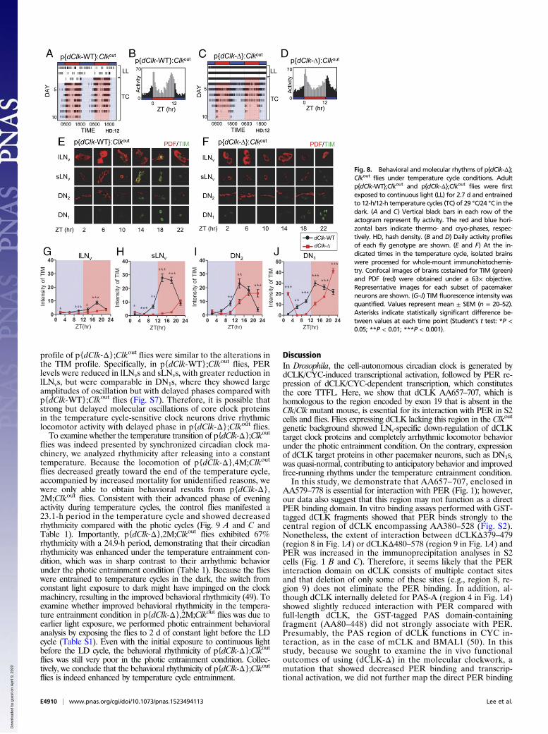
profile of p{dClk-Δ};Clkout flies were similar to the alterations inthe TIM profile. Specifically, in p{dClk-WT};Clkout flies, PERlevels were reduced in lLNvs and sLNvs, with greater reduction inlLNvs, but were comparable in DN1s, where they showed largeamplitudes of oscillation but with delayed phases compared withp{dClk-WT};Clkout flies (Fig. S7). Therefore, it is possible thatstrong but delayed molecular oscillations of core clock proteinsin the temperature cycle-sensitive clock neurons drive rhythmiclocomotor activity with delayed phase in p{dClk-Δ};Clkout flies.To examine whether the temperature transition of p{dClk-Δ};Clkout
flies was indeed presented by synchronized circadian clock ma-chinery, we analyzed rhythmicity after releasing into a constanttemperature. Because the locomotion of p{dClk-Δ},4M;Clkout
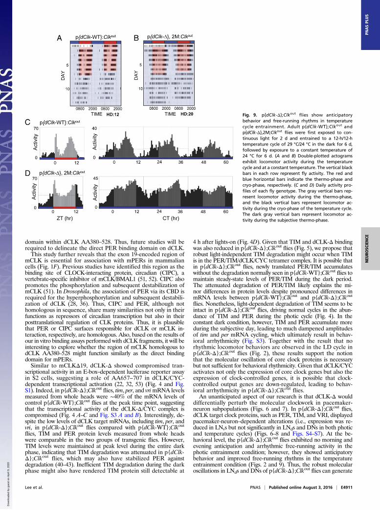
flies decreased greatly toward the end of the temperature cycle,accompanied by increased mortality for unidentified reasons, wewere only able to obtain behavioral results from p{dClk-Δ},2M;Clkout flies. Consistent with their advanced phase of eveningactivity during temperature cycles, the control flies manifested a23.1-h period in the temperature cycle and showed decreasedrhythmicity compared with the photic cycles (Fig. 9 A and C andTable 1). Importantly, p{dClk-Δ},2M;Clkout flies exhibited 67%rhythmicity with a 24.9-h period, demonstrating that their circadianrhythmicity was enhanced under the temperature entrainment con-dition, which was in sharp contrast to their arrhythmic behaviorunder the photic entrainment condition (Table 1). Because the flieswere entrained to temperature cycles in the dark, the switch fromconstant light exposure to dark might have impinged on the clockmachinery, resulting in the improved behavioral rhythmicity (49). Toexamine whether improved behavioral rhythmicity in the tempera-ture entrainment condition in p{dClk-Δ},2M;Clkout flies was due toearlier light exposure, we performed photic entrainment behavioralanalysis by exposing the flies to 2 d of constant light before the LDcycle (Table S1). Even with the initial exposure to continuous lightbefore the LD cycle, the behavioral rhythmicity of p{dClk-Δ};Clkoutflies was still very poor in the photic entrainment condition. Collec-tively, we conclude that the behavioral rhythmicity of p{dClk-Δ};Clkoutflies is indeed enhanced by temperature cycle entrainment.
DiscussionIn Drosophila, the cell-autonomous circadian clock is generated bydCLK/CYC-induced transcriptional activation, followed by PER re-pression of dCLK/CYC-dependent transcription, which constitutesthe core TTFL. Here, we show that dCLK AA657–707, which ishomologous to the region encoded by exon 19 that is absent in theClk/Clk mutant mouse, is essential for its interaction with PER in S2cells and flies. Flies expressing dCLK lacking this region in the Clkoutgenetic background showed LNv-specific down-regulation of dCLKtarget clock proteins and completely arrhythmic locomotor behaviorunder the photic entrainment condition. On the contrary, expressionof dCLK target proteins in other pacemaker neurons, such as DN1s,was quasi-normal, contributing to anticipatory behavior and improvedfree-running rhythms under the temperature entrainment condition.In this study, we demonstrate that AA657–707, enclosed in
AA579–778 is essential for interaction with PER (Fig. 1); however,our data also suggest that this region may not function as a directPER binding domain. In vitro binding assays performed with GST-tagged dCLK fragments showed that PER binds strongly to thecentral region of dCLK encompassing AA380–528 (Fig. S2).Nonetheless, the extent of interaction between dCLKΔ379–479(region 8 in Fig. 1A) or dCLKΔ480–578 (region 9 in Fig. 1A) andPER was increased in the immunoprecipitation analyses in S2cells (Fig. 1 B and C). Therefore, it seems likely that the PERinteraction domain on dCLK consists of multiple contact sitesand that deletion of only some of these sites (e.g., region 8, re-gion 9) does not eliminate the PER binding. In addition, al-though dCLK internally deleted for PAS-A (region 4 in Fig. 1A)showed slightly reduced interaction with PER compared withfull-length dCLK, the GST-tagged PAS domain-containingfragment (AA80–448) did not strongly associate with PER.Presumably, the PAS region of dCLK functions in CYC in-teraction, as in the case of mCLK and BMAL1 (50). In thisstudy, because we sought to examine the in vivo functionaloutcomes of using (dCLK-Δ) in the molecular clockwork, amutation that showed decreased PER binding and transcrip-tional activation, we did not further map the direct PER binding
Fig. 8. Behavioral and molecular rhythms of p{dClk-Δ};Clkout flies under temperature cycle conditions. Adultp{dClk-WT};Clkout and p{dClk-Δ};Clkout flies were firstexposed to continuous light (LL) for 2.7 d and entrainedto 12-h/12-h temperature cycles (TC) of 29 °C/24 °C in thedark. (A and C) Vertical black bars in each row of theactogram represent fly activity. The red and blue hori-zontal bars indicate thermo- and cryo-phases, respec-tively. HD, hash density. (B and D) Daily activity profilesof each fly genotype are shown. (E and F) At the in-dicated times in the temperature cycle, isolated brainswere processed for whole-mount immunohistochemis-try. Confocal images of brains costained for TIM (green)and PDF (red) were obtained under a 63× objective.Representative images for each subset of pacemakerneurons are shown. (G–J) TIM fluorescence intensity wasquantified. Values represent mean ± SEM (n = 20–52).Asterisks indicate statistically significant difference be-tween values at each time point (Student’s t test: *P <0.05; **P < 0.01; ***P < 0.001).
E4910 | www.pnas.org/cgi/doi/10.1073/pnas.1523494113 Lee et al.
Dow
nloa
ded
by g
uest
on
Apr
il 9,
202
0
domain within dCLK AA380–528. Thus, future studies will berequired to delineate the direct PER binding domain on dCLK.This study further reveals that the exon 19-encoded region of
mCLK is essential for association with mPERs in mammaliancells (Fig. 1F). Previous studies have identified this region as thebinding site of CLOCK-interacting protein, circadian (CIPC), avertebrate-specific inhibitor of mCLK/BMAL1 (51, 52). CIPC alsopromotes the phosphorylation and subsequent destabilization ofmCLK (51). In Drosophila, the association of PER via its CBD isrequired for the hyperphosphorylation and subsequent destabili-zation of dCLK (28, 36). Thus, CIPC and PER, although nothomologous in sequence, share many similarities not only in theirfunctions as repressors of circadian transcription but also in theirposttranslational regulation of CLK proteins. Thus, it is plausiblethat PER or CIPC surfaces responsible for dCLK or mCLK in-teraction, respectively, are homologous. Also, based on the results ofour in vitro binding assays performed with dCLK fragments, it will beinteresting to explore whether the region of mCLK homologous todCLK AA380–528 might function similarly as the direct bindingdomain for mPERs.Similar to mCLKΔ19, dCLK-Δ showed compromised tran-
scriptional activity in an E-box–dependent luciferase reporter assayin S2 cells, suggesting a role of AA657–707 in dCLK/CYC-dependent transcriptional activation (22, 32, 53) (Fig. 4 and Fig.S1). Indeed, in p{dClk-Δ};Clkout flies, tim, per, and vrimRNA levelsmeasured from whole heads were ∼40% of the mRNA levels ofcontrol p{dClk-WT};Clkout flies at the peak time point, suggestingthat the transcriptional activity of the dCLK-Δ/CYC complex iscompromised (Fig. 4 A–C and Fig. S3 A and B). Interestingly, de-spite the low levels of dCLK target mRNAs, including tim, per, andvri, in p{dClk-Δ};Clkout flies compared with p{dClk-WT};Clkoutflies, TIM and PER protein levels measured from whole headswere comparable in the two groups of transgenic flies. However,TIM levels were maintained at peak level during the entire darkphase, indicating that TIM degradation was attenuated in p{dClk-Δ};Clkout flies, which may also have stabilized PER againstdegradation (40–43). Inefficient TIM degradation during the darkphase might also have rendered TIM protein still detectable at
4 h after lights-on (Fig. 4D). Given that TIM and dCLK-Δ bindingwas also reduced in p{dClk-Δ};Clkout flies (Fig. 5), we propose thatrobust light-independent TIM degradation might occur when TIMis in the PER/TIM/dCLK/CYC tetramer complex. It is possible thatin p{dClk-Δ};Clkout flies, newly translated PER/TIM accumulateswithout the degradation normally seen in p{dClk-WT};Clkout flies tomaintain steady-state levels of PER/TIM during the dark period.The attenuated degradation of PER/TIM likely explains the mi-nor differences in protein levels despite pronounced differences inmRNA levels between p{dClk-WT};Clkout and p{dClk-Δ};Clkoutflies. Nonetheless, light-dependent degradation of TIM seems to beintact in p{dClk-Δ};Clkout flies, driving normal cycles in the abun-dance of TIM and PER during the photic cycle (Fig. 4). In theconstant dark condition, however, TIM and PER accumulate moreduring the subjective day, leading to much dampened amplitudesof tim and per mRNA cycling, which ultimately result in behav-ioral arrhythmicity (Fig. S3). Together with the result that norhythmic locomotor behaviors are observed in the LD cycle inp{dClk-Δ};Clkout flies (Fig. 2), these results support the notionthat the molecular oscillation of core clock proteins is necessarybut not sufficient for behavioral rhythmicity. Given that dCLK/CYCactivates not only the expression of core clock genes but also theexpression of clock-controlled genes, it is possible that clock-controlled output genes are down-regulated, leading to behav-ioral arrhythmicity in p{dClk-Δ};Clkout flies.An unanticipated aspect of our research is that dCLK-Δ would
differentially perturb the molecular clockwork in pacemaker-neuron subpopulations (Figs. 6 and 7). In p{dClk-Δ};Clkout flies,dCLK target clock proteins, such as PER, TIM, and VRI, displayedpacemaker-neuron–dependent alterations (i.e., expression was re-duced in LNvs but not significantly in LNds and DNs in both photicand temperature cycles) (Figs. 6–8 and Figs. S4–S7). At the be-havioral level, the p{dClk-Δ};Clkout flies exhibited no morning andevening anticipation and arrhythmic free-running activity in thephotic entrainment condition; however, they showed anticipatorybehavior and improved free-running rhythms in the temperatureentrainment condition (Figs. 2 and 9). Thus, the robust molecularoscillations in LNds and DNs of p{dClk-Δ};Clkout flies can generate
Fig. 9. p{dClk-Δ};Clkout flies show anticipatorybehavior and free-running rhythms in temperaturecycle entrainment. Adult p{dClk-WT};Clkout andp{dClk-Δ},2M;Clkout flies were first exposed to con-tinuous light for 2 d and entrained to a 12-h/12-htemperature cycle of 29 °C/24 °C in the dark for 6 d,followed by exposure to a constant temperature of24 °C for 6 d. (A and B) Double-plotted actogramsexhibit locomotor activity during the temperaturecycle and at a constant temperature. The vertical blackbars in each row represent fly activity. The red andblue horizontal bars indicate the thermo-phase andcryo-phase, respectively. (C and D) Daily activity pro-files of each fly genotype. The gray vertical bars rep-resent locomotor activity during the thermo-phase,and the black vertical bars represent locomotor ac-tivity during the cryo-phase of the temperature cycle.The dark gray vertical bars represent locomotor ac-tivity during the subjective thermo-phase.
Lee et al. PNAS | Published online August 3, 2016 | E4911
NEU
ROSC
IENCE
PNASPL
US
Dow
nloa
ded
by g
uest
on
Apr
il 9,
202
0
behavioral rhythmicity in temperature entrainment conditions, butnot in photic entrainment conditions. Importantly, previous reportsdemonstrate that DN1-generated evening activity is suppressed byhigh-intensity light (20, 54) and enhanced by warm temperature(20). Therefore, it might be that DN-generated rhythmic outputsare suppressed in p{dClk-Δ};Clkout flies, resulting in arrhythmicbehaviors under the standard (high-intensity light) photic entrain-ment condition used in this study. It is also important to note thatseveral lines of evidence support the idea that DNs play importantroles to control circadian behaviors in temperature entrainmentconditions (18–21). Consistently, in p{dClk-Δ};Clkout flies, rela-tively intact molecular clockwork in DNs deduced from strong ex-pression of dCLK target genes entrains to the temperature cycle,but with phase delay, and generates the delayed anticipatory be-havior of temperature transition, ultimately contributing to im-proved free-running rhythms compared with the rhythms underphotic cycle entrainment conditions.Given that dCLK-Δ showed reduced transactivation of dCLK
target genes, we investigated whether reduction in transcriptionalactivity such as seen in p{dClk-Δ};Clkout flies might have lessereffects on locomotor behavior in temperature cycles than in photiccycles. Flies harboring Clkar, a hypomorphic allele of dClk, showcompromised dCLK activity and levels leading to behavioralarrhythmicity under photic conditions, albeit with persistent butreduced amplitudes in PER and TIM molecular rhythms (55). Thus,we compared circadian locomotor behaviors of Clkar in the photicand temperature entrainment conditions. Unlike p{dClk-Δ};Clkoutflies, Clkar flies displayed no anticipatory activity of temperaturetransition and showed arrhythmicity under both entrainmentconditions (Fig. S8). This observation suggests that the reductionin transcriptional activity of dCLK itself does not necessarilyimpose less of an effect on behavioral rhythms in temperaturecycles than in photic cycles. Nonetheless, the possibility remainsthat the extent of the reduction in transcriptional activity of Clkaris greater than the extent of the reduction in transcriptional ac-tivity of dCLK-Δ, leading to arrhythmic behavior under photicand temperature cycle conditions.In p{dClk-Δ};Clkout flies, the LNv-specific reduction in dCLK
target protein expression likely resulted from compromisedtranscription, because all of the evaluated dCLK target proteinsin separate loops of the TTFL (e.g., TIM, PER, VRI) displayedreduced expression in LNvs. What would lead to these pacemaker-neuron–dependent alterations in clock protein abundances inp{dClk-Δ};Clkout flies? Given that the AA657–707 region ofdCLK may play roles in interactions with multiple factors, it istempting to speculate that cell type-specific circadian transcrip-tional activation occurs via the engagement of cell type-specifictranscriptional coactivators and/or other transcription factorsthrough this domain. Interestingly, in tissue-specific circadian tran-scription programs, context-dependent transcription factors recruitthe dCLK/CYC complexes to actively transcribed genes by binding totarget enhancer elements (56). Our results further suggest that al-though dCLK/CYC plays a central role in circadian transcription, the
exact functional architecture of dCLK/CYC-containing transcrip-tional complexes is cell type-specific. Our current hypothesis isthat LNv-specific coactivators and/or transcription factors are re-quired for dCLK/CYC-dependent transcriptional activation inLNvs and that these associations are mediated through theAA657–707 domain of dCLK. In contrast, factors associated viadCLK AA657–707 might not be required for circadian transcrip-tion in LNds and DNs, or the association between factors anddCLK might involve other regions of dCLK in those neurons.Future studies will be required to address the mechanisms un-derlying cell-specific circadian transcription.Animals exhibit direct light-induced responses called masking.
In positive masking, dim light increases activity, and in negativemasking, bright light suppresses activity (46, 47). Our resultsshowed that p{dClk-Δ};Clkout flies exhibited reduced responses toboth masking effects, because the lights-on/off transition-inducedstartle response was very weak (or absent), and the suppression ofactivity induced by constant light exposure was insignificant (Figs. 2and 8). The role of dCLK in light-mediated responses has beenreported previously (35, 55, 57, 58). Given that dCLK-Δ exhibitedrobust defect in LNvs in our study, intact dCLK in light-sensitiveLNvs might be more relevant for masking responses. Interestingly,Clk/Clk mice show reduced masking in responses to light (59–61).Taken together, these data suggest that light-induced direct be-havioral responses in Drosophila and mice might use a similarunderlying mechanism.In summary, our results demonstrate that the AA657–707 region
of dCLK and the region encoded by exon 19 of mCLK are essential forPER interaction and circadian transactivation. The p{dClk-Δ};Clkoutflies manifested more pronounced defects in standard photiccycle-induced circadian behavior, which was accompanied bymore severe perturbations in molecular clockwork in LNvs thanin DNs. Based on these intriguing observations, we propose thatthe dCLK/CYC-controlled TTFL operates differently dependingon the cellular context, which likely endows pacemaker neuronswith distinct circadian attributes, such as varying sensitivity tolight- and temperature-entraining cues.
Materials and MethodsAll materials and methods used in this study are detailed in SI Materials andMethods. Materials include plasmids, fly strains, and antibodies. Methodsinclude generation of dClkΔ657–707-expressing transgenic flies, behavioralanalyses, quantitative real-time PCR, immunoblotting, immunofluorescenceanalysis, and in vitro binding assays.
ACKNOWLEDGMENTS. We thank Paul Hardin for sharing the anti-VRI anti-body. Work in the laboratory of E.Y.K. was supported by the National ResearchFoundation of Korea (NRF) (Grant NRF-2014R1A2A1A11051765) and the ChronicInflammatory Disease Research Center (Grant NRF-2012R1A5A048183), whichwas funded by the Korean government. E.C. was supported by the NRF (GrantNRF-2013R1A1A2060533). Work in the laboratory of Z.C. was supported by TheRobert A. Welch Foundation (Grant AU-1731) and the NIH/National Institute onAging (Grant R01 AG045828). Work in the laboratory of S.-H.Y. was supported bythe NIH/ National Institute of General Medical Sciences (Grant R01 GM114424).
1. Hardin PE (2011) Molecular genetic analysis of circadian timekeeping in Drosophila.Adv Genet 74:141–173.
2. Mohawk JA, Green CB, Takahashi JS (2012) Central and peripheral circadian clocks inmammals. Annu Rev Neurosci 35:445–462.
3. Cyran SA, et al. (2003) vrille, Pdp1, and dClock form a second feedback loop in theDrosophila circadian clock. Cell 112(3):329–341.
4. Glossop NR, et al. (2003) VRILLE feeds back to control circadian transcription of Clockin the Drosophila circadian oscillator. Neuron 37(2):249–261.
5. Partch CL, Green CB, Takahashi JS (2014) Molecular architecture of the mammaliancircadian clock. Trends Cell Biol 24(2):90–99.
6. Grima B, Chélot E, Xia R, Rouyer F (2004) Morning and evening peaks of activity relyon different clock neurons of the Drosophila brain. Nature 431(7010):869–873.
7. Ripperger JA, Schibler U (2006) Rhythmic CLOCK-BMAL1 binding to multiple E-boxmotifs drives circadian Dbp transcription and chromatin transitions. Nat Genet 38(3):369–374.
8. Stoleru D, Peng Y, Agosto J, Rosbash M (2004) Coupled oscillators control morningand evening locomotor behaviour of Drosophila. Nature 431(7010):862–868.
9. Chung BY, Kilman VL, Keath JR, Pitman JL, Allada R (2009) The GABA(A) receptor RDLacts in peptidergic PDF neurons to promote sleep in Drosophila. Curr Biol 19(5):386–390.
10. Shang Y, Griffith LC, Rosbash M (2008) Light-arousal and circadian photoreception
circuits intersect at the large PDF cells of the Drosophila brain. Proc Natl Acad Sci USA
105(50):19587–19594.11. Sheeba V, et al. (2008) Large ventral lateral neurons modulate arousal and sleep in
Drosophila. Curr Biol 18(20):1537–1545.12. Sheeba V, Fogle KJ, Holmes TC (2010) Persistence of morning anticipation behavior
and high amplitude morning startle response following functional loss of small
ventral lateral neurons in Drosophila. PLoS One 5(7):e11628.13. Rieger D, Shafer OT, Tomioka K, Helfrich-Förster C (2006) Functional analysis of cir-
cadian pacemaker neurons in Drosophila melanogaster. J Neurosci 26(9):2531–2543.14. Yoshii T, Rieger D, Helfrich-Förster C (2012) Two clocks in the brain: An update of the
morning and evening oscillator model in Drosophila. Prog Brain Res 199:59–82.15. Glaser FT, Stanewsky R (2005) Temperature synchronization of the Drosophila circa-
dian clock. Curr Biol 15(15):1352–1363.16. Pittendrigh CS (1954) On temperature independence in the CLOCK system controlling
emergence time in Drosophila. Proc Natl Acad Sci USA 40(10):1018–1029.17. Yoshii T, Sakamoto M, Tomioka K (2002) A temperature-dependent timing mecha-
nism is involved in the circadian system that drives locomotor rhythms in the fruit fly
Drosophila melanogaster. Zoolog Sci 19(8):841–850.
E4912 | www.pnas.org/cgi/doi/10.1073/pnas.1523494113 Lee et al.
Dow
nloa
ded
by g
uest
on
Apr
il 9,
202
0
18. Miyasako Y, Umezaki Y, Tomioka K (2007) Separate sets of cerebral clock neurons areresponsible for light and temperature entrainment of Drosophila circadian locomotorrhythms. J Biol Rhythms 22(2):115–126.
19. Busza A, Murad A, Emery P (2007) Interactions between circadian neurons controltemperature synchronization of Drosophila behavior. J Neurosci 27(40):10722–10733.
20. Zhang Y, Liu Y, Bilodeau-Wentworth D, Hardin PE, Emery P (2010) Light and tem-perature control the contribution of specific DN1 neurons to Drosophila circadianbehavior. Curr Biol 20(7):600–605.
21. Picot M, Klarsfeld A, Chélot E, Malpel S, Rouyer F (2009) A role for blind DN2 clockneurons in temperature entrainment of the Drosophila larval brain. J Neurosci 29(26):8312–8320.
22. Darlington TK, et al. (1998) Closing the circadian loop: CLOCK-induced transcriptionof its own inhibitors per and tim. Science 280(5369):1599–1603.
23. Lee C, Bae K, Edery I (1998) The Drosophila CLOCK protein undergoes daily rhythms inabundance, phosphorylation, and interactions with the PER-TIM complex. Neuron21(4):857–867.
24. Chen R, et al. (2009) Rhythmic PER abundance defines a critical nodal point fornegative feedback within the circadian clock mechanism. Mol Cell 36(3):417–430.
25. Kiyohara YB, et al. (2006) The BMAL1 C terminus regulates the circadian transcriptionfeedback loop. Proc Natl Acad Sci USA 103(26):10074–10079.
26. Menet JS, Abruzzi KC, Desrochers J, Rodriguez J, Rosbash M (2010) Dynamic PER re-pression mechanisms in the Drosophila circadian clock: From on-DNA to off-DNA.Genes Dev 24(4):358–367.
27. Sato TK, et al. (2006) Feedback repression is required for mammalian circadian clockfunction. Nat Genet 38(3):312–319.
28. Sun WC, et al. (2010) Two distinct modes of PERIOD recruitment onto dCLOCK reveala novel role for TIMELESS in circadian transcription. J Neurosci 30(43):14458–14469.
29. Chang DC, Reppert SM (2003) A novel C-terminal domain of drosophila PERIOD in-hibits dCLOCK:CYCLE-mediated transcription. Curr Biol 13(9):758–762.
30. King DP, et al. (1997) The mouse Clock mutation behaves as an antimorph and mapswithin the W19H deletion, distal of Kit. Genetics 146(3):1049–1060.
31. Chang DC, et al. (2003) Constructing a feedback loop with circadian clock moleculesfrom the silkmoth, Antheraea pernyi. J Biol Chem 278(40):38149–38158.
32. Shimomura K, et al. (2013) Usf1, a suppressor of the circadian Clock mutant, revealsthe nature of the DNA-binding of the CLOCK:BMAL1 complex in mice. eLife 2:e00426.
33. Lee E, et al. (2014) Phosphorylation of a central clock transcription factor is requiredfor thermal but not photic entrainment. PLoS Genet 10(8):e1004545.
34. Mahesh G, et al. (2014) Phosphorylation of the transcription activator CLOCK regu-lates progression through a ∼ 24-h feedback loop to influence the circadian period inDrosophila. J Biol Chem 289(28):19681–19693.
35. Allada R, White NE, So WV, Hall JC, Rosbash M (1998) A mutant Drosophila homologof mammalian Clock disrupts circadian rhythms and transcription of period andtimeless. Cell 93(5):791–804.
36. Kim EY, Edery I (2006) Balance between DBT/CKIepsilon kinase and protein phos-phatase activities regulate phosphorylation and stability of Drosophila CLOCK pro-tein. Proc Natl Acad Sci USA 103(16):6178–6183.
37. Yu W, Zheng H, Houl JH, Dauwalder B, Hardin PE (2006) PER-dependent rhythms inCLK phosphorylation and E-box binding regulate circadian transcription. Genes Dev20(6):723–733.
38. Kim EY, Ko HW, YuW, Hardin PE, Edery I (2007) A DOUBLETIME kinase binding domain onthe Drosophila PERIOD protein is essential for its hyperphosphorylation, transcriptionalrepression, and circadian clock function. Mol Cell Biol 27(13):5014–5028.
39. Yu W, Zheng H, Price JL, Hardin PE (2009) DOUBLETIME plays a noncatalytic role tomediate CLOCK phosphorylation and repress CLOCK-dependent transcription withinthe Drosophila circadian clock. Mol Cell Biol 29(6):1452–1458.
40. Ko HW, Jiang J, Edery I (2002) Role for Slimb in the degradation of Drosophila Periodprotein phosphorylated by Doubletime. Nature 420(6916):673–678.
41. Price JL, et al. (1998) Double-time is a novel Drosophila clock gene that regulatesPERIOD protein accumulation. Cell 94(1):83–95.
42. Price JL, Dembinska ME, YoungMW, Rosbash M (1995) Suppression of PERIOD proteinabundance and circadian cycling by the Drosophila clock mutation timeless. EMBO J14(16):4044–4049.
43. Suri V, Lanjuin A, Rosbash M (1999) TIMELESS-dependent positive and negativeautoregulation in the Drosophila circadian clock. EMBO J 18(3):675–686.
44. Myers MP, Wager-Smith K, Rothenfluh-Hilfiker A, Young MW (1996) Light-induceddegradation of TIMELESS and entrainment of the Drosophila circadian clock. Science271(5256):1736–1740.
45. Zeng H, Qian Z, Myers MP, Rosbash M (1996) A light-entrainment mechanism for theDrosophila circadian clock. Nature 380(6570):129–135.
46. Golombek DA, Rosenstein RE (2010) Physiology of circadian entrainment. Physiol Rev90(3):1063–1102.
47. Mrosovsky N (1999) Masking: History, definitions, and measurement. Chronobiol Int16(4):415–429.
48. Boothroyd CE, Wijnen H, Naef F, Saez L, Young MW (2007) Integration of light andtemperature in the regulation of circadian gene expression in Drosophila. PLoS Genet3(4):e54.
49. Saunders DS (2002) Insect Clocks (Elsevier, Amsterdam), 3rd Ed.50. Huang N, et al. (2012) Crystal structure of the heterodimeric CLOCK:BMAL1 tran-
scriptional activator complex. Science 337(6091):189–194.51. Yoshitane H, et al. (2009) Roles of CLOCK phosphorylation in suppression of E-box-
dependent transcription. Mol Cell Biol 29(13):3675–3686.52. Zhao WN, et al. (2007) CIPC is a mammalian circadian clock protein without in-
vertebrate homologues. Nat Cell Biol 9(3):268–275.53. Lowrey PL, Takahashi JS (2011) Genetics of circadian rhythms in mammalian model
organisms. Adv Genet 74:175–230.54. Zhang L, et al. (2010) DN1(p) circadian neurons coordinate acute light and PDF inputs
to produce robust daily behavior in Drosophila. Curr Biol 20(7):591–599.55. Allada R, Kadener S, Nandakumar N, Rosbash M (2003) A recessive mutant of Dro-
sophila Clock reveals a role in circadian rhythm amplitude. EMBO J 22(13):3367–3375.56. Meireles-Filho AC, Bardet AF, Yáñez-Cuna JO, Stampfel G, Stark A (2014) cis-regulatory
requirements for tissue-specific programs of the circadian clock. Curr Biol 24(1):1–10.57. Lu B, Liu W, Guo F, Guo A (2008) Circadian modulation of light-induced locomotion
responses in Drosophila melanogaster. Genes Brain Behav 7(7):730–739.58. Kim EY, et al. (2002) Drosophila CLOCK protein is under posttranscriptional control
and influences light-induced activity. Neuron 34(1):69–81.59. Redlin U, Hattar S, Mrosovsky N (2005) The circadian Clock mutant mouse: Impaired
masking response to light. J Comp Physiol A Neuroethol Sens Neural Behav Physiol191(1):51–59.
60. Spoelstra K, Oklejewicz M, Daan S (2002) Restoration of self-sustained circadianrhythmicity by the mutant clock allele in mice in constant illumination. J Biol Rhythms17(6):520–525.
61. Vitaterna MH, et al. (2006) The mouse Clock mutation reduces circadian pacemakeramplitude and enhances efficacy of resetting stimuli and phase-response curve am-plitude. Proc Natl Acad Sci USA 103(24):9327–9332.
62. Kume K, et al. (1999) mCRY1 and mCRY2 are essential components of the negativelimb of the circadian clock feedback loop. Cell 98(2):193–205.
63. Jeong K, et al. (2015) Dual attenuation of proteasomal and autophagic BMAL1degradation in Clock Δ19/+ mice contributes to improved glucose homeostasis. SciRep 5:12801.
64. Groth AC, Fish M, Nusse R, Calos MP (2004) Construction of transgenic Drosophila byusing the site-specific integrase from phage phiC31. Genetics 166(4):1775–1782.
65. Venken KJ, He Y, Hoskins RA, Bellen HJ (2006) P[acman]: A BAC transgenic platformfor targeted insertion of large DNA fragments in D. melanogaster. Science 314(5806):1747–1751.
66. Sidote D, Majercak J, Parikh V, Edery I (1998) Differential effects of light and heat onthe Drosophila circadian clock proteins PER and TIM. Mol Cell Biol 18(4):2004–2013.
Lee et al. PNAS | Published online August 3, 2016 | E4913
NEU
ROSC
IENCE
PNASPL
US
Dow
nloa
ded
by g
uest
on
Apr
il 9,
202
0


![Disturbance-1 Uvod.ppt [režim kompatibility] · Datování disturbančních událostí –paleobotanika, 14C, 210Pb, dendrometrie, letecké snímky Dendrochronologie a dendrogeomorfologie](https://img.pdfslide.tips/doc/110x75/5e44d55b6645c5138e33b361/disturbance-1-uvodppt-reim-kompatibility-datovn-disturbannch-udlost.jpg)