Embed Size (px)
Citation preview
Title
ACUTE AND CHRONIC DISTURBANCE OFCONSCIOUSNESS AFTER MESENCEPHALICDESTRUCTION BY MEANS OFELECTROCOAGULATION
Author(s) YAMASAKI, TAKEHIRO
Citation 日本外科宝函 (1958), 27(4): 843-860
Issue Date 1958-07-01
URL http://hdl.handle.net/2433/206668
Right
Type Departmental Bulletin Paper
Textversion publisher
Kyoto University
原 著
ACUTE AND CHRONIC DISTURBANCE OF CONSCIOUSNESS
AFTER MESENCEPHALIC DESTRUCTION BY MEANS OF
ELECTROCOAGULATION by
T AKEHIRO YAMASAKI
From the 1st Surgical Division, Kyoto University Medical School (Director : Prof. Dr. CarsATO ARAKI)
Received for publication Mar. 11, 1958
INTRODUCTION
843
Over the past fourteen years a series of experiments has been carried out in our laboratory with regard to experimental disturbances of consciousness under the name“coma puncture”. The results indicate that the central grey matter of the midbrain, particularly in the region extending from the oculomotor to the trochlear nucleus may be the most important region in the maintenance of consciousness. However, severe experimental disturbances of consciousness in the past have been
transient lasting only from a few minutes to 20 minutes in almost all cases. It is very difficult加 comparesuch short experimental disturbances of consciousness with coma in patients having severe head injuries in whom coma may last from a few hours to a few days or longer.
About ten years ago T AKETOMO caused a prolonged coma (lasting about 50 minutes) in a rabbit with electrocoagulation of the ponto-mesencephalic region using the old machine of Bovie. Thus in the present study, we have electrocoagu・lated the mesencephalic region in cats using the Bovie's machine in order to induce prolonged unconsciousness of half an hour or more duration. ¥Ve have studied the behavioral and EEG changes through the entire survival periods of十heexperimental animals in order to better understand prolonged coma. In addition we have studied the difference in the effect of destruction of the central grey matter of the midbrain, which we have felt to be essential for the maintenance of consciousness, and the destruction of the reticular formation of the midbrain, which ::¥IAGOUN has stated is the seat of the regulatir:ig mechanism of consciousness.
Definition of disturbance of consciousness is the most important problem in animal experi-ments of this type. In our laboratory we difine this回 follows:
I) Unresponsiveness Ist degree (semicoma Ist degree). In this state, the postural reflex is somewhat feeble. The searching-reflex to light and
sound and the reactive motion phenomenon induced by touch or pressure to the body surface are almost abolished. But the animal still reacts to painful and smell stimuli very well. (GIRNDT II-IV).
2) Unresponsiveness 2nd degree (semicoma 2nd degree). The postural reflex is completely abolished. The active movements and . the re日exesto
smell stimuli are also gone. The reflexes to visual, acoustic, and touch sense are of cour:oe absent. The reflex to painful stimuli against the body surface is attenuated but partially
844 日本外科宝函第27巻第4号
remains. The sneeze reflex by stimulating septum nasi, the reflex to painful stimuli of nasal tip and the vomiting reflex due to stimuli of pharyngeal mucosa remain. (GIRNDT IV-V).
3) Unresponsiveness 3rd degree (coma). The reflex due to stimuli of nasal tip and septum nasi and the vomiting reflex due to
stimuli of pharyngeal mucosa are absent. The corneal reflex, the pupilary reflex, and the pinna reflex are sometimes present. Patellar reflex remains. (Over GIRNDT V).
EXPERIMENT AL l¥IETHOD
A mature cat was fixed on a hammock in prone position and the four limbs
were allowed to hang down naturally. After installation of the stereotaxic instru-
ment (hang down type) was carried out under ether anesthesia, four silver ball electrodes for recording the EEG were inserted passing through the skull and fixed
to the level of dura. Location of the silver ball electrodes were as follows:“1”・the anterior part of the left hemisphere,“2”: the same as 1 on the right side,
“3”: posterior part of the left hemisphere,“4”: the same as 3 on the right side.
After the cat had awakened fully, electrocoagulation in various parts of the midbrain was carried out. The insertion of the electrode for electrocoagulation
W剖 done in two ways. In one the electrode was inserted vertically under the guidance of HoRsLEY-CLARKE’s stereotaxic instrument through the parietal hole
trephined. In the other the ele:::trode was inserted through an opening in the
occipito・atlantoidligament and then through the 4th ventricle along the aqueduct of the midbrain.
The destroying electrodes consisted of iron-chrome wires of 15cm in length
and 0.3mm in diameter, each of which was insulated except for 0.1・・3.0mmof the
tip by means of specific lacquer (h6R6) or glasstube cover and could be used as a bipolar or unipolar electrode. The destruction of the tissue by electrocoagulation
was made with an electric current of 90・100volts and 80-1001¥'IA or more and
during the period of 1 second-1 minute. In some experiments an electric resistance of 1.5・3kiloohms was used.
The degree of disturbance of consciousness due to these electrocoagulation was examined according to criteria described above and behavioral and EEG changes were studied throughout the survival time of the animal.
After the death of the experimental animals, the brain stems were fixed in alcohol absolutus, packed in celloidin, sectioned serially and the areas of destruction
were confirmed b~' histological examination using N1ssL stain, myelin sheath stain
and iron-carmine stain. Anatomical guidance was obtained from vVINKLER and PoTTER (1914), JASPER and AJOMONE-}lARSAN (1955).
RESULTS
Elcc:trocoagulation was performed on the central grey matter of the midbrain
in 21 cats, the reticular formation of the miclbrain in 7 cats, both of them in 5 cats,
lamina quadrigemina in 5 cats and other parts in 14 cats, that is in a total of 52 cats.
The experimental animals are divided into a coma group and a non-coma group.
ACUTE AND CHRONIC DISTURBANCE OF CONSCIOUSNESS 845
,
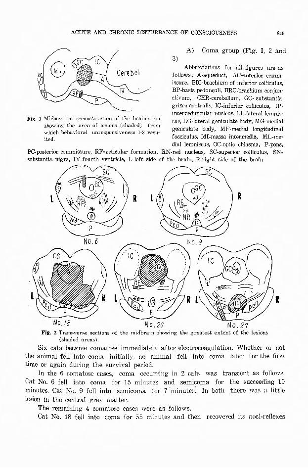
Fig. 1 Midsagittal reconstruction of the brain stem showing the area of lesions (shaded) from which behavioral unresponsiveness I・3resu-lted.
A) Coma group (Fig. I, 2 and
3)
Abbreviations for all figures are as
follows: A-aqueduct, AC-anterior comm-issure, BIC-brachium of inferior colliculus, BP-basis pedunculi, BRC-brachium conjun-ctivum, CER-cerebellum, GC-substantia grisea centralis, IC-inferior colliculus, IP-interpeduncular nucleus, LL-lateral lemnis-cu~. LG-lateral geniculate body, MG-medial geniculate body, MF-medial longitudinal fasciculus, MI-massa intermedia, ML-me-dial lemni:ocus, OC-optic chiasma, P-pons,
PC-posterior commissure, RF-reticular formation, RN・rednucleus, SC-superior colliculus, SN-substantia nigra, IV-fourth ventricle, L-left side of the brain, R-right side of the brain.
R R
No. 6
No.18 No. 20 No.2クFig. 2 Transverse sections of the midbrain showing the greatest extent of the lesions
(shaded areas).
I
Six cats became comatose immediatelY after electrocoagulation. Whether or not
the animal fell into coma initially, no animal fell into coma later for the first
time or again during the survival period.
In the 6 comatose cases, coma occurring in 2 cats was transient as follows.
Cat No. 6 fell into coma for 15 minutes and semicoma for the succeeding 10
minutes. Cat No. 9 fell into semicoma for 7~minutes'. In both there was a little
lesion in the central grey matter.
The remaining 4 comatose cases were as follows.
Cat No. 18 fell into coma for 55 minutes and then recovered its noci-reflexes

846 日本外科宝函第27巻第4号
for a while. Then it became comatose again and died 5 hours after electrocoagu-
lation. This animal had almost all the region of the central g1匂’ matterand a
large portion of the reticular formation of the midbrain destroyed.
Cat No. 20 fell into coma for 2 hours and died without awakening accompanied
by a tonic spasm of the whole body immediately before death. This animal had almost all the region of the central grey matter of the midbrain extending from
the oculomotor to the trochlear nucleus destroyed. Cat No. 27 was electrocoagulated 2 times. At fir叫 thisanimal had the nucleus
interpeduncularis and its surroundings destroyed. At this time, this animal did not show an:i" disturbance of consciousness. Eight days later I・e・electrocoagulationwas done in the portion of crus cerebri. Imme:liately after this experimental destruction,
this animal fell into coma and died one hour and 20 minutes later without awak-
ening, exhibiting extreme myosis, Cheyne-Stoke’s respiration and clonic spasm in
the left limbs. From autops:-・ findings the death was apparently due to subarachnoid
bleeding.
A detailed description of thE: change of behavior in cat No. 50 (male, 3Kg) is given as follows.
Before eledrocoagulation he was wild, snarled and cried in a raging manner
and his no::i-reflex was completely normal. There was no change in behavior with insertion of the ele:'.trode. The electrocoagulation was carried out for 10 seconds.
During this period of ele:::trocoagulation, tonic spasm of the whole bo〔lyand mydri-
asis appeared. Immediately following electrocoagulation he became mild and the
corneal reflex, pinna reflex, pharyngeal reflex, reflexes of all four limbs disappeared.
The head hung down and the four limbs became flaccid. The animal was unable
to avoid the test smoke. All ties were loosened. Fifteen minutes later, the four limbs were flaccid, the escape reflex of all four limbs, the reflex of the tip of the nose
to painful stimuli and the corneal reflex ・were still absent, but the pinna reflex
and the pharyngeal reflex reappearect to a small extent and the animal was able
to a¥'oid the test smoke. The animal clicl not mew or move at all. His head
remained hung down. Thirty minutes post injury the corneal reflex and postural reflex
reappeard and the other nod-reflexes were recovered normally. He was able to
walk down from the hammock unaided but his walk was ataxic. At that time the cat did not mew and did not show any interest in food or water. On the 2nd
postoperative da~· , the cat was sitting apathetic to objects or the environment ih
his cage. He did not mew or show any interest in water or food. His pupils showed moderate dilatation. The pupillar:-・ reaction to light was prompt and the
nod-reflexes were all normal. ¥Vhen the cat was taken out from his cage, he
walked aimlessly and slowly, but by this time his walk was not ataxic. When the
nourishment Ii:-・ a stomach tube was done, he raged, bared his teeth and spit out
the milk leaked into the pharynx from a bite hole of his feeding tube together with saliva. On the 3rd postoperative day, the animal appeared generally to behave as on the day before, but he was beginning to hide himself slowly if stimulated. Without stimulation, he sat immobile ancl <lid not pay attention to anything in his environment. On subsequent days the cat’s behavior resembled that described
ACUTE AND CHRONIC DISTURBANCE OF CONSCIOUSNESS 847
above until he died on the 8th postoperative day. Throughout the postoperative
course the noci-reflexes and postural reflex were maintained. Also the cat showed
no evidence of emotion.
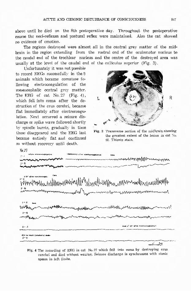
The regions destroyed were almost all in the central grey matter of the mid-
brain in the region extending from the rostral end of the oculomotor nucleus to
the caudal end of the trochlear nucleus and the centre of the destroyed area was
usually at the level of the caudal end of the colliculus superior (Fig. 3).
L
J 叫』‘.,.
R
--
Unfortunately it was not possible
to record EEGs successfullv in the 5
animals which became comatose fo・
llowing electrocoagulation of the
mesencephalic central gre~’ matter.
The EEG of cat No. 27 (Fig. 4),
which fell into coma after the de-
struction of the crus cerebri, became
flat immediately after electrocoagu-'
lation. Next occurred a seizure dis-
charge or spike wave followed shortly
by spindle bursts, gradually in time
these disappeared and the EEG last
became entirely flat and continued
so without recovery until death.
Fig. 3 Transverse section of the midbrain showing
No.L「io・'"・・"色、 r・"・""・"・‘ ,.‘...... "匂・?” r・l≪te・e・噌.,,山岨
the greatest extent of the lesion in cat 1¥o・50. Thionin stain.
1山川U州刷州州内川~w州川附州州f'//h州州iト
1-2
ーへ叫L
1-2
・1・....・,.(t・.......,陣岨・J-1ト
、れ門小、/•t、
創刊,.20' "色骨 l'le<:t.r-oco・.,,.,..,.. "
,. ,, .. _,,
Fig・. 4 The recording of EEG in cat No. 27 which fell into coma by destroying crus cerebri and died without waking. Seizure discharge is synchronous with clonic spasm in left limbs.

841¥ 日本外科宝函第'27巻第l号
B) Non-coma group
The 23 animals of the non-coma group livect an average of 5.6 da;,・s. The
longest lived 12 da~·s. Five animals that lived more than 7 days were recoagulated
and in these animals death was thus hastened. Behavioral changes had relation to
the degree and location of the midbrain destruction.
1) Those animals in which the central g1匂’ matterwas largel~’ destroyed.
Experiments were done in 9 cats.
Course of the experiment in cat No. 47 (female, 2kg) :
She mewed and moved actively before the operation. There were no behavioral
changes seen due to insertion of the electrode. \司'iththe onset of coagulation the
cat cried once and then became quiet. During coagulation a tonic spasm of the
entire body occurred. At that time the pupils became dilated. After operation the
animal did not mew and did not move at all. From time to time she licked her
li1孔・ Theanimal seemed to be semicomatose and all 4 limbs were flaccid, but she
retainetl all her noci-retlexes. When released the cat la;,・ down on the floor and had
not postural reflex. At times she struggled, but she did not eat and did not
respond to visual, auditor;,-, tactile or olfactory stimuli. On the 2nd postoperative
day, she laid down herself and at times moved her four limbs as if she tried to
walk, but she did not mew nor show a川’ interestin food or water. In spite
of warming and nourishing by a
stomach tube, the symptoms did not
improve :and she died on the 4th postoperative da;,・.
In the EEG obtained from this
animal, both flattening and spindle
bursts occurred. The portion of her
brain destro;,・ed was the central grey
in the region extending from the
rostral end of colliculus sup. to the
rostral end of colliculus inf. and the
L R
centre of destruction was at the level L
of the oculomotor nucleus (Fig. 5).
The postoperative courses in cats
No. 52 and No. 53 were the same
Fh・. 5 Transverse section of the midbrain showing the greatest extent of the lesion in cat No. 47. Thionin stain.
as the course of No. 50 of the coma group. After the 2nd day, they licked milk a
little if placed before their noses, but they would not drink any more and they
died on the 5th and 4th postoperative day respectively. In each cat the portions
destroyed were the central grey matter of the midbrain in the region extending
from the oculomotor nucleus to the trochlear nucleus (Figs. 6 and 7).
In the EEG obta~ned from cat No.は thefast waves present before operation were flattened 1mmed1ately after electrocoagulation and then slow waves appeared. Next the spindle bursts appeared intermittently. In the EEG of cat No. 52. the fast waves continued after operation (Fig. 8) and intermittently mixed ’with spindle bursts.

ACUTE AND CHRONIC DISTURBANCE OF CONSCIOUSNESS
Fig・. 6 Transverse section of the midbrain showing the greatest extent of the lesion (shaded area) in cat No. 52.
849
_,,,,
Fig. 7 Transverse section of the midbrain showing the greatest extent of the lesion in cat No・53.Thionin stain.
国国U・”""” F・a・.. ~ ..婦叫.
情吋州桝榊ザ酬酬納 一 一一一、W
W別刷111"""仲 trv.・
,,,・‘.....叫・'"似叫rv1.
1-3
,_‘’ ~旬、,、Aぺ,,
.. .,胸...・・d副館・制"'・1-3
,_今
e’M
•,U II・’・C• < J o , \.< .~凶 ・
1-l
A刊.
2一?
九《~・...,...,.,v、f
哨村山 叫判榊4州内山
If<'格帯内...
~““. .,, d‘"・unt•co吋"'
hザ~叫同州'\/-,ー一一ャー「,-Y"甲"""">'
.'(¥NW州制九叫怖い州.,,.,wy帰山
”~~
R’.,,...、ー」~--h再
...r..J';!も
却....., a陪岨N•I・ 6‘”・孟”.、帽穐酔"
l叫州哨ヤ抑叫似d仲
」:....__J~~
Fig. 8 The recording of EEG in cat l¥'o. 53 and No. 52.
Course of cat No. 46 (male, 3. Skg):
This cat became quiet entirely and seemed to be semicomatose in spite of that
he was wild before electrocoagulation. He retained all his noci-reftexes. After 2 or
3 minutes, he began to mew, snarled and became wild as though seeing imagi-
nary menaces. When he was loosened from his白xings,he tried to walk at once,
but he could not walk, because he fell down to the right. He lifted his head despite
lying down and at the same time he made walking-like or running-like movements
in all four limbs, especially in his front limbs. Five minutes later, he sat up and
850 日本外科宝函第27巻第4号
began to walk and climb up a door. He never showed any interest in food or
water. On the 2nd postoperative day he was sitting apathetically, inertly and
silently and turned his head to the left, looked up and stared vacantly into space
with his eyelids opened. He never meweヨunlikethe day before. Pupils were dilated
on both sides, but especially markedly on the right and his pupillary reaction to light was prompt. All noci-refiexes were retained normally, but he had no appetite.
When he was taken out, he walked slowly into the cage and sat silently. On the
3rd postoperative day his condition was unchanged. On the 4th postoperative day
his eyelids remained open still, but his eyes showed more evidence of alertness. He
stopped to stare vacantly in旬 space,but still did not mew or show any interest
in food and water. He died on the 5th postoperative day in spite of nourishment
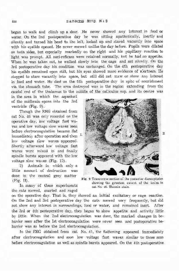
via the stomach tube. The area destroyed was in the region extending from the
caudal end of the thalamus to the middle of the colliculus sup. and its centre was
in the area in which the aqueduct of the midbrain opens into the 3rd
ventricle (Fig. 9).
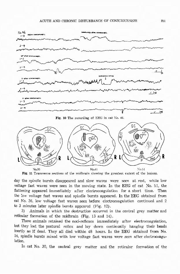
Though the EEG obtained from
cat No. 46 was onlv recorded on the
operative day, low voltage fast wa-
ves and low voltage slow waves seen before electrocoagulation became fiat immediately after operation and then L
low voltage slow waves appeared.
Shortly afterward low voltage fast
waves were mixed in and finally
spindle bursts appeared with the low
voltage slow waves (Fig. 10).
R
2) Animals in which only a
little amount of destruction was
done in the central grey matter (Fig. II).
In many of these experiments
the cats mewed, snarled and raged
Fig-. 9 Transverse section of the posterior diencephalon showing the greatest extent of the lesion in cat No. 46. Thionin stain.
on the operative day. That is, they showed an initial excitatory or rage reaction.
On the 2nd and 3rd postoperative day the cats mewed very frequently, but did
not show any interest in surroundings, food or water, and remained inert. After
the 3rd or 4th postoperative day, they began to show appetite and activity little
by little. When the 2nd electrocoagulation was done, the marked changes in be・
havior seen after the Ist electrocoagulation were never seen and postoperative be-
havior was as before the 2nd electrocoagulation.
In the EEG obtained from cat No. 41, the flattening appeared immediately
after electrocoagulation and soon low voltage fast waves similar to those seen
before electrocoagulation as well as spindle bursts appeared. On the 4th postoperative
ACUTE AND CHRONIC DISTURBANCE OF CONSCIOUSNESS 851
No.+b 1・3 ""'"・ M山崎申温
a・......_ .. 、ー岨ー・咽・事1.
』-t
~ヘ~ヘ、>-'!-
__!一一J.s”。臥・・・1U1Ot"'1r4'
l'rt•・r ・2・・..・向・,, l 1-3
句J"'--1・~吋\ね
‘' ,,世,.,偶企蜘・-弘L
』- 3
2 -~ト
一」.:....__J~
Fig・. 10 The recording of EEG in cat No. 46.
No.36 No.41 1¥o.51
Fig・. 11 Transverse sections of the midbrain showing the greatest extent of the lesions.
day the spindle bursts disappeared and slow waves were seen at rest, while low voltage fast waves were seen in the moving state. In the EEG of cat No. 51, the flattening appeared immediately after electrocoagulation for a short time. Then
the low voltage fast waves and spindle bursts appeared. In the EEG obtained from
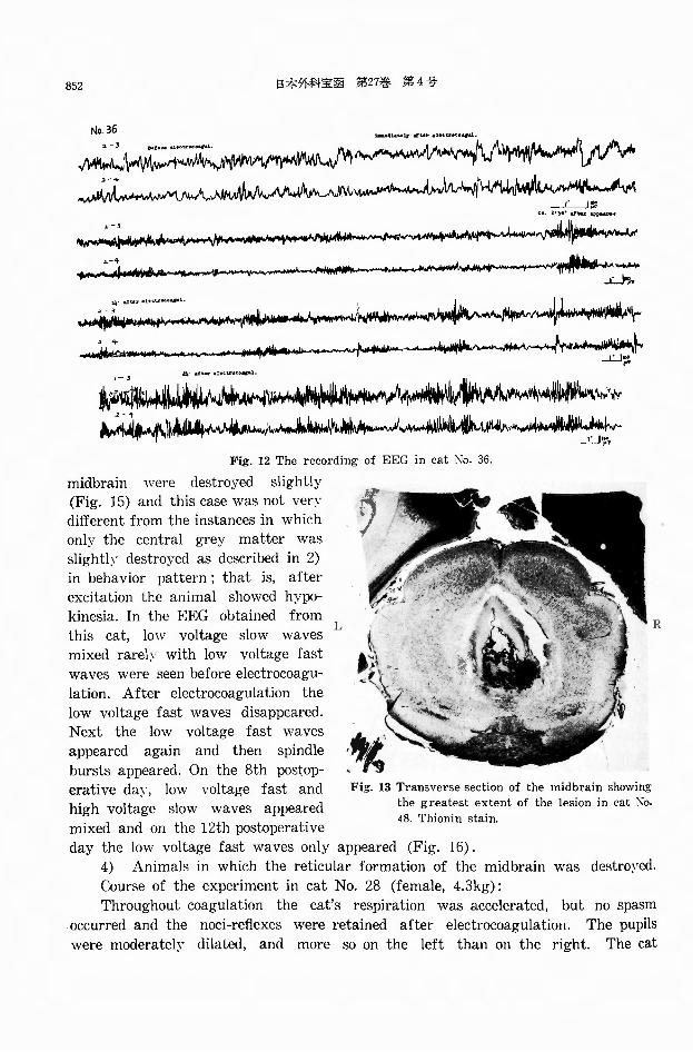
cat No. 36, low voltage fast waves seen before electrocoagulation continued and 2
to 3 minutes later spindle bursts appeared (Fig. 12). 3) Animals in which the destruction occurred in the central grey matter and
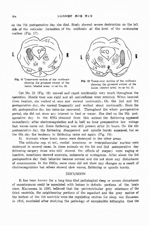
reticular formation of the midbrain (Fig. 13 and 14).
These animals retained the noci-reflexes immediately after electrocoagulation,
but they lost the postural reflex and lay down continually hanging their heads
inertly as if dead. They all died within 48 hours. In the EEG obtained from No.
54, spindle bursts mixed with low voltage fast waves were seen after electrocoagu-lation.
In cat No. 30, the central grey matter and the reticular formation of the
852 日本外科宝函第27巻第4号
内~:~広)榊叩州向/川~川帆ハW仇~川ω伽い叫~人岬ルル仇~仙川向仏仰吋》刈仇M岬凧Aバ
,・3
i-令
酬崎御判断叫内内仙3
州伽糾................ 岬蜘申叫帥
..i:.....Joo,..
lゃ
い時制榊ト
i刈4州~~·..v
Fig. 12 The recording of EEG in cat No. 36.
midbrain were destroyed slightly
(Fig. 15) and this case was not ven
different from the instances in which
only the central grey matter was
slightly destroyed as described in 2)
in behavior pattern ; that is, after
excitation the animal showed hypo-
kinesia. In the EEG obtained from
this cat, low voltage slow waves L
mixed rarely’ with low voltage fast
waves were seen before electrocoagu-
lation. After electrocoagulation the
low voltage fast waves disappeared.
Next the low voltage fast waves
appeared again and then spindle
bursts appeared. On the 8th postop-
erative day, low voltage fast and
high voltage slow waves appeared
mixed and on the 12th postoperative
;続Fig. 13 Transverse section of the midbrain showing
the greatest extent of the lesion in cat No. 48. Thionin stain.
day the low voltage fast waves only appeared (Fig. 16).
4) Animals in which the reticular formation of the midbrain was destroyed.
Course of the experiment in cat No. 28 (female, 4.3kg):
Throughout coag~lation the cat’s respiration was accelerated, but no spasm
occurred and the noc1-reflexes were retained after electrocoagulation. The pupils
were moderately dilated, and more so on the left than on the right. The cat
R

ACUTE A :.ID CI:RG:でrr’DISTUHBANCEOF CON3CIOU.3NESS 853
Fig. 14 Transverse section of the midbrain showing the greatest extent of the lesion in cat No. 54 Thionin stain.
M噌..” V・R・rNo.30 • ' I-之 . .,・"・ lettr•包・....,. .,.ぺroieaz叫i • ' a句
、,-、、M 仰れ由脚V申、制H’,,.,,,、A.r・-《""守ザ『 F噂円,,』eふ..l¥Jv.Jo
3-+ ~~←__ ...... J、~γ旬、ν..,,..J.ん日』L咋
1-2. " ..... """・"'叫両個岨凶
3-'t
R
Fi~. 15 Transverse section of the midbrain showing the greatest exent of the lesion (shaded area〕incat :¥io. 30.
,. l叩
守‘
一ー」入」剛戸
Fig. 16 The recording of EEG in cat i'¥o. 30
became very quiet and did not move or mew. She lay down wherever she was
placed, lying inertly on the floor and hanging her head down as if she had di吋,
when she was loosened from her ties. Her postural reflex had disappeared. She
did not show an:.’interest in food or water. On the 2nd postoperative dayア shelay where placed in the same posture in the cage as on the day before and seemed
unaware of ordinary environmental stimuli. She never moved or mewed and seemed
to be dead. Irritation which would cause an immediate cry out from a normal cat,
provoked only a feeble movement of the head and a stepping movement of the
extremities. But she retained her noci-reflexes. She did not show any interest in
food or water. Throughout her survival period, the same behavior continued and
854 日本外科宝函第27巻 第4号
on the 7th postoperative day she died. Study showed severe destruction on the left
side of the reticular formation of the midbrain at the level of the oculomotοr
nucleus (Fig. 17).
R
Fig. 17 Transverse section of the midbrain showing the greatest extent of the lesion (shaded area) in cat No. 28.
R
Fjg, 18 Transverse section of the midbrain showing the greatest extent of the lesion (shaded area) in cat No. 32.
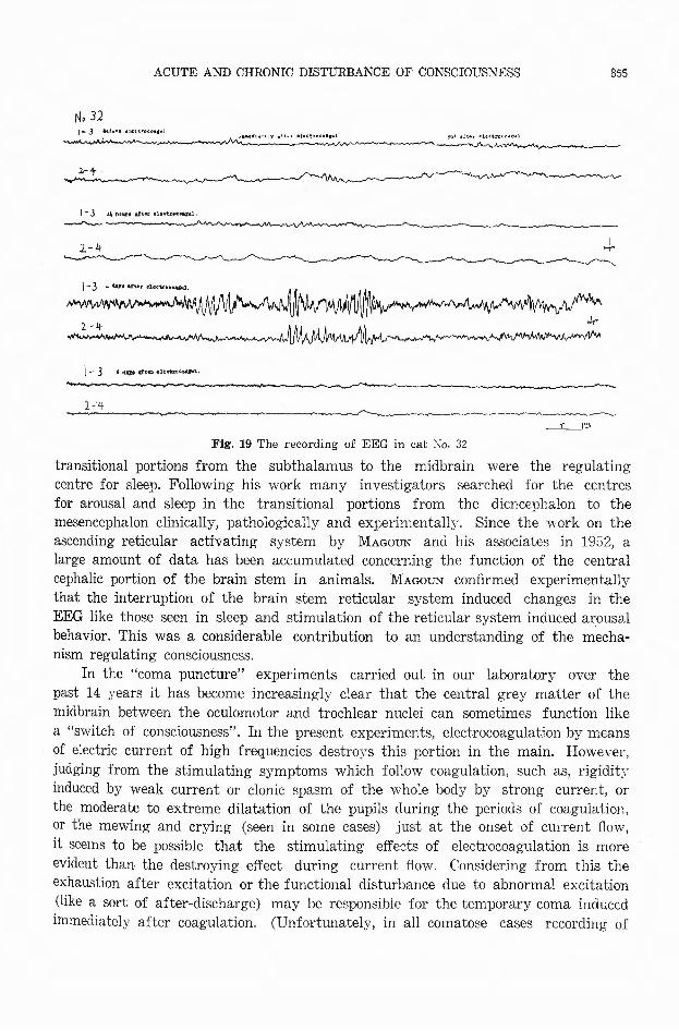
Cat No. 32 (Fig. 18) mewed and raged continually very much throughout the
operation. ::VIuscle tone was rigid and all noci-reflexes were retained. When loosened
from fixation, she walked at once and mewed continually. On the 2nd and 3rd
postoperative day, she mewed frequently and walked about continually. Since the
4th postoperative day her behavior recovered. Throughout the whole postoperative
period, she did not show any interest in food or water. She died on the 9th post-
operative day. In the EEG obtained from this animal the flattening appeared
immediate!;.’ after electrocoagulation and in half an hour preoperative low voltage
fast waves came out. Some flattening was still present after 24 hours. On the 4th
postoperati;.’e day, the flattening disappeared and spindle bursts appeared, but on
the 6th day the tendency to flattening came out again (Fig. 19). 5) Animals whose brain stems were destroyed in the other areas.
The colliculus sup. et inf., medial lemniscus or interpeduncular nucleus were destroyed in several cases. In these animals on the Ist and 2nd postoperative day following surgery those who still showed the e百ectsof surgery were raging or
inactive, sometimes showed anorexia, anisocoria or nystagmus. After about the 3rd postoperative day their behavior became normal and did not show any disturbance
of consciousness. In the EEGs, some cases did not show any changes as a result of electrocoagulation but others showed slow waves, flattening or spindle bursts.
DISCUSSION
It has been known for a long time that pathological sleep or severe disturbance of consciousness could be associated with lesions in definite portions of the brain
stem. MAUTHNER, in 1890, believed that the periventricular grey substance of the third ventricle, the neighbouring portions of the aqueduct and the grey matter of
the bottom of the 4th ventricle were the regulating centres for sleep. von EcoNoMo,
in 1916, concluded after studying the pathology of encephalitis lethargica that the
ACUTE AND CHRONIC DISTURBANCE OF CONSCIOUSNESS
N. 32 1島 3 ・・hn•IC'‘...ι・...』
1.-t
1・3 LI h•剛山・E ・E・・回,..,.,___,.、『,,
ユ,斗
ト 3 H 刷出@叫れ相同岨岨.
2-斗
地則前・’.' ........ "'"・句叫 円・'"・,叫射訂以 ・4似 a-一一............,、へん、
Fig. 19 The re巴ordingof EEG in cat No. 32
855
・マ,、
_i__J';>
transitional portions from the subthalamus to the midbrain were the regulating centre for sleep. Following his work many investigators searched for the centres for arousal and sleep in the transitional portions from the diencephalon to the mesencephalon clinically, pathologically and experin~entally. Since the work on the ascending reticular activating system by MAGOUN and his associates in 1952, a large amount of data has been accumulated concerning the function of the central cephalic portion of the brain stem in animals. MAGOUN confirmed experimentally that the interruption of the brain stem reticular system induced changes in the EEG like those seen in sleep and stimulation of the reticular system induced arousal behavior. This was a considerable contribution 句 anunderstanding of the mecha-nism regulating consciousness.
In the “coma puncture" experiments carried out in our laboratoryア overthe past 14 years it has become increasingly clear that the central grey matter of the midbrain between the oculomotor and trochlear nuclei can sometimes function like a "switch of consciousness”. In the present experiments, electrocoagulation by means of electric current of high frequencies destroys this portion in the main. However, judging from the stimulating symptoms which follow coagulation, such as, rigidit~・induced by weak current or clonic spasm of the whole body by strong current, or the moderate to extreme dilatation of the pupils during the periods of coagulation, or the mewing and crying (seen in some cases) just at the onset of current司ow,it seems to be possible that the stimulating effe~ts of electrocoagulation is more evident than the destroying e古ect during current自ow.Considering from this the exhaustion after excitation or the functional disturbance due to abnormal excitation (like a sort of after吋ischarge)may be responsible for the temporary coma induced immediately after coagulation. (Unfortunately, in all comatose cases recording of
856 日本外科宝函第27巻第4号
the EEG at the time of coma immediately after coagulation could not be done
except in a comatose case due to subarachnoid bleeding.) The fact that no animals
fell into coma when stimulating e百ectsexisting during and immediately after coag-
ulation disappeared and the local destroying e百ects, that is, the local functional
disruption appeared, would suggest that local partial functional disruption in the
midbrain alone did not凶 usecoma. In 5 of the 6 animals which became comatose,
pathological examination revealed destruction of the central grey matter or both
the central grey matter and the reticular formation. In the 6th comatose cat it
seems likely that coma occurred secondarily to basal hemorrage. This would indicate
that experimental coma defined above as the unresponsiveness to all external (es-
pecially nociceptive) stimuli is in most cases associated with abnormal stimulation
of the central grey matter in the region extending from the oculomotor nucleus to
the trochlear nucleus. However, as has also been shown by other workers in our laboratory all inju-
ries of this portion of the brain do not al ways induce coma. Therefore it seems
that some secondar~・ change rather than mere abnormal stimulation is necessary in
order to induce coma. \\アeare forced to the conclusion that the most reasonable explanation for coma is to assume a sort of seizure discharge arising and spreading
over the entire midbrain adequately from the area of electrocoagulation.
It was very difficult to induce prolonged coma purposed primarily. However,
when a large amount of the central grey matter was destroyed, the spontaneous
‘ movement and mewing were absent and the animal had anorexia. The animal was seen either sitting down apathetically oblivious to the ordinary environmental
stimuli with the postural reflex still functioning or lying motionless on its side
hanging its head down with no postural reflex. This occurrence is similar to the
“simulating coma in man" delineated by MAGOUN. In this state the animal appeared
as if dead except for the fact it was breathing. It was however not comatose
according to our criteria because the noci-reflex was retained. At this point we
cannot decide with certainty which takes the responsibility for this behavior
between (i) the disturbance of consciousness, and (ii) extreme general exhaustion
or a sort of exhaustive break down of all sympathetic nervous system. In this
simulating comatose behavior of experimental animals, there was greater or less
change in relation旬 greateror less degree of destruction. Whether or not coma
(absence of noci-reflexes) was induced immediately’ after coagulation made no differ-
ence in this change. The greater the degree of destruction the less chance for the
animal to recover destroyed vital function and the fewer postoperative survivals.
On the other hand when the destruction was slight, the cat became excited
and mewed frequently, occasionlly snarling and raging on the Ist postoperative day.
On the following day it became inert and apathetical to environmental stimuli
except for mewing and did not show any interest in food or water. These behav-
ioral abnormalities recovered graduall:-・ in 3 or 4【lay日 and appetite, spontaneous
movement and interest in environment reappeared. When the reticular formation
was destroyed, if the destruction was large, the cat la>・ on its side hanging its
ACUTE AND CHRONIC DISTURBANCE OF CONSCIOUSNESS 851
head down on the floor, with no postural reflex and no mewing or spontaneous
movement except an occasional stepping movement. The appetite was lost immedi-
ately after electrocoagulation. If, on the other hand, the destruction was small,
almost all the cats were active and walked ordinarilγ,mewed frequently and raged
wildly. But the noci-reflexes were held in every’ case to the end and coma accord-ing to our criteria was never seen. That is, the behavioral di百erencebetween the
large destruction of the central grey and that of the reticular formation was not
seen except the occasional absence of noci-reflexes following operation was induced
(in the former) or not (in the latter). When with reoperation 7 to 8 days after
initial operation further destruction was added to the central grey matter, the reticular formation or other areas there was no di仔erence made in be ha vi or and
the postoperative behavior was similar to that immediately prior 句 the 2nd coagulation.
SHINOZAKI, in 1937, reported that electrocoagulation of the periaqueductal tissue
caused some disturbance of consciousness if the destruction was extensive but did
not alter the state of consciousness if the destruction was small in amount or situated ventrally. In the present experiment and other experiments in our labora-初ry,i.e. local nicotine injection or local application of electronarcosis, the ventral portion of the central grey matter seemed more important than the other portions.
BAILEY and DAvis experimented with localized electrolytic lesions of the peri-
aqueductal grey matter in cats (1942) and monkeys (1945). The behavior of these animals resembled strikingly the syndrome seen in human patients after lesions in
the brain stem described by BAILEY and etc. as “arrest of consciousness'’, ancl by CAIRNS and his coworkers as“akinetic mutism". It was similar 句 ourresult. But this work did not report EEG change or disappearance of noci-reflexes which might have been seen immediately after operation.
LINDSLEY, ScHREINER, KNOWLES and MAGOUN, in 1950, in cats, and FRENCH and
MAGOUN, in 1952, in monkeys, produced the chronic destructive lesions of the central cephalic brain stem and outlined clearly in cats the behavior pattern follow-ing injury of the midbrain tegmentum (chronic somnolence and hypo】dnesis,which we feel might correspond t-0 the state of simulating coma seen in our animals).
But伺 udallesion on the periaqueductal grey matter in their experiments left the animal awake and alert from the first postoperative da~’ with no greater disposition
句 sleepthan that exhibited by a normal cat. In the only case of rostral lesion,
the animal appeared asleep or drowsy from the first postoperative day. The animal exhibited little or no spontaneous activity and could not be aroused by a百erentstimulation. In our experiments the caudal lesion produced the simulating coma too.
The EEG in the MAGOUN’s experiment showed large slow wayes and spindle bursts and could not be activated when the animals were in somnolence or asleep. In the
case of caudal lesions the EEG showed increased activity. On the contrary, in our
experiments, the EEG did not show any characteristic form associated with the
destruction of di百erentmesencephalic areas. In these lesions made b~· :VIAGOUN ancl
etc. of the reticular formation and central grey matter, it seems that neither coma
858 日本外科宝函第27巻 第4号
according to our criteria nor the flattening of the EEG tracings seen in our experi-ments was produced. In the experiments of MAGOUN and his co-workers the reticular
formation was destroyed very largely and bilaterally. Localized destruction of one
side such as we have done was not reported. In our experiments when occasionally a large amount of the reticular formation was destroyed although unilaterally, we observed hypersomnolence and hypokinesis or akinesis. But we did not trace the
large slow waves seen in the EEG in the experiments of MAGOUN.
BAILEY and DAvis, in 1942, described an interesting experiment,“the syndrome
of obstinate progression in the cat”. When the nucleus interpeduncularis was de-stroyed the cats in all cases began to progress obstinately forward making a peculiar low cry and never turned aside from any obstacle. This behavior continued as long
as the animals survived, usually about 3 days. In our cat No. 27, the nucleus interpeduncularis and its neighbouring structures were destroyed, and during the week it survived, it did not show these peculiar behavioral changes.
SUMMARY
1) With the intention of producing prolonged coma (absence of nod-reflex回)experimental lesions were made in several portions of the midbrain in 52 cats.
2) In 4 cases, prolonged coma, lasting half an hour or more, developed immediatel~’ after operation and wlls in each ca~e seen to be associated with electrocoagulation of the central grey matter, coagulation of both the central grey matter and the reticular formation, or coagulation of the crus of the cerebrum. However, electrocoagulation of all these parts described above did not always cause prolonged coma. On the contrary, prolonged coma occurred only in a few cases.
3) In most cases the appearance of the cats with the destruction of a large
amount of the central grey matter resembled the behavior pattern called “arrest of consciousness" with nod-reflexes retained. Unfortunately, it is impossible to decide
whether such behavior is due to a disturbance of consciousness per se or due to exhaustive inactivity. In the animals in which coma did not occur immediately following electrocoagulation, the course over the period of survival was similar to the prolonged course in those animals which did develop coma (absence of noci-re-flexes) immediate I~-, if the location and amount of coagulation were similar.
4) The behavioral difference between the large destruction of the central grey
and that of the reticular formation was not seen except the occasional lack of noci-reftexes following operation was induced (in the former) or not (in the latter).
5) The EEG did not show any characteristic pattern associated with the
destruction of di百erentareas of the brain stem. In many cases, the EEG became flat immediately after electrocoagulation and in a short time moderate voltage slow
waves appeared followed in 2 to 3 minutes b~· spindle bursts. In some cases low voltage fast waves continued as before operation and shortl:: after injury spindle
bursts appeared. In other cases these fast waves slowed down and a short time later spindle bursts appear℃cl as well. In all cases spindle bursts appeared 2 or 3
minutes after electrocoagulation and there was a tendenc;,’ to synchronization. The
ACUTE AND CHRONIC DISTURBANCE OF CONSCIOUSNESS 859
EEG recovered the preoperative low voltage fast "’aves from 5 to 8 da;.ァs after
surgery, but showed a tendenc>’ to flattening again little by little before death.
6) These experiments show that the central grey matter may be especially
concerned with consciousness and it also seems that coma occurs as a result of
abnormal excitement associated with the excessive stimulation due to coagulation.
However, a similar amount of stimulation (a similar amount of electrocoagulation)
does not always cause coma. Actually, coma appears to depend on some other
secondary change. For example, the most reasonable explanation might be to assume
a sort of seizure discharge originating at the site of the damaged mesencephalon
and causing a widespread loss of function as it spreads over the midbrain.
References I) Araki : Diencephalon and Disturbance of Consciousness. Saishinigaku, 8, 41, 1953戸2) Araki : Cerebral Trauma and Disturbance of Conscionsness. Sinryo, 9, l, 1956ヌ3) Araki : Disturbance of Consciousness due
to Head Injury, The Vlth Anual Meeting of the Japan EEG Society Symposia & Proceed. 1957. 4) Araki, C., Sakata, K. and Kuroki, T. : Pontine or“Reticular"' Epilepsy, Report of a Case, EEG Clin. Neurophisiol. Journal, 8, 4. 1956. 5)Araki, C., Taketomo, T. and Toda, T.: Coma puncture. Acta Sch. Med. UniY. Kioto, '%/, 205, 1949ヌ6)Araki, C., Sakata, K. and Matsunaga, M.: Recruiting Response-Like EEG Changes Induced with Extrathalamic Stimulation of Cat. Acta Srholae Med Univers. Kioto Jap., 32, 32・51,1956. 7〕Asai,S.: Elec-tronarcosis by Direct Stimulation of the Brain Stem. Arch. Jap. Chir., 27, l, 1958. 8) Bailey, P. and Davis, E. W.: Effects of Lesions of the Periaqueductal Grey Matter in the Cat. Proceedings of the Society for Experimental Biol. & Medicine, 51, 305, 1943. 9) Bailey, P. and Davis, E. W. : Effects of Lesions of the Periaqueductal Grey Matter in Cats and Monkeys. Abst. Arch. Neurol. &
Psychiat., 53, (4), 325, 1945. 10) Bailey, P.
and Davis, E.羽T.:“TheSyndrome of Obstin-ate Progression in the Cat.. Proc. of the So-ciety for Experimental Biol. and Medicine, 51, 307, 1942. 11) Bernhaut, M., Gellhorn, E.
and Raumussen, A. T. : Experimental Contr-ibutions to Problem of Conscioasness. Journal Neurophisiolog., 16, 21, 1953. 12) Davison, C. and Demuth, E. : Disturbances in Sleep Me-
chanism: A Clinico・PathologicStudy. IV. Les-ions at the Mesencephalometencephalic Level. 53, (!), 126, 1945. 13) French, J. D. : Brain
Lesions Associated with Prolonged Unconscio-usness. Arch. Nearol. Psychiat., Chicago, 68,
727, 1952. 14) French, J. D. and Magoun, H. W. : Effects of Chronic Lesions in Central Cephalic Brain Stem of '.¥lonkeys. Arch. Ne-urol. Psychiat., Chicago, 68, 591, 1952. 15) French, J. D., Verzeano, M. and Magoun
H. W.: An Extralemniscal Sensory System in the Brain. Arch. Neurol. Psychiat .. Chica-go, 69, 505, 1953. 16) Gerard, R. W., l¥Iachall, 、TV.H. and Saul, L. J .. Electrical Activity of the Cat's Brain. Arch. Neurol. and Psychiat., 36: 675, 1936. 17) Ishii, S.: Coma Puncture (Monograph). Kyoto, 1944タ 18)Jasper, H. H. and Cosimo Ajmone-Marsan : A Stereotaxic
Atlas of the Diencephalon of the C'at. Montr-eal Neurological Institute of l¥Ic Gill Unive1~
sity. 19) Lindsley, D. B., Schreiner, L. H., Knowles, W. B.加 dMagoun, H. W.: Behavi-oral and EEG Changes Following Chronic Brain Stem Lesions in the Cat. Electroencep・halog. & Clin. l¥'europhisiolog., 2: 483, 1950. 20) Lindsley, D. B., Bowden, J. W. and Ma-goun, H,羽T.: Effects upon the EEG of Acute
Injury to the Brain Stem Acti,・ating System. Electroencephalog. and Clin. Neurophisiol., 1, 475, 1949. 21) Magoun, H. W.: The Ascending Reticular Acti、atingSystem. Research Public司
ations, Associations for Research in l¥'ervous and Mental Diseases, XXX. 480, 1952. 22) Matsunaga, M.: Experimental Studies on
Coma Induced by Stimulation o! Various Par-ts of the Brain in Cat. Being prepared for publication. 23) Mauthner, L. : Zur Patholo・gie und Physiologie des Schlafes. Wien Klin. Wchnschr., 3, 445, 1890. 24) l¥Ioriyasu, H.: To be published shortly. 25) Starzl, T. E., Taylor
C. W. and Magoun, H. W. : Ascending Cond-uction in Reticular Activating System, with Special Reference to the Diencephalon. Journ-al ~curophisiolog., 14, 461, 1950. 26〕Yabuno,
S.: Coma Puncture by Means of Nicotinizat司
ion. Acta Scholac Med. Univers. Kioto Jap., 32,
860 日本外科宝函第27巻第4号
32・51,1954. 27) Wjnkler, C. and Potter, A.: An Anatomical Guide to Experimental Rese-arches on the Cat"s Brain. Amsterdam, W.
和文抄録
Verluys. 発 writtenin Japanese
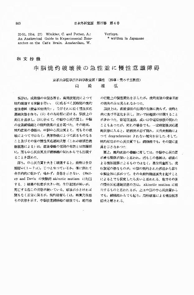
中脳焼灼破壊後の急性並に慢性意識障碍
京都大学医学部外科学教室第 I講座 (指導:荒木千里教授〉
山崎雄弘
私達は,成熟猫の中脳各所をp 高周波電流によって の行動上の慢性変化を示したが,焼均直後の侵害反射
焼灼破壕する実験を行"" (i)成るべく長時間の焼灼 の消失のみは見られなかった.
直後昏陵(侵害反射消失ハ及びそれに続く慢主主反応 脳波上は,直後昏怪の出現の有無に拘らず,焼灼と
遅鈍状態を作り,(ii)その各時期に於ける,脳波上の 共に先づ平坦化を示し,次いで紡縫波の出現すること
変化を追求し,(iii)併せてp 中脳中心灰白質と,中脳 が多かった.低電位速波,或いは中電位徐波の現れた
の被蓋網様織との焼灼効果の差を調べた.その結果, こともあったがp 何れの場合でもp 一定時間後Jjf,1応遅
焼灼直後の昏径は,中脳中心灰白質より,而もその破 鈍状態に入ると,紡経波が必ず現れ,叉外来刺戟はよ
壊によってではなくp 異常利戟によって起るものなる って desynchronizeされない傾向を示した.そして,
こと及びその後の慢性反応遅鈍状態(これは破綾性機 焼灼部位が中心灰白質でもF 網様織でも,その聞に差
能脱落による)は,直後昏陵の発現の有無とは無関係 異を示さなかった
に,而も中心灰白質及び網様織の何れからでも出現す 要之,焼灼直後の昏睡に対しては,中脳中心灰白賞
ることを認めた. ’ が最も関係、が深いと思われ,而もこの昏睡は,破嬢に
即ち,中心灰白質を大きく破壊するとp 動物は生存 よる機能脱落によるものではなく, 異常興奮即ちp 発
期間中(1~7日)'じっと坐っているか,績に倒れて 作放電の様なものがp 中脳の焼灼された部位から起り
合目的的に動かず,鳴かず,食慾を示さない. (Baiト 中脳全体に鉱がってP その全般的機能衷失を起すこと
ey and Davisの実験的 akineticmutism に相当 によるとでも仮定したら良いと恩われる.他方その後
する.)破壊の程度が大きい程p 生存期苅が短いが, の慢性反応遅鈍伏態の方は, akinetic mutismに相
死亡する迄この状態が続いている.敏壊が小さければ 当するものと思われるが,之は中脳の中心灰由貿から
間もなく正常に戻るが,焼灼後暫らくは,興奮乃至怒 でも,網様織からでも起り,局所破壊による機能脱怒
りの状態を示す.中脳被蓋網様織の破壕でも,略問機 症状と解される.