Embed Size (px)
Citation preview
JOURNAL OF HEMATOLOGY& ONCOLOGY
Pecci et al. Journal of Hematology & Oncology (2015) 8:71 DOI 10.1186/s13045-015-0169-6
RESEARCH ARTICLE Open Access
Particulate cytoplasmic structures with highconcentration of ubiquitin-proteasomeaccumulate in myeloid neoplasms
Alessandro Pecci1*, Vittorio Necchi2,3, Serena Barozzi1, Agostina Vitali2, Emanuela Boveri4, Chiara Elena5,Paolo Bernasconi5, Patrizia Noris1 and Enrico Solcia2,4Abstract
Background: Increased plasma levels of proteasome have been associated with various neoplasms, especiallymyeloid malignancies. Little is known of the cellular origin and release mechanisms of such proteasome.We recently identified and characterized a novel particulate cytoplasmic structure (PaCS) showing selectiveaccumulation of ubiquitin-proteasome system (UPS) components. PaCSs have been reported in some epithelialneoplasms and in two genetic disorders characterized by hematopoietic cell dysplasia and increased risk ofleukemia. However, no information is available about PaCSs in hematopoietic neoplasms.
Methods: PaCSs were investigated by ultrastructural, immunogold, and immunofluorescence analysis of bonemarrow (BM) biopsies and peripheral blood (PB) cell preparations of 33 consecutive, untreated, or relapsed patientsaffected by different hematopoietic neoplasms. BM and PB samples from individuals with non-neoplastic BM orhealthy donors were studied as controls. Granulocytes and platelet proteasome content was measured byimmunoblotting and plasma proteasome levels by ELISA.
Results: PaCSs with typical, selective immunoreactivity for polyubiquitinated proteins and proteasome werewidespread in granulocytic cells, megakaryocytes, and platelets of patients with myeloproliferative neoplasms(MPN). In acute myeloid leukemia and myelodysplastic syndromes (MDS), PaCSs were only occasionally detected inblast cells and were found consistently in cells showing granulocytic and megakaryocytic maturation. Conversely,PaCSs were poorly represented or absent in non-neoplastic hematopoietic tissue or lymphoid neoplasms. In MPNgranulocytes and platelets, the presence of PaCSs was associated with increased amounts of proteasome in celllysates. PaCSs were often localized in cytoplasmic blebs generating PaCSs-filled plasma membrane vesicles observablein the BM intercellular space. In MPN and MDS, accumulation of PaCSs was associated with significant increase inplasma proteasome. Immunogold analysis showed that PaCSs of myeloid neoplasia selectively concentrated thechaperone proteins Hsp40, Hsp70, and Hsp90.
Conclusions: PaCSs accumulate in cells of myeloid neoplasms in a lineage- and maturation-restricted manner; inparticular, they are widespread in granulocytic and megakaryocytic lineages of MPN patients. PaCSs development wasassociated with excess accumulation of polyubiquitinated proteins, proteasome, and chaperone molecules, indicatingimpairment of the UPS-dependent protein homeostasis and a possible link with Hsp90-related leukemogenesis. Amechanism of PaCSs discharge by leukemic cells could contribute to increased plasma proteasome of MPN and MDS.
Keywords: Ubiquitin/proteasome system, Proteasome, Polyubiquitinated proteins, Chaperone molecules, Myeloidneoplasia, Myeloproliferative neoplasms
* Correspondence: [email protected] of Internal Medicine, IRCCS Policlinico San Matteo Foundationand University of Pavia, Pavia, ItalyFull list of author information is available at the end of the article
© 2015 Pecci et al. This is an Open Access art(http://creativecommons.org/licenses/by/4.0),provided the original work is properly creditedcreativecommons.org/publicdomain/zero/1.0/
icle distributed under the terms of the Creative Commons Attribution Licensewhich permits unrestricted use, distribution, and reproduction in any medium,. The Creative Commons Public Domain Dedication waiver (http://) applies to the data made available in this article, unless otherwise stated.
Pecci et al. Journal of Hematology & Oncology (2015) 8:71 Page 2 of 11
BackgroundIncreased cellular expression and activity of proteasomehave been reported in a variety of neoplasms, includinghematological, epithelial, and neurological tumors [1–5].This has led to the proposal of proteasome inhibitors asantineoplastic therapy, with clinically relevant results insome tumors, such as plasma cell myeloma and mantlecell lymphoma [6, 7]. Both epithelial and hemopoieticneoplasms have been associated with increased plasmalevels of proteasome which were disease type, stage, andtherapy sensitive [8–11]. However, little is known aboutthe intracellular origin and release mechanisms of thisincrease in plasma proteasome in neoplastic diseases. Itremains unclear whether the proteasome in neoplasticcells is passively released during cell lysis and apoptosis,or whether some specific, more regulated, release mech-anism exists, which may better account for the selectivityof the changes in plasma proteasome level [9].Proteasome has been reported in exosomes [12], which
are small (50–100 nm) vesicles of endosomal origin thatare released by several cell types, including non-pathological blood cells [13, 14] and neoplastic cells [15].The involvement in proteasome release of other extracel-lular vesicles, such as plasma-membrane-derived mic-rovesicles, also called ectosomes or microparticles, hasrecently been suggested [16]. Besides proteasome compo-nents, neoplastic cells accumulate excessive amounts ofpolyubiquitinated proteins [2, 4, 5, 17], the proteasomeelective substrate. Limited information is available on thefate of such proteins and whether they are totally de-graded inside neoplastic cells or at least in part dischargedextracellularly together with proteasome components.We recently identified and characterized a novel par-
ticulate cytoplasmic structure (PaCS) that concentratespolyubiquitinated proteins and proteasome in someepithelial neoplasms and related preneoplastic lesions[5, 7, 17, 18], as well as in two genetic disorders charac-terized by hematopoietic cell dysplasia and increased riskof leukemia [19, 20]. However, the presence of PaCSs inhematological neoplasms has never been investigated.Here, we demonstrate that PaCSs accumulate in differ-
ent forms of myeloid neoplasia in a maturation- andlineage-specific manner. Our findings suggest that a mech-anism of PaCSs discharge by leukemic cells contribute toincreased plasma proteasome in myeloid neoplasms.
ResultsPaCSs in hematological neoplasmsCombined ultrastructural and immunogold analysisshowed that PaCSs were extensive in bone marrow (BM)of patients with myeloproliferative neoplasms (MPN),including chronic myelogenous leukemia (CML), polycy-themia vera (PV), essential thrombocythemia (ET), andprimary myelofibrosis (PMF). PaCSs appeared as focal
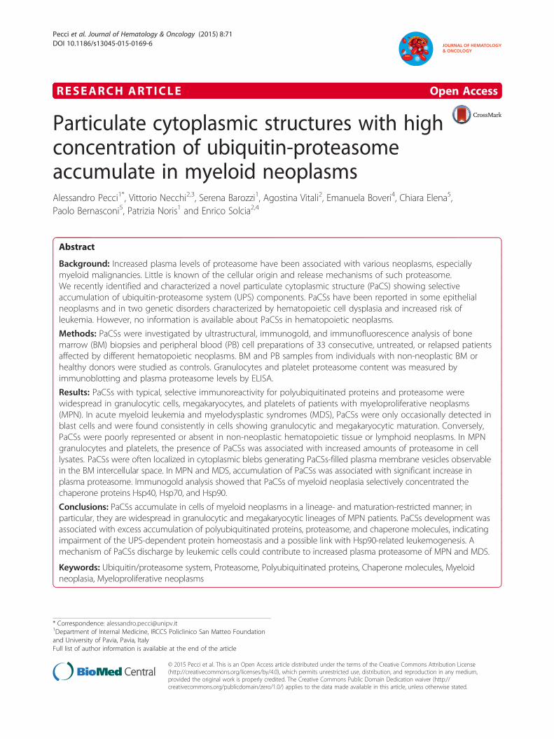
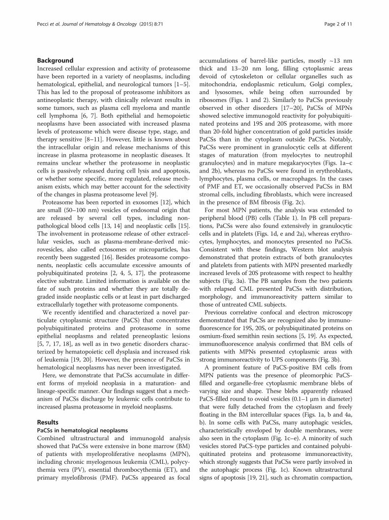
accumulations of barrel-like particles, mostly ~13 nmthick and 13–20 nm long, filling cytoplasmic areasdevoid of cytoskeleton or cellular organelles such asmitochondria, endoplasmic reticulum, Golgi complex,and lysosomes, while being often surrounded byribosomes (Figs. 1 and 2). Similarly to PaCSs previouslyobserved in other disorders [17–20], PaCSs of MPNsshowed selective immunogold reactivity for polyubiquiti-nated proteins and 19S and 20S proteasome, with morethan 20-fold higher concentration of gold particles insidePaCSs than in the cytoplasm outside PaCSs. Notably,PaCSs were prominent in granulocytic cells at differentstages of maturation (from myelocytes to neutrophilgranulocytes) and in mature megakaryocytes (Figs. 1a–cand 2b), whereas no PaCSs were found in erythroblasts,lymphocytes, plasma cells, or macrophages. In the casesof PMF and ET, we occasionally observed PaCSs in BMstromal cells, including fibroblasts, which were increasedin the presence of BM fibrosis (Fig. 2c).For most MPN patients, the analysis was extended to
peripheral blood (PB) cells (Table 1). In PB cell prepara-tions, PaCSs were also found extensively in granulocyticcells and in platelets (Figs. 1d, e and 2a), whereas erythro-cytes, lymphocytes, and monocytes presented no PaCSs.Consistent with these findings, Western blot analysisdemonstrated that protein extracts of both granulocytesand platelets from patients with MPN presented markedlyincreased levels of 20S proteasome with respect to healthysubjects (Fig. 3a). The PB samples from the two patientswith relapsed CML presented PaCSs with distribution,morphology, and immunoreactivity pattern similar tothose of untreated CML subjects.Previous correlative confocal and electron microscopy
demonstrated that PaCSs are recognized also by immuno-fluorescence for 19S, 20S, or polyubiquitinated proteins onosmium-fixed semithin resin sections [5, 19]. As expected,immunofluorescence analysis confirmed that BM cells ofpatients with MPNs presented cytoplasmic areas withstrong immunoreactivity to UPS components (Fig. 3b).A prominent feature of PaCS-positive BM cells from
MPN patients was the presence of pleomorphic PaCS-filled and organelle-free cytoplasmic membrane blebs ofvarying size and shape. These blebs apparently releasedPaCS-filled round to ovoid vesicles (0.1–1 μm in diameter)that were fully detached from the cytoplasm and freelyfloating in the BM intercellular spaces (Figs. 1a, b and 4a,b). In some cells with PaCSs, many autophagic vesicles,characteristically enveloped by double membranes, werealso seen in the cytoplasm (Fig. 1c–e). A minority of suchvesicles stored PaCS-type particles and contained polyubi-quitinated proteins and proteasome immunoreactivity,which strongly suggests that PaCSs were partly involved inthe autophagic process (Fig. 1c). Known ultrastructuralsigns of apoptosis [19, 21], such as chromatin compaction,
Fig. 1 PaCSs are widespread in hematopoietic cells of patients with untreated CML. a Several BM cells of the granulocytic lineage displaycytoplasmic collections of barrel-like particles, enlarged in a1 and a2 to show the particles and their 20S proteasome (β5i subunit) immunogoldreactivity. PaCS-filled blebs (arrowheads) and cell-detached vesicles (arrows) are devoid of cytoplasmic organelles. A cytoskeleton-rich cytoplasmicnetwork (arrowheads in a1) separates PaCSs from the cytoplasmic membrane; the cytoskeletal network is largely lost in some detached vesicles(arrow in a1) undergoing degeneration while still preserving proteasome reactivity. b A PaCS-filled cellular bleb (b) and isolated vesicles (b1, b2)in BM extracellular space show PaCS-restricted immunogold reactivity for polyubiquitinated proteins (FK1 antibody). c A myelocyte shows large,FK1-reactive PaCSs and several FK1-negative autophagic vesicles. The largest vesicle is enlarged in c1 to show its enveloping double membraneand its unusual storage of PaCS-type particles, as well as FK1-immunoreactive polyubiquitinated proteins comparable with those of adjacentPaCSs; a likely sign of ongoing PaCS autophagy. The asterisk indicates an erythroblast showing no PaCSs; s indicates stromal cell processes.d, e PB granulocytic cell (d, enlarged in d1) and platelet (e) from a CML patient showing PaCSs reactive for polyubiquitinated proteins
Pecci et al. Journal of Hematology & Oncology (2015) 8:71 Page 3 of 11
nuclear membrane loss, and dense and homogeneous cyto-plasm with no recognizable organelles were observed onlyoccasionally in MPN cells, irrespectively of the presence ofPaCS, blebs/ectosomes, or autophagic vesicles.To gain insight into the respective role of PaCSs and
autophagic vesicles, we performed in MPN cells cyto-chemical assays for proteins known to be associated withUPS function or autophagy. Hsp40, Hsp70, and Hps90chaperones were selectively concentrated inside PaCSs(Fig. 4a–c), while the co-chaperone Bag 6 had sparsecytoplasmic reactivity, without preferential concentra-tion in PaCSs (Fig. 4d). Autophagic vesicles showedreactivity for LC3A protein, a known autophagy marker[22], to which PaCSs showed no reactivity (Fig. 4e).Unlike MPN cells, BM blasts of the 8 patients with
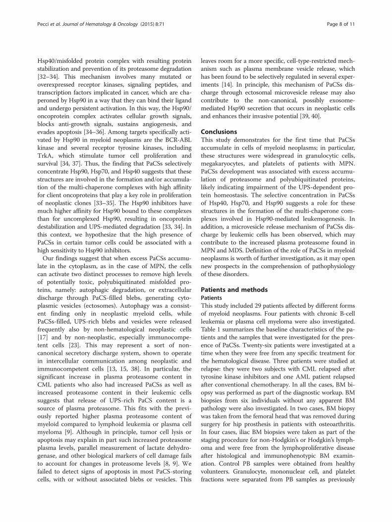
acute myeloid leukemia (AML) had no PaCSs or only afew small PaCSs, and they had no autophagic vesicles orPaCS-filled blebs (Fig. 5a–c) In BM biopsies of AMLpatients, only the cells with morphological features of
granulocytic precursors or megakaryocytes showedPaCSs. The findings on the relapsed AML patient weresimilar to those of the 7 untreated cases. Among the 8patients with myelodysplastic syndromes (MDS), PaCSswere found in BM cells of the granulocytic and megakar-yocytic lineages, as well as in PB granulocytes and plate-lets of all the analyzed patients (Fig. 5d, e). A notableexception was represented by BM granulocytic andmegakaryocytic precursors with most prominent signs ofcytoplasmic immaturity (abundant ribosomes, paucity ofcytoplasmic organelles, and reduced specific secretorygranules), which usually lacked or showed only occa-sional small PaCSs. Similarly, blast cells of the patientswith refractory anemia with excess blasts had no PaCSs(data not shown). These findings suggest that, within themyeloid neoplastic clones, PaCSs are widespread in cellswith preserved, although aberrant, maturation towardgranulocytic or megakaryocytic lineages, whereas theydo not develop in cells with maturation arrest at the
Fig. 2 PaCSs are widespread in cells of Philadelphia-negative MPN. a PaCSs in PB platelets of a patient with PV, enlarged in the inset to showbarrel-like particles and FK1 immunoreactivity. b BM megakaryocyte with small FK1-reactive peripheral PaCSs, enlarged in the inset, from a patientwith ET. c A fibroblast (f) with FK1-positive PaCSs and two intrasinusoidal erythroblasts (asterisks) are found in the BM of an individual with PMF.The fibroblast is enlarged in c1 and c2 to show the intracellular procollagen granules (arrows), juxtafibroblast collagen fibers (arrowheads), andFK1-positive PaCSs
Pecci et al. Journal of Hematology & Oncology (2015) 8:71 Page 4 of 11
early stage of blast cells or of very immature dysplasticprecursor. PaCSs of MDS presented the same immunore-activity pattern of those observed in MPN; in particular,PaCSs of MDS patients also showed selective concentra-tion of Hsp40, Hsp70, and Hps90 (data not shown). More-over, the presence of PaCS-filled cytoplasmic membraneblebs, associated with PaCS-filled vesicles freely floating inthe intercellular space, has been frequently observed alsoin BM of the MDS subjects (Fig. 5d).There were no PaCSs in BM biopsies of 4 patients
with chronic B-cell leukemia or plasma cell myeloma(Additional file 1: Figure S1). Myeloma plasma cellsshowed sparse proteasome immunoreactivity in closeassociation with the extensively developed rough endo-plasmic reticulum (RER) cisternae, some of which weredilated and filled with compact, amorphous material oc-casionally forming Russel or Dutcher bodies (Additionalfile 1: Figure S1e, f ).
PaCSs in non-neoplastic BM and PBUltrastructural analysis of non-neoplastic BM biopsiesfrom 6 individuals, combined with immunogold labelingfor polyubiquitinated proteins and 20S and 19S prote-asome, showed no PaCSs in hematopoietic or stromalcells (Additional file 1: Figure S2a–e). There were occa-sional small PaCSs in cells of the granulocytic or megakar-yocytic lineages inside areas with increased macrophages,eosinophils, plasma cells, or lymphocytes, likely due tofocal inflammation. There was increased accumulation ofpolyubiquitinated proteins in the cytoplasm of some
mature BM granulocytes, sometimes concentrated incytoplasmic blebs; particularly in minute clear, non-particulate “areolae,” as previously described [19](Additional file 1: Figure S2f). PB from healthy volunteersshowed small scanty PaCSs in 10–20 % of granulocytesand platelets (Table 2; Additional file 1: Figure S2g, h),consistent with previous observations on PB granulocytesand platelets obtained from healthy subjects [19, 20].
Quantification of PaCSs in PB cells from CML patients andhealthy controlsTo confirm and quantify the increased presence of PaCSsas a feature of hematopoietic cells in MPN, we performedsoftware-assisted image analysis of electron microscopypreparations of PB granulocytes and platelets from 4 con-secutive CML patients and three healthy subjects. Themorphometric features of PaCSs are reported in Table 2.In particular, the mean area of cytoplasm occupied byPaCSs was 6.5-fold higher in CML than control granulo-cytes (P < 0.01) and 8.4-fold higher in CML than controlplatelets (P < 0.001). For both granulocytes and platelets,the increased presence of PaCSs in CML patientscompared with healthy subjects resulted from a higherpercentage of cells with PaCSs and a higher number ofPaCSs per cell.
Plasma proteasome levels in patients with MPN, MDS,and healthy controlsPlasma proteasome levels were measured in 6 patientswith CML (patients 3–8 in Table 1), 4 subjects with PV
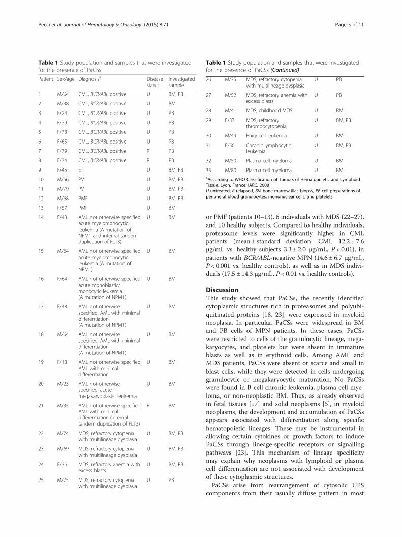
Table 1 Study population and samples that were investigatedfor the presence of PaCSs
Patient Sex/age Diagnosisa Diseasestatus
Investigatedsample
1 M/64 CML, BCR/ABL positive U BM, PB
2 M/38 CML, BCR/ABL positive U BM
3 F/24 CML, BCR/ABL positive U PB
4 F/79 CML, BCR/ABL positive U PB
5 F/78 CML, BCR/ABL positive U PB
6 F/65 CML, BCR/ABL positive U PB
7 F/79 CML, BCR/ABL positive R PB
8 F/74 CML, BCR/ABL positive R PB
9 F/45 ET U BM, PB
10 M/56 PV U BM, PB
11 M/79 PV U BM, PB
12 M/68 PMF U BM, PB
13 F/57 PMF U BM
14 F/43 AML not otherwise specified,acute myelomonocyticleukemia (A mutation ofNPM1 and internal tandemduplication of FLT3)
U BM
15 M/64 AML not otherwise specified,acute myelomonocyticleukemia (A mutation ofNPM1)
U BM
16 F/64 AML not otherwise specified,acute monoblastic/monocytic leukemia(A mutation of NPM1)
U BM
17 F/48 AML not otherwisespecified, AML with minimaldifferentiation(A mutation of NPM1)
U BM
18 M/64 AML not otherwisespecified, AML with minimaldifferentiation(A mutation of NPM1)
U BM
19 F/18 AML not otherwise specified,AML with minimaldifferentiation
U BM
20 M/23 AML not otherwisespecified, acutemegakaryoblastic leukemia
U BM
21 M/35 AML not otherwise specified,AML with minimaldifferentiation (internaltandem duplication of FLT3)
R BM
22 M/74 MDS, refractory cytopeniawith multilineage dysplasia
U BM, PB
23 M/69 MDS, refractory cytopeniawith multilineage dysplasia
U BM, PB
24 F/35 MDS, refractory anemia withexcess blasts
U BM, PB
25 M/75 MDS, refractory cytopeniawith multilineage dysplasia
U PB
Table 1 Study population and samples that were investigatedfor the presence of PaCSs (Continued)
26 M/75 MDS, refractory cytopeniawith multilineage dysplasia
U PB
27 M/52 MDS, refractory anemia withexcess blasts
U PB
28 M/4 MDS, childhood MDS U BM
29 F/37 MDS, refractorythrombocytopenia
U BM, PB
30 M/49 Hairy cell leukemia U BM
31 F/50 Chronic lymphocyticleukemia
U BM, PB
32 M/50 Plasma cell myeloma U BM
33 M/80 Plasma cell myeloma U BMaAccording to WHO Classification of Tumors of Hematopoietic and LymphoidTissue. Lyon, France: IARC, 2008U untreated, R relapsed, BM bone marrow iliac biopsy, PB cell preparations ofperipheral blood granulocytes, mononuclear cells, and platelets
Pecci et al. Journal of Hematology & Oncology (2015) 8:71 Page 5 of 11
or PMF (patients 10–13), 6 individuals with MDS (22–27),and 10 healthy subjects. Compared to healthy individuals,proteasome levels were significantly higher in CMLpatients (mean ± standard deviation: CML 12.2 ± 7.6μg/mL vs. healthy subjects 3.3 ± 2.0 μg/mL, P < 0.01), inpatients with BCR/ABL-negative MPN (14.6 ± 6.7 μg/mL,P < 0.001 vs. healthy controls), as well as in MDS indivi-duals (17.5 ± 14.3 μg/mL, P < 0.01 vs. healthy controls).
DiscussionThis study showed that PaCSs, the recently identifiedcytoplasmic structures rich in proteasomes and polyubi-quitinated proteins [18, 23], were expressed in myeloidneoplasia. In particular, PaCSs were widespread in BMand PB cells of MPN patients. In these cases, PaCSswere restricted to cells of the granulocytic lineage, mega-karyocytes, and platelets but were absent in immatureblasts as well as in erythroid cells. Among AML andMDS patients, PaCSs were absent or scarce and small inblast cells, while they were detected in cells undergoinggranulocytic or megakaryocytic maturation. No PaCSswere found in B-cell chronic leukemia, plasma cell mye-loma, or non-neoplastic BM. Thus, as already observedin fetal tissues [17] and solid neoplasms [5], in myeloidneoplasms, the development and accumulation of PaCSsappears associated with differentiation along specifichematopoietic lineages. These may be instrumental inallowing certain cytokines or growth factors to inducePaCSs through lineage-specific receptors or signallingpathways [23]. This mechanism of lineage specificitymay explain why neoplasms with lymphoid or plasmacell differentiation are not associated with developmentof these cytoplasmic structures.PaCSs arise from rearrangement of cytosolic UPS
components from their usually diffuse pattern in most
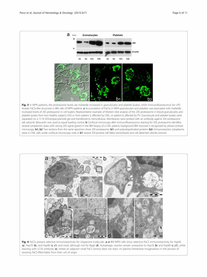
Fig. 3 In MPN patients, the proteasome levels are markedly increased in granulocytes and platelet lysates, while immunofluorescence for UPSreveals PaCSs-like structures in BM cells of MPN patients. a Accumulation of PaCSs in MPN granulocytes and platelets was associated with markedlyincreased levels of 20S proteasome in cell lysates. Representative example of Western blot analysis of the 20S proteasome in blood granulocytes andplatelet lysates from two healthy subjects (HS) or from patient 3, affected by CML, or patient 8, affected by PV. Granulocyte and platelet lysates wereseparated on a 12 % SDS/polyacrylamide gel and transferred to nitrocellulose. Membranes were probed with an antibody against 20S proteasome(α6 subunit). Beta-actin was used as equal loading control. b Confocal microscopy after immunofluorescence staining for 20S proteasome identifiesseveral cytoplasmic areas with strong 20S signal (green) in the BM biopsy of a CML patient; background BM structure is recognized by phase-contrastmicroscopy. b1, b2 Two sections from the same specimen show 20S proteasome (b1) and polyubiquitinated proteins (b2) immunoreactive cytoplasmicareas in CML cells under confocal microscopy; note in b1 several 20S-positive cell blebs (arrowheads) and cell detached vesicles (arrows)
Fig. 4 PaCSs present selective immunoreactivity for chaperone molecules. a–e BM MPN cells show selective PaCS immunoreactivity for Hsp40(a), Hsp70 (b), and Hsp90 (c, c1, and inset), although not for Bag6 (d). Autophagic vesicles remain unreactive to Hsp70 (b) and Hsp90 (c, c1), whilereacting with LC3A antibody (e), where an adjacent small PaCS (arrow) does not react. mi plasma membrane invaginations in the process ofsevering PaCS-filled blebs from their cell of origin
Pecci et al. Journal of Hematology & Oncology (2015) 8:71 Page 6 of 11
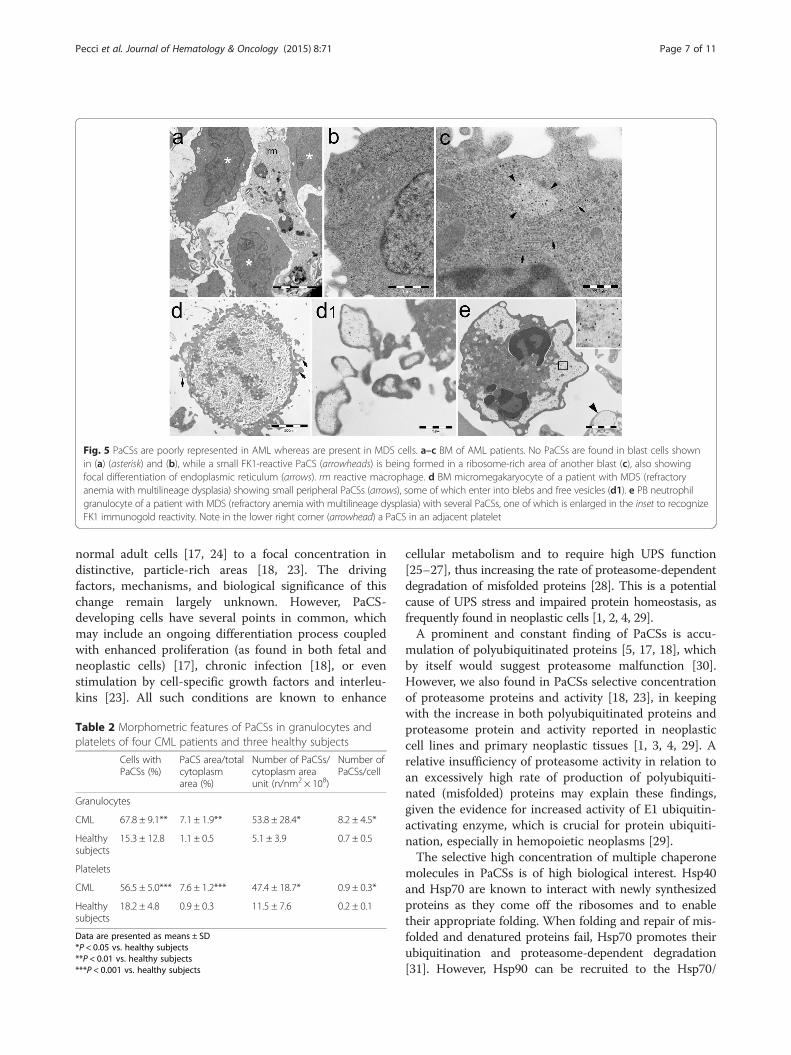
Fig. 5 PaCSs are poorly represented in AML whereas are present in MDS cells. a–c BM of AML patients. No PaCSs are found in blast cells shownin (a) (asterisk) and (b), while a small FK1-reactive PaCS (arrowheads) is being formed in a ribosome-rich area of another blast (c), also showingfocal differentiation of endoplasmic reticulum (arrows). rm reactive macrophage. d BM micromegakaryocyte of a patient with MDS (refractoryanemia with multilineage dysplasia) showing small peripheral PaCSs (arrows), some of which enter into blebs and free vesicles (d1). e PB neutrophilgranulocyte of a patient with MDS (refractory anemia with multilineage dysplasia) with several PaCSs, one of which is enlarged in the inset to recognizeFK1 immunogold reactivity. Note in the lower right corner (arrowhead) a PaCS in an adjacent platelet
Pecci et al. Journal of Hematology & Oncology (2015) 8:71 Page 7 of 11
normal adult cells [17, 24] to a focal concentration indistinctive, particle-rich areas [18, 23]. The drivingfactors, mechanisms, and biological significance of thischange remain largely unknown. However, PaCS-developing cells have several points in common, whichmay include an ongoing differentiation process coupledwith enhanced proliferation (as found in both fetal andneoplastic cells) [17], chronic infection [18], or evenstimulation by cell-specific growth factors and interleu-kins [23]. All such conditions are known to enhance
Table 2 Morphometric features of PaCSs in granulocytes andplatelets of four CML patients and three healthy subjects
Cells withPaCSs (%)
PaCS area/totalcytoplasmarea (%)
Number of PaCSs/cytoplasm areaunit (n/nm2 × 108)
Number ofPaCSs/cell
Granulocytes
CML 67.8 ± 9.1** 7.1 ± 1.9** 53.8 ± 28.4* 8.2 ± 4.5*
Healthysubjects
15.3 ± 12.8 1.1 ± 0.5 5.1 ± 3.9 0.7 ± 0.5
Platelets
CML 56.5 ± 5.0*** 7.6 ± 1.2*** 47.4 ± 18.7* 0.9 ± 0.3*
Healthysubjects
18.2 ± 4.8 0.9 ± 0.3 11.5 ± 7.6 0.2 ± 0.1
Data are presented as means ± SD*P < 0.05 vs. healthy subjects**P < 0.01 vs. healthy subjects***P < 0.001 vs. healthy subjects
cellular metabolism and to require high UPS function[25–27], thus increasing the rate of proteasome-dependentdegradation of misfolded proteins [28]. This is a potentialcause of UPS stress and impaired protein homeostasis, asfrequently found in neoplastic cells [1, 2, 4, 29].A prominent and constant finding of PaCSs is accu-
mulation of polyubiquitinated proteins [5, 17, 18], whichby itself would suggest proteasome malfunction [30].However, we also found in PaCSs selective concentrationof proteasome proteins and activity [18, 23], in keepingwith the increase in both polyubiquitinated proteins andproteasome protein and activity reported in neoplasticcell lines and primary neoplastic tissues [1, 3, 4, 29]. Arelative insufficiency of proteasome activity in relation toan excessively high rate of production of polyubiquiti-nated (misfolded) proteins may explain these findings,given the evidence for increased activity of E1 ubiquitin-activating enzyme, which is crucial for protein ubiquiti-nation, especially in hemopoietic neoplasms [29].The selective high concentration of multiple chaperone
molecules in PaCSs is of high biological interest. Hsp40and Hsp70 are known to interact with newly synthesizedproteins as they come off the ribosomes and to enabletheir appropriate folding. When folding and repair of mis-folded and denatured proteins fail, Hsp70 promotes theirubiquitination and proteasome-dependent degradation[31]. However, Hsp90 can be recruited to the Hsp70/
Pecci et al. Journal of Hematology & Oncology (2015) 8:71 Page 8 of 11
Hsp40/misfolded protein complex with resulting proteinstabilization and prevention of its proteasome degradation[32–34]. This mechanism involves many mutated oroverexpressed receptor kinases, signaling peptides, andtranscription factors implicated in cancer, which are cha-peroned by Hsp90 in a way that they can bind their ligandand undergo persistent activation. In this way, the Hsp90/oncoprotein complex activates cellular growth signals,blocks anti-growth signals, sustains angiogenesis, andevades apoptosis [34–36]. Among targets specifically acti-vated by Hsp90 in myeloid neoplasms are the BCR-ABLkinase and several receptor tyrosine kinases, includingTrkA, which stimulate tumor cell proliferation andsurvival [34, 37]. Thus, the finding that PaCSs selectivelyconcentrate Hsp90, Hsp70, and Hsp40 suggests that thesestructures are involved in the formation and/or accumula-tion of the multi-chaperone complexes with high affinityfor client oncoproteins that play a key role in proliferationof neoplastic clones [33–35]. The Hsp90 inhibitors havemuch higher affinity for Hsp90 bound to these complexesthan for uncomplexed Hsp90, resulting in oncoproteindestabilization and UPS-mediated degradation [33, 34]. Inthis context, we hypothesize that the high presence ofPaCSs in certain tumor cells could be associated with ahigh sensitivity to Hsp90 inhibitors.Our findings suggest that when excess PaCSs accumu-
late in the cytoplasm, as in the case of MPN, the cellscan activate two distinct processes to remove high levelsof potentially toxic, polyubiquitinated misfolded pro-teins, namely: autophagic degradation, or extracellulardischarge through PaCS-filled blebs, generating cyto-plasmic vesicles (ectosomes). Autophagy was a consist-ent finding only in neoplastic myeloid cells, whilePaCSs-filled, UPS-rich blebs and vesicles were releasedfrequently also by non-hematological neoplastic cells[17] and by non-neoplastic, especially immunocompe-tent cells [23]. This may represent a sort of non-canonical secretory discharge system, shown to operatein intercellular communication among neoplastic andimmunocompetent cells [13, 15, 38]. In particular, thesignificant increase in plasma proteasome content inCML patients who also had increased PaCSs as well asincreased proteasome content in their leukemic cellssuggests that release of UPS-rich PaCS content is asource of plasma proteasome. This fits with the previ-ously reported higher plasma proteasome content ofmyeloid compared to lymphoid leukemia or plasma cellmyeloma [9]. Although in principle, tumor cell lysis orapoptosis may explain in part such increased proteasomeplasma levels, parallel measurement of lactate dehydro-genase, and other biological markers of cell damage failsto account for changes in proteasome levels [8, 9]. Wefailed to detect signs of apoptosis in most PaCS-storingcells, with or without associated blebs or vesicles. This
leaves room for a more specific, cell-type-restricted mech-anism such as plasma membrane vesicle release, whichhas been found to be selectively regulated in several exper-iments [14]. In principle, this mechanism of PaCSs dis-charge through ectosomal microvesicle release may alsocontribute to the non-canonical, possibly exosome-mediated Hsp90 secretion that occurs in neoplastic cellsand enhances their invasive potential [39, 40].
ConclusionsThis study demonstrates for the first time that PaCSsaccumulate in cells of myeloid neoplasms; in particular,these structures were widespread in granulocytic cells,megakaryocytes, and platelets of patients with MPN.PaCSs development was associated with excess accumu-lation of proteasome and polyubiquitinated proteins,likely indicating impairment of the UPS-dependent pro-tein homeostasis. The selective concentration in PaCSsof Hsp40, Hsp70, and Hsp90 suggests a role for thesestructures in the formation of the multi-chaperone com-plexes involved in Hsp90-mediated leukemogenesis. Inaddition, a microvesicle release mechanism of PaCSs dis-charge by leukemic cells has been observed, which maycontribute to the increased plasma proteasome found inMPN and MDS. Definition of the role of PaCSs in myeloidneoplasms is worth of further investigation, as it may opennew prospects in the comprehension of pathophysiologyof these disorders.
Patients and methodsPatientsThis study included 29 patients affected by different formsof myeloid neoplasms. Four patients with chronic B-cellleukemia or plasma cell myeloma were also investigated.Table 1 summarizes the baseline characteristics of the pa-tients and the samples that were investigated for the pres-ence of PaCSs. Twenty-six patients were investigated at atime when they were free from any specific treatment forthe hematological disease. Three patients were studied atrelapse: they were two subjects with CML relapsed aftertyrosine kinase inhibitors and one AML patient relapsedafter conventional chemotherapy. In all the cases, BM bi-opsy was performed as part of the diagnostic workup. BMbiopsies from six individuals without any apparent BMpathology were also investigated. In two cases, BM biopsywas taken from the femoral head that was removed duringsurgery for hip prosthesis in patients with osteoarthritis.In four cases, iliac BM biopsies were taken as part of thestaging procedure for non-Hodgkin’s or Hodgkin’s lymph-oma and were free from the lymphoproliferative diseaseafter histological and immunophenotypic BM examin-ation. Control PB samples were obtained from healthyvolunteers. Granulocyte, mononuclear cell, and plateletfractions were separated from PB samples as previously
Pecci et al. Journal of Hematology & Oncology (2015) 8:71 Page 9 of 11
reported [20]. The study subjects or their legal guardiansgave informed consent for this study, which was approvedby the Institutional Review Board of the IRCCS PoliclinicoSan Matteo Foundation, Pavia, Italy.
Electron microscopy and immunogold analysisBM biopsies or PB cell preparations were fixed immedi-ately after sampling in 2.5 % glutaraldehyde and 2 % para-formaldehyde in pH 7.3 cacodylate buffer for 4 h at 4 °C,followed by post-fixation in 1.5 % osmium tetroxide for 1 hat room temperature, and Epon-Araldite embedding [18].Ultrathin sections (~70 nm thick) were cut from the resinblocks and stained with uranyl acetate/lead citrate for con-ventional electron microscopy or underwent immunogoldstaining as previously reported [17, 23]. Sections wereincubated in 10 % normal goat or bovine serum (Dako,Glostrup, Denmark) for 1 h at room temperature, followedby incubation with primary antibodies (as specified below)and secondary anti-mouse, anti-rabbit, or anti-goat IgG orIgM labeled with 6-, 10-, 15-, or 20-nm gold particles (Aur-ion ImmunoGold Reagents, Wageningen, Netherlands; orBB International, Cardiff, UK). Sections were counter-stained with uranyl acetate/lead citrate. Specimens wereanalyzed by a Jeol JEM-1200 EX II (JEOL Ltd, Tokyo,Japan) transmission electron microscope equipped with anOlympus Mega View III CCD camera (Tokyo, Japan). Thefollowing primary antibodies were used: mouse FK1 mono-clonal against polyubiquitinated proteins [41] (Enzo LifeSciences International, Plymouth Meeting, PA, USA);rabbit polyclonal against the 20S proteasome, αβ or β5isubunits (Calbiochem, La Jolla, CA, USA); rabbit poly-clonal against 19S proteasome, S2 subunit (Calbiochem);rabbit polyclonal against Hsp40 (LS Bio, Seattle, WA,USA); goat polyclonal against Hsp70 (Santa Cruz Biotech-nology, Santa Cruz, CA, USA); mouse 4F3-E8 monoclonalagainst Hsp90 (Novus Biologicals, Littleton, CO, USA);rabbit polyclonal against BAG6 (Santa Cruz Biotechnol-ogy); rabbit polyclonal against LC3A (Abgent, San Diego,CA, USA). The specificity of immunogold labeling wasevaluated by omitting the specific antibodies in the firstlayer of the procedure or by substituting them with non-immune IgG or IgM (Santa Cruz Biotechnology) or unre-lated antibodies.
Software-assisted image analysisImage analysis was carried out on PB granulocyte andplatelet electron microscopy preparations from 4 con-secutive patients affected by CML (patients 3–6 inTable 1) and three healthy subjects. Patient samples werecollected and processed for TEM simultaneously withcontrol samples in three different experiments. Sectionswere analyzed after immunogold staining with the FK1antibody against polyubiquitinated proteins. Images weretaken at a 25,000× magnification, and a mean number of
25.7 ± 1.8 granulocyte sections and 65 ± 10.2 plateletsections were analyzed for each subject. Morphometricobservations were carried out by the ITEM soft imagingsystem (Olympus Soft Imaging Solutions GmbH). Thefollowing parameters were directly measured: presence,number, and area of each individual PaCS; area of thecytoplasm of each analyzed cell; and number of immuno-gold particles inside PaCSs and in the cytoplasm outsidePaCSs. Data were expressed as means ± SD. Statisticalanalysis was performed by two-tailed Student’s t test.
Immunofluorescence analysisSemithin (about 0.1 μm) ultramicrotomic sections wereobtained from the resin blocks and used for immuno-fluorescence confocal and phase contrast microscopy.After washing with PBS, sections were incubated over-night with primary and then with secondary antibodiesas described [23]. Primary antibodies against 19S, 20S,and polyubiquitinated proteins are the same used forelectron microscopy. Secondary antibodies were AlexaFluor 488-conjugated anti-mouse or anti-rabbit IgG (Lifetechnologies, Paisley, UK). Specimens were analyzed by aTCS SP5II confocal laser scanning microscope equippedwith PL APO 40x/1.25 NA and 63/1.40 NA oil-immersionobjectives (Leica, Heidelberg, Germany).
Western blotting analysisGranulocytes and platelets lysates were prepared anddissociated as previously described [20].Equal amounts of samples were separated in a 12 %
polyacrylamide SDS-PAGE gel and transferred to nitrocel-lulose (Bio-Rad, Hercules, CA, USA). After blocking with5 % not-fat milk, membranes were probed with mousemonoclonal MCP against proteasome 20S α6 subunit(Enzo Life Sciences) or mouse monoclonal AC-15 againstβ-actin (Sigma, St. Louis, MO, USA). Membranes werethen incubated with horseradish peroxidase-conjugatedanti-mouse antibody, and protein bands were visualizedby an enhanced chemiluminescence method (GE Health-care, Waukesha, WI, USA).
ELISA of plasma proteasome levelsPlasma samples were obtained by centrifugation of wholeblood anti-coagulated with citrate for 15 min at 1000 gand maintained at –20 °C until analysis. The proteasomelevels were measured by the 20S/26S Proteasome ELISAKit (Enzo Life Sciences). Plasma samples were diluted 1:8with the ELISA buffer provided by the kit and assayed induplicate. Optical density was read at 450 nm on amicroplate reader (Model 680; Bio-Rad, Hercules, CA,USA). Data are representative of three separate mea-surements. Statistical analysis was performed by two-tailed Student’s t test.
Pecci et al. Journal of Hematology & Oncology (2015) 8:71 Page 10 of 11
Additional file
Additional file 1: Supplementary figures. Figure S1. No PaCSs arefound in chronic B-cell leukemia and plasma cell myeloma. Figure S2.PaCSs are not found in non-neoplastic BM cells, whereas some PaCSs areidentified in PB granulocytes and platelets from healthy subjects.
Competing interestsThe authors declare that they have no competing interests.
Authors’ contributionsAP and ES designed the study, investigated patients, analyzed data, andwrote the manuscript. VN and SB performed the study and analyzed data.AV performed the study. EB, CE, PB, and PN investigated patients andperformed the study. All the authors critically revised and approved the finalversion of the manuscript. All authors read and approved the final manuscript.
AcknowledgementsThis work was supported by grants n. RF-2010.2310098 from the Italian Ministryof Health and n. 2011.0485 and n. 2012.0529 from Cariplo Foundation to IRCCSPoliclinico San Matteo Foundation and University of Pavia. We are indebted toDr. Alessandro Corso, Prof. Luca Malcovati, Prof. Umberto Magrini, andDr. Vittorio Rosti for helpful discussion about clinical and hematologicalfeatures of some of the investigated patients and to Dr. Rita Zappatorefor technical assistance.
Author details1Department of Internal Medicine, IRCCS Policlinico San Matteo Foundationand University of Pavia, Pavia, Italy. 2Department of Molecular Medicine,University of Pavia, Pavia, Italy. 3Centro Grandi Strumenti, University of Pavia,Pavia, Italy. 4Pathologic Anatomy Section, Department of DiagnosticMedicine, IRCCS Policlinico San Matteo Foundation, Pavia, Italy. 5HematologySection, Department of Oncology and Hematology, IRCCS Policlinico SanMatteo Foundation, Pavia, Italy.
Received: 18 February 2015 Accepted: 5 June 2015
References1. Kumatori A, Tanaka K, Inamura N, Sone S, Ogura T, Matsumoto T, et al.
Abnormally high expression of proteasomes in human leukemic cells.Proc Natl Acad Sci U S A. 1990;87:7071–5.
2. Kanayama H, Tanaka K, Aki M, Kagawa S, Miyaji H, Satoh M, et al. Changes inexpressions of proteasome and ubiquitin genes in human renal cancer cells.Cancer Res. 1991;51:6677–85.
3. Magill L, Lynas J, Morris TC, Walker B, Irvine AE. Proteasome proteolyticactivity in hematopoietic cells from patients with chronic myeloid leukemiaand multiple myeloma. Haematologica. 2004;89:1428–33.
4. Bazzaro M, Lee MK, Zoso A, Stirling WL, Santillan A, Shih IM, et al.Ubiquitin-proteasome system stress sensitizes ovarian cancer toproteasome inhibitor-induced apoptosis. Cancer Res. 2006;66:3754–63.
5. Necchi V, Sommi P, Vanoli A, Manca R, Ricci V, Solcia E. Proteasome particle-rich structures are widely present in human epithelial neoplasms: correlativelight, confocal and electron microscopy study. PLoS One. 2011;6, e21317.
6. Rajkumar SV, Richardson PG, Hideshima T, Anderson KC. Proteasomeinhibition as a novel therapeutic target in human cancer. J Clin Oncol.2005;23:630–9.
7. Fisher RI, Bernstein SH, Kahl BS, Djulbegovic B, Robertson MJ, de Vos S, et al.Multicenter phase II study of bortezomib in patients with relapsed orrefractory mantle cell lymphoma. J Clin Oncol. 2006;24:4867–74.
8. Wada M, Kosaka M, Saito S, Sano T, Tanaka K, Ichihara A. Serumconcentration and localization in tumor cells of proteasomes in patientswith hematologic malignancy and their pathophysiologic significance.J Lab Clin Med. 1993;121:215–23.
9. Lavabre-Bertrand T, Henry L, Carillo S, Guiraud I, Ouali A, Dutaud D, et al.Plasma proteasome level is a potential marker in patients with solid tumorsand hemopoietic malignancies. Cancer. 2001;92:2493–500.
10. Jakob C, Egerer K, Liebisch P, Türkmen S, Zavrski I, Kuckelkorn U, et al.Circulating proteasome levels are an independent prognostic factor forsurvival in multiple myeloma. Blood. 2007;109:2100–5.
11. Ma W, Kantarjian H, Bekele B, Donahue AC, Zhang X, Zhang ZJ, et al.Proteasome enzymatic activities in plasma as risk stratification of patientswith acute myeloid leukemia and advanced-stage myelodysplasticsyndrome. Clin Cancer Res. 2009;15:3820–6.
12. Lai RC, Tan SS, Teh BJ, Sze SK, Arslan F, de Kleijn DP, et al. Proteolyticpotential of the MSC exosome proteome: implications for anexosome-mediated delivery of therapeutic proteasome. Int J Proteomics.2012;2012:971907.
13. Théry C, Ostrowski M, Segura E. Membrane vesicles as conveyors ofimmune responses. Nat Rev Immunol. 2009;9:581–93.
14. Raposo G, Stoorvogel W. Extracellular vesicles: exosomes, microvesicles,and friends. J Cell Biol. 2013;200:373–83.
15. Taylor DD, Gerçel-Taylor C. Tumour-derived exosomes and their role incancer-associated T-cell signalling defects. Br J Cancer. 2005;92:305–11.
16. Bochmann I, Ebstein F, Lehmann A, Wohlschlaeger J, Sixt SU, Kloetzel PM,et al. T lymphocytes export proteasomes by way of microparticles: apossible mechanism for generation of extracellular proteasomes. J Cell MolMed. 2014;18:59–68.
17. Necchi V, Sommi P, Vitali A, Vanoli A, Savoia A, Ricci V, et al.Polyubiquitinated proteins, proteasome, and glycogen characterize theparticle-rich cytoplasmic structure (PaCS) of neoplastic and fetal cells.Histochem Cell Biol. 2014;141:483–97.
18. Necchi V, Sommi P, Ricci V, Solcia E. In vivo accumulation of Helicobacterpylori products, NOD1, ubiquitinated proteins and proteasome in a novelcytoplasmic structure. PLoS One. 2010;5, e9716.
19. Necchi V, Minelli A, Sommi P, Vitali A, Caruso R, Longoni D, et al.Ubiquitin-proteasome-rich cytoplasmic structures in neutrophils of patientswith Shwachman-Diamond syndrome. Haematologica. 2012;97:1057–63.
20. Necchi V, Balduini A, Noris P, Barozzi S, Sommi P, di Buduo C, et al.Ubiquitin/proteasome-rich particulate cytoplasmic structures (PaCSs) in theplatelets and megakaryocytes of ANKRD26-related thrombocytopenia.Thromb Haemost. 2013;109:263–71.
21. Galluzzi L, Maiuri MC, Vitale I, Zischka H, Castedo M, Zitvogel L, et al. Celldeath modalities: classification and pathophysiological implications. CellDeath Differ. 2007;14:1237–43.
22. Sivridis E, Koukourakis MI, Zois CE, Ledaki I, Ferguson DJ, Harris AL, et al.LC3A-positive light microscopy detected patterns of autophagy andprognosis in operable breast carcinomas. Am J Pathol. 2010;176:2477–89.
23. Sommi P, Necchi V, Vitali A, Montagna D, De Luigi A, Salmona M, et al.PaCS is a novel cytoplasmic structure containing functional proteasomeand inducible by cytokines/trophic factors. PLoS One. 2013;8, e82560.
24. Rivett AJ, Palmer A, Knecht E. Electron microscopic localization of themulticatalytic proteinase complex in rat liver and in cultured cells.J Histochem Cytochem. 1992;40:1165–72.
25. Giaccia AJ, Simon MC, Johnson R. The biology of hypoxia: the role ofoxygen sensing in development, normal function, and disease. Genes Dev.2004;18:2183–94.
26. Kuboki Y, Shiratori K, Hatori T, Fujita I, Kimijima A, Yamamoto M, et al.Association of epidermal growth factor receptor and mitogen-activatedprotein kinase with cystic neoplasms of the pancreas. Mod Pathol.2010;23:1127–35.
27. Lunt SY, Vander Heiden MG. Aerobic glycolysis: meeting the metabolicrequirements of cell proliferation. Annu Rev Cell Dev Biol. 2011;27:441–64.
28. Yewdell JW, Nicchitta CV. The DRiP hypothesis decennial: support,controversy, refinement and extension. Trends Immunol. 2006;27:368–73.
29. Xu GW, Ali M, Wood TE, Wong D, Maclean N, Wang X, et al. The ubiquitin-activating enzyme E1 as a therapeutic target for the treatment of leukemiaand multiple myeloma. Blood. 2010;115:2251–9.
30. Navon A, Ciechanover A. The 26 S proteasome: from basic mechanisms todrug targeting. J Biol Chem. 2009;284:33713–8.
31. Wang RE. Targeting heat shock proteins 70/90 and proteasome for cancertherapy. Curr Med Chem. 2011;18:4250–64.
32. Hernández MP, Sullivan WP, Toft DO. The assembly and intermolecularproperties of the hsp70-Hop-hsp90 molecular chaperone complex.J Biol Chem. 2002;277:38294–304.
33. Kamal A, Thao L, Sensintaffar J, Zhang L, Boehm MF, Fritz LC, et al. Ahigh-affinity conformation of Hsp90 confers tumour selectivity on Hsp90inhibitors. Nature. 2003;425:407–10.
34. Moulick K, Ahn JH, Zong H, Rodina A, Cerchietti L, Gomes DaGama EM,et al. Affinity-based proteomics reveal cancer-specific networks coordinatedby Hsp90. Nat Chem Biol. 2011;7:818–26.
Pecci et al. Journal of Hematology & Oncology (2015) 8:71 Page 11 of 11
35. Neckers L. Heat shock protein 90: the cancer chaperone. J Biosci.2007;32:517–30.
36. Miyata Y, Nakamoto H, Neckers L. The therapeutic target Hsp90 and cancerhallmarks. Curr Pharm Des. 2013;19:347–65.
37. Rao R, Nalluri S, Fiskus W, Balusu R, Joshi A, Mudunuru U, et al. Heat shockprotein 90 inhibition depletes TrkA levels and signaling in human acuteleukemia cells. Mol Cancer Ther. 2010;9:2232–42.
38. Sixt SU, Dahlmann B. Extracellular, circulating proteasomes and ubiquitin-incidence and relevance. Biochim Biophys Acta. 1782;2008:817–23.
39. McCready J, Sims JD, Chan D, Jay DG. Secretion of extracellular hsp90alphavia exosomes increases cancer cell motility: a role for plasminogenactivation. BMC Cancer. 2010;10:294.
40. Wang X, Song X, Zhuo W, et al. The regulatory mechanism of Hsp90alphasecretion and its function in tumor malignancy. Proc Natl Acad Sci U S A.2009;106:21288–93.
41. Fujimuro M, Sawada H, Yokosawa H. Production and characterization ofmonoclonal antibodies specific to multi-ubiquitin chains of polyubiquitinatedproteins. FEBS Lett. 1994;349:173–80.
Submit your next manuscript to BioMed Centraland take full advantage of:
• Convenient online submission
• Thorough peer review
• No space constraints or color figure charges
• Immediate publication on acceptance
• Inclusion in PubMed, CAS, Scopus and Google Scholar
• Research which is freely available for redistribution
Submit your manuscript at www.biomedcentral.com/submit