Embed Size (px)
DESCRIPTION
dalam presentasi ini akan dijalankan apa itu buta warna bagaimana cara penurunannya dan bagaimana cara mendiagnosa. selamat membaca...
Citation preview
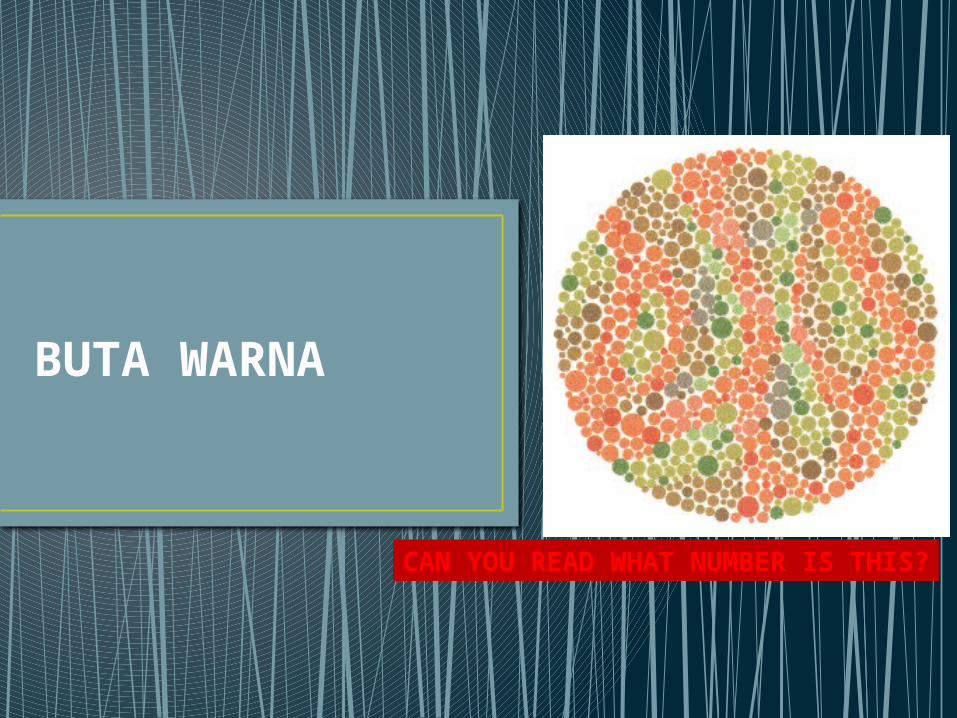
BUTA WARNA
CAN YOU READ WHAT NUMBER IS THIS?
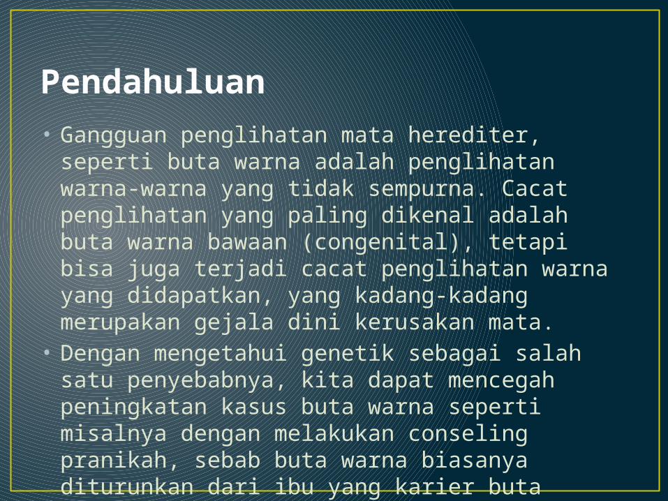
Pendahuluan• Gangguan penglihatan mata herediter, seperti
buta warna adalah penglihatan warna-warna yang tidak sempurna. Cacat penglihatan yang paling dikenal adalah buta warna bawaan (congenital), tetapi bisa juga terjadi cacat penglihatan warna yang didapatkan, yang kadang-kadang merupakan gejala dini kerusakan mata.
• Dengan mengetahui genetik sebagai salah satu penyebabnya, kita dapat mencegah peningkatan kasus buta warna seperti misalnya dengan melakukan conseling pranikah, sebab buta warna biasanya diturunkan dari ibu yang karier buta warna.
Anatomi Bola Mata
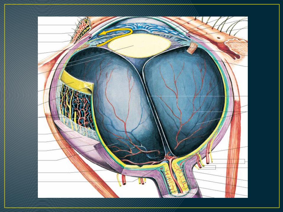
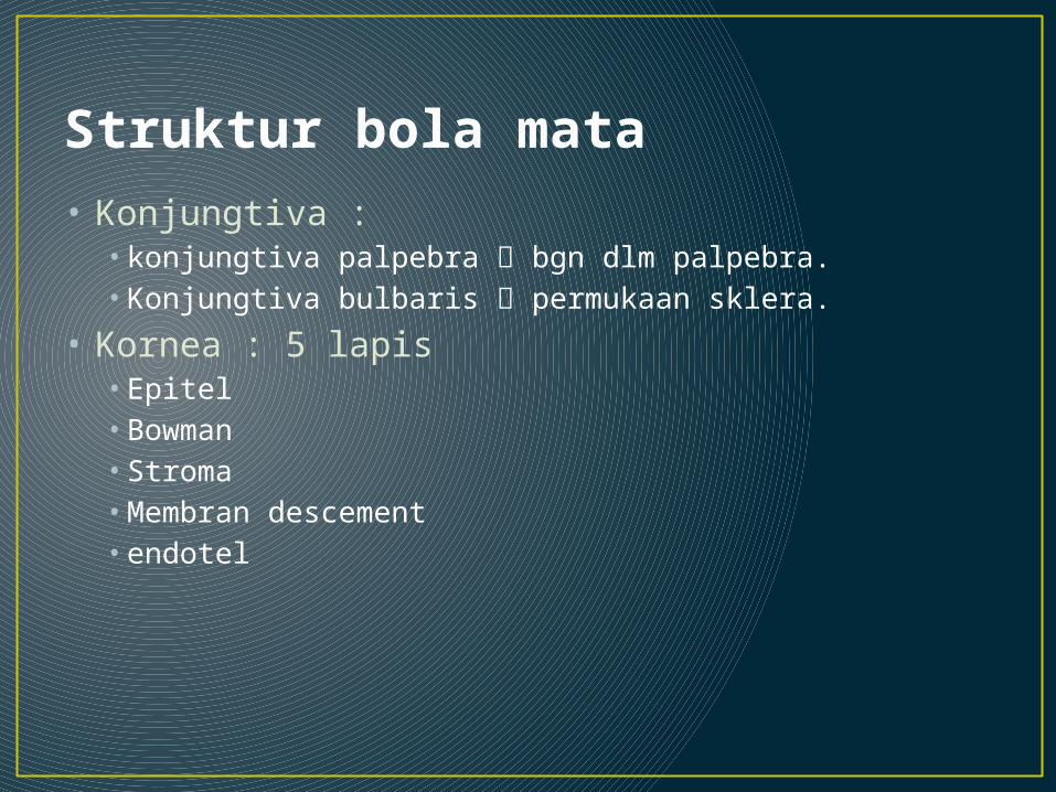
Struktur bola mata • Konjungtiva :
• konjungtiva palpebra bgn dlm palpebra. • Konjungtiva bulbaris permukaan sklera.
• Kornea : 5 lapis • Epitel • Bowman• Stroma • Membran descement • endotel

Struktur bola mata • Bilik mata depan :
• antara kornea perifer dan pangkal iris
• Pupil : daerah hitam di tengah iris • Iris : perpanjangan korpus siliare pisahkan BMD
dan BMB.• Lensa : bikonveks, avaskular, transparan
difiksasi oleh zonula zinii.• Sclera : jaringan fibrous tdd kolagen
berbatasan dengan kornea di anterior dan duramater n.II diposterior
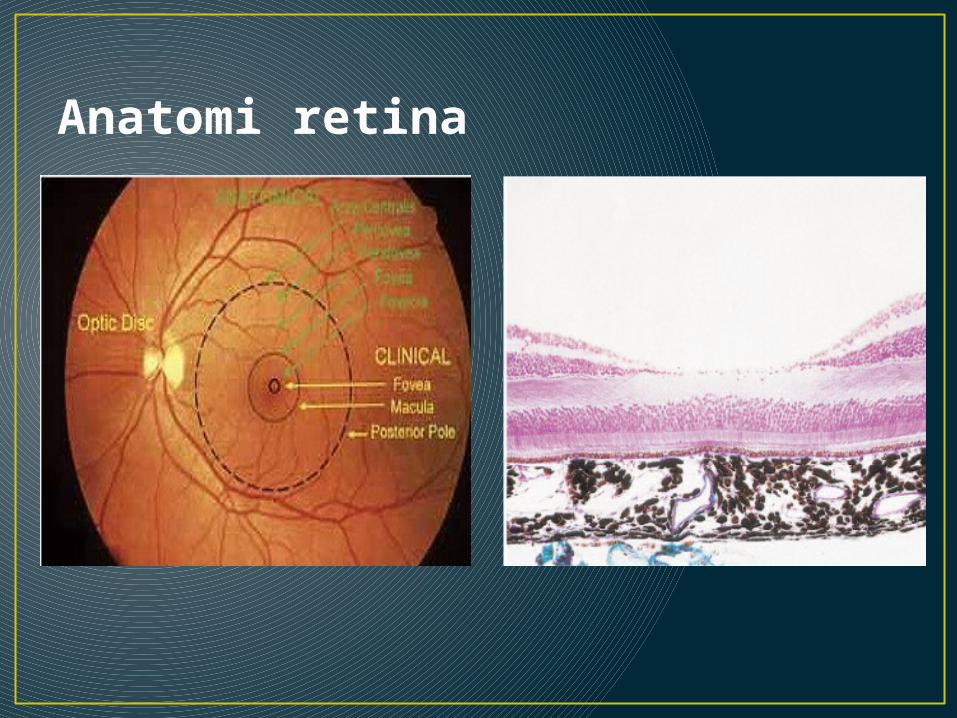
Anatomi retina
• Retina jaringan saraf melapisi bagian dalam 2/3 posterior dinding bola mata mengandung reseptor penerima rangsangan cahaya.
• Retina bagian mata yang peka terhadap cahaya karena mngandung sel-sel kerucut
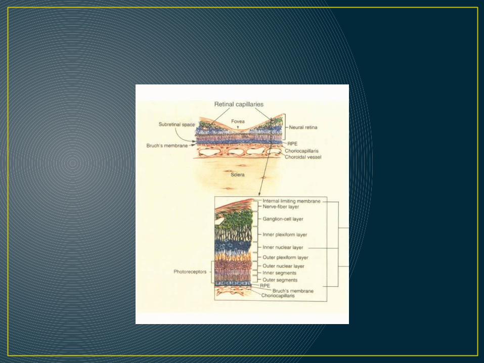
• Lapisan-lapisan retina(mulai dari sisi dalam) : 1. membran limitans interna 2. lapisan serat saraf akson-akson sel
ganglion.3. lapisan sel ganglion4. lapisan pleksiformis sambungan- sambungan sel ganglion dngan sel amakrin dan sel bipolar. 5. lapisan inti dalam sel2 bipolar, amakrin dan horizontal.6. lapisan pleksiformis luar sambungan sel bipolar dan sel horisontal dengan fotoreseptor. 7. lapisan inti luar sel fotoreseptor
8. membran limitans eksterna 9. lapisan fotoreseptor segmen dalam dan luar batang dan kerucut. 10. epitel pigmen retina.
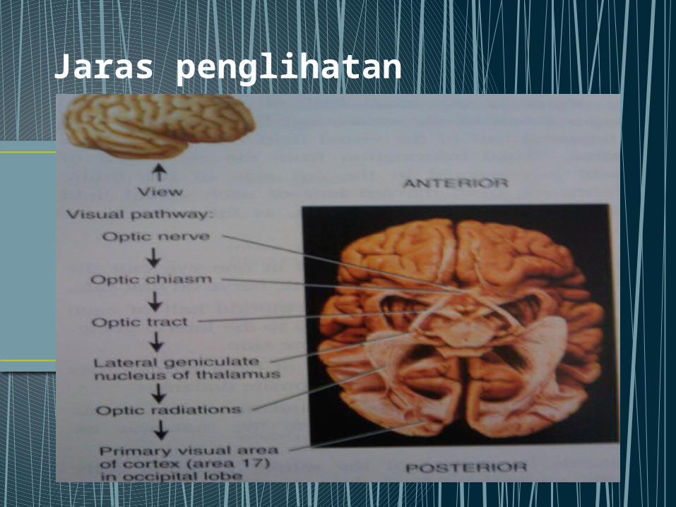
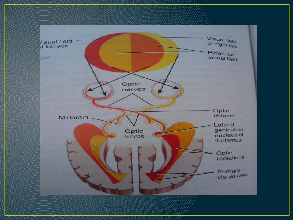
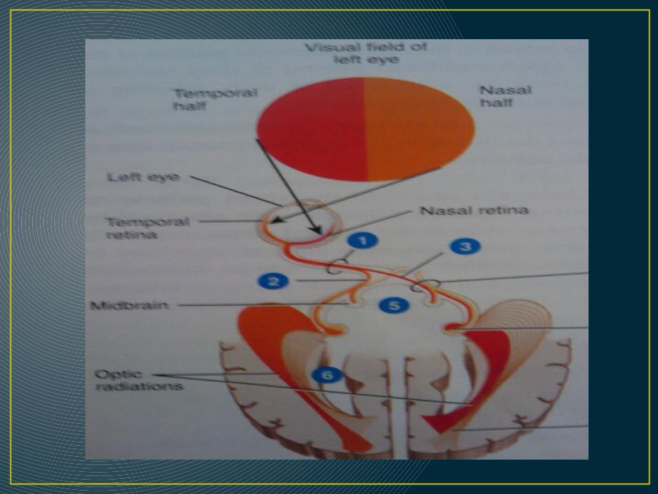
Jaras penglihatan
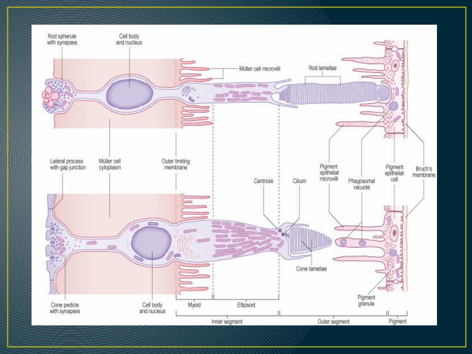
Sel batang dan kerucut
1• Segmen luar
2• Segmen dalam
3
• Inti
4• Badan Sinaptik
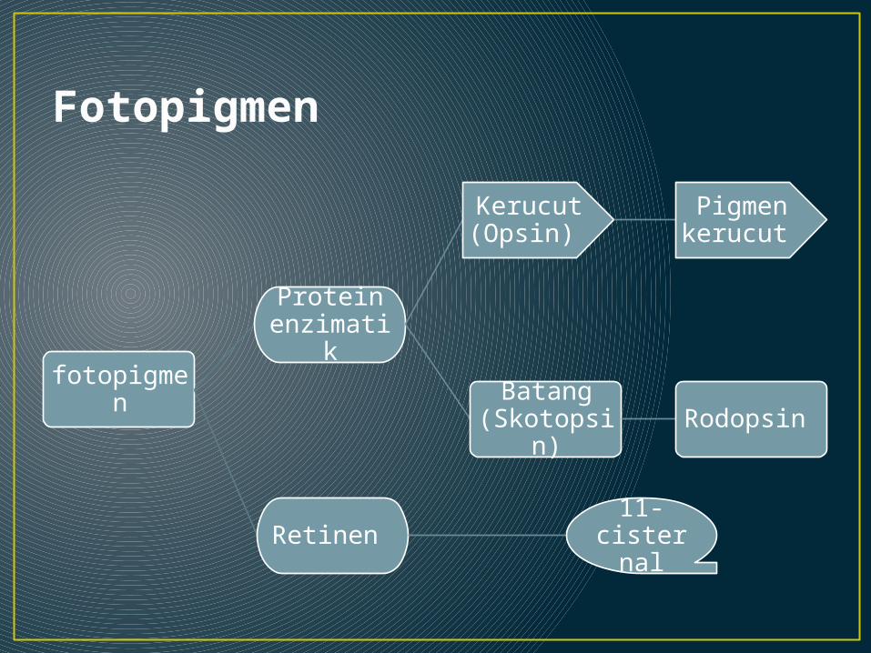
Fotopigmen
fotopigmen
Protein enzimatik
Kerucut (Opsin)
Pigmen kerucut
Batang (Skotopsin) Rodopsin
Retinen 11-
cisternal
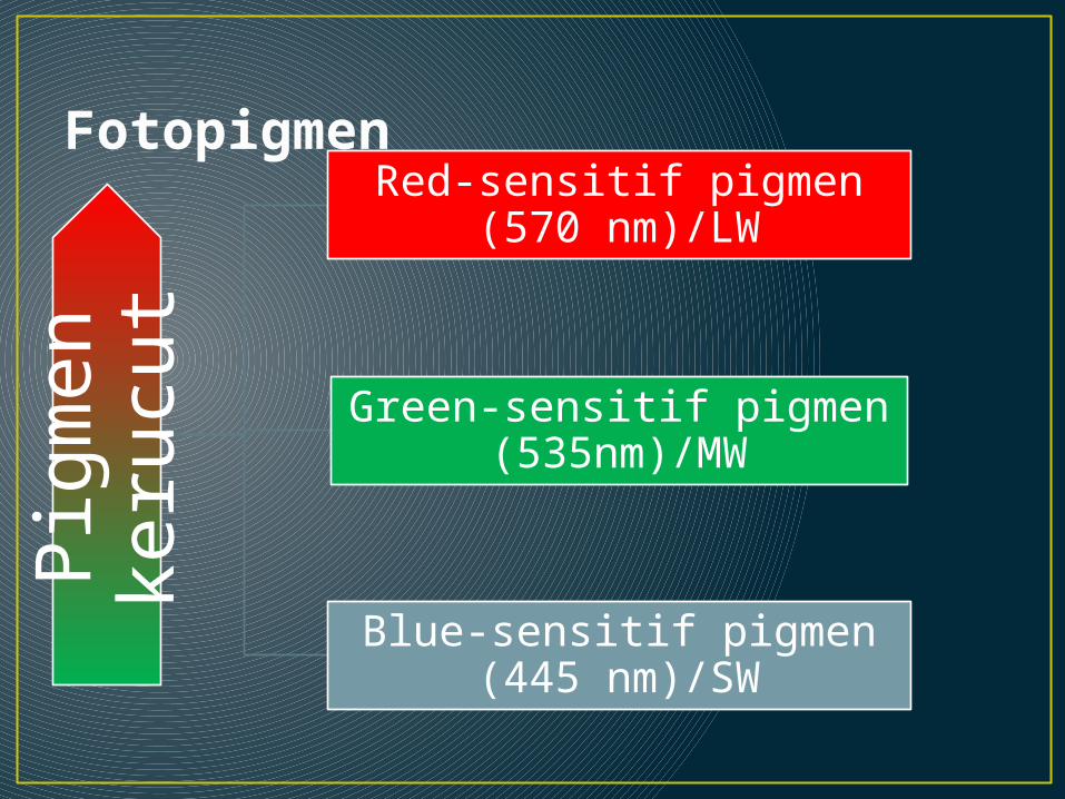
Fotopigmen Pig
men
keru
cut
Red-sensitif pigmen (570 nm)/LW
Green-sensitif pigmen (535nm)/MW
Blue-sensitif pigmen (445 nm)/SW
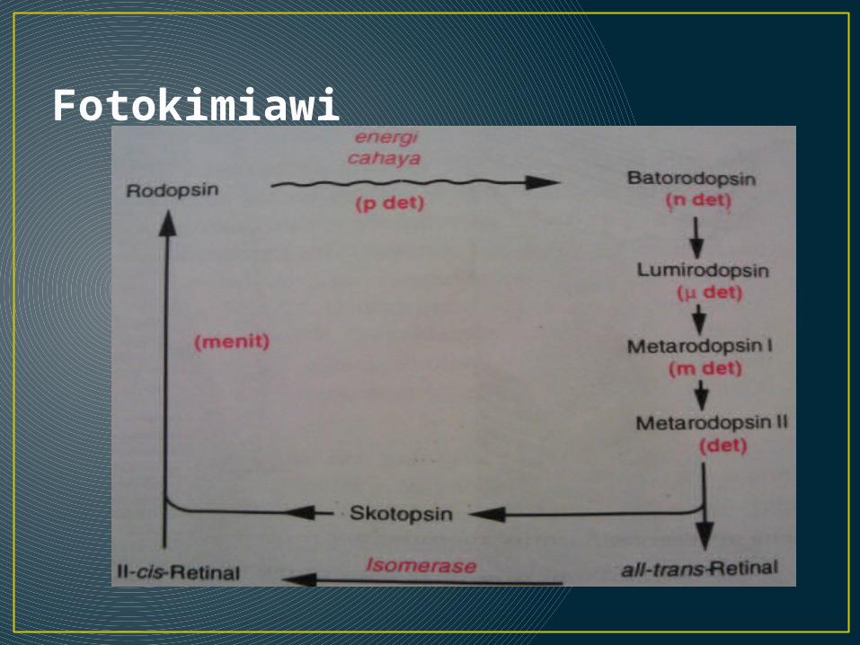
Fotokimiawi
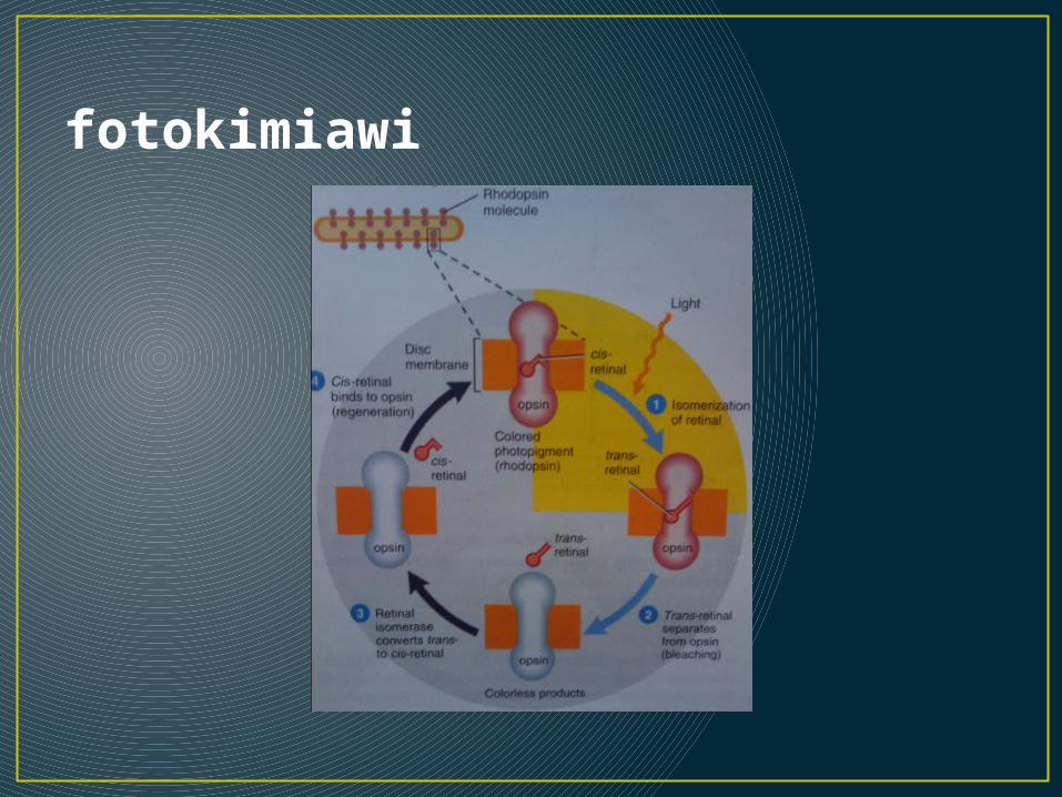
fotokimiawi
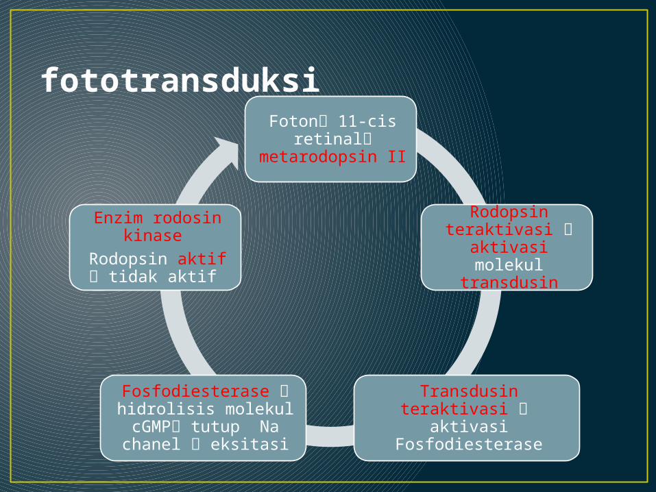
fototransduksiFoton 11-cis
retinal metarodopsin II
Rodopsin teraktivasi
aktivasi molekul transdusin
Transdusin teraktivasi aktivasi Fosfodiesterase
Fosfodiesterase hidrolisis molekul cGMP tutup Na chanel eksitasi
Enzim rodosin kinase
Rodopsin aktif tidak aktif
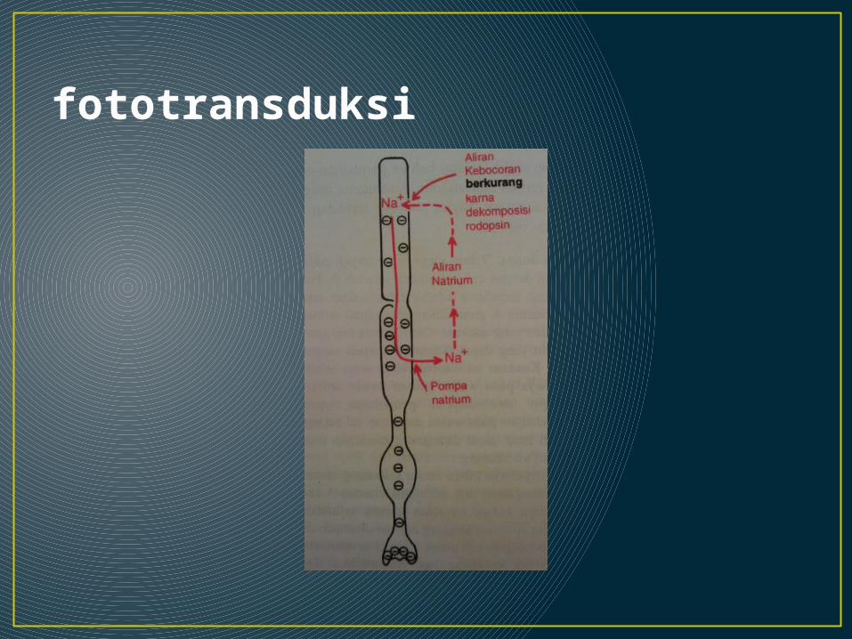
fototransduksi
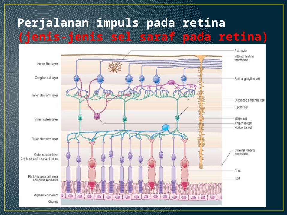
Perjalanan impuls pada retina(jenis-jenis sel saraf pada retina)

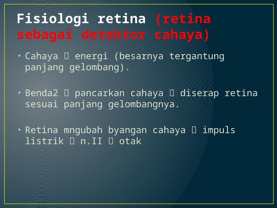
Fisiologi retina (retina sebagai detektor cahaya)• Cahaya energi (besarnya tergantung panjang
gelombang).
• Benda2 pancarkan cahaya diserap retina sesuai panjang gelombangnya.
• Retina mngubah byangan cahaya impuls listrik n.II otak
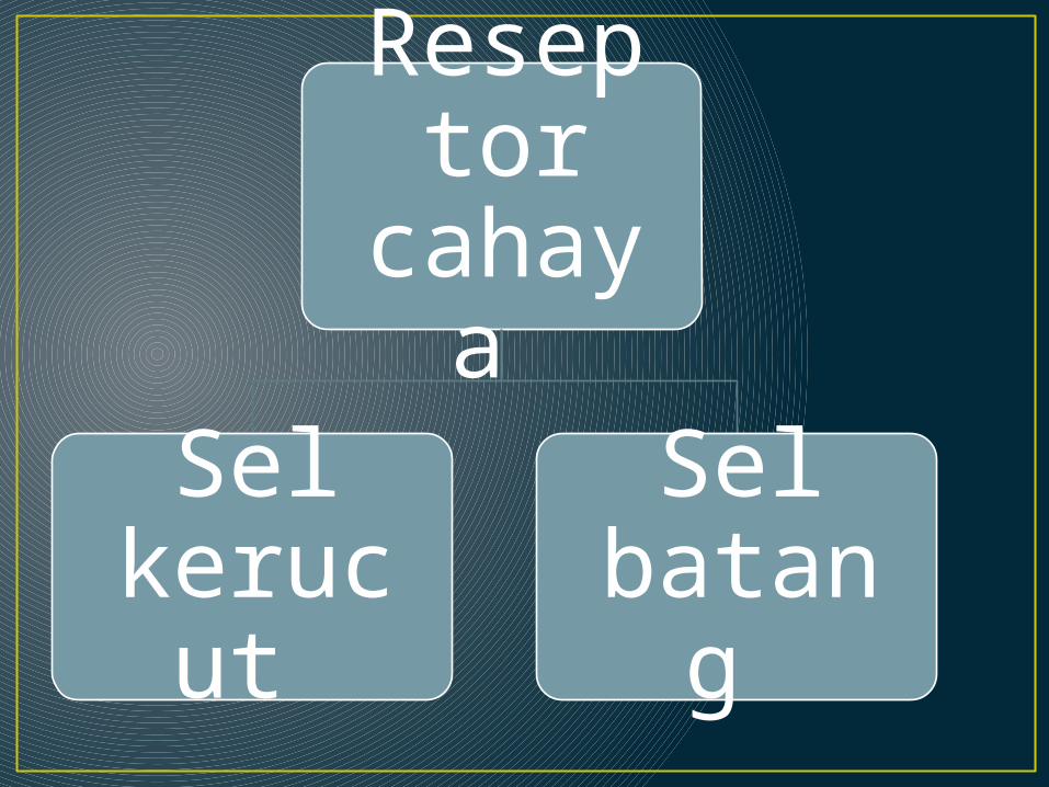
Reseptor
cahaya
Sel kerucu
t
Sel batang
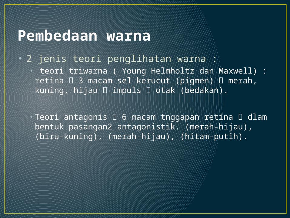
Pembedaan warna • 2 jenis teori penglihatan warna :
• teori triwarna ( Young Helmholtz dan Maxwell) : retina 3 macam sel kerucut (pigmen) merah, kuning, hijau impuls otak (bedakan).
• Teori antagonis 6 macam tnggapan retina dlam bentuk pasangan2 antagonistik. (merah-hijau), (biru-kuning), (merah-hijau), (hitam-putih).
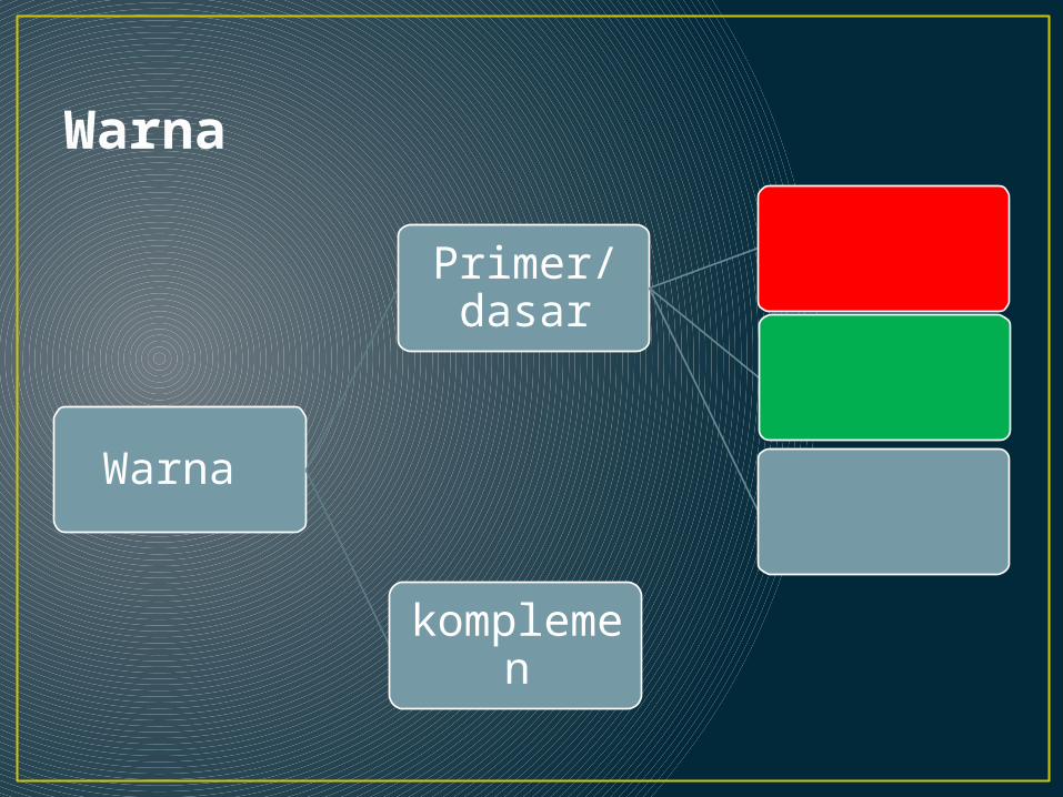
Warna
Warna
Primer/dasar
komplemen

Panjang gelombang masing2 warna
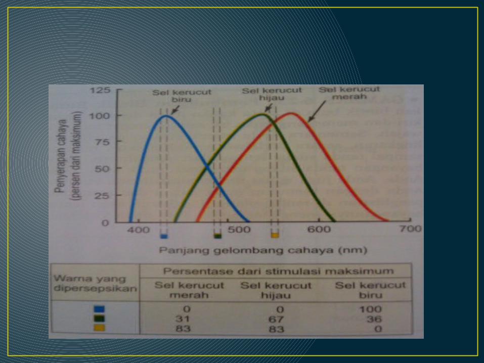
Pembedaan warna • Jingga 580 nm merah 99%: hijau 42 % : biru 0
otak jingga.
• Biru 450 nm merah 0 : hijau 0 : biru 97 % otak biru
• Putih stimulasi (sama besar + bersamaan) merah, hijau, biru putih.
Buta warna
Definisi • Kelainan pnglihatan sel2 kerucut retina tdk
mampu menangkap spektrum warna ttt. bkan warna sbnarnya.
• Ktidakmampuan herediter bedakan warna absence atau defisiensi 1 atau lebih dri tiga tipe.
Etiologi
Defek warna yg didapat - Kelainan pada macula (didapat)- Penyakit saraf (tekanan saraf optik massa)
Defek warna yg diturunkan- total - parsial
Klasifikasi (secara genetik)• Monokromat
buta warna total hanya 1 pigmen cones / tdk fungsi smua pnglihatan warna 1 dimensi.
- rod monocromat krusakan sel kerucut photofobia, nistagmus , visus menurun Pmx : pigmen mcula abnormal
- cone monokromat 1 macam sel kerucut aktivitas visual baik (visus normal).
• Dikromat salah satu dari tiga sel kerucut tdk ada/tdk fungsi warna 2 dimensi.berdasarkan pigmen yg rusak :
1. protanopia tdk ada gen opsin u/ pigmen L -
pigmen merah

2. deuteranopia tdk punya gen opsin u/ pigmen M - pigmen hijau. ksulitan bedakan
corak wrna merah-hijau.
3. tritanopia tdk punya gen opsin u/ pigmen S - pigmen biru. ksulitan bedakan
wrna biru dan kuning buta wrna biru-kuning.
• Trikromat anomali 3 sel kerucut lengkap trjadi kerusakan mkanisme sensitivitas (salah satu dari 3 sel reseptor warna) dapat melihat warna tetapi dengan interpretasi berbeda.
1. protonomali kelainan gen pigmen L terdpat pigmen S dan 2 pigmen M (pigmen yg mnyerupai
M). rendahnya sensitifitas trhadap warna merah dan spektrum warna merah buram ggg penlihatan merah-hijau.
• dasar genetik : delesi gen yang mnyandi pigmen jenis L normal gen hybrid atau chimeric sbagian susunan L diganti M.
• Gen chimerik : varian gen pigmen L dan M pertukaran segmen gen pigmen M dan L.
2. deuteranomali (paling sering)ada tiga pigmen: S dan 2 subtype spektrum pigmen L spt protanomaly (tpi klainan
pda rseptor hijau).dasar genetik : ada 2 gen berbeda yang
mnyandi pigmen L. 2 hipotesa :
- L “kedua” ganti gen pigmen M - pigmen L & M diekspresikan 1 sel kerucut
• Diagnosis1. Tes ishihara2. Color pencil discrimination3. Anomaloscope4. Tes Farnsworth-Munsell(corak,saturasi,derajat
iluminasi)
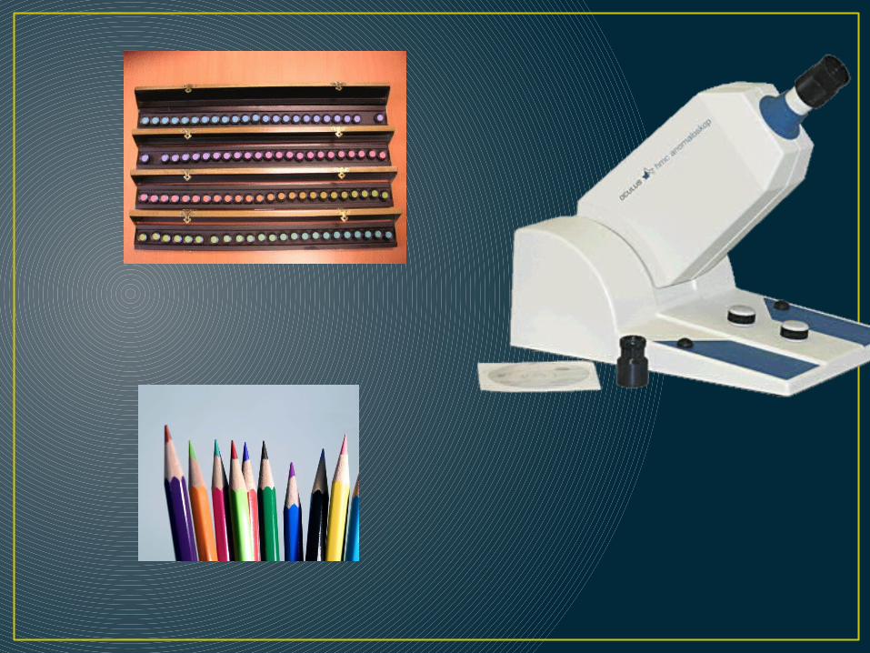

• TES ISHIHARA
• History1.Dr. Shinobu Ishihara• Definisi- Kumpulan kartu bergambar- -diagnosa defisiensi warna- Berupa satu/ lebih angka arab ataupun pola yang
tersusun dari bintik-bintik warna yang berbeda.- Pseudokromatik
Background (INSTRUCTION)Most cases of colour vision deficiency are characterised by a red-green deficiency which can be classed into two types;1.a protan(merah) type which may be absolute (protanopia MS) or partial (protanomalia (MMS) 2.a deutan(Hijau) type which may be absolute (deuteranopia LS) or partial (deuteranomalia LLS)
As a result of this red-green colour vision deficiencies show blue and yellow colours clearer than red and green colours.
•Those who suffer from typical total colour blindness show a complete failure to discriminate any colour variations, usually associated with impairment of central vision with photophobia and nystagmus.
•With atypical total colour blindness, the sensitivity to red and green, as well as to yellow and blue is so low that only very clear colours may be perceived. There are, however, no further abnormalities in the visual functions.
Images obtained from: Tests for Colour Blindness
byDr. Shinobu Ishihara
Professor Emeritus of the University of TokyoKanehara Shuppan Co., Ltd
Tokyo, Kyoto (1962)
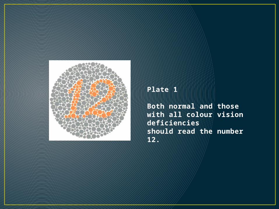
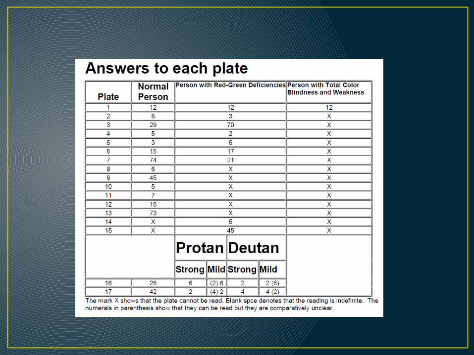
Plate 1
Both normal and those with all colour vision deficiencies should read the number 12.
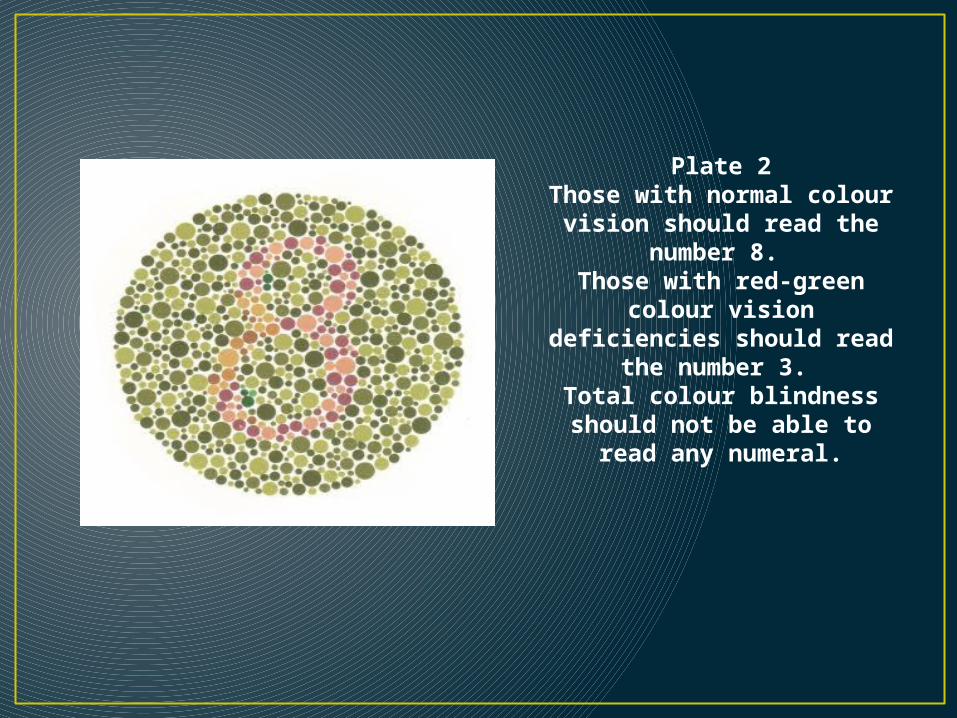
Plate 2Those with normal colour
vision should read the number 8.
Those with red-green colour vision deficiencies
should read the number 3. Total colour blindness
should not be able to read any numeral.
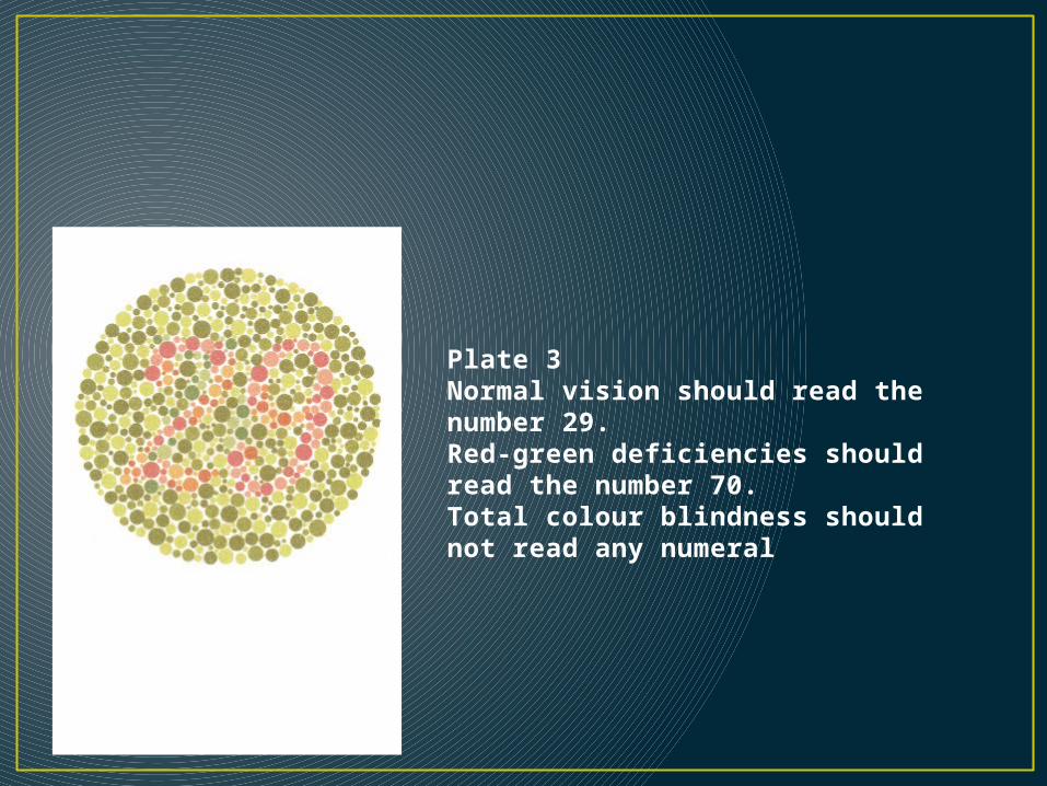
Plate 3Normal vision should read the number 29. Red-green deficiencies should read the number 70.Total colour blindness should not read any numeral
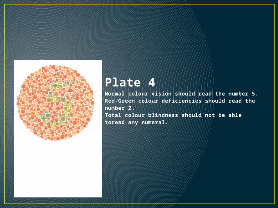
Plate 4Normal colour vision should read the number 5.Red-Green colour deficiencies should read the number 2.Total colour blindness should not be able toread any numeral.
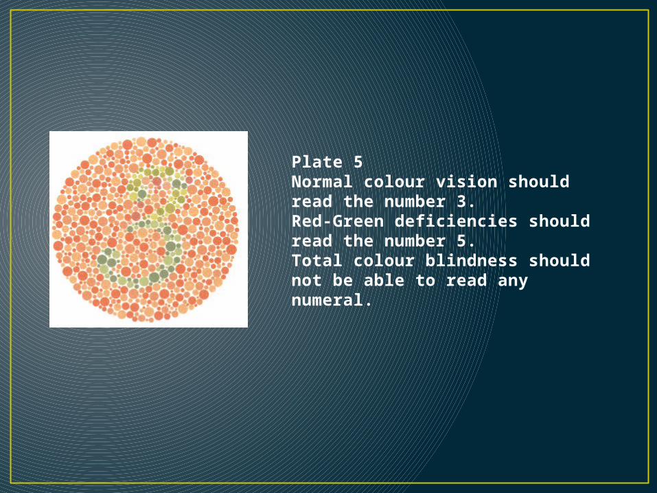
Plate 5Normal colour vision should read the number 3.Red-Green deficiencies should read the number 5.Total colour blindness should not be able to read any numeral.
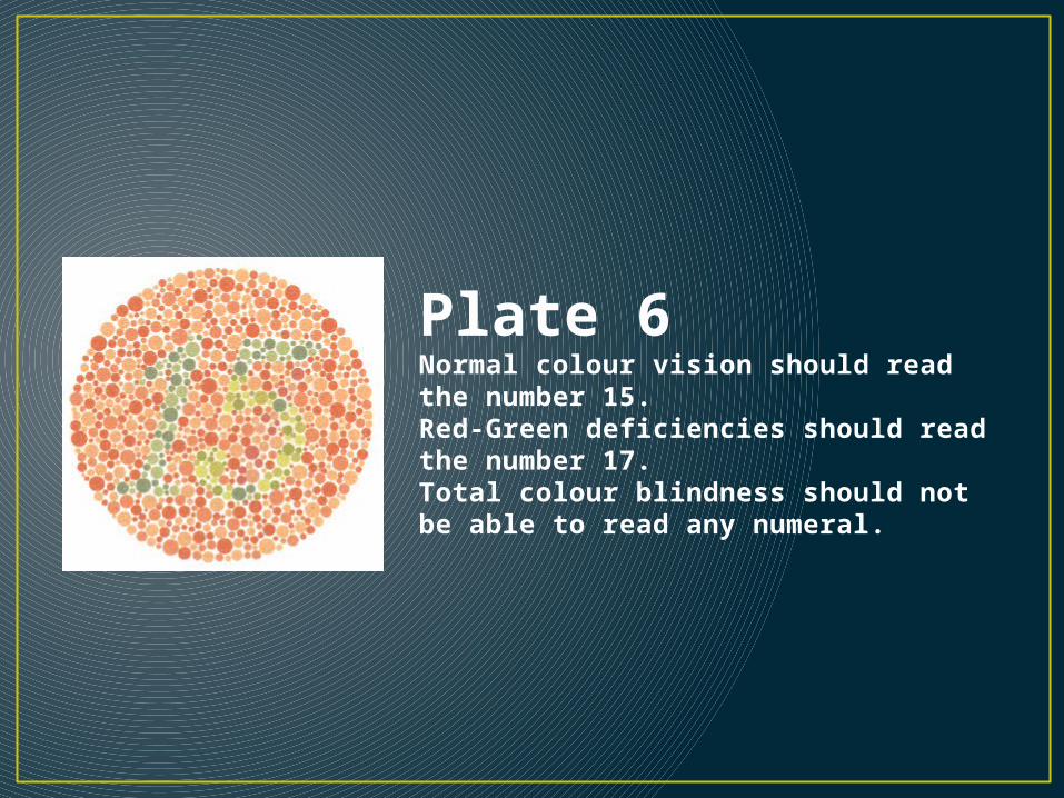
Plate 6Normal colour vision should read the number 15.Red-Green deficiencies should read the number 17.Total colour blindness should not be able to read any numeral.
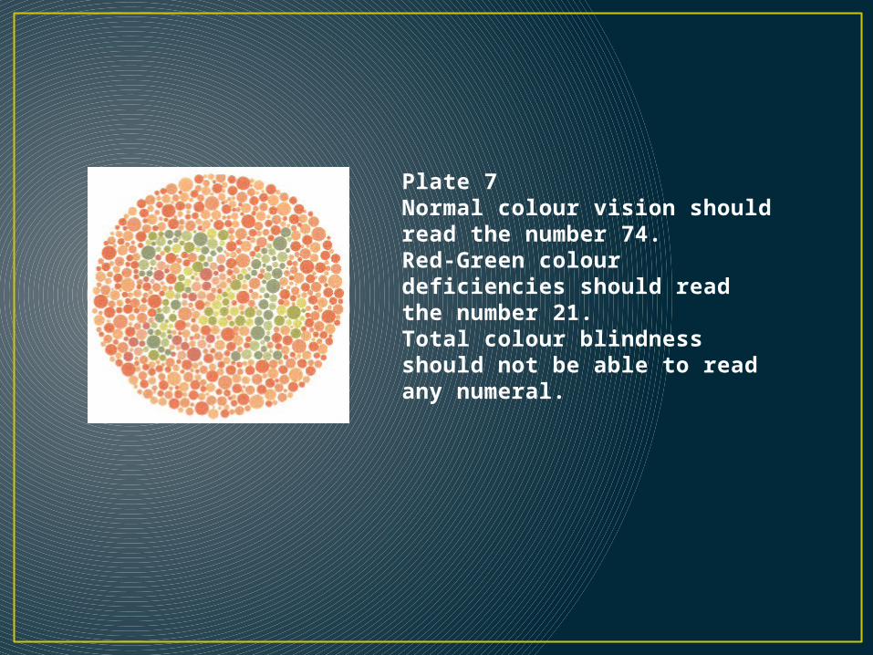
Plate 7Normal colour vision should read the number 74.Red-Green colour deficiencies should read the number 21.Total colour blindness should not be able to read any numeral.
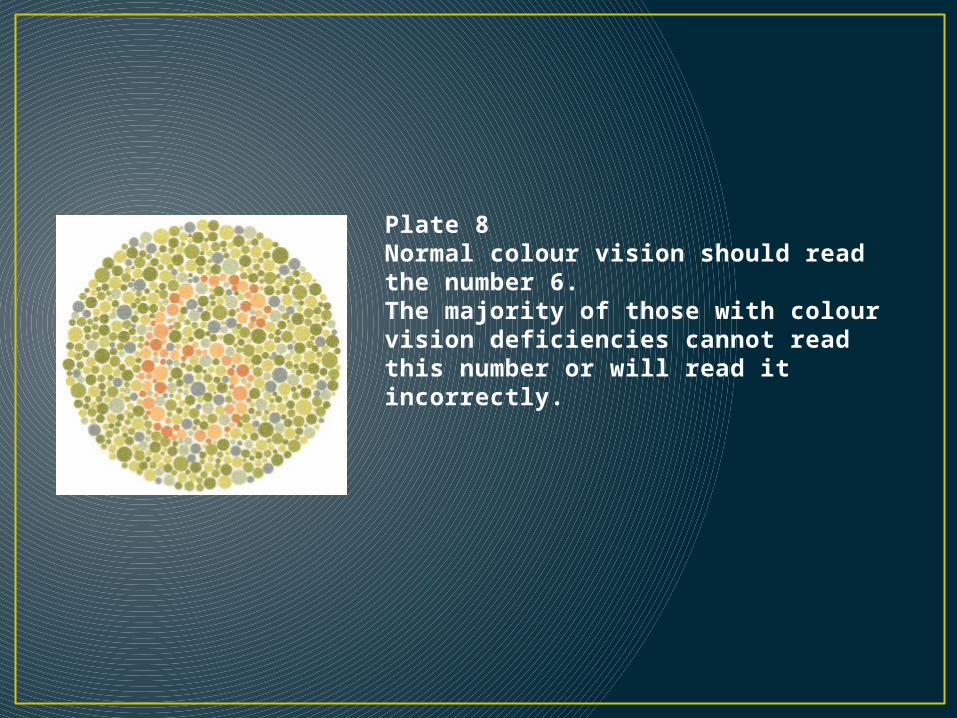
Plate 8Normal colour vision should read the number 6.The majority of those with colour vision deficiencies cannot read this number or will read it incorrectly.
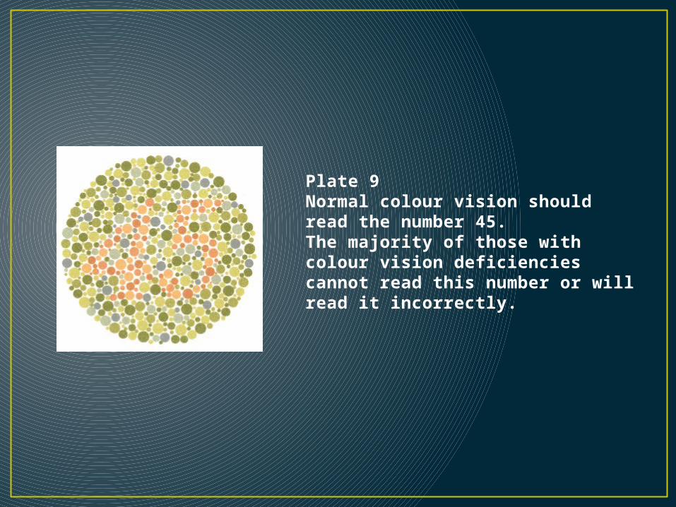
Plate 9Normal colour vision should read the number 45.The majority of those with colour vision deficiencies cannot read this number or will read it incorrectly.
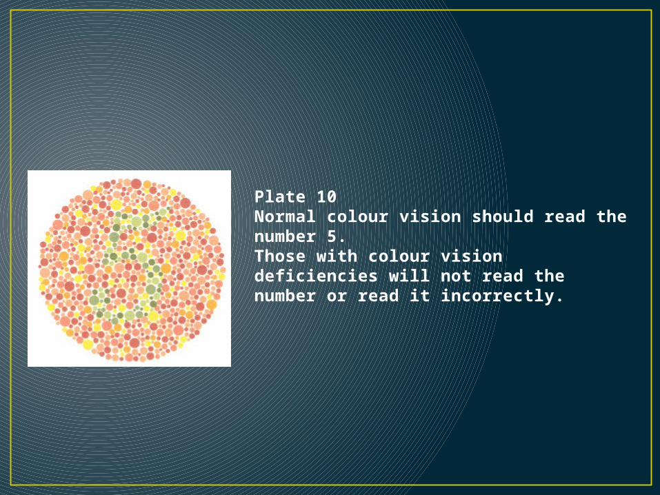
Plate 10Normal colour vision should read the number 5. Those with colour vision deficiencies will not read the number or read it incorrectly.
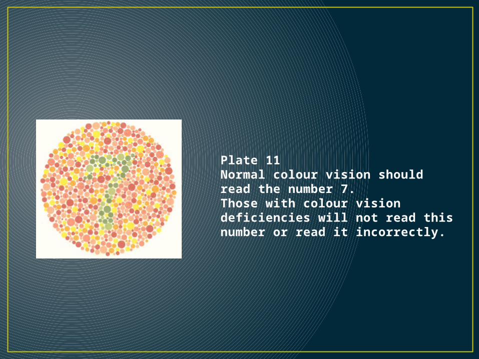
Plate 11Normal colour vision should read the number 7.Those with colour vision deficiencies will not read this number or read it incorrectly.
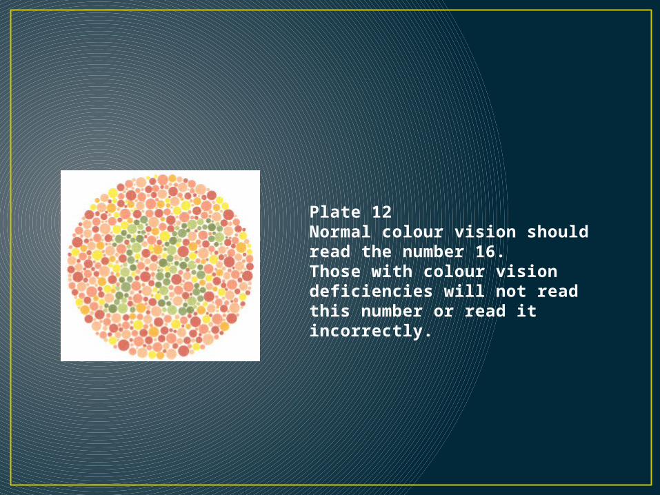
Plate 12Normal colour vision should read the number 16.Those with colour vision deficiencies will not read this number or read it incorrectly.
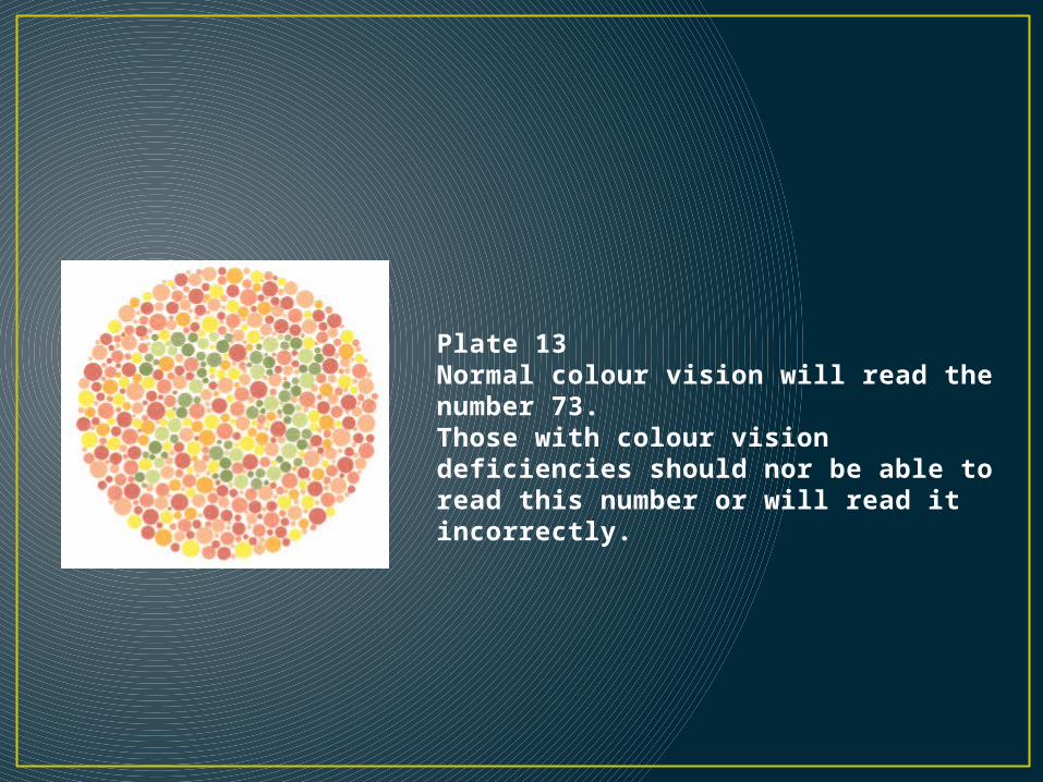
Plate 13Normal colour vision will read the number 73.Those with colour vision deficiencies should nor be able to read this number or will read it incorrectly.
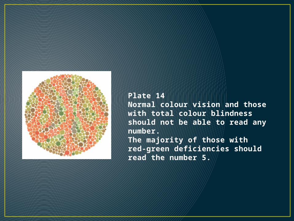
Plate 14Normal colour vision and those with total colour blindness should not be able to read any number.The majority of those with red-green deficiencies should read the number 5.
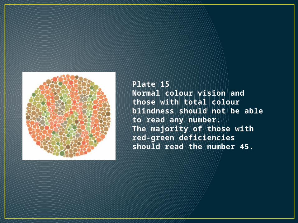
Plate 15Normal colour vision and those with total colour blindness should not be able to read any number.The majority of those with red-green deficiencies should read the number 45.
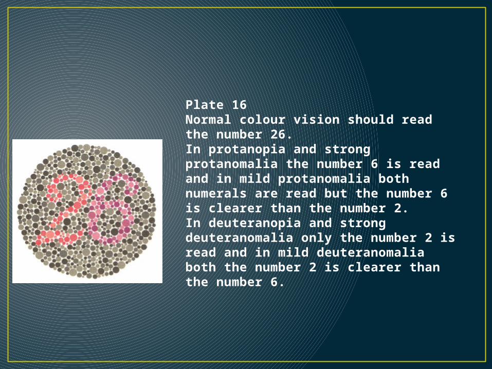
Plate 16Normal colour vision should read the number 26.In protanopia and strong protanomalia the number 6 is read and in mild protanomalia both numerals are read but the number 6 is clearer than the number 2.In deuteranopia and strong deuteranomalia only the number 2 is read and in mild deuteranomalia both the number 2 is clearer than the number 6.
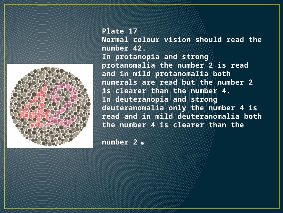
Plate 17Normal colour vision should read the number 42.In protanopia and strong protanomalia the number 2 is read and in mild protanomalia both numerals are read but the number 2 is clearer than the number 4.In deuteranopia and strong deuteranomalia only the number 4 is read and in mild deuteranomalia both the number 4 is clearer than the
number 2.
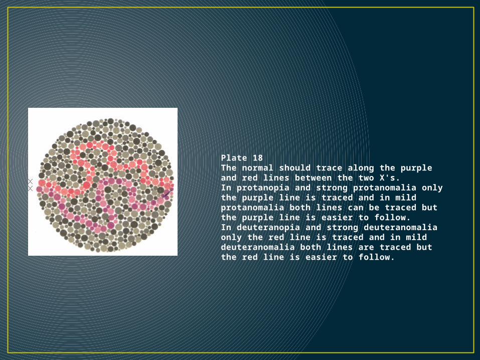
Plate 18The normal should trace along the purple and red lines between the two X's.In protanopia and strong protanomalia only the purple line is traced and in mild protanomalia both lines can be traced but the purple line is easier to follow.In deuteranopia and strong deuteranomalia only the red line is traced and in mild deuteranomalia both lines are traced but the red line is easier to follow.
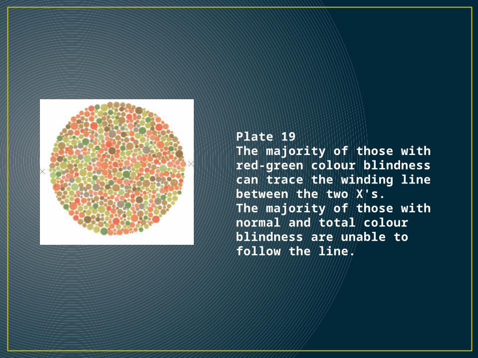
Plate 19The majority of those with red-green colour blindness can trace the winding line between the two X's.The majority of those with normal and total colour blindness are unable to follow the line.
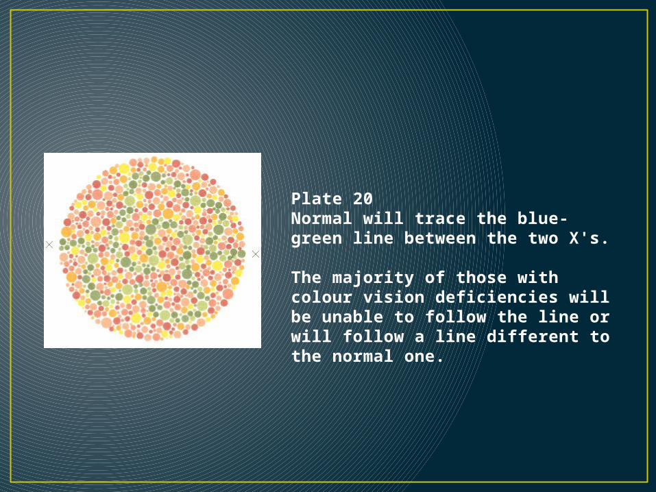
Plate 20Normal will trace the blue-green line between the two X's. The majority of those with colour vision deficiencies will be unable to follow the line or will follow a line different to the normal one.
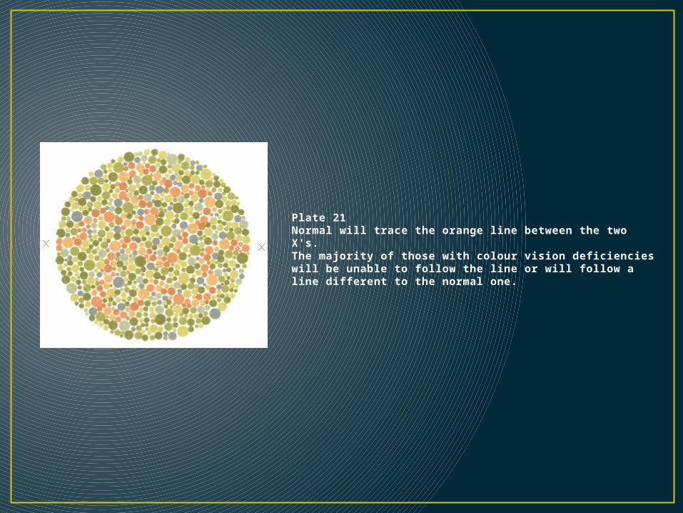
Plate 21Normal will trace the orange line between the two X's. The majority of those with colour vision deficiencies will be unable to follow the line or will follow a line different to the normal one.
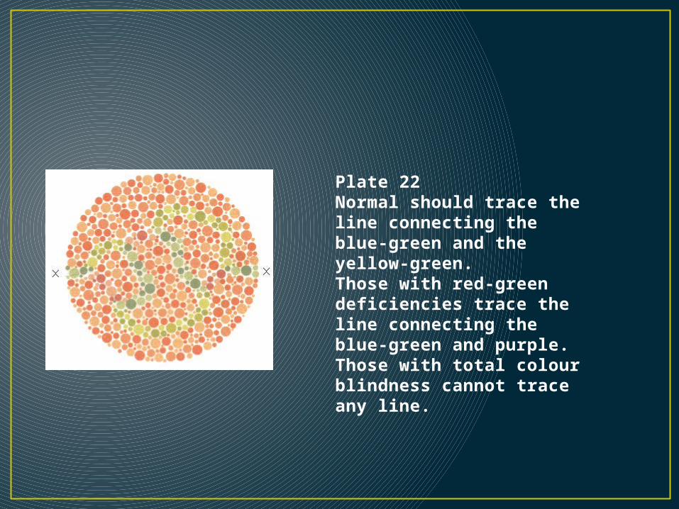
Plate 22Normal should trace the line connecting the blue-green and the yellow-green.Those with red-green deficiencies trace the line connecting the blue-green and purple.Those with total colour blindness cannot trace any line.
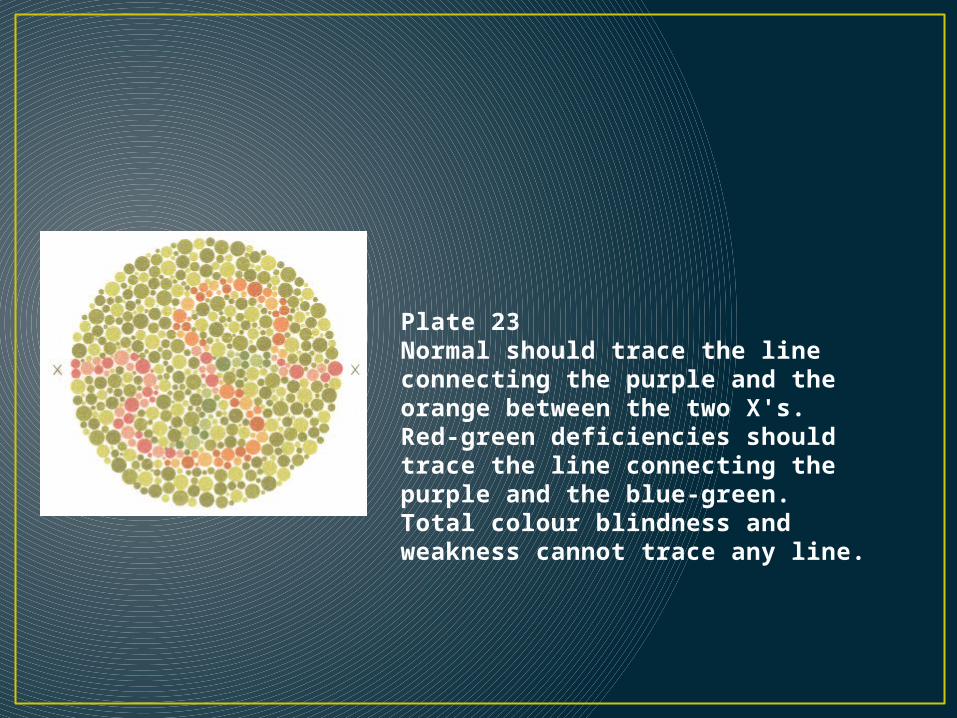
Plate 23Normal should trace the line connecting the purple and the orange between the two X's.Red-green deficiencies should trace the line connecting the purple and the blue-green.Total colour blindness and weakness cannot trace any line.
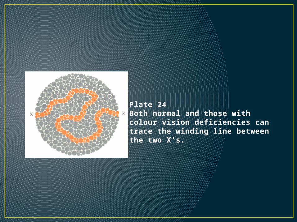
Plate 24Both normal and those with colour vision deficiencies can trace the winding line between the two X's.
Terapi• - Tidak ada pengobatan• - System• -Pengajaran dalam pengenalan warna dengan
benda langsung.• - tes spesifik dalam pekerjaan tertentu.
Pencegahan• Konseling pra-nikah
Kesimpulan• Buta merupakan penyakit keturunan yang terekspresi pada pria, tetapi
jarang pada wanita. Wanita secara genetic sebagai karier. Pada umumnya, terjadinya buta warna disebabkan oleh adanya reseptor warna dalam retina mata yang kurang berfungsi secara normal (malfungtion). Pada dasarnya, didalam retina mata kita terdapat 3 tipe reseptor warna, yaitu merah, biru, dan hijau.
• Buta warna adalah menurunnya kemampuan seseorang untuk membedakan warna, dimana orang lain dapat melakukannya. Buta warna ini sebagian besar disebabkan oleh kelainan genetic, tetapi juga bias diakibatkan adanya kelainan pada mata, saraf, otak, dank arena trauma kimia. Buta warna congenital muncul dalam 2 bentuk utama yaitu total dan parsial. Bentuk yang paling sering muncul pada kelainan congenital ini adalah buta warna merah-hijau, hal ini disebabkan karena kelainan pada kromosom X dan bersifat herediter. Insidensi pada pria lebih besar dari wanita karena wanita memiliki 2 kromosom X dan pria memiliki 1 kromosom X dan 1 kromosom Y. bentuk buta warna yang lain adalah buta warna biru-kuning, akan tetapi kelainan ini sangat jarang terjadi.