Embed Size (px)
Citation preview

Proximal Vertebral Body Fracture after 4-Level Fusion Using L1 as the Upper Instrumented Vertebra for Lumbar Degenerative
Disease: Report of 2 Cases with Literature Review
Takao Yasuharaa,b*, Yuichi Takahashia, Shinji Kumamotoa, Masayuki Nakaharaa, Kotaro Yonedaa, Tatsuomi Niimuraa, Takashi Tanouea, Akira Kusumegia, Takashi Sennaria, Yasukazu Hijikataa, Hiroaki Manabeb, Yasuyuki Miyoshib,
Isao Dateb, Koichi Ogawaa, and Kenki Nishidaa
aDepartment of Spinal Surgery, Shinkomonji Hospital, Kitakyushu, Fukuoka 800-0057, Japan, and Department of Neurological Surgery, Okayama University Hospital 700-8558, Japan
Some cases with lumbar degenerative diseases require multi-level fusion surgeries. At our institute, 27 and 4 procedures of 3- and 4-level fusion were performed out of a total 672 posterior lumbar interfu-sions (PLIFs) on patients with lumbar degenerative disease from 2005 to 2010. We present 2 osteo-porotic patients who developed proximal vertebral body fracture after 4-level fusion. Both cases presented with gait disability for leg pain by degenerative lumbar scoliosis and canal stenosis at the levels of L1/2-4/5. After 4-level fusion using L1 as the upper instrumented vertebra, proximal verte-bral body fractures were found along with the right pedicle fractures of L1 in both cases. One of these patients, aged 82 years, was treated as an outpatient using a hard corset for 24 months, but the frac-tures were exacerbated over time. In the other patient, posterolateral fusion was extended from Th10 to L5. Both patients can walk alone and have been thoroughly followed up. In both cases, the frac-ture of the right L1 pedicle might be related to the subsequent fractures and fusion failure. In consid-eration of multi-level fusion, L1 should be avoided as an upper instrumented vertebra to prevent junctional kyphosis, especially in cases with osteoporosis and flat back posture.
Key words: degenerative lumbar scoliosis, osteoporosis, pedicle fracture, posterior lumbar interbody fusion, vertebral body fracture
n lumbar degenerative diseases, some cases require multi-level (3 or more levels) surgeries
using posterior lumbar interbody fusion (PLIF). In the past 6 years from 2005 to 2010, 27 procedures of 3-level fusion (mean follow-up period, 21.2 months) and 4 of 4-level fusion (mean follow-up period, 31.5 months) were performed on patients with lumbar
degenerative diseases at our institute. The percent-age of 3- or 4-level fusion cases comprised 4.5オ of our 672 PLIF cases in this period. Recently, we experienced 2 osteoporotic patients with proximal vertebral body fractures after 4-level fusion surgeries in which L1 had been used as the upper instrumented vertebra. We report these 2 cases with a literature review to examine the mechanisms of the fractures.
I
Acta Med. Okayama, 2013Vol. 67, No. 3, pp. 197ン202CopyrightⒸ 2013 by Okayama University Medical School.
Case Report http ://escholarship.lib.okayama-u.ac.jp/amo/
Received October 17, 2012 ; accepted January 15, 2013.*Corresponding author. Phone : +81ン86ン235ン7336; Fax : +81ン86ン227ン0191E-mail : [email protected] (T. Yasuhara)

Case Reports
Informed consent was obtained from the patients and families. The institutional ethical committee approved this report. Case 1. An 82-year-old woman presenting with gait inability for leg pain visited our institute. She was diagnosed with severe degenerative lumbar scolio-sis and lumbar canal stenosis at the levels of L1/2- 4/5 with flat back posture. We performed 4-level PLIF and pedicle screw fixation. Two weeks after surgery, with rehabilitation starting a few days after surgery, she could walk using a walker with a decreased level of leg pain (Fig. 1). However, L1 and Th12 vertebral body fractures were found at 2 and 4 weeks, respectively. Retrospectively, we also found that computed tomography (CT) images obtained 5 days after surgery might have demonstrated the frac-ture of the right L1 pedicle. Because of her advanced age and lack of deterioration, she was treated as an outpatient using a hard corset for 24 months, although the fractures were exacerbated over time without fusion (Fig. 1). Case 2. A 65-year-old man presenting with
severe gait disturbance for leg pain visited our insti-tute. He was diagnosed with mild degenerative lumbar scoliosis and severe lumbar canal stenosis at the levels of L1/2-4/5 with spondylolisthesis at the level of L4/5 (Fig. 2). We performed PLIF at the levels of L1/2 and 4/5, lumbar laminoplasty at L2/3 and 3/4, and pedicle screw fixation from L1 to L5 (Fig. 2). The degree of leg pain decreased and he started gait exercise in a few days after surgery. At 20 days after surgery, he complained of low back pain. CT images demonstrated L1 and Th12 vertebral body fractures along with fracture of the right L1 pedicle. His pain worsened despite conservative therapy using a hard corset. At 50 days after the initial surgery, we extended the posterolateral fusion from Th10 to L5 (Fig. 2). One week after surgery, he started gait training. He was transferred to another hospital for rehabilitation at 30 days after the second surgery. After discharge from the hospital, he could stay at home by himself using a walking stick. CT images obtained at 7 months after surgery revealed a new L5 fracture. We performed thorough follow-up for 12 months after the initial surgery, although he had no further complaint of pain.
198 Acta Med. Okayama Vol. 67, No. 3Yasuhara et al.
A B C D E
F
G H I J K L
*
Fig. 1 Neuroradiological investigations of Case 1. A-D Radiographs of lumbar spine before (A, B) and after surgery (C, D) demon-strate that the scoliosis was corrected by 4-level fusion (A and C: AP view; B and D: lateral view), E-I Reconstructed sagittal CT images reveal the development of proximal vertebral-body fracture over time (E: 5 days, F: 2 weeks, G: 4 weeks, H: 6 weeks, I: 8 weeks, J: 12 weeks, K: 6 months, I: 20 months after surgery;* L1 vertebral body; arrowhead: initial pedicle fracture).
Summary of 4 cases of 4-level fusion associ-ated with degenerative lumbar scoliosis (Table 1). We performed 4-level fusion on 4 patients with degenerative lumbar scoliosis and canal stenosis. In order to explore some reasonable causes of the frac-tures in our cases, we evaluated age/sex, level of PLIF and fusion, type of scoliosis, degree of lordo-sis, degree and laterality of scoliosis, involvement of spondylolisthesis, sagittal rotational angle and bone mineral density (femur) of the 4 cases. The common points of case 1 and 2 were osteoporosis, scoliosis with lateral spondylolisthesis of L4/5, and the fact that the apical vertebra was L3. There were no apparent differences between cases with and without fractures after 4-level fusion, either in the factors we selected or in the rotation of the vertebral body, shape of disc, or degeneration of disc or facet,
although the number of 4-level fusion cases was small. Regarding pedicle fracture after surgery, we checked CT images of a total of 254 pedicle screws in the 31 cases of 3-or 4-level fusion. We found 1 wrong inser-tion of the screw, but no apparent screw-insertion-related pedicle fractures other than the 2 fractures of the present 2 cases. Case 3 and 4, the 4-level cases without proximal vertebral body fracture, were fol-lowed as outpatients with no neurological deficits for 3 and 5 years, respectively.
Discussion
In this manuscript, we described 2 osteoporotic patients with proximal vertebral body fractures after 4-level fusion using L1 as the upper instrumented vertebra. Both presented with severe gait disturbance
199Proximal Vertebral Body Fracture after Lumbar FusionJune 2013
A B C D E
F G H I J
*
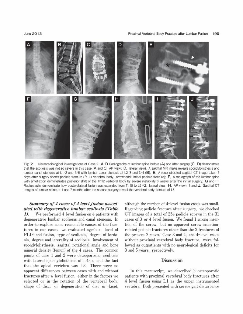
Fig. 2 Neuroradiological investigations of Case 2. A-D Radiographs of lumbar spine before (A) and after surgery (C, D) demonstrate that the scoliosis was not so severe in this case (A and C, AP view; D, lateral view). A sagittal MR image reveals spondylolisthesis and lumbar canal stenosis at L1/2 and 4/5 with lumbar canal stenosis at L2/3 and 3/4 (B); E, A reconstructed sagittal CT image taken 5 days after surgery shows pedicle fracture (*: L1 vertebral body; arrowhead: initial pedicle fracture); F, A radiograph of the lumbar spine with anteflexion demonstrates posterior shift of the Th12 vertebral body by severe instability 6 weeks after the initial surgery; G and H, Radiographs demonstrate how posterolateral fusion was extended from Th10 to L5 (G, lateral view; H, AP view), I and J, Sagittal CT images of lumbar spine at 1 and 7 months after the second surgery reveal the vertebral body fracture of L5.

with leg pain as symptoms of degenerative lumbar scoliosis and canal stenosis, which was treated with 4-level fusion using PLIF. Gait exercise was started in a few days after the surgery, and symptoms were ameliorated within 2 weeks. Retrospectively, CT image demonstrated the fracture of the right L1 pedicle within 5 days after surgery. Subsequently, fractures of the L1 vertebral body, and then the Th12 vertebral body occurred due to the screw loosening resulting from the instability (Fig. 3).
Proximal vertebral body fracture after fusion surgery. There are several risk factors of proxi-mal fracture after lumbar fusion: namely, obesity, older age [1], osteopenia, preoperative co-morbidi-ties, and severe global sagittal imbalance including flat back posture [2]. Marked correction of sagittal mala-lignment might be a risk factor of instrumented verte-bra collapse, although in our cases, the degree of pre/post-operative sagittal malalignment did not affect the results. In another report, proximal vertebral body fracture and development of focal kyphosis were found in about 20 and 40 percent of cases receiving multi-level lumbar fusion from L1 to L5 or S1, respectively. To minimize the incidence of these problems, limited instrumentation or fusion above the thoraco-lumbar junction might be preferred [3]. Recently, a retrospective study on the selection of the upper instrumented vertebra was reported [4]. The authors concluded that the upper instrumented verte-bra must be above the upper end vertebra and that fusion to Th11 or 12 is acceptable when the upper instrumented vertebra is above the upper end vertebra in adult degenerative lumbar scoliosis. Alternatively, prophylactic vertebral augmentation might be a cost-effective intervention in elderly female patients [5]. Etebar and colleagues performed a retrospective analysis of degeneration of the proximal adjacent seg-ment after lumbar fusion. Fifteen percent of their 125 patients developed symptomatic adjacent-segment
200 Acta Med. Okayama Vol. 67, No. 3Yasuhara et al.
Table 1 The summary of 4 cases of 4 levels-fusion
Case 1 Case 2 Case 3 Case 4
age/sex 82/F 65/M 77/F 54/FPLIF level L1/2-4/5 L1/2, 4/5 L1/2, 2/3, 4/5 L4/5Fusion level L1-5 L1-5 L1-5 L1-5 Type of scoliosis L4/5 lateral spondylolisthesis (+) L4/5 wedged disc (+) L1-S1(lordosis) 4° 33° 6° 29°PLC; Cobb angle; AV left; 14°; L3 right; 37°; L3 left; 27°; L2 right; 44°; L2Th12/L1 listh.; lat. list (-); (-) Meyerding I; (-) (-); (-) (-); 2mmSag. Rot. Ang. (Th12/L1) 12.9° N/A 0.8° 0°BMD (YAM) 71% 78% 85% N/A
Case 1 and 2 are the present cases. There are almost no common characters in Case 1 and 2, except for osteoporosis, the fusion level and scoliosis type. Other than the data shown in the table, there are no significant differences in vertebral body rotation, shape of disc, and degeneration of disc and facet in 4 cases. AV, apical vertebra; lat. listh, lateral spondylolisthesis; listh., spondylolisthesis; PLC, primary lumbar curve; Sag. Rot. Ang., sagittal rotation angle; BMD, bone mineral density; YAM, young adult male
A B
C
Fig. 3 Possible mechanisms of proximal vertebral body fracture in our cases, A, Slight pedicle fractures might occur intraopera-tively or within a few days of surgery; B, Screw loosening with vertebral body fracture near the pedicle might follow; C, Due to the increased instability, the antero-inferior edge of the proximal verte-bral body might be damaged.

degeneration at a previously asymptomatic level within 45 months after surgery. Smoking and postmeno-pausal status in women were shown to be high risk factors for adjacent segment disease after fusion sur-gery. Adjacent segment diseases included spondylolis-thesis (39オ), spinal canal stenosis due to disc hernia-tion and/or facet hypertrophy (33オ), stress fracture of the adjacent vertebral body (28オ), and scoliosis (17オ) [6]. In a review of adjacent segment disease by Park, the rate of symptomatic disease was higher in patients with transpedicular instrumentation (12.2- 18.5オ) compared with patients fused with other forms of instrumentation or without instrumentation (5.2- 5.6オ). Instrumentation, fusion length, sagittal malalignment, facet injury, age, and pre-existing degenerative changes might be possible risk factors for adjacent segment diseases. Decompression of the cauda equina with the extension of fusion might resolve such situations [7]. In our cases, acute adjacent segment failure was recognized in 2 of 4 cases of 4-level fusion from L1 to L5. However, we experienced no acute adjacent seg-ment failure in 27 cases of 3-level fusion. The high rate of proximal vertebral body fractures in our 4-level fusion cases might be strongly related to the selection of L1 as the upper instrumented vertebra, to which a strong force is applied after fixation at the thoraco-lumbar junction, although there might be mechanical differences between 3- and 4-level fusion at the proximal junction. We usually attempt to limit fusion levels and perform short fusion for lumbar degenerative diseases. However, especially for some patients of lumbar canal stenosis with degenerative lumbar scoliosis, multi-level fusion might be consid-ered. To prevent adjacent segment diseases, long fusion over the thoraco-lumbar junction might be a possible solution, although the surgical invasiveness might be greater. Vertebroplasty in advance at the initial surgery might be an alternative strategy. Pedicle fractures after screw insertion.Pedicle fractures after screw insertion occurred intraoperatively or early after surgery at L1 in our 2 cases. In other cases, intraoperative fractures of the pedicle were not found. It is possible that the use of a hard corset for early rehabilitation after surgery might cause early pedicle fractures. It is impossible to know genuinely how and when the pedicle fracture happened in our 2 cases. Nonetheless, the instability
due to the pedicle fracture might be one of the first steps to the catastrophic acute collapse through verte-bral body fracture of the proximal and adjacent verte-bral bodies (Fig. 3). After lumbar fusion surgery, fractures at the isthmus portion are sometimes seen, but pedicle frac-tures are rare. Usually the junction of pedicles and vertebral bodies is at the fracture site, as in our cases. Pedicle weakening by screws and cantilever motion after fusion might be contributing factors [8]. Amato and colleagues revealed that intraoperative pedicle fractures occurred in 2 percent of their 102 consecutive patients receiving lumbar fusion surgery (using a total of 424 pedicle screws) [9]. Pedicle fractures might lead to instability with lumbago or leg pain and may necessitate extension of the fusion or stabilization with wiring of the fractured pedicle [10]. Loosening of screws and pseudarthrosis are known to be the main complications at the ends of long fusions [11]. Pedicle fractures might be one of the critical reasons for the failure of fusions. In conclusion, we experienced 2 cases of proximal vertebral body fracture after multi-level fusion sur-gery with L1 as the upper instrumented vertebra. The reasons for the fractures were likely osteoporosis and the selection of L1 as the upper instrumented verte-bra, although fracture of the right L1 pedicle might have been involved in the subsequent fractures and fusion failure. In consideration of multi-level fusion below the thoraco-lumbar segment, these types of phenomena are need-to-know complications for spinal surgeons. L1 should be avoided as upper instru-mented vertebra for lumbar degenerative diseases to prevent junctional kyphosis, especially in cases with osteoporosis and flat back posture. For clarification of the efficacy and complications, we also need to fol-low patients and explore their systemic problems including sagittal balance and hip joint issues.
References
1. OセLeary PT, Bridwell KH, Lenke LG, Good CR, Pichelmann MA, Buchowski JM, Kim YJ and Flynn J: Risk factors and outcomes for catastrophic failures at the top of long pedicle screw constructs: a matched cohort analysis performed at a single cen-ter. Spine (Phila Pa 1976) (2009) 34: 2134-2139.
2. Watanabe K, Lenke LG, Bridwell KH, Kim YJ, Koester L and Hensley M: Proximal junctional vertebral fracture in adults after spinal deformity surgery using pedicle screw constructs: analysis of morphological features. Spine (Phila Pa 1976) (2010) 35: 138-
201Proximal Vertebral Body Fracture after Lumbar FusionJune 2013

145. 3. Simmons ED, Huckell CB and Zheng Y: Proximal kyphosis,
“Topping off syndrome”, and retrolisthesis secondary to multi-level lumbar fusion in the elderly patients. J Bone Joint Surg (2008) 90-B (Supple 68).
4. Cho KJ, Suk SI, Park SR, Kim JH and Jung JH: Selection of proximal fusion level for adult degenerative lumbar scoliosis. Eur Spine J (2012) Epub ahead of print.
5. Hart RA, Prendergast MA, Roberts WG, Nesbit GM and Barnwell SL: Proximal junctional acute collapse cranial to multi-level lumbar fusion: a cost analysis of prophylactic vertebral augmentation. Spine J (2008) 8: 875-881.
6. Etebar S and Cahill DW: Risk factors for adjacent-segment failure following lumbar fixation with rigid instrumentation for degenerative instability. J Neurosurg (1999) 2 Suppl: 163-169.
7. Park P, Garton HJ, Gala VC, Hoff JT and McGillicuddy JE: Adjacent segment disease after lumbar or lumbosacral fusion:
review of the literature. Spine (Phila Pa 1976) (2004) 29: 1938-1944.
8. Ha KY and Kim YH: Bilateral pedicle stress fracture after instru-mented posterolateral lumbar fusion: a case report. Spine (Phila Pa 1976) (2003) 28: 158-160.
9. Amato V, Giannachi L, Irace C and Corona C: Accuracy of pedi-cle screw placement in the lumbosacral spine using conventional technique: computed tomography postoperative assessment in 102 consecutive patients. J Neurosurg Spine (2010) 12: 306-313.
10. Lattig F, Fekete TF and Jeszenszky D: Management of fractures of the pedicle after instrumentation with transpedicular screws: A report of three patients. J Bone Joint Surg (2010) 92: 98-102.
11. Mok JM, Cloyd JM, Bradford DS, Hu SS, Deviren V, Smith JA, Tay B and Berven SH: Reoperation after primary fusion for adult spinal deformity: rate, reason, and timing. Spine (Phila Pa 1976) (2009) 34: 832-839.
202 Acta Med. Okayama Vol. 67, No. 3Yasuhara et al.