Embed Size (px)
Citation preview
Instructions for use
Title Purification, characterization and amino acid sequence of a novel enzyme, D-threo-3-hydroxyaspartate dehydratase,from Delftia sp. HT23
Author(s) Maeda, Takayuki; Takeda, Yuki; Murakami, Tomoko; Yokota, Atsushi; Wada, Masaru
Citation The Journal of Biochemistry, 148(6): 705-712
Issue Date 2010-12
Doc URL http://hdl.handle.net/2115/47109
RightsThis is a pre-copy-editing, author-produced PDF of an article accepted for publication in The Journal of Biochemistryfollowing peer review. The definitive publisher-authenticated version, J Biochem (2010) 148 (6): 705-712, is availableonline at: http://jb.oxfordjournals.org/content/148/6/705
Type article (author version)
File Information JoB148-6_705-712.pdf
Hokkaido University Collection of Scholarly and Academic Papers : HUSCAP
1
Regular Paper
Field; Biochemistry (Topic; Enzymology)
Purification, characterization, and amino acid sequence of a novel enzyme, D-threo-3-
hydroxyaspartate dehydratase, from Delftia sp. HT23
Takayuki Maeda, Yuki Takeda, Tomoko Murakami, Atsushi Yokota, and Masaru Wada*
Division of Applied Bioscience, Research Faculty of Agriculture, Hokkaido University, Kita-9, Nishi-
9, Kita-ku, Sapporo, 060-8589, Japan
Running title: D-threo-3-hydroxyaspartate dehydratase from Delftia
--------------------------------------------------------------------------------------
*Correspondence to Masaru Wada, Division of Applied Bioscience, Research Faculty of Agriculture,
Hokkaido University, Kita-9, Nishi-9, Kita-ku, Sapporo, 060-8589, Japan
Tel: +81-11-706-4185; Fax: +81-11-706-4961; E-mail: [email protected]
Abbreviations:
DH, dehydratase; D-EHA, D-erythro-3-hydroxyaspartate; D-THA, D-threo-3-hydroxyaspartate; EDTA,
ethylenediaminetetraacetic acid; IPTG, isopropyl-β-D-thiogalactopyranoside; LB, Luria–Bertani; L-
EHA, L-erythro-3-hydroxyaspartate; L-THA, L-threo-3-hydroxyaspartate; MALDI-TOF-MS, matrix-
assisted laser desorption/ionization time-of-flight mass spectrometry; NADH, nicotinamide adenine
dinucleotide; PAGE, polyacrylamide gel electrophoresis; PCR, polymerase chain reaction; PLP,
pyridoxal 5’-phosphate; SDS, sodium dodecyl sulfate; TLC, thin layer chromatography.
2
Summary
D-threo-3-hydroxyaspartate dehydratase (D-THA DH) was purified from the cell-free extract of the
soil-isolated bacterium Delftia sp. HT23. The enzyme exhibited dehydratase activity toward D-threo-
3-hydroxyaspartate, L-threo-3-hydroxyaspartate, L-erythro-3-hydroxyaspartate, and D-serine.
Absorption of the purified enzyme at 412 nm suggests that it contains pyridoxal 5'-phosphate (PLP) as
a cofactor. The NH2-terminal and internal amino acid sequences showed significant similarity to
hypothetical alanine racemase of genome-sequenced Delftia acidovorans SPH-1; however, the
purified enzyme showed no alanine racemase activity. Using the sequence information of Delftia
acidovorans SPH-1, the gene encoding D-THA DH was cloned. The deduced amino acid sequence,
which belongs to the alanine racemase family, shows significant (26-36%) similarity to D-serine
dehydratase of both Saccharomyces cerevisiae and chicken. In order to obtain purified D-THA DH
efficiently, the gene was expressed in Escherichia coli. The recombinant enzyme was highly
activated by divalent cations, such as Mn2+, Co2+, and Ni2+. Site-directed mutagenesis experiment
revealed that lysine 43 is an important residue involved in PLP binding and catalysis. This is the first
reported enzyme that acts on D-THA. In addition, this enzyme is the first example of a prokaryotic
dehydratase belonging to the fold-type III PLP-dependent enzyme family.
Key words: D-threo-3-hydroxyaspartate dehydratase, alanine racemase, pyridoxal 5 ′-phosphate,
Delftia sp. HT23
3
3-Hydroxyaspartate and its derivatives have attracted the attention of biochemists because they are
competitive blockers of the excitatory glutamate/aspartate transporters of the mammalian nervous
system (1, 2). However, 3-hydroxyaspartate has two chiral centers, and their four stereoisomers, i.e.,
D-threo-3-hydroxyaspartate (D-THA), L-threo-3-hydroxyaspartate (L-THA), D-erythro-3-
hydroxyaspartate (D-EHA), and L-erythro-3-hydroxyaspartate (L-EHA), have been difficult to
synthesize (3).
The biochemical activity of 3-hydroxyaspartate has been investigated in considerable detail
(4). Nevertheless, little is known about the enzymes that act on 3-hydroxyaspartate isomers, although
two microbial enzymes, erythro-3-hydroxyaspartate aldolase (EC 4.1.3.14) (5, 6) and erythro-3-
hydroxyaspartate dehydratase (EC 4.3.1.20) (7), were identified many years ago. Recently, we
reported that L-threo-3-hydroxyaspartate dehydratase (EC 4.3.1.16) isolated from the soil-isolated
bacterium Pseudomonas sp. T62 exhibits dehydratase activity specifically toward L-THA and not
toward other 3-hydroxyaspartate isomers (8, 9). The amino acid sequence and detailed biochemical
features, including side reactions, of this enzyme have also been reported (9). However, no enzyme
acting on the D-threo form of 3-hydroxyaspartate has been reported. The enzyme degrading D-THA
might be useful for enzymatic optical resolution of DL-THA to produce optically pure L-THA.
Because there has been no report about enzyme acting on D-THA so far, in order to obtain the amino
acid sequence information of D-THA converting enzyme, enzyme purification from microorganism
that produces D-THA converting enzyme was necessary. Thus, we screened microorganisms that can
utilize D-THA as a sole carbon source and found that a newly isolated bacterium, Delftia sp. HT23,
which produces an enzyme that catalyzes the dehydratase reaction of D-THA to oxaloacetate. We
designated this enzyme as D-threo-3-hydroxyaspartate dehydratase (D-THA DH; D-threo-3-
hydroxyaspartate ammonia-lyase).
4
We report here the purification, partial characterization, and amino acid sequence of this
novel enzyme, D-THA DH, from Delftia sp. HT23. The comparison of amino acid sequence of this
enzyme with known eukaryotic D-serine dehydratases is also discussed. This information may
provide useful clues for understanding mechanisms of these PLP-dependent enzymes.
Materials and Methods
Materials.
The L-THA was purchased from Tocris Cookson, Ltd. (Bristol, UK); L-EHA, from Wako Pure
Chemicals (Osaka, Japan); and DL-THA, from Tokyo Kasei Kogyo (Tokyo, Japan). D-THA was
prepared from DL-THA by enzymatic resolution using L-THA dehydratase from Saccharomyces
cerevisiae (10). Restriction endonucleases were obtained from Nippon Gene (Toyama, Japan). All
other chemicals were of analytical grade and commercially available.
Screening of D-THA DH-producing microorganisms from soil.
Basal agar medium contained 3 g of D-THA, 1 g of (NH4) 2SO4, 1 g of KH2PO4, 1 g of K2HPO4, 0.5 g
of yeast extract, 0.2 g of MgSO4・7H2O, and 2% agar in 1 L of tap water, pH 7.0. Each of the soil
samples corrected from Sapporo city area was suspended in 0.85% NaCl solution, streaked onto the
basal agar medium, and incubated at 30°C for 48 h. Strains forming colonies were isolated and
transferred to the same agar medium, and cultured at 30°C until growth of the microorganisms was
apparent. The isolated colonies were then transferred to liquid medium containing 10 g of glucose, 3
g of D-THA, 1 g of KH2PO4, 1 g of K2HPO4, 0.5 g of yeast extract, 0.2 g of MgSO4・7H2O in 1 L of
tap water (pH 7.0), and cultivated 30°C for 16 h with shaking. The cells harvested by centrifugation
were used for the reaction. Each reaction mixture contained 50 mM D-THA, 0.5 mM MgSO4・7H2O,
5
and 50 mM potassium phosphate buffer (pH 7.0). The reaction was carried out at 30°C for 16 h with
shaking. Degradation of D-THA in the reaction mixture was monitored by thin layer chromatography
(TLC) using the developing solvent ethanol/28% ammonia solution/water=7/1/2 (v/v/v), and was
visualized by ninhydrin. D-THA DH activity of the cell-free extract prepared from the strains
showing high D-THA degrading activity on TLC was measured as described below. Determination of
16S rDNA was done using a MicroSeq 500 16S rDNA Bacterial Identification Sequencing kit
(Applied Biosystems, CA, USA).
Microorganism and cultivation.
Delftia sp. HT23, isolated from soil and identified in our laboratory, was used. Bacteria were grown
aerobically in medium containing 10 g of DL-THA, 10 g of glucose, 1 g of KH2PO4, 1 g of K2HPO4,
0.5 g of yeast extract, and 0.2 g of MgSO4・7H2O in 1 L of tap water, pH 7.0. A loopful of Delftia sp.
HT23 cells was inoculated into a test tube (16.5 × 165 mm) containing 5 mL of medium and was
cultivated for 24 h at 30°C. The culture was transferred to a 2-L flask containing 400 mL of medium
and grown for 48 h at 30°C, with shaking. Escherichia coli JM109 was used as the host cell for the
cloning and expression of the D-THA DH gene (dthadh). The E. coli cells were grown at 37°C in
Luria-Bertani (LB) medium containing 1% polypeptone, 0.5% yeast extract, and 1% NaCl (pH 7.0).
When necessary, 100 µg mL-1 ampicillin were added to the medium.
Enzyme assays.
The activity of 3-hydroxyaspartate dehydratase was determined as described previously (8). One unit
of the enzyme was defined as the amount capable of catalyzing the oxidation of 1 µmol of NADH per
min. Serine dehydratase activity (11) and alanine, serine, and aspartate racemase activities (12-14)
6
were measured as described previously. Protein concentrations were determined by the dye-binding
method of Bradford (15) with a Bio-Rad protein assay kit, using bovine serum albumin as the
standard.
Purification of the enzyme.
All purification procedures were carried out at 4°C in 10 mM Tris-HCl buffer (pH 8.0) containing
0.01 mM PLP, 0.1 mM MnCl2, and 0.1 mM dithiothreitol, unless otherwise stated. Delftia sp. HT23
cells (30 g wet weight) obtained from a 2.4-L culture were disrupted with an ultrasonic oscillator.
After centrifugation (8,000 × g for 40 min), the supernatant was fractionated with solid ammonium
sulfate. The precipitate obtained at 40–60% saturation was collected, dialyzed against 10 L of the
buffer for 18 h, and applied to a HiPrep Q FF 16/10 column (1.6 × 10 cm; GE healthcare, UK)
equilibrated in the buffer. The enzyme was eluted with a linear gradient of 0 to 1.0 M NaCl in 160 mL
of the buffer at a flow rate of 2.0 mL/min. The enzyme eluted at approximately 0.55 M NaCl.
The concentration of (NH4)2SO4 was adjusted to 1 M by the addition of solid (NH4)2SO4, and
the enzyme solution was loaded onto a HiTrap Phenyl FF column (high sub, 1.0 × 10 cm; GE
Healthcare), previously equilibrated with buffer containing 1 M (NH4)2SO4, connected to a FPLC
system (Pharmacia Biotech, Sweden). The enzyme was eluted with a linear gradient of 1 to 0 M
(NH4)2SO4 in 230 mL of buffer at a flow rate of 1 mL/min. The activity-containing fractions, which
eluted with approximately 0.25 M (NH4)2SO4, were pooled and dialyzed against 3 L of buffer for 8 h.
The concentrated enzyme solution was applied to a Superdex-200 HR10/30 column (1.0 × 30
cm; GE Healthcare) equilibrated with buffer containing 0.15 M NaCl, and the enzyme was eluted with
the same buffer.
7
The enzyme solution was then applied to a RESOURCE Q column (0.5 × 5 cm) previously
equilibrated with the same buffer and was eluted by FPLC with a linear gradient of 0 to 0.8 M NaCl
in 35 mL of buffer at a flow rate of 1 mL/min. The activity-containing fractions, eluting with
approximately 0.3 M NaCl, were collected.
The (NH4)2SO4 concentration of the enzyme solution was adjusted to 1 M by the addition of
solid (NH4)2SO4, and the enzyme solution was applied to a HiTrap Butyl FF column (1.0 × 10 cm; GE
Healthcare) previously equilibrated with the buffer containing 1 M (NH4)2SO4. The enzyme was
eluted by FPLC with a linear gradient of 1 to 0 M (NH4)2SO4 in 230 mL of buffer at a flow rate of 2
mL/min. The activity-containing fractions, which eluted with approximately 0.3 M (NH4)2SO4, were
pooled, dialyzed against 3 L of the buffer for 8 h, and used as the purified enzyme.
Molecular weight determination.
SDS-PAGE was performed using a 4.5% acrylamide stacking gel and 12.5% acrylamide separation
gel. For molecular weight determination of the enzyme subunit by SDS-PAGE, the following
molecular weight standards were used: phosphorylase b (Mr = 97,400), bovine serum albumin
(66,300), aldolase (42,400), carbonic anhydrase (30,000), trypsin inhibitor (20,100), and lysozyme
(14,400).
The molecular weight of the native protein was determined by gel-permeation liquid
chromatography on TSKgel Super-SW3000 (Tosoh, Japan) using glutamate dehydrogenase
(Mr = 290,000), lactate dehydrogenase (142,000), enolase (67,000), myokinase (32,000), and
cytochrome c (12,400) as molecular weight standards. The column was equilibrated and eluted with
100 mM potassium phosphate buffer (pH 6.7) containing 0.1 M Na2SO4 , and 0.05 % (w/v) NaN3 at a
flow rate of 0.35 mL/min.
8
The molecular weight of the recombinant enzyme was also estimated using a MALDI-TOF-
MS (Voyager Biospectrometry, Applied Biosystems) in linear mode at 25-kV acceleration voltage,
with sinapic acid as the matrix.
Amino acid sequence analysis.
The N-terminal and internal peptide sequences were determined as described previously (16) with a
modification. After lysyl endopeptidase digestion of the enzyme, the peptides were separated by
HPLC (L-2000 system; Hitachi-Hitec Corp., Tokyo, Japan) on a CAPCELL PAK C18 MGIII column
(4.6 × 250 mm; SHISEIDO, Tokyo, Japan). Edman degradation was performed at the Center for
Instrumental Analysis at Hokkaido University.
Construction of the expression plasmid.
Total DNA was isolated from Delftia sp. HT23 using Isoplant II (Nippon Gene, Toyama, Japan). For
the expression of dthadh in E. coli, a DNA fragment containing the open reading frame of dthadh was
prepared by PCR, using Delftia sp. HT23 genomic DNA as a template and oligonucleotide sense (5′-
ATGCGGATCCATGCAAGACACACTTCTGAC-3′) and antisense primers (5′-
ATATAAGCTTTCACCAGCCATGGAGCCGCT-3′ (the underlined sequences are BamHI and
HindIII sites, respectively). The PCR mixture (50 µL) contained 10 pmol of each primer, 0.2 mM of
each dNTP, and 1.25 U of PrimeSTAR HS DNA polymerase (Takara Bio, Ohtsu, Japan). The thermal
cycler program was 98°C for 10 s, 55°C for 5 s, and 72°C for 1.5 min. The unique amplified band
corresponding to about 1,100 bp was digested with BamHI and HindIII, and then ligated into the
BamHI and HindIII sites of pQE30 (Qiagen, Hilden, Germany) to obtain pQE30dthadh. The
expression vector was introduced into E. coli JM109 cells, and the nucleotide sequence of the insert
9
was confirmed.
Expression and purification of recombinant D-THA DH.
The transformed E. coli JM109 cells carrying pQE30dthadh were grown at 37°C in 50 mL of LB
medium containing ampicillin. To induce gene expression, 0.1 mM IPTG was added to the culture
medium when the absorbance at 600 nm reached 0.3. After cultivation for another 8 h at 37°C, cells
were harvested by centrifugation. All purification procedures were carried out at 4°C in 10 mM Tris-
HCl buffer (pH 8.0) containing 0.01 mM PLP, 0.1 mM MnCl2, and 0.1 mM dithiothreitol. The cells
(0.62 g wet weight) obtained from a 50-mL culture were disrupted with an ultrasonic oscillator. After
centrifugation (8,000 × g for 15 min), the supernatant was applied to a HisTrap HP column (0.7 × 2.5
cm; GE Healthcare, UK) equilibrated with buffer supplemented with 20 mM imidazole. The enzyme
was eluted by FPLC with a step-wise gradient of 20 to 500 mM imidazole. Fractions showing activity,
which eluted with approximately 150 mM imidazole, were collected, dialyzed against the buffer, and
used as the enzyme for characterization.
Site-directed mutagenesis.
A mutant enzyme, K43A, was prepared according to the protocol for a PrimeSTAR Mutagenesis
Basal Kit (Takara Bio). The nucleotide substitutions were confirmed by DNA sequencing. The mutant
enzyme was produced in E. coli JM109 cells and purified by the same procedure as that used for the
wild-type recombinant enzyme.
EDTA-treated enzyme.
Recombinant D-THA DH was dialyzed against 10 mM Tris-HCl buffer (pH 8.0) containing 0.01 mM
10
PLP, 0.1 mM dithiothreitol, and 5 mM EDTA for 12 h at 4°C. To remove EDTA, the enzyme solution
was further dialyzed against the same buffer without EDTA for 12 h at 4°C.
Nucleotide sequence accession number.
The nucleotide sequence of the dthadh gene of Delftia sp. HT23 has been deposited in the
DDBJ/EMBL/GenBank database under accession number AB433986.
Bioinformatic analysis.
A homology search was performed with the FASTA program at DDBJ
(http://www.ddbj.nig.ac.jp/search/fasta-j.html) (17). The amino acid sequence alignment was
performed using ClustalW 1.83 and BOXSHADE 3.21 (18).
Results
Result of screening
D-THA-utilizing microorganisms were screened, and strains with high D-THA-degrading activity
were isolated from soil. Four hundred thirty seven strains were isolated as D-THA-utilizing
microorganisms, and 17 strains were selected by TLC as D-THA-degrading microorganisms. Most of
these 17 strains showed 0.02 – 0.2 unit (mg protein)-1 activity of D-THA DH, which produces
oxalacetate from D-THA (8). Among these strains, strain HT23, which is rod-shaped, and gram-
negative bacterium, was selected as the enzyme source, because HT23 showed the highest D-THA
DH activity (around 0.2 unit (mg protein)-1) among the strains tested.
11
The most variable region of the 16S rDNA sequence (460 bp) revealed 99.9% identity to
Delftia acidovorans (19). These results indicate that strain HT23 belongs to the genus, Delftia sp.
Thus, we designated this strain as Delftia sp. HT23.
Induction of the enzyme.
When Delftia sp. HT23 was cultured in the medium described above, the cell-free extract showed an
enzyme activity of approximately 0.2 unit (mg protein)-1. However, when DL-THA in the medium was
replaced by an equal amount of D-serine, D-threonine, D-aspartate, or peptone, the cell-free extract
showed no or only a trace [<0.02 unit (mg protein)-1] of enzyme activity. These results suggest that
the enzyme was induced by 3-hydroxyaspartate in the medium.
Purification and molecular weight determination of D-threo-3-hydroxyaspartate dehydratase.
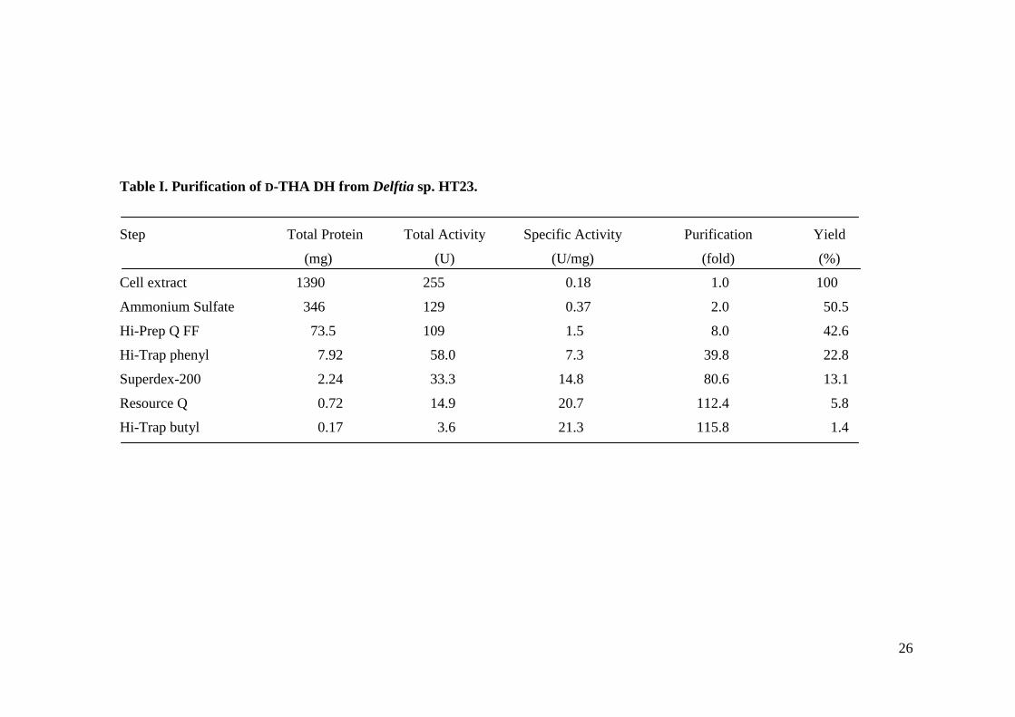
With the purification procedures described above, the enzyme was purified approximately 116-fold to
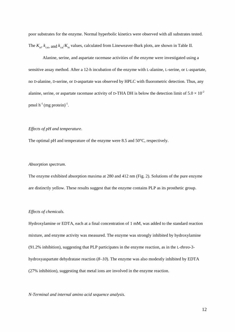
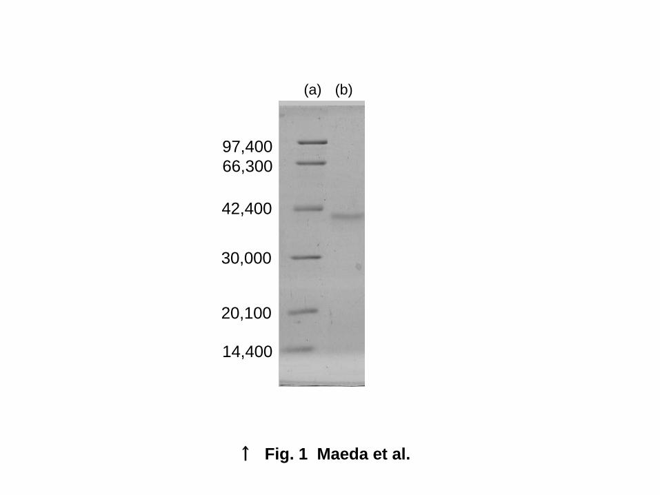
homogeneity, with approximately 1.4% recovery (Table I). The purified enzyme preparation gave a
single band on SDS-PAGE (Fig. 1). Furthermore, by high-performance gel-permeation liquid
chromatography, the enzyme gave a single symmetrical protein peak.
Using a calibrated TSKgel Super-SW3000 column, the relative molecular weight of the
enzyme was estimated to be 36,000. By SDS-PAGE, the relative molecular weight of the subunit was
estimated to be about 41,000, suggesting that the enzyme is a monomer.
Substrate specificity of the enzyme.
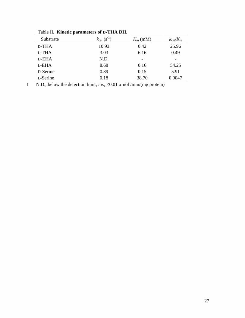
The enzyme showed broad specificity toward 3-hydroxyaspartate isomers. Table II shows the
substrate specificity and kinetic parameters of D-THA DH. In addition, D- and L-serine reacted as
12
poor substrates for the enzyme. Normal hyperbolic kinetics were observed with all substrates tested.
The Km, kcat, and kcat/Km values, calculated from Lineweaver-Burk plots, are shown in Table II.
Alanine, serine, and aspartate racemase activities of the enzyme were investigated using a
sensitive assay method. After a 12-h incubation of the enzyme with L-alanine, L-serine, or L-aspartate,
no D-alanine, D-serine, or D-aspartate was observed by HPLC with fluorometric detection. Thus, any
alanine, serine, or aspartate racemase activity of D-THA DH is below the detection limit of 5.0 × 10⁻²
pmol h-1 (mg protein)-1.
Effects of pH and temperature.
The optimal pH and temperature of the enzyme were 8.5 and 50°C, respectively.
Absorption spectrum.
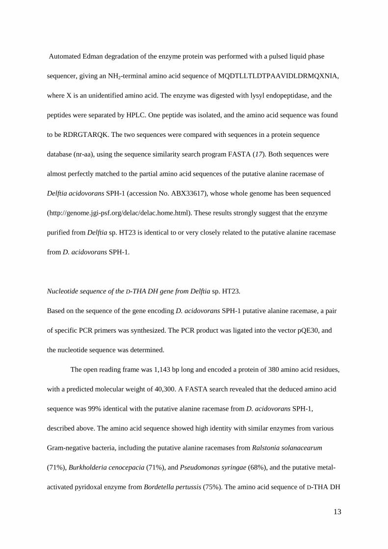
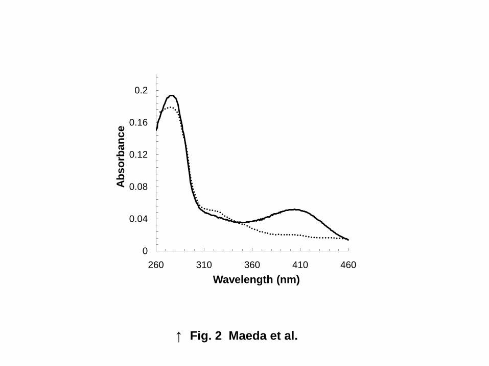
The enzyme exhibited absorption maxima at 280 and 412 nm (Fig. 2). Solutions of the pure enzyme
are distinctly yellow. These results suggest that the enzyme contains PLP as its prosthetic group.
Effects of chemicals.
Hydroxylamine or EDTA, each at a final concentration of 1 mM, was added to the standard reaction
mixture, and enzyme activity was measured. The enzyme was strongly inhibited by hydroxylamine
(91.2% inhibition), suggesting that PLP participates in the enzyme reaction, as in the L-threo-3-
hydroxyaspartate dehydratase reaction (8–10). The enzyme was also modestly inhibited by EDTA
(27% inhibition), suggesting that metal ions are involved in the enzyme reaction.
N-Terminal and internal amino acid sequence analysis.
13
Automated Edman degradation of the enzyme protein was performed with a pulsed liquid phase
sequencer, giving an NH2-terminal amino acid sequence of MQDTLLTLDTPAAVIDLDRMQXNIA,
where X is an unidentified amino acid. The enzyme was digested with lysyl endopeptidase, and the
peptides were separated by HPLC. One peptide was isolated, and the amino acid sequence was found
to be RDRGTARQK. The two sequences were compared with sequences in a protein sequence
database (nr-aa), using the sequence similarity search program FASTA (17). Both sequences were
almost perfectly matched to the partial amino acid sequences of the putative alanine racemase of
Delftia acidovorans SPH-1 (accession No. ABX33617), whose whole genome has been sequenced
(http://genome.jgi-psf.org/delac/delac.home.html). These results strongly suggest that the enzyme
purified from Delftia sp. HT23 is identical to or very closely related to the putative alanine racemase
from D. acidovorans SPH-1.
Nucleotide sequence of the D-THA DH gene from Delftia sp. HT23.
Based on the sequence of the gene encoding D. acidovorans SPH-1 putative alanine racemase, a pair
of specific PCR primers was synthesized. The PCR product was ligated into the vector pQE30, and
the nucleotide sequence was determined.
The open reading frame was 1,143 bp long and encoded a protein of 380 amino acid residues,
with a predicted molecular weight of 40,300. A FASTA search revealed that the deduced amino acid
sequence was 99% identical with the putative alanine racemase from D. acidovorans SPH-1,
described above. The amino acid sequence showed high identity with similar enzymes from various
Gram-negative bacteria, including the putative alanine racemases from Ralstonia solanacearum
(71%), Burkholderia cenocepacia (71%), and Pseudomonas syringae (68%), and the putative metal-
activated pyridoxal enzyme from Bordetella pertussis (75%). The amino acid sequence of D-THA DH
14
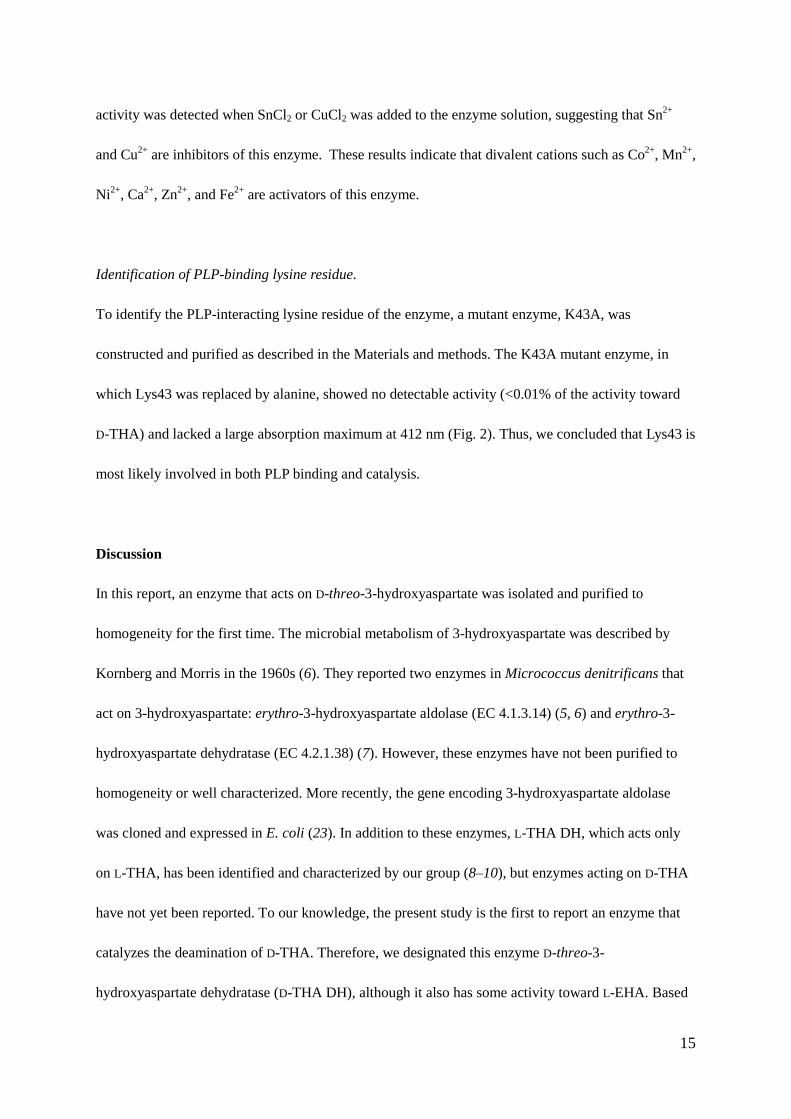
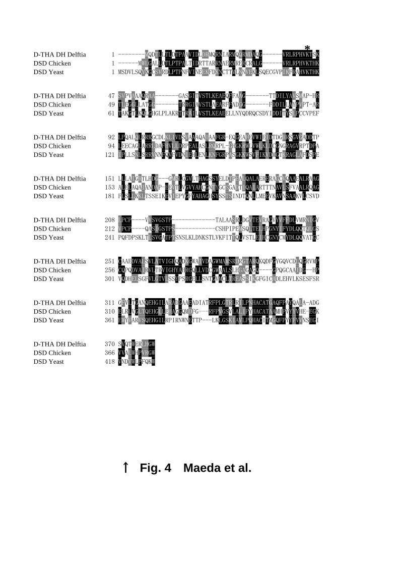
had relatively low, but significant, similarity to two eukaryotic D-serine dehydratases, those from
Gallus gallus (chicken, 36%) (20) and S. cerevisiae (26%) (21), as shown in the alignment in Fig. 4.
The PROSITE database (22) (http://au.expasy.org/prosite/) could not predict the PLP-binding site of
D-THA DH; however, the three enzymes share a common motif, R(P/A)HVKT, in their N-terminal
regions. In D-serine dehydratase from S. cerevisiae, the lysine residue in this motif binds PLP (21).
Thus, Lys43 of D-THA DH is probably a PLP-binding residue.
Characterization of the recombinant enzyme.
We purified recombinant His-tagged D-THA DH from E. coli cells and characterized its enzymatic
properties. The molecular weight determined by SDS-PAGE analysis (41,000) and that determined by
MALDI-TOF-MASS analysis (41,600) were in agreement with that calculated from the deduced
amino acid sequence of the recombinant enzyme (40,900). The first 15 N-terminal amino acid
residues sequenced in recombinant His-tagged enzyme perfectly matched the deduced amino acid
sequence. Moreover, the purified recombinant enzyme showed high activity, with specific activity of
around 20 units (mg protein)-1 toward D-THA. From these results, we concluded that this protein is
recombinant D-THA DH.
Effect of metal ions on the recombinant enzyme.
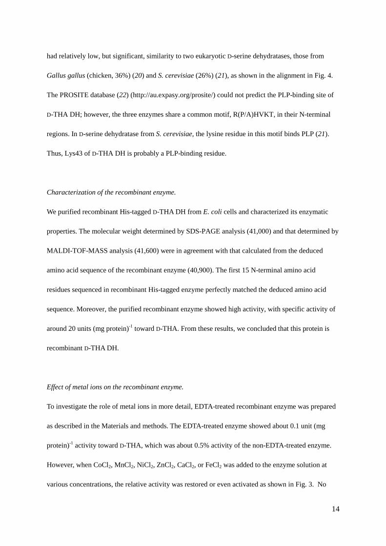
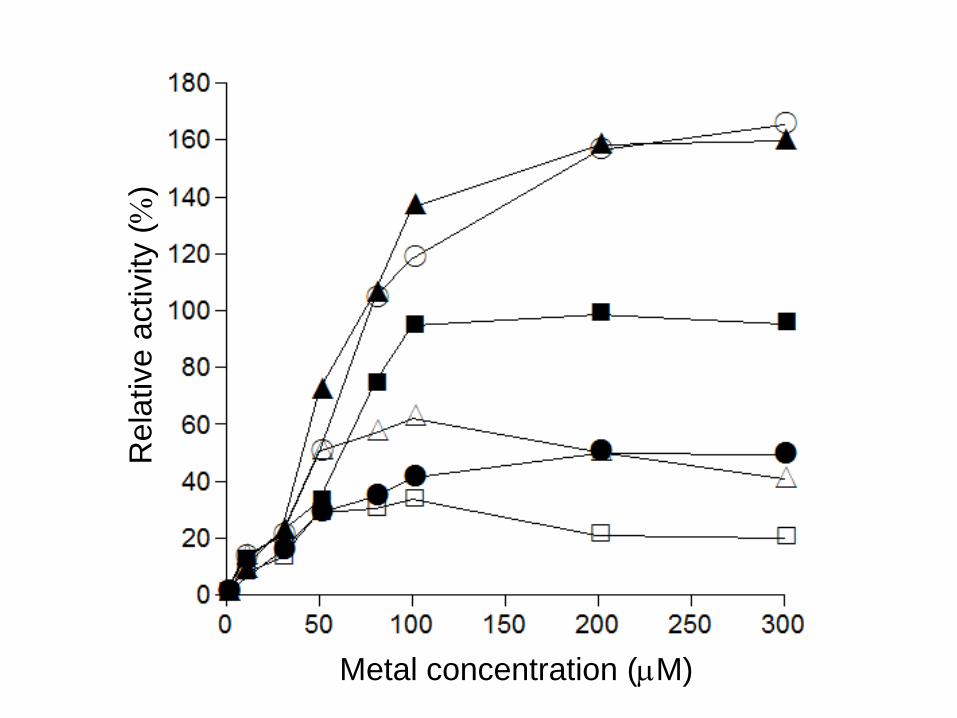
To investigate the role of metal ions in more detail, EDTA-treated recombinant enzyme was prepared
as described in the Materials and methods. The EDTA-treated enzyme showed about 0.1 unit (mg
protein)-1 activity toward D-THA, which was about 0.5% activity of the non-EDTA-treated enzyme.
However, when CoCl2, MnCl2, NiCl2, ZnCl2, CaCl2, or FeCl2 was added to the enzyme solution at
various concentrations, the relative activity was restored or even activated as shown in Fig. 3. No
15
activity was detected when SnCl2 or CuCl2 was added to the enzyme solution, suggesting that Sn2+
and Cu2+ are inhibitors of this enzyme. These results indicate that divalent cations such as Co2+, Mn2+,
Ni2+, Ca2+, Zn2+, and Fe2+ are activators of this enzyme.
Identification of PLP-binding lysine residue.
To identify the PLP-interacting lysine residue of the enzyme, a mutant enzyme, K43A, was
constructed and purified as described in the Materials and methods. The K43A mutant enzyme, in
which Lys43 was replaced by alanine, showed no detectable activity (<0.01% of the activity toward
D-THA) and lacked a large absorption maximum at 412 nm (Fig. 2). Thus, we concluded that Lys43 is
most likely involved in both PLP binding and catalysis.
Discussion
In this report, an enzyme that acts on D-threo-3-hydroxyaspartate was isolated and purified to
homogeneity for the first time. The microbial metabolism of 3-hydroxyaspartate was described by
Kornberg and Morris in the 1960s (6). They reported two enzymes in Micrococcus denitrificans that
act on 3-hydroxyaspartate: erythro-3-hydroxyaspartate aldolase (EC 4.1.3.14) (5, 6) and erythro-3-
hydroxyaspartate dehydratase (EC 4.2.1.38) (7). However, these enzymes have not been purified to
homogeneity or well characterized. More recently, the gene encoding 3-hydroxyaspartate aldolase
was cloned and expressed in E. coli (23). In addition to these enzymes, L-THA DH, which acts only
on L-THA, has been identified and characterized by our group (8–10), but enzymes acting on D-THA
have not yet been reported. To our knowledge, the present study is the first to report an enzyme that
catalyzes the deamination of D-THA. Therefore, we designated this enzyme D-threo-3-
hydroxyaspartate dehydratase (D-THA DH), although it also has some activity toward L-EHA. Based
16
on this substrate specificity, the enzyme is clearly distinct from the erythro-3-hydroxyaspartate
dehydratase (EC 4.2.1.38) reported by Gibbs and Morris (7), which acts only on L-EHA and not on D-
THA.
D-THA DH had approximately 30% amino acid sequence similarity to two eukaryotic D-
serine dehydratases, from chicken (20) and S. cerevisiae (21) (Fig. 4). Although D-THA DH acts on
D-serine, the relative activity toward D-serine was only about 23% of the activity toward D-THA.
Furthermore, the production of D-THA DH in Delftia sp. HT23 cells was not induced by D-serine, but
was induced by D-THA in the medium. From these results, we concluded that this enzyme is a D-
threo-3-hydroxyaspartate dehydratase, rather than a D-serine dehydratase.
The absorption spectrum of the purified enzyme revealed that D-THA DH contains PLP, as do
other bacterial dehydratases, although the amino acid sequence of D-THA DH was not similar to that
of L-THA DH, which catalyzes the same reaction of L-THA to oxaloacetate (9, 10). Despite
catalyzing the dehydratase reaction of D-THA, D-THA DH does not belong to the family of
serine/threonine dehydratases, which contains most bacterial dehydratases (24). Based on their folding
patterns, the PLP-dependent enzymes are classified into five groups, designated fold types I to V (25).
The L-THA DH enzymes of Pseudomonas sp. T62 and S. cerevisiae belong to the fold type II group
(9, 10), whereas D-THA DH belongs to the fold type III group. Eukaryotic D-serine dehydratases,
which are also fold-type III PLP-dependent enzymes, have been reported from chicken and from S.
cerevisiae (20, 21). However, D-THA DH is the first example of a prokaryotic dehydratase belonging
to the fold type III PLP-dependent enzyme family.
The high amino acid sequence identities between D-THA DH and several putative alanine
racemases and putative metal-activated pyridoxal enzymes from various Gram-negative bacteria,
including enzymes from Bordetella pertussis (75%), Ralstonia solanacearum (71%), Burkholderia
17
cenocepacia (71%), and Pseudomonas syringae (68%), suggest that these putative enzymes may have
D-THA DH activity. D-THA DH is expected to be present in other organisms, especially the Gram-
negative soil bacteria mentioned above.
Site-directed mutagenesis experiment revealed that lysine 43 is an important residue involved
in PLP binding and catalysis (Fig. 2), however, the K43A mutant enzyme still has weak absorption
peaks around 320 nm and 420 nm. The reason why K43A mutant enzyme has these absorption peaks
remains unknown, however, non-covalently bound PLP may cause these absorption peaks as in the
case of aspartate aminotransferase of E. coli (26). Another explanation is also possible; Lys residue
other than 43 (for example, Lys46) may bind PLP weakly, and gave catalytically inactive enzyme-PLP
complex with absorption around 320 nm, and 420 nm. But, further investigation is required to
elucidate this phenomenon.
Like L-THA DH from Pseudomonas sp. T62, D-THA DH requires divalent cations for its
activation (8, 9), but the activation pattern differed between the two enzymes. Both Co2+ and Zn2+ act
as inhibitors of L-THA DH, whereas these divalent cations are activators of D-THA DH. The pattern
of D-THA DH activation by divalent cations was similar to that of D-threonine aldolase from
Arthrobacter sp. DK-38 (27, 28), which is also activated by Mn2+, Co2+, Ni2+, Fe2+, and Ca2+. D-
Threonine aldolase from Arthrobacter sp. DK-38 also belongs to the fold-type III PLP-dependent
enzymes (28) and shares around 30% amino acid identity to D-THA DH (data not shown). D-
Threonine aldolase from Arthrobacter sp. DK-38 can bind 1 mol of Mn2+ ion per mol of subunit (28),
however, the amount of metal ion, which binds to D-THA DH, has not yet been determined.
Although the reason why activity of the EDTA-treated enzyme added with 100 µM MnCl2 is
higher than that of the non-EDTA-treated enzyme dialyzed against the same concentration of MnCl2 is
not clear (Fig. 3), the following explanation might be possible; the EDTA-treated enzyme is
18
somewhat more stable than the non-EDTA-treated enzyme, i.e., the non-EDTA-treated enzyme
(containing Mn2+) might be partially inactivated during the dialysis, however, the EDTA-treated
enzyme (containing no or very little amount of metal ions) may become more active than the non-
EDTA-treated enzyme when Mn2+ is added just before activity measurement. Effect of the metal ions
on stability of the enzyme, however, still needs to be elucidated.
Although the physiological function of this enzyme remains to be clarified, the dehydratase
reaction catalyzed by D-THA DH may be one of the reactions enabling Delftia sp. HT23 to grow on
medium containing D-THA as the sole carbon source. This hypothesis is also supported by the fact
that D-THA DH is inducible in Delftia sp. HT23; however, the details have not been elucidated.
To analyze the detailed reaction mechanism and 3D structure of this enzyme, an efficient
expression system is necessary. Unfortunately, the expression level of D-THA DH in E. coli was low.
The specific activity of the cell-free extract of E. coli expressing the D-THA DH gene was about 0.2
unit (mg protein)-1, which is almost equal to that of the cell-free extract of the original strain, Delftia
sp. HT23. The reason for poor expression in E. coli is unknown; however, the high GC content of the
gene (71.9%) is one possible explanation. Currently, we are trying to improve the expression level of
D-THA DH in E. coli.
Funding
This work was supported in part by a Grant-in-Aid for Scientific Research from the Ministry of
Education, Science, Sports, and Culture of Japan (KAKENHI, No. 19580074 to MW).
Conflict of interest
None declared.
19
Acknowledgments
The MALDI-TOF-MS analysis was carried out at the OPEN FACILITY, Hokkaido
University Sousei Hall.
20
References
1. Balcar, V. J., Johnston, G. A., and Twitchin, B. (1977) Stereospecificity of the inhibition of
L-glutamate and L-aspartate high affinity uptake in rat brain slices by threo-3-
hydroxyaspartate. J. Neurochem. 28, 1145-1146
2. Shigeri, Y., Shimamoto, K., Yasuda-Kamatani, Y., Seal, R. P., Yumoto, N., Nakajima, T.,
and Amara, S. G. (2001) Effects of threo-β-hydroxyaspartate derivatives on excitatory amino
acid transporters (EAAT4 and EAAT5). J. Neurochem. 79, 297-302
3. Kaneko, T. and Katsura, H. (1963) The synthesis of four optical isomers of β-hydroxyaspartic
acid. Bull. Chem. Soc. Jpn. 36, 899-903
4. Lebrun, B., Sakaitani, M., Shimamoto, K., Yasuda-Kamatani, Y., and Nakajima, T. (1997)
New β-hydroxyaspartate derivatives are competitive blockers for the bovine
glutamate/aspartate transporter. J. Biol. Chem. 272, 20336-20339
5. Gibbs, R. G. and Morris, J. G. (1964) Assay and properties of β-hydroxyaspartate aldolase
from Micrococcus denitrificans. Biochim. Biophys. Acta 85, 501-503
6. Kornberg, H. L. and Morris, J. G. (1965) The utilization of glycollate by Micrococcus
denitrificans: the β-hydroxyaspartate pathway. Biochem. J. 95, 577-586
7. Gibbs, R. G. and Morris, J. G. (1965) Purification and properties of erythro-β-
hydroxyaspartate dehydratase from Micrococcus denitrificans. Biochem. J. 97, 547-554
8. Wada, M., Matsumoto, T., Nakamori, S., Sakamoto, M., Kataoka, M., Liu, J.Q., Itoh, N.,
Yamada, H., and Shimizu, S. (1999) Purification and characterization of a novel enzyme, L-
threo-3-hydroxyaspartate dehydratase, from Pseudomonas sp. T62. FEMS Microbiol. Lett.
179, 147-151
21
9. Murakami, T., Maeda, T., Yokota, A., and Wada, M. (2009) Gene cloning and expression of
pyridoxal 5'-phosphate-dependent L-threo-3-hydroxyaspartate dehydratase from
Pseudomonas sp. T62, and characterization of the recombinant enzyme. J. Biochem. 145,
661-668
10. Wada, M., Nakamori, S., and Takagi, H. (2003) Serine racemase homologue of
Saccharomyces cerevisiae has L-threo-3-hydroxyaspartate dehydratase activity. FEMS
Microbiol. Lett. 225, 189-193
11. Robinson, W. G. and Labow, R. (1971) D-Serine dehydratase (Escherichia coli). Meth.
Enzymol. 17B, 356-360.
12. Panizzutti, R., de Souza Leite, M., Pinheiro, C. M., and Meyer-Fernandes, J. R. (2006) The
occurrence of free D-alanine and an alanine racemase activity in Leishmania amazonensis.
FEMS Microbiol. Lett. 256,16-21
13. Uo, T., Yoshimura, T., Shimizu, S., and Esaki, N. (1998) Occurrence of pyridoxal 5'-
phosphate-dependent serine racemase in silkworm, Bombyx mori. Biochem. Biophys. Res.
Commun. 246, 31-34
14. Shibata, K., Watanabe, T., Yoshikawa, H., Abe, K., Takahashi, S., Kera, Y., and Yamada, R.
(2003) Purification and characterization of aspartate racemase from the bivalve mollusk
Scapharca broughtonii. Comp. Biochem. Physiol. Part B 134, 307-314
15. Bradford, M. M. (1976) A rapid and sensitive method for quantitation of microgram
quantities of proteins utilizing the principle of protein-dye binding. Anal. Biochem. 72, 248-
254
22
16. Wada, M., Yoshizumi, A., Nakamori, S., and Shimizu, S. (1999) Purification and
characterization of monovalent cation-activated levodione reductase from Corynebacterium
aquaticum M-13. Appl. Environ. Microbiol. 65, 4399-4403
17. Pearson, W. R., and Lipman, D. J. (1988) Improved tools for biological sequence
comparison. Proc. Natl. Acad. Sci. USA 85, 2444-2448
18. Thompson, J. D., Higgins, D. G., and Gibson, T. J. (1994) CLUSTAL W: Improving the
sensitivity of progressive multiple sequence alignment through sequence weighting, position-
specific gap penalties and weight matrix choice. Nucleic Acids Res. 22, 4673-4680
19. Wen, A., Fegan, M., Hayward, C., Chakraborty, S., Sly, L.I. (1999) Phylogenetic
relationships among members of the Comamonadaceae, and description of Delftia
acidovorans (den Dooren de Jong 1926 and Tamaoka et al. 1987) gen. nov., comb. nov. Int.
J. Syst. Bacteriol., 49, 567-576
20. Tanaka, H., Yamamoto, A., Ishida, T., and Horiike, K. (2008) D-Serine dehydratase from
chicken kidney: a vertebral homologue of the cryptic enzyme from Burkholderia cepacia. J.
Biochem. 143, 49-57
21. Ito, T., Hemmi, H., Kataoka, K., Mukai, Y., and Yoshimura, T. (2008) A novel zinc-
dependent D-serine dehydratase from Saccharomyces cerevisiae. Biochem. J. 409, 399-406
22. Hulo, N., Bairoch, A., Bulliard, V., Cerutti, L., De Castro, E., Langendijk-Genevaux, P. S.,
Pagni, M., and Sigrist, C. J. A. (2006) The PROSITE database. Nucleic Acids Res. 34, D227-
D230
23
23. Liu, J. Q., Dairi, T., Itoh, N., Kataoka, M., and Shimizu, S. (2003) A novel enzyme, D-3-
hydroxyaspartate aldolase from Paracoccus denitrificans IFO 13301: purification,
characterization, and gene cloning. Appl. Microbiol. Biotechnol. 62, 53-60
24. Eliot, A. C, Kirsch, J. F. (2004) Pyridoxal phosphate enzymes: mechanistic, structural, and
evolutionary considerations. Annu. Rev. Biochem. 73, 383-415
25. Schneider, G., Käck, H., and Lindqvist, Y. (2000) The manifold of vitamin B6 dependent
enzymes. Structure 8, R1-6
26. Kuramitsu, S., Inoue, Y., Tanase, S., Morino, Y., and Kagamiyama, H. (1987) Substitution
of an arginyl residue for the active site lysyl residue (Lys258) of aspartate aminotransferase.
Biochem. Biophys. Res. Commun., 146, 416-421
27. Kataoka, M., Ikemi, M., Morikawa, T., Miyoshi, T., Nishi, K., Wada, M., Yamada, H., and
Shimizu, S. (1997) Isolation and characterization of D-threonine aldolase, a pyridoxal-5'-
phosphate-dependent enzyme from Arthrobacter sp. DK-38. Eur. J. Biochem. 248, 385-393
28. Liu, J.-Q., Dairi, T., Itoh, N., Kataoka, M., Shimizu, S., and Yamada, H. (1998) A novel
metal-activated pyridoxal enzyme with a unique primary structure, low specificity D-
threonine aldolase from Arthrobacter sp. strain DK-38. J. Biol. Chem. 273, 16678-16685
24
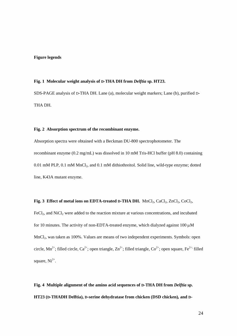
Figure legends
Fig. 1 Molecular weight analysis of D-THA DH from Delftia sp. HT23.
SDS-PAGE analysis of D-THA DH. Lane (a), molecular weight markers; Lane (b), purified D-
THA DH.
Fig. 2 Absorption spectrum of the recombinant enzyme.
Absorption spectra were obtained with a Beckman DU-800 spectrophotometer. The
recombinant enzyme (0.2 mg/mL) was dissolved in 10 mM Tris-HCl buffer (pH 8.0) containing
0.01 mM PLP, 0.1 mM MnCl2, and 0.1 mM dithiothreitol. Solid line, wild-type enzyme; dotted
line, K43A mutant enzyme.
Fig. 3 Effect of metal ions on EDTA-treated D-THA DH. MnCl2, CaCl2, ZnCl2, CoCl2,
FeCl2, and NiCl2 were added to the reaction mixture at various concentrations, and incubated
for 10 minutes. The activity of non-EDTA-treated enzyme, which dialyzed against 100 µM
MnCl2, was taken as 100%. Values are means of two independent experiments. Symbols: open
circle, Mn2+; filled circle, Ca2+; open triangle, Zn2+; filled triangle, Co2+; open square, Fe2+; filled
square, Ni2+.
Fig. 4 Multiple alignment of the amino acid sequences of D-THA DH from Delftia sp.
HT23 (D-THADH Delftia), D-serine dehydratase from chicken (DSD chicken), and D-
25
serine dehydratase from S. cerevisiae (DSD Yeast).
The alignment was generated with Clustal W 1.83 and BOXSHADE 3.21. The numbers on the
left are the residue numbers for each sequence. White letters on a black background indicate
identical residues, and white letters on a gray background indicate similar residues. The asterisk
indicates the PLP-binding residue.
26
Table I. Purification of D-THA DH from Delftia sp. HT23.
Step Total Protein Total Activity Specific Activity Purification Yield
(mg) (U) (U/mg) (fold) (%)
Cell extract 1390 255 0.18 1.0 100
Ammonium Sulfate 346 129 0.37 2.0 50.5
Hi-Prep Q FF 73.5 109 1.5 8.0 42.6
Hi-Trap phenyl 7.92 58.0 7.3 39.8 22.8
Superdex-200 2.24 33.3 14.8 80.6 13.1
Resource Q 0.72 14.9 20.7 112.4 5.8
Hi-Trap butyl 0.17 3.6 21.3 115.8 1.4
27
Table II. Kinetic parameters of D-THA DH. Substrate kcat (s-1) Km (mM) kcat/Km D-THA 10.93 0.42 25.96 L-THA 3.03 6.16 0.49 D-EHA N.D. - - L-EHA 8.68 0.16 54.25 D-Serine 0.89 0.15 5.91 L-Serine 0.18 38.70 0.0047
N.D., below the detection limit, i.e., <0.01 µmol /min/(mg protein) 1
97,40066,300
42,400
30,000
20,100
14,400
(a) (b)
↑ Fig. 1 Maeda et al.
0
0.04
0.08
0.12
0.16
0.2
260 310 360 410 460
Abso
rban
ce
Wavelength (nm)
↑ Fig. 2 Maeda et al.
Rel
ativ
e ac
tivity
(%)
Metal concentration (µM)
D-THA DH Delftia 1 --------MQDTLLTLDTPAAVIDLDRMQRNIARMQQRMDAQG------VRLRPHVKTSK DSD Chicken 1 ------MWLGALLDTLPTPALTIDRTTARRNAERMRERCRALG------VRLRPHVKTHK DSD Yeast 1 MSDVLSQYKGCSVRDLPTPNFVINEEKFDKNCTTMLNNVEKLSQECGVPIKFRAHVKTHK D-THA DH Delftia 47 SVPVAAAQRAA-------GASGITVSTLKEAEQFFAAG-------TTDILYAVSMAP-HR DSD Chicken 49 TLEGGLLATGG-------TRRGIAVSTLAEARFFADGG-------FDDILLAYPVPT-AR DSD Yeast 61 TAKGTLKQLGHGLPLAKRTTRAILVSTLKEAEELLNYQDRQCSDYIDDITYSLPCCVPEF D-THA DH Delftia 92 LPQALQLRRRGCDLKLIVDSVAAAQAIAAFGR-EQGEAFEVWIEIDTDGHRSGVGADDTP DSD Chicken 94 LEECAGLARRLDAFHVLLDRPEALASLRQRPL-GHGKRWLVWLKLDCGNGRAGVRPTDPA DSD Yeast 121 IPLLSNLSRRVNNFQVFVDNIEHLENLKNFGRPASGKKWSVFIKVDMGTKRAGLAFDSPE D-THA DH Delftia 151 LLLAIGRTLHDG---GMRLGGVLTHAGSSYELDTPEALQALAERERAGCVQAAEALRAAG DSD Chicken 153 ALELAQAIANDAP-EEVTLVGVYAHCGNTYGCSGADTIQAIARTTTNAVLSFVAALRQAG DSD Yeast 181 FLSLLKKLTSSEIKEVIEPYGFYAHAGHSYSSTSINDTQNLLMEEVKAVNSAAKVLCSVD D-THA DH Delftia 208 LPCP----VVSVGSTP-------------TALAASRLDGVTEVRAGVYVFFDLVMRNIGV DSD Chicken 212 VPCP----QASIGSTPS------------CSHPIPEMSQLTELHPGNYIFYDLQQTQLGS DSD Yeast 241 PQFDPSKLTLSVGATPTSNSLKLDNKSTLVKFITTQLVSTLEIHCGNYCMYDLQQVATGC D-THA DH Delftia 251 CAAEDVALSVLATVIGHQADKGWAIVDAGWMAMSRDRGTARQKQDFGYGQVCDLQGRVMP DSD Chicken 256 CQPQDVAIRVLTRVIGHYAHRGQLLVDCGWAALSLHGAGAGQ----GPQGCAAIDG--HP DSD Yeast 301 VQDHELSGFVLGTVLSSYPSRGELLSNTGVMCLTREASSIKGFGICADLEHVLKSESFSR D-THA DH Delftia 311 GFVLTGANQEHGILARADGAAEADIATRFPLGTRLRILPNHACATGAQFPAYQALA-ADG DSD Chicken 310 ELRLVGLTQEHGLLEHAGGQMDFG---RFPVGSVLALIPYHACATAAMHPVYYVHE-EGK DSD Yeast 361 EWYVARVSQEHGILRPIRNWNETTP---LKLGSKIAVLPQHACITMGQFPYYFVVNSEGI D-THA DH Delftia 370 SVQTWERLHGW DSD Chicken 366 VVALWHPVRGW DSD Yeast 418 VNDVWLPFQKW
↑ Fig. 4 Maeda et al.
*