Embed Size (px)
Citation preview

Radiographers’ Professional Competence
Development of a context-specific instrument
Bodil T Andersson
Akademisk avhandling För avläggande av doktorsexamen i Omvårdnad som med tillstånd av Nämnden för
utbildning och forskarutbildning vid Högskolan i Jönköping framläggs till offentlig granskning fredag den 9 november 2012 kl.13.00 i Forum Humanum,
Hälsohögskolan i Jönköping.
Opponent: Professor Kerstin Öhrling
Department of Health Sciences, Division of Nursing, Health and Rehabilitation, Luleå University of Technology
Forskarskolan Hälsa och Välfärd, Hälsohögskolan, Högskolan i Jönköping, 551 11 Jönköping
Jönköping 2012

Abstract
Title: Radiographers‟ Professional Competence
Author: Bodil T Andersson
Opponent: Professor Kerstin Öhrling, Department of Health Sciences, Division of Nursing, Health and Rehabilitation, Luleå University of Technology
Language: English
Keywords: Professional competence, Radiographer perspective, Patient perspective, Nursing, Radiography, Instrument development, Self-assessment, Psychometric evaluation, RCS
ISBN: 978-91-85835-33
Aims: The overall aim of this thesis was to explore and describe radiographers‟ professional competence based on
patients‟ and radiographers‟ experiences and to develop a context-specific instrument to assess the level and
frequency of use of radiographers‟ professional competence.
Methods: The design was inductive and deductive. Both qualitative and quantitative methods were used. The data
collection methods comprised interviews (Studies I-II) and questionnaires (Studies III-IV). The subjects were
patients in study I and radiographers in studies II-IV. In study I, 17 patients were interviewed about their experiences
of the encounter during radiographic examinations and treatment. The interviews were analysed using qualitative
content analysis. In study II, 14 radiographers were interviewed to identify radiographers‟ areas of competence. The
critical incident technique was chosen to analyse the interviews. Studies III and IV were based on a national cross-
sectional survey of 406 randomly selected radiographers. Study III consisted of two phases; designing the
Radiographer Competence Scale (RCS) and evaluation of its psychometric properties. A 42-item questionnaire was
developed and validated by a pilot test (n=16) resulting in the addition of 12 items. Thus the final RCS comprised a
54-item questionnaire, which after psychometric tests was reduced to 28 items. In study IV, the 28-item
questionnaire served as data. The level of competencies was rated on a 10-point scale, while their use was rated on a
six-point scale.
Results: In study I, the female patients‟ comprehensive understanding was expressed as feelings of vulnerability. The
encounters were described as empowering, empathetic, mechanical and neglectful, depending on the radiographers‟
skills and attitudes. Study II revealed two main areas of professional competence, direct patient-related and indirect
patient-related. The first focused on competencies in the care provided in close proximity to the patient and the
second on competencies used in the activities of the surrounding environment. Each of the two main areas was
divided into four categories and 31 sub-categories that either facilitated or hindered good nursing care. In study III
the analysis condensed the 54-item questionnaire in two steps, firstly by removing 12 items and secondly a further 14
items, resulting in the final 28-item RCS questionnaire. Several factor analyses were performed and a two factor-
solution emerged, labelled; “Nurse initiated care” and “Technical and radiographic processes”. The psychometric
tests had good construct validity and homogeneity. The result of study IV demonstrated that most competencies in
the RCS received high ratings both in terms of level and frequency of use. Competencies e.g. „Adequately informing
the patient‟, „Adapting the examination to the patient‟s prerequisites and needs‟ and „Producing accurate and correct
images‟ were rated the highest while „Identifying and encountering the patient in a state of shock‟ and „Participating
in quality improvement regarding patient safety and care‟ received the lowest ratings. The total score of each of the
two dimensions had a low but significant correlation with age and years in present position. The competence level
correlated with age and years in present position in both dimensions but not with the use of competencies in the
“Nurse initiated care” dimension.
Conclusion: This thesis has shown that professional competence is important in the encounter between patient and
radiographer. It has also demonstrated that radiographers‟ self-rated professional competence is based on nursing,
technological and radiographic knowledge. From a radiographer‟s perspective, „Nurse initiated care‟ and „Technical
and Radiographic processes‟ are two core dimensions of Radiographer Competence Scale. The 28-item questionnaire
regarding level and frequency of use of competence is feasible to use to measure radiographers‟ professional
competence.

Original papers
This thesis is based on the following papers, which are referred to by their
roman numerals in the text:
Paper I
Andersson BT, Fridlund B, Elgán C & Axelsson ÅB. Female patients‟ encounters with the radiographer in the course of recurrent radiographic examinations – a qualitative study. Submitted for publication.
Paper II
Andersson BT, Fridlund B, Elgán C & Axelsson ÅB. Radiographers‟ areas of professional competence related to good nursing care. Scandinavian Journal of Caring Sciences 2008; 22: 401-409.
Paper III
Andersson BT, Christensson L, Fridlund B, Broström A. Development and Psychometric Evaluation of the Radiographers‟ Competence Scale. Open Journal of Nursing 2012; 2: 85-96.
Paper IV
Andersson BT, Christensson L, Jakobsson U, Fridlund B, Broström A. Radiographers‟ Self-Assessed Level and Use of Competence – a National Survey. Accepted for publication in Insights Imaging 2012. DOI: 10.1007/s13244-012-0194-8.
Published papers are reproduced with the kind permission of the respective
journals.


Contents
Introduction ............................................................................................. 13 Background .............................................................................................. 14
The diagnostic radiology context ........................................................................ 14
A high technological environment ................................................................ 14
The patient ........................................................................................................ 15
Caring ................................................................................................................ 16
Encounters ........................................................................................................ 17
The radiographer ............................................................................................. 19
Radiography ...................................................................................................... 20
Measuring competence ................................................................................... 21
Conceptual standpoints........................................................................... 24
Evidence-based practice (EBP) ..................................................................... 24
Patient safety .................................................................................................... 25
Ethics ................................................................................................................. 26
Clinical habitus ................................................................................................. 27
Professional competence ................................................................................ 28
Rationale for the thesis ............................................................................ 30 Overall and specific aims ......................................................................... 31 Methodological framework ..................................................................... 32
Epistemology and ontology ................................................................................. 32
Qualitative content analysis ............................................................................ 33
Critical incident technique .............................................................................. 34
Developing the RCS ........................................................................................ 34
Methods ................................................................................................... 36
Design of the thesis ............................................................................................... 36
Participants ............................................................................................................. 37
Data collection ....................................................................................................... 40
Interviews .......................................................................................................... 40
Questionnaire ................................................................................................... 41
Data analysis ........................................................................................................... 42

Qualitative Content Analysis (Study I)..........................................................42
Critical Incident Technique (Study II) ..........................................................44
Questionnaire ....................................................................................................46
Development and psychometric tests (Study III) .....................................46
Comparisons between groups (Study IV) ..................................................47
Ethical considerations ............................................................................. 48 Summary of the results ............................................................................ 51
The patient ..............................................................................................................51
Vulnerability (Study I) ......................................................................................51
Encounters (Study I) ........................................................................................51
The radiographer ....................................................................................................52
Radiographers areas of professional competence (Study II) .....................52
Designing an instrument – the RCS (Study III) ..........................................53
Psychometric evaluation of the RCS (Study III) .........................................54
The level and frequency of use of professional competencies) (Study IV ............................................................................................................55
Discussion ................................................................................................ 58
Methodological considerations ............................................................................58
General discussions of the findings.....................................................................65
Patients‟ experiences ........................................................................................65
Vulnerability (Study I) ....................................................................................65
Encounters (Study I) ......................................................................................66
Radiographers‟ perspectives ...........................................................................68
Radiographers‟ areas of professional competence (Study II) ..................68
Development of the RCS for measuring professional competence (Study III) ........................................................................................................71
The level and frequency of use of professional competencies (Study IV) ........................................................................................................71
Comprehensive understanding ................................................................ 75 Conclusions .............................................................................................. 77 Clinical implications ................................................................................ 78 Research implications .............................................................................. 79 Summary in Swedish ................................................................................ 80 Acknowledgements .................................................................................. 86 References ................................................................................................ 88 Appendix. 28-item Questionnaire

13
Introduction
The patient arrives at the diagnostic radiology department to undergo a
radiographic examination and/or radiological intervention expecting to be
cared for and encountered as a human being by a professional radiographer
with appropriate competence. The radiographer plays a central role, as she/he
cares for the patient before, during and after the radiographic examination
and/or radiological intervention. It is of paramount importance that the
radiographer is familiar with the problems involved and can support the patient
during the radiographic procedure.1 Patients, especially those who are
chronically ill, are vulnerable and in need of caring when in hospital.2, 3 Hence,
the radiographer requires knowledge of nursing care in addition to specialised
radiography competence. Radiography is the area of professional knowledge,
research and responsibility.4 Caring encompasses someone who cares, a
relationship of caring and someone who is cared for and rests upon
understanding human thoughts and feelings.5-7 Nursing care in a diagnostic
radiology context includes interacting with patients while respecting their
privacy and personal space, focusing on patients‟ safety, comfort and dignity in
addition to dealing with their fear and anxiety.8, 9 The professional radiology
healthcare team; radiographer, radiologist and assistant nurse, plays a vital role
in assisting the patient physically and psychologically as well as helping her/him
to understand her/his feelings and reactions related to the examination.
Although nursing care is one of the primary functions of a radiographer, the
time available for the performance of a complete radiographic examination is
limited.
The work of radiographers in Sweden differs from that of their peers in some
other countries. In Sweden, registered radiographers are responsible for
performing the entire radiographic examination, thus they have to take care of
the patient as well as dealing with the medical technology, e.g. injections,
catheterizing and medical technical equipment.4 In most countries, registered
nurses are responsible for patient care, while the radiographer or corresponding
professional is in charge of the radiological equipment.

14
Diagnostic radiology departments are characterised by high-technology
comprising medical technical equipment, radiographic examination and
radiological interventions.10, 11 A profession such as that of radiographer is
situated in a field of great tension; encountering the patient, performing a
radiographic examination or intervention, achieving internal and external goals
and strategies while simultaneously providing nursing care.12 There is a need to
develop tools to measure professional competence in the radiographers‟
profession in order to ensure patient safety and high quality care. A
questionnaire adapted to a high-technological environment such as diagnostic
radiology could contribute to the development of radiographers‟ areas of
professional competence. Furthermore, deeper knowledge of and insights into
these areas are significant in terms of their responsibility for the patient in the
diagnostic radiology department.13 There is an increasing need for evidence-
based research and evidence-based practice (EBP) related to clinical
radiographer practice14, 15 in order to ensure patient safety and to analyse
ingrained habitus. A prerequisite for patient safety and effective health care
practice is professional competence and knowledge.
Background
The diagnostic radiology context
A high technological environment
The culture of a diagnostic radiology department is characterized by advanced
high technology, which was developed to help patients in their striving for
health and recovery but also to make radiological work safer and easier to
handle.16 The purpose of a radiology department is to provide a service to the
health care system for in- and out patients as well as the patients of general
practitioners and can be organised into conventional and specialised radiology.
The most common conventional examinations comprise the skeleton, chest,
gastrointestinal and urinary tracts, while computed tomography (CT), magnetic
resonance imaging (MRI), mammography, ultrasound, vascular examinations

15
and radiological interventions are considered specialized radiological
examinations.
The high-technological environment has evolved due to the implementation of
digital systems, which have totally changed the radiology department. The rapid
growth of technical and scientific knowledge has changed not only the
environment but also the requirements on competence for many professional
categories and the health care system as a whole.17 High technology has
transformed the physical appearance of the diagnostic radiology department
and influenced its social structure.12 This technical development process has
impacted on the radiographers‟ work in that it allows little human control in the
organisation.18 Traditionally, knowledge and skills were often developed by trial
and error and learnt from colleagues through habitus. Today, there is a great
demand for evidence.15
All parts of a radiological examination or intervention involve advanced high
technology and today, many patients go directly from e.g. the emergency ward,
to the radiology department. Historically, technological advances have been
considered a major indicator of human progress, especially in the areas of
medical science and health care.19 Technology can distance the personnel from
the patient, e.g. monitoring machines instead of using sensitivity and
reflection.20 Although technology can facilitate health care and enhance
professional development,16, 19, 21 there is a need to understand the humanistic
interaction involved.2, 12
High technology is very much a natural part of radiographers‟ work and the
importance they attribute to the technical equipment plays a vital role in their
competence. At the same time as radiographers have had to assume a more
demanding role and higher level of responsibility due to the technical evolution,
they are also required to take care of patients.22 Technology is not neutral in
radiography or nursing practice, but a pervasive reality.21
The patient
When a patient attends the hospital to undergo a radiographic examination,
she/he is in need of professional competence. Most patients have been referred
and do not require advanced medical support. However, this situation will

16
probably change in the future due to the critically ill trauma patients as well as
the increasing number of elderly patients suffering from chronic conditions.
Despite advances in medical technology, cancer remains one of the leading
causes of death globally. Approximately one third of the population in the
developed world will experience cancer in their lives.23 Cancer diagnosis is
central to the radiography profession, as radiographers assist in the diagnostic
process and follow up examinations.1 Furthermore, patients require physical
and psychological support when undergoing a radiographic examination or
complex radiological intervention.
Vulnerability of patients in today‟s health care system has been described by
several theorists.3, 24-27 Armstrong28 pointed out that the vulnerable patient can
lose control and enter into a state of powerlessness, which may influence
her/his participation in the radiographic examination. The relationship between
feelings of vulnerability and mammography examinations has been studied by
Lupton.29 A follow-up examination is very frequently associated with fear of
recurrence30, 31 and in such stressful circumstances a person is sensitive to what
is happening around her/him. Andersson32 and von Post33 highlighted the
patient‟s vulnerability and total dependence upon the professionals. The
patient‟ integrity may be violated in the encounter with professionals who treat
her/his body in a non ethical way or who intrude upon her/his personal
space.34 The experience of dignity influences a person‟s well-being.35 A situation
that threatens to violate dignity can be transformed into a non-violating action
through e.g. sensitivity, involvement and attention.36, 37 Irrespective of how
difficult the circumstances may be, respect for dignity and knowledge of the
nature of true dignity are essential.35
Caring
Caring is the human mode of being in every relationship. Professional
competence in caring is a prerequisite in the patient-radiographer interaction.2,38
Caring for the patient is a fundamental aspect of diagnostic radiology and also a
part of radiographers‟ education. In radiography, caring is derived from and
comparable with the nursing profession, although the context is quite different.
Caring is a rich concept reflecting genuine concern for another person,
attentiveness and respect.6 The radiographer has a unique position in terms of

17
encountering, supporting and protecting the patient and next of kin and is
responsible for the patient during the entire radiographic examination and stay
at the radiology department.4 Caring also involves acting as an advocate for
patients exposed to poor practice as well as facilitating contact between the
patient and the radiologist.4 Stolberg et al.39 pointed out that the patient more
often dares to approach the radiographer than the doctor. When positioning
the patient and assisting during radiological interventions there is a need for
physical closeness in the form of touching for examination purposes. However,
touch, eye contact and standing close to her/him as well as avoiding technical
language are important caring behaviours,40 as well as being aware of the
importance of mutuality in the caring of the patient.38
The core of good nursing care is human, interactive action comprising the
following categories: the actor, her/his characteristics, task and human oriented
activities and ways of acting, preconditions and aims.41 Leino-Kilpi 41 described
the most important and frequent elements of good nursing care as interaction,
comprehensiveness, need-centredness, initiative, technical procedures and
knowledge-base. In other words, good nursing care takes place in interaction
that demands initiative on the part of the radiographer and collaboration with
colleagues in technical procedures as well as being comprehensive, need-
centred and requiring a knowledge-base. Good nursing care has also been
described as decisions about correct or appropriate care, sensitive listening and
consideration of the different ways in which the patient‟s condition can
manifest itself.42 As the medical technology requires a great deal of a
radiographer‟s time, it should be borne in mind that good care is necessary to
ensure patient safety in the encounter.
Encounters
Despite the fact that the main purpose of the encounter is a radiographic
examination or radiological intervention, it is essential to treat the patient in a
professional caring way, taking account of her/his circumstances and
psychological needs. Encounters between the patient and the radiographer in
the course of radiographic examinations are short and intensive. Information
about the patient is sparse and usually she/he is unknown to the radiographer.
Croona43 has shown that despite an heavy workload it is necessary to allow time
to encounter the patient in a caring way.

18
Encounters in settings other than diagnostic radiology have been described
from the perspective of both patients and nurses.25, 26, 44-47 Wiman et al.47 found
that high quality encounters take place when caregivers are capable of shifting
their way of being with the patient between the instrumental and attentive
mode in accordance with the demands of the situation. Travelbee48 described
the encounter as a process made up of five phases where rapport constitutes
the genuine encounter. A good care encounter was described by Attre,49 as
patient-focused, individualised and related to the patient‟s needs.
Halldórsdóttir50 defined the essential structure of a caring and an uncaring
encounter from the perspective of people who had been diagnosed with and
treated for cancer. Later, Halldórsdóttir and Hamrin51 and Halldórsdóttir and
Karlsdóttir26 identified the concept of competence within nursing and health
care, which they labelled professional caring. In the latter study the participants
emphasised the need for professional competence as well as a genuine
encounter. Competence in professional caring is significant and lack of
professional caring affects the encounter in a negative way.26
Patients are usually provided with written information before a radiographic
examination. Such information contains knowledge, but does not necessarily
create trust or provide consolation. Thus, there is a need for a face to face
encounter. The language used and mode of speaking are of great importance to
the patient. There is a gap between medical jargon and everyday speech, which
can be difficult for a patient to understand.43 Two of the most important issues
in health care are ensuring that medical information is understandable and not
hindering patient participation during examinations and interventional. Cancer
or chronic illness52 influences daily life and the patient‟s total context. Such
information is important for the radiographer and has to be taken into account
when caring for a patient in the course of a radiographic examination. It is
necessary to ensure that professional competence in the encounter benefits all
patients.53, 54 A radiographer has an ethical responsibility to invite the patient
into a caring relationship and create confidence in the encounter.55

19
The radiographer
Radiographers have different national titles in almost all European countries,
but documents in English usually refer to them under the collective noun
„radiographer‟. This term covers health care workers throughout Europe with
comparable tasks in the professional fields of Diagnostic Radiology, Nuclear
medicine and Radiotherapy.56 In documents presented by international
organizations; the International Society of Radiographers and Radiological
Technologies (ISRRT) and the European Federation of Radiographers’
Societies (EFRS), patients‟ physical and psychosocial well-being prior to, during
and following examinations or treatment is highlighted.56, 57 Outside Europe,
members of the profession are mainly called Radiological technologists because
of the profession‟s primarily technological nature. Radiographers work in both
therapeutic and diagnostic settings. A therapeutic radiographer performs
radiation therapy, while a diagnostic radiographer is responsible for diagnostic
radiography. This thesis focuses on the latter. Diagnostic radiographers work
independently, with responsibility for nursing care as well as for performing
safe and accurate radiographic examinations and assist in radiological
interventions.13 They use a wide range of sophisticated equipment and
techniques and are responsible for radiation safety and diagnostic image
quality.4 In most European countries these techniques not only include the use
of X-rays, but also high frequency sound (Ultrasound), strong magnetic fields
(Magnetic Resonance Imaging, MRI) and radioactive tracers (Nuclear
Medicine).
A profession is characterised by being sanctioned by society, having its own
culture and authority, ethical code and systematic theory.17-19 The radiography
profession is concerned with serving people in order to meet their individual
needs and has its own code of ethics.55 In Sweden, the ethical code is set out in
laws and guidelines pertaining to health care in general.58 The radiographer is
registered, i.e. fully qualified, to assume responsibility and make use of her/his
knowledge.59
Professional identity is a vital part of the radiography profession and has been
reported by nurses as important for patient care.60-62 It defines values and
guides health care professionals in their thoughts, actions and interaction with a
patient.61 Building a strong professional identity helps to develop mastery of the

20
profession.63 In a study by Niemi and Paasivaara,22 technical, safety and
professional have been highlighted as three different types of discourse
describing radiographers‟ professional identity. The radiographer has a dual
professional identity; scientific-mechanical and mastery of the humane in
humanistic nursing.22 Nursing care is a significant dimension of radiographers‟
work, which cannot be carried out by other nursing staff.22
Nevertheless, the radiography profession is young (recognised in approx. 1960)
and the number of professionals as well as the level of scientific activity in the
educational institutions is limited. However, in Sweden, the academic field of
radiography has grown stronger since 2001.11, 64-67 This can be related to a more
“innovative” education at a number of universities, including both a
professional radiographer degree and an academic degree with the opportunity
to pursue higher academic studies in radiography. The academic status of the
profession today implies greater social authority and possibilities to influence
related social issues.68
Radiography
Diagnostic radiography covers all imaging modalities and the integration of
these with the best quality and the most appropriate diagnostic examination(s)
in a person-centred way, whilst minimising harm to and maximising the safety
of patients, staff and others; and assisting referring doctors to make the best
possible use of imaging for patient management and treatment. Furthermore,
radiography comprises the peri-radiographic process, i.e. pre-, intra- and post-
procedural care, which is contingent on proficiency in the nursing process and
reflection skills.4
The Swedish Society of Radiographers has defined radiography as an
interdisciplinary field of competencies that draws knowledge from nursing,
imaging and functional medicine, radiation physics and medicine.4 In a study by
Ahonen,69 radiography in relation to the health sciences, has been defined as
the radiographers‟ expertise in the use of radiation, which is dual, dynamic,
social, situation-related and typically based on versatile synthesis.

21
The radiographic knowledge base was developed through research activities in
medicine and natural science and still employs knowledge generated by other
disciplines.70 Radiography research-based knowledge has above all been
generated in and inspired by the field of nursing. This process is part of “the
new generations of a professionalized process”71 and involves the need to
achieve status for the profession and influence society. Openness to and
collaboration with other professions in sharing good practice and evaluating
research and education are necessary in order to increase professionalism72 and
develop a true radiography profession.73 Today, it is of great interest as well as a
high priority to increase knowledge through research networking and specific
radiography research in addition to building arenas where theoretical and
clinical practice and research can meet. However, as a health science,
radiography has been described as a new field and during the past decade has
been introduced into many universities in the Nordic countries.69, 74
Measuring competence
Measuring radiographers‟ competence is a professional matter and fundamental
to patient outcomes. In many countries, professional bodies regulate practice
and in Sweden this process is based on the Competency Standards for
Registered Radiographers.4 To date, no instrument is available for assessing
clinical radiographers‟ professional competence. On the other hand,
instruments have been developed in the nursing area and the value of
competence assessment is universally recognised in the nursing literature.75
Instruments can be generic, measuring competence by focusing on general
competence or domain-related measuring competence by focusing on a specific
aspect. Examples of instruments used in nursing, is showed in Table 1 below.
Examples of areas measuring competence include planning, patient care,
assessment, evaluation, decision making, social participation and research
awareness.76, 77 The Six-Dimension Scale (6-D) measures leadership, teaching/
collaboration, planning/evaluation, interpersonal relations/communication and
professional development78 and has been used by several researchers to
demonstrate concurrent validity.79, 80 Another scale, the Scale Nurse Evaluating
Form (SNEF), was developed for measuring clinical performance in practice,
administration, education, research and professional responsibilities.80 The most
used and well-known instrument is the Nurse Competence Scale (NCS)
developed by Meretoja et al.79 for measuring e.g. work and helping roles,

22
teaching-coaching, diagnostic functions, ensuring quality and managing
situations. The International Council of Nurses (ICN) developed the
Competence Inventory for Registered Nurses (CIRN).81, 82 Self-assessment
tools pertaining to competence have also been developed in specific contexts
e.g. oncology,83 the operating theatre84 and critical care specialist.85
Despite the fact that there is a fairly large number of instruments for self-
assessment of competence in the area of nursing, none are applicable in the
area of diagnostic radiology. There is an extensive limitation in using generic
tools or those developed for other specific nursing areas because of their
inability to capture the very specific contextual nuances that characterise clinical
practice in specialist areas. Instruments such as a self-assessment tool are
essential for facilitating clinical radiographers, managers and researchers to
better understand and improve their professional competence and can also
contribute to creating a culture of patient safety and quality nursing care in
diagnostic radiology departments.

23
Table 1. Examples of instruments used to measure generic and domain-related
nursing competence. Instrument Author Items Assessment aim
Six-Dimension Scale (6-D)
Schwirian PM,78
McClosney JC & McCain B80
52 to assess nursing competence in different client care settings
Staff Nurse Evaluation Form (SNEF)
McClosney JC &
McCain B,80
Gardner DL86
49 7
to assess the performance of practicing nurse competence
Nurse Competence Scale (NCS)
Meretoja R., Isoaho H, Leino-Kilpi H79
73 to assess the level of nurse competence in different hospital environments
Competence Inventory for Registered Nurses (CIRN)
Liu M, Kunaiktikul W, Senaratana W, Tonmukayakul O, Eriksen L81
59 to assess clinical competence
Focused on Oncology Nursing Care
Brixey MJ, Mahon SM83
139 to identify knowledge deficits in fundamental oncology concepts and assess competence
Perceived Perioperative Competence Scale-Revised (PPCS-R)
Gillespie BM, Polit DF, Hamlin L,
Chaboyer W84
40 to assess nurses‟ perceived perioperative competence
Focused on specialist Critical Care
Dunn SV, Lawson D, Robertson S, Underwood M, Clark R, Valentine T, Walker N, WilsonRow C, Crowder K, Herewane D85
20 to develop competency standards for critical care nurse specialist

24
Conceptual standpoints
Evidence-based practice (EBP)
Evidence-based practice (EBP) means the use of best available evidence when
making clinical decisions about individual patient care. EBP is derived from
evidence-based medicine (EBM), a definition of which was first presented by
Sackett:87 “Evidence-based medicine is the conscientious, explicit and judicious
use of current best evidence in making decisions about the care of the
individual patient”.
Sackett‟s87 definition has been adjusted for the health care sciences and allied
professions. “The Sicily statement on evidence-based practice” covering all
health care professionals,88 was adopted by delegates at an international
conference in Sicily in September 2003 (Signposting the future of EBC”)89.
Evidence-based care (EBC) is a key skill for all professional categories and
cultures. However, good practice includes both explicit knowledge such as
research evidence and non-research knowledge i.e. tacit knowledge and
knowledge based on experience.88 Tacit knowledge is derived from the wisdom
of experience and can be more difficult to share compared to research
evidence. The way in which the information is applied by means of actions in a
specific setting makes sense of the evidence.90
In radiography it is necessary to actively search for new evidence in order to
remain up to date in the profession and in a position to ensure high quality
care, patient safety and an ethical stance.15 However, radiographers do not
routinely use EBP and instead, reliance on tradition and subjective experience
has often been the norm.74 Recently, a new concept based on EBP called
Evidence-Based Radiography (EBR) has been presented, defined as
“radiography informed and based on the combination of clinical expertise and
the best available research-based evidence, patient preferences and available
resources”.14 The use of evidence can form the foundation for professional
competence in different radiography working fields and there is a clear and

25
obvious connection between them.90 EBP and EBR are prerequisites for
professional competence and the safety of the patient in the radiology
department.
Patient safety
Patient safety has been defined by the WHO91 as “the reduction of risk of
unnecessary harm associated with healthcare to an acceptable minimum”.
Competency in radiology departments has a direct influence on the health and
safety of all patients.92 The competencies required to fully address patient safety
go beyond educating health care professionals; a patient safety culture is also
required. Patient safety and care do not rely solely on the competencies of the
individual radiographer, but also on those of the team, made up of individuals
from multiple disciplines.93 The lack of competency may lead to serious medical
errors and consequences for the patient. Errors can be found in the active
delivery of care when doing something wrong or failing to take the right course
of action.91 This threat to patient safety increases when health care providers
work in an organisational culture that might hinder the preconditions of
professional competence. To resolve this problem it is necessary to bridge the
gap between theory and practice by combining professional competence and
improved knowledge.94, 95
The concept of clinical audit has been highlighted as an important part of
patient safety, quality improvement and patient care outcomes.96 Clinical audit
is “a systematic examination or review of medical radiological procedures”.97
The European Commission has published guidelines related to clinical audits
for radiological practice, including all investigations and therapies involving
ionizing radiation.97 These guidelines are in line with the European Atomic
Energy Community‟s (EURATOM) responsibility to establish uniform safety
standards to protect workers and the general public from the dangers of
ionizing radiation.98, 99 The guidelines are aimed at improving the quality and
outcome of patient care through structured review, whereby radiological
practices, procedures and results are examined in the light of agreed standards.
Modifications are implemented where indicated and new standards applied if
necessary. Clinical audit is a multi-professional activity that is both scheduled
and systematic. It should be able to identify the strengths of a radiology
department as well as areas in need of improvement.100 The objectives of

26
clinical audit in diagnostic radiology are quality improvement of patient care,
promotion of the effective use of resources, enhancement of the provision and
organisation of clinical services and further education and training.
Ethics
Ethics and morality are two concepts often referred to in the literature. Ethics
describes the motivation behind the activity or philosophical system of values,
while morality is generally used in relation to concrete activities.101 Beauchamp
and Childress102 identified four moral aspects as the main principles of health
care work: autonomy, beneficence, non-maleficence and justice. Each of them
are morally binding, equal and can be weighed against each other in a specific
situation in order to decide which principle is the best to act upon.102 What
patients want can be morally relevant in line with the principle of respect for
autonomy. Respecting a person‟s autonomy is morally good, while withdrawing
from a situation in which a patient is being violated is morally bad, irrespective
of the outcome. The presence of another person requires a relationship to
her/him as a fellow human being.103 However, the principle of ethics and
theoretical analysis are not always sufficient. Relational ethics, which focuses on
the encounter and relationship with other human beings, could constitute a
„bridge‟ between ethics and health care practice104, 105 and might facilitate the
ongoing dialogue between patient and radiographer.
The essence of relational ethics is the encounter and interaction between
people.104 Bergum and Dosseter105 described relational ethics as comprising
four components: Mutual respect, Engagement, Embodiment and
Environment. The ethical demand appears in the encounter, which is also
characterised by inter-dependence, a mutual obligation and a changeable
relationship of vulnerability and power.105 The radiographer can choose how to
use the power, by taking or not taking care of the other person. The ethical
demand is silent; it is invisible to the other person.104 The power in this inter-
dependent encounter does not mean that one person can assume responsibility
for the other. Each individual is responsible for her/his own life. In an
encounter, trust also emerges. If trust is met by an attitude of rejection, it can
become mistrust. As relational ethics is based on the ethical practice in the
relationship, it is in the relationship that the caregivers determine how to be and

27
act.105 Acting ethically does not only concern making the right decision in
critical situations, but involves how the radiographer relates to patients in
everyday practice and the commitment to them.
There are critical as well as non-critical situations in a radiology department.
Critical situations can be challenging for all involved: patients, next of kin,
radiographers and other staff. Reflecting on ethical issues related to critical
situations could lead to a deeper understanding of the experiences of those
involved and increase professional competence.
Clinical habitus
A habitus is based on experience and presupposes the active presence of
previous experiences found in every human being in the form of perception,
thought and behaviour patterns.106 Habitus is the product of history and subject
to constant transition; new experiences are added to earlier ones and thus
modify the habitus. Bourdieu107 described the way in which people experience
and recognise the social structure of which they are a part, reproducing it again
and again as habitus. As a social structure, behaviour depends on the linguistic
„cultural capital‟ and the habitual competencies involved in every encounter.107
Cultural capital can help the patient and radiographer to understand each other
and how to build an encounter.
The meaning inherent in the words depends on the willingness to understand
and accept the other person. These conditions of acceptability, which constitute
the habitus, concern not only the linguistic capital but also our physical
appearance and schema.107 Even if an experienced radiographer possesses
theoretical knowledge, routines in the clinical situation are often based on
unconscious habitual behaviours, which could influence newly qualified
radiographers‟ possibility to implement their theoretical knowledge in clinical
practice, in turn impacting on the development and growth of professional
competence. Individuals who have developed habitual behaviours become less
willing to act on new information and may even avoid it if it challenges their
present behaviour.106
Habitual behaviours generally emerge in stable contexts,108 such as a radiology
department, from conscious intention or routine repetitions of past activities

28
without reflection.108 The latter can negatively influence patients‟ experience of
a radiographic examination. Shaping the optimal situation through good habitus
is important for patient safety and nursing care.
Nilsson et al.109 found that all healthcare professionals tend to develop efficient
and automatically activated habits, which are self-created. However, habitus can
be a fluctuating entity. An awareness that social structure stimulates the habitus
provides the opportunity for change. Radiographers who adopt a critical and
reflective approach to clinical practice can be seen as key players in the future
organisation staffed by co-workers with high and appropriate competence.
Professional competence
Competence is a complex concept that has been frequently discussed
internationally.110, 111 In the literature, the definitions of the concept of
competence vary; in particular the concepts of competence and performance
give rise to confusion.112-114 Competence has been described as closely related
to „being able to‟ and „having the ability to‟ do something, but there is no
common agreement as to whether competence implies a greater level of ability
or capacity.115 However, nursing studies have explored competence in different
ways and there is general consensus that it is based on a combination of
components that reflect knowledge, understanding and judgement, cognitive,
technical and interpersonal skills and personal attitudes.111 Competence in
general has been defined by Benner116 as “the ability to perform the task with
desirable outcomes under the varied circumstances of the real world”.
Professional competence includes the way of acting in a specific context, in this
case a diagnostic radiology department and its traditions. People do not possess
the same knowledge although they may work in the same field. Knowledge can
be so deeply embedded that a radiographer with vast experience intuitively
carries out her/his duties. This is called “tacit knowledge” and seems to be
nebulous and difficult to capture.117 A central premise of tacit knowledge is that
“we know more than we can express”.118 Furthermore, tacit knowledge is in the
background and not our primary focus. According to Benner116 and Dreyfus et
al.,119 five levels of professional pathways “from novice to expert” are described
as the basis for achieving increased skills and competencies. The lower stage of

29
competence depends on rules and guidelines, while expert knowledge is
apparent by intuition and a general grasp of a situation rather than being able to
articulate the reasons for the response.119 However, a higher level of
competence does not automatically result in expertise. Understanding and
judging situations are the key skills in complex human activities.120 Benner116
described the nurse‟s evidence-based knowledge as derived from actual nursing
situations in an emergency context. The findings from Benner‟s study were
further developed by Benner et al.,121 who emphasised a more holistic view of
caring behaviour.

30
Rationale for the thesis
Radiographers‟ professional competence has a direct influence on patient care
and safety in the course of radiographic examinations and interventions. Lack
of competence may lead to serious diagnostic mistakes, resulting in severe
consequences for the patient. The safety of patients receiving medical care is
clearly associated with the competence of the healthcare providers and quality
care can only be achieved if the providers are considered competent to deliver
the best possible standard of care.122 It is vital to approach patients‟ as well as
radiographers‟ experiences of care when identifying areas of professional
competence. Hence, the rationale behind this thesis was to explore and describe
radiographers‟ professional competence based on patients‟ and radiographers‟
experiences.
No available instruments related to radiographers‟ professional competence
were applicable and therefore a second rationale was to develop a context-
specific instrument for radiographers‟ self-assessment of professional
competence, its level and frequency of use. It is vital to be able to measure
competence in order to create a stable knowledge base for organisational and
educational interventions aimed at optimising clinical practice. The lack of a
systematic and structured measurement instrument also makes it difficult to
compare different groups of radiographers in various contexts and evaluate the
effects of level and frequency of use of competencies. In addition to developing
a context-specific instrument for competence self-assessment it is essential to
expand the existing understanding of and deepen the insights into the content
of radiographers‟ professional competence. A third rationale was to investigate
the level and frequency of use of radiographers‟ professional competence. The
main reason for this thesis is to increase our knowledge of radiographers‟
professional competence. Such knowledge can be useful in caring for the
patient during radiographic examinations, for management and education of
staff at radiological departments as well as serving as a basis for future
longitudinal studies.

31
Overall and specific aims
The overall aim of this thesis was to explore and describe radiographers‟
professional competence based on patients‟ and radiographers‟ experiences and
to develop a context-specific instrument for radiographers‟ self-assessment of
competence.
The specific aims of the studies were to:
explore experiences of encounters between female patients diagnosed
with breast cancer and radiographers in the course of recurrent
radiographic follow-up examinations (Study I).
describe radiographers‟ areas of professional competence related to
good nursing care, based on critical incidents that occur in the course
of radiological examinations and treatment (Study II).
develop and psychometrically test a specially designed instrument, the
Radiographer Competence Scale (RCS) (Study III).
describe radiographers‟ self-assessed level and use of competencies as
well as how socio-demographic and situational factors are associated
with these competencies, particularly in relation to work experience
(Study IV).

32
Methodological framework
Qualitative explorative and descriptive methods were used in studies I and II,
including qualitative content analysis and the critical incident technique (CIT).
In studies III and IV, quantitative methods such as questionnaires were
employed. A paradigm is a set of beliefs that define the researcher‟s worldview
and their ultimate truthfulness can never be established.123 Paradigm
encompasses three elements: epistemology, ontology and methodology.
Epistemology concerns how we know the world and the relationship between
the questioner and the known, while ontology involves the nature of reality.123
Methodology focuses on how we gain knowledge of the world.123 Different
research methodologies have been used in this thesis.
Epistemology and ontology
The diagnostic radiology setting is strongly influenced by the positivistic
tradition. At one end of the continuum, natural science is deeply rooted in a
positivistic epistemology that emphasises hard data, objectivity and unbiased
findings.124 At the other, the human and social sciences are positioned in an
interpretative framework where the emphasis is on context, description, soft
data and understanding.124 In a dynamic profession such as radiology,
technology has a major impact on the professionals‟ competence, where their
response to and responsibility for meeting the patient‟s needs are important.
The ontological perspective of this thesis is inspired by humanism and a holistic
view of the human being, where all caring actions are grounded in the intention
to „do good‟.125
The clinical work is focused upon the interaction between the radiographer and
the patient with a range of methods and technologies designed to diagnose
and/or treat disease. In everyday health care work, radiographers, their
colleagues and patients collaborate in an environment driven by medical-
technical orientation and best outcome. Radiographers are mostly occupied

33
with practical matters of great urgency related to the situation. They are
expected to achieve the highest standards of image quality and the most
appropriate diagnostic examinations in a limited period of time, whilst
minimising harm to and maximising safety of patients and staff. An
individualised perspective takes account of the feelings, behaviours and cultures
of those involved and reveals the essence of the radiographic examination. This
is an ontological question and ontology provides the foundation for the
epistemology of the discipline, which is the basis of knowledge development.126
As the purpose of studies I and II was to epistemologically explore and
describe the social reality of the participants, an inductive process was used,
while a deductive process was employed in studies III and IV to develop a
questionnaire. Psychometric tests and amendments of the questionnaire to
maximise its relevance to radiographers‟ competence produced knowledge that
can be used when approaching practitioners on an individual basis.
Qualitative content analysis
The patient perspective is crucial for understanding patients‟ experience of the
encounter with health care professionals. Content analysis has been defined as a
research technique that employs a set of procedures to make valid inferences
from a text.124 Based on communication and system theory, it can describe both
the manifest and the latent message.127 Content analysis lacks a philosophical
framework for the interpretation of texts. Although there is a distinction
between descriptive and interpretative analysis of a text128, content analysis
makes use of both approaches. It initially dealt with objective systematic and
quantitative descriptions of manifest communication, but over time expanded
to include interpretations of latent content and has been applied to a variety of
data, as well as various depths of interpretation.129 Manifest content analysis
describes the visible components in the text. In contrast, latent content analysis
concerns what the texts talk about and deals with relationships, as well as an
interpretation of the underlying meaning.130 Burnard131 described content
analysis as a coding and categorisation system containing a number of steps,
briefly described as: (1) writing notes and memos after every interview, (2)
reading verbatim and making notes on meaning units, (3) repeatedly reading
and making notes about topics related to the aim of the study, (4)
categorisation, which involves describing all aspects of the content, a stage

34
known as open coding.132 (5) Re-reading the text as a whole, (6) condensing
based on the content, (7) coding, (8) grouping similar codes together, (9)
naming and (10) listing categories and sub-categories.
Critical incident technique
The critical incident technique (CIT) was used in order to obtain a description
of radiographer‟s professional competence in relation to good nursing care.
CIT is a systematic, inductive method employed in solving practical problems133
as well as a multifaceted and flexible approach to nursing research134 and
teaching professionalism.72 It was developed by the United States Air Force
Psychology Program in the early 1950s, where it was used to improve the
selection of pilots and pilot training programmes.135 Flanagan136 described CIT
as a method comprising five key steps; (1) establishing general aims, (2)
working out plans and specifications for collecting incidents regarding the
activity in question, (3), collecting data in different ways e.g. interviews,
observations and written self-reports, (4) analysing the data in an objective way
and (5) interpreting and reporting the requirements of the activity. An incident
is defined as “any observable human activity that is sufficiently complete in
itself to permit inferences and predictions to be made about the person
performing the act” and, in order to be critical, “an incident must occur in a
situation where the purpose or intent of the act seems fairly clear to the
observer and where its consequences are sufficiently definite to leave little
doubt concerning its effects”.136 (p. 327f) The number of incidents required
varies and depends on the complexity of the problem. Generally, 100 critical
incidents are sufficient for a classification.137 The limitation of the CIT is the
participants‟ inability to recall detailed information about previous situations in
their working lives.
Developing the RCS
In study III the Radiographer Competence Scale (RCS), a questionnaire to
measure the level and use of competence was developed. Developing and
designing items for inclusion in the questionnaire is an important task, as
statistical manipulations after the fact cannot compensate for a poorly chosen
question.138 The development of this specific questionnaire involved seven

35
methodological aspects and was guided by Streiner and Norman,138 DeVellis,139
and Burns and Grove.140
1) the concept of radiographer competence
2) the choice of items in the categories
3) the choice of items in the sub-categories
4) description of the categories
5) grouping of the categories
6) test of the face and content validity of the instrument
7) selecting an appropriate scale format for the competence assessment
The first aspect in the development process is to examine what others have
done in order to obtain a proper theoretical knowledge base, in addition to
which radiographer competence should be conceptualised. Then, the items in
the categories and sub-categories must be selected on the basis of theoretical
knowledge. Furthermore, the categories must be described and a statement
made before the grouping of the categories can be decided. Face and content
validation of an instrument is of great importance in order to collect accurate
and relevant data. One of the most appropriate methods of achieving this is to
use the criteria recommended by Lynn.141 These criteria include content,
relevance, clarity, concreteness, understandability and readability of the scale.
To judge the relevance of the items both individually and as a set, experts have
to rate the content relevance of each item using a 4-point rating scale (from 1
“not relevant”, to 4 “very relevant”). In addition, experts must identify
important areas not included in the instrument, items dealing with
competencies and the association between the items and competencies. Finally,
missing items or competencies and suggested additional items need to be
identified.

36
Methods
Design of the thesis
In this thesis an inductive qualitative design was used in studies I and II and a
deductive cross-sectional design in studies III and IV. The overall design
comprises five steps. First, an exploration of female patients‟ experiences of
patient-radiographer encounters by means of individual interviews. Second, an
investigation of radiographers‟ areas of professional competence related to
good nursing care was undertaken using individual interviews. Third, a
questionnaire was designed and a pilot test carried out, after which validity was
tested. Fourth, a web-based questionnaire aimed at measuring radiographers‟
professional competence was developed. In the fifth and final step, a self-
assessment of professional competence in terms of level and frequency of use
took place. An overview of the study design, samples, data collection and
analysis employed in the studies included in this thesis is presented in Table 2
below.

37
Table 2. Overview of the study design, samples, data collection and a data analysis in the four papers
Study I II III IV
Design Inductive, qualitative
Inductive, qualitative
Deductive, cross-sectional
Deductive, cross-sectional
Sample 17 female patients, aged 33-80 years, diagnosed breast cancer, strategic sampling
14 registered radiographers, strategic sampling
406 registered radiographers, aged 22-66 years, randomly selected, national register data
406 registered radiographers, aged 22-66 years, randomly selected, national register data
Data collection
Open ended interviews
Semi-structured interviews
Questionnaire, 54 items
Questionnaire, RCS, 28 items
Data analysis
Qualitative Content Analysis
Qualitative, Critical Incident Technique
Descriptive statistics, Item-total correlation, Explorative factor analysis (Varimax type, Kaisers‟ Normalization), Cronbach‟s Alpha
Descriptive statistics, Chi-square test, One-way analysis of variance, Spearman‟s rank order correlation Linear regression analyses
Participants
A total of 17 female patients with varying degrees of breast cancer and at
different stages of treatment were included in study I. They were selected from
the same oncology department. The inclusion criterion was females who
attended recurrent follow-up radiographic examinations, i.e. mammography,
skeletal radiography, lungs, ultrasound, CT and MRI. The patients were selected
in order to obtain a wide range of experiences (age, various stages of cancer and
treatment, relapse, duration for diagnosis). The age of the participants ranged
from 33 to 80 and the median was 56 years (Table 3).
In order to study professional competence related to good nursing care in study
II, 14 registered radiographers based at different hospitals in Sweden were

38
recruited. The inclusion criterion was that, at the time of the study, the
radiographer had worked in a radiology department for at least one year. The
radiographers were selected in order to obtain a wide range of experiences (sex,
age, educational background, number of years in the profession, size of the
workplace). It was decided to limit the number of interviews to three per day.
The number of years in the profession ranged from one to over 25. Four
radiographers were men and 10 were women. Their age ranged from 23 to 54
years and the median was 39 (Table 3).
In study III and IV the sample was drawn from a nationwide register
administered by the Swedish Association of Health Professionals (SAHP) set in
120 medical imaging departments at University (30 %), County (34 %) and
District hospitals (36 %). The SAHP is a trade union and professional
organization for radiographers, nurses, midwives and biomedical scientists.
Based on the register, a computer randomly generated a list of 1,000 registered
radiographers who were invited to participate. The inclusion criterion was that
they should be clinically active as radiographers. Of the 3,592 Swedish
registered radiographers and diagnostic radiology nurses listed in the SAHP
register, 2,167 were members of the SAHP at the time of the study. Out of
these, 1,772 (82 %) met the inclusion criterion (Table 3).
39
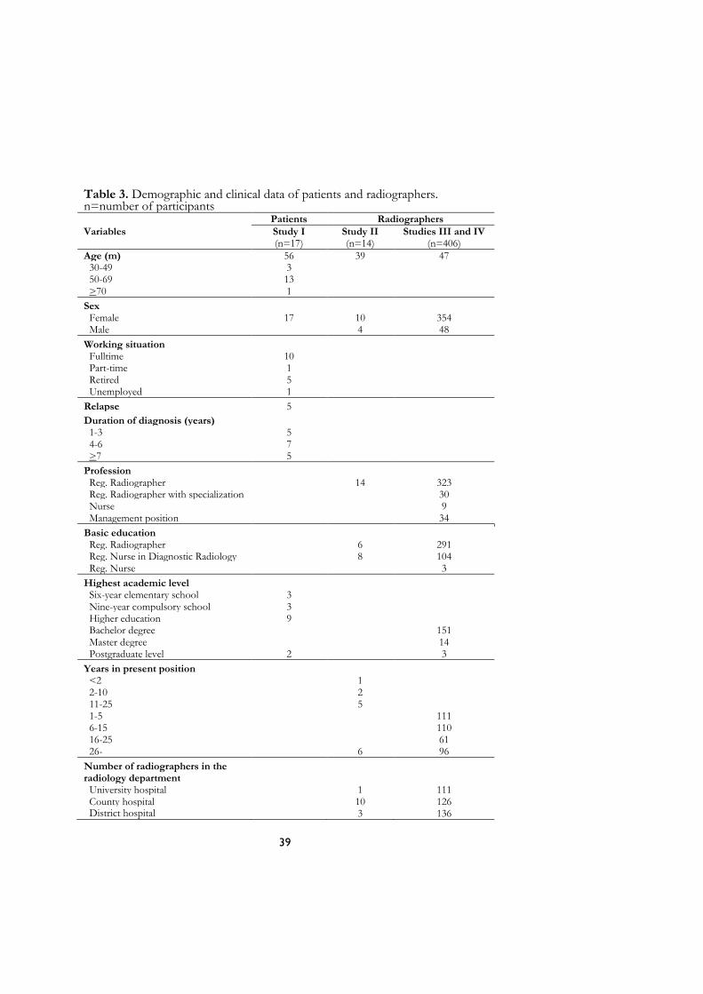
Table 3. Demographic and clinical data of patients and radiographers. n=number of participants Patients Radiographers
Variables Study I (n=17)
Study II (n=14)
Studies III and IV (n=406)
Age (m) 56 39 47 30-49 3 50-69 13 >70 1 Sex Female 17 10 354 Male 4 48 Working situation Fulltime 10 Part-time 1 Retired 5 Unemployed 1 Relapse 5 Duration of diagnosis (years) 1-3 5 4-6 7 >7 5 Profession Reg. Radiographer 14 323 Reg. Radiographer with specialization 30 Nurse 9 Management position 34 Basic education Reg. Radiographer 6 291 Reg. Nurse in Diagnostic Radiology 8 104 Reg. Nurse 3 Highest academic level Six-year elementary school 3 Nine-year compulsory school 3 Higher education 9 Bachelor degree 151 Master degree 14 Postgraduate level 2 3 Years in present position <2 1 2-10 2 11-25 5 1-5 111 6-15 110 16-25 61 26- 6 96 Number of radiographers in the radiology department
University hospital 1 111 County hospital 10 126 District hospital 3 136

40
Data collection
Interviews
In study I, open-ended interviews were conducted in order to explore
experiences of encounters between female patients diagnosed with breast
cancer and radiographers in the course of recurrent radiographic follow-up
examinations in radiology departments. The patients were approached by the
researcher in the waiting room of the radiology department before the
examination. After the examination they received brief verbal information
about the study as well as written information along with an informed consent
form and gave the author permission to contact them by phone. An
appointment for the interview was made when the author phoned them. The
majority of the interviews were conducted in a quiet private room in the
radiology department, while a few were held in the informant‟s home or place
of work and one took place over the telephone. Open-ended interviews focus
on a specific topic but do not have a fixed sequence of questions formulated
prior to the interview occasion.142 Such interviews encourage the informant to
focus on and define what is important to her/him rather than being guided by
the researcher‟s idea of what is relevant.124 Adequate time must be provided to
permit a full response.124 The interview began with a general question: “Could
you please describe one negative or positive encounter in a radiology
department that you remember especially well?” In order to gain a deeper
understanding, follow up questions were posed: “What do you mean?” “Could
you explain?” “How did you feel?"
In study II semi-structured interviews were used to explore radiographers‟
professional competence in relation to good nursing care, as this method allows
the informants to describe critical incidents in their own words. According to
Polit and Beck,124 a semi structured interview is used when the researchers want
to ensure that a specific set of topics is covered. Prior to the interview, the
radiographers were informed about the study, that it would focus on critical
incidents that facilitated and/or hindered good nursing care and where the
outcome was identifiable. All interviews took place in a quiet room close to the
radiographers‟ workplace. Informed consent was signed. The intention was to
collect data on 100 or more critical incidents from different areas in the

41
radiological setting. The CIT concerns a factual incident, which may be defined
as an observable and integral episode of human behaviour 124. The technique
thereafter concentrates on something specific about which respondents can be
expected to testify as expert witnesses. The questions are presented in Table 4.
Brief information concerning the data collection procedure was provided
before the interview and a critical incident was defined as a major event of great
importance to the radiographer. The data collection continued until a sufficient
amount of material with rich descriptions of critical incidents related to good
nursing care had been collected.
Table 4. Questions about critical incidents related to radiographic examinations and interventional. Could you please tell me about an ordinary day in your professional life? Could you please describe a successful and positive work situation in as much detail as possible? Could you please describe an incident in which you handled and/or did not handle a situation successfully? Could you please describe in as much detail as possible a difficult, complex and hard-to-manage work situation? Could you please describe an incident in which you handled and/or did not handle a difficult, complex and hard-to-manage situation successfully? At what time of the day and/or night did the incident occur and how long ago? Where did the incident occur? What was it that made the incident critical for you? What was it that made the incident positive? What was it that made the incident negative?
Questionnaire
In late November 2010, a link to the web-based questionnaire, comprising of
the RCS was e-mailed to 500 participants (Studies III and IV). This resulted in
200 responses, a response rate of 40%. A first reminder was sent after one
week and a second after two weeks. As the number of responses was
considered low, a new computer generated list of 500 participants was chosen
from the SAHP register and a reminder sent after two weeks. In total, 1,000
questionnaires were distributed, resulting in 406 responses (40.6%).
Background questions concerning age, sex, present position, basic education,
highest academic level and work place were included in the questionnaire. An
accompanying letter was distributed, containing information about the study

42
and ethical aspects. Informed consent was implied as the participant completed
the questionnaire.
Data analysis
Qualitative Content Analysis (Study I)
Qualitative content analysis was performed in accordance with Burnard131 to
explore experiences of encounters between female patients diagnosed with
breast cancer and radiographers at recurrent radiographic follow-up
examinations. The interviews were first read individually and then as a whole in
order to identify words and phrases containing important meaning units in the
statements by female patients with breast cancer about their experiences of
encounters with radiographers. A naive reading was undertaken and notes were
made about topics that emerged from the data. Then the texts were reread as a
whole and questions such as “what is it about?”, “what does it mean?” and
“what does it stand for?” were posed. After open coding of the text as a whole,
it was condensed and rewritten into codes. Similar codes were grouped together
and a sense of a structure with categories and sub-categories was developed. In
this phase each subcategory was critically analysed, questioned, read and
compared in order to arrive at a reasonable interpretation and, if possible, to
identify categories. This part of the analysis process, which is concerned with
interpreting the meaning of the text, guided the choice of category names. The
analysis process was a movement between the whole and the parts of the text.
Table 5 provides an example of the process in the data analysis process.

43
Table 5. Example of the data analysis process comprising quotations, condensed codes, sub-categories and principal categories. Examples of quotations What is it
about/meaning Examples of condensed codes
Sub-category Principal Category
“ … uncertainty about the cancer … “
Feelings of being out of control. Feelings of fear about having to face another unpleasant diagnosis.
Loneliness, isolation
Vulnerability
“I met someone who dared to encounter me with a smile, gave me hope and a feeling that I was important … if somebody dares to meet me like that, I suppose it won‟t be that bad.” (I 4)
Feelings of being confirmed. Feelings of self-reliance.
Strength The empowering encounter
"… the nurse held my hand the whole time …" (I 3)
Feelings of trust and confidence.
Trustfulness The empathetic encounter
"They informed me that I should just lie still and hold my breath, they told me … nothing else …" (I 10)
Feelings of being an object. Feelings of not being allowed to talk.
Objectified The mechanical encounter
"… I was very frightened and they were a bit indifferent and callous …" (I 5)
Feelings of not being confirmed. Feelings of loneliness and being invisible.
Marginalised The neglectful encounter

44
Critical Incident Technique (Study II)
The analysis of the data was based upon the critical incident technique as
described by Flanagan136 and aimed to identify radiographers‟ professional areas
related to good nursing care. The analysis process comprised four steps. First, a
careful and repeated reading of the transcribed interviews took place in order to
gain an overall understanding and become immersed in the data. The interviews
were then processed by condensing the data, after which the incidents could be
identified and considered critical if related to the aim of the study and the
interview question. A total of 277 critical incidents were identified from the
transcripts. The number of incidents varied between one and 23 per informant,
i.e. each informant did not provide a critical incident for every item, while some
informants reported more than one incident for some items. In the initial part
of the categorisation, the incidents were extracted from the text, sorted, labelled
according to their specific content and then classified into groups. The groups
of incidents were reformulated into different kinds of behaviour, resulting in 31
subcategories that were allocated to eight categories and two main areas. The
main areas describe the overall structure of the critical incidents. Table 6
provides an example of the process of the data analysis process.
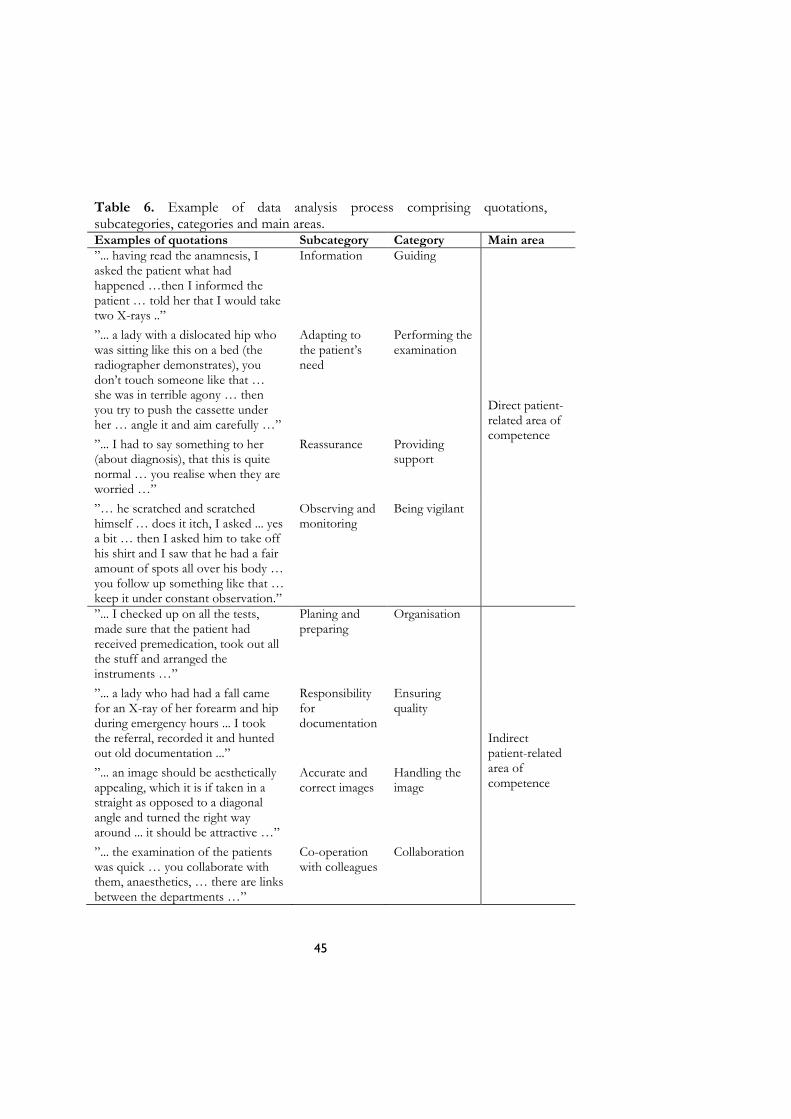
45
Table 6. Example of data analysis process comprising quotations, subcategories, categories and main areas. Examples of quotations Subcategory Category Main area
”... having read the anamnesis, I asked the patient what had happened …then I informed the patient … told her that I would take two X-rays ..”
Information Guiding Direct patient-related area of competence
”... a lady with a dislocated hip who was sitting like this on a bed (the radiographer demonstrates), you don‟t touch someone like that … she was in terrible agony … then you try to push the cassette under her … angle it and aim carefully …”
Adapting to the patient‟s need
Performing the examination
”... I had to say something to her (about diagnosis), that this is quite normal … you realise when they are worried …”
Reassurance Providing support
”… he scratched and scratched himself … does it itch, I asked ... yes a bit … then I asked him to take off his shirt and I saw that he had a fair amount of spots all over his body … you follow up something like that … keep it under constant observation.”
Observing and monitoring
Being vigilant
”... I checked up on all the tests, made sure that the patient had received premedication, took out all the stuff and arranged the instruments …”
Planing and preparing
Organisation
”... a lady who had had a fall came for an X-ray of her forearm and hip during emergency hours ... I took the referral, recorded it and hunted out old documentation ...”
Responsibility for documentation
Ensuring quality
Indirect patient-related area of competence
”... an image should be aesthetically appealing, which it is if taken in a straight as opposed to a diagonal angle and turned the right way around ... it should be attractive …”
Accurate and correct images
Handling the image
”... the examination of the patients was quick … you collaborate with them, anaesthetics, … there are links between the departments …”
Co-operation with colleagues
Collaboration

46
Questionnaire
Development and psychometric tests (Study III)
The questionnaire, the RCS, was developed and psychometric tested in study
III. The questionnaires were in Swedish. The development involved seven
methodological phases and was guided by Streiner and Norman,138 DeVellis,139
and Burns and Grove.140 A pilot test of the questionnaire was performed by an
expert group. When developing a new instrument face and content validity are
of importance. Face validity indicates whether, on the face of it, the instrument
appears to be assessing the desired qualities. Content validity consist of a
judgement whether the instrument samples all the relevant or important
content or domains.138 Face and content validity was conducted in line with
Lynn‟s Criteria.141 Depending on the type of variable as well as on the
distributional characteristics parametric or non-parametric methods were used
in this thesis.143 Variables are categorized in four levels of measurements,
nominal, ordinal, interval and ratio. Parametric methods are based on measures
of means, standard deviation and differences among the means which can be
interpreted from an interval and ratio scales such as age and years in present
position. Non-parametric methods are based on measures of percentage or
median on nominal and ordinal scales, regarded as frequencies in individual
categories.
The item reduction was performed in two steps. Firstly, corrected item-total
correlation and Cronbach‟s alpha if item deleted was conducted on the initial
54-items. In this step all items in the questionnaire, one at a time, were
correlated with the total number of points. Items with correlations below 0.5
were removed one at a time. The total number of points was adjusted so that
the item that was correlated with the total number of points was not involved
in the total number of points at the time when the analysis was performed.144
Secondly, several explorative factor analysis were conducted on the remaining
42-items. A principal components analysis providing preliminary information of
the construct was performed before the final factoring. To decide the number
of factors to be included in the construct, factors with an eigenvalue above 1.0
were included. The items factor loading was >0.50 and were considered
sufficient, according to Field.145 Catell‟s scree test139 was used to control the
number of tentative factors to be retained. Data were examined with Bartlett‟s

47
test of sphericity, and had to be significant (p<0.001). A factor rotation with
Varimax type was used in order to simplify the factor structure.140 A two-factor
solution was identified explaining 53.8 % of the total variance. Homogeneity,
was measured with Cronbach‟s alpha146 and with regard to developing a new
instrument, the lowest value for Cronbach‟s alpha coefficient was set at 0.70.147
Comparisons between groups (Study IV)
In study IV, four groups were created based on years of work experience: short
(0-5 years), medium (5-15 years), long-term (15-25 years) and longest period of
work experience (>25 years). The groups were then compared regarding the
sample characteristics, namely level and use of competence.
The choice of appropriate method of analysis was based on the number of the
groups and sample, the type of data, the distribution of data and the objective
of the analysis.143 To compare data from three groups or more One-way
analysis of variance (ANOVA) was used when the data was continuous or
normally distributed. Pearson‟s chi-square test was used when the data was
categorical or not normally distributed. Variations among the means of three or
more groups increase the risk for Type I error.144 P-values at <0.05 were
considered statistically significant, except when multiple comparisons were
made, when a reduced p-value was used. Due to multiple comparisons, the
Bonferroni correction, a reduced p-value was calculated (p<0.008) and used to
control for the risk of mass-significance.148 The correction was made by
dividing the P-value at 0.05 by six (the number of group comparisons).
Correlation is a method of analyses to study the possible linear association
between variables and the Spearman‟s rank-order correlation coefficient, a non-
parametric calculation was used. The correlation coefficient indicates the
strength of the association as a single number but does not describe the relation
between the variables.144 There are differences between correlations and
regression. Regression is a method for the prediction of one variable to
another.144 In the analysis one of the variables is considered dependent (the
outcome) and the other as independent (predictor). As independent variables,
age, years in present position and work place were entered. Variables at nominal
and ordinal level were made to dummy variables.143 To identify the linear

48
relation between variables linear regression analyses was used when the
dependent variable was continuous.
All data were computerized and analysed using SPSS 18.0 for Windows (SPSS
Inc., Chicago, Illinois, USA). Choice of appropriate method of analyses is
shown in Table 2.
Ethical considerations
The studies in this thesis were conducted in accordance with the requirements
pertaining to research on human beings as set out in the Helsinki declaration,149
Ethical guidelines for nursing research in the Nordic countries,150 as well as in
the Swedish ethical approval legislation concerning research on humans.151
Ethical clinical research is based upon respect for autonomy, providing
beneficence, ensuring non-maleficence, avoiding risk as well as promoting
justice.102 Ethical considerations are basically a matter of finding a reasonable
balance between different interests such as the search for knowledge, individual
privacy and protection against various forms of harm or risk of harm. Ethical
approval and permission to undertake the studies I and II were granted by the
Ethics Committees at Örebro University, Sweden (No. 2002/135), and Lund
University, Sweden (M:G 35, 42/05). In studies III and IV it was not
considered necessary to apply for permission from the regional research ethics
committee. According to the Swedish Act concerning the ethical review of
research involving human beings, permission is not required when studies
involve staff members.151
Autonomy
The principle of autonomy is related to respect for the person, her/his right to
know or not to know and the freedom to make decisions (to participate in, not
participate in or withdraw from the research).102 Important ethical issues related
to autonomy were found to be those concerning informed consent.151 A freely
made decision to participate in the studies was significant. When requesting
informed consent, the researcher was particularly careful to ensure that the

49
participants were not in a dependent relationship. The female patients (Study I)
and radiographers (Studies II-IV) took part voluntarily and were not in a
position of dependency on the researchers. In studies I and II the participants
were informed of the aim, utility and confidentiality of the study, their right to
integrity, the voluntary nature of participation and the possibility to withdraw at
any time. Written informed consent was obtained from all female patients and
radiographers. In study I the female patients were invited to participate and
received oral and written information directly after the radiographic
examination. They were requested to choose the time and place for the
interview and given sufficient opportunity to reflect on whether or not to
participate. They were assured that participation would not affect their care or
treatment. In study II the radiographers were selected and contacted by the
head of the radiology department on behalf of the researcher. An introductory
letter containing information about the study was sent by post. They were given
time to consider the question of participation and asked to choose the time and
place for the interview during certain weeks. The researcher then phoned the
participants to make the final arrangements. In studies III and IV, completing a
questionnaire implied informed consent. A supplement, with information about
the aim and utility of the study, confidentiality, the voluntary nature of
participation and how to contact the researcher, was sent attached in an email
to the participants.
Beneficence and Non-Maleficence
Every person attending healthcare and a radiology department is in a vulnerable
situation, especially patients undergoing recurrent follow-up examinations. The
principle of beneficence states the ethical obligation to maximise possible
benefits, while the principle of non-maleficence sets out the ethical obligation
to minimise possible harm.102 There is always a risk that participants can feel
that their personal integrity is threatened. Conducting research involves ethical
gains and costs. The risk of distress or violation of integrity was deemed
significant in studies I and II, but negligible in studies III and IV.
A potential ethical cost was the risk of distress and harm if unpleasant
memories were aroused during the interview. To minimise this risk, the
interviewer ensured that there was sufficient time for the participants to talk
about difficult and painful situations. Several of the participants expressed that

50
they found it beneficial to be in focus, have the opportunity to talk about
themselves and their experiences and to know that somebody was actively
listening to them. The introductory letter contained the interviewer‟s telephone
numbers, which afforded opportunities for contact after the interview. The
interviewer also assured the female patients and the radiographers that there
were other professionals and/or networks available to support them; almoner,
psychologist and the occupational health service department. Permission to use
a tape recorder and to transcribe the interviews verbatim was obtained (Studies
I and II). The findings from this thesis could be of benefit to future patients by
contributing to improved patient care at the radiology department.
Furthermore, the research could also enhance the knowledge and development
of the radiographers‟ professional competence.
To protect the confidentiality of the female patients and the radiographers, no
names were included in the stored recordings and transcribed interviews in
studies I and II, thus no connection to any person could be made. In
quotations in the transcriptions, any names mentioned were altered so that no
connection can be made to specific persons. In studies III and IV the data were
collected from a national register administered by the SAHP and the sample
was randomised by an employee. A web-based questionnaire was linked to the
participants. Web-based tools always imply a certain security risk such as
hacking. To protect the participants no documentation contained the
radiographers‟ names, and coded numbers were used throughout the whole
data collection. All material was kept safe, analysed and described in a way that
makes it impossible to identify or link specific information to individual
patients or radiographers. The research team reflected deeply on issues of
security and integrity.
Justice
The principle of justice requires the sampling of participants in a non-
discriminatory way with regard to age, sex and nationality.102 This implies that
vulnerable groups should be protected from exposure as well as discrimination.
The participants were selected regardless of sex, religion, race, political opinion,
etc.

51
Summary of the results
The patient
The experiences of encounters between female patients diagnosed with breast
cancer and radiographers at recurrent radiographic follow-up examinations
were categorized into five groups; the women‟s comprehensive understanding
and four encounters.
Vulnerability (Study I)
Women‟s comprehensive understanding was described as feelings of
vulnerability. Their uncertainty about the cancer had an impact on the female
patients‟ experience of the encounter. Prior to an examination, they described
feelings of anxiety, fear of pain and the risk of having to face another
unpleasant diagnosis and being out of control. They described feelings of panic
manifested in the form of palpitations, an icy sensation in the neck, a restless
body or losing control of their emotions. During the examination, patients
experienced pain and unpleasant feelings of being exposed when undressed.
After the examination, patients who had to wait for the result experienced
tension and stress. At times they objected to the recurrent radiographic follow-
ups in order to cope with the situation, as they felt that they needed to protect
themselves.
Encounters (Study I)
The empowering encounter optimised the patients‟ own capacity and participation,
as the radiographer viewed her as an expert on her own circumstances and
listened attentively to her. There was a mutual information flow, which made it
possible for the patient to take decisions and cope with different situations
during the examination. The radiographer was a supportive resource, although
treating the patient as an equal. The patient had a feeling of being strengthened,
confidence in her own ability as well as being in control of her own life and
circumstances. In the empathetic encounter concern about the patient's diagnosis
and illness was emphasised. She was the subject of the radiographer‟s attention

52
and the communication had a comforting and caring character. The patient was
listened to and treated with respect. The radiographer showed insight and
understanding of the discomfort that the patient experienced throughout the
radiographic examination. The patient was left with an experience of having her
dignity and identity respected as well as a sense of well-being. In the mechanical
encounter the patient was treated in a distanced manner and the radiographer was
mainly concerned about the images and being as quick as possible. The patient
was an object and the organ demanded the radiographer‟s attention. It was a
mechanical, routine based approach guided by the radiographer's professional
task of focusing on correct projections and positioning. The patient was not
asked about possible discomfort or pain in the various positions and left with
an experience of being treated in an impersonal manner during the entire
encounter. In the neglectful encounter, the patient‟s feelings were disregarded and
ignored. She was invisible, abandoned and treated by the radiographer as being
of no significance, a nobody. There was lack of respect for the patient as an
autonomous human being and no social communication or physical contact,
which left the patient with an experience of being invisible and marginalised.
The radiographer
Radiographers areas of professional competence (Study II)
In the radiographer study (Study II) the descriptions of competence were
grouped into two main areas, each with four sub-groups. The direct patient-related
area of competence focused on the closeness to the patient and The indirect-patient-
related area of competence concerned the surroundings. In the first area the nursing
perspective was more obvious while in the latter, the medical-technical
dominated.
In the direct-patient-related area of competence, the competency termed
guiding, was described as being with the patient and helping her/him to assume
an active role in the examination. In addition to informing the patient, the
radiographers needed to obtain information and asked relevant questions
related to the referral. When performing the radiographic examination, the various
descriptions involved acting in a safe and secure manner regarding radiation

53
protection and taking the individual patient‟s condition and needs into account.
Furthermore, physical touch was used to locate anatomical structures as well as
the existence of any pain. Within providing support, the description reflected
caring for both the patient and the next-of-kin. The radiographers encouraged
the anxious patient to undergo the planned examination and at the same time
showed respect for her/his integrity. The descriptions of being vigilant involved
ensuring the safety of patients who developed an allergic reaction after the
injection of contrast medium and sometimes from an unnecessarily high dose
of radiation. The radiographers also prevented injuries when deciding to
terminate examinations and saved lives by intervening at short notice in
difficult and potentially fatal situations.
In the indirect patient-related area of competence, organising was described
as planning and preparing for the radiographic examination and treatment,
which included administrative as well as practical matters. The radiographers
took responsibility for ensuring that the results were communicated by a
radiologist on conclusion of the radiographic examination. When ensuring quality,
the participants reported that the identity of the patient as well as the
correspondence between the referral and the patient‟s symptom was confirmed.
The technical equipment and documentation were also verified. The
description of handling the medical images mainly focused on obtaining correct and
accurate medical images, but also involved scrutinizing and assessing the images
as well as transmitting them to other units and hospitals. In the competence
Collaborating internally and externally, the radiographers supervised all staff
categories and strived to work as a team, especially in critical situations. Their
strive to become more efficient could support the patient by reducing the time
at both the diagnostic radiology department and the hospital.
Designing an instrument – the RCS (Study III)
In study III, an instrument for measuring the level and frequency of use of
competence was developed and by the use of factor analyses, two
psychometrically sound factors were identified. These were labelled „Nurse
initiated care‟ and „Technical and radiographic processes‟. „Nurse initiated care‟
comprised 18 items and „Technical and radiographic processes‟ 10 items (See
Appendix 1).

54
The initial version of the RCS consisted of 42 items, each of which was
described as a radiographer competency and distributed between eight
categories. By means of a pilot test, the first set of experts (N=16) evaluated the
logical consistency as well as the relevance of the items, assessing and
quantifying their validity both individually and as a set. Some revisions were
recommended e.g. the order and rewording of certain items and additional
areas were suggested, thus after the pilot test, 12 items were added to the initial
version of the RCS. A two part-scale was considered appropriate, one of which
focused on the level of radiographer competence and the other on the
frequency of its use. The level of competence was measured on a 10-point scale
(1-10) where one was the lowest and 10 the highest grade. The frequency of
using the competencies was measured by the following response alternatives:
“never used”, “very seldom used”, “sometimes used”, “often used”, “very
often used” and “always used”. No changes were made to the final 54-item
RCS after the second set of experts (N=4) had evaluated the logical consistency
of the competencies and the number of items to be included.
Psychometric evaluation of the RCS (Study III)
A total of 406 radiographers with clinical experience in diagnostic radiology
departments in Sweden, completed the questionnaire, representing a response
rate of 40.6 %. The mean age of the participants was 47 years, ranging between
22 and 66 years and 88 % were women (Table 3).
The number of items was reduced from 54 to 28 in two phases. First, a corrected
item-total correlation and Cronbach‟s alpha if item deleted, was conducted on
the 54-item questionnaire. Items with low correlations, i.e. <0.5, were removed
one at a time and new item-total statistics calculated on each occasion, resulting
in a reduction of 12 items. Second, repeated explorative factor analyses were
conducted on the remaining 42 items. Principal component analyses were
conducted to obtain the solution with optimal scale variance. Items with
correlations of <0.5 were removed from the factor one by one, which led to the
removal of a further 14 items, resulting in a 28-item scale. As the factor structure
decided by the Kaiser criteria was not simple and no clear factor solutions was
obtained, the items were forced into a two factor solution. The items in the two
identified factors had fairly good communality values of >0.40 with the
55
exception of one; „optimizing of radiation doses to patient and personnel‟,
which had 0.34. There were 18 items in factor 1 and 10 items in factor 2. Factor
1 was labelled „Nurse initiated care‟ and factor 2 „Technical and radiographic
processes‟. The two factors appeared to be clearly defined and quite different
from each other. Initially, data were examined by means of Bartlett‟s test of
sphericity as well as the measure of sample adequacy in each variable and
overall. The number of factors extracted was based on the Kaiser criteria with
an Eigenvalue of <1.0.
Homogeneity was assessed using Cronbach‟s alpha coefficient in each of the two
factors and the scale in its entirety. The value was 0.94 for the first factor, 0.89
for the second and 0.87 for the entire 28-item scale. The corrected item-total
correlations varied between 0.52 and 0.78. In factor 1, the item with the highest
correlation was; „Alleviating the patient‟s anxiety‟ while the item with the lowest
correlation was; „Participating in quality improvement regarding patient safety
and care‟. In factor 2, the item with the highest correlation was; „Evaluating the
quality of the medical image in relation to the referral and question stated
therein‟. The item with lowest correlation was; „Minimising radiation doses for
patient and staff‟.
Missing data and the proportion of floor and ceiling effects (people obtaining
minimum and maximum scores respectively) among the items were also
examined. The distribution of scores spanned the entire range (i.e. 1-10) with
no floor and ceiling effect problems identified. The results indicated that the
number of participants did not exceed 15% of the anticipated floor and ceiling
effects. The response rate for each item in the 28-item instrument was high
(range: 93.6% to 95.8%).
The level and frequency of use of professional competencies
(Study IV)
In study IV the data from the 24-item questionnaire was used to measure the
level and frequency of use of competence.
Nurse initiated care. The radiographers rated ‘Adequately informing the patient’ as
the highest competency and ‘Identifying and encountering the patient in a state of shock’
and ‘Participating in quality improvement regarding patient safety and care’ as the lowest.

56
Differences between those with short and medium-term experience and those
with the longest period were seen in ‘Adequately informing the patient’. This
competence was rated more highly by those with the longest period of work
experience (p<0.001). Differences between those with a short period of work
experience and those with the longest period were seen in ‘Alleviating the patient’s
anxiety‟ and ‘Judging the risk of leaving the patient unattended’. The level of the
competencies was significantly lower in the group with a short period of work
experience (p<0.001).
The most highly rated competency in terms of use was ‘Adequately informing the
patient’, while the lowest was ‘Identifying and encountering the patient in a state of
shock’. The competencies; ‘Participating in quality improvement regarding patient safety
and care’ as well as ‘Reporting to internal as well as external colleagues and other
professionals’ had low ratings in all four groups. Differences between those with a
short period of work experience compared to those with long-term experience
were seen in ‘Collaborating with other internal and external professionals’. This
competency was significantly more used by those with short-term work
experience (p<0.001). Those with long-term experience employed ‘Applying
ethical guidelines’, ‘Guiding the patient’s relatives’, ‘Encouraging and supporting the patient’
and ‘Alleviating the patient’s anxiety’ more frequently compared to those with a
short period of work experience (p<0.001).
Technical and radiographic processes. The radiographers rated the
competency ‘Adapting the examination to the patient’s prerequisites and needs’ the
highest whereas the lowest was ‘Preliminary assessment of images’. Differences
between those with a short, medium and long-term period of work experience
and those with the longest period of work experience were seen in the
competencies; ‘Organising and planning taking account of the clinical situation’ and
‘Independently planning and preparing work on the basis of existing documentation’. A
significantly higher level was reported by those with the longest period of work
experience (p<0.001). The ratings were the same in ‘Prioritising patients in the work
flow’, ‘Producing accurate and correct images’ and ‘Optimising the quality of the image’.
‘Producing accurate and correct images’ and ‘Adapting the examination to the patient’s
prerequisites’ were rated as the most frequently used competencies. Differences
between those with long and short-term experience were seen concerning the

57
competencies; ‘Organising and planning taking account of the clinical situation’,
‘Prioritising patients in the work flow’, ‘Minimising radiation doses for patient and staff’,
‘Optimising the quality of the image’ and Preliminary assessment of images’. These
competencies were significantly more used by those with long-term experience
(p<0.001).
The total score for „Nurse initiated care‟ had a low but significant correlation
with age (r=0.265; p<0.001) and years in present position (r=0.217; p<0.001).
The total score for „Technical and radiographic processes‟ also had a low yet
significant correlation with age (r=0.278; p<0.001) and years in present position
(r=0.287; p<0.001). The competence level was significantly associated with age
in „Nurse initiated care‟ and „Technical and radiographic processes‟ as well as
with the total score of the RCS. The use of competence was significantly
associated with years in present position in „Technical and radiographic
processes‟ and the total score of the RCS, but not in „Nurse initiated care‟.
58
Discussion
Methodological considerations
Trustworthiness (Studies I and II)
Qualitative content analysis was the methodological choice for exploring
patient perceptions of radiographic experiences (Study I), while the critical
incident technique was chosen to describe radiographers‟ areas of professional
competence (Study II). The findings in the interview studies described the
experiences of the patients and the radiographers, with no information about
what really happened or how frequent or rare the experiences were. Lincoln
and Guba152 suggested four concepts as for establishing trustworthiness in
qualitative studies: credibility, dependability, transferability and confirmability.
Credibility refers to the confidence in the findings based on the data.124, 152
Recommendations to establish credibility involve actions that make the
emergence of credible data and interpretations more likely.124, 152 In study I, the
patients constituted a well-defined group of women living in the south of
Sweden who attended regular follow-up radiographic examinations, i.e.
mammography, skeletal radiography, lungs, ultrasound, CT and MRI. In order
to obtain variation, strategic sampling was used for data collection in line with
qualitative content analysis. The female patients were approached by the
researcher in the waiting room before and after the radiographic examination.
After the examination, information about the study was provided and a time
for a phone call arranged. The patient was given time to reflect on her
willingness to participate. As the female patients were interviewed a couple of
days after the examination, it increased the chance of obtaining her reflected
experience. To establish credibility two of the researchers read the interview
texts and took part in the whole analysis process, while all four researchers
independently reviewed the categories and sub-categories.

59
In study II the registered radiographers constituted a carefully chosen, well-
defined group living in a specific area in Sweden with the latest technology in
the radiology department. Choosing participants with different experiences
increases the possibility of shedding light on the research questions from a
variety of perspectives.128 In order to obtain variation, strategic sampling was
used for the data collection in line with the critical incident technique. The
critical incident technique, which employs semi structured interviews as the data
collection method, was chosen in order to describe how critical incidents
influenced the radiographer in terms of either facilitating or hindering good
nursing care. The categorisation of critical incidents is the most controversial
part of the CIT and has been criticised for being both subjective and difficult.153
The objectivity of the method lies in the attempt to capture direct raw data that
are free from assumptions. Although the data can be categorised in more than
one way, it is always possible to refer back to the critical incidents
themselves.153 The rich material in studies I and II also strengthen credibility.
The interviewer tried to create a peaceful and positive atmosphere in order to
encourage the participants to express their personal opinions, which is
important in data collection.
Dependability refers to the stability of data over time and in relation to various
conditions.124, 152 There is a risk of inconsistency when dealing with a large
amount of data and when the data collection takes place over a long period of
time. It is important to ask all participants the same initial question. In this
thesis the researchers remained close to the original meaning and context by
referring back to the audio-tapes and transcripts of the interviews during the
analysis and writing process. The informants and the main researcher were very
familiar with the research area. To establish dependability in study I, one
researcher in addition to the main researcher was involved in the analysis
process, at first individually and then together, after which the third and fourth
researchers also became involved. To establish dependability in study II, the
main researcher conducted the analysis process while the second took part at
the end of the analysis and the third and fourth researchers verified the
plausibility of the findings.
Transferability refers to the degree to which the findings can be transferred to
other settings or groups.124, 152 A limitation of qualitative research is that it does

60
not permit the use of a large sample in order to obtain data. However, the
reason for using a qualitative approach was to obtain as much meaningful
information as possible with regard to the aim of the studies as opposed to
generalising as in the quantitative approach.128 To facilitate transferability it is
important to provide a clear description of the culture and context, selection
and participant characteristics, data collection and analysis process. The
findings of study I are not intended to be generalised but can be seen as useful
information for clinical practice while, the findings of study II can be
transferred to other radiological settings, as they pertain to critical incidents that
influence the radiographer, either facilitating and/or hindering good nursing
care.
Confirmability refers to the objectivity or neutrality of the data124, 152 and is
concerned with ensuring that interpretations of the findings are clearly derived
from the material. To strengthen confirmability, careful descriptions of the
research process and the inclusion of quotations presented in studies I and II
elucidate the interpretations of the original text. Negotiated consensus was
employed to establish confirmability, which meant that the researchers
continuously discussed the interpretations until consensus was reached.154
Validity and reliability (Studies III and IV)
When considering the quality of studies III and IV, both validity and reliability
need to be discussed. To evaluate a study‟s strengths and weaknesses in
quantitative research, Kazdin155 has outlined four types of validity that can be
used to convey key aspects of the research: internal, external, construct and
statistical conclusion validity. The reliability of an instrument can be evaluated
in various ways by e.g. stability and homogeneity.124, 152
Internal validity relates to the degree to which the findings can be explained by
the independent variable rather than the observed effects being due to
uncontrolled confounders.155 A threat to internal validity can be non-
responders. In the present studies it was decided to use a random sample of at
least 500 out of the 1,772 SAHP members who were radiographers. This was
done in order to reduce systematic sampling bias, increase the validity of the
study140 and ensure that all radiographers listed in the national register had an

61
equal chance of being selected. A total of 406 out of the 1,000 randomly
selected radiographers completed the questionnaire. The external drop-out
comprised 594 radiographers (59.4%) who declined to participate for unknown
reasons. It is possible that the number of items in the questionnaire was too
great or that they were difficult to understand. Another possibility is that the
radiographers did not have time during their working day. A third suggestion is
that only the most interested and perhaps experienced radiographers completed
the questionnaire. A relevant issue is therefore whether the participants are
representative of the population intended to be investigated. It is a weakness
that no drop-out analyses can be made of those who declined participation. On
the other hand, there was great variation with regard to the number of socio-
demographic and clinical characteristics, indicating that the participants
represented a wide range of radiographers in Sweden.
External validity refers to the extent to which findings can be generalised beyond
the conditions of an experiment.155 Threats to external validity include various
characteristics of the sample and context.138, 140 The study population comprised
adults with variation in terms of age, sex, education, years in present position
and work place. Since the items in the RCS were selected from theoretical
studies and pilot tested by an expert group, the sample chosen for the
development of the RCS and the psychometric testing was representative of the
target population and the results might therefore be generalised to both general
and specific settings among the Radiographer population. Further proof of the
validity of the findings with regard to the structure of the RCS and its construct
validity require additional studies.
Another threat to validity concerns the way in which instructions and
information about the study are given to the participants in the web-based
questionnaire.155 It is important that the information provided is correct, well-
structured and easy to understand. The invitation to participate should be open,
friendly and hopefully promote willingness to take part. There was no state of
dependence on the researchers or the SAHP official responsible for the
national register. The same information and instructions were provided to all
participants in the random sample. The SAHP official was informed both orally
and in writing about how to invite participants, what information to give them

62
and also received a detailed written description of the study and its aims and
objectives.
Examinations of validity and reliability also concern whether instruments used
in the study actually measure what they are intended to measure. When the RCS
was tested for internal validity, 28 out of 54 items fitted a two-factor structure.
The face validity and content validity of the RCS were enhanced by using the
results from theoretical studies including the qualitative study (Study II) as
items on the scale. The content of the questionnaire should be representative of
the phenomenon under investigation and be appropriate for the respondents.138
Pilot testing of the face and content validity was conducted in line with the well
structured Lynn‟s Criteria141. To strengthen internal validity, a strategically
selected group of experts was formed to judge the relevance of the items.
Third, face and content validity were established based on agreement between
the four authors (Study III).
Construct validity refers to the degree to which the measuring procedure actually
measures what it is intended to measure.155 The key construct validity question
with regard to measurement is “What is this instrument really measuring and
does it validly measure the abstract concept of interest?”. In study III construct
validity was measured by means of explorative factor analysis which is a factor
analysis undertaken to explore the underlying dimensionality of a set of
variables.
Validity is also related to the statistical power of the analyses.124, 152 Based on
estimated number of participants, no power analysis was performed in these
studies. The sample size in studies III and IV was 406, which was considered
sufficient to examine the psychometric properties of the initial 54-item
questionnaire. Various recommendations defining the ratio between variables
used in multivariate analysis and sample size have been discussed. Calculation
of an adequate sample size for factor analysis is a complicated issue with no
straightforward scientific answer.147 A number of alternatives have been
presented. Nunnally147 recommended a 10 to 1 ratio, i.e. ten cases for each item
included to be factor analysed. Others hold that five cases for each item are
adequate in most analyses.156 In studies III and IV, the number of participants
in relation to the number of questions was sufficient, a ratio of almost 10 to 1.

63
Statistical conclusion validity refers to the extent to which statistical relations and
their influence on the conclusions can be detected.155 This type of validity is
often viewed and interpreted from two standpoints: the risk of Type I (α) and
Type II error (β). Type I error is the risk of rejecting the null hypothesis when it
is true, i.e. the researcher concludes that a significant difference exists when in
fact there is none. This type of risk is indicated by the p-value and often set at
5%. When multiple comparisons are made, a reduced p-value must be
calculated to minimise the risk of mass significance.148 In study III a reduced p-
value was used due to multiple comparisons across four groups to control for
the risk of mass significance. Apart from that, the statistical significance was set
at 0.05. Type II error is the risk of not rejecting the null hypothesis when it is
false; i.e. concluding that no relationship exists when in fact there is one. Power
analysis is a way to estimate the sample size so the risk of Type II error can be
reduced but, as already mentioned, no power analysis was used in these studies.
The reliability of an instrument is the degree of stability and homogeneity with
which it measures what it is supposed to measure.124 Assessment of reliability
means determining that a scale or measurement yields reproducible and
homogeneous results. Reliability testing examines the amount of random error
associated with the measurement technique.140 Stability concerns repeated
measures of the same attribute with the same instrument e.g. test-retest
reliability. However, this was not used in these studies with consideration to the
design and methods used for data collection. One way of measuring reliability is
to use homogeneity, which refers to the extent to which the items are
interrelated. A common method for measuring homogeneity is Cronbach‟s
alpha,146 which is a function of both the average inter-item correlation and the
number of items in a scale, where the alpha increases if either of the above
increases. Theoretically, Alpha=0 should be the result if the items are
uncorrelated and Alpha=1 when all items are identical.146 However, Cronbach‟s
alpha can underestimate true reliability and is therefore a conservative
measure.147 An Alpha above 0.7 is generally regarded as acceptable, although it
is often recommended that values above 0.8 and sometimes even 0.9 should be
used.138, 147 A too high alpha (above 0.9) can indicate that some items are
unnecessary.138 In this thesis (Study III) an Alpha >0.70 was used since the RCS
is a newly developed instrument and considered acceptable.140 The results of
64
the item analysis were found to be homogeneous, and the RCS as a whole was
satisfactory homogeneous. Cronbach‟s alpha was considered high for the 28-
item questionnaire, as well as for each of the two dimensions. The distribution
of scores ranged over the whole span (i.e. 1-10) for all of the items.
Structural equation modelling could be another statistical method for assessing
associations between different variables and competencies.

65
General discussions of the findings
Patients’ experiences
Vulnerability (Study I)
For most female patients diagnosed with breast cancer, diagnosis starts at the
radiology department. In this study, the comprehensive understanding of the
patient-radiographer encounter was that the female patients who had been
diagnosed with breast cancer experienced vulnerability before, during and after
the radiographic examination. Vulnerability before the examination due to
uncertainty about the diagnosis threatened patients‟ self-control and influenced
their way of handling the situation. Lindop and Cannon157 stated that the stages
of diagnosis and pre-diagnosis can be similar to a post-traumatic stress reaction
associated with a sense of loss and insecurity about the future. Feelings of being
vulnerable due to fear of cancer recurrence (FCR) related to follow-up
examinations have been described by Allen.30 Thewes et al.158 found that
younger women are more vulnerable to FCR than their older counterparts.
Another study indicated that only a small number of patients attending the
radiology department could identify what kind of examination they were going
to have, as they placed more attention on the diagnosis.159 The radiographic
examination was considered routine and would come to an end unlike the
cancer. Patients suffering from cancer can be extremely sensitive to every
gesture or sign that indicates a change in their diagnosis.26 During the
examination, the female patients‟ vulnerability was obvious in the pain caused
by the examination and being, which was also found by Poulus and
Llewellyn.160 After the examination, vulnerability was experienced as being
lonely, isolated and a feeling of strain due to having to wait for the result.
According to Thorne et al.,161 to handle this situation, they focused on getting
through their day. One negative coping strategy was to object to the recurrent
radiographic examinations, which can be related to earlier experiences of
vulnerability in connection with such examinations.162 Healthcare providers can
assist the patient by suggesting coping strategies, as well as providing necessary
information and promoting a positive interpretation of events through
reframing.

66
Encounters (Study I)
Four different types of encounter emerged in study I; Empowering,
Empathetic, Mechanical and Neglectful. The Empowering and the Empathetic
encounters gave rise to positive feelings in the female patients and were
interpreted as caring encounters, while the Mechanical and the Neglectful
encounters engendered negative feelings and were thus interpreted as uncaring
encounters.
The caring encounter was achieved through empowering the patient, thus
strengthening and providing her with a sense of being in control of her life and
circumstances. The findings revealed that in this encounter the patient is seen
as a person who is actively listened to and acknowledged during the
examination. The prerequisites have been described in other studies.163 Being
encountered as an equal was achieved through mutual respect and
responsibility. Mutual respect105, 164, 165 involves respecting differences and
allows the care provider to work with people, even those about whom she/he
has a negative attitude, because everyone is deserving of respect.166 Together
with the patient, the radiographers created a caring encounter in which they
assumed responsibility for the former‟s well-being. Self-determination is a key
concept in the empowerment process.167 In the caring encounter, the
radiographers consciously strove to encourage the patients to participate and
understand the course of the examination. Patients need to be heard and
listened to as well as being active participants in the discourse.6 The encounter
was characterised by purposefulness in addition to a mutual understanding
between the patients and radiographers, as both parties were involved in what
was happening in the examination room, where the radiographers encouraged
the patients to play an active part in the examination. It is vital that professional
caregivers are knowledgeable about all aspects of integrity and that interaction
with patients is characterised by dignity and confidentiality.168 Involvement in
caring as well as patients‟ participation in the care has been discussed in nursing
research and reported to be fundamental for understanding the patient.169, 170 In
study I the caring and the uncaring encounters in the course of radiographic
examinations can be compared to Halldorsdottir et al.26, 50, 171 51
The caring encounter was also characterised by empathy made visible by
connectedness and concern for the patient. Empathy has been described as the

67
central „helping component‟ in nurse-patient interactions.172 In the caring
encounter it was significant to be present for the patient. The radiographers
remained close to the women, waited and were prepared to intervene if
necessary. The radiographers‟ personal qualities had a major influence on the
caring, which is in line with Bassett.173 The radiographers‟ communication was
based on mutuality, which made the patients feel respected as human beings.
This confirms that dialogue is important and that one-way communication
should be avoided.1, 8 The caring encounter was person-centred.174 In this study
it meant that the radiographers focused on the persons as individuals and did
not reduce the patients to diseases or organs. It also implied that patients play
an active part in their own care and decision-making process.174
The uncaring encounter was understood as mechanical and emotionless, which
made the female patients feel objectified and invisible. The women were not
treated as individuals but as objects or an organ, e.g. a lung, breast or stomach.
The radiographers responded in a mechanical way that focused on the
equipment and the need to obtain optimal images. Caregivers who act in this
manner may appear uncaring; at best their behaviour can be deemed
absentminded kindness and at worst callousness.26, 54 In this study, it meant that
the radiographers did not engage with the female patients and that feelings were
blocked or ignored. According to Wiman and Wikblad,46 communication was
poor or non-existent.
The uncaring encounter was also interpreted as neglectful due to the total
indifference on the part of the radiographers who failed to show emotional
insight, which led to lack of ability to respond to the patients‟ needs. According
to Morse et al.,172 some feelings are so intense that radiographers would be
unable to function if they permitted themselves to experience them. However,
uncaring encounters can have a devastating effect on patients.26 The patients
experienced powerlessness and stress in the course of the radiographic
examination. This occurred in situations when personal contact was blocked
and the patients‟ needs were totally ignored. Gadow175 stressed that
involvement allows nurses to attend to the patient without reducing them to
the level of an object, while at the same time allowing them to remain at the
centre of their experience.

68
The caring and uncaring encounters in the course of radiographic examinations
in this study are in line with Halldorsdottir‟s theory,26 which consists of two
major metaphors: the bridge and the wall, where the patient is in between. The
bridge symbolises professional caring while the wall is non-professional. There
is a need for further research from the patient perspective to develop a theory
related to the person-radiographer encounter in the course of radiography
examinations in order to make such examinations and treatment as pleasant as
possible.
Radiographers’ perspectives
Radiographers’ areas of professional competence (Study II)
There was an apparent awareness of the importance of knowledge and
competence related to good nursing care. The radiographers‟ competence was
two-fold; that of a nurse and that of a medical technologist, both of which are
significant. This was visible in the direct as well as in the indirect patient-related
areas of competence described by the radiographers, where the first area
focused on closeness to the patient while the second was concerned with the
surroundings. In the first, humanistic values were more obvious, while in the
second, the natural sciences had a more prominent place.
Guiding, including the provision of information, was one of the radiographers‟
areas of professional competence. Providing basic information about the
examination was central and concerned transferring information in one
direction, from the radiographers to the patients. However, to achieve the goal
of the radiographic examination, radiographers need to request information
from the patients.1, 8 Radiographers play a major role in helping patients obtain
and understand information pertinent to the examination or IR.176 The
radiographers‟ most important role is to ensure that the information provided is
accurate and complete and that the patient has understood it. Each patient
should be carefully informed 8, 176 Communication helped the patients to be
involved and participate in the examination. Detailed information about what is
going to happen next can be of great value to patients experiencing fear and
panic.176 Information built on rapport can calm and support patients, even in a
short space of time.177 How the radiographer-patient interaction develops is
influenced by the characteristics of both parties.8

69
The radiographers‟ competence in providing support ensured patient safety and
security in the radiology department. When a patient arrives at the department,
feelings of fear, anxiety and insecurity may be difficult to handle, thus one
radiographer competency is assisting patients to assume command in situations
in which they feel out of control. The patient can experience reassurance when
respected, understood and supported to recognise and develop an inner
strength that can facilitate the restoration of self-confidence178. Reassurance can
help patients to understand the consequences of a critical illness and resolve
doubts and fears 179. When undergoing a radiographic examination, patients
could still be in a state of shock due to the diagnosis of e.g. cancer and might
not be receptive to the information provided by the radiographer, a situation
that should be faced by providing reassurance. Patients are totally dependent
on the radiographer and entrust their body into her/his hands. When providing
support, protection concerned respecting the patients‟ integrity and dignity,
both of which guide radiography practice.55 However, maintaining integrity, can
be difficult, as it requires upholding values and principles during both calm and
turbulent times.55 Powerful forces determine our actions, thus considerable
courage is required to act according to our beliefs in everyday work.
When performing examinations, radiation protection was described as important.
Radiation dose measurement in diagnostic radiology is considered a critical
factor for optimising radiation protection for patients as well as health care
practitioners and the general public.180 The reduction of patient radiation doses
is a very important area of radiographers‟ competence.181 Furthermore, being
responsive to the patients‟ needs and trying to relieve the pain is essential. The
radiographers endeavoured to use their competence to gain a deep
understanding of the overall situation. An important competency was being
vigilant. Patients rely on radiographers‟ competence both from a caring
perspective and with regard to handling the technological equipment. Being
vigilant in complex and emergency situations by means of observation and
surveillance ensures patient security.182 Intervening at short notice in difficult
and potentially fatal situations was remarkable proof of radiographers‟
competence. Although patients are monitored and observed, they often have
the impression of being invisible due to the high-tech environment in which
patients and machines form a unit.182 Radiographers were vigilant in emergency
situations and when confronted with the risk of the patient dying, acted to save

70
lives or in some cases refrained from action by the doctor who had referred the
patient. It was challenging for radiographers to care for patients who had no
chance of survival, as they wished to help such patients die with dignity. This
constitutes an ethical dilemma, as the patient needs a quiet place with her/his
relatives close by.183
Planning and preparing for the radiological examination and treatment is one of
the radiographers‟ areas of competence. It includes organisational ability and
highlights the importance of anticipating the administrative, practical and
technical preparation required before the patient‟s arrival. Being well prepared
implies optimal planning for taking care of patients and performing radiological
examinations in a professional manner.66 Ensuring quality was related to
professional competence in the technical and administrative area, which at the
same time ensured patient safety. One extremely important task was
establishing that the patient information contained in the referral was correct
and agreed with the patients‟ own account. The radiographers‟ area of
responsibility includes confirming patients‟ identity based on the referral as well
as correct documentation of the medical images.184 Lack of attention on the
part of the radiographer can result in incorrect patient identification and
incomplete radiological examinations. Correcting mistakes requires time, and
the problems are often due to the fact that there is insufficient time available
for performing examinations. Although an organisation can appear structured
and well-functioning, there is always the human factor, and one way of
reducing human error is by means of clinical audit.96
Handling the medical image comprised a specialist competency. A clear difference
in relation to other professionals is skills in the production of medical images,
identifying patterns, making comparisons with previous images and performing
assessments.11, 66, 185 In line with Reeves,186 radiographers‟ competence in the
area of image production was described as an art form, mainly created under
difficult conditions. Collaboration illuminated professional competence related to
co-operation, education and a well-functioning care chain. It was characterised
by team work, support from colleagues as well as distinct and clear
communication channels. Knowledge of supervision and education of different
staff categories constituted an integral part of radiographers‟ work.11, 185 The
importance of collaboration in the context of healthcare has been underlined in

71
several studies.187-189 Radiographers‟ skill in clinically assessing patients‟ acute
nursing needs resulted in shorter and more efficient care. The willingness to co-
operate across professional boundaries and between departments demonstrates
respect for and understanding of each other‟s knowledge.190 Common
understanding through teamwork and collaboration can facilitate
communication in clinical settings and is vital for patient care.191 Teamwork is
an essential component of reliability, particularly in health care organisations.192
These areas of competence are necessary in an often stressful environment with
special demands, thus highlighting the need to keep abreast of the rapid
advances in technology and to work towards increased efficiency and patient
safety.
Development of the RCS for measuring professional
competence (Study III)
The RCS was developed in order to identify the level and frequency of use of
radiographers‟ professional competencies. Developing a new questionnaire is a
challenging process where consideration must be given to methodological
issues and psychometric testing.138
The level and frequency of use of professional competencies
(Study IV)
The development of the RCS resulted in a questionnaire that measures two
competence areas; nurse initiated care as well as technical and radiographic
processes. The radiographers considered that they had high competence in
providing patient information and the item; „Adequately informing the patient‟
had a high mean score, irrespective of work experience. The importance of this
competence is confirmed by other studies.79, 122 The patients are unknown to
the radiographers, who rarely encounter the same patients a second time, which
makes the encounter particularly transitory and the provision of information
significant.193, 194 Studies focusing on nursing in general have emphasised
adequate information as an important competence79, 116 and a requirement for
increased patient participation.195 However, others have found teaching-
coaching to be a poorly rated competency among operating room nurses, who
in the same way as radiographers encounter the patient for a short period of
time, compared to other nurses.196

72
The radiographers rated „Identifying and encountering the patient in a state of
shock‟ low, which may demonstrate that this complex clinical situation requires
relevant education, clinical training and many years of experience. Furthermore,
the radiographers considered that they had low competence in „Identifying pain
and pain reactions‟. Many patients experience pain during their hospital stay,
and departments often have inadequate pain assessment routines.197 Caring for
patients in pain can be a challenge, as the patient is unknown to the
radiographer. To minimise pain, patients with e.g. a hip fracture are given
higher priority at the medical imaging department.198 Being vigilant with regard
to pain and in emergency situations can be of vital importance and involves
competencies and requirements based on skill and flexibility. The lowest level
was found among those with short experience, which indicates that the length
of work experience may play a crucial role in relation to this competency.
Management of different situations is a vital competence.199-201 The
radiographers considered themselves highly competent in „Adapting the
examination to the patient‟s prerequisites and needs‟. Radiographers often have
to perform examinations on patients who are unable to play an active part in
the procedure (e.g. critically injured patients, those suffering from dementia or
orthopaedic patients in plaster). The competency „to adapt‟ can, however, be
seen as contradictory in relation to the self-assessed low competencies
pertaining to „Identifying and encountering the patient in a state of shock‟ and
„Identifying pain and pain reactions‟. Radiographers often face anaphylactic
reactions in relation to contrast medium and should therefore be confident
about managing patients in a state of shock. However, the low score on both of
these competencies can be interpreted as a lack of knowledge related to other
medical causes of shock (e.g. severe internal bleeding leading to haemodynamic
reactions) as well as the absence of assessment and pharmacological treatment
of pain. Proper professional training from the beginning of radiographers‟
education in relation to these topics is essential. Bearing this in mind, the
education system, both on basic and advanced levels, as well as quality
improvement projects in clinical practice should place more emphasis on these
important aspects.
Both age and years in present position had a low but significant correlation with
the competencies in the areas of „Nurse initiated care‟ and „Technical and

73
radiographic processes‟ as well as with the RCS as a whole. However, the R2
values in the linear regressions were considered very low, and years in present
position and work place were not significant in the two dimensions or in
relation to the RCS as a whole. When examining the literature, no previous
studies regarding factors associated with radiographers‟ self-assessed
competence were found. According to Benner116 and Dreyfus et al.,119 five
levels of the professional pathway „from novice to expert‟ form the basis for
gaining increased skills and competencies. The progress from a novice to an
expert is almost always combined with many years of experience. However, the
number of years of experience does not automatically mean that the individual
will reach the competent, proficient or expert level.116
The low significance between self-assessed competence and age, years in
present position and work place indicates that there are several other variables
that should be taken into consideration, such as the radiographers‟ level of
knowledge and/or competence as well as the use of evidence-based knowledge
in the department. An experienced radiographer working at a university
hospital (i.e. using evidence-based knowledge) who has in-depth knowledge of
both „Nurse initiated care‟ and „Technical and radiographic processes‟ might be
better able to evaluate lack of competence compared to a newly qualified
radiographer with little experience working at a district hospital. A multi-rater
feed-back (i.e. 360 degree feed-back) could be used as an additional description
of radiographers‟ competencies.202
The variation regarding years in present position was good, despite the fact that
one of the four groups of years in present position contained a smaller number
of participants. This might reflect the situation in Sweden where there is
ambivalence between different educational organisations and a decreasing
number of qualified radiographers. Most participants had a university/high
school education. However, no comparisons were made between participants
with different levels and types of education or frequencies of use since so few
had a higher education (i.e. master or doctoral degree). The predominance of
female participants, 88 %, in the present study reflects the sex distribution
among Swedish radiographers.

74
Effects of the education system or the individual radiographer‟s habitual
behaviour in clinical practice might be other possible factors of importance.
Despite the fact that radiographers have theoretical knowledge, routines in the
clinical situation are often based on unconscious habitual behaviours,203 which
might influence newly qualified radiographers‟ opportunity to implement
theoretical knowledge in clinical practice, thus affecting the self-assessed level
and use of competence. However, a habitus can also be used consciously
towards a specific goal204 to encourage improvement.

75
Comprehensive understanding
Radiographers‟ professional competence is of vital importance for patients at
the radiology department as well as for the entire health care process, as the
outcome of radiographic examinations contributes to patient management and
treatment. In clinical practice, radiographers encounter patients in different
conditions and various stages of disease. The encounter occurs before the
diagnosis, during radiographic and follow-up examinations related to curative
treatment as well as after treatment when a radiographic examination confirms
recovery or relapse. The radiographer should make the examination or
intervention more understandable and manageable by providing patients with
information, thus facilitating them to assist in the procedure.12 While this
approach could strengthen patients, it also places demands on the
radiographers to reflect on ethical issues in order to increase their professional
competence. Interacting with patients should receive as much focus as the
technology. Radiographers‟ professional competence is two-fold; nursing care
as well as technical and radiographic processes. Radiographers‟ clinical practice
comprises advanced technology, where it is essential to deliver optimal images
and interventions in addition to maximising the safety of patients and staff in a
responsible way. Radiographers have to change between a medical and a
humanistic perspective.22 Patient care is holistic and radiographic examinations
are adjusted to the patients‟ needs and requirements on the basis of the
intention to do what is best, i.e. person-centred care.174 Hence, the performance
of radiographic examinations and treatment in a high-tech environment
combined with nursing care is challenging for the radiographer due to the
ethical demands involved in combining advanced medical technology and good
nursing care. In the course of a radiographic examination, the medical
technology is always present. However, nursing care can be an active choice.
The approach the radiographer takes regarding nursing care, technique and
radiography needs to be simultaneous and interactive within the frame of the
short encounter. An instrument for evaluating radiographers‟ professional
competence must capture both the nursing care and the technical radiographic
aspects. The RCS developed in this thesis includes two dimensions comprising

76
these aspects and can serve as a self-assessment instrument to measure the level
and frequency of use of competencies. This is the first instrument aimed at
measuring radiographers‟ self-assessed competence. The reason for the lack of
a previous instrument may be the radiographers‟ dual and extended role in an
advanced technical environment. The two dimensions in the RCS successfully
cover the core of radiographers’ competence.
Radiographers must rely on EBP and not be trapped by clinical habitus.109
However, EBP combined with long work experience including well-developed
intuition can be extremely important. There is a need to develop evidence-
based guidelines for radiography practice.14, 15 Multi- and interdisciplinary
teamwork is necessary93 in order to manage the various factors related to
patients‟ health status during radiographic examinations in general and
interventional radiography, trauma and emergency situations in particular.
Nevertheless, radiographers frequently work alone when on duty and therefore
have to rely on their own competencies and resources. In such situations, the
RCS can be a useful tool for facilitating reflection as well as the development
and improvement of various competencies.
It is not advisable to force patients to fit into the high-tech environment and
specialised examinations or interventions. Instead, radiographers and all health
care professionals should strive to offer person-centred care adapted to the
specific context in question. Furthermore, it is necessary for radiographers to
consider ongoing professional development as a natural part of their work,
which requires transparency in the organisation as well as evidence-based
guidelines.
77
Conclusions
Conclusions from this thesis are:
The encounter between the patient and radiographer vary, with some
variations being more acceptable than others (Study I).
Short encounters, related to radiographic examinations and
interventions, demand specific competence (Studies I and II).
Radiographers‟ competence is two-fold, with emphasise on the care in
the close proximity to the patient and activities in the surrounding
environment (Study II).
A 28-item self-assessment questionnaire regarding level and frequency
of use of competence was shown to be feasible to measure
radiographers‟ professional competence (Study III).
The two dimensions: „nurse initiated care‟ and „technical and
radiographic processes‟ were proved useful in the 28-item self-
assessment questionnaire named Radiographers Competence Scale
(RCS) (Study III).
A big sample of Swedish radiographers has self-assessed their
professional competence on a generally high level (Study IV).
Long-term experience as a radiographer is one factor of importance for
self-assessed level of competence in several of the studied areas
(Study IV).

78
Clinical implications
Clinical implications from this thesis are as following:
to have knowledge and understanding of patient‟s experiences; before,
during and after a radiographic examination and/or interventional
radiography is of importance in order to plan, perform, assess and
evaluate the radiographic processes
it is important to approach the patient with a care in a person-centered
way
the RCS is a short and time-saving instrument with a well-arranged
number of questions in two distinct dimensions that can be useful for
radiographers when reflecting over self-assessed competence and the
role of radiographer and possibilities to develop different parts of
competencies
the RCS can be useful for managers and administrators to plan for
improvement and increasing professional competence
the RCS can be used as a baseline assessment when evaluating the
work of the radiographer
during supervision, it is of importance to initiate a dialogue among
radiographers to illuminate issues such as encountering, ethics,
relations, patient safety, habits and communication skills
79
Research implications
Research implications should address the following areas:
Continued analysis of the two-fold concept of radiographers‟
competence as well as the dimensions of nurse initiated care and
technical and radiographic processes.
Further tests of the psychometric properties of the RCS among
radiographers in different radiology work settings.
Tests of the psychometric properties of the RCS among radiographers
internationally to identify cultural differences in the level and use of
competencies.
A study on comparisons between self and third party assessment of
clinical competence among radiographers based on health-care
personnel and/or patients.
A national study of clinical radiographers, their managers and head of
the department to identify similarities and differences.
A longitudinal study to measure changes over time, regarding the level
and frequency of use of competence among students after one, two
and three years of studies, as well as after one, three and five years of
work experience.
A longitudinal study to measure changes over time regarding the level
and use of competence among newly registered radiographers with
follow-up after one year, three and five years.

80
Summary in Swedish
Röntgensjuksköterskans professionella kompetens, utveckling av ett kontext-
specifikt instrument för att mäta kompetens.
Röntgensjuksköterskans kompetens har betydelse för omvårdnaden och
patientsäkerheten i samband med röntgenundersökningar och behandlingar.
Vårdkulturen på en röntgenavdelning präglas av avancerad teknik och ofta hög
belastning i vårdarbetet. Det är därför av stor vikt att i takt med den tekniska
komplexiteten och ett utökat ansvar för röntgensjuksköterskan, säkerställa bästa
möjliga kompetens. När kompetensen är för låg eller saknas helt, kan detta leda
till allvarliga konsekvenser för patienten och den fortsatta vården. Ett
grundläggande antagande för att vara en professionell yrkesutövare är att
röntgensjuksköterskan har det yrkeskunnande och den kompetens som krävs
för att arbeta inom specialområdet bild- och funktionsmedicin (diagnostisk
radiologi). I röntgensjuksköterskans kunskapsområde radiografi, ingår
omvårdnad, bild- och funktionsmedicin, strålningsfysik och medicin.
Det övergripande syftet med denna avhandling var att kvalitativt och
kvantitativt undersöka och beskriva röntgensjuksköterskans professionella
kompetens baserad på kvinnliga patienters och röntgensjuksköterskors
upplevelser och erfarenheter i samband med olika röntgenundersökningar och
behandlingar. Vidare syfte var att utveckla och psykometriskt testa ett kontext-
specifikt frågeformulär, Röntgensjuksköterskans Kompetens Skala (RCS), för
självskattning av kompetens avseende nivå och användning.
Datainsamlingen skedde både induktivt med intervjuer och deduktivt med
frågeformulär för att söka det unika i varje enskilt fall, samt för att kunna
studera gruppkorrelationer. Intervjuerna analyserades med Critical Incident
Technique (CIT) (Delstudie I) och kvalitativ innehållsanalys (Delstudie II). Det
instrument som användes för samla in data till studierna III-IV var ett
nyutvecklat frågeformulär (RCS), som utvecklades för syftet i denna avhandling.

81
Studiernas deltagare bestod av kvinnor: studie I, n=17, röntgensjuksköterskor:
studie II, n=14, studierna III IV, n= 406.
Delstudie I hade en kvalitativ ansats och en deskriptiv, tolkande design. Med
syfte att undersöka och belysa kvinnors upplevelser av mötet med
röntgensjuksköterskan genomfördes kvalitativa intervjuer med kvinnor med
diagnostiserad bröstcancer. För att få en bred variation av erfarenheter gjordes
ett strategiskt urval (ålder, varierande stadier av cancer och behandling, hur lång
tid kvinnorna levt med bröstcancer, samt antal upprepade
kontrollröntgenundersökningar). Deltagarnas ålder varierade mellan 33 och 80
år och medianen var 56 år. Intervjuerna analyserades med kvalitativ
innehållsanalys och fem kategorier identifierades; kvinnornas övergripande förståelse
av sårbarhet före, under och efter en röntgenundersökning och fyra olika möten;
det kraftgivande, det empatiska, det mekaniska och det försumliga mötet.
Kvinnornas övergripande förståelse av sårbarhet var relaterad till bland annat
osäkerhet om sin sjukdom och att detta påverkade kvinnornas upplevelser av
mötet med röntgensjuksköterskan i samband med återkommande
röntgenundersökningarna. Före undersökningen upplevde kvinnorna känslor av
oro, rädsla för smärta och hot om att ännu en gång få ett obehagligt besked om
återfall och kanske en ny diagnos och att då förlora kontrollen över sin
situation. Under undersökningen kunde kvinnorna uppleva smärta och obehag
av att vara naken inför andra personer. Efter undersökningen, när hon
inväntade resultatet från undersökningen, upplevdes stress och en ökad press.
Det kunde inträffa att kvinnan lämnade återbud eller uteblev från kontroll-
röntgenundersökningen. Detta var en strategi för att skydda sig själv mot det
okända.
Det kraftgivande mötet karakteriserades av ömsesidighet, värdighet och en
medveten närvaro. Kvinnorna blev bemötta som en unik person med
individuella behov. När hon lämnade röntgenavdelningen var det med en
upplevelse av tilltro till sig själv och en känsla av att ha kontroll över sitt liv och
omständigheterna omkring sin sjukdom. Det empatiska mötet karakteriserades av
känslomässig närhet och värme. Kvinnan blev bemött som en person som var i
behov av vård och omsorg. Hon lämnade röntgenavdelningen med en känsla av
välbefinnande och tillfredsställelse. Det mekaniska mötet karakteriserades av

82
objektifiering och distansering. Kvinnan blev bemött som ”ett organ”, där
organet fick all uppmärksamhet. När hon lämnade röntgenavdelningen var det
med en känsla av ha blivit opersonligt behandlad och reducerad till ett objekt.
Det försumliga mötet karakteriserades av känsla av övergivenhet och brist på fysisk
och mental kontakt. När kvinnan avvisades upplevdes osäkerhet och känsla av
isolering. När hon lämnade röntgenavdelningen var det med en känsla av att
blivit marginaliserad.
Delstudie II hade en kvalitativ ansats och en deskriptiv design. Med syfte att
undersöka och belysa röntgensjuksköterskans professionella
kompetensområden genomfördes kvalitativa intervjuer med
röntgensjuksköterskor, 10 kvinnor och 4 män. Ett strategiskt urval av
röntgensjuksköterskor ingick i studien (kön, ålder, utbildningsbakgrund, antal
yrkesverksamma år och arbetsplatsens storlek) för att få en bred variation av
erfarenheter. Deltagarnas ålder varierade mellan 23 och 54 år med en median på
39 år. Fokus i intervjuerna var inriktade mot betydelsefulla situationer som
påverkade röntgensjuksköterskans kompetens på ett sätt som antingen gynnade
eller hindrade god vård. Intervjuerna analyserades med CIT och två
huvudområden identifierades; direkt patientrelaterade och indirekt patientrelaterade
kompetensområden.
Det direkt patientrelaterade kompetensområdet belyste händelser i den
omedelbara närheten till patienten och beskrevs i fyra kategorier; ‟Att vägleda‟,
‟Att genomföra en undersökning och behandling‟, Att stödja‟, och ‟Att vara
handlingsberedd‟. Det indirekt patientrelaterade kompetensområdet beskrevs i
fyra kategorier som belyste händelser som pågick i flödet runt patienten; ‟Att
organisera‟, Att kvalitetsförbättra‟, Att hantera den medicinska bilden‟ och ‟Att
samverka‟. Händelser som utgjorde hinder för god vård fanns inom samtliga
kategorier i det direktrelaterade kompetensområdet. När informationen brast
resulterade det i att patienten inte förstod vad som skulle hända och att
patientens delaktighet i röntgenundersökningen uteblev. När
röntgensjuksköterskan inte visade respekt för patientens integritet var detta
också ett utryck för hinder för god vård. Bristande handlingsberedskap i att
uppmärksamma patientens behov var ytterligare ett hinder. När patienten skulle
genomgå en röntgenundersökning ”ända in i det sista” ställdes
röntgensjuksköterskan inför svåra etiska dilemman, vilket kunde vara uttryck

83
för hinder för god vård. Händelser som utgjorde hinder för god vård inom det
indirekta patientrelaterade komptensområdet var relaterat till
säkerhetskontroller, röntgensjuksköterskans bristande uppmärksamhet med
förväxling av patienter samt ofullständigt genomförd röntgenundersökning. En
annan händelse var relaterad till ensamarbete och till tids- och känslomässig
press. Den känslomässiga pressen uttrycktes när den medicinska bilden visat en
icke tidigare känd cancersjukdom.
Delstudie III och IV hade en deduktiv och tvärsnittlig design. Deltagarna
valdes slumpmässigt utifrån ett nationellt register administrerat av
Vårdförbundet. Deltagarnas medelålder var 47 år (354 kvinnor och 48 män)
med arbetslivserfarenhet som varierade från 2 till >26 år. Den valda gruppen
var kliniskt verksamma röntgensjuksköterskor (n=406) på någon av de cirka
120 bild- och funktionsmedicinska avdelningarna (diagnostisk radiologi) i
Sverige, fördelade mellan universitetssjukhus (30 %), länssjukhus (34 %) och
länsdelssjukhus (36 %).
Syftet med delstudie III var att dels utveckla och dels psykometriskt testa ett
kontext-specifikt frågeformulär, ett självskattningsinstrument av
röntgensjuksköterskans kompetens avseende nivå och användning;
Röntgensjuksköterskans Kompetens Skala (RCS). Det ursprungliga
frågeformuläret bestod av 42 frågor fördelade inom åtta kategorier och från
fyra till sju frågor i varje kategori. Skattning av kompetensnivå gjordes på en 10-
gradig skala där 1 var lägsta nivån och 10 den högsta. Skattning av användning
av respektive kompetens gjordes på en sex-gradig skala utifrån följande
svarsalternativ: ‟används aldrig‟, ‟används mycket sällan‟, ‟används ibland‟,
‟används ofta‟, ‟används mycket ofta‟ och ‟används alltid‟. En pilottest
genomfördes, där frågeformuläret yt- och innehållsvaliderades av en
expertgrupp, strategiskt utvalda personer (N=16). Deltagarna bedömde
frågorna utifrån en 4-gradig skala (från 1=icke relevant, till 4= mycket relevant).
Dessutom gjordes en bedömning om ytterligare frågor skulle inkluderas i
frågeformuläret. Resultatet av valideringen blev en utökning med 12 frågor.
Frågeformuläret webbaserades och distribuerades till 1000
röntgensjuksköterskor i Sverige. Svarsfrekvensen var 40.6%. Den
psykometriska testningen gjordes på de 54 frågorna som blev resultatet av
pilotstudien. Antal frågor reducerades, dels genom item-total korrelation och
84
dels genom upprepade explorativa faktoranalyser. Resultatet blev en reduktion
från 54 till 28 frågor och en två-faktorlösning. Den första faktorn, benämnd
‟Sjuksköterskeinitierad vård‟, innefattade 18 frågor medan den andra faktorn,
benämnd ‟Tekniska och radiografiska processer‟, innefattade 10 frågor. Den
psykometriska analysen visade på god begreppsvaliditet och homogenitet.
I delstudie IV användes data utifrån de 28-frågorna i RCS. Frågorna omfattade
två dimensioner av röntgensjuksköterskans kompetens, ‟Sjuksköterskeinitierad
vård‟ och ‟Tekniska och radiografiska processer‟. Syftet med studien var att
beskriva röntgensjuksköterskans självskattning av kompetens avseende nivå
och användning samt om det var några skillnader i kompetens mellan olika
åldersgrupper (0-5 år; 5-15 år; 15-25 år; >25 år) avseende ålder, antal år i
nuvarande befattning och arbetsplats. De flesta av de 28 kompetenserna
skattades högt inom båda dimensionerna. Inom ‟Sjuksköterskeinitierad vård‟
skattades ‟Att informera patienten på ett korrekt sätt‟ högst, medan den lägst
skattade kompetensen var ‟Att identifiera och bemöta patienter i chock‟. I
dimensionen ”Tekniska och radiografiska processer” skattades ‟Att anpassa
undersökningen efter patientens tillstånd och behov‟ högst medan ‟Att
preliminärgranska bilder‟ skattades lägst. Ålder, år i nuvarande befattning samt
arbetsplats förklarade endast en liten del av röntgensjuksköterskans kompetens,
vilket betyder att det kan finnas andra faktorer som anses viktiga att undersöka.
Sammanfattningsvis:
Mötet mellan patient och röntgensjuksköterska upplevdes variera på
olika sätt där vissa variationer är mer accepterade än andra (Studie I).
Korta möten som är relaterade till röntgenundersökningar och
behandlingar kräver specifik kompetens. (Studierna I och II)
Röntgensjuksköterskans kompetens beskrivs som tvåfaldig, dels med
tonvikt på aktiviteter i patients omedelbara närhet och dels till
aktiviteter i den omgivande miljön (Studie II).
Det är möjligt att mäta röntgensjuksköterskans professionella
kompetens utifrån en 28-gradig skala dels utifrån nivå och dels
användning av kompetens (Studie III).
De båda dimensionerna; ‟Sjuksköterskeinitierad vård‟ och ‟Tekniska
och radiografiska processer‟ var användbara utgångspunkter i ett

85
självskattat frågeformulär med 28 frågor, Röntgensjuksköterskans
Kompetens Skala (RCS) (Studie III).
Ett stort antal svenska röntgensjuksköterskor skattar sin professionella
kompetetens högt (Studie IV).
Lång yrkeserfarenhet som röntgensjuksköterska är en viktig faktor som
kan bidra till en självskattad ökad kompetensnivå inom många
områden (Studie IV).

88
References
1. Paterson A. Cancer: Implications for pre-registration radiography curricula. Radiography. 2012;18: 47-50.
2. Halldorsdottir S. The dynamics of the nurse-patient relationship: introduction of a synthesized theory from the patient's perspective. Scand J Caring Sci. 2008;22(4):643-52.
3. Halldorsdottir S, Hamrin E. Experiencing existential changes: the lived experience of having cancer. Cancer Nurs. 1996;19(1):29-36.
4. Description of radiographers' professional competence. 2011; Available from: http://www.swedrad.com/images/stories/kompetensbeskrivning/
5. Marcum JA. Care and competence in medical practice: Francis Peabody confronts Jason Posner. Med Health Care Philos. 2010;14(2):143-53.
6. Sumner J. Caring in nursing: a different interpretation. J Adv Nurs. 2001;35(6): 926-32.
7. Watson J. Postmodern nursing and beyond. Edinburgh: Churchill Livingstone; 1999.
8. Booth L. The radiographer-patient relationship: Enhancing understanding using a transactional analysis approach. Radiography. 2008;14:323-331.
9. Paterson A. An Observational Study of Smiling, Touching and Social Speech Behaviours of Diagnostic Radiographers during Contact with their Patients. Surrey: University of Surrey; 1991.
10. Aspelin P. New imaging techniques, new demands. Precised problems and subspecialty competency needed for good results. Lakartidningen. 2006;103(42):3156.
11. Fridell K, Aspelin P, Edgren L, Lindsköld L; Lundberg N. PACS influence the radiographer's work. Radiography. 2008;15(2):121-133.
12. Murphy FJ. The paradox of imaging technology: A review of the literature Radiography. 2006;12:169-174.
13. Reeves PJ, Decker, S. Diagnostic radiography: A study in distancing. Radiography. 2012;18(2):78-83.
14. Hafslund B, Clare J, Graverholt B, Wammen Nortwedt M. Evidence-based radiography. Radiography. 2008;14:343-348.
15. Ahonen SM, Liikanen E. Radiographers' preconditions for evidence-based radiography. Radiography. 2010;16:217-222.
16. Lundberg N. IT in healthcare: artefacts, infrastructures and medical practices. (Dissertation). Gothenburg University: Gothenburg; 2000.
17. Barnard A. Technology and nursing: an anatomy of definition. Int J Nurs Stud. 1996;33(4):433-41.

89
18. Fridell K, Edgren L, Lindsköld L, Aspelin P, Lundberg N. The Impact of PACS on Radiologists' Work Practice. Journal of Digital Imaging. 2007;20:411-421.
19. Barnard A, Gerber R. Understanding technology in contemporary surgical nursing: a phenomenographic examination. Nurs Inq. 1999;6(3):157-66.
20. Sandelowski M. Tools of the trade: analyzing technology as object in nursing. Sch Inq Nurs Pract. 1996;10(1):5-16.
21. Barnard A. A critical review of the belief that technology is a neutral object and nurses are its master. J Adv Nurs. 1997;26(1):126-31.
22. Niemi A, Paasivaara L. Meaning contents of radiographers' professional identity as illustrated in a professional journal - A discourse analytical approach. Radiography. 2007;13:258-264.
23. Socialstyrelsen. Cancer Incidence in Sweden 2010 - Cancerförekomst i Sverige 2010. Available in: http://www.socialstyrelsen.se/publikationer2011/2011-12-15
24. Benner P, Wrubel J. Caring comes first. Am J Nurs. 1988;88(8):1072-5.
25. Berg L. Vårdande relation i dagliga möten: en studie av samspelet mellan patienter med långvarig sjukdom och sjuksköterskor i medicinsk vård. (Caring relationship in daily encounters between patients with long-term illness and their nurses.) (Dissertation). Gothenburg University: Gothenburg; 2006.
26. Halldorsdottir S. Caring and uncaring encounters in nursing and health care: developing a theory. (Dissertation). Linköping University: Linköping; 1996.
27. Watson J. Nursing: human science and human care. A theory of nursing. NLN Publ. 1988;(15-2236):1-104.
28. Armstrong AE. Towards a strong virtue ethics for nursing practice. Nurs Philos. 2006;7(3):110-24.
29. Lupton D. Femininity, responsibility, and the technological imperative: discourses on breast cancer in the Australian press. Int J Health Serv. 1994;24(1):73-89.
30. Allen A. The meaning of the breast cancer follow-up experience for the women who attend. Eur J Oncol Nurs. 2002;6(3):155-61.
31. Mathers SA, McKenzie GA, Robertson EM. A necessary evil: The experiences of men with prostata cancer undergoing imaging procedures. Radiography. 2011;17:284-291.
32. Andersson M. Integritet som begrepp och princip: en studie av ett vårdetiskt ideal i utveckling. (Dissertation). Åbo Akademi University: Åbo; 1994.
33. Von Post I. Professionell naturlig vård ur anestesi- och operationssköterskors perspektiv. (Dissertation). Åbo Akademi University: Åbo; 1999.
34. Von Post I, Eriksson K. A hermeneutic textual analysis of suffering and caring in the peri-operative context. J Adv Nurs. 1999;30(4):983-9.

90
35. Edlund M, Människans värdighet : ett grundbegrepp inom vårdvetenskapen. (Human dignity. A basic caring science concept.); (Dissertation). Åbo Akademi University: Åbo; 2002.
36. Moody TW, Leyton J, Gozes I, Lang L, Eckelman WC. VIP and breast cancer. Ann N Y Acad Sci. 1998;865:290-6.
37. Soderberg A, Gilje F, Norberg A. Dignity in situations of ethical difficulty in intensive care. Intensive Crit Care Nurs. 1997 13(3): 135-44.
38. Lundgren SM, Berg L. The meanings and implications of receiving care. Scand J Caring Sci. 2010 25(2): 235-42.
39. Stolberg HO, Nahmias C, Trop I. Interpretation of diagnostic tests: diagnostic accuracy efficacy and effectiveness. Canadian Association of Radiologists Journal. 2003;54(1):18-22.
40. Eriksson K. Caring science in a new key. Nurs Sci Q. 2002;15(1):61-5.
41. Leino-Kilpi H. Good nursing care: on what basis? (Dissertation). Turku University: Turko; 1990.
42. Nortvedt P. Sensitive judgment: nursing, moral philosophy, and an ethics of care. Oslo: Tano Ascheoug; 1996.
43. Croona G. Etik och utmaning: om lärande av bemötande i professionsutbildning. (Dissertation). Växjö: Växjö University; 2003.
44. Johansson IM, Skarsater I, Danielson E. Encounters in a locked psychiatric ward environment. J Psychiatr Ment Health Nurs. 2007;14(4):366-72.
45. Westin L, Danielson E. Encounters in Swedish nursing homes: a hermeneutic study of residents' experiences. J Adv Nurs. 2007;60(2):172-80.
46. Wiman E, Wikblad K. Caring and uncaring encounters in nursing in an emergency department. J Clin Nurs. 2004;13(4):422-9.
47. Wiman E, Wikblad K, Idvall E. Trauma patients' encounters with the team in the emergency department--a qualitative study. Int J Nurs Stud. 2007;44(5):714-22.
48. Travelbee J. Interpersonal aspects of nursing. 2nd ed. Philadelphia: Davis; 1971.
49. Attree M. Patients' and relatives' experiences and perspectives of 'Good' and 'Not so Good' quality care. J Adv Nurs. 2001;33(4):456-66.
50. Halldorsdottir S. The essential structure of a caring and an uncaring encounter with a teacher: the perspective of the nursing student. NLN Publ. 1990;(41-2308):95-108.
51. Halldorsdottir S, Hamrin E. Caring and uncaring encounters within nursing and health care from the cancer patient's perspective. Cancer Nurs. 1997;20(2):120-8.
52. Berg L, Skott C, Danielson E. An interpretive phenomenological method for illuminating the meaning of caring relationship. Scandinavian Journal of Caring Sciences. 2006;20:42-50.

91
53. Eriksson U, Svedlund M. Struggling for confirmation--patients' experiences of dissatisfaction with hospital care. J Clin Nurs. 2007;16(3):438-46.
54. Nystrom M, Dahlberg K, Carlsson G. Non-caring encounters at an emergency care unit--a life-world hermeneutic analysis of an efficiency-driven organization. Int J Nurs Stud. 2003,40(7):761-9.
55. Code of Ethics for Radiographer. 2008. Available from: https://www.vardforbundet.se/Documents/Trycksaker%20-%20egna/Nationella/Foldrar%20Broschyrer/Yrkesetisk%20kod%20for%20rontgensjukskoterskor_0809.pdf.
56. European Federation for Radiographers Societies (EFRS). Available from: http://efrs.eu/the-profession/.
57. International Society of Radiological Technologists and Radiographers (ISRRT). Available from: http://www.isrrt.org/isrrt/default.asp/.
58. SFS 2010:659. Svensk författningssamling. Patientsäkerhetslag. Stockholm: Socialdepartmentet.
59. HFS 1998:1003. Högskoleförordning. (The Higher Education Ordinance). Stockholm: National Agency for Higher Education.
60. Fagerberg I. Registered Nurses' work experiences: personal accounts integrated with professional identity. J Adv Nurs. 2004;46(3):284-91.
61. Fagermoen MS. Professional identity: values embedded in meaningful nursing practice. J Adv Nurs. 1997;25(3):434-41.
62. Randle J. The effect of a 3-year pre-registration training course on students' self-esteem. J Clin Nurs. 2001;10(2):293-300.
63. Ohlen J, Segesten K. The professional identity of the nurse: concept analysis and development. J Adv Nurs. 1998;28(4):720-7.
64. Björkman B, Nilsson B, Sigstedt S, Enskär K. Children‟s pain and distress while undergoing an acute radiographic examination. Radiography. 2012;18(3):191-196.
65. Fatahi N, Mattsson B, Lundgren SM, Hellstrom M. Nurse radiographers' experiences of communication with patients who do not speak the native language. J Adv Nurs. 2010;66(4):774-83.
66. Larsson W, Lundberg N, Hillergård K. Use your good judgement - Radiographers' knowledge in image production work. Radiography. 2009;15:e11-e21.
67. Tornqvist E, Mansson A, Larsson EM, Hallstrom I. It's like being in another world--patients' lived experience of magnetic resonance imaging. J Clin Nurs. 2006;15(8):954-61.
68. Bentling S, Jonsson B. Vårdpedagogiska utmaningar. 1st ed. Stockholm: Liber; 2010.
69. Ahonen SM. Radiography - A conceptual approach. Radiography. 2008;14:288-293.

92
70. Fridell K. The wind of change: individuals change when technology change. (Dissertation). Stockholm: Karolinska Institution; 2007.
71. Selander S. Kampen om yrkesutövning, status och kunskap : professionaliseringens sociala grund. 1989, Lund: Studentlitteratur.
72. Rademacher R, Simpson D, Marcdante K. Critical incidents as a technique for teaching professionalism. Med Teach. 2010;32(3) 244-9.
73. Nixon S. Professionalism in radiography. Radiography. 2001;7:31-35.
74. Pakarinen R, Jussila AL. Radiography - a new field among health sciences in Finland. Radiography. 2007;13:210-3.
75. Cowan DT, Norman I, Coopamah VP. Competence in nursing practice: a controversial concept--a focused review of literature. Nurse Educ Today. 2005;25(5):355-62.
76. Clinton M, Murrells T, Robinson S. Assessing competency in nursing: a comparison of nurses prepared through degree and diploma programmes. J Clin Nurs. 2005;14(1):82-94.
77. Cowin LS, Hengstberger-Sims C, Eagar SC, Gregory L, Andrew S, Rilley J. Competency measurements: testing convergent validity for two measures. J Adv Nurs. 2008;64(3):272-7.
78. Schwirian PM. Evaluating the performance of nurses: a multidimensional approach. Nurs Res. 1978;27(6):347-51.
79. Meretoja R, Isoaho H, Leino-Kilpi H. Nurse competence scale: development and psychometric testing. J Adv Nurs. 2004;47(2):124-33.
80. McCloskey JC, McCain B. Variables related to nurse performance. Image J Nurs Sch. 1988;20(4):203-7.
81. Liu M, Kunaiktikul W, Senaratana W, Tonmukayakul O, Eriksen L. Development of competency inventory for registered nurses in the People's Republic of China: scale development. International Journal of Nursing Studies. 2007;44(5):805-13.
82. Liu M, Yin L, Ma E, Lo S, Zeng L. Competency Inventory for Registered Nurses in Macao: instrument validation. J Adv Nurs. 2009 65(4): 893-900.
83. Brixey MJ, Mahon SM. A self-assessment tool for oncology nurses: preliminary implementation and evaluation. Clin J Oncol Nurs. 2010;14(4):474-80.
84. Gillespie BM, Polit DF, Hamlin L, Chaboyer W. Developing a model of competence in the operating theatre: psychometric validation of the perceived perioperative competence scale-revised. Int J Nurs Stud. 2011;49(1):90-101.
85. Dunn SV, Lawson D, Robertson S, Underwood M, Clark R, Valentine T, Walker N, Wilson-Row C, Crowder K, Herewane D. The development of competency standards for specialist critical care nurses. J Adv Nurs. 2000;31(2):339-46.
86. Gardner DL. Career commitment in nursing. J Prof Nurs. 1992;8(3):155-60.

93
87. Sackett DL, Rosenberg WM, Gray JA, Haynes RB, Richardson WS. Evidence based medicine: what it is and what it isn't. BMJ;1996;312(7023):71-2.
88. Dawes M, et al. Sicily statement on evidence-based practice. BMC Med Educ. 2005;5(1):1.
89. Gruppo Italiano per la Medicina Basata sulle Evidenze ( GIMBE). (2003). Available from: http://www.ebhc.org.
90. Lejonqvist GB, Eriksson K, Meretoja R. Evidence of clinical competence. Scand J Caring Sci. 2012;26(2):340-8.
91. WHO, Conceptual Framework for the International Classification for Patient Safety. Final Technical report. World Health Organization. 2009.
92. Worrall J. Evidence in Medicine and Evidence-Based medicine. Philosophy Compass. 2007;2(6):981-1022.
93. Sargeant J. Theories to aid understanding and implementation of interprofessional education. J Contin Educ Health Prof. 2009;29(3):178-84.
94. Batalden PB, Stoltz PK. A framework for the continual improvement of health care: building and applying professional and improvement knowledge to test changes in daily work. Jt Comm J Qual Improv. 1993;19(10): 424-47.
95. Sandborg M, Althen JN, Gustafsson A. Efficient quality assurance programs in radiology and nuclear medicine in Ostergotland, Sweden. Radiat Prot Dosimetry. 2010;139(1-3):410-7.
96. Donabedian A. Evaluating the quality of medical care. Milbank Mem Fund Q. 1966;44(3):Suppl:166-206.
97. Radiation Protection. NO 159 – European Commission Guidelines on Clinical Audit for Medical Radiological Practices. 2011. Available from: http://ec.europa.eu/energy/nuclear/radiation_protection/doc/publication/159.pdf
98. Council Directive 96/29/Euratom of 13 May 1996. Available from: http://ec.europa.eu/energy/nuclear/radioprotection/doc/legislation/9629_en.pdf
99. Council Directive 97/43/Euratom of 30 June 1997. Available from: http://ec.europa.eu/energy/nuclear/radioprotection/doc/legislation/9743_en.pdf
100. Faulkner K, Järvinen H, Butler P, McLean ID, Pentecost M, Rickard M, Abdullah B. A clinical audit programme for diagnostic radiology: the approach adopted by the International Atomic Energy Agency. Radiat Prot Dosimetry. 2010;139(1-3):418-21.
101. Hermerén G. Kunskapens pris: forskningsetiska problem och principer i humaniora och samhällsvetenskap. 2nd ed. Stockholm: Humanistisk-samhällsvetenskapliga forskningsrådet (HSFR); 1996.
102. Beauchamp TL, Childress JF. Principles of biomedical ethics. 6th ed. New York: Oxford University Press; 2009.

94
103. Levine ME. Nursing ethics and the ethnical nurse. Am J Nurs. 1977;77(5):845-9.
104. Løgstrup KE, Brandby-Cöster M. Det etiska kravet. Göteborg: Daidalos; 1994.
105. Bergum V, Dossetor, J. Relational Ethics. The full Meaning of Respect. Hagerstown: University Publishing Group; 2005.
106. Verplanken B. Beyond frequency: habit as mental construct. Br J Soc Psychol. 2006;45(Pt 3):639-56.
107. Broady D. Kapital, habitus, fält: några nyckelbegrepp i Pierre Bourdieus sociologi. Arbetsrapport/UHÄ, FoU-Universitets- och högskoleämbetet. Forskning och utveckling för högskolan 1989:2. Stockholm: UHÄ; 1989.
108. Wood W, Quinn JM, Kashy DA. Habits in everyday life: thought, emotion, and action. J Pers Soc Psychol. 2002;83(6):1281-97.
109. Nilsen P, Roback K, Brostrom A, Ellstrom PE. Creatures of habit: accounting for the role of habit in implementation research on clinical behaviour change. Implement Sci. 2012;7(1):53.
110. McCready T. Portfolios and the assessment of competence in nursing: a literature review. Int J Nurs Stud. 2007;44(1):143-51.
111. Redfern S, Norman I, Calman L, Watson R, Murrells T. Assessing competence to practice in nursing: a review of the literature. Research Paper Education. 2002;17:51-77.
112. Ramritu PL, Barnard A. New nurse graduates' understanding of competence. International Nursing Review. 2001;48(1):47-57.
113. Watson R, Calman L, Norman I, Redfern S, Murrells T. Assessing clinical competence in student nurses. Journal of Clinical Nursing. 2002;11(4):554-5.
114. While AE. Competence versus performance: which is more important? Journal of Advanced Nursing. 1994;20(3):525-31.
115. Eraut M, Germain J, James J. Evaluation of vocational training of science graduates in the NHS. A Research Report. Brighton: University of Sussex School of Education; 1999.
116. Benner P. From novice to expert : excellence and power in clinical nursing practice. Menlo Park Calif: Addison-Wesley; 1984.
117. Johannessen KS. Praxis och tyst kunnande. Stockholm: Dialoger; 1999.
118. Polanyi M. The tacit dimension. Garden City NY:Doubleday; 1967.
119. Dreyfus HL, Dreyfus SE, Athanasiou T. Mind over machine: the power of human intuition and expertise in the era of the computer. Oxford: Basil Blackwell; 1986.
120. Dreyfus HL, Hall H. (Eds). Husserl, intentionality and cognitive science. Cambridge MA: MIT Press;1982.
121. Benner PE, Chesla CA, Tanner CA. Expertise in nursing practice: caring, clinical judgment, and ethics. New York: Springer Pub Co; 1996.

95
122. Axley L. Competency: a concept analysis. Nurs Forum. 2008;43(4):214-22.
123. Denzin N, Lincoln Y. The landscape of qualitative research: theories and issues. 2nd ed. Ca SAGE: Thousand Oaks; 2003.
124. Polit DF, Beck CT. Nursing research: generating and assessing evidence for nursing practice. 8 ed. Philadelphia: Wolters Kluwer Health/Lippincott Williams & Wilkins; 2008.
125. Kalkas H. Nursing as a scientific discipline. Scand J Caring Sci. 1990;4(1):5-7.
126. Pilkington FB. Exploring the ontology of space, place, and meaning. Nurs Sci Q. 2006;19(2):88.
127. Eriksson K. Broar: Introduktion i vårdvetenskaplig metod. [Bridges. Introduction to the methods of caring science. Vaasa: Åbo akademi; 1992.
128. Patton MQ. Qualitative research & evaluation methods. 3rd ed. London: SAGE; 2002.
129. Graneheim UH, Lundman B. Qualitative content analysis in nursing research: concepts, procedures and measures to achieve trustworthiness. Nurse Educ Today. 2004;24(2):105-12.
130. Downe-Wamboldt B. Content analysis: method, applications, and issues. Health Care Women Int. 1992;13(3):313-21.
131. Burnard P. Interpreting Text: An alternative to some current forms of textual analysis in qualitative research. Social Sciences in Health. 1995:1:236-245.
132. Berg B. Qualitative research methods for the social sciences. New York: Allyn and Bacon; 1989.
133. Kemppainen JK. The critical incident technique and nursing care quality research. J Adv Nurs. 2000:32(5):1264-71.
134. Keatinge D. Versatility and flexibility: attributes of the Critical Incident Technique in nursing research. Nurs Health Sci. 2002;4(1-2):33-9.
135. Cormack D. Psychiatric Nursing Described. Edinburgh: Churchill Livingstone; 1983.
136. Flanagan JC. The critical incident technique. Psychol Bull. 1954;51:327-358.
137. Care WD. Identifying the learning needs of nurse managers: application of the Critical Incident Technique. Journal of Nursing Staff Dev. 1996; 12:27-30.
138. Streiner DL, Norman GR. Health measurement scales: a practical guide to their development and use. 4th ed. Oxford: Oxford University Press; 2008.
139. DeVellis RF. Scale development: theory and applications. 2nd ed. Applied social research methods series 26. Newbury Park: Sage. 2003.
140. Burns N, Grove SK. The practice of nursing research: conduct, critique & utilization. 4th ed. Philadelphia: Saunders; 2001.
141. Lynn MR. Determination and quantification of content validity. Nursing Research. 1986;35(6):382-5.
142. Kvale S. Den kvalitativa forskningsintervjun. Oslo: Gyldendal akademisk; 2006.

96
143. Altman DG. Practical statistics for medical research. London: Chapman & Hall; 1995.
144. Jakobsson U. Forskningens termer & begrepp - en ordbok. Lund: Studentlitteratur; 2011.
145. Field A. Discovering Statistics Using SPSS. 3rd ed. ISM introducing statistical methods. London: SAGE; 2009.
146. Cronbach LJ, Warrington WG. Time-limit tests: estimating their reliability and degree of speeding. Psychometrika. 1951;16(2):167-88.
147. Nunnally JC, Bernstein IH. Psychometric theory. 3rd ed. New York: McGraw-Hill; 1994.
148. Bland JM, Altman DG. Multiple significance tests: the Bonferroni method. BMJ. 1995;310(6973):170.
149. World Medical Association Declaration of Helsiniki - Ethical Principles for Medical Research Involving Human Subjects. Available from: http://www.wma.net/en/30publications/10policies/b3/.
150. Sykepleiernes Samarbeid i Norden. Etiska riktlinjer för omvårdnadsforskning i Norden. (Ethical recommendations for nursing research in the Nordic countries.) Available from: http://www.ssn-nnf.org/vard/index.html.
151. CODEX rules and guidelines for research. Available from: http://www.codex.uu.se/en/index.shtml.
152. Lincoln YS, Guba EG. Naturalistic inquiry. Beverly Hills California: SAGE Publ Inc; 1985.
153. Andersson BE, Nilsson SG. Studies in the reliability and validity of the Critical Incident Technique. Journal of Appl Psychol. 1964;48:398-403.
154. Göransson A, Dahlgren LO, Lennerstrand G. Changes in conceptions of meaning effects and treatment of amblyopic. A phenomenographic analysis of interview data from parents of amblyopic children. Patient Educ Couns 1998;34: 213-225.
155. Kazdin A. Research design in clinical psychology. 4th ed. Boston: Allyn & Bacon; 2003.
156. Tabachnick BG, Fidell LS. Experimental designs using ANOVA. Belmont, CA: Thomson/Brooks/Cole; 2007.
157. Lindop E, Cannon S. Experiences of women with a diagnosis of breast cancer: a clinical pathway approach. Eur J Oncol Nurs. 2001;5(2):91-9.
158. Thewes B, et al. Fear of cancer recurrence: a systematic literature review of self-report measures. Psychooncology. 2012;21(6):571-87.
159. Chesson R, McKenzie G, Mathers S. What do patients know about ultrasound, CT and MRI? Clin Radiol. 2002;57(6):477-82.
160. Poulus A, Llewellyn G. Mammography discomfort: a holistic perspective derived from women‟s experiences. Radiography. Radiography. 2005;11:17-25.

97
161. Thorne S, Balneaves L, Reimer J. Practical support for women with breast cancer. Can Oncol Nurs J. 1999;9(2):74-7.
162. Lampic C, Thurfjell E, Bergh J, Sjoden PO. Short- and long-term anxiety and depression in women recalled after breast cancer screening. Eur J Cancer. 2001;37(4):463-9.
163. Rudolfsson G, von Post I, Eriksson K. The expression of caring within the perioperative dialogue: a hermeneutic study. Int J Nurs Stud. 2007;44(6):905-15.
164. Austin W. Relational ethics in forensic psychiatric settings. J Psychosoc Nurs Ment Health Serv. 2001;39(9):12-7.
165. Austin W. Ethics in a time of contagion: a relational perspective. Can J Nurs Res. 2008;40(4):10-24.
166. Freshwater D. Dialogue, conversation and discussion: reflection upon languaging relationship. J Psychiatr Ment Health Nurs. 2007;14(5):431-2.
167. Nordgren S, Fridlund B. Patients' perceptions of self-determination as expressed in the context of care. J Adv Nurs. 2001;35(1):117-25.
168. Widang I, Fridlund B. Self-respect, dignity and confidence: conceptions of integrity among male patients. J Adv Nurs. 2003:42(1):47-56.
169. Berg L, Skott C, Danielson E. Caring relationship in a context: fieldwork in a medical ward. Int J Nurs Pract. 2007;13(2):100-6.
170. Skott C. Caring narratives and the strategy of presence: narrative communication in nursing practice and research. Nurs Sci Q. 2001;14(3):249-54.
171. Halldorsdottir S. Five basic modes of being with another. NLN Publ. 1991;15-2401):37-49.
172. Morse JM, Bottorff J, Anderson G, O'Brien B, Solberg S. Beyond empathy: expanding expressions of caring. 1991. J Adv Nurs. 2006;53(1):75-87.
173. Bassett C. Nurses' perceptions of care and caring. Int J Nurs Pract. 2002;8(1):8-15.
174. Ekman I, et al. Person-centered care--ready for prime time. Eur J Cardiovasc Nurs. 2011;10(4):248-51.
175. Gadow S. An ethical case for patient self-determination. Semin Oncol Nurs. 1989;5(2):99-101.
176. Tornqvist E, Mansson A, Larsson EM, Hallstrom I. Impact of extended written information on patient anxiety and image motion artifacts during magnetic resonance imaging. Acta Radiol. 2006;47(5):474-80.
177. Bull R, FitzGerald M. Nursing in a technological environment: nursing care in the operating room. Int J Nurs Pract. 2006;12(1):3-7.
178. Gregg D. Reassurance. American Journal of Nursing. 1955; 55:171-174.

98
179. Pattison NA, Dolan S, Townsend P, Townsend R. After critical care: a study to explore patients' experiences of a follow-up service. J Clin Nurs. 2007;16(11):2122-31.
180. Engel-Hills P. Radiation protection in medical imaging. Radiography. 2006;12:153-160.
181. Williams PL, Berry JE. What is competence? A new model for diagnostic radiographers: Part 1. Radiography. 1999;20:2210-235.
182. Almerud S, Alapack RJ, Fridlund B, Ekebergh M. Beleaguered by technology: care in technologically intense environments. 2008;9:55-61.
183. Stabell A, Naden D. Patients' dignity in a rehabilitation ward: ethical challenges for nursing staff. Nurs Ethics. 2006;13(3):236-48.
184. SOSFS 2011:9. Socialstyrelsens författningssamling. Socialstyrelsens föreskrifter och allmänna råd om ledningssystem för systematiskt kvalitetsarbete. Stockholm: Socialstyrelsen; 2011.
185. Price RC, Le Mausurier SB. Longitudinal changes in extended roles in radiography: A new perspective. Radiography. 2007;13 (1):18-29.
186. Reeves PJ. Models of Care for Diagnostic Radiography and their use in the education of undergraduate and postgraduate radiographers. (Dissertation). Bangor: University of Wales; 1999.
187. Benner P. Från novis till expert: mästerskap och talang i omvårdnadsarbetet. (Svensk översättning: Grundberg T. Sakgranskning: Liselotte Rooke.). Lund: Studentlitteratur; 1993.
188. Hindsen U, Fridlund B. The nurse in clinical practice--a qualitative analysis of nursing competence. Scand J Caring Sci. 1995;9(3):139-44.
189. Idvall E, Rooke L. Important aspects of nursing care in surgical wards as expressed by nurses. J Clin Nurs. 1998;7(6):512-20.
190. Meretoja R, Eriksson E, Leino-Kilpi H. Indicators for competent nursing practice. Journal of Nursing Management. 2002;10:95-102.
191. Xyrichis A, Ream E. Teamwork: a concept analysis. J Adv Nurs. 2008;61(2):232-41.
192. Baker DP, Day R, Salas E. Teamwork as an essential component of high-reliability organizations. Health Serv Res. 2006;41(4 Pt 2):1576-98.
193. Salonen AH, Kaunonen M, Meretoja R, Tarkka MT. Competence profiles of recently registered nurses working in intensive and emergency settings. J Nurs Manag. 2007 15(8): 792-800.
194. Dellai M, Mortari L, Meretoja R. Self-assessment of nursing competencies--validation of the Finnish NCS instrument with Italian nurses. Scand J Caring Sci. 2009;23(4):783-91.
195. Meretoja R, Leino-Kilpi H. Comparison of competence assessments made by nurse managers and practising nurses. J Nurs Manag. 2003;11(6):404-9.

99
196. Meretoja R, Leino-Kilpi H, Kaira AM. Comparison of nurse competence in different hospital work environments. J Nurs Manag. 2004;12(5):329-36.
197. Wadensten B, Frojd C, Swenne CL, Gordh T, Gunningberg L. Why is pain still not being assessed adequately? Results of a pain prevalence study in a university hospital in Sweden. J Clin Nurs. 2011;20(5-6):624-34.
198. Hommel A, Ulander K, Thorngren KG. Improvements in pain relief, handling time and pressure ulcers through internal audits of hip fracture patients. Scand J Caring Sci. 2003;17(1):78-83.
199. Bahreini M, Shahamat S, Hayatdavoudi P, Mirzaei M. Comparison of the clinical competence of nurses working in two university hospitals in Iran. Nurs Health Sci. 2011;13(3):282-8.
200. Frank C, Fridlund B, Baigi A, Asp M. Patient participation in the emergency department: an evaluation using a specific instrument to measure patient participation (PPED). J Adv Nurs. 2011;67(4):728-35.
201. Istomina N, et al. Competence of nurses and factors associated with it. Medicina (Kaunas). 2011;47(4):230-7.
202. Lockyer J. Multisource feedback in the assessment of physician competencies. J Contin Educ Health Prof. 2003;23(1):4-12.
203. Benner P, Tanner C. Clinical judgment: how expert nurses use intuition. Am J Nurs. 1987;87(1):23-31.
204. Green C. Nursing intuition: a valid form of knowledge. Nurs Philos. 2012;13(2):98-111.

100


![PERHIMPUNAN RADIOGRAFER INDONESIA (PARI) INDONESIAN … · 2021. 1. 4. · PERHIMPUNAN RADIOGRAFER INDONESIA (PARI) [INDONESIAN SOCIETY OF RADIOGRAPHERS] ... (25%) dan Pengcab (50%)](https://img.pdfslide.tips/doc/110x75/60dfa9d4e1de8524df46097a/perhimpunan-radiografer-indonesia-pari-indonesian-2021-1-4-perhimpunan-radiografer.jpg)