Embed Size (px)
Citation preview

Simplakidine A, a Unique PyridiniumAlkaloid from the Caribbean SpongePlakortis simplex†
Claudio Campagnuolo,‡ Caterina Fattorusso,‡ Ernesto Fattorusso,‡Angela Ianaro,§ Barbara Pisano,§ and Orazio Taglialatela-Scafati*,‡
Dipartimento di Chimica delle Sostanze Naturali and Dipartimento di FarmacologiaSperimentale, UniVersita di Napoli “Federico II”, Via D. Montesano 49,I-80131 Napoli, Italy
Received December 9, 2002
ABSTRACT
Simplakidine A, a unique 4-alkyl-substituted pyridiunium alkaloid, has been isolated from the Caribbean sponge Plakortis simplex. Thestereostructure of simplakidine A has been determined using MS and NMR data, molecular mechanics, and an extension of the J-basedconfiguration analysis. Data about the growth-inhibition activity of simplakidine A are reported.
During the last six years, our research group has devotedconsiderable effort to the chemical investigation of theCaribbean spongePlakortis simplex, resulting in the discov-ery of many structurally unique and biologically activemetabolites.1 Significant examples include the immuno-suppressor glycosphingolipids plakosides,1a the antimalarialcycloperoxide plakortin,1b,k and all of the series of cytotoxic
polyketides of the plakortin family.1g Recently, we startedon the analysis of the most polar fractions obtained fromthe organic extract ofP. simplexand isolated plakohypa-phorines A-C,1l the first natural iodoindoles. Further inspec-tion of these fractions led to the isolation of a novelpyridinium alkaloid named simplakidine A (1); its stereo-structure elucidation and biological activity are reportedherein.
A specimen ofP. simplex(Demospongiae, family Pla-kinidae, order Homosclerophorida; 57 g, dry weight afterextraction) was homogenized and exhaustively extracted firstwith methanol and then with chloroform. The methanolextract was partitioned betweenn-BuOH and water, andsubsequently the combined organic phases were subjectedto chromatography over reverse-phase silica (RP18). Themost polar fractions were first separated over silica gel andthen rechromatographed by reverse-phase HPLC to finallyyield 1.4 mg of pure simplakidine A (1).
The molecular formula C24H37NO6 was assigned to sim-plakidine A (1) on the basis of the HR-nanospray-MSspectrum2 acquired in the positive ion mode ([M+ H]+:
† This paper is dedicated to the memory of Professor D. John Faulkner.‡ Dipartimento di Chimica delle Sostanze Naturali.§ Dipartimento di Farmacologia Sperimentale.(1) (a) Costantino, V.; Fattorusso, E.; Mangoni, A.; Di Rosa, M.; Ianaro,
A. J. Am. Chem. Soc.1997, 119, 12465. (b) Cafieri, F.; Fattorusso, E.;Taglialatela-Scafati, O.; Ianaro, A.Tetrahedron1999, 55, 7045. (c) Cafieri,F.; Fattorusso, E.; Taglialatela-Scafati, O.; Ianaro, A.Tetrahedron1999,55, 13831. (d) Costantino, V.; Fattorusso, E.; Mangoni, A.; Di Rosa, M.;Ianaro, A. Bioorg. Med. Chem. Lett.1999, 9, 271. (e) Costantino, V.;Fattorusso, E.; Mangoni, A.; Di Rosa, M.; Ianaro, A.Tetrahedron2000,56, 1393. (f) Costantino, V.; Fattorusso, E.; Imperatore, C.; Mangoni, A.Tetrahedron2000, 56, 3781. (g) Fattorusso, E.; Taglialatela-Scafati, O.;Di Rosa, M.; Ianaro, A.Tetrahedron2000, 56, 7959. (h) Costantino, V.;Fattorusso, E.; Imperatore, C.; Mangoni, A.Tetrahedron2001, 57, 4045.(i) Costantino, V.; Fattorusso, E.; Imperatore, C.; Mangoni, A.Eur. J. Org.Chem. 2001, 4457. (j) Campagnuolo, C.; Fattorusso, E.; Taglialatela-Scafati,O.; Ianaro, A.; Pisano, B.Eur. J. Org. Chem, 2002, 61. (k) Fattorusso, E.;Parapini, S.; Campagnuolo, C.; Basilico, N.; Taglialatela-Scafati, O.;Taramelli, D.J. Antimicrob. Chemother.2002, 50, 833. (l) Campagnuolo,C.; Fattorusso, E.; Taglialatela-Scafati, O.Eur. J. Org. Chem.2003, 284.
ORGANICLETTERS
2003Vol. 5, No. 5
673-676
10.1021/ol027437r CCC: $25.00 © 2003 American Chemical SocietyPublished on Web 02/08/2003
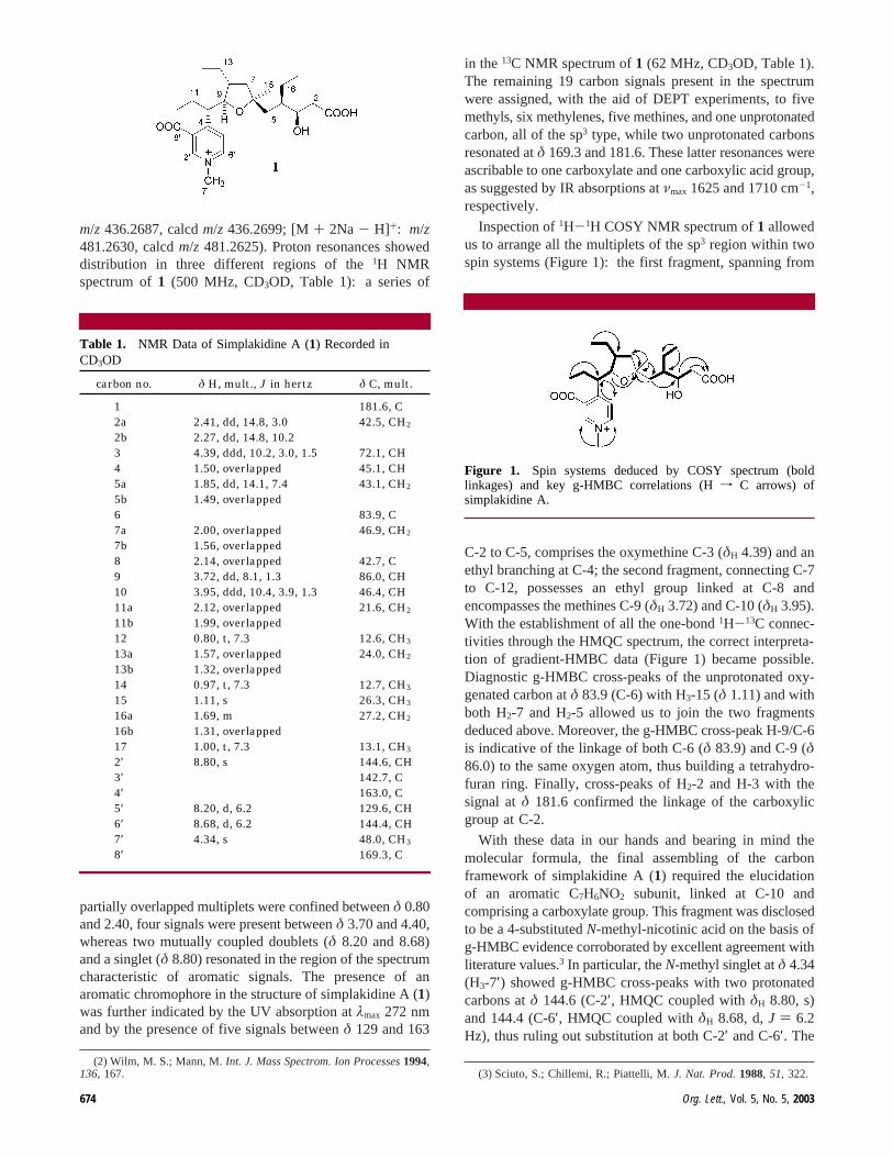
m/z 436.2687, calcdm/z 436.2699; [M+ 2Na - H]+: m/z481.2630, calcdm/z 481.2625). Proton resonances showeddistribution in three different regions of the1H NMRspectrum of1 (500 MHz, CD3OD, Table 1): a series of
partially overlapped multiplets were confined betweenδ 0.80and 2.40, four signals were present betweenδ 3.70 and 4.40,whereas two mutually coupled doublets (δ 8.20 and 8.68)and a singlet (δ 8.80) resonated in the region of the spectrumcharacteristic of aromatic signals. The presence of anaromatic chromophore in the structure of simplakidine A (1)was further indicated by the UV absorption atλmax 272 nmand by the presence of five signals betweenδ 129 and 163
in the13C NMR spectrum of1 (62 MHz, CD3OD, Table 1).The remaining 19 carbon signals present in the spectrumwere assigned, with the aid of DEPT experiments, to fivemethyls, six methylenes, five methines, and one unprotonatedcarbon, all of the sp3 type, while two unprotonated carbonsresonated atδ 169.3 and 181.6. These latter resonances wereascribable to one carboxylate and one carboxylic acid group,as suggested by IR absorptions atνmax 1625 and 1710 cm-1,respectively.
Inspection of1H-1H COSY NMR spectrum of1 allowedus to arrange all the multiplets of the sp3 region within twospin systems (Figure 1): the first fragment, spanning from
C-2 to C-5, comprises the oxymethine C-3 (δH 4.39) and anethyl branching at C-4; the second fragment, connecting C-7to C-12, possesses an ethyl group linked at C-8 andencompasses the methines C-9 (δH 3.72) and C-10 (δH 3.95).With the establishment of all the one-bond1H-13C connec-tivities through the HMQC spectrum, the correct interpreta-tion of gradient-HMBC data (Figure 1) became possible.Diagnostic g-HMBC cross-peaks of the unprotonated oxy-genated carbon atδ 83.9 (C-6) with H3-15 (δ 1.11) and withboth H2-7 and H2-5 allowed us to join the two fragmentsdeduced above. Moreover, the g-HMBC cross-peak H-9/C-6is indicative of the linkage of both C-6 (δ 83.9) and C-9 (δ86.0) to the same oxygen atom, thus building a tetrahydro-furan ring. Finally, cross-peaks of H2-2 and H-3 with thesignal atδ 181.6 confirmed the linkage of the carboxylicgroup at C-2.
With these data in our hands and bearing in mind themolecular formula, the final assembling of the carbonframework of simplakidine A (1) required the elucidationof an aromatic C7H6NO2 subunit, linked at C-10 andcomprising a carboxylate group. This fragment was disclosedto be a 4-substitutedN-methyl-nicotinic acid on the basis ofg-HMBC evidence corroborated by excellent agreement withliterature values.3 In particular, theN-methyl singlet atδ 4.34(H3-7′) showed g-HMBC cross-peaks with two protonatedcarbons atδ 144.6 (C-2′, HMQC coupled withδH 8.80, s)and 144.4 (C-6′, HMQC coupled withδH 8.68, d,J ) 6.2Hz), thus ruling out substitution at both C-2′ and C-6′. The
(2) Wilm, M. S.; Mann, M.Int. J. Mass Spectrom. Ion Processes1994,136, 167. (3) Sciuto, S.; Chillemi, R.; Piattelli, M.J. Nat. Prod.1988, 51, 322.
Table 1. NMR Data of Simplakidine A (1) Recorded inCD3OD
carbon no. δ H, mult., J in hertz δ C, mult.
1 181.6, C2a 2.41, dd, 14.8, 3.0 42.5, CH2
2b 2.27, dd, 14.8, 10.23 4.39, ddd, 10.2, 3.0, 1.5 72.1, CH4 1.50, overlapped 45.1, CH5a 1.85, dd, 14.1, 7.4 43.1, CH2
5b 1.49, overlapped6 83.9, C7a 2.00, overlapped 46.9, CH2
7b 1.56, overlapped8 2.14, overlapped 42.7, C9 3.72, dd, 8.1, 1.3 86.0, CH10 3.95, ddd, 10.4, 3.9, 1.3 46.4, CH11a 2.12, overlapped 21.6, CH2
11b 1.99, overlapped12 0.80, t, 7.3 12.6, CH3
13a 1.57, overlapped 24.0, CH2
13b 1.32, overlapped14 0.97, t, 7.3 12.7, CH3
15 1.11, s 26.3, CH3
16a 1.69, m 27.2, CH2
16b 1.31, overlapped17 1.00, t, 7.3 13.1, CH3
2′ 8.80, s 144.6, CH3′ 142.7, C4′ 163.0, C5′ 8.20, d, 6.2 129.6, CH6′ 8.68, d, 6.2 144.4, CH7′ 4.34, s 48.0, CH3
8′ 169.3, C
Figure 1. Spin systems deduced by COSY spectrum (boldlinkages) and key g-HMBC correlations (Hf C arrows) ofsimplakidine A.
674 Org. Lett., Vol. 5, No. 5, 2003

spatial proximity of H3-7′ with both H-2′ and H-6′, evidencedthrough the ROESY experiment, further supported the aboveconclusion.
Since H-6′ exhibited vicinal coupling with the signal atδ8.20 (H-5′, d, J ) 6.2 Hz), only the 3,4-disubstitutionremained possible. The g-HMBC cross-peaks H-10/C-5′ andH-5′/C-10 allowed us to link C-10 at the pyridinium carbonC-4′ and, therefore, the remaining carboxylate group mustbe placed at C-3′.
Taking into account all the above data, the planar structureof simplakidine A (1) was completely assembled. Thenonaromatic part of this molecule closely parallels thestructure of plakortethers A-E (e.g., plakortether B,2),1j aclass of polyketides structurally related to plakortin that werecently obtained from the apolar fractions of the organicextract ofP. simplex.
Of the sp3 carbon atoms of simplakidine A (1), 6 of the17 are asymmetric centers, and determination of theirstereochemistry represented a particularly challenging task.Unfortunately, only one of these carbons (C-3) appeared tobe amenable to derivatization with chiral auxiliary reagents,a procedure generally used to determine the absolute stereo-chemistry of natural products.4
As a consequence, stereochemical elucidation of simplaki-dine A (1) might greatly rely on the comparison of its spectraldata with those of a model compound, plakortether B (2),whose (3R,4R,6R,8R,9S)-configuration has been unambigu-ously defined by a three-step semisynthesis from plakortin.1j
In particular, we observed that the small coupling constantJH-3/H-4 (1.5 Hz) of simplakidine A (1), as well as thechemical shift values of the relevant protons, are almostidentical to the corresponding parameters measured forplakortether B (2) in the same solvent. This stronglysuggested that simplakidine A (1) and the model compoundplakortether B (2) actually share the same relative config-uration about the C-3/C-4 bond. The relative configurationwas promoted to the absolute configuration since standardapplication of a modified Mosher method5 to the secondaryalcohol C-3 of simplakidine A (see Supporting Information)enabled us to assign the (R)-configuration to this center, thesame of plakortether B (2). Consequently, the (R)-configu-ration at C-4 of the model compound2 was extended to C-4of simplakidine A (1).
The relative configuration at the three tetrahydrofuranasymmetric centers C-6, C-8, and C-9 of1, easily deducedon the basis of the ROESY cross-peaks of H3-15 with bothH-9 and H2-13, is the same as that detected for2. Also inthis case, useful information to upgrade the relative config-
uration of this region to the absolute configuration came fromcomparison of1H and13C NMR spectra of1 with those ofthe model compound, plakortether B (2). In this regard,1Hcoupling constants and1H and 13C chemical shifts of theentire C-3/C-6 fragment of simplakidine A (1) appeared tobe almost superimposable to the parallel values obtained forplakortether B (2) in the same solvent (see SupportingInformation). This is a strong indication that the twomolecules share the same relative configuration of thisfragment. Therefore, the establishment that both C-3 and C-4of simplakidine A (1) possess the same absolute configura-tion as plakortether B (2) allowed the absolute configurationof C-6 and, consequently, of C-8 and C-9 of1 to be assignedas that of the corresponding carbons of2 (6R,8R,9R).
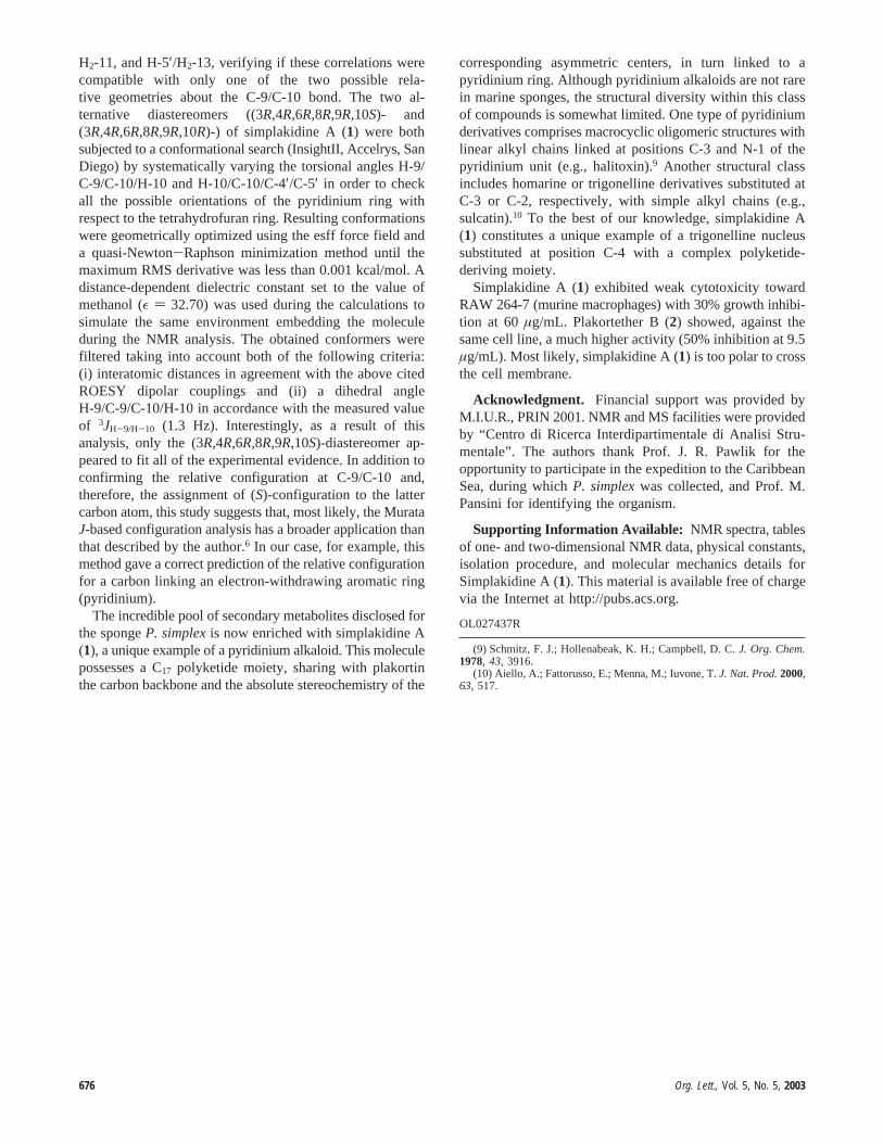
Determination of relative geometry around the C-9/C-10bond required additional spectral analysis. The small valueof 3JH-9/H-10 (1.3 Hz) indicates that a dominant staggeredrotamer exists around the C-9/C-10 axis, and this constitutesthe first prerequisite for applying theJ-based configurationanalysis recently developed by Murata et al.6 This NMR-based method allows elucidation of the relative configurationin acyclic structures on the basis of3JH,H and2,3JC,H values.In our case, the four required heteronuclear couplingconstants (Figure 2) were qualitatively evaluated throughanalysis of the PS-HMBC spectrum.7
The obtained pattern ofJ values appeared perfectlyconsistent and indicated athreostereochemical relationshipbetween C-9 and C-10 of simplakidine A (1), as reported inFigure 2, thus implying the (S)-stereochemistry at C-10.Anyway, since the Murata method has been originallydeveloped and reported only to determine relative stereo-chemistries between oxygen- and methyl-substituted6 (andsuccessively extended to halogen-substituted8) methines, theabove application needed to be verified by additionalindependent experimental evidence.
To this aim, we tried to take advantage of some key cross-peaks of the ROESY spectrum of1, namely, H-5′/H-9, H-5′/
(4) Seco, J. M.; Quin˜oa, E.; Riguera, R.Tetrahedron: Asymmetry2001,12, 2915.
(5) Ohtani, I.; Kusumi, T.; Kashman, Y.; Kakisawa, H.J. Am. Chem.Soc. 1991, 113, 4092.
(6) Matsumori, N.; Kaneno, D.; Murata, M.; Nakamura, H.; Tachibana,K. J. Org. Chem.1999, 64, 866.
(7) Matsumori, N.; Murata, M.; Tachibana, K.Tetrahedron1995, 51,12229.
(8) Ciminiello, P.; Fattorusso, E.; Forino, M.; Di Rosa, M.; Ianaro, A.;Poletti, R.J. Org. Chem.2001, 66, 578.
Figure 2. Application of Murata’s method to the C-9/C-10 bondof simplakidine A (1).
Org. Lett., Vol. 5, No. 5, 2003 675
H2-11, and H-5′/H2-13, verifying if these correlations werecompatible with only one of the two possible rela-tive geometries about the C-9/C-10 bond. The two al-ternative diastereomers ((3R,4R,6R,8R,9R,10S)- and(3R,4R,6R,8R,9R,10R)-) of simplakidine A (1) were bothsubjected to a conformational search (InsightII, Accelrys, SanDiego) by systematically varying the torsional angles H-9/C-9/C-10/H-10 and H-10/C-10/C-4′/C-5′ in order to checkall the possible orientations of the pyridinium ring withrespect to the tetrahydrofuran ring. Resulting conformationswere geometrically optimized using the esff force field anda quasi-Newton-Raphson minimization method until themaximum RMS derivative was less than 0.001 kcal/mol. Adistance-dependent dielectric constant set to the value ofmethanol (ε ) 32.70) was used during the calculations tosimulate the same environment embedding the moleculeduring the NMR analysis. The obtained conformers werefiltered taking into account both of the following criteria:(i) interatomic distances in agreement with the above citedROESY dipolar couplings and (ii) a dihedral angleH-9/C-9/C-10/H-10 in accordance with the measured valueof 3JH-9/H-10 (1.3 Hz). Interestingly, as a result of thisanalysis, only the (3R,4R,6R,8R,9R,10S)-diastereomer ap-peared to fit all of the experimental evidence. In addition toconfirming the relative configuration at C-9/C-10 and,therefore, the assignment of (S)-configuration to the lattercarbon atom, this study suggests that, most likely, the MurataJ-based configuration analysis has a broader application thanthat described by the author.6 In our case, for example, thismethod gave a correct prediction of the relative configurationfor a carbon linking an electron-withdrawing aromatic ring(pyridinium).
The incredible pool of secondary metabolites disclosed forthe spongeP. simplexis now enriched with simplakidine A(1), a unique example of a pyridinium alkaloid. This moleculepossesses a C17 polyketide moiety, sharing with plakortinthe carbon backbone and the absolute stereochemistry of the
corresponding asymmetric centers, in turn linked to apyridinium ring. Although pyridinium alkaloids are not rarein marine sponges, the structural diversity within this classof compounds is somewhat limited. One type of pyridiniumderivatives comprises macrocyclic oligomeric structures withlinear alkyl chains linked at positions C-3 and N-1 of thepyridinium unit (e.g., halitoxin).9 Another structural classincludes homarine or trigonelline derivatives substituted atC-3 or C-2, respectively, with simple alkyl chains (e.g.,sulcatin).10 To the best of our knowledge, simplakidine A(1) constitutes a unique example of a trigonelline nucleussubstituted at position C-4 with a complex polyketide-deriving moiety.
Simplakidine A (1) exhibited weak cytotoxicity towardRAW 264-7 (murine macrophages) with 30% growth inhibi-tion at 60µg/mL. Plakortether B (2) showed, against thesame cell line, a much higher activity (50% inhibition at 9.5µg/mL). Most likely, simplakidine A (1) is too polar to crossthe cell membrane.
Acknowledgment. Financial support was provided byM.I.U.R., PRIN 2001. NMR and MS facilities were providedby “Centro di Ricerca Interdipartimentale di Analisi Stru-mentale”. The authors thank Prof. J. R. Pawlik for theopportunity to participate in the expedition to the CaribbeanSea, during whichP. simplexwas collected, and Prof. M.Pansini for identifying the organism.
Supporting Information Available: NMR spectra, tablesof one- and two-dimensional NMR data, physical constants,isolation procedure, and molecular mechanics details forSimplakidine A (1). This material is available free of chargevia the Internet at http://pubs.acs.org.
OL027437R
(9) Schmitz, F. J.; Hollenabeak, K. H.; Campbell, D. C.J. Org. Chem.1978, 43, 3916.
(10) Aiello, A.; Fattorusso, E.; Menna, M.; Iuvone, T.J. Nat. Prod.2000,63, 517.
676 Org. Lett., Vol. 5, No. 5, 2003