Embed Size (px)
Citation preview
STROKE ASSESSMENT POCKET GUIDE
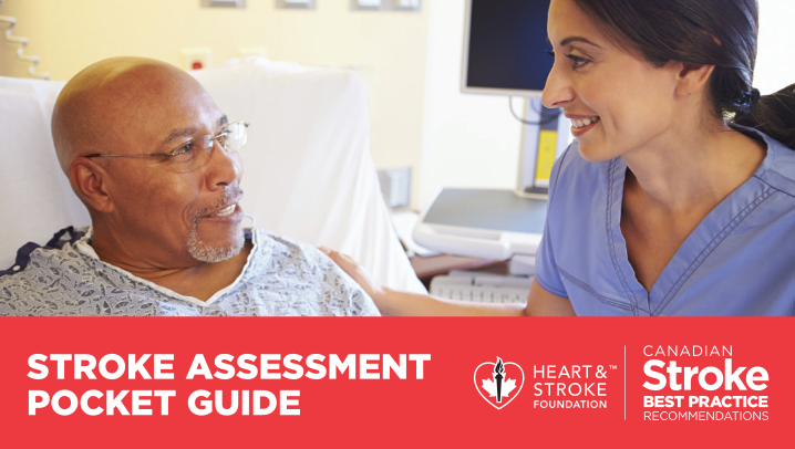
COMMON SIGNS AND SYMPTOMS OF STROKE SYNDROMESAnterior Cerebral
StrokeMiddle Cerebral
StrokePosterior
Cerebral StrokeVertebro Basilar
StrokeThalamic
StrokeLacunar Stroke
Four Types
• Contralateral sensorimotor deficit: foot and leg
• Arm paresis
• Gait ataxia
• Bladder incontinence
• Personality and behaviour changes
• Flat affect, distractible
• Perservation and amnesia
• Contralateral sensorimotor deficit: face, arm, leg
• Contralateral homonymous hemianopsia
• Contralateral hemi- spatial neglect or inattention (usually in Right Hemispheric Strokes)
• Aphasia, alexia, agraphia (usually in Left Hemispheric Stroke or dominant hemisphere)
• Gaze deviation towards affected hemisphere
• Dysarthria
• Pure homonymous hemianopsia
• Nausea
• Vomiting
• Ataxia
• Vertigo
• Weakness
• Sensory loss
• Dysarthria
• Vertigo
• Limb and gait ataxia
• Cranial nerve dysfunction
• Coma at onset
• Diplopia
• Cross sensory loss
• Bilateral motor deficits
• Isolated field defect
• Pure motor/ sensory loss
• Dysarthria
• Dysphagia
• Alteration in senses (except smell)
• Alteration in pain, crude touch (loss)
• Alteration in
temperature
• Contralateral hemiplegia
• Hyper-sensitivity to stimulus
• Vertical and lateral gaze deficits
• Short-term memory loss
Pure motor hemiparesis
• Contralateral hemiparesis of face, arm and leg
Ataxic Hemiparesis• Ipsilateral paresis
of leg
• Arm and leg ataxia
Dysarthria and Clumsy Hand Syndrome• Dysarthia
• Weakness of hand
• Impaired manual dexterity
Pure Sensory Stroke• Impairments in
pain, temperature, touch, position and vibration
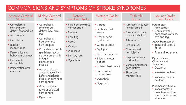
COMMON SYMPTOMS IN STROKE PATIENTS
The effects of a stroke depend on several factors including the location of the obstruction and how much brain tissue is affected. However, because one side of the brain controls the opposite side of the body, a stroke affecting one side will result in neurological complications on the opposite side.
Right Hemispheric Stroke Left Hemispheric Stroke
If the stroke occurs in the brain’s right side, the left side of the body will be affected, which could produce any or all of the following:
If the stroke occurs in the left side of the brain, the right side of the body will be affected, producing some or all of the following:
• Contralateral face, arm and leg weakness or hemiparesis• Contralateral arm and/or leg sensory loss or extinction• Hemispatial neglect or inattention• Deficit and/or neglect of left visual field• Right gaze preference• Impulsive or overestimation of abilities (risk for injury)
• Contralateral face, arm and leg weakness or hemiparesis• Contralateral face, arm and/or leg sensory loss• Aphasia, alexia, agraphia• Slow and cautious behaviour• Deficits in right visual field• Left gaze preference
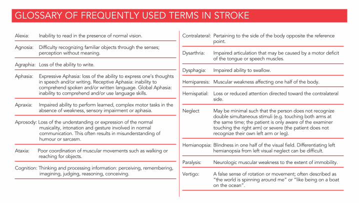
GLOSSARY OF FREQUENTLY USED TERMS IN STROKE
Alexia: Inability to read in the presence of normal vision.
Agnosia: Difficulty recognizing familiar objects through the senses; perception without meaning.
Agraphia: Loss of the ability to write.
Aphasia: Expressive Aphasia: loss of the ability to express one’s thoughts in speech and/or writing. Receptive Aphasia: inability to comprehend spoken and/or written language. Global Aphasia: inability to comprehend and/or use language skills.
Apraxia: Impaired ability to perform learned, complex motor tasks in the absence of weakness, sensory impairment or aphasia.
Aprosody: Loss of the understanding or expression of the normal musicality, intonation and gesture involved in normal communication. This often results in misunderstanding of humour or sarcasm.
Ataxia: Poor coordination of muscular movements such as walking or reaching for objects.
Cognition: Thinking and processing information: perceiving, remembering, imagining, judging, reasoning, conceiving.
Contralateral: Pertaining to the side of the body opposite the reference point.
Dysarthria: Impaired articulation that may be caused by a motor deficit of the tongue or speech muscles.
Dysphagia: Impaired ability to swallow.
Hemiparesis: Muscular weakness affecting one half of the body.
Hemispatial: Loss or reduced attention directed toward the contralateral side.
Neglect May be minimal such that the person does not recognize double simultaneous stimuli (e.g. touching both arms at the same time; the patient is only aware of the examiner touching the right arm) or severe (the patient does not recognize their own left arm or leg).
Hemianopsia: Blindness in one half of the visual field. Differentiating left hemianopsia from left visual neglect can be difficult.
Paralysis: Neurologic muscular weakness to the extent of immobility.
Vertigo: A false sense of rotation or movement; often described as “the world is spinning around me” or “like being on a boat on the ocean”.
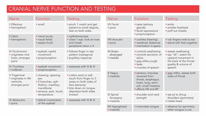
Nerve Function Testing
I Olfactory• hemispheric
• smell • pinch 1 nostril and get patient to smell objects, test on both sides
II Optic • hemispheric
• visual acuity• visual fields• assess fundi
• opthalmoscope• close 1 eye, look at nose
and cheek peripheral vision x 4
III Oculomotor • originates mid-
brain, emerges pons
• eyeball, eyelid movement
• proprioception
• follows finger in star pattern (nystagnus)
• pupillary response
IV Trochlear • midbrain
• eyeball movement• proprioception
• assessed with III & VI
V Trigeminal• originates in 4th ventricle,
emerges pons
• chewing, opening jaw
• 3 branches, oph-thalmic, maxillary, mandibular
• sensory: pain, touch, temperature
• cotton wool or soft touch from finger to 3 areas on both sides of face (sensory)
• bite down on tongue depressor-both sides (motor)
VI Abducens• pons
• lateral movements of the eyeball
• assessed with III & IV
Nerve Function Testing
VII Facial• pons
• taste (salivary glands)
• facial expressions• proprioception
• smile• wrinkle forehead• puff out cheeks
VIII Acoustic• pons
• cochlea (hearing)• vestibular (balance)• orientation is space
• rub fingers nest to ear• stand with feet together
IX Glosso-pharyngeal
• medulla
• controls swallowing• controls secretion of
saliva• gag reflex,cough• taste• muscles of speech
• assess swallowing• say “ah”, watch for
upward movement in the back of the throat-quality & volume of speech
X Vagus• medulla
• sensory: impulses received from throat, esophagus, heart, lung, stom-ach, small intestine
• affects HR and BP
• gag reflex, assess both sides of throat
XI Spinal Accessory
• medulla
• shoulder and neck strength
• ask pt to shrug shoulders (symmetry)
• rotate head
XII Hypoglossal• medulla
• innervates tongue • observe for symmetry when tongue is out
CRANIAL NERVE FUNCTION AND TESTING
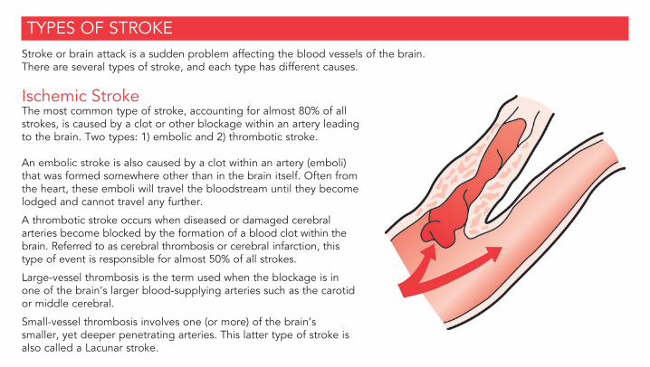
TYPES OF STROKEStroke or brain attack is a sudden problem affecting the blood vessels of the brain. There are several types of stroke, and each type has different causes.
Ischemic StrokeThe most common type of stroke, accounting for almost 80% of all strokes, is caused by a clot or other blockage within an artery leading to the brain. Two types: 1) embolic and 2) thrombotic stroke.
An embolic stroke is also caused by a clot within an artery (emboli) that was formed somewhere other than in the brain itself. Often from the heart, these emboli will travel the bloodstream until they become lodged and cannot travel any further.
A thrombotic stroke occurs when diseased or damaged cerebral arteries become blocked by the formation of a blood clot within the brain. Referred to as cerebral thrombosis or cerebral infarction, this type of event is responsible for almost 50% of all strokes.
Large-vessel thrombosis is the term used when the blockage is in one of the brain’s larger blood-supplying arteries such as the carotid or middle cerebral.
Small-vessel thrombosis involves one (or more) of the brain’s smaller, yet deeper penetrating arteries. This latter type of stroke is also called a Lacunar stroke.
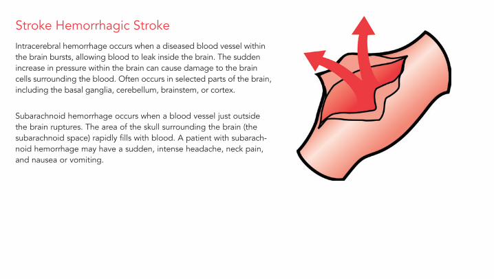
Stroke Hemorrhagic StrokeIntracerebral hemorrhage occurs when a diseased blood vessel within the brain bursts, allowing blood to leak inside the brain. The sudden increase in pressure within the brain can cause damage to the brain cells surrounding the blood. Often occurs in selected parts of the brain, including the basal ganglia, cerebellum, brainstem, or cortex.
Subarachnoid hemorrhage occurs when a blood vessel just outside the brain ruptures. The area of the skull surrounding the brain (the subarachnoid space) rapidly fills with blood. A patient with subarach-noid hemorrhage may have a sudden, intense headache, neck pain, and nausea or vomiting.
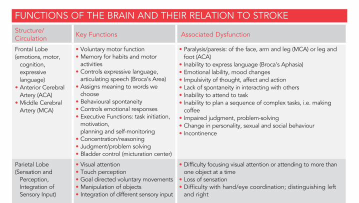
FUNCTIONS OF THE BRAIN AND THEIR RELATION TO STROKEStructure/Circulation Key Functions Associated Dysfunction
Frontal Lobe (emotions, motor,
cognition, expressive language)
• Anterior Cerebral Artery (ACA)
• Middle Cerebral Artery (MCA)
• Voluntary motor function• Memory for habits and motor
activities• Controls expressive language,
articulating speech (Broca’s Area)• Assigns meaning to words we
choose• Behavioural spontaneity• Controls emotional responses• Executive Functions: task initiation,
motivation, planning and self-monitoring
• Concentration/reasoning• Judgment/problem solving• Bladder control (micturation center)
• Paralysis/paresis: of the face, arm and leg (MCA) or leg and foot (ACA)
• Inability to express language (Broca’s Aphasia)• Emotional lability, mood changes• Impulsivity of thought, affect and action• Lack of spontaneity in interacting with others• Inability to attend to task• Inability to plan a sequence of complex tasks, i.e. making
coffee• Impaired judgment, problem-solving• Change in personality, sexual and social behaviour• Incontinence
Parietal Lobe (Sensation and
Perception, Integration of Sensory Input)
• Visual attention• Touch perception• Goal directed voluntary movements• Manipulation of objects• Integration of different sensory input
• Difficulty focusing visual attention or attending to more than one object at a time
• Loss of sensation• Difficulty with hand/eye coordination; distinguishing left
and right
• Anterior Cerebral Artery
• Middle Cerebral Artery
• Posterior Cerebral Artery
• The ability to sense the position, location, orientation and movement of the body and its parts (Proprioception)
• Inability to perceive objects normally (Agnosia)• Neglecting part of the body or space (contralateral neglect/
difficulties with ADLs)• Difficulty reading, writing (Agraphia), drawing, constructing,
naming objects, calculating• Denial of deficits (Anosagnosia)
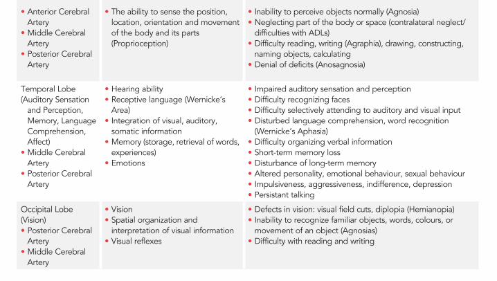
Temporal Lobe (Auditory Sensation
and Perception, Memory, Language Comprehension, Affect)
• Middle Cerebral Artery
• Posterior Cerebral Artery
• Hearing ability• Receptive language (Wernicke’s
Area)• Integration of visual, auditory,
somatic information• Memory (storage, retrieval of words,
experiences)• Emotions
• Impaired auditory sensation and perception• Difficulty recognizing faces• Difficulty selectively attending to auditory and visual input• Disturbed language comprehension, word recognition
(Wernicke’s Aphasia)• Difficulty organizing verbal information• Short-term memory loss• Disturbance of long-term memory• Altered personality, emotional behaviour, sexual behaviour• Impulsiveness, aggressiveness, indifference, depression• Persistant talking
Occipital Lobe (Vision)• Posterior Cerebral
Artery• Middle Cerebral
Artery
• Vision• Spatial organization and
interpretation of visual information• Visual reflexes
• Defects in vision: visual field cuts, diplopia (Hemianopia)• Inability to recognize familiar objects, words, colours, or
movement of an object (Agnosias)• Difficulty with reading and writing
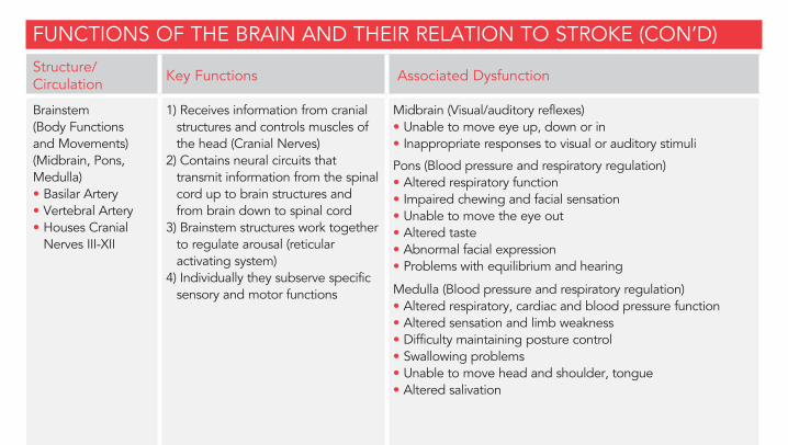
FUNCTIONS OF THE BRAIN AND THEIR RELATION TO STROKE (CON’D)Structure/Circulation Key Functions Associated Dysfunction
Brainstem (Body Functions and Movements) (Midbrain, Pons, Medulla)• Basilar Artery• Vertebral Artery• Houses Cranial
Nerves III-XII
1) Receives information from cranial structures and controls muscles of
the head (Cranial Nerves)2) Contains neural circuits that
transmit information from the spinal cord up to brain structures and from brain down to spinal cord
3) Brainstem structures work together to regulate arousal (reticular activating system)
4) Individually they subserve specific sensory and motor functions
Midbrain (Visual/auditory reflexes)• Unable to move eye up, down or in• Inappropriate responses to visual or auditory stimuli
Pons (Blood pressure and respiratory regulation)• Altered respiratory function• Impaired chewing and facial sensation• Unable to move the eye out• Altered taste• Abnormal facial expression• Problems with equilibrium and hearing
Medulla (Blood pressure and respiratory regulation)• Altered respiratory, cardiac and blood pressure function• Altered sensation and limb weakness• Difficulty maintaining posture control• Swallowing problems• Unable to move head and shoulder, tongue• Altered salivation
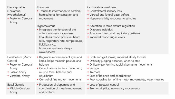
Diencephalon (Thalamus, Hypothalamus)• Posterior Cerebral
Artery
Thalamus• Transmits information to cerebral
hemispheres for sensation and movement
Hypothalamus• Integrates the function of the
autonomic nervous system (maintains blood pressure, heart rate, respiratory rate, temperature, fluid balance, hormone synthesis, sleep-wakefulness)
Contralateral weakness• Contralateral sensory loss• Vertical and lateral gaze deficits• Hypersensitivity response to stimulus
• Alteration in temperature regulation• Diabetes insipidus• Abnormal heart and respiratory patterns• Impaired blood sugar levels
Cerebellum (Motor Control)• Posterior Cerebral
Artery• Basilar Artery• Vertebral Artery
• Regulates movements of eyes and limbs; helps maintain posture and balance
• Coordinates voluntary movement, muscle tone, balance and equilibrium
• Control of fine motor movements
• Limb and gait ataxia; impaired ability to walk• Difficulty judging distance, when to stop• Difficulty performing rapid alternating movements• Vertigo• Tremors• Loss of balance and coordination• Poor coordination of fine motor movements, weak muscles
Basal Ganglia• Middle Cerebral
Artery
• Production of dopamine and coordination of muscle movement and posture
• Loss of postural control• Tremor, rigidity, involuntary movements
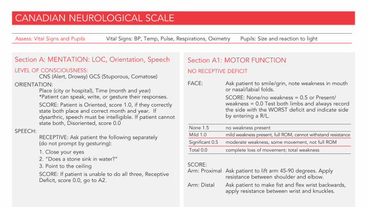
CANADIAN NEUROLOGICAL SCALE
Assess: Vital Signs and Pupils Vital Signs: BP, Temp, Pulse, Respirations, Oximetry Pupils: Size and reaction to light
Section A: MENTATION: LOC, Orientation, Speech
LEVEL OF CONSCIOUSNESS: CNS (Alert, Drowsy) GCS (Stuporous, Comatose)ORIENTATION: Place (city or hospital), Time (month and year) *Patient can speak, write, or gesture their responses. SCORE: Patient is Oriented, score 1.0, if they correctly
state both place and correct month and year. If dysarthric, speech must be intelligible. If patient cannot state both, Disoriented, score 0.0
SPEECH: RECEPTIVE: Ask patient the following separately
(do not prompt by gesturing):
1. Close your eyes 2. “Does a stone sink in water?” 3. Point to the ceiling SCORE: If patient is unable to do all three, Receptive
Deficit, score 0.0, go to A2.
Section A1: MOTOR FUNCTION
NO RECEPTIVE DEFICIT FACE: Ask patient to smile/grin, note weakness in mouth
or nasal/labial folds. SCORE: None/no weakness = 0.5 or Present/
weakness = 0.0 Test both limbs and always record the side with the WORST deficit and indicate side by entering a R/L.
None 1.5 no weakness presentMild 1.0 mild weakness present, full ROM, cannot withstand resistance
Significant 0.5 moderate weakness, some movement, not full ROM
Total 0.0 complete loss of movement; total weakness
SCORE: Arm: Proximal Ask patient to lift arm 45-90 degrees. Apply
resistance between shoulder and elbow. Arm: Distal Ask patient to make fist and flex wrist backwards,
apply resistance between wrist and knuckles.

EXPRESSIVE:
1. Show patient 3 items separately (pencil, watch, key) and ask patient to name each object.
2. Ask patient what each object is used for while holding each up again, i.e. “What do you do with a pencil?”
SCORE: If patient is able to state the name and use of all 3 objects, Normal Speech, score 1.0.
If patient is unable to state the name and use of all 3 objects, Expressive Deficit, score 0.5.
*If patient answers all questions correctly but speech is slurred and intelligible, score Normal Speech and record “SL” along with the score.
Leg: Proximal In supine, ask patient to flex hip to 90 degrees, apply pressure to mid thigh.
Leg: Distal Ask patient to dorsiflex foot, apply resistance to top of foot.
Section A2: MOTOR RESPONSE
RECEPTIVE DEFICIT PRESENTFACE: Have patient mimic your smile. If unable, note facial
expression while applying sternal pressure.ARMS: Demonstrate or lift patient’s arms to 90 degrees,
score ability to maintain equal levels (>5 secs). If unable to maintain raised arms, apply nail bed
pressure to assess reflex response.LEGS: Lift patient’s hip to 90 degrees, score ability
to maintain equal levels (>5 secs). If unable to maintain raised position, apply nail bed pressure to assess reflex response.
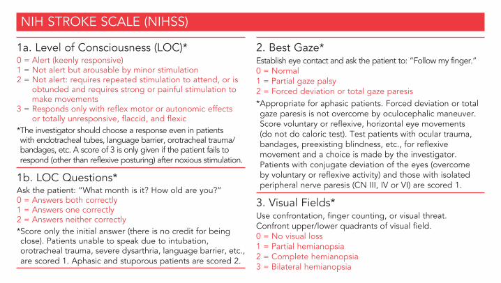
1a. Level of Consciousness (LOC)*0 = Alert (keenly responsive)1 = Not alert but arousable by minor stimulation2 = Not alert: requires repeated stimulation to attend, or is
obtunded and requires strong or painful stimulation to make movements
3 = Responds only with reflex motor or autonomic effects or totally unresponsive, flaccid, and flexic
* The investigator should choose a response even in patients with endotracheal tubes, language barrier, orotracheal trauma/bandages, etc. A score of 3 is only given if the patient fails to respond (other than reflexive posturing) after noxious stimulation.
1b. LOC Questions*Ask the patient: “What month is it? How old are you?”0 = Answers both correctly 1 = Answers one correctly 2 = Answers neither correctly* Score only the initial answer (there is no credit for being close). Patients unable to speak due to intubation, orotracheal trauma, severe dysarthria, language barrier, etc., are scored 1. Aphasic and stuporous patients are scored 2.
2. Best Gaze*Establish eye contact and ask the patient to: “Follow my finger.”0 = Normal 1 = Partial gaze palsy2 = Forced deviation or total gaze paresis* Appropriate for aphasic patients. Forced deviation or total gaze paresis is not overcome by oculocephalic maneuver. Score voluntary or reflexive, horizontal eye movements (do not do caloric test). Test patients with ocular trauma, bandages, preexisting blindness, etc., for reflexive movement and a choice is made by the investigator. Patients with conjugate deviation of the eyes (overcome by voluntary or reflexive activity) and those with isolated peripheral nerve paresis (CN III, IV or VI) are scored 1.
3. Visual Fields*Use confrontation, finger counting, or visual threat. Confront upper/lower quadrants of visual field.0 = No visual loss 1 = Partial hemianopsia2 = Complete hemianopsia 3 = Bilateral hemianopsia
NIH STROKE SCALE (NIHSS)
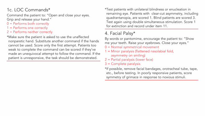
1c. LOC Commands*Command the patient to: “Open and close your eyes. Grip and release your hand.”0 = Performs both correctly 1 = Performs one correctly 2 = Performs neither correctly* Make sure the patient is asked to use the unaffected nonparetic hand. Substitute another command if the hands cannot be used. Score only the first attempt. Patients too weak to complete the command can be scored if they’ve made an unequivocal attempt to follow the command. If the patient is unresponsive, the task should be demonstrated.
* Test patients with unilateral blindness or enucleation in remaining eye. Patients with clear-cut asymmetry, including quadrantanopia, are scored 1. Blind patients are scored 3. Test again using double simultaneous stimulation. Score 1 for extinction and record under item 11.
4. Facial Palsy*By words or pantomime, encourage the patient to: “Show me your teeth. Raise your eyebrows. Close your eyes.”0 = Normal symmetrical movement 1 = Minor paralysis (flattened nasolabial fold,
asymmetry on smiling) 2 = Partial paralysis (lower face)3 = Complete paralysis* If possible, remove facial bandages, orotracheal tube, tape, etc., before testing. In poorly responsive patients, score symmetry of grimace in response to noxious stimuli.
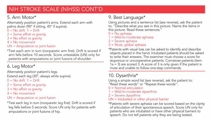
5. Arm Motor*Alternately position patient’s arms. Extend each arm with palms down (90° if sitting, 45° if supine).0 = No drift 1 = Drift 2 = Some effort vs gravity 3 = No effort vs gravity 4 = No movement UN = Amputation or joint fusion* Test each arm in turn (nonparetic arm first). Drift is scored if arm falls before 10 seconds. Score untestable (UN) only for patients with amputations or joint fusions of shoulder.
6. Leg Motor*Alternately position patient’s legs. Extend each leg (30°, always while supine).0 = No drift 1 = Drift 2 = Some effort vs gravity 3 = No effort vs gravity 4 = No movement UN = Amputation or joint fusion* Test each leg in turn (nonparetic leg first). Drift is scored if leg falls before 5 seconds. Score UN only for patients with amputations or joint fusions of hip.
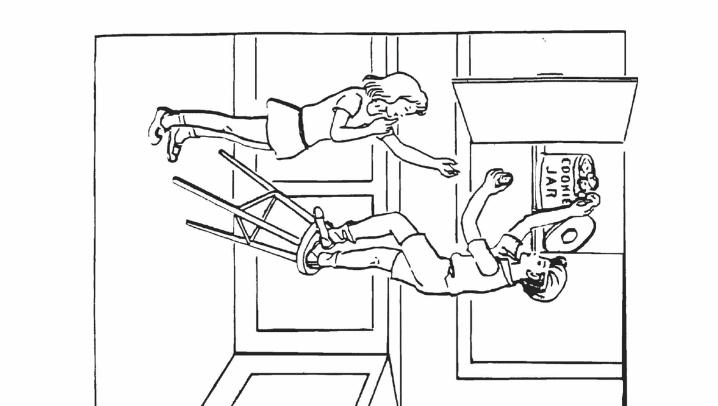
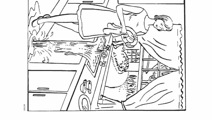
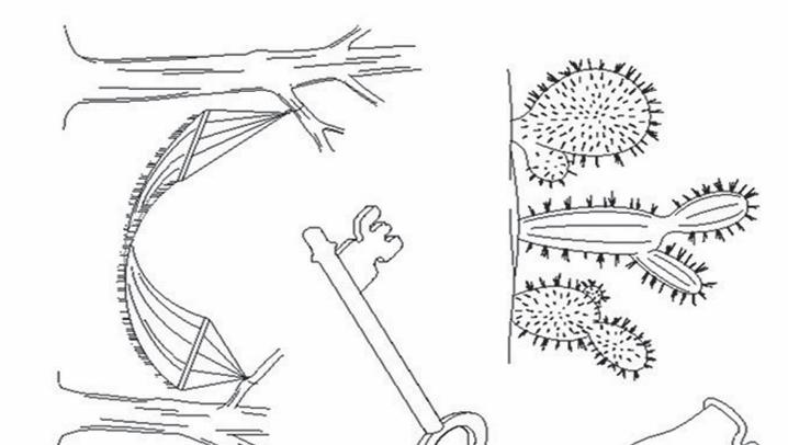
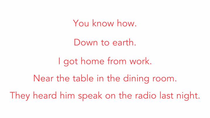
9. Best Language*Using pictures and a sentence list (see reverse), ask the patient to: “Describe what you see in this picture. Name the items in this picture. Read these sentences.”0 = No aphasia 1 = Mild-to-moderate aphasia 2 = Severe aphasia 3 = Mute, global aphasia* Patients with visual loss can be asked to identify and describe objects placed in the hand. Intubated patients should be asked to write their answers. The examiner must choose a score for stuporous or uncooperative patients. Comatose patients (item 1a = 3) are scored 3. A score of 3 is only given if the patient is mute and unable to follow one-step commands.
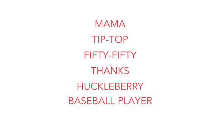
10. Dysarthria*Using a simple word list (see reverse), ask the patient to: “Read these words” or “Repeat these words”.0 = Normal articulation 1 = Mild-to-moderate dysarthria2 = Severe dysarthria UN = Intubated or other physical barrier* Patients with severe aphasia can be scored based on the clarity of articulation of their spontaneous speech. Score UN only for patients who are intubated or have other physical barriers to speech. Do not tell patients why they are being tested.
NIH STROKE SCALE (NIHSS) CONT’D
7. Limb Ataxia*Ask patient (eyes open) to: “Touch your finger to your nose. Touch your heel to your shin.”0 = Absent 1 = Present in one limb2 = Present in two or more limbs UN = Amputation or joint fusion* Perform finger-nose and heel-shin tests on both sides to determine unilateral cerebellar lesion. Score 0 for patients who are paralyzed or cannot understand. Score 1 or 2 only if ataxia is disproportionate to weakness. Score UN only for patients with amputations or joint fusions.
8. Sensory*Test as many body parts as possible (arms [not hands], legs, trunk, face) for sensation using pinprick or noxious stimulus (in the obtunded or aphasic patient).0 = Normal 1 = Mild-to-moderate sensory loss2 = Severe-to-total sensory loss* Score sensory loss due to stroke only. Stuporous and aphasic patients are scored 0 or 1. Patients with brainstem stroke and bilateral sensory loss, quadriplegic patients who do not respond, and comatose patients (item 1a = 3) are scored 2. A score of 2 is only given when severe or total sensory loss is demonstrated.
11. Extinction and Inattention*Sufficient information to determine these scores may have been obtained during the prior testing. 0 = No abnormality1 = Visual, tactile, auditory, spatial, or personal inattention2 = Profound hemi-inattention or extinction to more than one
modality* Lack of patient response and inattention may already be evident from the previous items. Score 0 if the patient has a severe visual loss preventing visual double simultaneous stimulation, but the response to cutaneous stimuli is normal, or if the patient has aphasia but does not appear to attend to both sides. The presence of visual spatial attention or anosognosia may also be evidence of abnormality.
You know how.
Down to earth.
I got home from work.
Near the table in the dining room.
They heard him speak on the radio last night.
MAMA
TIP-TOP
FIFTY-FIFTY
THANKS
HUCKLEBERRYBASEBALL PLAYER
The information contained in this document supports the Canadian Stroke Best Practice Recommendations
For detailed recommendations and updates visit:strokebestpractices.ca
TIME IS BRAIN. DON’T WASTE EITHER ONE.
HP6311EV3.4