Embed Size (px)
Citation preview

Structure of a Ca 2+/CaM:Kv7.4 (KCNQ4) B-HelixComplex Provides Insight into M Current Modulation
Qiang Xu1†, Aram Chang1†, Alexandra Tolia1 and Daniel L. Minor Jr.1,2,3,4
1 - Cardiovascular Research Institute, University of California, San Francisco, CA 94158-2156, USA2 - Departments of Biochemistry and Biophysics, and Cellular and Molecular Pharmacology, University of California, San Francisco,CA 94158-2156, USA3 - California Institute for Quantitative Biosciences, University of California, San Francisco, CA 94158-2156, USA4 - Physical Biosciences Division, Lawrence Berkeley National Laboratory, Berkeley, CA 94720, USA
Correspondence to Daniel L. Minor: [email protected]://dx.doi.org/10.1016/j.jmb.2012.11.023Edited by J. Bowie
Abstract
Calmodulin (CaM) is an important regulator of Kv7.x (KCNQx) voltage-gated potassium channels. Channelsfrom this family produce neuronal M currents and cardiac and auditory IKS currents and harbor mutations thatcause arrhythmias, epilepsy, and deafness. Despite extensive functional characterization, biochemical andstructural details of the interaction between CaM and the channel have remained elusive. Here, we show thatboth apo-CaM and Ca2+/CaM bind to the C-terminal tail of the neuronal channel Kv7.4 (KCNQ4), which isinvolved in both hearing and mechanosensation. Interactions between apo-CaM and the Kv7.4 tail involve twoC-terminal tail segments, known as the A and B segments, whereas the interaction between Ca2+/CaM andthe Kv7.4 C-terminal tail requires only the B segment. Biochemical studies show that the calcium dependenceof the CaM:B segment interaction is conserved in all Kv7 subtypes. X-ray crystallographic determination of thestructure of the Ca2+/CaM:Kv7.4 B segment complex shows that Ca2+/CaM wraps around the Kv7.4 Bsegment, which forms an α-helix, in an antiparallel orientation that embodies a variation of the classic 1-14Ca2+/CaM interaction motif. Taken together with the context of prior studies, our data suggest a model formodulation of neuronal Kv7 channels involving a calcium-dependent conformational switch from an apo-CaMform that bridges the A and B segments to a Ca2+/CaM form bound to the B-helix. The structure presentedhere also provides a context for a number of disease-causing mutations and for further dissection of themechanisms by which CaM controls Kv7 function.
© 2012 Elsevier Ltd. All rights reserved.
Introduction
Pore-forming subunits of the Kv7.x (KCNQx)potassium channel family (Kv7.1–Kv7.5) (KCNQ1-KCNQ5)1–3 form the classically studied M currentin neurons (Kv7.2–Kv7.5)4–6 and IKS current(Kv7.1) in heart, vestibular, and auditory cells.2,7–9
Kv7 channels open at subthreshold membranepotentials, do not inactivate, and, therefore, providea strong brake on membrane excitation.4,10 Conse-quently, a variety of signaling pathways tune electri-cal excitability by regulating Kv7 function.3,4,6
Commensurate with their pivotal role in controllingexcitation, Kv7 channels are targets for the develop-
0022-2836/$ - see front matter © 2012 Elsevier Ltd. All rights reserve
ment of modulators to treat diseases involvingneuronal hyperexcitability such as epilepsy andneuropathic pain.11–14 Further, mutations in Kv7channels have been linked to various humandiseases, including cardiac arrhythmias, deafness,and epilepsy.2,14,15
Kv7 channels have the canonical six-transmem-brane architecture found throughout the voltage-gated ion channel family16 (Fig. 1a). The Kv7cytoplasmic C-terminal tail (~300–400 residues)comprises approximately half of the total residuesof the pore-forming subunit and is composed of fourconserved elements known as segments A–D (Fig.1b; Supplementary Fig. S1).17,18 This region forms
d. J. Mol. Biol. (2013) 425, 378–394

379Ca2+/CaM:Kv7.4 (KCNQ4) B-Helix Complex Structure
an important, multifunctional entity that is central tochannel assembly, gating, regulation, and the for-mation of complexes with regulatory factors17
including calmodulin (CaM).19–23 Although Kv7function has been characterized extensively,4,6,10,14
structural knowledge for this channel class is limitedto a portion of the cytoplasmic C-terminal tail, calledthe D-helix or A domain tail,18,24,25 which is importantfor subunit specific assembly. Strikingly, over 60% ofthe known Kv7 disease mutations occur in thecytoplasmic C-terminal tail where their direct func-tional consequences are not obvious.18,26–28 Thus,defining the structure that underlies this hub of Kv7regulation remains a key objective both for under-standing the basic mechanisms by which Kv7channels are regulated and assembled, and forunderstanding how disease mutations cause chan-nel dysfunction.One of the key Kv7 modulation pathways involves
interaction between the calcium sensor CaM19,29,30
and the Kv7 C-terminal cytoplasmic domain.29 CaMaffects the function of all Kv7 subtypes19–21,29,31,32
by binding to distinct regions within the cytoplasmicC-terminal tail.19,22,23 Two of these, the A and Bsegments, have sequence features that have beenproposed as CaM binding motifs, an IQ motif in the Asegment and two adjacent 1–5–10 motifs in the Bsegment.23 Studies of the calcium dependency ofthe CaM interaction with Kv7.118,20,21 and Kv7.2,Kv7.3, and Kv7.433 C-terminal tails indicate that bothforms of CaM, apo-CaM and Ca2+/CaM, bind to thechannel. However, the details of the precise in-teractions and calcium dependencies remaincontroversial17,22,34 and exactly how CaM binds tothe channel and how CaM-mediated calcium signalsaffect channel activity remain unclear.Here, in an effort to unravel the structural details of
the CaM–Kv7 association, we examined the in-teractions of CaM with the Kv7.4 C-terminal domain.Kv7.4 is a neuronal channel that has been shown tobe important for mechanosensation in cochlear outerhair cells35,36 and touch-sensitive dorsal root ganglianeurons37 and carries mutations causing autosomaldominant hearing loss known as nonsyndromicsensorineural deafness type 2 (DNFA2).36 System-atic delineation of the Ca2+/CaM interaction siteshows that that the B segment alone constitutes aminimal binding module for Ca2+/CaM. Determina-tion of the crystal structure of the Ca2+/CaM:Kv7.4B-helix complex at 2.60 Å resolution revealed thatCa2+/CaM binds the B-helix in an antiparallelorientation that uses a novel variation of the 1–14Ca2+/CaM binding motif. This structure together withour biochemical studies suggests that there is aconformational switch between the apo-CaM andCa2+/CaM states that involves a change in bindingmode from a cross-bridged form using the A and Bsegments to an antiparallel binding interactionfocused on the B-helix.
Results
CaM binds to the Kv7.4 C-terminal tailindependently of calcium
To investigate the interaction of CaM with theKv7.4 C-terminal tail, we co-expressed CaM and aconstruct bearing the Kv7.4 cytoplasmic C-terminuselements A–D (residues 319–645) fused C-terminalto a His6-maltose binding protein (MBP)-tobaccoetch virus (TEV) protease tag, HMT (termed ‘HMT-Q4AD’) in Escherichia coli and purified the complexunder two conditions: high calcium (1 mM CaCl2)and low calcium [1 mM ethylene glycol tetraaceticacid (EGTA)]. Consistent with previous results forthe purified Kv7.1 C-terminal tail18 and with pull-down experiments reported for various glutathioneS-transferase (GST) fusions of Kv7.2, Kv7.3, andKv7.4 C-terminal tails,33 we found by size-exclusionchromatography (SEC) measurements that thepurified CaM:Q4AD complexes remained associat-ed independent of the presence of calcium (Fig. 1c).In the SEC experiments, the purified complexes
elute much earlier than what would be predicted for aglobular protein of the same size (apparent molec-ular mass ~380 kDa versus the expected molecularmass for a 4:4 complex of 211 kDa). Consideringthat the apparent molecular mass from SEC relies onthe hydrodynamic radius and is, thus, directlyaffected by the shape of the protein in question,38
we set out to assess the oligomerization state of thecomplex more accurately by equilibrium sedimenta-tion experiments, which provide shape-independentmass information.39,40 Under low-calcium conditions[1 mM ethylenediaminetetraacetic acid (EDTA)], weobserved a mass distribution that could be fit by asingle-species model and that revealed an apparentmolecular mass of 208±17 kDa (Fig. 1d), a valuehaving excellent agreement with the expectedmolecular mass of a 4:4 CaM:Q4AD complex(221 kDa). This behavior was maintained over arange of starting concentrations (3–40 μM). The 4:4nature of this complex and the similarity of the SECbehavior between the 1 mM EDTA and 1 mM Ca2+
conditions suggest that both the apo- and calcium-bound forms of the CaM:Q4AD complex have a 4:4stoichiometry.
Identification of Ca2+–CaM and apo-CaM bindingsites on the Kv7 C-terminal tail
Previous studies indicated that the Kv7 C-terminaltail A and B segments are candidate CaM bindingelements6,18,20–23,33; however, opposing observa-tions about the specific role of these domains in theinteraction have prevented the proposal of aconsensus model. In order to address the contribu-tion of each of the segments to CaM binding within

Fig. 1. Kv7 C-terminal tail and interactions with CaM. (a) Kv7 cartoon diagram. Two of the four transmembrane subunitsare shown. Transmembrane segments are labeled S1–S6. The four C-terminal tail segments, A–D, are indicated in darkgreen, red, orange, and purple, respectively. The D segment/A domain tail is shown in cartoon form using the Kv7.4 Dsegment/A domain tail structure.24 Broken lines indicate non-conserved C-terminal end. (b) Sequence comparisons of Kv7tail A, B, C, and D segments. Colors are as in (a). Secondary structure of the B segment is indicated. Blue and green circlesindicate Ca2+/N-lobe and Ca2+/C-lobe anchors, respectively. Black open circles indicate the 1–10–14 motif. Gray andblack filled circles under the B segments indicate the two suggested 1–5–10 motifs from Yus-Najera et al.23 (c)Superdex200 chromatography of the CaM:Q4AD complexes in the presence (top) and absence (bottom) of calcium. Insetshows peak fraction components. Cartoon represents the state of the investigated complex. (d) Exemplar equilibriumsedimentation data from 238 nm for the 20-μM apo-CaM:Q4AD complex at 7000 rpm. Inset shows residuals from a single-species fit. Blue, green, yellow, and red curves show the expected distributions for 4:4, 3:4, 2:4, and 1:4 CaM:Q4ADcomplexes, respectively. Cartoon represents the state of the investigated complex. CaM binding orientations in allcartoons are for illustrative purposes only.
380 Ca2+/CaM:Kv7.4 (KCNQ4) B-Helix Complex Structure
the Kv7.4 (KCNQ4) C-terminal tail, we co-expressedCaM with Kv7.4 (KCNQ4) constructs bearing helicesB–D (residues 522–645, denoted as ‘Q4BD’) or C–D(residues 546–645, denoted ‘Q4CD’) to comparetheir CaM binding behaviors with the Q4AD con-struct. Pull-down experiments of the HMT-Q4BDfusion co-expressed with CaM revealed an obviousdifference from the Q4AD construct. Althoughcapture of HMT-Q4BD in the presence of 1 mMCaCl2 brought down CaM (Fig. 2a), similar experi-ments in the presence of 1 mM EDTA failed to pulldown CaM even though sufficient amounts had beenco-expressed with the HMT-Q4BD fusion (Fig. 2b).Experiments with the HMT-Q4CD construct, whichlacks both potential CaM binding elements, showed
an absence of interaction with CaM in either thepresence or the absence of calcium (Fig. 2c). Thesedata indicate that the B segment is indispensable forthe interaction and agree with similar resultsobtained for a previously reported GST-fusionconstruct of a similar portion of Kv7.2.23 Thus,together with the experiments on the Q4AD con-struct, our data support the notion that the B segmenthas a prominent role in Ca2+/CaM binding (Fig. 2aand c), whereas the A element appears essential forapo-CaM binding (Figs. 1c and d and 2b), and are inagreement with prior studies of these elements.19,23
We were able to purify the Ca2+/CaM:Q4BDcomplex and analyze its properties by SEC. Theseexperiments showed that in the presence of calcium,

381Ca2+/CaM:Kv7.4 (KCNQ4) B-Helix Complex Structure
the two components associated in an assembly thatwas at least as large as a 4:4 complex (Fig. 2d). Incontrast, addition of EDTA to the purified Ca2+/CaM:Q4BD complex just prior to SEC resulted in a singlepeak that was much smaller than the complex andthat contained only CaM (Fig. 2d). Under theseconditions, the Q4BD portion of the sample precip-itated once CaM was disengaged, explaining theabsence of the Q4BD component. This behavior isdecidedly different from the Q4AD construct in whichCaM remained associated in both Ca2+/CaM andapo-CaM states (Fig. 1c) and is consistent with theinability of the HMT-Q4BD fusion to pull down apo-CaM (Fig. 2b). Assessment of the oligomerizationstate of the Ca2+/CaM:Q4BD complex by equilibri-um sedimentation experiments using a range of
Fig. 2. Kv7.4 B–D interactions with CaM. (a) SDS-PAGE anQ4BD). (b) SDS-PAGE analysis of pull down of apo-CaM anddown of Ca2+/CaM and apo-CaM with Kv7.4 C-D (HMT-Q4CDQ4BD complex in the presence of 1 mM CaCl2 (blue) and 1 mequilibrium sedimentation data for the Ca2+/CaM:Q4BD compcurves show expected mass distributions for a 4:4, 3:4, 2:4, ananalysis of pull-downs showing the calcium-dependent interacKv7.2 (Q2BD), Kv7.3 (Q3BD), and Kv7.5 (Q5BD). Cartoons in
starting concentrations (10–40 μM) revealed a mo-lecular mass of 124±4 kDa, indicating a 4:4 Ca2+/CaM:Q4BD stoichiometry (expected 125 kDa) (Fig.2e). This stoichiometry matches that of the Ca2+/CaM:Q4AD complex and confirms that helix A isdispensable for tetramerization of the C-terminal tail.Pull-down experiments using similar BD constructsfrom the other Kv7 isoforms demonstrate that all arecapable of interacting with Ca2+/CaM (Fig. 2f) andindicate that the Ca2+/CaM:Kv7 B segment interac-tion is common, is calcium dependent in all Kv7subtypes, and is shared by the entire Kv7 family.SEC of these constructs shows that similar to Q4BD,both Ca2+/CaM:Q2BD and Ca2+/CaM:Q5BD com-plexes produce clean, well-behaved, tetramericmaterial (Supplementary Fig. S2).
alysis of a pull-down of Ca2+/CaM with Kv7.4 B–D (HMT-Kv7.4 B–D (HMT-Q4BD). (c) SDS-PAGE analysis of a pull). (d) Superdex200 SEC showing the behavior of the CaM:M EDTA (black). Inset shows peak fractions. (e) Exemplarlex. Inset shows residuals. Blue, green, orange, and redd 1:4 Ca2+/CaM:Q4BD complex. (f) Exemplar SDS-PAGEtion of CaM with each of the Kv7 isoforms: Kv7.1 (Q1BD),dicate the species investigated in each panel.

382 Ca2+/CaM:Kv7.4 (KCNQ4) B-Helix Complex Structure
Kv7 segment B is necessary and sufficient forCa2+–CaM binding
To define the Ca2+/CaM binding site better, weexamined C-terminal deletions of the C and Dregions for their ability to bind Ca2+/CaM. Trunca-tion of either the D segment/A domain tail (Q4BC,522-593) or both the C segment and D segment/Adomain tail (Q4B, 522-557) failed to disrupt Ca2+/CaM binding (Fig. 3a). Further investigation bySEC coupled with multiangle light scattering (SEC-MALS)41,42 showed that at 35 μM, the Ca2+/CaM:Q4BC (residues 522–593) complex forms a mono-disperse complex having a 1:1 stoichiometry (Fig.3b). This result differs from a previous report thatindicated that the Kv7.1 C helix alone is dimeric at
Fig. 3. Characterization of Ca2+/CaM:Kv7.4 B–D interactionindicated Kv7.4 BD truncations. Cartoons indicate the domain sMALS characterization of (b) the Ca2+/CaM:Kv7.4 B–C (Q4Superdex200 column run in a buffer of 200 mMKCl, 1 mMCaCB (Q4B) complex loaded at a concentration of 80 μM on a Supeand 10 mM Hepes, pH 7.4. Insets show molecular weight distrianalysis. MALS-derived parameters and expected molecular m
concentrations below 100 μM.18 This discrepancymay be due to the fact that the Kv7.1 C segment isdifferent in sequence from that of Kv7.4 (Fig. 1b) orthat the presence of Ca2+/CaM perturbs the abilityof the C segment to self-associate. SEC-MALSinvestigation of the Ca2+/CaM:Q4B (residues 522–557) complex found a monodisperse entity havinga 1:1 Ca2+/CaM:Q4B stoichiometry (Fig. 3c). Theabsence of higher-order complexes provides fur-ther evidence that the C and D segments arenecessary for oligomerization18,24,43–45 and showsthat Ca2+/CaM binding is independent of theoligomeric state of the channel C-terminal tail.The data further demonstrate that the B segment isboth necessary and sufficient for Ca2+/CaMbinding.
s. (a) SDS-PAGE analysis of a pull-down of Ca2+/CaM withtructure of the various constructs. (b and c) Exemplar SEC-BC) complex loaded at a concentration of 35 μM on al2, and 10 mMHepes, pH 7.4, and (c) the Ca2+/CaM:Kv7.4rdex75 column run in a buffer of 200 mM KCl, 1 mMCaCl2,bution across the main peak and peak fraction SDS-PAGEasses for a 1:1 Ca2+/CaM:Kv7.4 complex are shown.

Table 1. X-ray data collection and refinement statistics
Ca2+/CaM:Kv7.4 B-helixcomplex
Data collectionWavelength (Å) 1.006Resolution (Å) 50–2.60 (2.74–2.60)Space group P6522Cell dimensionsa, b, c (Å) 104.01, 104.01, 113.72α, β, γ (°) 90, 90, 120Rsym (%) 10.3 (96.4)Rpim (%) 2.6 (28.3)I/σI 19.1 (2.8)Completeness (%) 100 (100)Redundancy 16.7(12.7)
RefinementNumber of reflections (work/test) 11,699/562Rwork/Rfree 24.0/26.5Number of atomsProtein 1340Water 18Average B-factor (Å2) 27.2RMSDBond lengths (Å) 0.003Bond angles (°) 0.778Ramachandran plotFavored region (%) 93.2Allowed region (%) 6.8
Values in parentheses are those for the highest-resolution shell.
383Ca2+/CaM:Kv7.4 (KCNQ4) B-Helix Complex Structure
Structure of the Ca2+–CaM/Kv7.4 (KCNQ4)B-helix complex
We were able to grow crystals from hanging dropsetups containing the Ca2+/CaM:Q4BC (Kv7.4 resi-dues 522–593) complex. The best of these was ahexagonal crystal form that diffracted X-rays to2.60 Å resolution (Table 1). Structure determinationby molecular replacement using the N-lobe polypep-tide from the Ca2+/CaM:smooth muscle myosin lightchain kinase (sMLCK) peptide complex46 showedthat the asymmetric unit contained only a singleCa2+/CaM:peptide complex comprising one Ca2+/
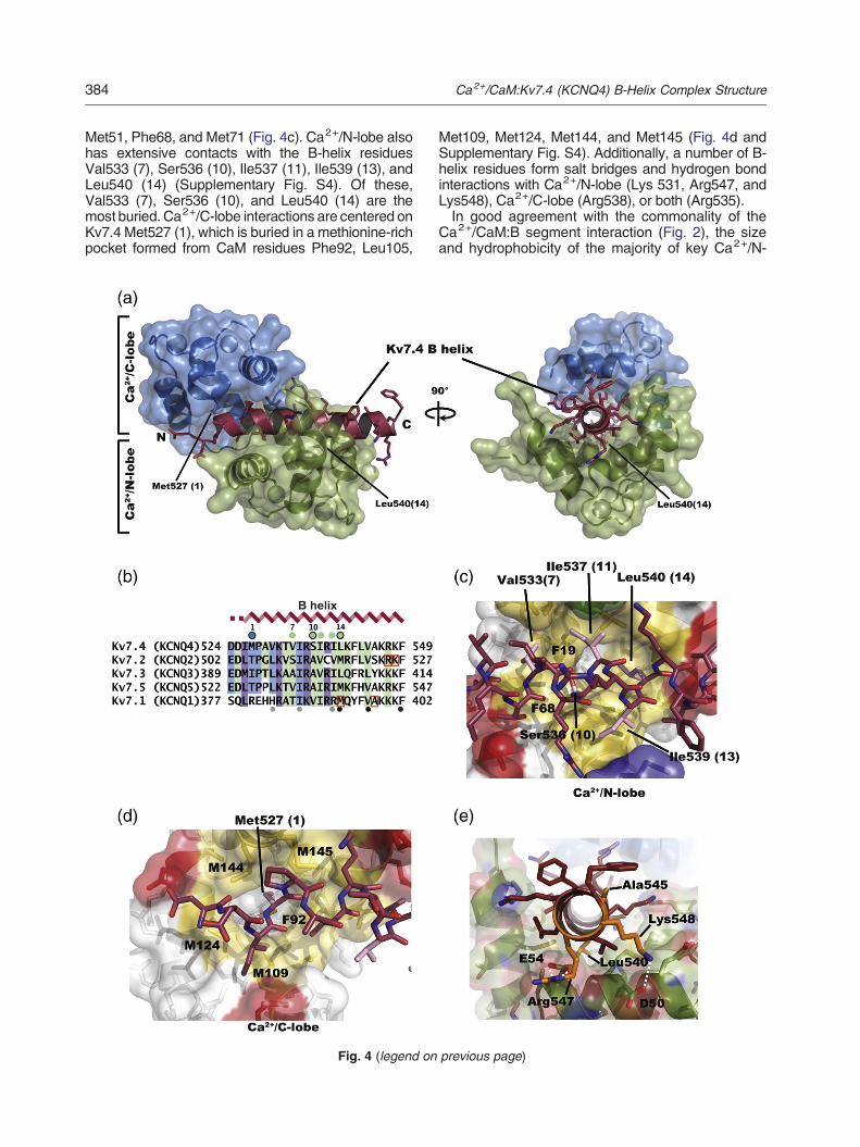
Fig. 4. Ca2+/CaM:Kv7.4 B-helix complex structure. (a) Ca2+
are shown in green and blue, respectively. Kv7.4 B-helix is shoterminal ends of the B-helix are labeled. Axial view is from theindicated. (b) Sequence comparison of Kv7 B-peptide segmeKv7.4 positions contacted by Ca2+/C-lobe, Ca2+/N-lobe, andindicate conserved contact residues. B-helix secondary structuand Ca2+/C-lobe anchors, respectively. Black open circles indthe alignment indicate the two suggested B segment 1–5–10positions of disease mutants in Kv7.1 and Kv7.2 B segments.Ca2+/N-lobe:Kv7.4 B-helix interactions. Ca2+/C-lobe and Ca2+
Hydrophobic, basic, acidic, and polar side chains fromCaM arehelix is shown as sticks and colored firebrick with nitrogen ananchor side chains are colored pink. In both panels, key residKv7.4 B-helix residues are labeled using the three-letter codindicated in parenthesis. (e) Locations of Kv7.1 and Kv7.2 diseletter code, mapped on the Ca2+/CaM:Kv7.4 B-helix structureatoms colored blue and red, respectively. CaM residues areindicate the electrostatic interactions for Arg547–Glu54 (2.1 a
CaM bound to an α-helix formed by Kv7.4 B segmentresidues Asp524-Phe549 (Fig. 4a and Supplemen-tary Fig. S3). Subsequent mass spectroscopy anal-ysis of the crystallization mixture revealed that theQ4BC construct had been truncated at residue 551during the process of crystallization leading to thehexagonal crystal. This deletion essentially removedthe entire C segment (cf. Materials and Methods) andexplains its absence from the structure.The structure revealed that Ca2+/CaM wraps
around the Kv7.4 B-helix in an antiparallel orientationin which the Ca2+/C-lobe and Ca2+/N-lobe bind tothe N-terminal and C-terminal portions of the targethelix, respectively. This interaction buries a substan-tial amount of surface area, 2831 Å2, of which1570 Å2 is hydrophobic. Kv7.4 B-helix residuesMet527 and Leu540 form the most distant Ca2+/CaM anchors and comprise a 1-14 motif. 47,48
Hence, we indicate each of the residues in helix Bby both the residue number and the indicator number1 through 14 with respect to this motif (Fig. 4b). Thisbinding motif was first described in Ca2+/CaMcomplexes with the M13 peptide from sMLCK46,49
and is regarded as a motif that is specific to Ca2+/CaM binding rather than apo-CaM binding.47 Priorstudies had suggested that the B segment had twopossible 1–5–10 Ca2+/CaM binding motifs (Figs. 1band 4b).23 Although some of the residues in theseputative 1–5–10 motifs interact with Ca2+/CaM,neither of these motifs forms the heart of the Ca2+/CaM:Kv7.4 B-helix interaction. Further, the con-served aromatic residues at the (16) and (23)positions (Kv7.4 Phe542 and Phe549), includingthe key (10) position of the second putative 1–5–10motif (Fig. 4b), do not interact with Ca2+/CaM.Of the two Ca2+–CaM lobes, Ca2+/N-lobe makes
more extensive interactions with the B-helix (1764 Å2
versus 1068 Å2 buried surface, for Ca2+/N-lobe andCa2+/C-lobe, respectively). The C-terminal anchor ofthe 1–14 motif, Leu540 (14), resides in a pocketformed by Ca2+/N-lobe residues Phe19, Leu32,
/CaM:Kv7.4 B-helix complex. Ca2+/CaMN-lobe and C-lobewn in firebrick with side chains shown as sticks. N- and C-B-helix C-terminal end. Positions of the 1–14 anchors arents. Residues shaded in blue, green, and purple indicateboth lobes, respectively. Colors on other Kv7 sequencesre is indicated. Blue and green circles indicate Ca2+/N-lobeicate the 1–10–14 motif. Gray and black filled circles undermotifs from Yus-Najera et al.23 Orange squares indicate(c and d) Details of (c) Ca2+/C-lobe:Kv7.4 B-helix and (d)/N-lobe are shown as semitransparent surfaces and sticks.colored yellow, blue, red, and green, respectively. Kv7.4 B-d oxygen atoms colored blue and red, respectively. Kv7.4ues of Ca2+/CaM are labeled using the single-letter code.e followed by the position number within the 1–14 motif,ase mutants, colored orange and indicated using the three-. Ca2+/N-lobe is colored green with nitrogen and oxygenindicated using the single-letter code. White broken linesnd 2.7 Å) and Lys548–Asp50 (4.2 Å).
384 Ca2+/CaM:Kv7.4 (KCNQ4) B-Helix Complex Structure
Met51, Phe68, and Met71 (Fig. 4c). Ca2+/N-lobe alsohas extensive contacts with the B-helix residuesVal533 (7), Ser536 (10), Ile537 (11), Ile539 (13), andLeu540 (14) (Supplementary Fig. S4). Of these,Val533 (7), Ser536 (10), and Leu540 (14) are themost buried. Ca2+/C-lobe interactions are centered onKv7.4 Met527 (1), which is buried in a methionine-richpocket formed from CaM residues Phe92, Leu105,
Fig. 4 (legend on
Met109, Met124, Met144, and Met145 (Fig. 4d andSupplementary Fig. S4). Additionally, a number of B-helix residues form salt bridges and hydrogen bondinteractions with Ca2+/N-lobe (Lys 531, Arg547, andLys548), Ca2+/C-lobe (Arg538), or both (Arg535).In good agreement with the commonality of the
Ca2+/CaM:B segment interaction (Fig. 2), the sizeand hydrophobicity of the majority of key Ca2+/N-
previous page)

385Ca2+/CaM:Kv7.4 (KCNQ4) B-Helix Complex Structure
lobe contact residues (positions 7, 10, 11, and 14)are well conserved among the Kv7 family (Fig. 4b).One Ca2+/N-lobe contact, position (13) (Kv7.4Ile539), is an arginine in Kv7.1, but as this residue isnot completely buried, it seems possible that both thehydrophobic and charged natures of the argininecould beaccommodatedby the bindingmode seen forKv7.4 (Fig. 4d). Besides the 1–14 anchors, the otherhighly buried position isSer536 (10). The γ-hydroxyl ofthis residue is buried in a hydrophobic environment(Supplementary Fig. S5) in which the nearest polarcontact is to the Val533 backbone carbonyl (2.7 Å).Notably, this position is conserved as a small residueamong the Kv7s (Fig. 4b). From this perspective, theKv7 B-helix Ca2+/CaM binding motif could beconsidered a 1–10–14 variation on the 1–14 motif.The observed interaction network is in line with
reported effects of aspartate mutants in the Kv7.2 Bsegment23 in which a Ser→Asp change at position (7)(Kv7.4 V533) interfered with Ca2+/CaM binding,23,34
whereas an Ala→Asp change at position (19) (Kv7.4Ala545), which makes no contacts, showed no effect.In contrast to the high conservation of Ca2+/N-lobeanchor sites, Ca2+/C-lobe contact sites (Fig. 4b) aremore varied. In particular, the identity of the (1)position is not well conserved and is a positivelycharged residue in Kv7.1. Other Ca2+/C-lobe contactpositions, Asp524, Pro528 (2), and Val529 (4), arewell conserved among the neuronal channels,Kv7.2–Kv7.5, but all have changes in charge inKv7.1. It is possible that these differences underlie theapparent difference in the CaM response of Kv7.1versus the other Kv7s.19–21
Location of B-helix disease mutants andregulatory sites
Kv7 channels bear a large number of humanmutations that cause cardiovascular and hearing
Fig. 5. Comparison of the Ca2+/CaM:Kv7.4 B-helix complexthe Ca2+/CaM:Kv7.4 B-helix complex with the M13 peptide of(1CDL).46 The sMLCK complex is shown in light blue and cyangray (sMLCK) spheres. Residues from sMLCK are labeled inboldface. (b–e) Comparisons of the Ca2+/CaM:Kv7.4 B-helixcase, the (1) position or (14) position residue is shown in stickssMLCK, eNOS, and DAPK structures. Kv7.4 residues are labesingle-letter code. In all panels, the Ca2+/CaM from the Kv7.4shown in firebrick. Nitrogen, oxygen, and sulfur atoms are colorC-lobe interactions surrounding the (1) position of the 1–14 motin light blue. sMLCK Trp800 is in cyan. sMLCK Ca2+/CaM Crespectively. (c) Comparison of the (14) position binding poColors are as in (b). sMLCK Leu813 is in cyan. Ca2+/CaM Nrespectively. (d) Comparison of the (1) position binding pockeCaM:Kv7.4 B-helix. The Ca2+/CaM:eNOS complex is shownRMSDs are 1.085 and 1.520 for Cα and all atoms, respectiveCa2+/CaM:DAPK (1YR5) complex and Ca2+/CaM:Kv7.4 B-helitan. Ca2+/CaM C-lobe RMSDs are 0.903 and 1.648 for Cα andpeptides from the Kv7.4, sMLCK, eNOS, and DAPK structureslobe anchors. Black open circles indicate the 1–10–14 motif.
disorders and epilepsies.15,26,50 The conservation ofthe key anchoring residues in the B-helix (Fig. 4b)allows the identification of the positions of a numberof known disease mutants from other Kv7 channelswithin the context of the Ca2+/CaM:Kv7.4 B-helixcomplex structure. Of the four reported B-helixdiseasemutants, three occur at positions that contactthe Ca2+/N-lobe. The Kv7.1 mutant M520R51 corre-sponds to anchor position (14) in the 1–14 motif,Leu540 in Kv7.4 (Fig. 4b and d). In accordance withthe buried nature of this position, the M520R changehas been reported to disrupt both channel functionand CaM binding. 51 Two Kv7.2 mutations,R533Q26,52 and K562N,53 correspond to Kv7.4positions Arg547 and Lys548 (Fig. 4b) that makeelectrostatic interactions with Ca2+/N-lobe residuesGlu54 and Asp50, respectively (Fig. 4e), and woulddisrupt these electrostatic contacts. The fourthreported mutant, Kv7.1 A525T,54 corresponds toKv7.4 Ala545, a residue having no contacts withCa2+/CaM (Supplementary Fig. S4). Because the Bsegment is involved in both apo-CaM and Ca2+/CaMbinding, it is not possible from the structure alone toknow whether the disease mutations act by affectingapo-CaM binding, Ca2+/CaM binding, or both.Nevertheless, the structure provides an importantcontext for further studies addressing this question.In addition to the effects that B-helix mutations
have on function, a pair of studies by Shapiro et al.has shown that N-ethylmaleimide (NEM) treatment,a channel opener that acts by covalent modificationof a cysteine residue that is part of the B segment,blunts the effects of Ca2+/CaM suppression ofKv7.2, Kv7.4, and Kv7.5 currents.29,55 This effectis competitive with CaM action, a result that hassuggested overlapping sites of action.29 Althoughthe cysteine responsible for this NEM effect was notpresent in the crystallized construct, it is just fiveresidues away from the N-terminal end of the B-helix
with other 1–14 Ca2+/CaM complexes. (a) Superposition ofsmooth muscle myosin light chain kinase (cyan), sMLCK. Calcium ions are indicated as white (Kv7.4 complex) anditalics. Residues from the Kv7.4 complex are labeled incomplex with other 1–14 Ca2+/CaM complexes. In eachand labeled. Residues in italics and surfaces come from theled in boldface. Ca2+/CaM residues are indicated using thecomplex is shown in marine and the Met527 side chain ised blue, red, and yellow, respectively. (b) View of the Ca2+/if for the Ca2+/CaM:sMLCK complex. Ca2+/CaM:sMLCK is-lobe RMSDs are 0.568 and 1.375 for Cα and all atoms,cket for Ca2+/CaM:sMLCK and Ca2+/CaM:Kv7.4 B-helix.-lobe RMSDs are 0.914 and 1.636 for Cα and all atoms,t for the Ca2+/CaM:eNOS complex (1N1W)57 and Ca2+/in orange. eNOS Phe465 is in olive. Ca2+/CaM C-lobe
ly. (e) Comparison of the position 1 binding pocket for thex.58 Ca2+/CaM:DAPK is shown in white. DAPK Trp305 is inall atoms, respectively. (f) Sequence comparison of target. Blue and green circles indicate Ca2+/N-lobe and Ca2+/C-

Fig. 5 (legend on previous page)
386 Ca2+/CaM:Kv7.4 (KCNQ4) B-Helix Complex Structure

387Ca2+/CaM:Kv7.4 (KCNQ4) B-Helix Complex Structure
(Supplementary Fig. S1). Examination of the struc-ture suggests that given the covalent constraints ofthe polypeptide backbone, this cysteine should benear enough to contact Ca2+/C-lobe and thusaccount for the interfering effects of NEM on Kv7function, CaM binding, and the antagonistic rela-tionship between NEM and CaM.29 Importantly,both Kv7.1 and Kv7.3, which are NEM insensitive,56
lack a cysteine at the equivalent position29 (Sup-plementary Fig. S1).
Comparison of the Ca2+/CaM:Kv7.4 B-helixstructure with canonical Ca2+/CaM:1–14motif structures
The original analysis of the B segment sequencepredicted two 1–5–10 motifs (Fig. 4b),23 neither ofwhich are used in Ca2+/CaM binding, but did notpredict the 1–14 motif observed in the Ca2+/CaM:Kv7.4 B-helix structure (Fig. 4a–d). Hence, we wereinterested to compare the Ca2+/CaM:Kv7.4 B-helixcomplex to other Ca2+/CaM:1–14 motif complexes toexamine points of commonality and differences.Structure comparison with the Ca2+/CaM:sMLCKpeptide structure46 that originally defined the 1–14motif showed that faithful to the 1–14 motif, the 14residue spacing of anchor positions, Trp800 (1) andLeu813 (14) for sMLCK and Met527 (1) and Leu540(14) for the Kv7.4 B-helix, places the Kv7.4 B-helixanchor positions onopposite facesof theα-helixwherethey can independently engage the two Ca2+/CaMlobes (Fig. 5a). Notably, comparison of the structuresreveals that theKv7.4B-helix is longer than the sMLCKhelix by approximately three turns and makes moreCa2+/N-lobe contacts. Although the overall Cα RMSDfor both complexes is 2.59 Å2, indicating some globaldifferences that are due in part to the longer targetpeptide in the Kv7.4 structure, the conformations of theindividual Ca2+/CaM lobes are very similar and haveCα RMSDs of 0.91 Å2 and 0.57 Å2 for Ca2+/C-lobeand Ca2+/N-lobe, respectively.Examination of the local environments of the (1)
and (14) anchor positions shows that these residuesbind pockets formed from the same Ca2+/CaM loberesidues in both the Kv7.4 and sMLCK structures(Fig. 5b and c). The (1) position, Kv7.4 Met527 andsMLCK Trp800, binds to a pocket formed by Ca2+/C-lobe residues Phe92, Met109, Met124, Met144, andMet145 (Fig. 5b). Notably, there is a rather largedifference in the way the (1) position residuesoccupy the Ca2+/C-lobe pocket. Kv7.4 Met527 ismuch closer to the wall of the pocket made by CaMresidue Phe92, whereas sMLCK Trp800 is morecentral to the pocket. In concert with the changesseen at the Kv7.4 (1) position anchor, CaM residueMet144 adopts a side-chain rotamer that reduces thepocket size relative to the sMLCK complex. Thisconformational difference is consistent with thechange from a large aromatic anchor, Trp800, to a
smaller and less constrained hydrophobic residue,Met527 (Fig. 5b). In contrast, the Kv7.4 and sMLCK(14) position anchors of residues Leu813 andLeu540, respectively, occupy a pocket formed byCa2+/N-lobe residues Phe68, Met71, Leu32, andPhe19 that has similar features in both structures(Fig. 5c).To see whether the differences in the 1–14
interaction were particular to the comparison withthe classic sMLCK interaction or more specificallyrelated to the Kv7.4 complex, we conducted a surveyto identify all current known 1–14 complexes. Thissearch identified 11 Ca2+/CaM:1–14 complexes(Supplementary Table 1). The difference in theplacement of the (1) position residue and theshape of the pocket around Ca2+/CaM Met144remain vis-à-vis the Kv7.4 B-helix interactions withrespect to all complexes from this set. Thesedifferences are exemplified by the Ca2+/CaM:endo-thelial nitrogen oxide synthase (eNOS) peptidecomplex [Protein Data Bank (PDB) code: 1NIW]57
(Fig. 5d) and a Ca2+/CaM complex with anautoregulatory domain peptide from the death-associated protein kinase DAPK (PDB code:1YR5)58 (Fig. 5e). The structure having a non-aromatic at position (1), Leu in human inducible nitricoxide synthase (iNOS)[PDB code:3HR4],59 has aCa2+/C-lobe pocket similar to the other structuresbut has a very different pitch of the target helix, whichdeviates from the other complexes by ~30° andresults in a placement of the (1) position residuemidway between the aromatic position from sMLCKTrp800 and the position occupied by Kv7.4 Met 527.These observations provide additional support forthe idea that the distinctive structural features wenote in the Kv7.4 complex relative to other 1–14complexes result from accommodation of a non-aromatic at the (1) position. The presence of the non-aromatic (1) position residue further stands out asaromatic residues occupy the Ca2+/C-lobe pocket ina large number of Ca2+/CaM complexes.60 In light ofthis analysis, and considering that the canonicalpattern of residues at the 1 and 14 positions isdefined by the presence of one of five hydrophobicresidues (Phe/Ile/Leu/Val/Trp) at the 1 and 14positions,47 it is clear how the 1–14 pattern couldbe missed. Notably, none of the Kv7s have acanonical residue at the (1) position (Fig. 4b).Undoubtedly, the well-known adaptability of CaMstands behind this observation and underscores theimportance of experimental determination of Ca2+/CaM:peptide structures.
Discussion
The ability to open at membrane potentials that arebelow those required to initiate action potentialstogether with a lack of inactivation sets Kv7 channels

388 Ca2+/CaM:Kv7.4 (KCNQ4) B-Helix Complex Structure
in a key position to control electrical excitability inboth the heart and nervous system.4,10 Hence, theyare subject to a wide range of regulatorypathways;3,4,6 have been identified as molecules inwhich misfunction underlies a variety of disorders inthe cardiovascular, hearing, vestibular, and nervoussystems;2,14,15 and are attractive targets for a rangeof hyperexcitability disorders.11–14 CaM is a keyregulator and affects the function of all Kv7 subtypesthrough interactions made in both its apo- andcalcium-bound forms with the C-terminal tail of thechannel.19–21,29,31,32 Although the importance ofCaM for Kv7 modulation has been long appreciated,the details of the interactions of CaMwith the channelhave remained unclear. Our data provide newbiochemical and structural information that directlybear on the question of how CaM interacts with thechannel in both apo- and calcium-bound forms.In agreement with previous evidence,22,23 we
found that apo-CaM binding to the Kv7.4 C-terminaltail strictly requires the presence of both the A and Bsegments (Figs. 1c and d and 2b). Importantly, weshow directly by sedimentation equilibrium studiesthat the interaction between apo-CaM and the Kv7.4construct spanning the A–D segments (Q4AD) has astoichiometry of 4:4 (Fig. 1d), a result that isconsistent with prior sedimentation equilibrium18
and chemical cross-linking studies of a similarKv7.1 C-terminal domain construct.20 This situationis striking as more than 100 amino acids separatethe A and B segments. Such a large insertionprecludes a contiguous binding site as first noted byYus-Najera et al.23 and suggests that apo-CaM mayuse a bidentate binding mode to interact with thesenon-contiguous sites. There are a number of pre-cedents that indicate that CaM is able to make thissort of bridged interaction,61–63 most notably, thosefrom the interaction of CaM and the intracellulardomains of small-conductance (SK) potassiumchannel intracellular domains.62,63 Although thereare no structural examples of a complete bridgedstructure formed by apo-CaM, such an interactionwould be very much in line with the ability of theindependent apo-CaM lobes to engage targets.61,64–66
It should be noted that our data do not distinguishwhether the apo-CaM interaction with the A and Bsegments is intrasubunit or intersubunit. Prior studiesof the oligomerization behavior of a Kv7.1 constructspanning just the A–B segments indicated thepresence of a 1:1 complex in both the presenceand absence of calcium and suggest that the apo-CaM cross bridge is intrasubunit.18 Nevertheless, inthe context of the full-length channel, both configu-rations are reasonable possibilities given the prox-imity of the intracellular domains, which areconstrained by both the interactions of the pore-forming regions in the membrane at one end andthose from the D segment/A domain tail18,24 at theother. Thus, based on the available data, we favor the
interpretation that the apo-CaM bridging occurswithin a single subunit. Nevertheless, further testsin the context of the full-length channel will berequired to address this issue definitively.In contrast to the complex interactions made by
apo-CaM, both our structural and biochemical dataindicate that Ca2+/CaM requires only the B segment(Figs. 3c and 4). This interaction involves a classic,contiguous Ca2+/CaM binding site formed by asingle α-helix (Fig. 4). Ca2+/CaM wraps around theB segment helix in an antiparallel orientation thatuses a variation of the well-described 1-14 bindingmotif47,48 in which the two most distant anchoringpositions are Met527 and Leu540. This bindingmode contrasts with the original suggestion thatCa2+/CaM might interact via 1–5–10 motifs found inthe B segment23 (Fig. 4b) and has a pattern of buriedand exposed residues that matches the reportedeffects of aspartate mutants on CaM binding.23
Consistent with the observation that constructsbearing the B-helix from all Kv7 isoforms are able tobind Ca2+/CaM (Fig. 2f), the main determinants ofthis interaction are conserved throughout the Kv7family (Fig. 4b). One feature that stands out fromother 1-14 Ca2+/CaM target structures is that ratherthan an aromatic residue at the 1 position, the Kv7.4B-helix has a methionine. This substitution of anaromatic for a long hydrophobic side chain is readilyaccommodated by a small rearrangement in theCa2+/C-lobe binding pocket. Interestingly, all Kv7isoforms have a non-aromatic residue at the (1)position of the 1-14 motif (Fig. 4b).Functional studies indicate that the neuronal
channels Kv7.2, Kv7.4, and Kv7.5 share a commonmechanism of calcium-induced current suppressionthat involves CaM.19,29 However, this property is notshared by all members of the family, and mostnotably, the calcium sensitivity of Kv7.1 has been apoint of controversy.17 Some studies have reportedCaM involvement in calcium-dependent activation ofKv7.1 channels,20,21,67 whereas other studies havefailed to find this response.29 Because the modula-tion of Kv channels is complex,3,4,6 it is possible thatthe reported differences are due to the effects ofother CaM-dependent pathways, such as the oneinvolved in S-nitrosylation signals.32,67 Regardless,it is clear that Kv7.1 behaves differently with respectto CaM than the neuronal Kv7s. It is notable thateven though Ca2+/CaM binds C-terminal constructsbearing the B-helix regardless of the Kv7 isoformtested (Fig. 2f), there is a substantial divergence inthe residues that would form the Ca2+/C-lobecontacts in Kv7.1 (KCNQ1), including a number ofcharged residues. Such differencesmay underlie thefunctional differences between the CaM-dependentresponses of Kv7.1 versus the neuronal Kv7s.Additionally, there are differences at the (1) positionthat could play a role in tuning the CaM responses ofthe neuronal Kv7s in a subunit-specific manner. Our

389Ca2+/CaM:Kv7.4 (KCNQ4) B-Helix Complex Structure
new structural data should help to guide furtherinvestigation of this phenomenon.Studies of the calcium dependency of CaM
interaction with Kv7.2, Kv7.3, and Kv7.4 C-terminaltails indicate that both apo-CaM and Ca2+/CaMbind.22,23,33 The presence of apo-CaM binding sitesin the A22,23 and B23 segments of Kv7.2, togetherwith the observations that the A/B region binds apo-CaM better than the individual segments23 and thatCa2+/CaM favors binding to the B segment in Kv7.2–Kv7.5,19 matches well with our observations of theinteraction seen using purified components. Anumber of previous proposals have suggested across-bridged binding mode for apo-CaM.22,23 Con-sideration of our new data together with this priorwork leads us to propose an extension of the CaMmodulation hypothesis to include a conformationalswitch between the apo-CaM and Ca2+/CaM statesthat involves a change from a cross-bridged form inthe apo-CaM state to a classic Ca2+/CaM–helixinteraction in the calcium-bound form as seen in ourcrystal structure of the Ca2+–CaM/Kv7.4 B-helixcomplex (Fig. 6). Because one of the parts of thebidentate binding site, the A segment, is very nearthe likely end of the pore-lining transmembranesegment S6, it is reasonable to posit that such aconformational change would affect channel func-tion. For example, the interaction of apo-CaM withthe A segment may affect S6 in a way that favorschannel opening and, in contrast, when CaMswitches to the Ca2+–CaM site on the B-helix, lossof the A segment interaction causes the current to be
Fig. 6. Model for the calcium-dependent interactions of neuproposed calcium-dependent switch from the cross-bridged a(right). CaM lobes are indicated as green (C-lobe) and blue (N-noted that the exact binding mode for the apo-CaM state is notomitted from the cartoon for clarity.
suppressed. This hypothesis would explain onemechanism for calcium-dependent suppression ofneuronal Kv7s. As mentioned above, the responseof Kv7.1 to calcium is complicated and unlike that ofthe neuronal Kv7s. Given that the Kv7.1 B-helixbinds Ca2+/CaM, even though the Ca2+/C-lobe siteis quite different, Kv7.1 may use some variant of thisproposal.The idea that there would be a structural transition
between a bidentate apo-CaM clamp to a calcium-bound form in which the Ca2+/CaM interactions arerestricted to the B-helix fits well with the observed,antagonistic effects of NEM alkylation at a B-helixcysteine29,55 and with the data showing that thecalcium sensitivity of the N-lobe is crucial.29 Such amodel would also be consistent with the knownimportance of theC-terminal tail region that spans theA and B segments for modulation of Kv7 channels byphosphatidylinositol 4,5-bisphosphate68 and S-nitrosylation32 and suggests that these variousinputs can be coupled in a way that makes the A–Bregion a signal integration domain. In this regard, it isstriking that the sequence that links the A and Bsegments varies among the Kv7 isoforms in com-parison to the A, B, C, and D segments (Fig. S1) andis the site of Kv7.4 splice variation that affectsfunction.69
Overall, our data are in agreement with theexistence of two CaM binding sites on Kv7 channels,one Ca2+ independent and one Ca2+ dependentthat confers Ca2+ sensitivity to the channel.17 Thestructure of the Ca2+/CaM:Kv7.4 B-helix complex
ronal Kv7 channels and CaM. Cartoon model depicts thepo-CaM binding state (left) to the Ca2+/CaM bound statelobe). Only two of the four subunits are shown. It should beknown. Sequences following the D-helix/A domain tail are

390 Ca2+/CaM:Kv7.4 (KCNQ4) B-Helix Complex Structure
shows that a number of Kv7 disease mutations fall atpositions that make direct contacts with Ca2+/CaM.However, as the B segment is also involved in apo-CaM binding22,23 and interactions with CaM mayhave important effects in channel folding andtrafficking,20,21,70,71 such mutations may interferewith multiple layers of CaM control of the channel.The structure presented here provides a key contextfor further dissection of the complex effects of Kv7disease mutants. Finally, the novel variation weobserve in the classic 1–14 Ca2+/CaM binding modeunderscores the exceptionally adaptive nature ofCa2+/CaM for target recognition.
Materials and Methods
Cloning, expression, and purification
Fragments of human Kv7.1–Kv7.5 (KCNQ1–KCNQ5)C-terminal domain were subcloned into either pSV272 (N.Pokala and T.M. Handel, unpublished results) or pET28-HMT vectors (Novagen) using NarI/HindIII or NdeI/HindIIIsites. Both vectors contain the sequence for a hexahisti-dine tag followed by an MBP and a TEV protease sitebefore the gene of interest. The only difference betweenthe two vectors is that the NarI site following the TEVcleavage site in pSV272 is replaced by an NdeI site inpET28-HMT. Full-length native CaM and mutants werecloned into pEGST without an affinity tag as previouslydescribed.72
Kv7 fragments co-expressed with CaM in E. coliBL21(DE3)pLysS grown in 2YT media at 37 °C for 3 to8 h. Cells containing constructs containing B–D segmentswere lysed by sonication in buffer containing 100 mM Tris(pH 8.8), 200 mM KCl, 10% sucrose, 1 mM MgCl2, 1 mMPMSF, 20 μg ml− 1 lysozyme, and 25 μg ml− 1 DNase I.1 mM CaCl2 or 1 mM EGTA was also added before celllysis depending on whether calcium-bound or calcium-freeforms were the subject of purification. Lysed cells werecentrifuged at 25,000g for 30 min to remove insolublematerial. For constructs spanning the A–D segments, the10% sucrose in the lysis buffer was replaced by 10%glycerol and the cell lysis was performed using anEmulsiflex C-5 homogenizer (Avestin).For all complexes, the soluble fraction containing the
Kv7 fragment and CaM was purified on a Poros20MC(PerSeptive Biosystems) column equilibrated in buffer A(250 mM KCl, 1 mM CaCl2, and 10 mM Hepes buffer,pH 7.4). When EGTA was used in the lysis buffer, a bufferof identical composition but lacking CaCl2 replaced bufferA. After loading the sample, the column was washed in twosteps: first using 3 column volumes of buffer A and thenusing 3 column volumes of buffer A plus 30 mM imidazoleto remove weakly bound contaminants. Bound protein wasthen eluted using an imidazole gradient from 30 mM to300 mM over 6 column volumes. After repeated spinconcentration using a 50-kDa-cutoff Amicon Ultra-15concentrator (Millipore) and dilution with buffer A toremove imidazole, the eluate from the Poros20MC columnwas loaded on to an amylose (New England Biolabs)column and washed with buffer A. The bound protein was
then eluted in buffer A plus 10 mM maltose. The purifiedprotein was digested with histidine-tagged TEV protease73
in the amylose column elution buffer overnight at roomtemperature and was subsequently passed over aPoros20MC and amylose column run in buffer A plus1 mM CaCl2 to remove the protease and uncleavedmaterial. Flow through was collected and concentratedusing an Amicon Ultra-15 concentrator with a 10-kDacutoff. During this process, the buffer was exchanged to abuffer of 10 mM KCl, 1 mM CaCl2, and 10 mM Tris,pH 8.8, through a cycle of dilution and concentration. Thesample was further purified using Resource Q or HiLoadQ(GE Healthcare) anion-exchange column and a gradientfrom 100 mMKCl to 350 mMKCl over 15 column volumes.This step is important for removal of residual His-MBP. Theconcentration of the purified complex was determined bymeasuring the absorption at 280 nm.74
Pull-down experiments
Various HMT-tagged Kv7 constructs were co-expressedwith CaM as described above. Cells were lysed bysonication and centrifuged at 25,000g for 30 min. Thesupernatant (500 μl) was incubated with the same amountof buffer A (250 mM KCl, 1 mM CaCl2 or EGTA, and10 mM Hepes, pH 7.4) and 500 μl of amylose resin andincubated for 30 min at 4 °C. The unbound proteins werewashed out using three washes with 5 ml of buffer A.Bound proteins were eluted by buffer A plus 10 mMmaltose and analyzed by SDS-PAGE.
Crystallization and data collection
The Ca2+/CaM:Kv7.4 B–C complex, containing Kv7.4residues 522–593, yielded crystals suitable for structuralstudies grown using hanging drop vapor diffusion from adrop containing equal volumes (1 μl each) of purifiedprotein (~20 mg/ml) and a reservoir solution of 1.6 Mammonium sulfate, 0.2 M sodium citrate (pH 6.0), 0.1 Msodium tartrate, and 4% isopropanol. Rectangular rod-shaped crystals appeared in 1–3 weeks at room temper-ature. Crystals having this morphology diffracted to ~2.8 Åbut suffered from high mosaicity, anisotropy, and a largeunit cell. One crystal having hexagonal plate morphologyappeared in the same conditions subsequent to a round ofcrystal harvesting from the well in which it grew. Thiscrystal diffracted sufficiently to be used for structuredetermination. Mass spectrometry analysis revealed thatthe protein in this particular well had been cleaved C-terminal to residue 551, leaving only the B segment. Incontrast, mass spectroscopy analysis of wells containingrod-shaped crystals showed the presence of the completeKv7.4 BC construct. Diffraction data were collected fromfrozen crystals at beamline 12-2 (Stanford SynchrotronRadiation Lightsource) and were indexed and processedin the space group P6522 using MOSFLM and SCALA inthe CCP4 package.75
Structure determination
The structure was determined by a molecular replace-ment search using the N-lobe without calcium ions from the
391Ca2+/CaM:Kv7.4 (KCNQ4) B-Helix Complex Structure
sMLCK complex (PDB code: 1CDL) using PHASER.76
Electron density indicating the Kv7.4 B-helix and Ca2+/C-lobe was readily visible in the initial maps. Structure was builtby iterative cycles of manual building and refinement usingREFMAC77 and PHENIX.78 TLS refinement using threegroups (Ca2+/N-lobe, Ca2+/C-lobe, and Kv7.4 helix) wasadded in the last few cycles. Residues 524–549 of Kv7.4 andall of CaM except for a few residues at the N-terminus and inthe loop interdomain linker (residues 78–82) were resolved.Structural quality was evaluated using MolProbity.79
SEC and MALS-RI analysis
Purified Kv7.4 C-terminal domain:CaM complexes wererun through either a Superdex200 10/300 GL or aSuperdex75 HR column depending on the size of thecomplex. The columns were calibrated using at least fourstandard protein molecular mass markers (HMW andLMW calibration kits, GE Healthcare). The running buffercontained 250 mM KCl, 1 mM CaCl2 or 1 mM EDTA, and10 mM Hepes, pH 7.4. When EDTA was used, EDTA wasadded to the sample to a final concentration of 5 mM andthe sample was incubated for 10 min before the run.MALS-refractive index (RI) analysis41,42 was performed
usingminiDAWN™ TREOS (LS) and Optilab® T-rEX™ (RI)(Wyatt Technology) for simultaneous detection of UV, RI,andMALS signals run in linewith a Superdex200 10/300GLor Superdex75 HR column in a buffer of 200 mMKCl, 1 mMCaCl2, and 10 mM Hepes, pH 7.4, calibrated using 0.2 mgbovine serum albumin as an internal standard. ASTRA® 6software package (Wyatt Technology) was used to computeprotein molecular mass (Mw) and protein monodispersity(Mw/Mn) from the UV, RI, and MALS outputs.
Equilibrium sedimentation
Equilibrium sedimentation experiment was carried out at4 ºC using a Beckman Optima XL-A analytical ultracentri-fuge (Beckman Coulter). For the CaM:Q4AD complex, thebuffer contained 250 mM KCl, 1 mM EDTA, and 10 mMTris, pH 8.0. For the CaM:Q4BD complex, the buffercontained 1 mM CaCl2 instead of EDTA. The purifiedprotein was dialyzed overnight in the final buffer. Thedialysis buffer was then used to dilute the protein to theintended concentration and as the reference cell sample.The data processing followed previously describedprocedures.24 The molecular mass of the complex wasobtained through a single exponential fit to the distributionof the complex over the radius.
Survey of Ca2+/CaM 1–14 structures
Structures of the Ca2+/CaM:Kv7.4 B-helix (4GOW) andCa2+/CaM:sMLCK (1CDL) 1–14 complexes were used assearch queries using the DALI server80 to identify similarCa2+/CaM complexes. Redundant structures were re-moved from the comparison and the remaining structureswere aligned and manually examined to identify thoseusing a 1–14 binding motif.
Accession numbers
Coordinates and structure factors have been depositedin the PDB with the following accession code: 4GOW.
Acknowledgements
This work was supported by grants from theNational Institutes of Health (R01 DC007664) andtheAmericanHeart Association (0740019N) toD.L.M.and by a European Molecular Biology Organizationlong-term fellowship to A.T. We thank Rachel E. Gateand Susan Shim for excellent technical support, FelixFindeisen for helpful comments on the manuscript,and members of the Minor laboratory for supportthroughout these studies.D.L.M. is anAmericanHeartAssociation Established Investigator.
Author Contributions. Q.X., A.C, A.T., andD.L.M. conceived the study. Q.X, A.C., and A.T.performed the experiments and analyzed data.D.L.M. analyzed data and provided guidance andsupport throughout. Q.X., A.C., A.T., and D.L.M.wrote the paper.
Conflict of Interest. The authors declare noconflict of interest.
Supplementary Data
Supplementary data to this article can be foundonline at http://dx.doi.org/10.1016/j.jmb.2012.11.023
Received 21 August 2012;Received in revised form 14 November 2012;
Accepted 15 November 2012Available online 23 November 2012
Keywords:Kv7 (KCNQ) voltage-gated potassium channel;
calmodulin;X-ray crystallography;
SEC-MALS;channel modulation
†Q.X. and A.C. contributed equally to this work.
Present address: Q. Xu, Institute of Nuclear-AgriculturalSciences, Zhejiang University, 268 Kaixuan Road,
310029 Hangzhou, China.
Abbreviations used:CaM, calmodulin; MBP, maltose binding protein;
TEV, tobacco etch virus; EDTA, ethylenediaminetetraaceticacid; EGTA, ethylene glycol tetraacetic acid;
GST, glutathione S-transferase; SEC, size-exclusionchromatography; MALS, multiangle light scattering;
RI, refractive index; sMLCK, smooth muscle myosin lightchain kinase; NEM, N-ethylmaleimide;
PDB, Protein Data Bank.

392 Ca2+/CaM:Kv7.4 (KCNQ4) B-Helix Complex Structure
References1. Gutman, G. A., Chandy, K. G., Adelman, J. P., Aiyar,
J., Bayliss, D. A., Clapham, D. E. et al. (2003).International Union of Pharmacology. XLI. Compendi-um of voltage-gated ion channels: potassium chan-nels. Pharmacol. Rev. 55, 583–586.
2. Jentsch, T. J. (2000). Neuronal KCNQ potassiumchannels: physiology and role in disease. Nat. Rev.Neurosci. 1, 21–30.
3. Jespersen, T., Grunnet, M. & Olesen, S. P. (2005).The KCNQ1 potassium channel: from gene tophysiological function. Physiology (Bethesda), 20,408–416.
4. Delmas, P. & Brown, D. A. (2005). Pathwaysmodulating neural KCNQ/M (Kv7) potassium chan-nels. Nat. Rev. Neurosci. 6, 850–862.
5. Wang, H.-S., Pan, Z., Shi, W., Brown, B. S., Wymore,R. S., Cohen, I. S. et al. (1998). KCNQ2 and KCNQ3potassium channel subunits: molecular correlates ofthe M-Channel. Science, 282, 1890–1893.
6. Hernandez, C. C., Zaika, O., Tolstykh, G. P. &Shapiro, M. S. (2008). Regulation of neural KCNQchannels: signalling pathways, structural motifs andfunctional implications. J. Physiol. 586, 1811–1821.
7. Barhanin, J., Lesage, F., Guillemare, E., Fink, M.,Lazdunski, M. & Romey, G. (1996). KVLQT1 and IsK(minK) proteins associate to form the IKs cardiacpotassium current. Nature, 384, 78–80.
8. Sanguinetti, M. C., Curran, M. E., Zou, A., Shen, J.,Spector, P. S., Atkinson, D. L. & Meating, M. T. (1996).Coassembly of KVLQT1 and minK (IsK) proteins to formcardiac IKs potassium channel. Nature, 384, 80–83.
9. Wang, Q., Curran, M. E., Splawski, I., Burn, T. C.,Millholland, J. M., VanRaay, T. J. et al. (1996).Positional cloning of a novel potassium channelgene: KVLQT1 mutations cause cardiac arrhythmias.Nat. Genet. 12, 17–23.
10. Brown, D. A. & Passmore, G. M. (2009). Neural KCNQ(Kv7) channels. Br. J. Pharmacol. 156, 1185–1195.
11. Barrese, V., Miceli, F., Soldovieri, M. V., Ambrosino,P., Iannotti, F. A., Cilio, M. R. & Taglialatela, M. (2010).Neuronal potassium channel openers in the manage-ment of epilepsy: role and potential of retigabine. Clin.Pharmacol. 2, 225–236.
12. Fritch, P. C., McNaughton-Smith, G., Amato, G. S.,Burns, J. F., Eargle, C. W., Roeloffs, R. et al. (2010).Novel KCNQ2/Q3 agonists as potential therapeuticsfor epilepsy and neuropathic pain. J. Med. Chem. 53,887–896.
13. Wickenden, A. D. & McNaughton-Smith, G. (2009).Kv7 channels as targets for the treatment of pain.Curr. Pharm. Des. 15, 1773–1798.
14. Soldovieri, M. V., Miceli, F. & Taglialatela, M. (2011).Driving with no brakes: molecular pathophysiology ofKv7 potassium channels. Physiology (Bethesda), 26,365–376.
15. Maljevic, S., Wuttke, T. V. & Lerche, H. (2008).Nervous system KV7 disorders: breakdown of asubthreshold brake. J. Physiol. 586, 1791–1801.
16. Yu, F. H., Yarov-Yarovoy, V., Gutman, G. A. &Catterall, W. A. (2005). Overview of molecular re-lationships in the voltage-gated ion channel super-family. Pharmacol. Rev. 57, 387–395.
17. Haitin, Y. & Attali, B. (2008). The C-terminus of Kv7channels: a multifunctional module. J. Physiol. 586,1803–1810.
18. Wiener, R., Haitin, Y., Shamgar, L., Fernandez-Alonso, M. C., Martos, A., Chomsky-Hecht, O. et al.(2008). The KCNQ1 (Kv7.1) COOH terminus, amultitiered scaffold for subunit assembly and proteininteraction. J. Biol. Chem. 283, 5815–5830.
19. Gamper, N. & Shapiro, M. S. (2003). Calmodulinmediates Ca2+-dependent modulation of M-type K+
channels. J. Gen. Physiol. 122, 17–31.20. Ghosh, S., Nunziato, D. A. & Pitt, G. S. (2006).
KCNQ1 assembly and function is blocked by long-QTsyndrome mutations that disrupt interaction withcalmodulin. Circ. Res. 98, 1048–1054.
21. Shamgar, L., Ma, L., Schmitt, N., Haitin, Y., Peretz, A.,Wiener, R. et al. (2006). Calmodulin is essential forcardiac IKS channel gating and assembly: impairedfunction in long-QTmutations.Circ.Res.98, 1055–1063.
22. Wen, H. & Levitan, I. B. (2002). Calmodulin is anauxiliary subunit of KCNQ2/3 potassium channels. J.Neurosci. 22, 7991–8001.
23. Yus-Najera, E., Santana-Castro, I. & Villarroel, A.(2002). The identification and characterization of anoncontinuous calmodulin-binding site in noninacti-vating voltage-dependent KCNQ potassium channels.J. Biol. Chem. 277, 28545–28553.
24. Howard, R. J., Clark, K. A., Holton, J. M. &Minor, D. L.,Jr. (2007). Structural insight into KCNQ (Kv7) channelassembly and channelopathy. Neuron, 53, 663–675.
25. Xu, Q. & Minor, D. L., Jr. (2009). Crystal structure of atrimeric form of the K(V)7.1 (KCNQ1) A-domain tailcoiled-coil reveals structural plasticity and contextdependent changes in a putative coiled-coil trimeriza-tion motif. Protein Sci. 18, 2100–2114.
26. Soldovieri, M. V., Miceli, F., Bellini, G., Coppola, G.,Pascotto, A. & Taglialatela, M. (2007). Correlating theclinical and genetic features of benign familial neonatalseizures (BFNS) with the functional consequences ofunderlying mutations. Channels (Austin), 1, 228–233.
27. Gardiner, M. (2006). Molecular genetics of infantilenervous system channelopathies. Early Hum. Dev.82, 775–779.
28. Singh, N. A., Westenskow, P., Charlier, C., Pappas,C., Leslie, J., Dillon, J. et al. (2003). KCNQ2 andKCNQ3 potassium channel genes in benign familialneonatal convulsions: expansion of the functional andmutation spectrum. Brain, 126, 2726–2737.
29. Gamper, N., Li, Y. & Shapiro, M. S. (2005). Structuralrequirements for differential sensitivity of KCNQ K+
channels to modulation by Ca2+/calmodulin.Mol. Biol.Cell, 16, 3538–3551.
30. Zaika, O., Tolstykh, G. P., Jaffe, D. B. & Shapiro, M. S.(2007). Inositol triphosphate-mediated Ca2+ signalsdirect purinergic P2Y receptor regulation of neuronalion channels. J. Neurosci. 27, 8914–8926.
31. Roden, D. M. (2006). A new role for calmodulin in ionchannel biology. Circ. Res. 98, 979–981.
32. Asada, K., Kurokawa, J. & Furukawa, T. (2009).Redox- and calmodulin-dependent S-nitrosylation ofthe KCNQ1 channel. J. Biol. Chem. 284, 6014–6020.
33. Bal, M., Zaika, O., Martin, P. & Shapiro, M. S. (2008).Calmodulin binding to M-type K+ channels assayed byTIRF/FRET in living cells. J. Physiol. 586, 2307–2320.

393Ca2+/CaM:Kv7.4 (KCNQ4) B-Helix Complex Structure
34. Gomez-Posada, J. C., Aivar, P., Alberdi, A., Alaimo,A., Etxeberria, A., Fernandez-Orth, J. et al. (2011).Kv7 channels can function without constitutive cal-modulin tethering. PLoS One, 6, e25508.
35. Kharkovets, T., Hardelin, J. P., Safieddine, S.,Schweizer, M., El-Amraoui, A., Petit, C. & Jentsch,T. J. (2000). KCNQ4, a K+ channel mutated in a formof dominant deafness, is expressed in the inner earand the central auditory pathway. Proc. Natl Acad.Sci. USA, 97, 4333–4338.
36. Kubisch, C., Schroeder, B. C., Friedrich, T., Lütjohan,B., El-Amaraoui, A., Marlin, S. et al. (1999). KCNQ4, anovel potassium channel expressed in sensory outerhair cells, is mutated in dominant deafness. Cell, 96,437–446.
37. Heidenreich, M., Lechner, S. G., Vardanyan, V.,Wetzel, C., Cremers, C. W., De Leenheer, E. M.et al. (2012). KCNQ4 K(+) channels tune mechano-receptors for normal touch sensation in mouse andman. Nat. Neurosci. 15, 138–145.
38. Minor, D. L., Jr. (2007). The neurobiologist's guide tostructural biology: a primer on why macromolecularstructure matters and how to evaluate structural data.Neuron, 54, 511–533.
39. Laue, T. M. (1995). Sedimentation equilibrium as athermodynamic tool. Methods Enzymol. 259, 427–453.
40. Laue, T. M., Bhairavi, B. D., Ridgeway, T. M. &Pelletier, S. L. (1992). Computer-aided interpretationof analytical sedimentation data for proteins. InAnalytical Ultracentrifugation in Biochemistry andPolymer Science (Harding, S. E., Rowe, A. J. &Horton, J. C., eds), Royal Society of Chemistry,Cambridge, UK.
41. Folta-Stogniew, E. & Williams, K. R. (1999). Determi-nation of molecular masses of proteins in solution:implementation of an HPLC size exclusion chroma-tography and laser light scattering service in a corelaboratory. J. Biomol. Tech. 10, 51–63.
42. Folta-Stogniew, E. (2006). Oligomeric states of pro-teins determined by size-exclusion chromatographycoupled with light scattering, absorbance, and refrac-tive index detectors. Methods Mol. Biol. 328, 97–112.
43. Maljevic, S., Lerche, C., Seebohm, G., Alekov, A. K.,Busch, A. E. & Lerche, H. (2003). C-terminalinteraction of KCNQ2 and KCNQ3 K+ channels. J.Physiol. 548, 353–360.
44. Schwake, M., Athanasiadu, D., Beimgraben, C.,Blanz, J., Beck, C., Jentsch, T. J. et al. (2006).Structural determinants of M-type KCNQ (Kv7) K+
channel assembly. J. Neurosci. 26, 3757–3766.45. Schwake, M., Jentsch, T. J. & Friedrich, T. (2003). A
carboxy-terminal domain determines the subunitspecificity of KCNQ K+ channel assembly. EMBORep. 4, 76–81.
46. Meador, W. E., Means, A. R. & Quiocho, F. A. (1992).Target enzyme recognition by calmodulin: 2.4 Åstructure of a calmodulin-peptide complex. Science,257, 1251–1255.
47. Rhoads, A. R. & Friedberg, F. (1997). Sequencemotifs for calmodulin recognition. Biochem. J. 11,331–340.
48. Hoeflich, K. P. & Ikura, M. (2002). Calmodulin inaction: diversity in target recognition and activationmechanisms. Cell, 108, 739–742.
49. Ikura, M., Barbato, G., Klee, C. B. & Bax, A. (1992).Solution structure of calmodulin and its complex with amyosin light chain kinase fragment. Cell Calcium, 13,391–400.
50. Maljevic, S., Wuttke, T. V., Seebohm, G. & Lerche, H.(2010). KV7 channelopathies. Pflugers Arch. 460,277–288.
51. Schmitt, N., Calloe, K., Nielsen, N. H., Buschmann,M., Speckmann, E. J., Schulze-Bahr, E. & Schwarz,M. (2007). The novel C-terminal KCNQ1 mutationM520R alters protein trafficking. Biochem. Biophys.Res. Commun. 358, 304–310.
52. Moulard, B., Picard, F., le Hellard, S., Agulhon, C.,Weiland, S., Favre, I. et al. (2001). Ion channelvariation causes epilepsies. Brain Res. Brain Res.Rev. 36, 275–284.
53. Borgatti, R., Zucca, C., Cavallini, A., Ferrario, M.,Panzeri, C., Castaldo, P. et al. (2004). A novelmutation in KCNQ2 associated with BFNC, drugresistant epilepsy, and mental retardation. Neurology,63, 57–65.
54. Larsen, L. A., Fosdal, I., Andersen, P. S., Kanters,J. K., Vuust, J., Wettrell, G. & Christiansen, M.(1999). Recessive Romano–Ward syndrome asso-ciated with compound heterozygosity for two muta-tions in the KVLQT1 gene. Eur. J. Hum. Genet. 7,724–728.
55. Li, Y., Gamper, N. & Shapiro, M. S. (2004). Single-channel analysis of KCNQ K+ channels reveals themechanism of augmentation by a cysteine-modifyingreagent. J. Neurosci. 24, 5079–5090.
56. Roche, J. P., Westenbroek, R., Sorom, A. J., Hille, B.,Mackie, K. & Shapiro, M. S. (2002). Antibodies and acysteine-modifying reagent show correspondence ofM current in neurons to KCNQ2 and KCNQ3 K+
channels. Br. J. Pharmacol. 137, 1173–1186.57. Aoyagi, M., Arvai, A. S., Tainer, J. A. & Getzoff, E. D.
(2003). Structural basis for endothelial nitric oxidesynthase binding to calmodulin. EMBO J. 22,766–775.
58. De Diego, I., Kuper, J., Bakalova, N., Kursula, P. &Wilmanns, M. (2010). Molecular basis of the death-associated protein kinase-calcium/calmodulin regula-tor complex. Sci. Signal. 3, ra6.
59. Xia, C., Misra, I., Iyanagi, T. & Kim, J. J. (2009).Regulation of interdomain interactions by calmodulinin inducible nitric-oxide synthase. J. Biol. Chem. 284,30708–30717.
60. Ataman, Z. A., Gakhar, L., Sorensen, B. R., Hell, J. W.& Shea, M. A. (2007). The NMDA receptor NR1 C1region bound to calmodulin: structural insights intofunctional differences between homologous domains.Structure, 15, 1603–1617.
61. Sarhan, M. F., Tung, C. C., Van Petegem, F. & Ahern,C. A. (2012). Crystallographic basis for calciumregulation of sodium channels. Proc. Natl Acad. Sci.USA, 109, 3558–3563.
62. Zhang, M., Abrams, C., Wang, L., Gizzi, A., He, L., Lin,R. et al. (2012). Structural basis for calmodulin as adynamic calcium sensor. Structure, 20, 911–923.
63. Schumacher, M. A., Rivard, A. F., Bächinger, H. P. &Adelman, J. P. (2001). Structure of the gating domainof a Ca2+ activatived K+ channel complexed withCa2+/calmodulin. Nature, 410, 1120–1124.
394 Ca2+/CaM:Kv7.4 (KCNQ4) B-Helix Complex Structure
64. Feldkamp, M. D., Yu, L. & Shea, M. A. (2011).Structural and energetic determinants of apo calmod-ulin binding to the IQ motif of the Na(V)1.2 voltage-dependent sodium channel. Structure, 19, 733–747.
65. Chagot, B. & Chazin, W. J. (2011). Solution NMRstructure of Apo-calmodulin in complex with the IQmotif of human cardiac sodium channel NaV1.5. J.Mol. Biol. 406, 106–119.
66. Schumacher, M. A., Crum, M. & Miller, M. C. (2004).Crystal structures of apocalmodulin and an apocal-modulin/SK potassium channel gating domain com-plex. Structure, 12, 849–860.
67. Bai, C. X., Namekata, I., Kurokawa, J., Tanaka, H.,Shigenobu, K. & Furukawa, T. (2005). Role of nitricoxide in Ca2+ sensitivity of the slowly activatingdelayed rectifier K+ current in cardiac myocytes.Circ. Res. 96, 64–72.
68. Hernandez, C. C., Zaika, O. & Shapiro, M. S. (2008). Acarboxy-terminal inter-helix linker as the site ofphosphatidylinositol 4,5-bisphosphate action on Kv7(M-type) K+ channels. J. Gen. Physiol. 132, 361–381.
69. Xu, T., Nie, L., Zhang, Y., Mo, J., Feng, W., Wei, D.et al. (2007). Roles of alternative splicing in thefunctional properties of inner ear-specific KCNQ4channels. J. Biol. Chem. 282, 23899–23909.
70. Shahidullah, M., Santarelli, L. C., Wen, H. & Levitan, I.B. (2005). Expression of a calmodulin-binding KCNQ2potassium channel fragment modulates neuronal M-current and membrane excitability. Proc. Natl Acad.Sci. USA, 102, 16454–16459.
71. Etxeberria, A., Aivar, P., Rodriguez-Alfaro, J. A.,Alaimo, A., Villace, P., Gomez-Posada, J. C. et al.(2008). Calmodulin regulates the trafficking of KCNQ2potassium channels. FASEB J. 22, 1135–1143.
72. Van Petegem, F., Chatelain, F. C. & Minor, D. L., Jr.(2005). Insights into voltage-gated calcium channel
regulation from the structure of the CaV1.2 IQ domain-Ca2+/calmodulin complex. Nat. Struct. Mol. Biol. 12,1108–1115.
73. Kapust, R. B., Tozser, J., Fox, J. D., Anderson, D.E., Cherry, S., Copeland, T. D. & Waugh, D. S.(2001). Tobacco etch virus protease: mechanism ofautolysis and rational design of stable mutants withwild-type catalytic proficiency. Protein Eng. 14,993–1000.
74. Edelhoch, H. (1967). Spectroscopic determination oftryptophan and tyrosine in proteins. Biochemistry, 6,1948–1954.
75. Collaborative Computational Project, No. 4 (1994).The CCP4 suite: programs for protein crystallography.Acta Crystallogr., Sect. D: Biol. Crystallogr. 50,760–763.
76. Storoni, L. C., McCoy, A. J. & Read, R. J. (2004).Likelihood-enhanced fast rotation functions. ActaCrystallogr., Sect. D: Biol. Crystallogr. 60, 432–438.
77. Murshudov, G. N., Vagin, A. A. & Dodson, E. J.(1997). Refinement of macromolecular structures bythe maximum-likelihood method. Acta Crystallogr.,Sect. D: Biol. Crystallogr. 53, 240–255.
78. Adams, P. D., Afonine, P. V., Bunkoczi, G., Chen, V.B., Davis, I. W., Echols, N. et al. (2010). PHENIX: acomprehensive Python-based system for macromo-lecular structure solution. Acta Crystallogr., Sect. D:Biol. Crystallogr. 66, 213–221.
79. Chen, V. B., Arendall, W. B., 3rd, Headd, J. J., Keedy,D. A., Immormino, R. M., Kapral, G. J. et al. (2010).MolProbity: all-atom structure validation for macromo-lecular crystallography. Acta Crystallogr., Sect. D:Biol. Crystallogr. 66, 12–21.
80. Holm, L. & Rosenstrom, P. (2010). Dali server:conservation mapping in 3D. Nucleic Acids Res. 38,W545–W549.