Embed Size (px)
Citation preview
Bulletin de la Société Royale des Sciences de Liège, Vol. 85, 2016, p. 912 - 934
912
Synthesis of γ-Alumina Nanoparticles with High-Surface-Area via Sol-Gel Method and their Performance for the Removal of Nickel from
Aqueous Solution
Seyed Mahdi SIAHPOOSH, Esmaeil SALAHI*, Fereidoun Alikhani HESSARI,and Imam MOBASHERPOUR
Materials and Energy Research Center (MERC), P.O. Box 14155-477, Tehran, Iran
Abstract
In the present investigation, γ-alumina nanoparticles with high specific surface area (351 m2/g) and relatively narrow pore size distribution was prepared using sol-gel method in thepresence of aluminum isopropoxide as an aluminum precursor, distilled water, acetic acid ashydrolysis rate controller and tert-butanol as solvent.
The calcined γ-alumina nanoparticles were characterized using X-ray diffractometer (XRD), scanning electron microscope (SEM) and nitrogen adsorption-desorption techniques.
Prepared γ-alumina was tested for the adsorption of Ni2+ from aqueous solution at toxicmetal concentrations, and isotherms were determined. The Ni2+ adsorption performance of theas-prepared samples was studied by atomic adsorption spectroscopy (AAS) method.According to the results, the prepared γ-alumina showed the great adsorption performance of Ni2+ with a high adsorption rate and adsorption capacity of 350 mg/g when the adsorptionreached to equilibrium for 3 min at room temperature and pH=5.5. The adsorbed amountincreased rapidly with pH variation from pH 3 to 9. The optimum contact time, initialconcentration of adsorbate, adsorbent mass and pH were determined and Langmuir, Freundlichand Tempkin adsorption models were obtained using metal ions concentrations in ranging from30 to 90 mg/L. Correlation coefficients (R2) of Langmuir, Freundlich and Tempkin adsorptionisotherms were 0.9961, 0.9831 and 0.9959, respectively, and Langmuir isotherm was moresuitable for adsorption on γ-alumina than others. Also presence of Ni2+ ions has been confirmedby infrared spectroscopy.
Efficient synthesis strategy, exceptionally high specific surface area and high adsorptionefficiency of these mesoporous γ-alumina nanoparticles showed an excellent ability to remove Ni2+ heavy metal ions from aqueous solution that could find potential utility in the purification ofpolluted water.
Keywords: γ-Alumina, Nanoparticles, Sol-gel, Adsorption, Nickel, Isotherm.
Bulletin de la Société Royale des Sciences de Liège, Vol. 85, 2016, p. 912 - 934
913
1. INTRODUCTIONPowder preparation is a great important step in ceramic processing [1]. Non-traditional
methods have advantages and disadvantages according to their nature. In the present study, sol-gel method was used for preparation of γ-alumina nanoparticles.
The usefulness of γ-alumina relies on its favorable combination of physical, textural, thermal and chemical properties [2]. α-alumina adsorption performance was investigated in many researches and various results have been reported, but properties of γ-alumina have been studying less than the others [3]. it could be noted that of the needs of a suitable solid adsorbenthigh specific surface area of its particles, controlled dimensions of particles, high capacity ofheavy metals adsorption, high speed adsorbent restoration and low manufacturing cost alsovarious factors such as type and concentration of adsorbates, type and concentration ofadsorbent, time and temperature while stirring adsorption, pH of solution affected on theadsorption rate.
GAO Jian-feng et al. [4] and Shen et al. [5] using the precursor of aluminum nitrate and inanother research D.Ma et al. [6] using aluminum chloride precursor, produced boehmite and γ-alumina nanoparticles through hydrothermal, the main limitation of them is the lack of highpurity powder. Fernandes et al. [7] and Sasani Ghamsari et al. [8] to reduce the cost of rawmaterials, prepared mesopore γ-alumina with sol-gel method using aluminum nitrate and aluminum chloride mineral salt inexpensive precursors, respectively, but the relatively high anduseful specific surface area for the adsorption process was not found.Zeng et al. [9], producedmeso porosity alumina, with high specific surface area using the precursor of tri-sec-butoxidealuminum.
Nowadays the preparation of nanostructures has gained significant importance with phasecharacteristics, size and shape of the particles and the controllable crystallinity, using methodssuch as solvothermal, sol-gel, hydrolysis, chemical vapor deposition, etc. [10 and 11]. M.Ma etal. [12] and Y.Zhu et al. [13], prepared γ-alumina powder as a bundle-like and rod-like morphology, respectively, using solvothermal method and with the precursor of aluminumchloride. Fajardo et al. [14] and Haung et al. [15] prepared mesoporous alumina spheres and γ-alumina membranes using sol-gel method, respectively. Wang et al. [16] prepared alumina nanofibers by hydrolyzing aluminum nitrate in the presence of hexamethylenetetramine.Shen et al. [17] developed a simple solid-phase method for the synthesis of high qualityboehmite and γ-alumina nanorods. Ji et al. [9] produced meso porosity alumina, with highspecific surface area using the precursor of tri-sec-butoxide aluminum. Peng et al. [18] producedγ-alumina with a specific surface area of 158 m2/g with a chemical deposition process at roomtemperature in an aqueous solution using various aluminum mineral salts.
Various properties of alumina adsorption were studied much less in comparison with otheradsorbent materials, yet. Mahmoud et al. [19] using a commercial alumina with specific surfacearea of about 155 m2/g, to investigation of heavy metal adsorption process and found tomaximize the adsorption capacity 29 mg/g. Providing γ-alumina from sodium aluminate derived from scrap aluminum, as a cheap precursor by precipitation with sulfuric acid is performed byAsencios et al. [20]. Obtained γ-alumina was used for the adsorption of cadmium, zinc and lead heavy metals from aqueous solutions. Bhaumik et al. [21] have prepared meso pore γ-alumina with specific surface area of about 250 m2/g using sodium salicylate and have studied its abilityto adsorb arsenic by it. Mousavi Zavvar et al. [22] also used γ-alumina obtained by solvent burning for the Zn heavy metal adsorption and reached to maximum adsorption capacity 58
Bulletin de la Société Royale des Sciences de Liège, Vol. 85, 2016, p. 912 - 934
914
mg/g. Yu et al. [23] investigated the impact of environmental conditions on the adsorptionbehavior of 63Ni(II) on the surface of commercial γ-alumina, in their study.
In the present study, it is focused on γ-alumina nanoparticles preparation based on sol-gel method at low temperature with specific surface area higher than 350 m2/g, high pore volumeand well defined narrow pore size distribution for Ni2+ adsorption. This research was alsoemphasized on using almost cheap raw materials with accepted level of purity.
2. EXPERIMENTAL PROCEDURE2.1. Starting Materials
Preparation of the good quality γ-alumina nanoparticles was done by sol-gel method from almost cheaper raw materials without environmental pollution which has a high purity.Aluminum isopropoxide alkoxide as aluminum precursor instead of aluminum inorganic salt willproduce γ-alumina powder with a higher specific surface area. On the other hand, this material is cheaper and easily available rather than many other alkoxides such as tri-sec-butoxide aluminum.Aluminum isopropoxide (AIP, Al(OCH(CH3)CH3)3, >98.0% wt%, MERCK Art. No.801079),tert-butanol ((CH3)3COH, >99.0% wt%, MERCK Art. No.822264), and acetic acid (AA,CH3CO(OH), >63.0% wt%, MERCK Art. No.62) were used as starting materials. In allpreparations distilled water was used.
All solutions for the Ni heavy metal sorption experiments were prepared using Nickel(II)nitrate hexahydrate salt (Ni(NO3)2.6H2O, ≥ 99% wt%, MERCK Art. No.106721). The aqueous solution initial pH was adjusted to the desired value using negligible amount of 0.01 or 0.1 Mnitric acid (HNO3, ≥65.0% wt%, MERCK Art. No.100443) or ammonia (NH3, ≥25.0% wt%, MERCK Art. No. 105432) solution.
All Materials were of analytical grade reagents and used as received without furtherpurification. All glassware was cleaned with nitric acid, rinsed thoroughly and dried before use.
2.2. Synthesis of γ-Alumina All experiments were conducted under air atmosphere. Aluminum isopropoxide was used as
an aluminum precursor, acetic acid as hydrolysis rate controller and tert-butanol as solventduring synthesis. The weight ratios of reactants AIP: Solvent, AIP: H2O, and AIP: AA were1:60, 1:1, and 40:1 during processing, respectively. Initially, 3 g (15 mmol) of AIP was added totert-butanol solvent to 0.065 molar AIP solution was prepared, under continuous and vigorousmagnetic stirring at room temperature for 3 h until all AIP particles dissolved. Then, the mixtureof 0.07 ml (1 mmol) acetic acid and 3 ml (166 mmol) distilled water was added drop-wise intothe above solution. The solution was magnetically stirred for 3 h for completion of hydrolysis.
In all time the reaction, solution was stirred at 150 rpm to form a uniform product. Oncompletion of addition of two solutions together, the final solution was placed in a glass vessel atroom temperature for 24 h resulted in formation of the gel material. Finally, the gel was dried inan oven at 120°C for 6 h in flow of air. Obtained white dry gel, was pulverized and passedthrough a 70 mesh sieve to the next test to be done on it.
Heat treatment for powder calcination was took place in a normal environment, using alaboratory chamber furnace which equipped with thermal string of silicon carbide and the abilityto reach 1500°C maximum temperature. The sample was poured in an alumina crucible and washeated to 600ºC with ramp rate of 2ºC/min and was maintained in that temperature for 6 hours sothat obtained γ-alumina white powder after slow and gradual cooling in the furnace. A ramp rate
Bulletin de la Société Royale des Sciences de Liège, Vol. 85, 2016, p. 912 - 934
915
of 2°C/min was used to (1) avoid rapid dehydration to ensure uniform pore construction and (2)ensure uniform heat transfer to achieve better homogeneity and avoid rapid grain growth [2].
Reactivity of precursors depends on the chemical properties of them [24]. Aluminumisopropoxide is sensitive to moisture and even air moisture is sufficient for the start of hydrolysisreaction. The performed final reaction at low temperature is shown in follow:
Al(OR)3 + 2H2O AlO(OH) + 3R(OH)In this reaction, R is the propyl group (–C3H7). Accordingly, the hydrolysis reaction is done
completely, due to high water content and high tendency of the aluminum isopropoxide toreaction with water. Using of aluminum isopropoxide precursor in solution, propylene glycol isformed as a by-product of the reaction as expected, in this research. But this compound isdecomposed and came out of the system during calcination of prepared powder; withoutundesirable effects on the final product.
2.3. Adsorption essayUptake solutions in initial concentrations of 30, 50, 70 and 90 mg/L of bivalent Ni2+
adsorbate in distilled water were prepared from the Ni(NO3)2.6H2O salt precursor.The samples were taken as control sample from these solutions, before beginning the study
of adsorption. 500 ml of solution were mixed with a certain amount of prepared γ-alumina nanoparticles powder in certain conditions, to investigate the process of adsorption. Mixingoperation was taking place in a clean glass jar on a magnetic stirrer (Heidolph MR 3001 K).
The adsorption isotherms were obtained under favorable conditions for the adsorption ofNickel, in contact time (adsorbate/adsorbent) of 3, 5, 10, 20, 30, 60 and 120 minutes, magneticstirring at 300 rpm, adsorbent mass in solution = 0.02, 0.04 and 0.06 g/L, initial pH of Nickelsolution = 3 to 9 and all solutions at room temperature (approximately 293 K).
Heavy metal concentration in final solution was measured using atomic absorptionspectrometry and the heavy metal adsorption and adsorption capacity per unit mass of adsorbentwere calculated by comparing that with the initial concentration. After adsorption process, solidand liquid phases (adsorbate/adsorbent) were separated by centrifuging at 5000 rpm for 30 minthen filtration by Whatman filter paper and dried at room temperature, finally. The resultingprecipitate was pulverized and passed through 70 mesh sieve until the experiments to beperformed on them.
2.4. Characterization2.4.1. X-Ray Diffraction (XRD)
Phase identification and crystallinity of the sample was done by X-ray diffraction usingSiemens D-500, semi-automatic, at room temperature with Cu-Kα radiation. The samplediffraction intensity is measured in the Bragg angle (2θ) range between 20-80°, a secondresidence time per step and 0.02 degrees of step size for each point. The data are collected withsample rotation. It is noteworthy that Cu-Kα radiation was obtained from a copper X-ray tubeoperated at 30 kV and 25 mA (λ= 1.5404 Å). Obtained phase were identified by comparing the diffraction angle of XRD peaks with the corresponding intensity values in the ASTM cards andPANalytical X'pert High Score Plus software, 2.2b version by 2006-11-01 release date. The fullwidth at half maximum (FWHM) is determined accurately after correcting for instrumentbroadening and the particle size is then estimated by using Scherer equation in nanometer [25]:
DXRD= (1)
Bulletin de la Société Royale des Sciences de Liège, Vol. 85, 2016, p. 912 - 934
916
λ is the wavelength of the incident radiation, β is the full width of diffraction peak at half maximum intensity (FWHM) and θ is the diffraction angle. This equation can be used when the crystalline particle size is less than 1000 Å. Calculated particle size using this method isestimated and applying the correct ratio can be improved the accuracy of this method. In order todetermine the particle size, the peaks of the pages with the maximum intensity were used, due tothey have the clear and appropriate separation than the other peaks. Distance values between thecrystal plates have been calculated by the equation (2), known as Bragg's law.
nλ=2dsinɵ (2)
2.4.2. Nitrogen Gas Adsorption/DesorptionThe specific surface area, the total pore volume, the average and distribution of pore
diameter of the calcined sample was measured using a nitrogen gas adsorption/desorptionisotherm from the curve data at liquid nitrogen temperature (77.4 K), using a Belsorp instrument(mini-ΙΙ version). The pore volumes were determined at a relative pressure P/Po= 0.99. Specificsurface areas were calculated using the Brunauer-Emmett-Teller (BET) equation, at P/P0 rangebetween 0.05-0.35. It should be noted that P is partial pressure in the adsorbed gas in equilibriumat 77.4 K by Pascal, and Po is the partial pressure in the adsorbed gas in experimental conditionsby Pascal. Pore size distribution of the sample obtained employing the Barrett-Joyner-Hatenda(BJH) model (N2 gas adsorption on silica as reference). The as-prepared sample was degassed at150°C in a vacuum flow for 12 h to remove the water and any impurities physisorbed on thesolid surface. In this study, adsorption is usually expressed by the isotherms which are equivalentto the amount of adsorbed material on the surface of the adsorbent [26]. The contrary, desorptionisotherms are obtained by measuring the amount of desorbed gas. Isotherms of Ι, ΙΙ and ΙΙΙ type is usually reversible, but Ι type could have a hysteresis loop. The hysteresis loop also can be seen in the type of ΙV and V. The hysteresis loop indicates on the presence of meso pores in the material and helps to achieve some information about the geometry of the pores. Assumingsphericity and same size of the particles, the measured specific surface area for the sample incrystallite forms was converted to equivalent particle size according to the equation (3) [27]:
DBET= (3)
DBET is the average particle size by nm, SBET is the specific surface area expressed in m2/gand ρ is the theoretical density expressed in g/cm3.
2.4.3. Scanning Electron Microscopy (SEM)The SEM images were obtained with a CAMBRIDGE-S360 scanning microscope operated
at an acceleration voltage of 20 kV and were used to study the surface of the adsorbent. Toprototyping, powder samples were suspended in acetone to form a dilute suspension. One to twodrops of the suspension, that its container was in the ultrasonic device, were dropped on cleanglassy plates. After drying, the plates were used to produce microscopic images.
2.4.4. Fourier Transform Infrared Spectroscopy (FTIRs)Infrared spectroscopy is an important tool in the identification of the functional groups that
may be present in different substances. FTIR spectra of samples dispersed on KBr disks wererecorded at room temperature using Perkin Elmer spectrometer (Spectrum 400, United States)over the range of 4000 to 400 cm−1 at a resolution of 4 cm−1 and 30 scans for each run. Diskswere dried at 373K for 24 h prior to recording the FTIR spectrum.
Bulletin de la Société Royale des Sciences de Liège, Vol. 85, 2016, p. 912 - 934
917
2.4.5. Atomic Absorption Spectroscopy (AAS)The initial and final concentrations of used heavy metal in the adsorption experiments were
determined using atomic absorption spectroscopy (GBC932 Plus) that provided with hollowcathode lamp for Nickel (λ= 232.0 nm). Ensuring the results analysis, the test is repeated triplicate for each sample and the average of them was reported as the concentration of thesolution.
In the study of adsorption process two fundamental and affecting factors are introduced [28].The first factor is the amount of adsorbed heavy metal onto the adsorbent material per unit massof adsorbent which is defined by the follow equation:
(4)
where Ci and Cf are the initial and final concentration of the adsorbent in solution (mg/L)respectively, V is the solution volume containing heavy metal ions (Ni2+) in liters, m is theadsorbent mass (γ-alumina) in grams and qt is the adsorption capacity (mg of adsorbate/g ofadsorbent).
Another factor is removal percentage that is defined as follow:
(5)
Reaching equilibrium, when the adsorption and desorption rate on the absorbent was equal,and there isn't any significant change in heavy metal ions concentration of the solution; qe (mg/g)and %Removal are defined as the amount of adsorbed heavy metal onto the adsorbent per unitmass of adsorbent and removal percentage in the equilibrium state, respectively.
2.4.6. Adsorption IsothermsAdsorption isotherms are offering quantitative relationships between the amount of the
adsorbate on the surface of a solid material and concentration of that in the solution phase at agiven temperature which investigation of them, is one of the most important ways to introduce asuitable adsorbent to heavy metals removal, in the adsorption process [20].
Langmuir, Freundlich and Tempkin isotherm models that are more use than other models areused in this study.
2.4.6.1. Langmuir IsothermThis model based on the assumption that all adsorption active sites are equivalent and is
homogeneous in terms of energy and has the same adsorption action [20]. In addition, adsorptionis restricted to a monolayer and there are no lateral interactions between adsorbed molecules.The linearized related to this isotherm is represented by the following equation:
(6)
Ce is the concentration of remaining adsorbate in solution at equilibrium (mg/L), qe is theamount of adsorbate per gram of adsorbent at equilibrium (mg/g), qmax (mg/g) and b (L/mg) areLangmuir constants related to the maximum adsorption capacity and adsorption energy,respectively.
In linear curve of versus Ce, values of and are slope and intercept, respectively;
which the Langmuir constants is calculated using them. The essential characteristic of Langmuirequation is prediction whether an adsorption system is ‘favorable’ or ‘unfavorable’ could be
Bulletin de la Société Royale des Sciences de Liège, Vol. 85, 2016, p. 912 - 934
918
expressed by dimensionless constant called equilibrium parameter RL, which is defined from thefollowing relation [23]:
RL = (7)
b is Langmuir constant (L/mg), and C0 is the initial concentration of adsorbate in solution(mg/L).
As described by Hall et al. [29], RL values indicate the shape of isotherm: (1) unfavorable(RL > 1), (2) linear (RL = 1), (3) favorable 0 < RL < 1, and (4) irreversible (RL = 0).
2.4.6.2. Freundlich IsothermThis isotherm based on the assumption that the adsorbing surface is energetically
heterogeneous and consisting of adsorption sites of various energies [20]. The linearized form ofthe Freundlich equation is as follows:
Lnqe = LnKf + LnCe (8)
qe is the amount of adsorbate per gram of adsorbent at equilibrium (mg/g), Ce is theconcentration of remaining adsorbate in solution at equilibrium (mg/L), Kf and n are Freundlichconstants related to the adsorption capacity (mg/g) and adsorption intensity (g/L), respectively.
In linear curve of Lnqe versus LnCe, values of and LnKf are slope and intercept,
respectively; which the Freundlich constants is calculated using them. is a dimensionless
constant between zero and one, which n=1 represents the completely linear behavior betweenadsorption capacity and concentration of remaining adsorbate in solution at equilibrium.Whatever the value of n is more than 1 indicates that highly adsorption occurs in the highequilibrium concentration.
2.4.6.3. Tempkin IsothermTempkin isotherm is given as [22]:
(9)
It can be expressed in the linear form as:(10)
Ce is the concentration of remaining adsorbate in solution at equilibrium (mg/L), qe is theamount of adsorbate per gram of adsorbent at equilibrium (mg/g), KT is the equilibrium bindingconstant corresponding to the maximum binding energy (mg/L) and constant B is related to theheat of adsorption (L/g). The isotherm constants are determined using curve of LnCe versus qe,which B and KT are calculated using slope and intercept, respectively.
3. RESULTS AND DISCUSSION3.1. X-Ray Diffraction
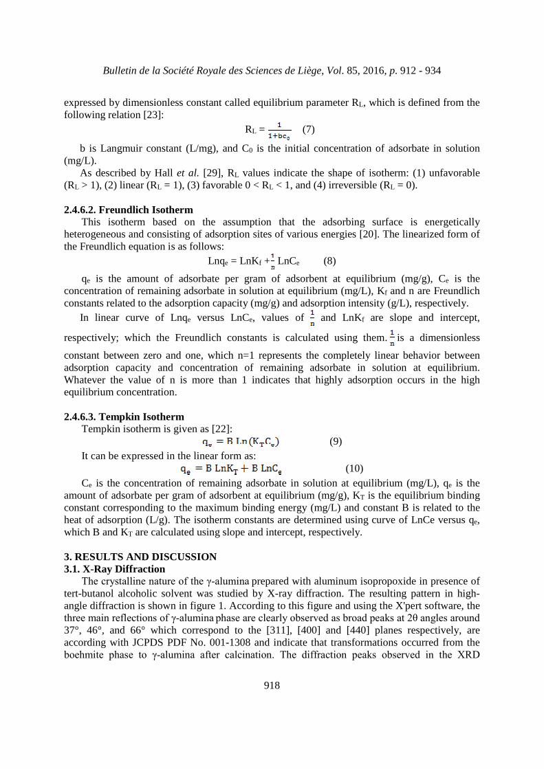
The crystalline nature of the γ-alumina prepared with aluminum isopropoxide in presence oftert-butanol alcoholic solvent was studied by X-ray diffraction. The resulting pattern in high-angle diffraction is shown in figure 1. According to this figure and using the X'pert software, thethree main reflections of γ-alumina phase are clearly observed as broad peaks at 2θ angles around 37°, 46°, and 66° which correspond to the [311], [400] and [440] planes respectively, areaccording with JCPDS PDF No. 001-1308 and indicate that transformations occurred from theboehmite phase to γ-alumina after calcination. The diffraction peaks observed in the XRD
Bulletin de la Société Royale des Sciences de Liège, Vol. 85, 2016, p. 912 - 934
919
pattern were broad, because the crystallites were very small. Such a size indicates to their partlyweak crystalline nature in the prepared γ-alumina. It should be noted that no peak of other phases of alumina is recognizable in the diffraction pattern of prepared sample and the γ-alumina phase is the only detectable phase.
The calculation was done for prepared γ-alumina based on Scherrer equation in the [440] planes with the most intensity of diffraction and 2θ angle value of 66° that the average particlesize (DXRD) was 5.4 nm. The prepared sample had nanometer structure due to less than 100 nmsize of the particles. Since the d-spacing could be considered a measure of the distance betweenlayers in a crystal structure, the values calculated by the Bragg's law for the above peaks of theprepared sample XRD pattern are 0.239 nm, 0.198 nm and 0.141 nm, respectively that indicatesthe decrease in this distance and thus reduction of the structure lattice parameter with increasingX-ray diffraction angle.
Fig. 1: X-ray diffraction pattern at high angles diffraction for prepared γ-alumina
3.2. Textural PropertiesTextural properties of BET analysis consist of particles specific surface area, total pore
volume and average pore diameter and also average particle size of the γ-alumina prepared via sol-gel method using aluminum isopropoxide in presence of tert-butanol solvent and acetic acidcatalyst are presented in table 1.
Table 1: Textural properties of prepared γ-alumina
Average particle
size (nm)
Average pore
diameter (nm)
Total pore
volume
(cm3/g)
Specific surface
area (m2/g)
5.3412.431.09351
Bulletin de la Société Royale des Sciences de Liège, Vol. 85, 2016, p. 912 - 934
920
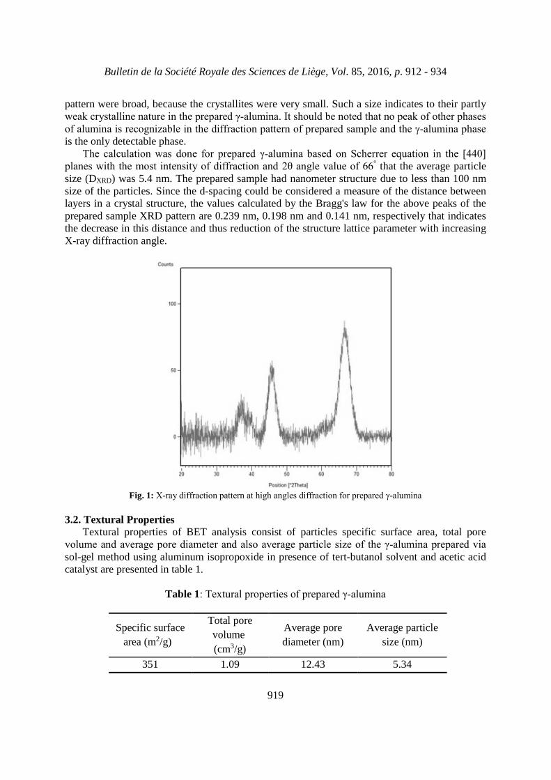
The nitrogen adsorption/desorption isotherm of the sol-gel derived γ-alumina is shown in figure 2. According to IUPAC classification, the obtained isotherm for this sample ischaracterized as V type. This isotherm has been extended in almost flat and stretched conditionuntil reach high relative pressures, which is the characteristic of meso porosity solids. Inaddition, the significant mutation occurs in the curve at high relative pressures of about P/P0 =0.9, which indicates presence of some macro pores in the structure of the sol-gel derived sample.
The shape of the hysteresis loops can be correlated with the change in pore structure, whichin this case can be a phase transformation from boehmite to γ-alumina with different morphologies [26]. In this study, hysteresis loop for prepared sample occurred at a relativepressure range of P/P0 = 0.4-0.98. It is H1 type that adsorption and desorption branches hasparallel mode completely, so the most of pores are in cylindrical shape. The ultimate amount ofadsorbed nitrogen by the sample is more than 600 cm3/g, indicating presence of large volumepores.
Fig. 2: Nitrogen adsorption/desorption isotherm for prepared γ-alumina
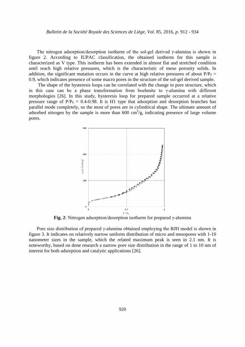
Pore size distribution of prepared γ-alumina obtained employing the BJH model is shown in figure 3. It indicates on relatively narrow uniform distribution of micro and mesopores with 1-10nanometer sizes in the sample, which the related maximum peak is seen in 2.1 nm. It isnoteworthy, based on done research a narrow pore size distribution in the range of 1 to 10 nm ofinterest for both adsorption and catalytic applications [26].
Bulletin de la Société Royale des Sciences de Liège, Vol. 85, 2016, p. 912 - 934
921
Fig. 3: BJH model of prepared γ-alumina
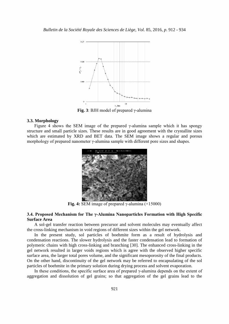
3.3. MorphologyFigure 4 shows the SEM image of the prepared γ-alumina sample which it has spongy
structure and small particle sizes. These results are in good agreement with the crystallite sizeswhich are estimated by XRD and BET data. The SEM image shows a regular and porousmorphology of prepared nanometer γ-alumina sample with different pore sizes and shapes.
Fig. 4: SEM image of prepared γ-alumina (×15000)
3.4. Proposed Mechanism for The γ-Alumina Nanoparticles Formation with High Specific Surface Area
A sol-gel transfer reaction between precursor and solvent molecules may eventually affectthe cross-linking mechanism in void regions of different sizes within the gel network.
In the present study, sol particles of boehmite form as a result of hydrolysis andcondensation reactions. The slower hydrolysis and the faster condensation lead to formation ofpolymeric chains with high cross-linking and branching [30]. The enhanced cross-linking in thegel network resulted in larger voids regions which is agree with the observed higher specificsurface area, the larger total pores volume, and the significant mesoporosity of the final products.On the other hand, discontinuity of the gel network may be referred to encapsulating of the solparticles of boehmite in the primary solution during drying process and solvent evaporation.
In these conditions, the specific surface area of prepared γ-alumina depends on the extent of aggregation and dissolution of gel grains; so that aggregation of the gel grains lead to the
Bulletin de la Société Royale des Sciences de Liège, Vol. 85, 2016, p. 912 - 934
922
formation of γ-alumina nanoparticles with higher specific surface area, and dissolution and decomposition of them bring formation of γ-alumina nanoparticles with lower specific surface area [31].
Intergrowth of the boehmite sol particles in the gel network occurred through hydrogenbonding and interspacing, in which alcohol as a solvent affected on the particle size and specificsurface area, so that the specific surface area decreases with increasing of chain length of usedalcohol, which could be caused by increasing pore sizes of the corresponding sample [32].
In acidic conditions, the encapsulated alkoxide particles decomposed upon calcination andresulting in further porosity, which may explain the formation of γ-alumina nanoparticles prepared from alkoxide. This mechanism supports the formation of mainly mesopores of smalleraverage diameter and the absence of noticeable amount of macropores in prepared γ-alumina nanoparticles, in presence of tert-butanol as a solvent. In this sample, pores were homogeneousand the final aggregates were ordered. This may be referred to the formation of small andhomogeneous sol particles of the boehmite precursor.
Acetic acid plays some crucial role in controlling the microstructure of γ-alumina nanoparticles due to moderate boehmite particle growth speeds in different modes. That isprobably through its selective adsorption on high-energy faces of boehmite particles during theaging process [33]. In these conditions, high energy levels would not be available for growth andtherefore crystal growth speeds are significantly reduced because of the strong interactionbetween CH3COO groups of acetic acid and high energy levels of boehmite particles. On theother hand, due to gradually nucleation of boehmite during the aging process where noadsorption of acetic acid takes place; energy levels minimized and boehmite nanoparticles withspecified morphology is formed.
3.5. Adsorption studies: Nickel removal by prepared γ-alumina nanoparticles3.5.1. Effect of Contact Time
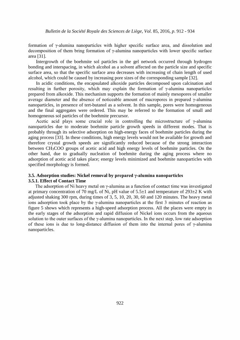
The adsorption of Ni heavy metal on γ-alumina as a function of contact time was investigated at primary concentration of 70 mg/L of Ni, pH value of 5.5±1 and temperature of 293±2 K withadjusted shaking 300 rpm, during times of 3, 5, 10, 20, 30, 60 and 120 minutes. The heavy metalions adsorption took place by the γ-alumina nanoparticles at the first 3 minutes of reaction as figure 5 shows which represents a high-speed adsorption process. All the places were empty inthe early stages of the adsorption and rapid diffusion of Nickel ions occurs from the aqueoussolution to the outer surfaces of the γ-alumina nanoparticles. In the next step, low rate adsorption of these ions is due to long-distance diffusion of them into the internal pores of γ-alumina nanoparticles.
Bulletin de la Société Royale des Sciences de Liège, Vol. 85, 2016, p. 912 - 934
923
Fig. 5: Effect of contact time with γ-alumina nanoparticles on concentration decreasing of Ni2+
ions (rpm=300, pH=5.5, T = 293 K)
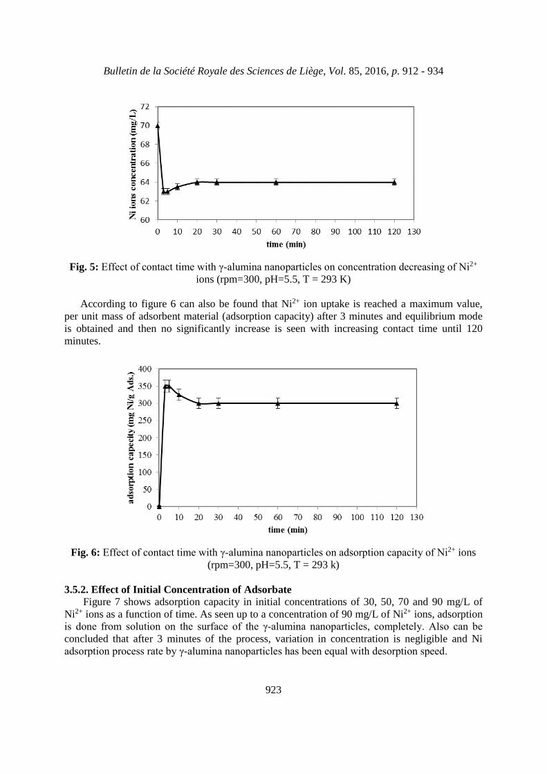
According to figure 6 can also be found that Ni2+ ion uptake is reached a maximum value,per unit mass of adsorbent material (adsorption capacity) after 3 minutes and equilibrium modeis obtained and then no significantly increase is seen with increasing contact time until 120minutes.
Fig. 6: Effect of contact time with γ-alumina nanoparticles on adsorption capacity of Ni2+ ions(rpm=300, pH=5.5, T = 293 k)
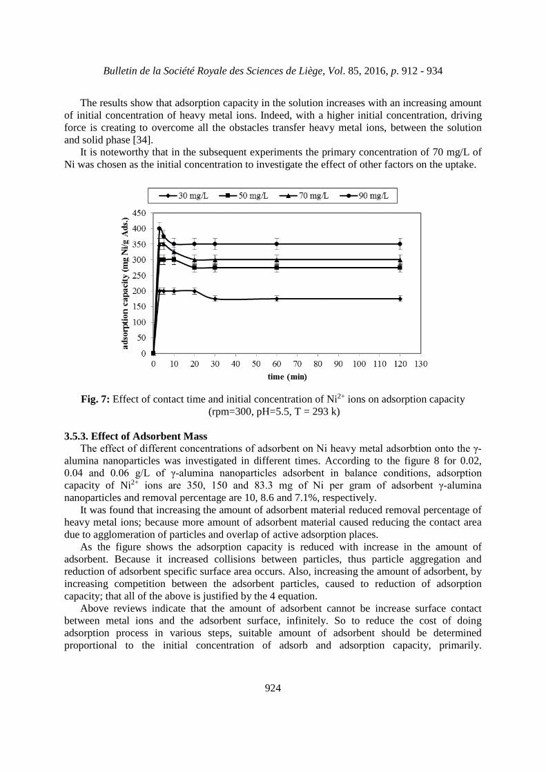
3.5.2. Effect of Initial Concentration of AdsorbateFigure 7 shows adsorption capacity in initial concentrations of 30, 50, 70 and 90 mg/L of
Ni2+ ions as a function of time. As seen up to a concentration of 90 mg/L of Ni2+ ions, adsorptionis done from solution on the surface of the γ-alumina nanoparticles, completely. Also can be concluded that after 3 minutes of the process, variation in concentration is negligible and Niadsorption process rate by γ-alumina nanoparticles has been equal with desorption speed.
Bulletin de la Société Royale des Sciences de Liège, Vol. 85, 2016, p. 912 - 934
924
The results show that adsorption capacity in the solution increases with an increasing amountof initial concentration of heavy metal ions. Indeed, with a higher initial concentration, drivingforce is creating to overcome all the obstacles transfer heavy metal ions, between the solutionand solid phase [34].
It is noteworthy that in the subsequent experiments the primary concentration of 70 mg/L ofNi was chosen as the initial concentration to investigate the effect of other factors on the uptake.
Fig. 7: Effect of contact time and initial concentration of Ni2+ ions on adsorption capacity(rpm=300, pH=5.5, T = 293 k)
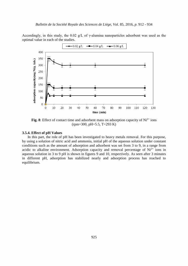
3.5.3. Effect of Adsorbent MassThe effect of different concentrations of adsorbent on Ni heavy metal adsorbtion onto the γ-
alumina nanoparticles was investigated in different times. According to the figure 8 for 0.02,0.04 and 0.06 g/L of γ-alumina nanoparticles adsorbent in balance conditions, adsorption capacity of Ni2+ ions are 350, 150 and 83.3 mg of Ni per gram of adsorbent γ-alumina nanoparticles and removal percentage are 10, 8.6 and 7.1%, respectively.
It was found that increasing the amount of adsorbent material reduced removal percentage ofheavy metal ions; because more amount of adsorbent material caused reducing the contact areadue to agglomeration of particles and overlap of active adsorption places.
As the figure shows the adsorption capacity is reduced with increase in the amount ofadsorbent. Because it increased collisions between particles, thus particle aggregation andreduction of adsorbent specific surface area occurs. Also, increasing the amount of adsorbent, byincreasing competition between the adsorbent particles, caused to reduction of adsorptioncapacity; that all of the above is justified by the 4 equation.
Above reviews indicate that the amount of adsorbent cannot be increase surface contactbetween metal ions and the adsorbent surface, infinitely. So to reduce the cost of doingadsorption process in various steps, suitable amount of adsorbent should be determinedproportional to the initial concentration of adsorb and adsorption capacity, primarily.
Bulletin de la Société Royale des Sciences de Liège, Vol. 85, 2016, p. 912 - 934
925
Accordingly, in this study, the 0.02 g/L of γ-alumina nanoparticles adsorbent was used as the optimal value in each of the studies.
Fig. 8: Effect of contact time and adsorbent mass on adsorption capacity of Ni2+ ions(rpm=300, pH=5.5, T=293 K)
3.5.4. Effect of pH ValuesIn this part, the role of pH has been investigated to heavy metals removal. For this purpose,
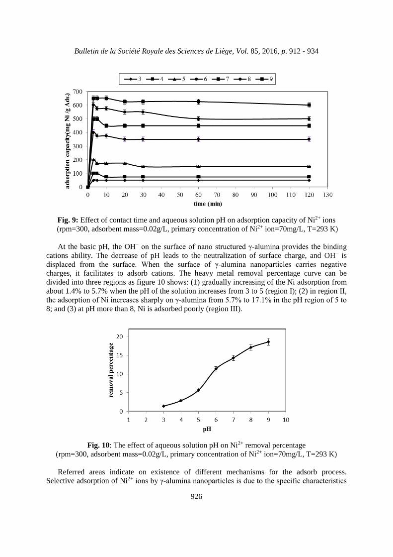
by using a solution of nitric acid and ammonia, initial pH of the aqueous solution under constantconditions such as the amount of adsorption and adsorbent was set from 3 to 9, in a range fromacidic to alkaline environment. Adsorption capacity and removal percentage of Ni2+ ions inaqueous solution in 3 to 9 pH is shown in figures 9 and 10, respectively. As seen after 3 minutesin different pH, adsorption has stabilized nearly and adsorption process has reached toequilibrium.
Bulletin de la Société Royale des Sciences de Liège, Vol. 85, 2016, p. 912 - 934
926
Fig. 9: Effect of contact time and aqueous solution pH on adsorption capacity of Ni2+ ions(rpm=300, adsorbent mass=0.02g/L, primary concentration of Ni2+ ion=70mg/L, T=293 K)
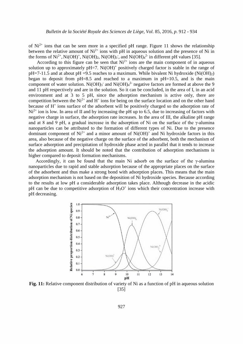
At the basic pH, the OH− on the surface of nano structured γ-alumina provides the binding cations ability. The decrease of pH leads to the neutralization of surface charge, and OH− isdisplaced from the surface. When the surface of γ-alumina nanoparticles carries negative charges, it facilitates to adsorb cations. The heavy metal removal percentage curve can bedivided into three regions as figure 10 shows: (1) gradually increasing of the Ni adsorption fromabout 1.4% to 5.7% when the pH of the solution increases from 3 to 5 (region I); (2) in region II,the adsorption of Ni increases sharply on γ-alumina from 5.7% to 17.1% in the pH region of 5 to 8; and (3) at pH more than 8, Ni is adsorbed poorly (region III).
Fig. 10: The effect of aqueous solution pH on Ni2+ removal percentage(rpm=300, adsorbent mass=0.02g/L, primary concentration of Ni2+ ion=70mg/L, T=293 K)
Referred areas indicate on existence of different mechanisms for the adsorb process.Selective adsorption of Ni2+ ions by γ-alumina nanoparticles is due to the specific characteristics
Bulletin de la Société Royale des Sciences de Liège, Vol. 85, 2016, p. 912 - 934
927
of Ni2+ ions that can be seen more in a specified pH range. Figure 11 shows the relationshipbetween the relative amount of Ni2+ ions with pH in aqueous solution and the presence of Ni inthe forms of Ni2+, Ni(OH)+, Ni(OH)2, Ni(OH)3
-, and Ni(OH)42- in different pH values [35].
According to this figure can be seen that Ni2+ ions are the main component of in aqueoussolution up to approximately pH=7. Ni(OH)+ positively charged factor is stable in the range ofpH=7-11.5 and at about pH =9.5 reaches to a maximum. While bivalent Ni hydroxide (Ni(OH)2)began to deposit from pH=8.5 and reached to a maximum in pH=10.5, and is the maincomponent of water solution. Ni(OH)3
- and Ni(OH)42- negative factors are formed at above the 9
and 11 pH respectively and are in the solution. So it can be concluded, in the area of I, in an acidenvironment and at 3 to 5 pH, since the adsorption mechanism is active only, there arecompetition between the Ni2+ and H+ ions for being on the surface location and on the other handbecause of H+ ions surface of the adsorbent will be positively charged so the adsorption rate ofNi2+ ion is low. In area of II and by increasing the pH up to 6.5, due to increasing of factors withnegative charge in surface, the adsorption rate increases. In the area of III, the alkaline pH rangeand at 8 and 9 pH, a gradual increase in the adsorption of Ni on the surface of the γ-alumina nanoparticles can be attributed to the formation of different types of Ni. Due to the presencedominant component of Ni2+ and a minor amount of Ni(OH)+ and Ni hydroxide factors in thisarea, also because of the negative charge on the surface of the adsorbent, both the mechanism ofsurface adsorption and precipitation of hydroxide phase acted in parallel that it tends to increasethe adsorption amount. It should be noted that the contribution of adsorption mechanisms ishigher compared to deposit formation mechanisms.
Accordingly, it can be found that the main Ni adsorb on the surface of the γ-alumina nanoparticles due to rapid and stable adsorption because of the appropriate places on the surfaceof the adsorbent and thus make a strong bond with adsorption places. This means that the mainadsorption mechanism is not based on the deposition of Ni hydroxide species. Because accordingto the results at low pH a considerable adsorption takes place. Although decrease in the acidicpH can be due to competitive adsorption of H3O+ ions which their concentration increase withpH decreasing.
Fig. 11: Relative component distribution of variety of Ni as a function of pH in aqueous solution[35]
Bulletin de la Société Royale des Sciences de Liège, Vol. 85, 2016, p. 912 - 934
928
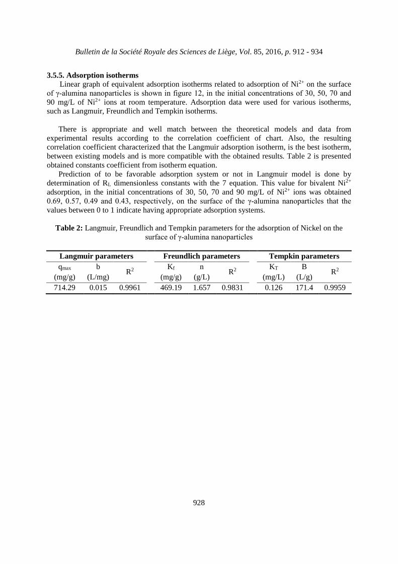
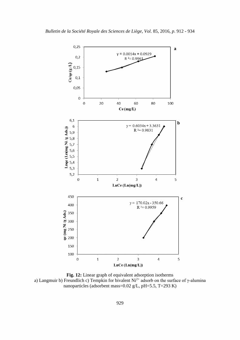
3.5.5. Adsorption isothermsLinear graph of equivalent adsorption isotherms related to adsorption of Ni2+ on the surface
of γ-alumina nanoparticles is shown in figure 12, in the initial concentrations of 30, 50, 70 and 90 mg/L of Ni2+ ions at room temperature. Adsorption data were used for various isotherms,such as Langmuir, Freundlich and Tempkin isotherms.
There is appropriate and well match between the theoretical models and data fromexperimental results according to the correlation coefficient of chart. Also, the resultingcorrelation coefficient characterized that the Langmuir adsorption isotherm, is the best isotherm,between existing models and is more compatible with the obtained results. Table 2 is presentedobtained constants coefficient from isotherm equation.
Prediction of to be favorable adsorption system or not in Langmuir model is done bydetermination of RL dimensionless constants with the 7 equation. This value for bivalent Ni2+
adsorption, in the initial concentrations of 30, 50, 70 and 90 mg/L of Ni2+ ions was obtained0.69, 0.57, 0.49 and 0.43, respectively, on the surface of the γ-alumina nanoparticles that the values between 0 to 1 indicate having appropriate adsorption systems.
Table 2: Langmuir, Freundlich and Tempkin parameters for the adsorption of Nickel on thesurface of γ-alumina nanoparticles
Langmuir parameters Freundlich parameters Tempkin parameters
qmax
(mg/g)
b
(L/mg)R2 Kf
(mg/g)
n
(g/L)R2 KT
(mg/L)
B
(L/g)R2
714.29 0.015 0.9961 469.19 1.657 0.9831 0.126 171.4 0.9959
Bulletin de la Société Royale des Sciences de Liège, Vol. 85, 2016, p. 912 - 934
929
Fig. 12: Linear graph of equivalent adsorption isothermsa) Langmuir b) Freundlich c) Tempkin for bivalent Ni2+ adsorb on the surface of γ-alumina
nanoparticles (adsorbent mass=0.02 g/L, pH=5.5, T=293 K)
Bulletin de la Société Royale des Sciences de Liège, Vol. 85, 2016, p. 912 - 934
930
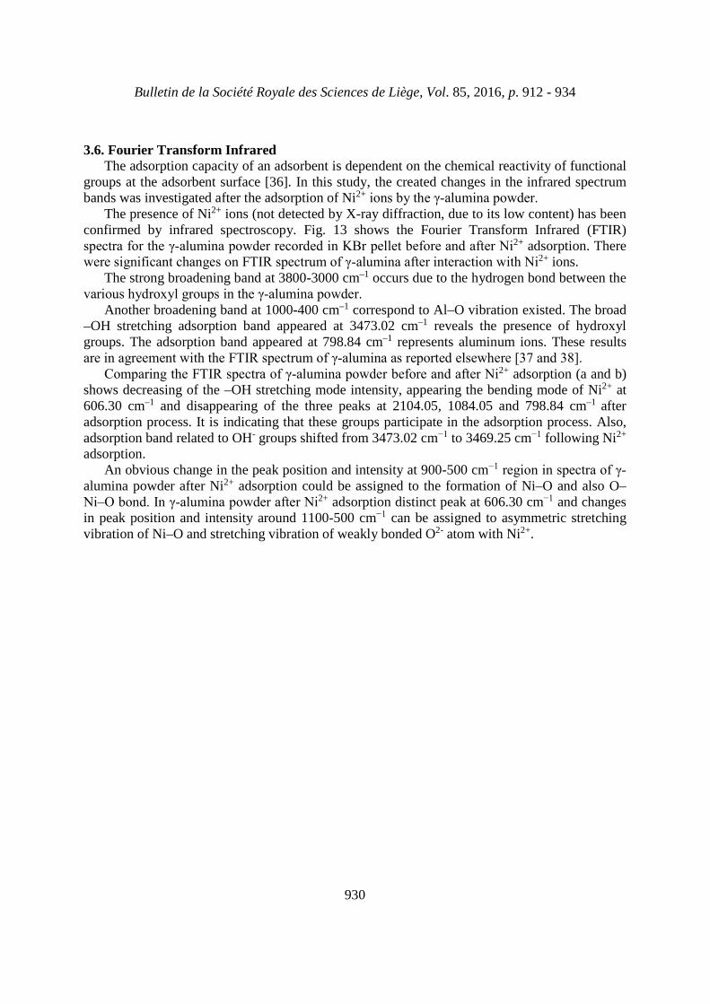
3.6. Fourier Transform InfraredThe adsorption capacity of an adsorbent is dependent on the chemical reactivity of functional
groups at the adsorbent surface [36]. In this study, the created changes in the infrared spectrumbands was investigated after the adsorption of Ni2+ ions by the γ-alumina powder.
The presence of Ni2+ ions (not detected by X-ray diffraction, due to its low content) has beenconfirmed by infrared spectroscopy. Fig. 13 shows the Fourier Transform Infrared (FTIR)spectra for the γ-alumina powder recorded in KBr pellet before and after Ni2+ adsorption. Therewere significant changes on FTIR spectrum of γ-alumina after interaction with Ni2+ ions.
The strong broadening band at 3800-3000 cm–1 occurs due to the hydrogen bond between thevarious hydroxyl groups in the γ-alumina powder.
Another broadening band at 1000-400 cm–1 correspond to Al–O vibration existed. The broad–OH stretching adsorption band appeared at 3473.02 cm–1 reveals the presence of hydroxylgroups. The adsorption band appeared at 798.84 cm–1 represents aluminum ions. These resultsare in agreement with the FTIR spectrum of γ-alumina as reported elsewhere [37 and 38].
Comparing the FTIR spectra of γ-alumina powder before and after Ni2+ adsorption (a and b)shows decreasing of the –OH stretching mode intensity, appearing the bending mode of Ni2+ at606.30 cm–1 and disappearing of the three peaks at 2104.05, 1084.05 and 798.84 cm–1 afteradsorption process. It is indicating that these groups participate in the adsorption process. Also,adsorption band related to OH- groups shifted from 3473.02 cm−1 to 3469.25 cm−1 following Ni2+
adsorption.An obvious change in the peak position and intensity at 900-500 cm−1 region in spectra of γ-
alumina powder after Ni2+ adsorption could be assigned to the formation of Ni–O and also O–Ni–O bond. In γ-alumina powder after Ni2+ adsorption distinct peak at 606.30 cm−1 and changesin peak position and intensity around 1100-500 cm−1 can be assigned to asymmetric stretchingvibration of Ni–O and stretching vibration of weakly bonded O2- atom with Ni2+.
Bulletin de la Société Royale des Sciences de Liège, Vol. 85, 2016, p. 912 - 934
931
Fig. 13: FTIR spectra of γ-alumina powder a) before and b) after Ni2+ adsorption
4. CONCLUSIONThe present study was successful in synthesizing porous γ-alumina nanoparticles using a
facile sol-gel process. The prepared γ-alumina in the presence of tert-butanol as a solvent even after calcining process of the boehmite at 600°C had a specific surface area of 351 m2/g, a porevolume of 1.09 cm3/g, particle size of 5.34 nm and suitable pore size distribution which aredesirable for heavy metal adsorption. This work also showed that prepared γ-alumina powder had the great adsorption performance of Ni2+ with a high adsorption rate and adsorption capacityof 350 mg/g when the adsorption reached equilibrium for 3 min at room temperature andpH=5.5.
Bulletin de la Société Royale des Sciences de Liège, Vol. 85, 2016, p. 912 - 934
932
ACKNOWLEDGEMENTSThe authors gratefully acknowledge from the Material and Energy Research Center (MERC)
and the Iranian Nanotechnology Initiative Council for their financial supports of this study.
REFRENCES[1] Hiroaki Masuda, Ko Higashitani, Hideto Yoshida, "Powder Technology: Handling andOperations, Process Instrumentation and Working Hazards", CRC Press. 2006.[2] Baiyu Huang, Calvin Bartholomew, Brian F. Woodfield, "Facile synthesis of mesoporous γ-alumina with tunable pore size: The effects of water to aluminum molar ratio in hydrolysis ofaluminum alkoxides", Journal of Microporous and Mesoporous Materials 183 (2014) 37-47.[3] César Lúcio Lopes de Faria, Tânia Keli Resende de Oliveira, Vera Lúcia dos Santos, CarlosAugusto Rosa, José Domingos Ardisson, Waldemar de Almeida Macêdo, Armindo Santos,"Usage of the sol-gel process on the fabrication of macroporous adsorbent activated-gammaalumina spheres", Journal of Microporous and Mesoporous Materials, 120 (2009) 228-238.[4] YI Jian-hong, SUN You-yi, GAO Jian-feng, XU Chun-yan, "Synthesis of crystalline γ-Al2O3
with high purity", Journal of Transactions of Nonferrous Metals Society of China, 19 (2009)1237-1242.[5] S.C. Shen, W.K. Ng, Q. Chen, X.T. Zeng, R.B.H. Tan, "Novel synthesis of lace-likenanoribbons of boehmite and γ-alumina by dry conversion method", Journal of Materials Letters 61 (2007) 4280-4282.[6] Y. Liu, D. Ma, X. Han, X. Bao, W. Frandsen, D. Wang, D. Su, "Hydrothermal synthesis ofmicroscale boehmite and gamma nanoleaves alumina", Journal of Materials Letters 62 (2008)1297-1301.[7] P. Morajkar, J. Fernandes, "A new facile method to synthesize mesoporous γ-Al2O3 of highsurface area and catalytic activity", Journal of Catalysis Communications 11 (2010) 414-418.[8] M. Sasani Ghamsari, Z. Ashor Said Mahzar, S. Radiman, A.M. Abdul Hamid, Sh. RahmaniKhalilabad, "Facile route for preparation of highly crystalline γ-Al2O3 nanopowder", Journal ofMaterials Letters 72 (2012) 32-35.[9] L. Ji, J. Lin, K.L. Tan, H.C. Zeng, "Synthesis of High-Surface-Area Alumina UsingAluminum Tri-sec-butoxide − 2,4-Pentanedione − 2-Propanol − Nitric Acid Precursors", Journal of Chemistry of Materials, 12 (2000) 931-939.[10] Y.W. Jun, J.S. Choi, J.W. Cheon, "Shape Control of Semiconductor and Metal OxideNanocrystals through Nonhydrolytic Colloidal Routes, A review", Journal of Angewandte Che-mie International Edition in English, 45 (2006) 3414-3439.[11] G.F. Zou, H. Li, Y.G. Zhang, K. Xiong, Y.T. Qian, "Solvothermal/hydrothermal route tosemiconductor nanowires", Journal of Nanotechnology, 17 (2006) S313.[12] Ming-Guo Ma, Jie-Fang Zhu, "A facile solvothermal route to synthesis of γ-alumina with bundle-like and flower-like morphologies", Journal of Materials Letters 63 (2009) 881-883.[13] M.G. Ma, Y.J. Zhu, Z.L. Xu, "A new route to synthesis of γ-alumina nanorods", Journal of Materials Letters 61 (2007) 1812-1815.[14] H.V. Fajardo, A.O. Martins, R.M. de Almeida, L.K. Noda, "Synthesis of mesoporous Al2O3
macrospheres using the biopolymer chitosan as a template: A novel active catalyst system forCO2 reforming of methane", Journal of Materials Letters, 59 (2005) 3963-3967.[15] T. C. Haung , H. I. Chen, "A STUDY ON THE PREPARATION OF NON-SUPPORTEDγ-ALUMINA MEMBRANES BY SOL-GEL METHOD", journal of Chemical Engineering Communications 132 (1995) 125-139.
Bulletin de la Société Royale des Sciences de Liège, Vol. 85, 2016, p. 912 - 934
933
[16] J. Wang, Y. Wang, M. Qiao, S. Xie, K. Fan, "A novel sol-gel synthetic route to aluminananofibers via aluminum nitrate and hexamethylenetetramine", Journal of Materials Letters, 61(2007) 5074-5077.[17] S. Shen, Q. Chen, P. Chow, G. Tan, X. Zeng, Z. Wang, R. Tan, "Steam-Assisted Solid Wet-Gel Synthesis of High-Quality Nanorods of Boehmite and Alumina", Journal of PhysicalChemistry: C, 111 (2007) 700-707.[18] Xun Liu, Tianyou Peng, Jinchun Yao, Hongjin Lv, Cheng Huang, "Synthesis and texturalevolution of alumina particles with mesoporous structures", Journal of Solid State Chemistry,183 (2010) 1448-1456.[19] M.E. Mahmoud, M.M. Osman, O.F. Hafez, A.H. Hegazi, E. Elmelegy, "Removal andpreconcentration of lead(II) and other heavy metals from water by alumina adsorbents developedby surface-adsobed-dithizone", Journal of Desalination, 251 (2010) 123-130.[20] Yvan J.O. Asencios, María R. Sun-Kou, "Synthesis of high-surface-area γ-Al2O3 fromaluminum scrap and its use for the adsorption of metals: Pb(II), Cd(II) and Zn(II)", Journal ofApplied Surface Science, 258 (2012) 10002-10011.[21] A. K. Patra, A. Dutta, A. Bhaumik, "Self-assembled mesoporous γ-Al2O3 sphericalnanoparticles and their efficiency for the removal of arsenic from water", Journal of HazardousMaterials, 201-202 (2012) 170-177.[22] A. Rahmani, H. Zavvar Mousavi, M. Fazli, "Effect of nanostructure alumina on adsorptionof heavy metals", Journal of Desalination, 253 (2010) 94-100.[23] H. Zhang, L. Chen, D. Zhang, S. Lu, X. Yu, "Impact of environmental conditions on theadsorption behavior of radionuclide 63Ni(II) on γ-Al2O3", Journal of Colloids and Surfaces A:Physicochemical and Engineering Aspects, 380 (2011) 16-24.[24] M. Burgos, M. Langlet, "The sol-gel transformation of TIPT coatings: a FTIR study",Journal of Thin Solid Films, 349 (1999) 19-23.[25] B.D. Cullity, S.R. Stock, "Elements of X-Ray Diffraction", U.S.A, Prentice Hall, 2001.[26] S. Lowell, J. E. Shields, "Powder Surface Area and Porosity", Chapman and Hall, Londonand New York, 1984.[27] Zahra Hosseini, Majid Taghizadeh, Fereydoon Yaripour, "Synthesis of nanocrystalline γ-Al2O3 by sol-gel and precipitation methods for methanol dehydration to dimethyl ether", Journalof Natural Gas Chemistry, 20 (2011) 128-134.[28] A.K. Bhattacharya, S.N. Mandal, S.K. Das, "Adsorption of Zn(II) from aqueous solution byusing different adsorbents", Journal of Chemical Engineering, 123 (2006) 43-51.[29]. Hall, K.R., Eagleton, L.C., Acrivos, A. and Vermeulen, T. Poreand, "Solid-DiffusionKinetics in Fixed-Bed Adsorption under Constant-Pattern Conditions", Journal of Industrial andEngineering Chemistry Fundamentals, 5 (1966) 212-223.[30] C. Brinker, G. Scherer, "Sol-Gel Science", Academic Press, 1989.[31] K. Hellgardt, D. Chadwick, "On the preparation of high surface area aluminas from nitratesolutions", Journal of Industrial and Engineering Chemistry Research, 37 (1998) 405-411.[32] S.J. Gregg, K.S.W. Sing, "Adsorption, surface area and porosity", Academic Press, 1982.[33] S.B. Deshpande, H.S. Potdar, Y.B. Khollam, K.R. Patil, R. Pasricha, N.E. Jacob,"Roomtemperature synthesis of mesoporous aggregates of anatase TiO2 nanoparticles", Journal ofMaterials Chemistry and Physics, 97 (2006) 207-212.[34] Z. Aksu, S. Tezar, "Biosorption of reactive dyes on the green alga Chlorella vulgaris",Journal of Process Biochemistry, 40 (2005) 1347-1361.
Bulletin de la Société Royale des Sciences de Liège, Vol. 85, 2016, p. 912 - 934
934
[35] Shitong Yang, Jiaxing Li, Dadong Shao, Jun Hu, Xiangke Wang, "Adsorption of Ni(II) onoxidized multi-walled carbon nanotubes: Effect of contact time, pH, foreign ions and PAA",Journal of Hazardous Materials, 166 (2009) 109-116.[36] Dayane J. Amorim, Hélen C. Rezende, Érica L. Oliveira, Ione L. S. Almeida, Nívia M. M.Coelho, Túlio N. Matos, Cleide S. T. Araújo, "Characterization of Pequi (Caryocar brasiliense)Shells and Evaluation of Their Potential for the Adsorption of Pb(II) Ions in Aqueous Systems",Journal of the Brazilian Chemical Society , 27 (2016) 616-623.[37] Z. Zeng, J. Yu, Z.X. Guo, "Preparation of functionalized core-shell alumina/polystyrenecomposite nanoparticles", Journal of Macromolecular Chemistry and Physics, 206 (2005) 1558-1567.[38] G. Busca, V. Lorenzelli, G. Ramis, R.J. Willey, "Surface sites on spinel-type and corundum-type metal oxide powders", Journal of Langmuir, 9(1993) 1492-1499.


![Synthesis of Photoluminescence Si Nanoparticles: Size … · 2019. 8. 2. · mechanochemical disproportionation of silicon monoxide (SiO) [63-67], microwave-assisted reduction of](https://img.pdfslide.tips/doc/110x75/60f69068a4170821fc7a79f2/synthesis-of-photoluminescence-si-nanoparticles-size-2019-8-2-mechanochemical.jpg)
![Synthesis of nano [alpha]-alumina powders using ... · PDF fileand ammonia solution) and α-alumina seeding on the transformation temperature ... transformation process to α phase](https://img.pdfslide.tips/doc/110x75/5ab848dd7f8b9ac10d8cd0da/synthesis-of-nano-alpha-alumina-powders-using-ammonia-solution-and-alumina.jpg)

![Synthesis of Cesium Tungsten Bronze Nanoparticles by ......tungsten bronze nanoparticles using a spray pyrolysis method [4,5]. 1.2 Methods for particle preparation In general, high](https://img.pdfslide.tips/doc/110x75/60f6ab01dd966c47d148a1dd/synthesis-of-cesium-tungsten-bronze-nanoparticles-by-tungsten-bronze-nanoparticles.jpg)