Embed Size (px)
Citation preview
TGF-b1 regulates the invasive and metastatic potential ofmucoepidermoid carcinoma cells
Jing Wang1,2, Jian Chen
3, Kailiang Zhang
1, Yuan Zhao
1, Jacques E. Nor
2,4,5, Junzheng Wu
6
1School of Stomatology, Lanzhou University, Gansu, China; 2Angiogenesis Research Laboratory, Department of Cariology,Restorative Sciences and Endodontics, University of Michigan School of Dentistry, Ann Arbor MI, USA; 3Department of PediatricSurgery, the First Hospital of Lanzhou University, Gansu, China; 4Department of Biomedical Engineering, College of Engineering,University of Michigan, Ann Arbor MI, USA; 5Department of Otolaryngology, School of Medicine; University of Michigan, AnnArbor MI, USA; 6Department of Oral Biology, College of Stomatology, Fourth Military Medical University, Shannxi, China
BACKGROUND: Patients with mucoepidermoid carci-
noma exhibit poor long-term prognosis because of the
lack of therapeutic strategies that effectively block tumor
progression. We have previously characterized the Ms
cells as a highly metastatic mucoepidermoid carcinoma
cell line that expresses high levels of transforming growth
factor b1 (TGF-b1). Here, we studied the effect of sup-
pressing TGF-b1 by RNA silencing on the invasive and
metastatic potential of mucoepidermoid carcinoma.
METHODS: Cell motility, substratum adhesion, and
transmembrane invasion were estimated by migration,
matrigel adhesion, and matrigel invasion assay. Matrix
metalloproteinase (MMP)-2 and MMP-9 activity were
determined using gelatin gel zymography. Balb ⁄ c nu ⁄ nu
nude mice lung metastatic model was used to test the
metastatic ability of the Ms cells. Lung metastatic tumors
were experimentally induced by mice tail vein inoculation
of cancer cells.
RESULTS: TGF-b1 silencing inhibits cell motility, sub-
stratum adhesion, and transmembrane invasion. In vivo,
a significant decrease in lung metastasis was observed
when mice received tail vein injections of TGF-b1-
silenced mucoepidermoid carcinoma cells, as compared
to controls.
CONCLUSION: These results unveil a critical role for
TGF-b1 in the progression of mucoepidermoid carcino-
mas and suggest that patients with this malignancy may
benefit from therapeutic inhibition of the effectors of the
TGF-b1 pathway.
J Oral Pathol Med (2011) 40: 762–768
Keywords: extracellular matrix; matrix metalloproteinase;
metastasis; salivary gland cancer; tumor progression
Introduction
Mucoepidermoid carcinoma is the most frequent sali-vary gland cancer, accounting to approximately 30% ofall salivary gland malignancies. Primary mucoepider-moid carcinomas are not sensitive to radiotherapy andchemotherapy (1). The most common cause of death isthe lack of response to therapy, leading to recurrence ordistant metastasis. As a consequence, the long-termprognosis for patients with this malignancy is invariablypoor (2). The understanding of mechanisms involved inthe invasive behavior of mucoepidermoid carcinomacells is critical for the development of mechanism-basedtherapies for this cancer.
Transforming growth factor-b (TGF-b) belongs to afamily of structurally related, dimeric, disulfide-linkedpolypeptides, which includes five TGF-b isoforms (TGFb1–5), activins, inhibins, Mullerian-inhibiting substance,bone morphogenetic proteins, and products of theXenopus Vg1 and Drosophila decapentaplegic genes. Inmammals, three isoforms of TGF b (TGF b1–3) havebeen identified (3). They are known to regulate cellproliferation and differentiation, positively or negativelydepending on the cell type, and have been implicatedin diverse physiological events such as angiogenesis,immune function, steroidogenesis, and tissue remodelingand repair. TGF-b plays a critical role in the progressionof many epithelial cancers. TGF-b1 is a potent growthinhibitor for epithelial cells, and this function contributesgreatly to its role in tumor suppression (4). Paradoxically,TGF-b1 is overexpressed in many malignant humantumors (5, 6) and in various cancers in experimentalanimals, including skin tumors (7). Studies have shownthat TGF-b1 overexpression at early stages of carcino-genesis provides tumor-suppressive effects primarily viagrowth inhibition (8), whereas TGF-b1 overexpression atlate stages promotes tumor progression, metastasis, andepithelial to mesenchymal transition (EMT), potentiallyvia loss of adhesion molecules, angiogenesis, proteinaseactivation, and immune suppression (9–12).
Correspondence: Junzheng Wu, DDS, MS, Professor of Dentistry,Fourth Military Medical University, 7 Kangfu Road, Xi’an, Shannxi710032, China. Tel: +86 029 83376169, Fax: +86 029 833762322,E-mail: [email protected] for publication April 12, 2011
J Oral Pathol Med (2011) 40: 762–768
ª 2011 John Wiley & Sons A/S Æ All rights reserved
wileyonlinelibrary.com/journal/jop
doi: 10.1111/j.1600-0714.2011.01051.x
Journal of
Oral Pathology & Medicine
It is known that TGF-b1 modulates the motilebehavior of several tumor cells including human glioma(13), oral squamous carcinoma (14), prostate cancer(15), breast cancer (16), and lung cancer cells (17).However, the role of TGF-b1 in salivary gland muco-epidermoid carcinoma cells is not currently well under-stood. We have observed that the metastatic Msmucoepidermoid carcinoma cell line expresses highlevels of TGF-ß1 (18). We hypothesize that TGF-ß1may contribute to the invasive and metastatic potentialof mucoepidermoid carcinomas. Here, we report thatsilencing of TGF-b1 effectively decreases invasion andmetastasis of Ms cells.
MethodsConstruction of a vector-expressing shRNA for TGF-b1The pWH1 was used for the construction of a vector-expressing shRNA for TGF-b1. Human TGF-b1-specific sequences 5¢-GATCCCCTGCTGCCGCTGCTGCTACCttcaagagaGGTAGCAGCAGCGGCAGCATTTTTGGAAA-3¢ and 5¢-AGCTTTTCCAAAAATGCTGCCGCTGCTGCTACCtctcttgaaGGTAGCAGCAGCGGCAGCAGGG-3¢ were cloned into Blg ⁄Hind sitesof the pWH1 vector, which was generously providedby Dr. Wu YM.
Cell culture and transfection conditionsMs cells (mucoepidermoid carcinoma spindle code met-astatic cell lines), established and characterized in ourlaboratory, showed a higher metastatic potential than theparentalMc3 cells’ in vitro and in vivo behavior (19). Cellswere maintained in Dulbecco’s modified Eagle’s medium(DMEM, Gibco, Langley, OK, USA) supplemented with10% fetal bovine serum (FBS, Gibco) and 100 lg ⁄mlstreptomycin and 100-U ⁄ml penicillin at 37�C in ahumidified 5% CO2 atmosphere. Cells were periodicallydetermined as mycoplasma free by Immu-maker myco-test kit. Twenty-four hours before transfection, cells wereseeded onto 24-well plates with antibiotics-free growthmedium at a density of 5 · 105 cells ⁄well, so that theconfluence would reach approximately 50–70% at thetime of transfection. Cells were transfectedwith 1 lg ⁄wellof shRNA vector targeting TGF-b1 using the Lipofec-termine2000 (Invitrogen, Carlsbad, CA, USA) reagentfollowing the protocol provided by the manufacturer.Control transfection was generated by transfectingpWH1-puro into Ms cells as shRNA-C. Control cellswere incubated with DMEM alone without shRNA orLiperfectamine2000 as control. After 6 h, medium wasreplaced by antibiotics-freeDMEM.Cells were harvestedand plated on 100-mm culture dish and cultured for 48 h,and clones stably expressing shRNA were selectedusing 600 lg ⁄ml G418 (Gibco) for 3 weeks. Cloneswere screened by RT-PCR, immunocytochemistry, andWestern blotting (20).
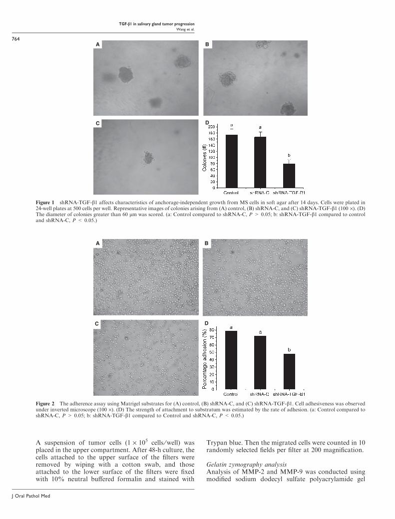
Soft agar colony formation assayTo evaluate whether cells were able to grow in soft agar,24-well plates were prepared with 0.8-ml feeder layer of5 g ⁄ l agar in medium plus 20% FBS and a 3 g ⁄ l agar
top layer in the same medium that contained recentlytrypsinized log-phase growing cells (500 cells ⁄well). Twoweeks after seeding, colonies with more than 60 lm indiameter were counted under an inverted microscope.
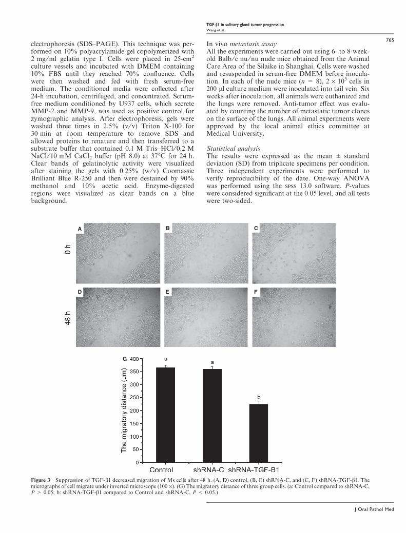
Matrigel adhesion assayThe effect of TGF-b1 on the adhesive capacity of Mscells was investigated in a Matrigel adhesion assay, asdescribed (21). Wells from 96-well plates were coveredwith five-times-diluted Matrigel Basement MembraneMatrix (Becton Dickinson, Franklin Lakes, NJ, USA),and incubated at room temperature for 1 h. Coated wellswere further incubated for 1 h with DMEM plus 10 g ⁄ lbovine serum albumin (BSA) to saturate any substrate-free space. Wells coated with 10 g ⁄ l BSA alone were usedas a substrate control. MTT was prepared in PBS(5 mg ⁄ml) and further diluted (10%) in DMEM. Toanalyze the ability of cells to adhere to Matrigel, 1 · 105
cells in 0.1 ml DMEMwith 10 g ⁄ l BSA were plated ontocoated wells. After 1 h of incubation at 37�C, each wellwas washed twice in PBS to remove non-adherent cells.Attached cells were treated with 250 ll sterile MTT dyefor 4 h at 37�C. The excess of MTT solution wasremoved and DMSO was added and thoroughly mixedfor 10 min. The absorbance of the DMSO solution wasmeasured spectrometry at 570 nm. The rate of adhesion(%) = (A in experiment group ⁄A in a BSA substratecontrol-1) · 100.
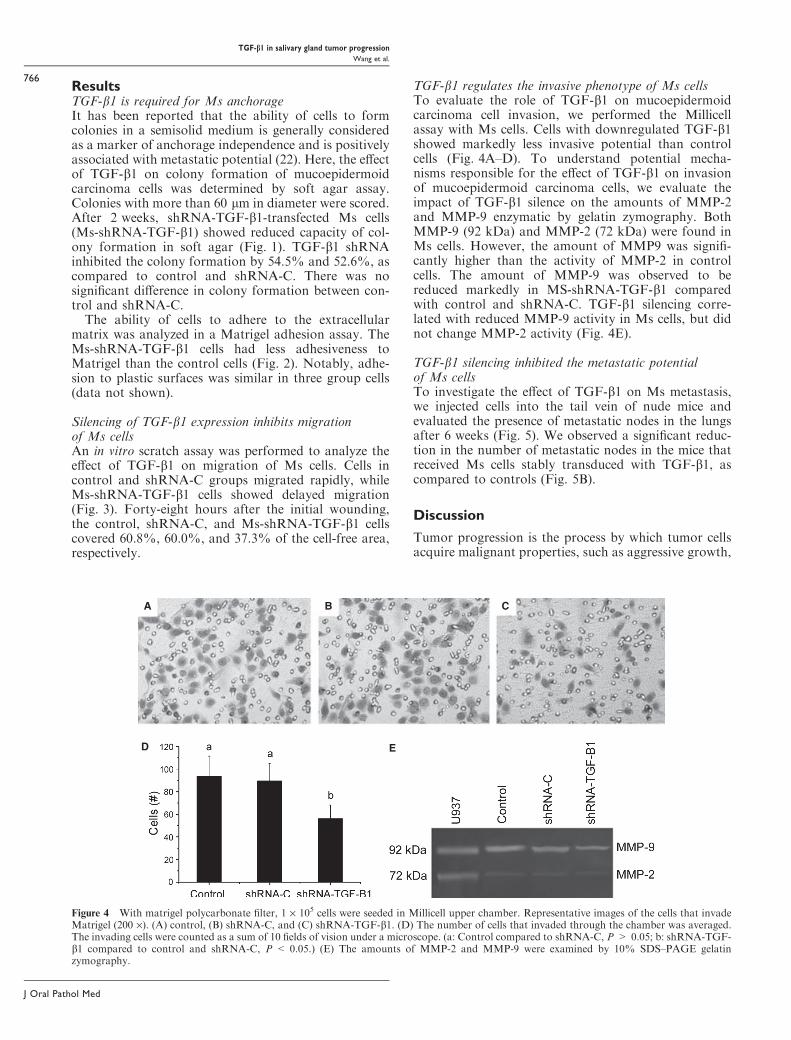
Migration assayTo determine the effects of TGF-b1 shRNA transfectionon the motility of Ms cells, an in vitro scratch assay wasused. Briefly, 1 · 105 cells were seeded into 96-wellplates in which were coated Matrigel in DMEMsupplemented with 10% FBS. When cells reachedconfluence, the cell monolayer was then wounded byscraping the surface with a plastic tip to leave anapproximately 600-lm-wide clearing. After wounding,cultures were washed in DMEM plus 10 g ⁄ l BSA threetimes to remove residual FBS and cells detached duringthe wounding process and then cultured in DMEM with10 g ⁄ l BSA. After 24-h incubation, medium wasreplaced with fresh DMEM supplemented with 10%FBS. Cell motility was determined by measuring thedistance of cell movement toward the wounded spaceafter 48-h culture.
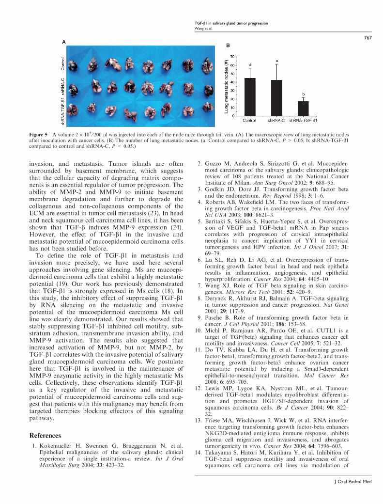
Matrigel invasion assayThe invasion ability of the cells was assayed using aMillicell (Millipore) separated by a polycarbonate filters(8 lm pore size). The polycarbonate filter was coatedwith 75 ll of Matrigel diluted (5 · ) in cold DMEM andincubated at room temperature for 1 h. The 600 llNIH3T3 fibroblast-conditioned medium (obtained byincubation of NIH3T3 cells for 24 h in serum-freeDMEM) was used as a chemoattractant in the lowercompartment. DMEM was used as negative control.The coated Millicell were further incubated for 1 h withassay medium (DMEM with 10 g ⁄ l BSA) to saturateany substrate-free space. Confluent cultures from threegroup cells were detached and resuspended in DMEM.
TGF-b1 in salivary gland tumor progression
Wang et al.
763
J Oral Pathol Med
A suspension of tumor cells (1 · 105 cells ⁄well) wasplaced in the upper compartment. After 48-h culture, thecells attached to the upper surface of the filters wereremoved by wiping with a cotton swab, and thoseattached to the lower surface of the filters were fixedwith 10% neutral buffered formalin and stained with
Trypan blue. Then the migrated cells were counted in 10randomly selected fields per filter at 200 magnification.
Gelatin zymography analysisAnalysis of MMP-2 and MMP-9 was conducted usingmodified sodium dodecyl sulfate polyacrylamide gel
A
C
B
D
Figure 1 shRNA-TGF-b1 affects characteristics of anchorage-independent growth from MS cells in soft agar after 14 days. Cells were plated in24-well plates at 500 cells per well. Representative images of colonies arising from (A) control, (B) shRNA-C, and (C) shRNA-TGF-b1 (100 ·). (D)The diameter of colonies greater than 60 lm was scored. (a: Control compared to shRNA-C, P > 0.05; b: shRNA-TGF-b1 compared to controland shRNA-C, P < 0.05.)
A B
C D
Figure 2 The adherence assay using Matrigel substrates for (A) control, (B) shRNA-C, and (C) shRNA-TGF-b1. Cell adhesiveness was observedunder inverted microscope (100 ·). (D) The strength of attachment to substratum was estimated by the rate of adhesion. (a: Control compared toshRNA-C, P > 0.05; b: shRNA-TGF-b1 compared to Control and shRNA-C, P < 0.05.)
TGF-b1 in salivary gland tumor progression
Wang et al.
764
J Oral Pathol Med
electrophoresis (SDS–PAGE). This technique was per-formed on 10% polyacrylamide gel copolymerized with2 mg ⁄ml gelatin type I. Cells were placed in 25-cm2
culture vessels and incubated with DMEM containing10% FBS until they reached 70% confluence. Cellswere then washed and fed with fresh serum-freemedium. The conditioned media were collected after24-h incubation, centrifuged, and concentrated. Serum-free medium conditioned by U937 cells, which secreteMMP-2 and MMP-9, was used as positive control forzymographic analysis. After electrophoresis, gels werewashed three times in 2.5% (v ⁄ v) Triton X-100 for30 min at room temperature to remove SDS andallowed proteins to renature and then transferred to asubstrate buffer that contained 0.1 M Tris–HCl ⁄ 0.2 MNaCl ⁄ 10 mM CaCl2 buffer (pH 8.0) at 37�C for 24 h.Clear bands of gelatinolytic activity were visualizedafter staining the gels with 0.25% (w ⁄ v) CoomassieBrilliant Blue R-250 and then were destained by 90%methanol and 10% acetic acid. Enzyme-digestedregions were visualized as clear bands on a bluebackground.
In vivo metastasis assayAll the experiments were carried out using 6- to 8-week-old Balb ⁄ c nu ⁄ nu nude mice obtained from the AnimalCare Area of the Silaike in Shanghai. Cells were washedand resuspended in serum-free DMEM before inocula-tion. In each of the nude mice (n = 8), 2 · 105 cells in200 ll culture medium were inoculated into tail vein. Sixweeks after inoculation, all animals were euthanized andthe lungs were removed. Anti-tumor effect was evalu-ated by counting the number of metastatic tumor cloneson the surface of the lungs. All animal experiments wereapproved by the local animal ethics committee atMedical University.
Statistical analysisThe results were expressed as the mean ± standarddeviation (SD) from triplicate specimens per condition.Three independent experiments were performed toverify reproducibility of the date. One-way ANOVAwas performed using the SPSS 13.0 software. P-valueswere considered significant at the 0.05 level, and all testswere two-sided.
A
D
G
E F
CB
Figure 3 Suppression of TGF-b1 decreased migration of Ms cells after 48 h. (A, D) control, (B, E) shRNA-C, and (C, F) shRNA-TGF-b1. Themicrographs of cell migrate under inverted microscope (100 ·). (G) The migratory distance of three group cells. (a: Control compared to shRNA-C,P > 0.05; b: shRNA-TGF-b1 compared to Control and shRNA-C, P < 0.05.)
TGF-b1 in salivary gland tumor progression
Wang et al.
765
J Oral Pathol Med
ResultsTGF-b1 is required for Ms anchorageIt has been reported that the ability of cells to formcolonies in a semisolid medium is generally consideredas a marker of anchorage independence and is positivelyassociated with metastatic potential (22). Here, the effectof TGF-b1 on colony formation of mucoepidermoidcarcinoma cells was determined by soft agar assay.Colonies with more than 60 lm in diameter were scored.After 2 weeks, shRNA-TGF-b1-transfected Ms cells(Ms-shRNA-TGF-b1) showed reduced capacity of col-ony formation in soft agar (Fig. 1). TGF-b1 shRNAinhibited the colony formation by 54.5% and 52.6%, ascompared to control and shRNA-C. There was nosignificant difference in colony formation between con-trol and shRNA-C.The ability of cells to adhere to the extracellular
matrix was analyzed in a Matrigel adhesion assay. TheMs-shRNA-TGF-b1 cells had less adhesiveness toMatrigel than the control cells (Fig. 2). Notably, adhe-sion to plastic surfaces was similar in three group cells(data not shown).
Silencing of TGF-b1 expression inhibits migrationof Ms cellsAn in vitro scratch assay was performed to analyze theeffect of TGF-b1 on migration of Ms cells. Cells incontrol and shRNA-C groups migrated rapidly, whileMs-shRNA-TGF-b1 cells showed delayed migration(Fig. 3). Forty-eight hours after the initial wounding,the control, shRNA-C, and Ms-shRNA-TGF-b1 cellscovered 60.8%, 60.0%, and 37.3% of the cell-free area,respectively.
TGF-b1 regulates the invasive phenotype of Ms cellsTo evaluate the role of TGF-b1 on mucoepidermoidcarcinoma cell invasion, we performed the Millicellassay with Ms cells. Cells with downregulated TGF-b1showed markedly less invasive potential than controlcells (Fig. 4A–D). To understand potential mecha-nisms responsible for the effect of TGF-b1 on invasionof mucoepidermoid carcinoma cells, we evaluate theimpact of TGF-b1 silence on the amounts of MMP-2and MMP-9 enzymatic by gelatin zymography. BothMMP-9 (92 kDa) and MMP-2 (72 kDa) were found inMs cells. However, the amount of MMP9 was signifi-cantly higher than the activity of MMP-2 in controlcells. The amount of MMP-9 was observed to bereduced markedly in MS-shRNA-TGF-b1 comparedwith control and shRNA-C. TGF-b1 silencing corre-lated with reduced MMP-9 activity in Ms cells, but didnot change MMP-2 activity (Fig. 4E).
TGF-b1 silencing inhibited the metastatic potentialof Ms cellsTo investigate the effect of TGF-b1 on Ms metastasis,we injected cells into the tail vein of nude mice andevaluated the presence of metastatic nodes in the lungsafter 6 weeks (Fig. 5). We observed a significant reduc-tion in the number of metastatic nodes in the mice thatreceived Ms cells stably transduced with TGF-b1, ascompared to controls (Fig. 5B).
Discussion
Tumor progression is the process by which tumor cellsacquire malignant properties, such as aggressive growth,
A B C
ED
Figure 4 With matrigel polycarbonate filter, 1 · 105 cells were seeded in Millicell upper chamber. Representative images of the cells that invadeMatrigel (200 ·). (A) control, (B) shRNA-C, and (C) shRNA-TGF-b1. (D) The number of cells that invaded through the chamber was averaged.The invading cells were counted as a sum of 10 fields of vision under a microscope. (a: Control compared to shRNA-C, P > 0.05; b: shRNA-TGF-b1 compared to control and shRNA-C, P < 0.05.) (E) The amounts of MMP-2 and MMP-9 were examined by 10% SDS–PAGE gelatinzymography.
TGF-b1 in salivary gland tumor progression
Wang et al.
766
J Oral Pathol Med
invasion, and metastasis. Tumor islands are oftensurrounded by basement membrane, which suggeststhat the cellular capacity of degrading matrix compo-nents is an essential regulator of tumor progression. Theability of MMP-2 and MMP-9 to initiate basementmembrane degradation and further to degrade thecollagenous and non-collagenous components of theECM are essential in tumor cell metastasis (23). In headand neck squamous cell carcinoma cell lines, it has beenshown that TGF-b induces MMP-9 expression (24).However, the effect of TGF-b1 in the invasive andmetastatic potential of mucoepidermoid carcinoma cellshas not been studied before.
To define the role of TGF-b1 in metastasis andinvasion more precisely, we have used here severalapproaches involving gene silencing. Ms are mucoepi-dermoid carcinoma cells that exhibit a highly metastaticpotential (19). Our work has previously demonstratedthat TGF-b1 is strongly expressed in Ms cells (18). Inthis study, the inhibitory effect of suppressing TGF-b1by RNA silencing on the metastatic and invasivepotential of the mucoepidermoid carcinoma Ms cellline was clearly demonstrated. Our results showed thatstably suppressing TGF-b1 inhibited cell motility, sub-stratum adhesion, transmembrane invasion ability, andMMP-9 activation. The results also suggested thatincreased activation of MMP-9, but not MMP-2, byTGF-b1 correlates with the invasive potential of salivarygland mucoepidermoid carcinoma cells. We postulatehere that TGF-b1 is involved in the maintenance ofMMP-9 enzymatic activity in the highly metastatic Mscells. Collectively, these observations identify TGF-b1as a key regulator of the invasive and metastaticpotential of mucoepidermoid carcinoma cells and sug-gest that patients with this malignancy may benefit fromtargeted therapies blocking effectors of this signalingpathway.
References
1. Kokemueller H, Swennen G, Brueggemann N, et al.Epithelial malignancies of the salivary glands: clinicalexperience of a single institution-a review. Int J OralMaxillofac Surg 2004; 33: 423–32.
2. Guzzo M, Andreola S, Sirizzotti G, et al. Mucoepider-moid carcinoma of the salivary glands: clinicopathologicreview of 108 patients treated at the National CancerInstitute of Milan. Ann Surg Oncol 2002; 9: 688–95.
3. Godkin JD, Dore JJ. Transforming growth factor betaand the endometrium. Rev Reprod 1998; 3: 1–6.
4. Roberts AB, Wakefield LM. The two faces of transform-ing growth factor beta in carcinogenesis. Proc Natl AcadSci USA 2003; 100: 8621–3.
5. Baritaki S, Sifakis S, Huerta-Yepez S, et al. Overexpres-sion of VEGF and TGF-beta1 mRNA in Pap smearscorrelates with progression of cervical intraepithelialneoplasia to cancer: implication of YY1 in cervicaltumorigenesis and HPV infection. Int J Oncol 2007; 31:69–79.
6. Lu SL, Reh D, Li AG, et al. Overexpression of trans-forming growth factor beta1 in head and neck epitheliaresults in inflammation, angiogenesis, and epithelialhyperproliferation. Cancer Res 2004; 64: 4405–10.
7. Wang XJ. Role of TGF beta signaling in skin carcino-genesis. Microsc Res Tech 2001; 52: 420–9.
8. Derynck R, Akhurst RJ, Balmain A. TGF-beta signalingin tumor suppression and cancer progression. Nat Genet2001; 29: 117–9.
9. Pasche B. Role of transforming growth factor beta incancer. J Cell Physiol 2001; 186: 153–68.
10. Michl P, Ramjaun AR, Pardo OE, et al. CUTL1 is atarget of TGF(beta) signaling that enhances cancer cellmotility and invasiveness. Cancer Cell 2005; 7: 521–32.
11. Do TV, Kubba LA, Du H, et al. Transforming growthfactor-beta1, transforming growth factor-beta2, and trans-forming growth factor-beta3 enhance ovarian cancermetastatic potential by inducing a Smad3-dependentepithelial-to-mesenchymal transition. Mol Cancer Res2008; 6: 695–705.
12. Lewis MP, Lygoe KA, Nystrom ML, et al. Tumour-derived TGF-beta1 modulates myofibroblast differentia-tion and promotes HGF ⁄ SF-dependent invasion ofsquamous carcinoma cells. Br J Cancer 2004; 90: 822–32.
13. Friese MA, Wischhusen J, Wick W, et al. RNA interfer-ence targeting transforming growth factor-beta enhancesNKG2D-mediated antiglioma immune response, inhibitsglioma cell migration and invasiveness, and abrogatestumorigenicity in vivo. Cancer Res 2004; 64: 7596–603.
14. Takayama S, Hatori M, Kurihara Y, et al. Inhibition ofTGF-beta1 suppresses motility and invasiveness of oralsquamous cell carcinoma cell lines via modulation of
AB
Figure 5 A volume 2 · 105 ⁄ 200 ll was injected into each of the nude mice through tail vein. (A) The macroscopic view of lung metastatic nodesafter inoculation with cancer cells. (B) The number of lung metastatic nodes. (a: Control compared to shRNA-C, P > 0.05; b: shRNA-TGF-b1compared to control and shRNA-C, P < 0.05.)
TGF-b1 in salivary gland tumor progression
Wang et al.
767
J Oral Pathol Med
integrins and down-regulation of matrix-metalloprotein-ases. Oncol Rep 2009; 21: 205–10.
15. Jones E, Pu H, Kyprianou N. Targeting TGF-beta inprostate cancer: therapeutic possibilities during tumorprogression. Expert Opin Ther Targets 2009; 13: 227–34.
16. Ganapathy V, Ge R, Grazioli A, et al. Targeting theTransforming Growth Factor-beta pathway inhibitshuman basal-like breast cancer metastasis. Mol Cancer2010; 9: 122–7.
17. Minamiya Y, Miura M, Hinai Y, et al. Transforminggrowth factor-b1 29T>C genetic polymorphism is asso-ciated with lymph node metastasis in patients withadenocarcinoma of the lung. Tumour Biol 2010; 31: 437–41.
18. Wang J, Wu JZ, Guo FP, et al. Expression of TGF-b1 insalivary gland mucoepidermoid carcinoma high and lowmetastatic cell lines. Stomatology 2006; 26: 87–90.
19. Yang HL, WU JZ, Liu B, et al. Establishment andcharacterization of a metastatic cell line from spinal cordmetastasis induced by injection of Mc3 cells in nude mice.J Pract Stomatol 2003; 19: 241–4.
20. Wang J, Wu JZ, Wei HY, et al. The effect of TGF-b1 genesilencing by shRNA on the proliferation of humanmucoepidermoid salivary gland carcinoma Ms cells. Chi-nese J Clin Oncol 2007; 4: 18–21.
21. Li DQ, Wang ZB, Bai J, et al. Effects of mifepristoneon invasive and metastatic potential of human gastric
adenocarcinoma cell line MKN-45 in vitro and in vivo.World J Gastroenterol 2004; 10: 1726–9.
22. Lim S, Lee HY, Lee H. Inhibition of colonization and cell-matrix adhesion after nm23-H1 transfection of humanprostate carcinoma cells. Cancer Lett 1998; 133: 143–9.
23. Zhang X, Liu Y, Gilcrease MZ, et al. A lymph nodemetastatic mouse model reveals alterations of metastasis-related gene expression in metastatic human oral carci-noma sublines selected from a poorly metastatic parentalcell line. Cancer 2002; 95: 1663–72.
24. Sinpitaksakul SN, Pimkhaokham A, Sanchavanakit N,et al. Pavasant P.TGF-beta1 induced MMP-9 expressionin HNSCC cell lines via Smad ⁄MLCK pathway. BiochemBiophys Res Commun 2008; 371: 713–8.
Acknowledgements
This work was supported by the grants from National Natural Science
Foundation of PR China (No. 30371551) the Fundamental Research
Funds for the Central University (No. Lzujbky-2009-96) and the Medical
Subject Fund of Lanzhou University (No. LZUYX200707).
Conflict of interest
No conflict of interest.
TGF-b1 in salivary gland tumor progression
Wang et al.
768
J Oral Pathol Med