Embed Size (px)
Citation preview
Composition of Cell- Wall at Apical Meristem of Stem and Root. 109
LITERATURE.
Bridges, C., ‘ Amer. Nat.,’ vol. 56 (1922).Davies, C. J., ‘Vet. Journ.,’ vol. 69 (1913).Goldschmidt, R., ‘ Mechanismus u. Physiologie d. Geschlechtsbestimmung ’ (1920). Lillie, F. R., ‘Journ. Exper. Zool.,’ vol. 23 (1917).Marshall, F. H. A., ‘Roy. Soc. Proc.,’ B, vol. 85 (1912).Minoura, T., ‘ Journ. Exper. Zool.,’ vol. 33 (1921).Sand, K., ‘ Journ. Physiol.,’ vol. 53 (1919).Steinach, W., ‘ Arch. Entwick.-Mech.,’ vol. 42 (1916).Tandler and Gross, ‘ Wien. Klin. Woch.,’ vol. 21 (1908).
The Composition of the Cell-Wall at the Apical Meristem ofStem and Root.
By B. M. T upper-Carey and J. H. P riestley, Department of Botany,University of Leeds.
(Communicated by Dr. F. F. Blackman, F.R.S. Received April 25, 1923.)
CONTENTS. page
Introduction ......................................................................................................... 109I. Microchemical Reactions of the Apical Meristems............................................ I l l
II. Cellulose and Protein in the Meristem Wall ............................................... 113A. Cellulose............................... ....................................................................... 113B. Protein ............... 115
III. Pectic Substances in the Meristem W a ll......................................................... 117IV. Fatty Substances in the Meristem Wall, their Relation to Calcium .......... 122
A. Fatty Acids ............................................................................................... 122B. Calcium in the Meristem and in Differentiated Tissue........................... 124
V. Discussion.............................................................................................. 126Summary ........................................................................................................................... 129
I ntroduction.
It is a striking feature of the growth of any highly organised plant body that the construction of new protoplasm and consequent formation of new cells is usually strictly localised to certain definite regions, known generally
, as the meristematic tissues. In the normal flowering plant, the main meristematic regions of the axis are found at the apices of stem and root as the apical or polar meristems and distributed in the intercalary region as two thin cylinders of cambial meristem, one between xylem and phloem, the vascular cambium, the other the cork phellogen, situated near the periphery.
on September 5, 2018http://rspb.royalsocietypublishing.org/Downloaded from
No tissues are more important in plant development than these meristematic regions, but so- far their study ’ has mainly been carried out by cytological methods, which have supplied much information as to the structural organisation of the protoplast, and especially of the nucleus. In the present paper, two of these meristematic regions, namely, the polar meristems of shoots and roots, are studied with reference only to the biochemical changes that proceed within the wall separating the protoplasts.
Originally these walls are extremely thin, and from general considerations, as well as from cytological observations upon the phenomena at the completion of anaphase, it would appear that these walls, commencing as interfaces in a protein-containing medium, may be regarded as composed at first mainly of protein. The original wall may be homogeneous in physical structure, but will be of extremely complex chemical nature. From the observations that follow it would appear that its subsequent history represents chemically a progressive simplification ; the constituent substances segregate into special lamellae as they are released, so that the change is accompanied by an increasing complexity of organisation, of which the distinction between middle lamella and inner wall is the first visible indication.
In the literature of plant micro-chemistry there are many scattered statements as to the nature and reaction of the walls of the meristematic regions; it has been our aim to correlate and extend these observations, so as to enable a detailed comparison to be made between the walls of the two types of polar meristem. Differences of behaviour in microchemial reaction are difficult to interpret, and whenever practicable the explanations advanced have been checked by macrochemical manipulation. This latter method involves the slow and laborious accumulations of material, and these first experiments are consequently still far from quantitative, although every effort has been made to make the macrochemical observations strictly comparable for the two main types of meristem under consideration. It W’as thought advisable to limit these macrochemical observations to one species, and although the microchemical observations they are designed to interpret are of very general occurrence amongst flowering plants, the macrochemical data refer only to the shoot and root of Viciafaba, L., of which two varieties were used, “ Windsor ” and “ Prolific Long-pod,” supplied by Messrs. Sutton and Sons, Eeading. Some very similar experiments have been carried out with the Scarlet Eunner bean, Phaseolus multijiorus, Willd., but they are only referred to because they have suggested that possibly the broad-bean may be exceptional in the very large amount of alkali soluble protein contained in the ungerminated radicles.
It was pointed out in an earlier paper (20), that owing to the relative
110 R. M. Tupper-Carey and J. H. Priestley. Composition of the
on September 5, 2018http://rspb.royalsocietypublishing.org/Downloaded from
Cell- Wall at the Apical Meristemof Stem ancl Root. I l l
impermeability of the protoplasts of the meristem and their need for a constant supply of organic solutes, if the active synthesis of protoplasm essential to growth is to be maintained, the walls separating the protoplasts become of very considerable importance, as they seem to be the natural channels by which the distribution of these solutes must take place in the meristem. Certain reasons were given in the earlier paper for the conclusion that some of the differences in the manner of growth of roots as compared with shoots, might be traced in part to the slower diffusion of such solutes in the ineristem of the root than of the shoot. I t is therefore of great interest that the present biochemical study reveals marked and essential differences between the walls of the two polar meristems. In the root meristem the walls contain a much greater proportion of protein and fatty acid than in the stem meristem, where cellulose and pectic substance preponderate ; the presence of fatty acid and protein would undoubtedly impede the diffusion of water-soluble solutes through the meristem wall.
In the course of this work the closer analysis of the meristem wall has made it necessary to distinguish five types of apical meristem. Eoot meristems are practically very much alike, but it is advisable to distinguish the meristem of(i) the ungerminated radicle from that of (ii) the growing root, whilst in the shoot essential differences have revealed themselves between (iii) the ungerminated plumule, (iv) the shoot apex grown in darkness, and (v) the shoot apex grown in light.
I. M icrochemical R eactions of the A pical M eristem .
The characteristic microchemical reaction of a differentiated tissue system, such as is found just behind the apical meristem of either stem or root, is the readiness with which the walls of the general ground tissue turn blue when treated with iodine in aqueous potassium iodide after hydrolysis with a suitable reagent. This is the characteristic cellulose reaction, and will be given by all tissues except specially differentiated xylem elements, in which the lignified wall reacts but slowly, and fat impregnated. protective layers (the Schtitzscheide of the German writers), such as a superficial epidermis or deeper seated endodermis. As the tissues grow older, the cellulose itself frequently becomes more resistant to hydrolysis, the walls are incrusted or impregnated with more resistant substances so that the cellulose reaction is less readily given, but the starting point of the present investigation was the observation that adult differentiated tissues give characteristic reactions, both for cellulose and pectin, far more readily than any meristematic tissue, except, perhaps, that of the normal shoot growing in the light.
on September 5, 2018http://rspb.royalsocietypublishing.org/Downloaded from
While the meristem walls of radicle, plumule and root are unchanged in appearance by treatment with iodine and sulphuric acid, those of the green and etiolated stem apices swell up and turn blue, the swelling being less marked in the etiolated than in the green stem. (It is important that only pure sulphuric acid be used, and reliable results are obtained using it at a strength of 70 per cent. Sulphuric acid frequently contains impurities capable of acting as oxidising catalysts, and the bine reaction is then immediately obtained. This may be associated with the fact that, after treatment with such an oxidising agent as Eau de Javelle, i.e., alkaline potassium hypochlorite (Molisch (15)), the blue reaction is given with iodine and pure sulphuric acid.)
The meristem of radicle, plumule and root of Yicia Eaba give, however, the cellulose reactions with iodine and 70 per cent, pure sulphuric acid after any of the following alternative treatments:—
(1) Sections soaked in ammonia (sp. gr. 0'88) 48 hours, followed by Eau de Javelle for 24 hours.
(2) Sections soaked in Eau de Javelle for two to three days.(3) Sections boiled with 10 per cent, aqueous caustic potash or caustic
soda for a few minutes.(4) Sections boiled in 10 per cent, sulphuric acid for a few minutes.(5) Sections boiled in 10 per cent, hydrochloric acid for a few minutes.
But still no reaction for cellulose is given with chloriodide of zinc, which throughout the work was used as two separate solutions, zinc chloride and iodine in aqueous potassium iodide, as suggested by Artschwager (1). Uniformity in the preparation and employment of this reagent is very essential if reliable results are to be obtained.
Further experiment showed, that to obtain the cellulose reaction with chloriodide of zinc at the meristem of root, radicle and plumule, one of two alternative preliminary treatments is necessary ; the sections must either be(1) boiled for a long period in strong (40 per cent.) aqueous potash or soda, or (2) boiled for a short time in alcoholic potash.
The walls of the meristem of the etiolated stem differ from the above types only in giving the cellulose reaction with iodine and sulphuric acid without previous treatment, and in the relatively shorter preliminary treatment with aqueous or alcoholic alkalis required to produce the reaction with chloriodide of zinc.
The meristem walls of the green stem, on the other hand, though they do not give the cellulose reaction with chloriodide of zinc direct, give it with much greater readiness, viz., after any of the following treatments :—
112 R. M. Tupper-Carey and J. H. Priestley. Composition of the
on September 5, 2018http://rspb.royalsocietypublishing.org/Downloaded from
Cell- Wall at the Apical Meristem of Stem and Boot. 113
(1) Sections 1 hour in Eau de Javelle (cold).(2) Sections boiled in 25 per cent, hydrochloric acid in alcohol.(3) Sections boiled in 5 per cent, aqueous hydrochloric acid.(4) Sections boiled in dilute (2 per cent.) aqueous potash or soda.(5) Sections boiled in alcoholic potash (an intense cellulose reaction
subsequently).(6) Sections soaked in cold concentrated potash (an intense reaction
subsequently).(7) Sections soaked in cold concentrated aqueous hydrochloric acid (slight
reaction subsequently).We see, then, that the reactions of the various meristems towards
cellulose reagents places them in three distinct categories, viz.:—(1) That of radicle, plumule and root.(2) That of etiolated stem.(3) That of normal stem growing in light.
The differences between these types of meristem will be analysed in a series of discussions of the presence and distribution of various substances found to play an important part in the construction of the meristem wall, viz., celluloses, protein and pectic substances, and fatty substances and their salts, notably the calcium soaps.
II. Cellulose and P rotein in the M eristem W all.
A. Cellulose.It has been shown in an earlier paper (20), that while a normal cellulose
wall dissolves relatively easily in concentrated pure sulphuric acid, the walls at the meristem of the root are comparatively resistant and dissolve only slowly, without giving the characteristic cellulose reaction with iodine reagents at any stage. The normal oxycellulose or decomposition product of cellulose, which stains with iodine, is not recognisable as the wall slowly breaks down and dissolves in the concentrated acid. The walls at the meristem of plumule and radicle have now been found to behave in this respect as those of the root. But the fact that in each case, after short treatment with boiling alkali, these walls dissolve readily, giving the blue reaction with iodine reagents, suggests that cellulose, or a precursor of cellulose, is present in the meristem wall; though probably so combined into a complex molecule, that as the molecule is slowly broken down by sulphuric acid, the cellulose is destroyed before it is freed from its associated linkages and while it is unable to show the characteristic reaction.
fhe following macrochemical experiment proves clearly that cellulose is
on September 5, 2018http://rspb.royalsocietypublishing.org/Downloaded from
liberated from the walls of the radicle, only as the residt of previous treatment with alkali. 16 grin, of dry radicles were separated from dry broad-beans, ground into a fine powder, and extracted for 48 hours with Schweizer’s solution (cupric hydrate dissolved in 088 ammonia (Haas and Hill (10))? The extract was filtered through Kahlbaum asbestos on a Buchner funnel, and the clear blue solution acidified with concentrated hydrochloric acid. The dense flocculent precipitate obtained was washed several times in water by decantation, and then collected on a filter paper in a Buchner funnel.
This precipitate slightly greyish-white in colour, tested with chloriodide of zinc, remained unchanged except for very occasional specks here and there which gave a strong blue reaction. The precipitate dissolved almost completely in ammonia, or in caustic soda and potash, the few white specks remaining undissolved all gave the cellulose reaction with chloriodide of zinc. The alkali-soluble precipitate gave magnificent reactions for protein, especially the tyrosin reaction (Millons and the xanthoproteic), a strong biuret reaction, very slight cystine reaction with lead acetate and caustic soda, and a very slight Molisch reaction for carbohydrate (see Plimmer (22)).
These results are compatible with the assumption that Schweizer’s solution dissolves out a large bulk of globulin-like protein, soluble in concentrated ammonia as well as in dilute aqueous alkali, together with the small traces of cellulose always present in the root cap and at the base of the radicle, where it joins the liypocotyl.
In view of the complete solubility in aqueous alkali and the slight reaction with a-naphthol and sulphuric acid, there seemed little likelihood of the presence of any cellulose complex in the precipitate. But as a considerable amount of the precipitate was available, it was dissolved slowly and with as little warming as possible, in 75 per cent, sulphuric acid, and then diluted down to 10 per cent, with distilled water. This solution was boiled for about 10 hours on a water-bath under a reflux condenser. A black and sandy precipitate separated out at an early stage during the process, and was obtained on every occasion when this hydrolysis was repeated. The solution was filtered, neutralised with solid calcium carbonate and the neutral solution sucked away from the paste on a Buchner funnel. When the solution was evaporated to dryness, no syrup was obtained, but only a slight organic residue which was soluble in alcohol, crystallising from alcoholic solution in clusters of very small needles and lsevo-rotatory in alcoholic solution. The concentrated aqueous extract did not reduce Pehling’s solution and gave no osazone on warming with phenyl hydrazine hydrochloride, sodium acetate and a drop of acetic acid. The precipitate
114 R. M. Tupper-Carey and J. H. Priestley. Composition of the
on September 5, 2018http://rspb.royalsocietypublishing.org/Downloaded from
Cell-Wall at the Apical Merof Stem and Root. 115
from the Schvveizer extract of the bean radicle is therefore probably mainly protein in nature and contains no cellulose complex.
The residue from the radicles, after extraction with Schweizer’s solution, was collected from the asbestos on the Buchner funnel and boiled for an hour with 100 c.c. of 10 per cent, sodium hydrate. I t was then filtered through asbestos, washed with water and the swollen slimy residue again extracted with 500 c.c. of Schweizer’s solution. A copious precipitate was again obtained on acidifying with hydrochloric acid, and in this case gave a strong cellulose reaction with chloriodide of zinc. The precipitate was hydrolysed in 10 per cent, sulphuric acid. No precipitate formed; during hydrolysis the solution remained clear and pale yellow in colour. After neutralisation with calcium carbonate, filtration and evaporation of the neutralised solution, a few cubic centimetres of a syrupy liquid were obtained, which reduced Fehling’s solution and gave the characteristic dextrosazone crystals in the hot solution. The aqueous liquid had a well- marked dextro-rotatory action on polarised light and there is little doubt that <f-glucose was present, formed by the hydrolysis of cellulose present in the residue from the radicles after boiling with alkali.
To prove the protein nature of the substance first extracted by Schweizer’s solution, 20 grin, of ground-up radicles were extracted for 48 hours, the solution filtered through Kahlbaum asbestos, and the filtrate acidified with concentrated hydrochloric acid. The precipitate was washed several times by decantation, then redissolved in 10 per cent, potassium hydrate to avoid the possible formation by the protein of an ammonium salt, The protein was reprecipitated from solution with hydrochloric acid, filtered, washed and dried in a vacuum desiccator. The total nitrogen, as estimated by Kjeldahl’s method, was 15'4 per cent. The protein extractable from 10 grm. of broad bean plumules by Schweizer’s solution was similarly treated, and yielded 16*5 per cent, nitrogen on estimation by a Kjeldahl. These figures leave little doubt that an alkali soluble protein is present in the meristems, which can be in part extracted by Schweizer’s solution, whilst this cellulose solvent fails to remove practically any cellulose unless these meristems are previously boiled with alkali, a treatment which extracts the greater part of this protein. In the next section grounds are given for thinking that a protein of this type is actually an important compound of the meristem wall in root, radicle, and plumule.
B. Protein in the Meristem Wall.A consideration of the.micro-chemical reactions on p. 114, shows that the
substance, which in combination with cellulose renders the latter so resistant
on September 5, 2018http://rspb.royalsocietypublishing.org/Downloaded from
116 R. M. Tnpper-Carey and J. H. Priestley. Composition of the
to sulphuric acid and prevents the formation of the usual decomposition products, can be removed from the meristem wall of root, radicle and plumule by certain reagents. After this treatment, the wall, when dissolved in sulphuric acid in the presence of iodine reagents, gives the blue colour characteristic of cellulose.
The fact that even then the cellulose reaction is not given with chloriodide of zinc, suggests that yet another substance is linked with the cellulose, which is removed by prolonged treatment with caustic alkalis, and is shown in a later section to be in all probability a fatty acid.
Mangin (13) has already suggested, that in the young cell-wall, as in the adult parenchymatous wall, cellulose exists in a state of combination with a pectic substance, but it is difficult to believe that this pectie complex can be responsible for the resistance to sulphuric acid, for the following reasons:—
(1) The ready hydrolysis of all pectic substances in strong sulphuric acid.(2) The fact that meristem walls of the etiolated stem, which give the
cellulose reaction immediately with iodine and sulphuric acid, contain if anything more pectin still more firmly combined.
(3) That the meristem walls do not stain with such a pectin stain as methylene blue, even after treatment with alcoholic hydrochloric acid, while the differentiated walls stain intensely.
On the whole, a combination of protein with the cellulose, seems the most pirobable explanation of this resistance to sulphuric acid. Such a protein- cellulose complex derives some support from the secondary considerations suggested by the chemical nature of the basal substance of the Casparian strip (Priestley and North (19)).
During an experiment to be fully described later (p. 123), 20 grm. of radicles, finely ground, after 72 hours’ extraction with cold Eau de Javelle and the removal of all fatty substances by various means, were boiled for three- quarters of an hour with 5 per cent, aqueous hydrochloric acid. The solution filtered hot, became opalescent as it cooled ; when neutralised with ammonia a precipitate formed which gave all the usual protein reactions (Plimmer (22) loc. cit., p. 365). The filtrate, after removal of the protein, poured into absolute alcohol, gave a copious precipitate of pectin.
This suggests how very intimately some of the protein constituents seem to be linked with the meristem wall. None of these considerations are conclusive, but they lead to the tentative suggestion that the meristem walls of the radicle and probably of the root and plumule also, owe their resistance to strong sulphuric acid and the failure to give the cellulose reaction with iodine and sulphuric acid, to the fact that cellulose, or a related carbohydrate, is present in complex union with a protein.
on September 5, 2018http://rspb.royalsocietypublishing.org/Downloaded from
That this protein is not the protein extracted by Schweizer’s solution or concentrated ammonia, is clear from the observation that sections of radicle and plumule left 24 hours in either Schweizer’s solution or ammonia, then washed and treated with iodine and strong sulphuric acid, do not give the cellulose reaction in their walls.
Some experiments in which equal weights of ground up dry radicles and plumules were extracted successively w ith: (1) Eau de Javelle, cold, 72 hours,(2) 0’5 per cent, ammonium oxalate on a boiling water bath for half-an-hour,(3) boiling 5 per aqueous hydrochloric acid, do not deserve description in detail but are noteworthy because they showed clearly that after the prolonged preliminary treatment with Eau de Javelle, the walls of the meristem of the radicle yielded in the successive extractions considerably larger quantities of protein than the walls of the plumule.
Microscopic control of material treated in this manner with Eau de Javelle makes it absolutely certain that the protein obtained in subsequent manipulations cannot arise from the cell contents. Tissue treated with this strong alkaline oxidising agent for such long periods, shows the cells completely cleared of all contents, so that nothing but a net-work of wall, remains to undergo further experimental treatment.
Cell-Wall at the Apical Meriof Stem and Root. 117
III.—P ectic Substances in the M eristem W all.
As these substances, in part anhydrides of pentose sugars, are not too well characterised, the group may be treated as including the following categories:—
(1) Pectin (= pectose of many authors—pectinogen of Schryver and Haynes (23)) forms a colloidal solution in water, very soluble in dilute ammonium oxalate, precipitated from solution by alcohol, or in presence of calcium precipitated as calcium pectate—in presence of cold dilute alkalis or of pectase, methyl alcohol and acetone are eliminated from the molecule (Tutin (27)).
(2) Pectic acid only very slightly soluble in water giving a colloidal solution, but the sodium and potassium salts very soluble, the calcium salt insoluble.
(3) Products of hydrolysis, more acid in nature, such as metapectic acid, probably identical with arabic acid, going into true solution in water.
Details as to chemical behaviour, microchemical reactions, etc., will be found in such monographs as Tollens (26) or in text-books such as Onslow (16) and Haas and Hill (10). Mangin (13) has shown that pectic substances are present in the meristem wall. The present problem is the explanation of differences in reaction towards pectic stains of the various meristems and the relative ease of extraction of the pectic substances during microchemical
on September 5, 2018http://rspb.royalsocietypublishing.org/Downloaded from
investigation. I t is here that differences first appear between the meristem of radicle and plumule.
Radicles and plumules were isolated from the broad-bean by breaking open the dry testa and pressing apart the cotyledons, then separating the two small dry structures with a knife. Although these structures were separated from many kilograms of dry seed, as their individual weights are of the order of plumule 3 to 4 mgrm., radicle 7 to 8 mgrm., the quantities available have set a limit to the macrochemical scale of operations throughout the investigation.
Equal weights of dry radicles and plumules from the broad-bean were ground up and extracted with a warm aqueous 0’5 per cent, solution of ammonium oxalate for half-an-hour.
[Note.—This type of comparative experiment occurs frequently. In each case equal weights of original substance were treated throughout in an identical manner with equal quantities of the same reagents, all apparatus used being in duplicate, so that roughly quantitative comparisons could be made.]
The solution was then filtered off through a cloth and absolute alcohol added to the filtrate ; about equal flocculent precipitates were obtained. Tested for protein with xanthoproteic, Millon and biuret reaction (Plimmer (22), loc. cit., p. 365, the same technique used wherever these reactions are quoted),the precipitate from the radicles gave better tyrosin reaction, but both precipitates gave good biuret reactions. The precipitates were then dissolved in dilute alkali, allowed to stand for 10-15 minutes and then tested for pectin with the phloroglucin and orcinol reaction (Onslow (16), loc. cit., p. 44), which gave positive results in both cases.
The radicles and plumules were then boiled for half-an-hour with 5 per cent, aqueous hydrochloric acid, filtered, and the pectin precipitated with alcohol. These gave in each case a slight biuret reaction, good xanthoproteic and Millons reaction, not quite so good from the plumules. The pectin reactions were as follows :—
118 R. M. Tupper-Carey and J. H. Priestley. Composition of the
It would seem from the above experiment that the pectic substances whether from the wall or the protoplast, are more readily removed from the radicles than from the plumule.
The following results suggest, that in addition to these pectic substances, a pectic complex is present in the wall of the radicle, but is not so firmly attached as the pectic complex in the plumule.
One grm. each of radicles and plumules was ground up and extracted for
Radicles. Plumules. Slight reaction. Good reaction.
rhloroglucin, slight reaction. Orcinol-Ferric chloride, no reaction.
on September 5, 2018http://rspb.royalsocietypublishing.org/Downloaded from
Cell-Wall at the Apical Meristemof Stem and Root. 119
17 hours with ammonia (sp. gr. 0-88), filtered, washed with distilled water, and the filtrates acidified with hydrochloric acid. A flocculent precipitate formed in each case, but greater from the radicles, to judge from the rate of settlement in an Esbach tube; the precipitates were in bulk as 3 :2. Examined precipitates with the following results
Radicles.Xanthoproteic, good reaction. Millons, good reaction.Biuret, slight reaction. Molisch, no reaction. Furfural,*' good reaction. Phloroglucin, no reaction. Orcinol, no reaction.
Plumules. Good reaction. Good reaction. Slight reaction. No reaction. Good reaction. No reaction.No reaction.
The acidified filtrates on neutralisation with lime water gave further precipitates which gave protein reactions.
The radicles and plumules were then extracted'for 72 hours with Eau de Javelle, filtered and washed with distilled water. Absolute alcohol added to the filtrate gave a slight precipitate, which gave no protein reaction but a good furfural reaction in both cases, and from the plumules only a good orcinol and phloroglucin reaction. Presumably the long exposure to Eau de Javelle, after concentrated ammonia, had so well oxidised and dissolved the proteins that the precipitate thrown down by alcohol in neither case shows a protein reaction.
The residues of the radicles and plumules were again extracted for half-an - hour on a water-bath with 0*5 per cent, ammonium oxalate, filtered and absolute alcohol added to the filtrates. Both gave precipitates of pectin only but nearly twice as much from the radicles. The residues were then left for three-quarters of an hour in cold 25 per cent, hydrochloric acid, filtered, washed with alcohol and warmed for half-an-hour on the water-bath with ammonium oxalate. The filtrates from the extraction were poured into absolute alcohol which precipitated pectin only from the plumule. The original residues of radicle and plumule were then boiled on a water-bath with 25 per cent, alcoholic hydrochloric acid, filtered, washed with alcohol, then re-extracted with 05 per cent, ammonium oxalate. On filtering, and pouring the extraction into alcohol, again a precipitate of pectic substance was given only by the plumules.
As it was not sufficiently clear that some of the pectic substance in the radicles was only removed after the protein of the wall had been loosened by Eau de Javelle, 1 grm. of radicles was extracted with successive quantities
* Onslow, loc. cit., p. 44.
on September 5, 2018http://rspb.royalsocietypublishing.org/Downloaded from
of warm 05 per cent, ammonium oxalate until no further precipitate of pectin was obtained with alcohol. The residue was then left for 72 hours in Eau de Javelle, the solution filtered off and after washing well with distilled water, the radicles were again extracted with warm ammonium oxalate. The filtered solution poured into absolute alcohol gave a precipitate of pectin alone. The difference then, between the plumule and radicle is in the combination of pectic substances in the wall. Both have a certain amount of pectin directly soluble in warm ammonium oxalate. Both again have a protein- pectin complex from which the pectin is only removed after treatment with Eau de Javelle, but more is obtained from the radicles, and this apparently is all the pectin the radicles contain. But in the plumule there is yet more pectic substance, in close connection with some other constituent of the wall, probably the cellulose, which is partially removed after treatment with cold strong acid, but only completely removed after boiling acid.
The following micro-chemical experiments support the macro-chemical results. Eresh sections of the meristem of radicle, plumule and root do not stain with methylene blue. This would suggest that even the pectin, which is directly removed by warm ammonium oxalate, is combined in some way and cannot show the usual staining reactions. After 24 hours in Eau de Javelle the walls of section of both radicles and plumules stain intensely with methylene blue. After short boiling in ammonium oxalate the walls of the radicles still stain deeply. The walls of the plumule stain also except in the plerome. After treatment with Eau de Javelle followed by ammonium oxalate, sections of the radicle do not stain at all with methylene blue; the walls of the plumules are slightly stained except in the plerome. Sections of radicles or plumules warmed for 1 hour in aqueous caustic soda and giving a good cellulose reaction with chloriodide of zinc, do not stain with methylene blue, but sections boiled for 5 minutes in alcoholic caustic potash stain deeply, except in the plerome.
The prolonged treatment with aqueous alkali evidently removes the water soluble pectin, and probably also breaks up the protein-pectin complex, removing the pectin in the form of soluble salts, while in alcoholic solution the pectin remains. I t seems possible that there is less pectic substance in the walls of the plerome, which are certainly thinner than the other walls of the radicle.
In the case of the plumule where the pectin is so firmly held, an alternative combination of the pectic substance might be as calcium pectate as in the middle lamella of the adult growing tissues, but this is negatived by the following microchemical observations. Longitudinal sections of the apical meristem of green and etiolated stems, of plumule and radicle, were left in
120 R. M. Tupper-Carey and J. H. Priestley. Composition of the
on September 5, 2018http://rspb.royalsocietypublishing.org/Downloaded from
25 per cent, alcoholic hydrochloric acid for 24 hours, washed, then stained with methylene blue. On examination it was seen th a t:
(1) In the green stem the middle lamella stained strongly in the walls behind the meristem but the wails of the meristem itself were entirely unstained.
(2) In the etiolated stem, while the middle lamella in adult tissues stained, the meristem remained unstained.
(3 and 4) Plumule and radicle—all walls quite unstained.The pectin complexes of the meristems of radicle and plumule are appa
rently quite stable towards cold alcoholic hydrochloric acid, but on germination, behind the growing point the middle lamella is of calcium pectate, or possibly, in the case of the etiolated stem, another salt of pectic acid (see p. 126); these are decomposed by the acid, and the pectin is released, to stain subsequently with methylene blue.
In the region of the meristem then, there is in no case a middle lamella of calcium pectate, but the ease with which the cells macerate, even of the meristem, each separate cell being still surrounded by a definite organised wall showing considerable structure, suggests that a middle lamella is present here also, but different in composition from that of the older differentiated cells.
Maceration can be produced by :—
1. Treatment with Eau de Javelle followed or preceded by ammonia.2. Cold concentrated sulphuric acid.3. Boiling acids.4. Boiling in lime water.5. Boiling in 0-5 per cent, ammonium oxalate.
Lau de Javelle followed by ammonia, would remove protein as well as pectin cold sulphuric acid would probably destroy protein—boiling acids, pectin boiling lime water might precipitate both pectin and protein, but chiefly pectin, while ammonium oxalate gives further indication of pectin and protein, and negatives the assumption that the cementing substance is of fatty nature. Probably, therefore, the middle lamella in the meristem is a combination of pectin and protein, with relatively more protein present in the root and radicle meristem, and relatively more pectin in the plumule and shoot.
This is further supported by the results of an experiment on the digestion of the cell-contents by certain enzymes. When sections of radicle and plumule, after a two days’ digestion with pepsin in acid solution, were transferred to a neutral solution of diastase for 48 hours, the sections were
VOL. XCV.— B.
Cell-Wall at the Apical Merisof Stem and Root. 121
K
on September 5, 2018http://rspb.royalsocietypublishing.org/Downloaded from
122 it. M. Tupper-Carey and J. H. Priestley. Composition of the
easily macerated, due to the digestion of the protein by pepsin and of the pectin by pectinase, probably contained in the malt diastase (Haas and Hill (10), loc. cit.,p. 146).
IV. F atty Substances in the M eristem W all, tiieir relation to
Calcium.
A. Fatty Acids in the Meristem Wall.About 5 per cent, of the dry weight of the radiclbs of broad-beans consists
of fatty substances extractable by boiling ether. These substances include the glycerides of saturated and unsaturated fatty acids and phytosterol. In addition, upon subsequent treatment with boiling alcohol, lipins giving characteristic myelin forms are extracted. These lipins are still under investigation.
In spite of this comparatively large percentage of fatty substances present in the radicles, the usual fat stains, Sudan III and osmic acid, give no clear indication of the distribution of these substances in a fresh section. Only after the disintegration of the protoplasts by Eau de Javelle and subsequent staining with Sudan III, do any definite red-stained bodies show in the meristem cells.
Czapek (8) has suggested a method for the microchemical investigation of the distribution of fat in the protoplasts ; pyridine is used as a solvent to coalesce the finely dispersed “ lipoid” into visible globules, which are stained by Sudan III dissolved in the solvent, the whole being mixed with tertiary amyl alcohol, to enable the penetration of the substance of the protoplast. With this method the cells of the meristem of the broad bean give obvious indication of these lipoids dispersed throughout the protoplasm.
On microtome sections various methods for staining and identifying fats have been tried (see Cranner, in Bolles Lee, loc. cit., pp. 356-369 (3)). Fixing agents containing osmic acid such as Flemming have been found useful as indicating the probable distribution of unsaturated fats. Plumules, radicles and roots have been examined by this method, and althbugh in the case of radicles and roots, blackened granules can be seen in the cells of the dermatogen in the developing leaves in the plumule, and in the vacuolated cells behind the growing point, in no case are these granules present in the cells of the meristem, which only show a diffuse fat stain with reagents containing osmic acid.
It is clear then, that the protoplasts of the meristem contain throughout their mass a considerable amount of “ lipoid,” including fats, phytosterol,and lipins, but in a fine state of dispersion. It is probable that these lipoid substances may tend to accumulate at the boundary surface, if free to move,
on September 5, 2018http://rspb.royalsocietypublishing.org/Downloaded from
Cell- Wall at the Apical Meristem of Stem and Root. 123
•and therefore form part of the substance of the wall from a very early stage. The recent papers of Hansteen Cranner (5, 6, and 7) have shown that fatty substances regularly form an integral part of the walls of normal parenchymatous cells. These considerations suggested that the failure of the meristem walls to give the cellulose reaction with chloriodide of zinc might be due to a •combination of fatty substances with the cellulose. This would explain the fact that prolonged treatment of these meristems with aqueous or alcoholic alkali (p. 114) was followed by prompt reaction with chloriodide of zinc. The greater solubility of most potassium soaps in alcohol would account for the greater efficacy of alcoholic potash in preparing the way for the cellulose reagent.
The following experiment supplies further confirmation of the presence of fatty acids in the meristem wall. Twenty grams of radicles were finely ground and extracted with cold Eau de Javelle for seventy-two hours, the residue then filtered, washed well with distilled water and extracted for two hours with boiling absolute alcohol under a reflux condenser. The alcohol extract was filtered hot and immediately after filtering became very opalescent. The filtrate was concentrated and the precipitate that settled on cooling was filtered off and well washed with cold alcohol. This precipitate probably consisted mainly of lipins, but was completely used up in an unavailing examination for galactose, after hydrolysis with 1 per cent, sulphuric acid, as the presence of cerebrosides (Maclean (14)) was suspected.
The residue from the radicles was dried and then extracted with boiling ether (redistilled from potash) for two hours. The ether extract on evaporation only gave a very slight oily residue. After this treatment, it seemed certain that any uncombined fat had now been removed. The protoplasts had been dissolved out by the preliminary treatment with Eau de Javelle, and any fat subsequently extracted must come from the walls. The residue was now boiled under a reflux condenser for two hours with a 5 per cent, solution of caustic potash in absolute alcohol, the solution becoming deep yellow during the extraction. The extract was filtered hot, and when acidified with hydrochloric acid after the addition of water, a dense precipitate appeared which was filtered off and redissolved in absolute alcohol. On evaporation, 045 grm. of a brown fatty substance was obtained with an iodine value determined by Wijs’ method (Leatlies (12)) of 104. Glycerol could not be detected in the original alcoholic filtrate. When this fatty substance, which consists in part of an unsaturated fatty acid, is removed from the wall by saponification, the wall gives for the first time the cellulose reaction with chloriodide of zinc, from which it is assumed that the presence of the fatty acid in combination with the cellulose protects the cellulose from the action
K 2
on September 5, 2018http://rspb.royalsocietypublishing.org/Downloaded from

124 B. M. Tupper-Carey and J. H. Priestley. Composition of the
of ehloriodide of zinc. Protein and pectin were extracted from the residue by boiling aqueous hydrochloric acid, as described on p. 121.
It is probable that the plumule and etiolated stem possess a similar combination of cellulose and fatty acid, which is only dissociated by saponification. The removal appears to be a little more ready, and this is provisionally assigned to the fact that in these walls there is less of the protein complex attached to the wall and the fatty acid is more accessible to saponification. On the other hand, in the green stem, the wall gives the chloriodide of zinc reaction so much more readily that it is impossible to assume the fatty acid to be definitely combined with the cellulose. I t is probably present in the wall in various combinations, in part as a calcium soap; hence the partial removal by cold concentrated hydrochloric acid (p. 120). This matter will be dealt with in the next section.
These conclusions as to the general distributions of fatty substances in the plant confirm the earlier findings of Hansteen Cranner, who gives evidence also for the presence of lipins of the lecithin type in the wall, but in the experimental work so far carried out, although lipins have frequently been found, no evidence has yet been obtained that shows these lipins to have been indubitably extracted from the wall and not from the substance of the protoplast. This seems to be true also of Cranner’s experiments so far as at present described.
B. Calcium in the Meristem and in Differentiated Tissue.The finely dispersed lipoid material present throughout the meristematic
protoplast, may be present as an inevitable bye-product in the active synthetic metabolism proceeding during growth (Priestley (18)), but there can be no question that its presence is of the utmost importance with reference to the entrance of water and solutes.
Some attention has, therefore, been paid to the distribution of calcium in the developing meristem, as undoubtedly the arrival of inorganic kations and notably of calcium, will have a most important bearing upon the lipoid- water relation of the protoplast. The experimental results obtained are given in this section; unfortunately the macrochemical data suffer from our inability to distinguish between calcium present in the wall or in the protoplast, but the data given have a certain significance in comparison with the amounts of calcium present in differentiated tissue, although it must be remembered that quite arbitrary limits between these tissues have to be chosen in isolating material for analysis. As a consequence the maceration experiments given at the end of the third section (p. 121) have perhaps greater significance.
The lipoids are dispersed throughout the protoplast, probably as a very
on September 5, 2018http://rspb.royalsocietypublishing.org/Downloaded from
Cell-Wall at the Apical Merio f Stem and Root. 125
finely divided and permanent emulsion, either an emulsion of oil in water or of water in oil. It has already been suggested (Priestley and Tupper- Carey (20)), that in the very early stage the meristem cell is practically impermeable to water and this would suggest a water-in-oil emulsion. Such an emulsion is quite possible and its stability would depend upon an emulsifying agent, that is a substance lowering surface-tension at the water-and-oil interface, which is more soluble in the oil than in the water (Bancroft (2), loc. cit., p. 262).
Such an emulsifying agent is a calcium soap, while excess of potassium and sodium soaps, more soluble in water than in oil, would favour the formation of an oil-in-water emulsion, which would presently leave the protoplast readily permeable to water. Continued synthetic activity would then be carried on under greater disabilities.
Palladin’s (17) observations are most suggestive in this connection. He placed the yellow leaves from 18-day old etiolated plants of L., in the light, in various nutrient solutions. He found that in distilled water, 0’3 per cent, calcium nitrate solution, and also in a 10 per cent, solution of cane sugar, the leaves all died, but that in 10 per cent, cane sugar, plus 03 per cent, calcium nitrate, they remained quite healthy. Other observations (Priestley and Ewing (21)) have given reasons for regarding the rudimentary leaf on an etiolated broad-bean as practically nothing but meristem, it is therefore striking that when the leaf is provided with organic nutriment, plus a calcium salt, it is able to live and remain healthy.
The meristematic protoplast in presence of the nutrient sap diffusing along the walls, does not remain in continuous equilibrium with it, and one reason for the change involved in differentiation may well be the constant loss of substance to the wall from the protoplast. This loss is probably the result of precipitation ; soluble salts will attain an equilibrium, out it is quite otherwise if these salts, whether pectate or soap, are left on the wall in an insoluble form. Then in the meristem cells nearest the supply of sap, the deposit of calcium salts as it proceeds, will hasten the time when the ectoplast, drained of lipoid material, loses its impermeability and, with the formation of bye-products having high osmotic power, allows the passage of water into the distending vacuoles within. Thus, if a trace of calcium enables the meristem cell to maintain its internal conditions so that they are suitable for synthetic metabolism, a continuous supply results in increased permeability and subsequent differentiation.
This assumption is strengthened by the following experiments:—The amount of calcium present in the various meristems was estimated by its
on September 5, 2018http://rspb.royalsocietypublishing.org/Downloaded from

precipitation as calcium oxalate and the subsequent titration of the free oxalic acid with potassium permanganate by a method described by A. T. Shohl and F. C. Pedley (24), with the exception that it was found necessary first to ash the dried material in a platinum crucible and then extract the residue with dilute hydrochloric acid. The percentage of calcium is given on the dry weight—green stem apices of broad-beans grown in. the light contained 0015 grm. per cent., while the apices of etiolated stem contain 00103 grm. per cent. A reverse difference was noticed when the dried material was extracted for 24 hours with distilled water. Here the extract from the green stem apices contained an average percentage of 0 004 grm. of calcium and the apices of the etiolated stems O'OOS grm. per. cent., which suggests that the etiolated stem apex contains more calcium uncombined with pectin or fatty acid, and therefore directly extractable with water. The dried radicles from the broad-bean seed contain 0‘0086 grm. per cent, of calcium. The tips of growing primary roots contain 0 012 grm. per cent., while primary roots without tips contain 0-014 grm. per cent. That roots and root tips show a higher percentage than stem apices is probably due to their position with regard to the source of calcium, and to their absorptive activities.
Discussion.In this section an attempt is made to sum up the results without further
discussion of their experimental basis, so as to indicate their bearing upon the development of the plant wall and upon the chemical and developmental differences between the various apical meristems. In view of the complexity of the problem, only the broadest and the most general statement is attempted. In the first place, it is assumed that the wall arises by gradual transition from a living protoplasmic interface to a series of relatively simple chemical compounds such as pectin, cellulose, fats, etc.
Historically, the development of the plant wall has been the subject of much controversy, but from a morphological point of view, and the opposing schools both seem to supply evidence in support of the preceding statement. Thus Wiesner (28) and Strasburger (25) support its protoplasmic origin, while Gleisberg (9) has recently reviewed its evolution from a protoplasmic-protein surface to carbohydrate lamellae, through various phylogenetic series of organisms. This review reveals the striking general similarity as to wall structure that exists between a simple colony of cells embedded in a common pectin gel, and the highly organised cells of the vascular plant, where the loose gel is replaced by the firm cement of the salts of pectic and fatty acids.
Incidentally, the long controversy aroused by Wiesner’s views as to the
126 R. M. Tupper-Carey and J. H. Priestley. Composition of the
on September 5, 2018http://rspb.royalsocietypublishing.org/Downloaded from

existence of protoplasm in the cell wall, has led to the accumulation of a considerable amount of microchemical evidence for the presence of proteins or their decomposition products, in the cell wall. The observations of Zacharias on the growth of root hairs, quoted by Strasburger, have demonstrated the presence of a substance staining only brown with cliloriodide of zinc, that precedes the formation of cellulose, and Krabbe’s ( l l ) work upon the development of sclerenchyma, points in the same direction. Even such a wholehearted opponent of Wiesner as Correns (4) has supplied evidence for the presence of tyrosin in the wall. Such microchemical evidence, though insufficient alone, may carry conviction when supported by the macrochemical results in the preceding sections.
If the meristem wall always commences as an essentially protein interface between two masses of protoplasm, the outstanding problem is to explain the difference in the walls of the meristems of stem and root, assuming this similar starting point for both, whilst the final product of differentiation is usually very similar also. When a normal parenchymatous tissue has formed behind the growing apex, whether of root or stem, the walls of the parenchyma give similar microchemical reactions ; in both cases cellulose is a main constituent of the inner wall and calcium pectate of the middle lamella, but this final result has evidently been attained in different ways, which may be associated with the different metabolism of the protoplasts in the two meristems. Lack of information prevents any attempt at explanation of the difference in terms of metabolism, and for the moment, the simplest method of stating the facts in a generalised manner, appears to be found in the assumption that in the growing point of the root the newly-formed walls of the meristem are more conservative in character, and retain their protein constituents and more complex chemical character for a longer time than the walls of the stem meristem. In the latter the transition to an essentially more heterogeneous, but less chemically complex system, takes place within the region of the meristem itself. As a result, the walls of the meristem region in root and stem show important chemical differences which may be briefly resumed as follows:—
The meristem walls of radicle and root apex are not far removed from the protoplasmic stage, and consist mainly of a protein-pectin and protein- cellulose complex, the cellulose also linked with fatty acid and with some pectin present less firmly combined and possibly not in the wall. The meristem walls of the plumule still contain the protein-pectin complex, but associated with a cellulose-pectin fatty acid complex. After a broad-bean has been soaked for 24 hours in water, the meristem walls of the plumule give the cellulose reaction with iodine and sulphuric acid, which is most simply
Cell- Wall at the Apical Meriof Stem and Root. 127
on September 5, 2018http://rspb.royalsocietypublishing.org/Downloaded from
explained as due to the loss of protein. The meristem walls of the etiolated stem seem therefore to be characterised mainly by a cellulose-pectin fatty acid complex. In the normal green stem apex this complex seems to be merely a cellulose-pectin, with possibly a certain amount of fatty acid as the soap of an inorganic kation. The middle lamella in the meristem is never found to be of calcium pectate, but probably as the separation of these complex molecules takes place in the adjacent wall the protein and pectin appear as the middle lamella.
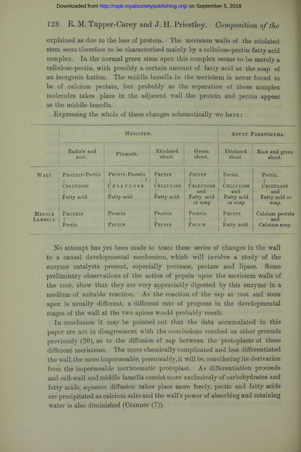
Expressing the whole of these changes schematically we have:
128 R. M. Tupper-Carey and J. H. Priestley. Composition of the
M e r i s t e m . A d u l t P a r e n c h y m a .
Eadicle and Plumule. Etiolated Green Etiolated Foot and greenroot.
ishoot. shoot. shoot. shoot.
W a l l P R O T E I N - P e c t i n PECTIN-Protein1 : ?
C e l l u l o s e
P e c t i nl
P e c t i n PectinI
Pectin.1
C e l l u l o s e
1C e l l u l o s e C e l l u l o s e
1C e l l u l o s e C e l l u l o s e
| | | and and andFatty acid Fatty acid Fatty acid Fatty acid Fatty acid Fatty acid or
or soap or soap soap.
M i d d l e P r o t e i n Protein Protein Protein P e c t i n Calcium pectateL a m e l l a i i i and
Pectin P e c t i n P e c t i n P e c t i n Fatty acid Calcium soap.
No attempt has yet been made to trace these series of changes in the wall to a causal developmental mechanism, which will involve a study of the enzyme catalysts present, especially protease, pectase and lipase. Some preliminary observations of the action of pepsin upon the meristem walls of the root, show that they are very appreciably digested by this enzyme in a medium of suitable reaction. As the reaction of the sap at root and stem apex is usually different, a different rate of progress in the developmental stages of the wall at the two apices would probably result.
In conclusion it may be pointed out that the data accumulated in this paper are not in disagreement with the conclusions readied on other grounds previously (20), as to the diffusion of sap between the protoplasts of these different meristems. The more chemically complicated and less differentiated the wall, the more impermeable, presumably,it will be, considering its derivation from the impermeable meristematic protoplast. As differentiation proceeds and cell-wall and middle lamella consist more exclusively of carbohydrates and fatty acids, aqueous diffusion takes place more freely, pectic and fatty acids are precipitated as calcium salts and the wall’s power of absorbing and retaining water is also diminished (Cranner (7)).
on September 5, 2018http://rspb.royalsocietypublishing.org/Downloaded from
Cell-Wall at the Apical Meristemof Stem and . 129
Summary.1. Differences in microchemical reaction between the apical meristems of
root and stem in the broad bean, suggested that a difference in chemical composition might exist and explain the difference in relative permeability found during an earlier investigation.
2. A detailed microchemical investigation of the reaction of the cell walls of the various meristems towards cellulose reagents led to their classification as follows:—
(1) Radicle, root and plumule, which do not give the cellulose reaction with iodine and sulphuric acid unless previously treated with strong acids or alkalis, and do not give the cellulose reaction with chloriodide of zinc unless well treated with aqueous or alcoholic alkalis.
(2) Etiolated stem,which gives the reaction with iodine and sulphuric acid direct, but require short treatment with boiling aqueous or alcoholic caustic alkalis before the reaction is given with chloriodide of zinc.
(3) Normal green stem, which gives the reaction with iodine and sulphuric acid direct, and only a short treatment with various reagents is necessary before the reaction is given with chloriodide of zinc.
3. Macrochemical experiments prove the existence of cellulose in the walls of the meristem, but its presence is masked by association with other substances.
4. Protein, closely linked to the cellulose, is found by macrochemical experiments to be most probably the substance which prevents the reaction with iodine and sulphuric acid.
5. Pectin is present in each case, though not directly linked to the cellulose in the meristem wall of radicle and root.
6. The middle lamella ‘in the meristem is never of calcium pectate but is probably a mixture of pectin and protein.
7. When all fat has been extracted from the meristem of the radicle by the use of the usual fat solvents, and the protoplasts themselves dissolved out with Eau de Javelle, the residue, after boiling with alcoholic potash, yields a small quantity of some fatty substance, presumably from the walls, with an iodine value of 104.
This fat, present in the walls of the meristem of root, radicle, plumule and etiolated stem, closely linked to the cellulose, is responsible for the failure of chloriodide of zinc to give the cellulose reaction.
8. As the impermeability of the meristematic protoplast depends, in part, upon the fats present in a finely divided state, the presence of calcium in the
on September 5, 2018http://rspb.royalsocietypublishing.org/Downloaded from
130 Composition of Cell- Wall at Apical of Stem and .
sap will cause precipitation, involving increased permeability and subsequent differentiation.
9. Estimation of the amount of calcium present in the various meristems show it to be greatest in the stem apex when grown in the light and least in the meristem of the radicle.
10. It is assumed that the plant wall arises by gradual transition from a. living protoplasmic interface, to a structural system of relatively simple chemical compounds.
Various writers incidentally support this view, so that the difference in chemical composition of the cell walls at the various meristems is explained by the suggestion that the developmental changes proceed most slowly at the root apex, more quickly in the plumule and etiolated stem, and most rapidly at the normal green stem apex, under the influence of enzyme catalysts.
REFERENCES.1. Artschwager, Ernst, “ Use of Chloriodide of Zinc in Plant Histology,” ‘Bot. Gaz.,’
vol. 71, p. 400 (1921).2. Bancroft, ‘Applied Colloid Chemistry,’ New York, 1921.3. Bolles Lee, * The Microtomist’s Yade Meeurn,’ 1921.4. Correns, C. E., “ Ueber die vegetabilisclie Zellmembran,” ‘ Jahrb. Wiss. Bot.,’ vol. 26,
pp. 587-673 (1894).5. Cranner, B. Hansteen, “ Ueber das Verhalten der Kulturpflanzen zu den Boden-
salzen III, Beitriige zur Biochemie und Physiologie der Zellvvand lebender Zellen,” ‘ Jahrb. Wiss. Bot.,’ vol. 53, pp. 536-598 (1914).
6. Cranner, B. Hansteen, “ Beitrage zur Biochemie und Physiologie der Zellwand undder plasmatischen Grenzschichten,” ‘ Ber. Deutsch. Bot. Ges.,’ vol. 37, pp. 380-391 (1919).
7. Cranner, B. Hansteen, “ Zur Biochemie und Physiologie der Grenzschichten lebenderPflanzeuzellen,” ‘Meldinger fra Noyes Landbrukshiskole,’ vol. 2, pp. 1-160 (1922).
8. Czapek, F., “ Zum Nachweis von Lipoiden in Pflanzenzellen,” ‘ Ber. Deutsch. Bot.Ges.,’ vol. 37, pp. 207-216 (1919).
9. Gleisberg, “ Der gegenwsirtige Stand der Membranforschung,” ‘ Beitr. zum Bot.Centr.,’ vol. 38,1., pp. 217-265 (1921).
10. Haas and Hill, ‘An Introduction to the Chemistry of Plant Products,’ vol. 1,London (1921).
11. Krabbe, “ Ein Beitrag zur Kenntniss der Structur und der Wachsthums vegetabi-lischer Zellbaute,” ‘Jahrb. wiss. Bot.,’ vol. 18 (1887).
12. Leathes, J. B., “ The Fats,” ‘ Monographs on Biochemistry,’ London (1910).13. Mangin, M.L., “Recherches anatomiques sur la Distribution des Composes Pectiques,”
Paris, 1893 (Extrait du ‘Journal de Botanique’).14. Maclean, Hugh, ‘ Lecithin and Allied Substances,’ London, 1918.15. Molisch, Hans, ‘Mikrochemie der Pflanze,’ 1913.16. Onslow, Muriel Wheldale, ‘ Plant Biochemistry,’ 1920.17. Palladin, W., “ Ergrunen und Wachstum der etiolirten Blatter,” ‘Ber. Deutsch. Bot.
Ges.,’ vol. 9, p. 229-232 (1891).18. Priestley, J. H., “ The Fundamental Fat Metabolism of the Plant,” ‘ New Phyto-
logist,’ 1923.
on September 5, 2018http://rspb.royalsocietypublishing.org/Downloaded from
Effect of X-Rays of Different Wave-Lengths. 131
19. Priestley, J. H., and North, Edith, “ Physiological Studies in Plant Anatomy.—III. The Structure of the Endodermis in Relation to its Function," ‘ New Phyto- logist,’ vol. 21, pp. 115-149 (1922).
20. Priestley, J. H., and Tupper-Carey, R. M., “ Physiological Studies in Plant Anatomy.IY. The Water Relations of the Plant Growing Point,” ‘New Phytologist, vol. 21, pp. 210-229 (1922).
21. Priestley, J. H., and Ewing, J., “ Physiological Studies in Plant Anatomy.—VI.Etiolation,” ‘New Phytologist,’ vol. 22, pp. 30-44 (1923).
22. Plimmer, R. H. A., ‘ Practical Organic and Biochemistry,’ London, 1918.23. Schryver, S. B., and Haynes, D., “ The Pectic Substances of Plants,” ‘ Biochem.
Journ.,’ vol. 10, pp. 539-547 (1916).24. Shohl, A. T., and Pedley, F. C., “ A Rapid and Accurate Method for Calcium in
Urine,” ‘ Journ. Bio. Chem.,’ vol. 50, pp. 537-544 (1922).25. Strasburger, E., “ Die pflanzlichen Zellhaute,” ‘ Jahrb. wiss. Bot.,’ vol. 29, pp. 511—
598 (1898).26. Tollens, B., ‘ Kurzes Handbuch der Kohlenhydrate,’ Leipzig, 1914.27. Tutin, F., “ The Behaviour of Pectin towards Alkalis and Pectase,” ‘ Biochem.
Journ.,’ vol. 15, pp. 494-497 (1921).28. Wiesner, J., “ Untersuchungen liber die Organisation der vegetabilischen Zellhaut,”
‘ Sitzungsber. K. Akad. Wiss. Wien (Math.-nat. Klasse),’ vol. 93 (1886).29. Wiesner, J., “ Ueber den Nachweis der Eiweisskorper in den Ptlanzenzellen,” ‘ Ber.
Deutsch. Bot. Ges.,’ vol. 6, pp. 187-195 (1888).30. Wiesner, J., “ Zur Eiweissreaction und Structur der Zellmembran,” ‘ Ber. Deutsch.
Bot, Ges.,’ vol. 6, pp. 33-36 (1888).
On the Effect of X-rays of Different Wave-Lengths upon some Animal Tissues.—Proof of Differential Action.
By S. Russ, D.Sc., F.Inst.P., Cancer Research Laboratories, MiddlesexHospital.
(Communicated by Prof. A. W. Porter, F.R.S. Received April 17th, 1923.)
CONTENTS.
1. Introduction ........................2. Physical Section....................3. Animal Experiments Section4. Discussion of Results .........
P age
131132 137 141
Introduction.In a paper (1) upon the Germicidal Action of Ultra-Violet rays, it is shewn that
a marked differential action exists in this part of the spectrum, electromagnetic disturbances have a different effect upon the same kind of organism
on September 5, 2018http://rspb.royalsocietypublishing.org/Downloaded from