Embed Size (px)
Citation preview
The Limping ChildTodd Milbrandt, MD
Division ChairPediatric OrthopaedicsMayo Clinic‐Rochester
Faculty Disclosure
•No disclosures relevant to this talk
Practice Gap
•Primary Care Providers are faced with limping children everyday
•The differential is long and can be confusing.•By creating a guiding algorithm, this can be a manageable problem.
Objectives
•Develop a differential diagnosis for the limping child
•Briefly describe the diagnostic and therapeutic options for those conditions
Limps defined
•A deviation in a child’s walking pattern•More commonly unilateral•Reasons fall into one of three categories
• Pain• Weakness• Structural abnormality
Painful conditions
•Osteoarticular infection•Neoplasia•Trauma
• Fracture
Weak Conditions
•Neuromuscular•Cerebral palsy
•Muscle weakness•Muscular Dystrophy

Structural Condition
• Leg length inequality• Joint stiffness•Articular surface deformity
Infection‐Septic Arthritis
•Who?•Young children and infants
•How present?• Joint pain, involuntary guarding, and muscle spasm
•Pseudo paralysis•May be toxic and febrile•But may not be!!

Infection‐Septic Arthritis•How Diagnose?
• Only definitive answer is ASPIRATION• Hip‐U/S can guide if leg pain due to septic arthritis• Must have a high index of suspicion• Labs help
• If WBC>12,000, ESR>40, inability to bear wt, and fever>38.5 C, then septic arthritis >90%
•Treatment• Surgical Drainage
•Remember the Psoas abscess that can mimic the septic arthritis!!!!

Infection‐Osteomyelitis
•Who?•From neonate to 10 years old
•How present?•Bone pain with inability to move extremities•Maybe a recent history of bacteremia•+/‐ fever•May tell you that they just fell down history of trauma
Infection‐Osteomyelitis
•Why do they get it?•Vascular anatomy
•Metaphyseal “sludge”•Really terminal branches that allow extravasation of cells and bacteria
•This forms the nidus of the infection•Local modulators
•Prostaglandins, TNF and interleukins all released by Staph cause inflammation response
Infection‐Osteomyelitis
•How Diagnose?•Three levels according to Morrey and Peterson
•Definite‐ “The pathogen is isolated from the bone or there is histological evidence of osteomyelitis”
•Probable‐ “A blood culture is positive in the setting of clinical or radiographic features
• Likely‐ “Typical clinical findings and definte radiographic evidence are present and there is a response to antibiotics”
Infection‐Osteomyelitis
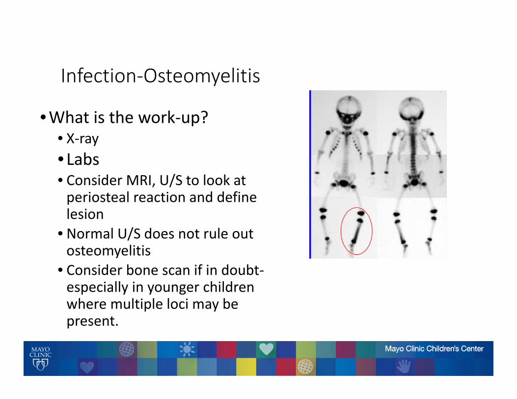
•What is the work‐up?• X‐ray• Labs• Consider MRI, U/S to look at periosteal reaction and define lesion
• Normal U/S does not rule out osteomyelitis
• Consider bone scan if in doubt‐especially in younger children where multiple loci may be present.
Infection‐Osteomyelitis
•Treatment•All loci aspirated• If no pus and no bone destruction on x‐ray then its ok to treat solely with antibiotics
• If pus on aspiration then treatment with surgical drainage and IV antibiotics
•The threshold for children under 1 may be less‐especially near the proximal femoral metaphysis.

Neoplasia•Who?
• Anywhere along the childhood spectrum•How present?
• Painful extremity• May have fever or not
•What is the work‐up?• X‐ray, labs• MRI now standard to delineate lesion• Biopsy can be tricky when considering future treatment and resections
•Treatment• Depends on the tumor‐should be reserved for specialist care

Trauma•Major fractures fairly obvious•Subtle fractures or Stress fractures
•Common locations include•Femoral neck/shaft•Tibial shaft•Metatarsals
•Usually history is one of recent increase in activity
•Work up with either bone scan or MRI of location
CEREBRAL PALSY
•Good Walkers: decreased step length, decreased step time, increased cadence, normal speed
•Poor Walkers: decreased step length, increased stride time, decreased speed
SPASTIC HEMIPLEGIA
•Usually present as unilateral toe‐walkers or “dragging one leg”
•Delay in age of walking ‐ 18 to 24 mos.•Early handedness a clue

SPASTIC HEMIPLEGIA
•Upper extremity spasticity• Ipsilateral lower extremity spasticity•Classic etiology ‐ intracerebral bleed
SPASTIC HEMIPLEGIA
• Look carefully for posturing of the ipsilateral arm•May elicit posturing during running•Check fine motor activities (use your keys)•Early handedness/ ignorance of contralateral hand
SPASTIC HEMIPLEGIA
•Ankle equinus•Possible foot drop•Crouch at knee•Possible stiff knee in swing phase•Varus of foot
SPASTIC HEMIPLEGIA
•Tight heel cord (Achilles tendon) leads to toe walking
• If mild, may lower heel to floor after contacting the ground with the toe
•May thrust knee into recurvatum to keep the foot on the ground in stance phase

ANKLE EQUINUS
•Early heel rise•Shorter step length• Limited push off power since the ankle is already plantarflexed

ANKLE EQUINUS
• Lose heel contact since foot already plantarflexed• Limited dorsiflexion in stance phase, therefore difficulty advancing limb over foot
ANKLE EQUINUS
•Compensatory knee recurvatum due to tight heel cord
SPASTIC HEMIPLEGIA
•TREATMENT ‐ first establish the diagnosis• If young ‐may require AFO bracing, physical therapy• If older and/or has contractures ‐ tendon lengthening of the Achilles tendon with or without hamstring lengthening
ANKLE EQUINUS
•Tendoachilles lengthening•Need to assess multiple joints for contractures•Can restore more normal kinematics and power to the ankle plantarflexors
SPASTIC HEMIPLEGIA•Measure the popliteal angle for hamstring tightness•The knee will bend either due to tight hamstrings or to compensate for toe‐walking

FOOT DROP
• Inability of ankle to dorsiflex in swing phase•Stub toe•Steppage gait•Ankle foot orthosis effective (AFO)
SPASTIC DIPLEGIA
• Involvement of both lower extremities with lesser involvement of the arms
•Usual etiology ‐ hypoxia, prematurity•Very delayed walkers ‐ 2 to 7 years• If the arms are completely normal, beware! Could be spinal cord pathology.
SPASTIC DIPLEGIA
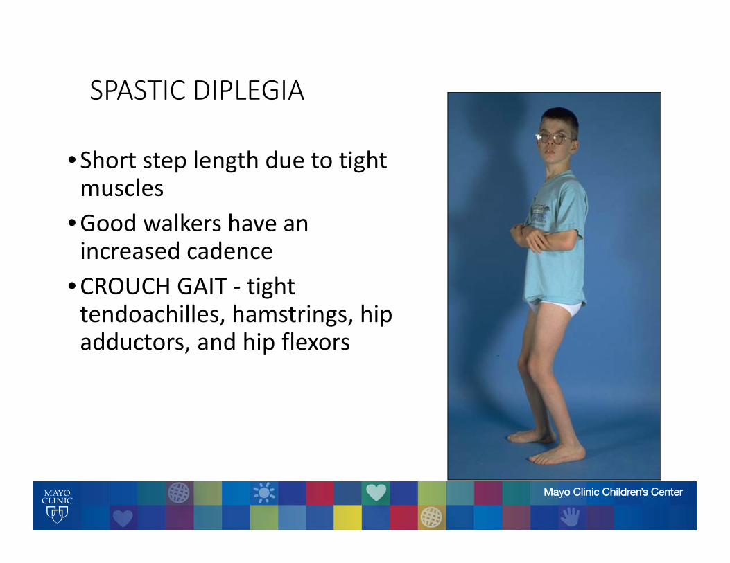
•Short step length due to tight muscles
•Good walkers have an increased cadence
•CROUCH GAIT ‐ tight tendoachilles, hamstrings, hip adductors, and hip flexors

SPASTIC DIPLEGIA
•Ankle equinus•Possible foot drop•Crouch at the knee +/‐ stiff knee
•Crouch at the hip or anterior pelvic tilt
• Intoeing (femoral anteversion)
CROUCH KNEE
•Due to hamstring spasticity• Increased popliteal angle•Places increased demands on quadriceps
•Shortens stride length
Weakness‐Muscular Dystrophy
•Usually present age 3 to 6 years•Complain of clumsy gait, stumbling, or toe walking•Normal birth history•Usually mildly delayed walking age (18 mos)
MUSCULAR DYSTROPHY
•May present as toe‐walking•BEWARE boys who toe‐walk with normal birth histories
•Perform the Gower’s test

GOWER’S SIGN
•Have the boy sit on the floor
• Instruct him to rise to standing as fast as possible
•Watch to see if they use their hands to lock back the knees and climb up their thighs.

MUSCULAR DYSTROPHY
•Proximal greater than distal muscle weakness
•Hips weaker than toes•Shoulders weaker than hands• Look for pseudohypertrophy (enlargement of gastrocsoleus as the muscle belly is replaced by fat)
MUSCULAR DYSTROPHY
•GAIT DISTURBANCE: •wide base of support•equinus of the ankles +/‐ varus•increased lumbar lordosis and anterior pelvic tilt (compensates for lack of hip extensor strength)
MUSCULAR DYSTROPHY
•TRENDELENBERG GAIT:•Hip abductors weak•Cannot stabilize pelvis during single limb stance phase
•Swing‐side hemipelvis drops•Body sways over stance phase leg to help the weak abductors out.
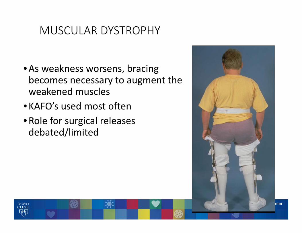
MUSCULAR DYSTROPHY
•As weakness worsens, bracing becomes necessary to augment the weakened muscles
•KAFO’s used most often•Role for surgical releases debated/limited
Structural‐Developmental Dysplasia of the Hips• Incidence of dislocated hips 1‐2/1000• Incidence of dislocatable hips 5‐10/1000• Incidence of dysplastic but stable hips ??

D.D.H.
•DYSPLASIA: a descriptive term for underdevelopment of the acetabulum
• Leads to early arthritis (teens, young adults)•Usually have normal gait as long as the hip doesn’t hurt
•Surgery may be necessary
D.D.H.
•Examination requires a quiet relaxed baby•Check one hip at a time
D.D.H.
•Diagnosis in the neonate: Ortolani and Barlow maneuvers, limited abduction.
•Diagnosis in the infant (>3‐6 mos): Limited abduction, Galleazzi sign, asymmetric thigh folds

D.D.H.
•Ortolani maneuver reduces a dislocated hip
D.D.H.
•Barlow maneuver dislocates a reduced hip.
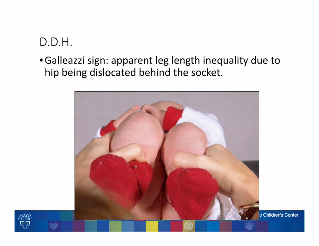
D.D.H.•Galleazzi sign: apparent leg length inequality due to hip being dislocated behind the socket.

D.D.H.• Limited abduction of the dislocated hip
D.D.H.
•EARLY DIAGNOSIS IS MANDATORY• Less treatment and no surgery•Once walking age, surgery is necessary
D.D.H.
•Diagnosis in the walking child: Waddling gait (Trendelenberg), limited abduction, increased lordosis, leg length inequality
D.D.H.
• If bilateral, the walking child will shift the body weight from side to side over the stance phase leg.
•TRENDELENBERG GAIT (waddling)
TRENDELENBERG GAIT
•Hip abductors of the stance phase leg fire to keep the pelvis level as the opposite leg leaves the ground.
• In hip conditions, the abductors are weak and the swing limb hemipelvis drops
•May compensate by throwing body over the stance phase limb to level the pelvis

D.D.H.•Once suspected, the diagnosis in a child of walking age is made from the xray.
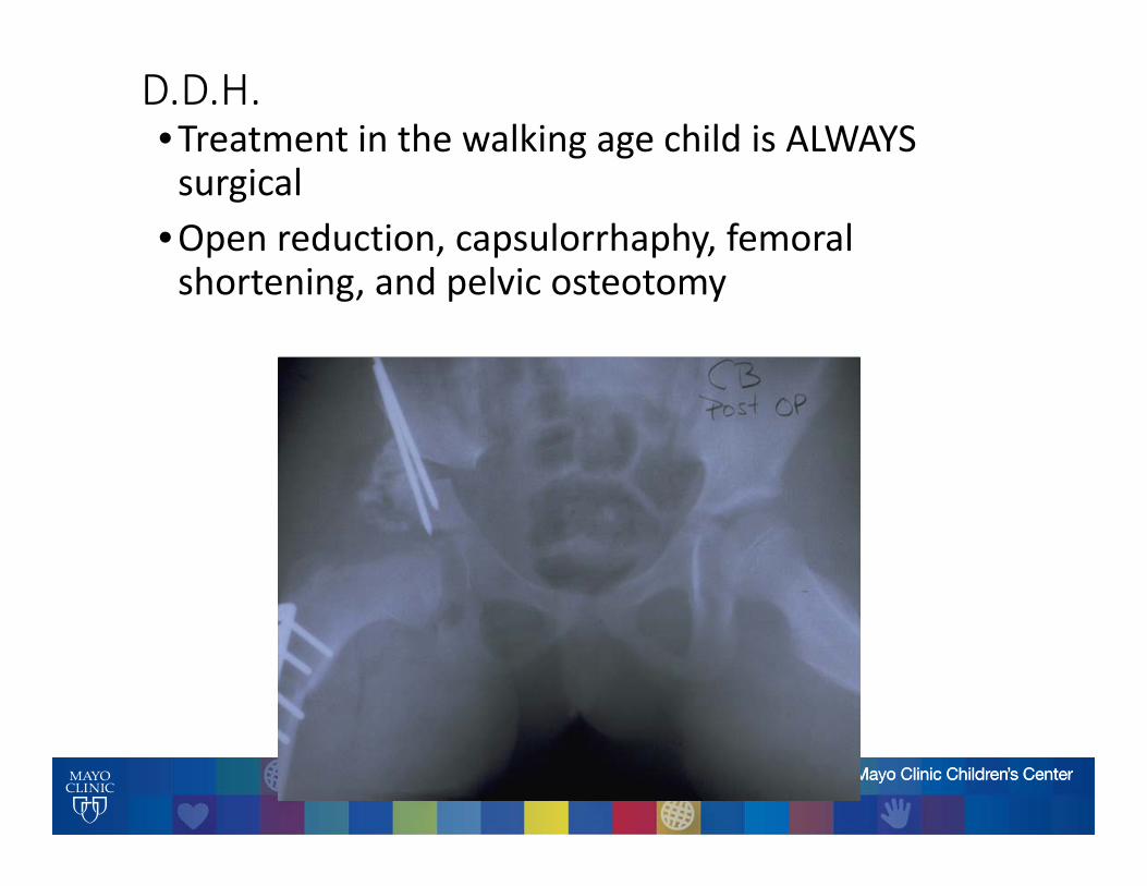
D.D.H.•Treatment in the walking age child is ALWAYS surgical
•Open reduction, capsulorrhaphy, femoral shortening, and pelvic osteotomy
Structural‐ S.C.F.E.
•Slipped capital femoral epiphysis•Posterior migration of the femoral head on the femoral neck
•Presents as pain in the hip, groin, buttock, or knee.•Usually 10‐14 years old
S.C.F.E.
•Usually overweight children.
S.C.F.E.
•Physical examination: • Limited internal rotation of the hip• Hip will externally rotate as you flex it• Pain with internal rotation of the hip
S.C.F.E.
•Radiographic findings: •AP view is usually normal in mild slips
•Physis appears wide
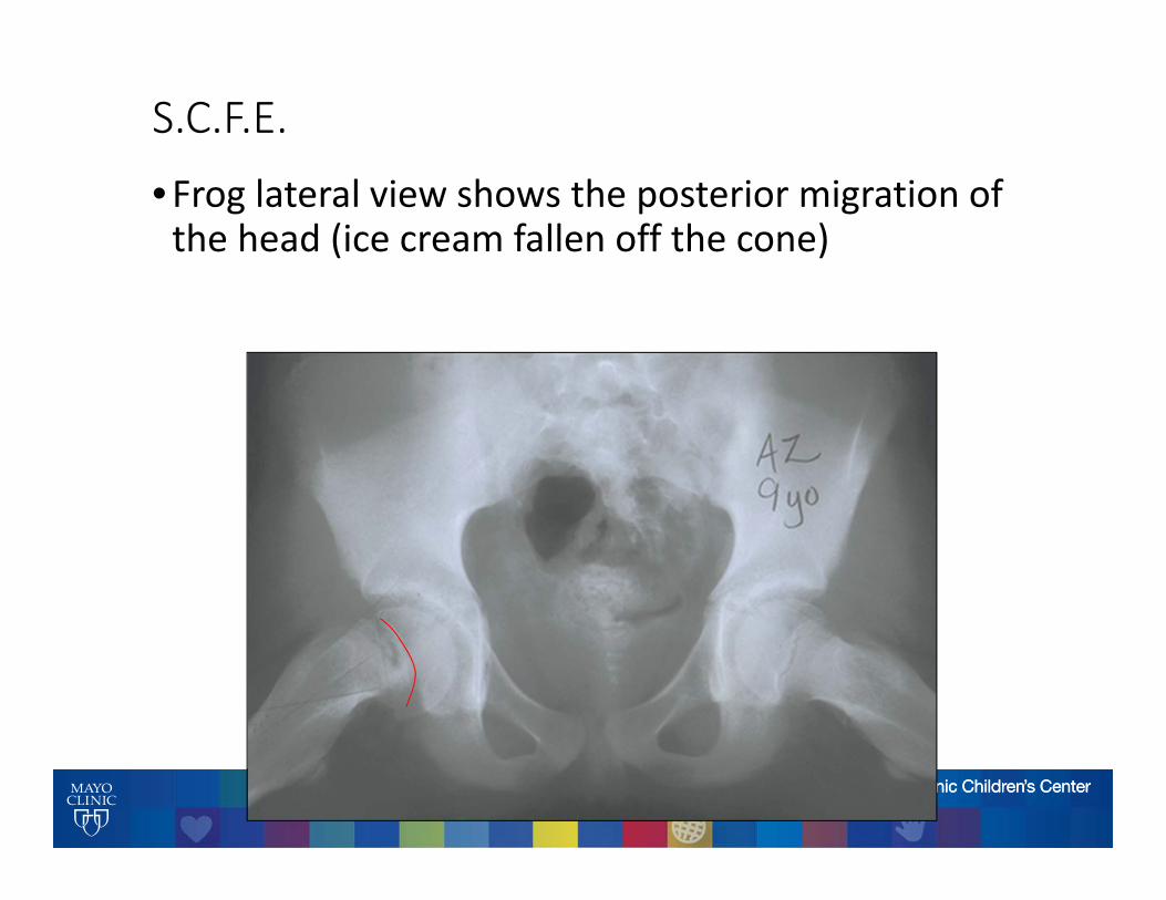
S.C.F.E.•Frog lateral view shows the posterior migration of the head (ice cream fallen off the cone)
S.C.F.E.
•Two varieties:•Stable slip: patient can walk on it; pain usually present for weeks to months; complain of pain and limp
•Unstable slip: acts like a hip fracture; come in by ambulance.
S.C.F.E.
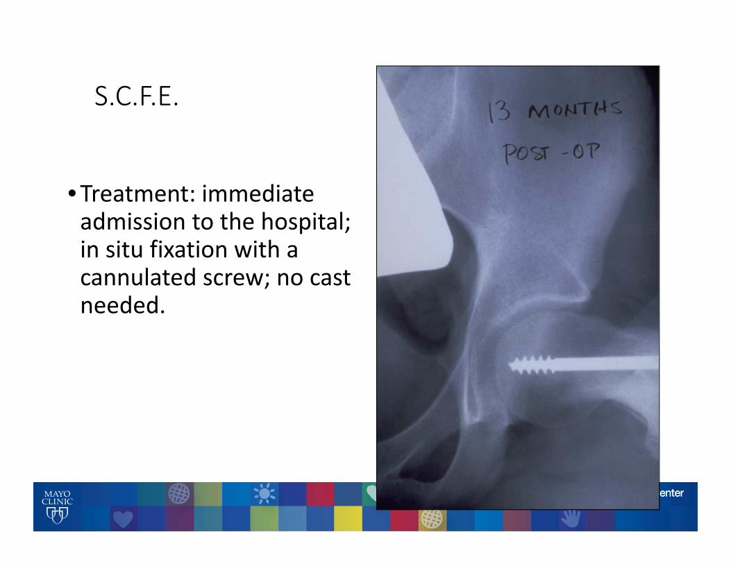
•Treatment: immediate admission to the hospital; in situ fixation with a cannulated screw; no cast needed.
S.C.F.E.
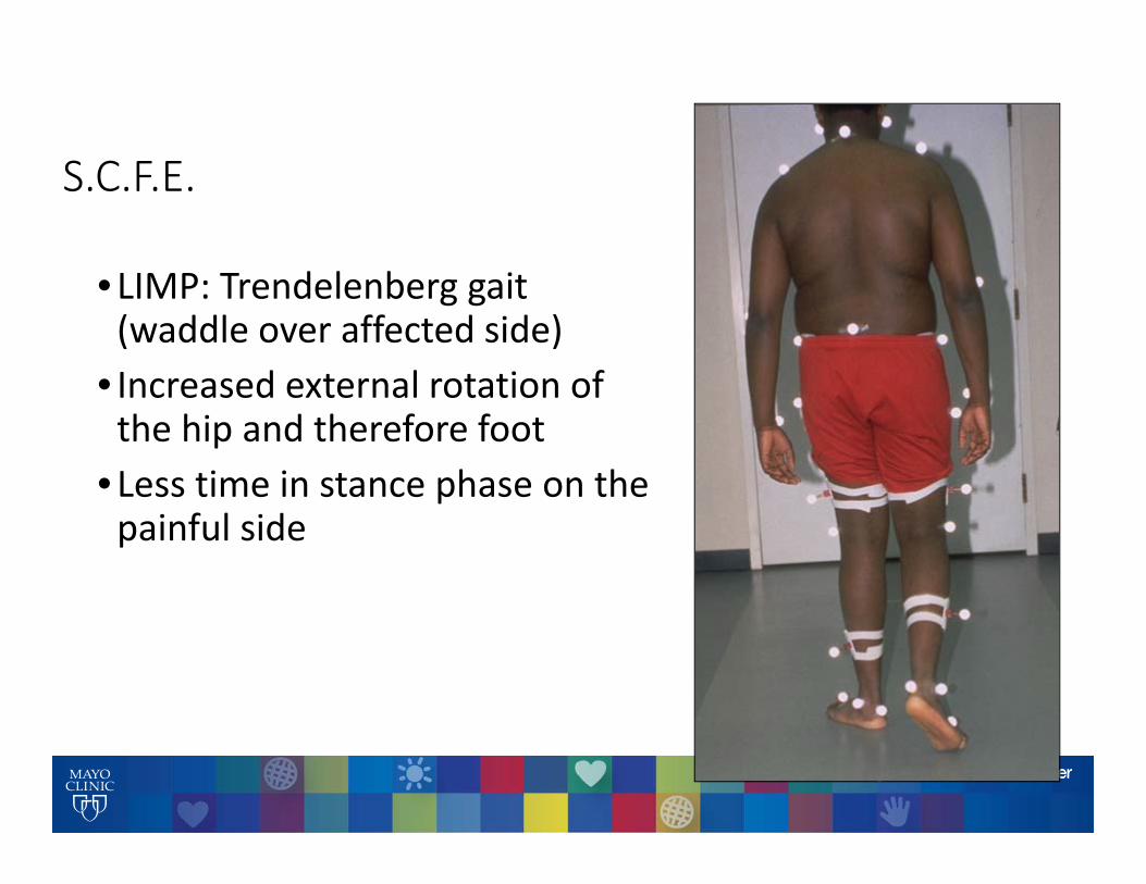
• LIMP: Trendelenberg gait (waddle over affected side)
• Increased external rotation of the hip and therefore foot
• Less time in stance phase on the painful side

S.C.F.E.
•OUTCOME:• The outcome of the hip (arthritis) is directly related to the severity of the displacement of the femoral head
• The slip continues to migrate over time without surgery• Therefore diagnosis is critical at presentation to the primary care physician
Take Home Message
•Be able to identify typical patient•Be alert to the variety of pain complaints
•Hip, thigh, groin or knee• Be aware of Atypical slips•Always obtain AP and Frog pelvis films
Structural‐PERTHES DISEASE
•Also known as Legg‐Calve‐Perthes disease• Idiopathic avascular necrosis of the head of the femur in an otherwise healthy child
•Usually 4 ‐ 8 years old, males > females•Usually thin and very active•Uncommon in black children
PERTHES DISEASE
•Parents complain of limp•Antalgic and Trendelenberg gait• Loss of range of motion of the hip (abduction most)
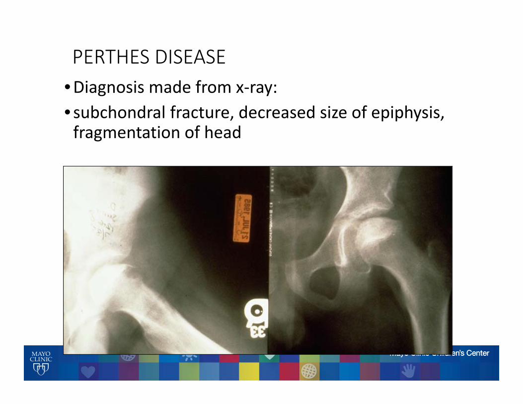
PERTHES DISEASE•Diagnosis made from x‐ray:• subchondral fracture, decreased size of epiphysis, fragmentation of head
PERTHES DISEASE
•Treatment is controversial•Surgery sometimes needed to decrease pressure on hip
•Bracing less common
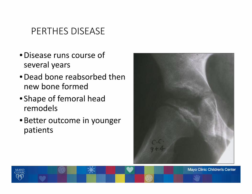
PERTHES DISEASE
•Disease runs course of several years
•Dead bone reabsorbed then new bone formed
•Shape of femoral head remodels
•Better outcome in younger patients

Antalgic Gait
Infectious-Like
Clinical Signs:Recent History of coldFever/ChillsErethema over extremityIll-appearing
Lab values:Elevated C-reactive ProteinElevated White Blood CellsElevated ESR
Focused Clinical Examination
Decreased Range of Motion of any Joint with Swelling:Test: Joint AspirationIf positive then septic jointIf negative then transientsynovitis or juvenile arthritis
Focused Pain or Erethema over an ExtremityTest: Plain RadiographIf positive then osteomyelitisIf negative and still with high index of suspicion then MRI
If Cannot Focus Examination:Test: Bone Scan to focus furtherevaluation
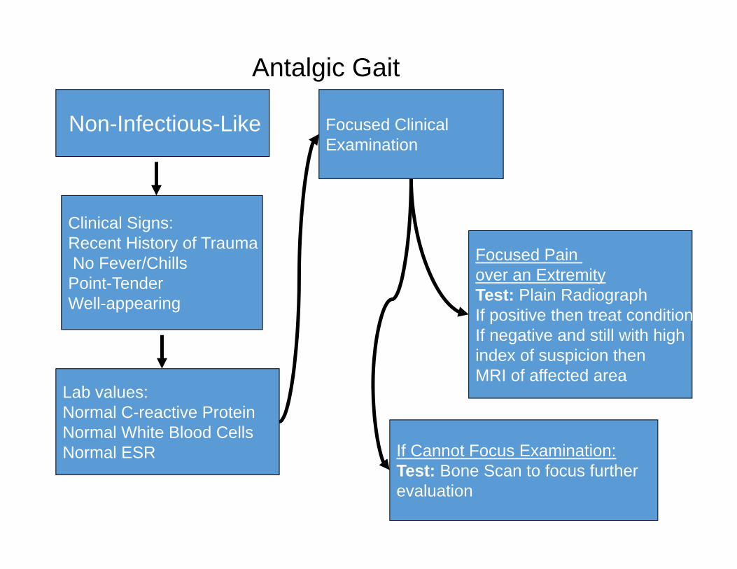
Antalgic Gait
Non-Infectious-Like
Clinical Signs:Recent History of TraumaNo Fever/ChillsPoint-TenderWell-appearing
Lab values:Normal C-reactive ProteinNormal White Blood CellsNormal ESR
Focused Clinical Examination
Focused Pain over an ExtremityTest: Plain RadiographIf positive then treat conditionIf negative and still with high index of suspicion then MRI of affected area
If Cannot Focus Examination:Test: Bone Scan to focus furtherevaluation

Non-Antalgic Gait
Toe walking
Physical Exam: Spasticity
Physical Exam: No Spasticity
Leg Length DifferenceTest: Plain Radiographs of HipsAnd Lower Extremities
Gait type Physical Exam or Test
Cerebral Palsy
Idiopathic Toe Walking
•DDH•Congenitally short femur•Congenitally short tibia
Diagnosis
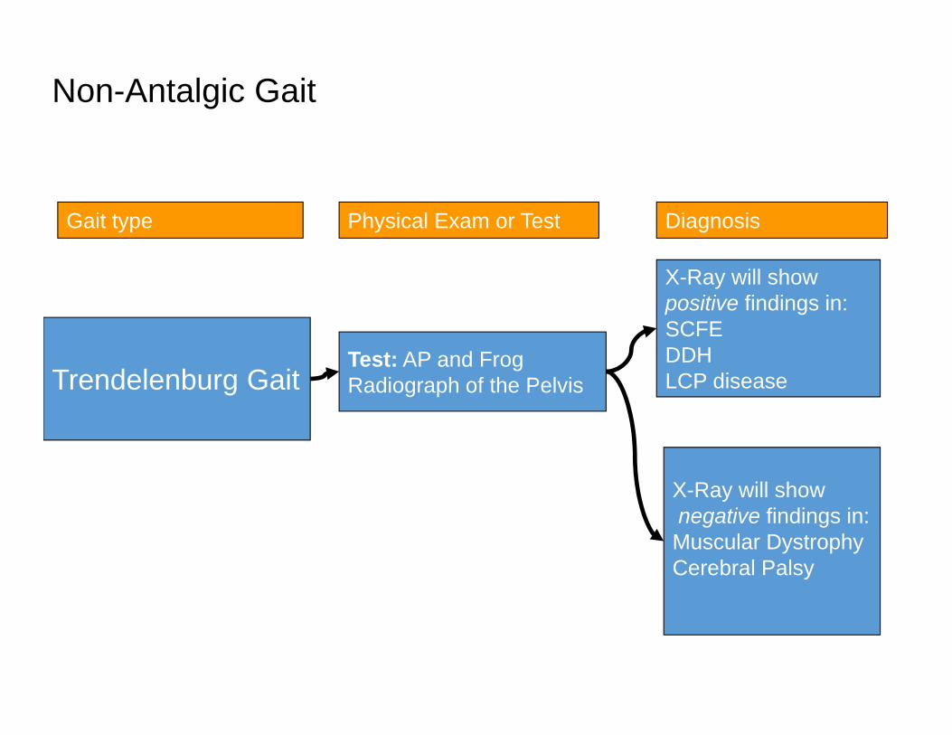
Non-Antalgic Gait
Trendelenburg GaitTest: AP and Frog Radiograph of the Pelvis
Gait type Physical Exam or Test Diagnosis
X-Ray will showpositive findings in:SCFEDDHLCP disease
X-Ray will shownegative findings in:Muscular DystrophyCerebral Palsy
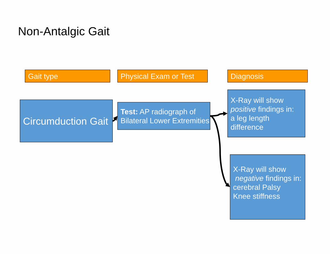
Non-Antalgic Gait
Circumduction Gait
Gait type Physical Exam or Test Diagnosis
Test: AP radiograph ofBilateral Lower Extremities
X-Ray will showpositive findings in:a leg lengthdifference
X-Ray will shownegative findings in:cerebral PalsyKnee stiffness