Embed Size (px)
Citation preview

Send Orders for Reprints to [email protected]
The Open Dentistry Journal, 2016, 10, 261-267 261
1874-2106/16 2016 Bentham Open
The Open Dentistry Journal
Content list available at: www.benthamopen.com/TODENTJ/
DOI: 10.2174/1874210601610010261
Chronic Maxillary Sinusitis Caused by Denture Lining Material
Tsutomu Sugiura1,2,*, Kazuhiko Yamamoto2, Chie Nakashima1,2, Kazuhiro Murakami2, YumikoMatsusue2, Satoshi Horita2, Go Sakagami3 and Tadaaki Kirita2
1Department of Oral and Maxillofacial Surgery, Narakasuga Hospital, Nara, Japan2Department of Oral and Maxillofacial Surgery, Nara Medical University, Nara, Japan3Department of Otolaryngology, Nara City Hospital, Nara, Japan
Received: February 02, 2016 Revised: April 27, 2016 Accepted: May 03, 2016
Abstract: We report a case of chronic maxillary sinusitis caused by denture lining material entering through an oroantral fistula aftertooth extraction. The patient was an 80-year-old female who visited us with a complaint of pus discharge from the right posteriormaxilla. She had extraction of the upper right second molar and had her upper denture relined with silicone lining material. Thepatient noticed swelling of the right cheek and purulent rhinorrhea 20 days before her first visit to our clinic. Oral examinationshowed an oroantral fistula with a diameter of 3 mm in the posterior alveolar ridge of the right maxilla. Computed tomographyrevealed a hyperdense foreign body in the right maxillary sinus and thickening of the mucosal lining. Under diagnosis of maxillarysinusitis caused by a foreign body, endoscopic maxillary surgery was performed simultaneously with the removal of the foreignbody. The foreign body removed was 12 × 6 mm in size, oval in shape, light pink in color, and compatible with silicone denturelining material. During the follow-up it was observed that the oroantral fistula closed spontaneously after the removal of the foreignbody. The maxillary sinus was in a good shape without recurrence of sinusitis seven months after surgery.
Keywords: Denture lining material, Endoscopic surgery, Foreign body, Maxillary sinusitis.
INTRODUCTION
Maxillary sinusitis is occasionally odontogenic because of the anatomic juxtaposition of the teeth and maxillarysinus. Odontogenic sources once accounted for approximately 10 to 12% of all maxillary sinusitis cases [1]. However,in recent publications, up to 30% to 40% of such cases have been attributed to dental causes such as an oroantralfistulae, foreign bodies, and periapical/periodontal lesions [2, 3]. Maxillary sinusitis secondary to the presence offoreign bodies is an unusual clinical complication [4]. Most foreign bodies, such as roots of teeth, root-filling materials,dental implants, and dental impression materials, are related to iatrogenic dental manipulation and/or the presence of anoroantral fistula [5 - 9].
Small foreign bodies in the maxillary sinus can be expelled spontaneously, but in most cases they require removalbecause they may cause chronic sinusitis [9]. A variety of techniques have been reported for the removal of foreignbodies such as endoscopic surgery and an intraoral direct approach through the anterior maxillary wall. In some cases, acombination of these approaches is necessary [6]. Therefore, the best treatment should be chosen according to the sizeand location of the foreign body and the condition of the maxillary sinus.
This paper reports a rare case of chronic maxillary sinusitis caused by denture lining material entering through anoroantral fistula, which was successfully treated endoscopically.
* Address correspondence to this author at the Department of Oral and Maxillofacial Surgery, Nara Medical University, 840 Shijo-cho, KashiharaCity, Nara 634-8522, Japan; Tel/Fax: +81-744-29-8875; E-mail: [email protected]

262 The Open Dentistry Journal, 2016, Volume 10 Sugiura et al.
CASE REPORT
An 80-year-old female was referred to the Department of Oral and Maxillofacial Surgery, Narakasuga Hospitalbecause of pus discharge from an oroantral fistula at the right posterior maxilla. The right upper second molar had beenextracted 43 days before because of chronic periodontitis. She noticed swelling of the right cheek, purulent rhinorrhea,and discharge through the oroantral fistula 20 days before. She was prescribed antibiotics for seven days by her familydoctor. The cheek swelling improved, but the discharge through the oroantral fistula persisted. She had a medicalhistory of hypertension and chronic hepatitis C, both of which were well controlled with an angiotensin II receptorblocker, calcium channel blocker, and ursodeoxycholic acid. The right chronic maxillary sinusitis was identified in X-ray findings by her family doctor several years ago. However, it was asymptomatic and was not treated. A physicalextra-oral examination was unremarkable. Oral examination revealed an oroantral fistula with a diameter of 3 mm in theposterior alveolar ridge of the right maxilla (Fig. 1). By irrigation of the right maxillary sinus through the oroantralfistula, purulent discharge was observed through the fistula.
Fig. (1). Intraoral findings at the first visit. An oroantral fistula was observed at the posterior alveolar ridge of the right maxilla(mirror image).
A panoramic radiograph showed a bone defect at the posterior alveolar ridge of the right maxilla (Fig. 2). Computedtomography (CT) showed partial thickening of the mucosal lining and the presence of a hyperdense foreign body with adensity of 240 Hounsfield units in the posterior lower part of the right maxillary sinus (Fig. 3). The patient was unawareof the presence of the foreign body. Because the upper complete denture was repaired with acrylic resin and relinedwith silicone soft lining material 36 days before (seven days after the tooth extraction), the foreign body was suspectedto be the denture lining material.
Fig. (2). Panoramic radiograph at the first visit. A bone defect was observed at the posterior alveolar ridge of the right maxilla.

Chronic maxillary sinusitis caused by denture lining material The Open Dentistry Journal, 2016, Volume 10 263
Fig. (3). CT scans at the first visit. Partial thickening of the mucosal lining and a hyperdense foreign body in the right maxillary sinuswere observed. (a) axial plane, (b) coronal plane, (c) sagittal plane.
Antibiotic therapy with cefditoren pivoxil was conducted for seven days. The oroantral fistula became almostclosed. The patient refused to undergo immediate surgical treatment. Low dose macrolide was administered to improvethe maxillary sinusitis for a month. However, CT examination showed complete opacification in the maxillary, ethmoidand frontal sinuses on the right side (Fig. 4).
Fig. (4). CT scans 6 weeks after the first visit. Complete opacification in the maxillary, ethmoid and frontal sinuses on the right sidewas observed. (a) axial plane, (b) coronal plane.

264 The Open Dentistry Journal, 2016, Volume 10 Sugiura et al.
The foreign body was removed by endoscopic surgery under general anesthesia 11 weeks after the first visit, andmaxillary antrostomy and ethmoidectomy were also performed. First, the maxillary antrostomy was performed. Themaxillary sinus was filled with inflamed hypertrophic mucosa and granulation tissue. The foreign body was found andremoved endoscopically (Fig. 5). After removal of the granulation tissue, markedly inflamed mucous membrane wasremoved and the residual mucous membrane was irrigated and preserved. Then, anterior ethmoidectomy and revision ofthe frontal recess were performed. The foreign body was 12 × 6 mm in size, oval in shape, light pink in color, andcompatible with silicone soft lining material (Fig. 6).
Fig. (5). Intra-operative endoscopic image. Inflamed hypertrophic mucosa and a foreign body (arrow) were observed.
Fig. (6). The foreign body removed.
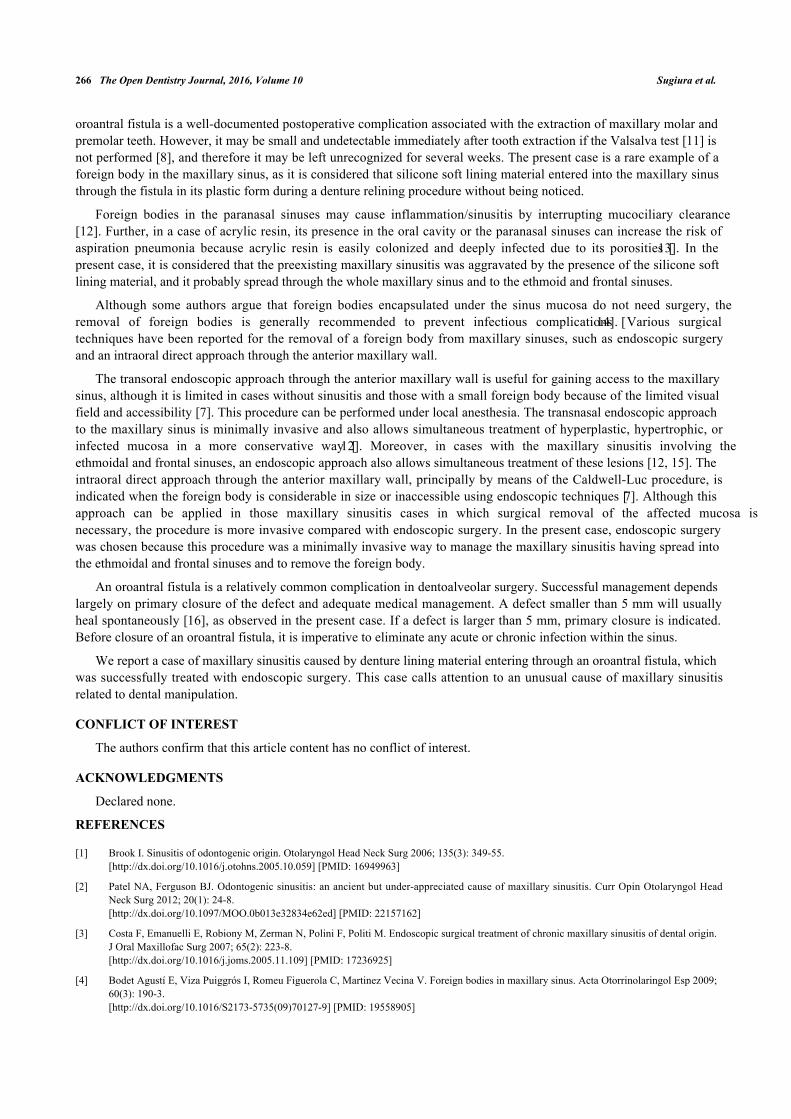
The oroantral fistula closed spontaneously four weeks after the surgery (Fig. 7). Macrolide antibiotics wereadministered for six months. The postoperative course was uneventful. After seven months of follow-up, CT scanningshowed complete aeration of the right maxillary sinus (Fig. 8).
Chronic maxillary sinusitis caused by denture lining material The Open Dentistry Journal, 2016, Volume 10 265
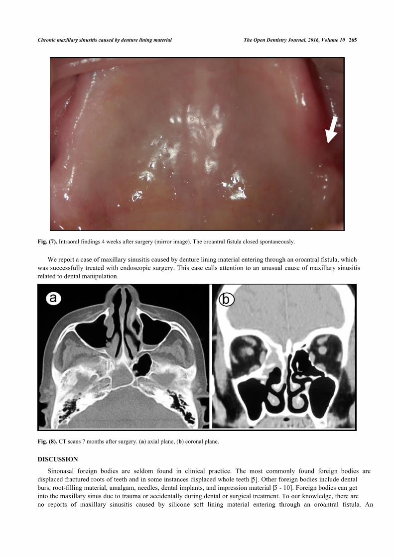
Fig. (7). Intraoral findings 4 weeks after surgery (mirror image). The oroantral fistula closed spontaneously.
We report a case of maxillary sinusitis caused by denture lining material entering through an oroantral fistula, whichwas successfully treated with endoscopic surgery. This case calls attention to an unusual cause of maxillary sinusitisrelated to dental manipulation.
Fig. (8). CT scans 7 months after surgery. (a) axial plane, (b) coronal plane.
DISCUSSION
Sinonasal foreign bodies are seldom found in clinical practice. The most commonly found foreign bodies aredisplaced fractured roots of teeth and in some instances displaced whole teeth [5]. Other foreign bodies include dentalburs, root-filling material, amalgam, needles, dental implants, and impression material [5 - 10]. Foreign bodies can getinto the maxillary sinus due to trauma or accidentally during dental or surgical treatment. To our knowledge, there areno reports of maxillary sinusitis caused by silicone soft lining material entering through an oroantral fistula. An
266 The Open Dentistry Journal, 2016, Volume 10 Sugiura et al.
oroantral fistula is a well-documented postoperative complication associated with the extraction of maxillary molar andpremolar teeth. However, it may be small and undetectable immediately after tooth extraction if the Valsalva test [11] isnot performed [8], and therefore it may be left unrecognized for several weeks. The present case is a rare example of aforeign body in the maxillary sinus, as it is considered that silicone soft lining material entered into the maxillary sinusthrough the fistula in its plastic form during a denture relining procedure without being noticed.
Foreign bodies in the paranasal sinuses may cause inflammation/sinusitis by interrupting mucociliary clearance[12]. Further, in a case of acrylic resin, its presence in the oral cavity or the paranasal sinuses can increase the risk ofaspiration pneumonia because acrylic resin is easily colonized and deeply infected due to its porosities [13]. In thepresent case, it is considered that the preexisting maxillary sinusitis was aggravated by the presence of the silicone softlining material, and it probably spread through the whole maxillary sinus and to the ethmoid and frontal sinuses.
Although some authors argue that foreign bodies encapsulated under the sinus mucosa do not need surgery, theremoval of foreign bodies is generally recommended to prevent infectious complications [14]. Various surgicaltechniques have been reported for the removal of a foreign body from maxillary sinuses, such as endoscopic surgeryand an intraoral direct approach through the anterior maxillary wall.
The transoral endoscopic approach through the anterior maxillary wall is useful for gaining access to the maxillarysinus, although it is limited in cases without sinusitis and those with a small foreign body because of the limited visualfield and accessibility [7]. This procedure can be performed under local anesthesia. The transnasal endoscopic approachto the maxillary sinus is minimally invasive and also allows simultaneous treatment of hyperplastic, hypertrophic, orinfected mucosa in a more conservative way [12]. Moreover, in cases with the maxillary sinusitis involving theethmoidal and frontal sinuses, an endoscopic approach also allows simultaneous treatment of these lesions [12, 15]. Theintraoral direct approach through the anterior maxillary wall, principally by means of the Caldwell-Luc procedure, isindicated when the foreign body is considerable in size or inaccessible using endoscopic techniques [7]. Although thisapproach can be applied in those maxillary sinusitis cases in which surgical removal of the affected mucosa isnecessary, the procedure is more invasive compared with endoscopic surgery. In the present case, endoscopic surgerywas chosen because this procedure was a minimally invasive way to manage the maxillary sinusitis having spread intothe ethmoidal and frontal sinuses and to remove the foreign body.
An oroantral fistula is a relatively common complication in dentoalveolar surgery. Successful management dependslargely on primary closure of the defect and adequate medical management. A defect smaller than 5 mm will usuallyheal spontaneously [16], as observed in the present case. If a defect is larger than 5 mm, primary closure is indicated.Before closure of an oroantral fistula, it is imperative to eliminate any acute or chronic infection within the sinus.
We report a case of maxillary sinusitis caused by denture lining material entering through an oroantral fistula, whichwas successfully treated with endoscopic surgery. This case calls attention to an unusual cause of maxillary sinusitisrelated to dental manipulation.
CONFLICT OF INTEREST
The authors confirm that this article content has no conflict of interest.
ACKNOWLEDGMENTS
Declared none.
REFERENCES
[1] Brook I. Sinusitis of odontogenic origin. Otolaryngol Head Neck Surg 2006; 135(3): 349-55.[http://dx.doi.org/10.1016/j.otohns.2005.10.059] [PMID: 16949963]
[2] Patel NA, Ferguson BJ. Odontogenic sinusitis: an ancient but under-appreciated cause of maxillary sinusitis. Curr Opin Otolaryngol HeadNeck Surg 2012; 20(1): 24-8.[http://dx.doi.org/10.1097/MOO.0b013e32834e62ed] [PMID: 22157162]
[3] Costa F, Emanuelli E, Robiony M, Zerman N, Polini F, Politi M. Endoscopic surgical treatment of chronic maxillary sinusitis of dental origin.J Oral Maxillofac Surg 2007; 65(2): 223-8.[http://dx.doi.org/10.1016/j.joms.2005.11.109] [PMID: 17236925]
[4] Bodet Agustí E, Viza Puiggrós I, Romeu Figuerola C, Martinez Vecina V. Foreign bodies in maxillary sinus. Acta Otorrinolaringol Esp 2009;60(3): 190-3.[http://dx.doi.org/10.1016/S2173-5735(09)70127-9] [PMID: 19558905]

Chronic maxillary sinusitis caused by denture lining material The Open Dentistry Journal, 2016, Volume 10 267
[5] Kumar N, Bhutani H, Jain P, et al. Accidental entry of foreign body in maxillary sinus-a case report. Open J Stomatol 2015; 5: 1-5.[http://dx.doi.org/10.4236/ojst.2015.51001]
[6] Chiapasco M, Felisati G, Maccari A, Borloni R, Gatti F, Di Leo F. The management of complications following displacement of oral implantsin the paranasal sinuses: a multicenter clinical report and proposed treatment protocols. Int J Oral Maxillofac Surg 2009; 38(12): 1273-8.[http://dx.doi.org/10.1016/j.ijom.2009.09.001] [PMID: 19781911]
[7] González-García A, González-García J, Diniz-Freitas M, García-García A, Bullón P. Accidental displacement and migration of endosseousimplants into adjacent craniofacial structures: a review and update. Med Oral Patol Oral Cir Bucal 2012; 17(5): e769-74.[http://dx.doi.org/10.4317/medoral.18032] [PMID: 22549685]
[8] Rodrigues MT, Munhoz ED, Cardoso CL, de Freitas CA, Damante JH. Chronic maxillary sinusitis associated with dental impression material.Med Oral Patol Oral Cir Bucal 2009; 14(4): E163-6.[PMID: 19333183]
[9] Dimitrakopoulos I, Papadaki M. Foreign body in the maxillary sinus: report of an unusual case. Quintessence Int 2008; 39(8): 698-701.[PMID: 19107258]
[10] Abe K, Beppu K, Shinohara M, Oka M. An iatrogenic foreign body (dental bur) in the maxillary antrum: A report of two cases. Br Dent J1992; 173(2): 63-5.[http://dx.doi.org/10.1038/sj.bdj.4807941] [PMID: 1503836]
[11] Valsalva AM. De aure humana tractatus. 1st ed. Bologna: Pisarri C 1704.
[12] Felisati G, Lozza P, Chiapasco M, Borloni R. Endoscopic removal of an unusual foreign body in the sphenoid sinus: an oral implant. ClinOral Implants Res 2007; 18(6): 776-80.[http://dx.doi.org/10.1111/j.1600-0501.2007.01409.x] [PMID: 17868385]
[13] Urushibara Y, Ohshima T, Sato M, et al. An analysis of the biofilms adhered to framework alloys using in vitro denture plaque models. DentMater J 2014; 33(3): 402-14.[http://dx.doi.org/10.4012/dmj.2013-325] [PMID: 24882112]
[14] Lopatin AS, Sysolyatin SP, Sysolyatin PG, Melnikov MN. Chronic maxillary sinusitis of dental origin: is external surgical approachmandatory? Laryngoscope 2002; 112(6): 1056-9.[http://dx.doi.org/10.1097/00005537-200206000-00022] [PMID: 12160273]
[15] Crovetto-Martínez R, Martin-Arregui FJ, Zabala-López-de-Maturana A, Tudela-Cabello K, Crovetto-de la Torre MA. Frequency of theodontogenic maxillary sinusitis extended to the anterior ethmoid sinus and response to surgical treatment. Med Oral Patol Oral Cir Bucal2014; 19(4): e409-13.[http://dx.doi.org/10.4317/medoral.19629] [PMID: 24608208]
[16] Simuntis R, Kubilius R, Vaitkus S. Odontogenic maxillary sinusitis: a review. Stomatologija 2014; 16(2): 39-43.[PMID: 25209225]
© Sugiura et al.; Licensee Bentham Open.
This is an open access article licensed under the terms of the Creative Commons Attribution-Non-Commercial 4.0 International Public License(CC BY-NC 4.0) (https://creativecommons.org/licenses/by-nc/4.0/legalcode), which permits unrestricted, non-commercial use, distribution andreproduction in any medium, provided the work is properly cited.