Embed Size (px)
Citation preview
Title ON THE THORACOLUMBAR DISC HERNIATION WITHSPECIAL REFERENCE TO ITS MYELOGRAM
Author(s) IZUMIDA, SHIGEO; IKEDA, AKIRA
Citation 日本外科宝函 (1963), 32(3): 375-382
Issue Date 1963-05-01
URL http://hdl.handle.net/2433/205532
Right
Type Departmental Bulletin Paper
Textversion publisher
Kyoto University
ON THE THORACOLUMBAR DISC HERNIATION WITH SPECIAL REFERENCE TO ITS MYELOGRAM
by
SHIGEO IzuMmA・AKIR孔 IKEDA
From the D::partrnent of Orthopaedic Surgery, School .,f Medicine, Keio-Gijuku University (Director : Prof. T. lwAHARA)
Received for publication Feb. 25. 1963
375
Since MrxTER and BARR in 1934 established the herniation of the inten・ertebral disc
as a distinct clinical entity, many works ha\七 htrn done on the pathology, clinical
features and treatment of the herniated cervical and lumbar disc. But the surgery of the
thoracic intervertebral disc herniation have been a relatively unknown field because of the
rarity of the lesion and difficulty in diagnosis contrasted with other spinal cord involve-
ments in the thoracic region. Only 4 cases in Japan and 108 cases in foreign lands have
been found in the literature. The result of surgical intervention of this lesion haw been
usually poor.
The herniation of thoracic intervertebral discs occur most often in the thoracolumbar
region. Two thirds of the involvements have been found in this region and they reveal
characteristic figure in myelogram.
The purpose of this paper is to report three cases of thoracolumbar disc herniation,
especially their characteristic myelograms, and to criticize some statistical data obtained by
review of the literature.
CASE REPORTS
Case 1. -A driver, aged 28 years, was admitted to Keio-Gijuku University Hospital
with the chief complaint of difficulty in walking. Four years previously he noticed
numbness and weakness of both lower extremities without any history of trauma. He
was treated by a physician under the diagnosis of beriberi or neuralgia without any
evidence of improvement. Three months ago he had low back pain radiating to the right
thigh. Gradually his difficulty in walking developed. Some frequency of micturition has
been noticed for more than one year.
On examination, spastic gait with drop feet, paraplegia, muscular atrophy and
fascicular twitchings in both lower limbs were observed. Knee and ankle jerks were
bilaterally exaggerated. Ankle clonus was present but BABl:--!SKI’S sign was negati\ピ・
Cutaneous sensory impairment and depressed vibration sense were found out up to the LI
dermatome on the right side and to the SI on the left. The abdominal reflex wa,; al〕ーヒnt
in the lower portion, but normal in the middle and upper portion. Cremaster reflex was
also normal.
At lumbar manometry pressure was 115mm of water with negative QuECKENSTEDT's
test. Cerebrospinal fluid was clear, 9 cells/3 per cubic millimeter and positive P刊 DYand
NONNE-APEL T's test.
376 日本外科宝函第32巻第3号
Roentgenograms showed moderately reduced Tll-12 and T12-Ll interspaces especially
at the posterior portion, ScHMORL’s nodules at T9, T 10 and T 11 and Kyphosis dorsalis
juvenilis (SCHEUERMANN) at the thoracolumbar vertebrae.
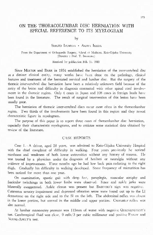
入lyelogramshowed incomplete block at Tll-12 and T12・Llinterspaces. The column
of the contrast medium stopped at TlO, Tl 1 and its leading edges lay just above the
Tll-12 disc space. A small amount of the contrast medium occupied the right side of
Tl2. Bilateral root sleeves were clearly discernible. The leading edges were clear cut
in the lateral borders but vague in the medial borders (Fig. la). The column of the
contrast medium displaced posteriorly in the lateral film (Fig. lb). Twenty hours later
the residual moljodol took a figure of “moustache”(Fig. 2).
Fig. 1 a Fig. 1 b Fig. 2
A.t operation herniation of Tl~-Ll disc was removed but also a slightly bulged
Tll-12 disc was left untouched. The spinal cord was found to be displaced posteriorly especially in T12-Ll.
Postoperatively low back pain soon disappeared. But the rどじoveryof the spinal cord palsy was unsatisfactory.
Case 2. 入 clerk, aged .33 years, has noted low back pain and weakness of lower
limbs for ten years. Disturbances of bladder and bowels have developed since seven months. There was no history of trauma.
Examination revealed kyphosis of slight degree. Muscular atrophy of the lower
limbs and claw-toes were observed. Knee jerks decreased and ankle jerks absent.
Positive LASEGUE's test. Cutaneous sensory depression up to the Sl-2 segment.
Roentgenograms showed a narrowing of the Tll・12disc space. In lateral films
concave lens shaped deformity and osteophyte of the adjoining vertebral bodies were
visible .. Myelogram showed a filling defect at the Tl l-12 disc space and the leading
edges of contrast medium were obscure. The root sleeve of the 11th thoracic nerve was percei~ed on the right side.
ON THE THORACOLUMBAR DISC HERNIATION 377
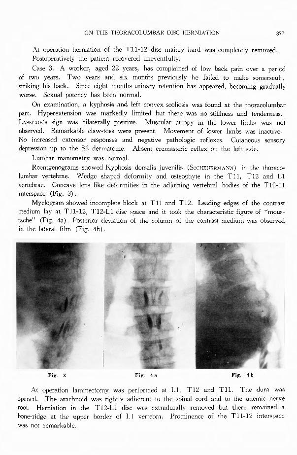
At operation herniation of the Tll-12 disc mainly hard was completely removed.
Postoperatively the patient recovered uneventfully.
Case 3. A worker, aged 22 years, has complained of low back pain over a period
of two years. Two years and six months previously he failed to make somersault, striking his back. Since eight months urinary retention has appeared, becoming gradually
worse. Sexual potency has been normal.
On examination, a kyphosis and left convex scoliosis was found at the thoracolumbar part. Hyperextension was markedly limited but there was no stiffness and tenderness.
LASEGUE’s sign was bilaterally positive. Muscular atropy in the lower limbs was not observed. Remarkable claw-toes were present. Movement of lower limbs was inactive.
No increased extensor responses and negative pathologic reflexes. Cutaneous sensory depression up to the S3 dermatome. Absent cremasteric reflex on the left side.
Lumbar manometry was normal.
Roentgenograms showed Kyphosis dorsalis juvenilis (ScCHEUERMANN) in the thoraco-
lumbar vertebrae. ¥Vedge shaped deformity and osteophyte in the Tll, T12 and Ll
vertebrae. Concave lens like deformities in the adjoining vertebral bodies of the TIO・11interspace (Fig. 3).
Myelogram showed incomplete block at Tl 1 and Tl2. Leadi_~g edges of the contrast medium lay at Tll-12, T12-Ll disc space and it took the characteristic figure of "mous-
tache" (Fig. 4a) . Posterior deviation of the column of the contrast medium was observed in the lateral film (Fig. 4b).
Fig. 3 Fig. 4a Fig. 4 b
At operation laminectomy was performed at Ll, T12 and Tll. The dura was
opened. The arachnoid was tightly adherent to the spinal cord and to the anemic nerve root. Herniation in the T12-Ll disc was extradurally removed but there remained a
bone-ridge at the upper border of Ll vertebra. Prominence of the Tll-12 interspace
was not remarkable.
378 日本外科宝函第32巻 第3号
Postoperatively no remarkable improvement was gained.
DISCUSSION
1. Statistical Analyses
Herniated thoracic discs are relatively rare conditions and only 112 casEs WEre found
m the literature. One hundred and fifteen heriniated discs were available for this
statistical analyses.
Incidence : From 1929 throught 1962 34 cases of cer¥・ical and 195 cases of lumbar
disc herniations have been verified at operation at our clinic. Three cases of the herniated
thoracolumbar disc in this series amount to 1.3 per cent of total 232 disc herniations.
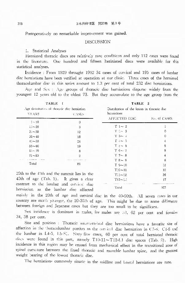
A氏eand Sex Agじ groupsof thoracic disc herniations disperse widely from the
youngest 12 years old to the oldest 73. But they accumulate to the age group from the
TABLE I
λge d1,trihution of thoracic disc herniation
YE主RS C.-¥SES
1-10 。1 l~20 3 21~30 12 31~40 18 41~50 24 51~60 19 61~70 8 71~80 2
Total 86
20th to the !Clth and the summit lies in the
40th of age (Tab. 1). It gives a clear
contrast to the lumbar and じげγIじ礼ldisc
herniation as the lumbar disc affected
TABLE 2
Distribntion of the lesion in thoracic disc herniations
人FFECTEDDISC j¥f,》 ofC¥SES
T l~ 2
l‘::- 3 。T 3~ 4 。T -l~ 5 4 T 5~ 6 5
T 6~ 7 9 T 7戸、 8 4
T 8~ 9 8
T 9~IO 12
TIO~1 1 12
T!l~12 36
T!Z~L1 17
Total 107
mainlv in the 20th of age and cervical disc in the 40・50th. All sevenじasesin our
country are mu:'.1 yoユロger,the 20-30ih of age. This might be due to some difference
between foreign and Japanese cases but they are too small to be significant.
刊とx incidence is dominant in males, for males are :-):3, 62 per cent and females
34, 38 per cent.
Site and p::isition Thoracic mtじr':げ tehrnldisc herniations have a favorite site of
affection in the thoracolumbar portion as the rげ VIι・;iidisc herniation in c:i-仇仁ト5and
the lumbar in L4司 5, L5-六1. メ1xtyfo-ぞ cases, 60 per cent of total herniated thoracic
dis.・メ werefound in this part, namely TI0-11~T12-Ll disc spaces (Tab. 2). High
incidence in this region may be ensued from mechanical effect in the transitional zone of
spinal curvature between the fixed thoracic and movable lumbar spine, and the greatest
weight bearing of the lowest thoracic disc.
The herniations commonly situate in the midline and |λter;il herniations are rare・
OP< TトIET!-IOR.¥l 'ULUMBλR DISC !-IERNL\’rrけN 379
2. Clincal features
Clinical features of thoracolumhar disc herniations are chronic progressive spinal
para pieεia identical with anterior extradural tumour of the spinal cord hut the uけursesare
more insidious and sometimes remittent.
Low hack pain, weakness and numbness of lower limbs are noticed at first, later
difficulty in walking is predominant. Sphincter disturbances and insufficiency of sexual
potency are sometimes remarkable, for this lesion lies at the medullary conus. Local
sings are mainly low back pain, kyphosis and scoliosis in the affected disc region.
Often it lacks radicular pain differing from extramedullary tumour and making a
striking contrast to the herniated lumbar disc. It may depend upon the facts that the
midline situated herniations sometimes make no irritation to the nen・e root and if it does
the compression of the spinal cord has already ιle¥・eloped. The radicular pain radiates to
the lower abdomen. It is sometimes mistaken for the referred pain of the abdominal
viscera. Even the operation would be performed under the misdiagnosis of appendicitis,
stones of the urinary tract etc. Several degrees of spinal cord involvements are the signs
of earlier stadium such as spastic palsy, atrophy of lower extremities, ankle and patellar
clonus and positive BABINSKI's sign. Sensory impairment may be appeared in the sacral
segment due to relationship of the thoracolumbar disc to the spinal segment.
3. Cerebrospinal fluid
Lumbar manometry may be normal or reveal incomplete and complete block according
to the degrees of herniation to the subarachnoid space. Cerebrospinal fluid may contain
increased protein and normal numbers of lymphocytes but without xanthochromia.
4. Plain roentgenogram
Many characteristic changes are seen on roentεenograms because the duration of the
involvement would be chronic.
These changes are summarized as follows, l. narrowing of the affected disc space,
2. localized osteoarthritic changes such as osteophyte on the posterior borders of vertebral
bodies. 3. wedεe shaped deformities of adjoining vertebrae (Kyphosis dorsalis juvenilis
SCHEUERMANN), 4. concave lens shaped deformity, 5. abnormality of spinal cun・ature,
6. calcification of nucleus pulposus, 7. ScHMORL's nodules in the other parts of vertebral
bodies.
We have not encountered to the calcified nucleus pulposus which was first detected
by LOGUE (1952). KYPHOSIS dorsalis juvenilis (山:HEUERMANN) was found in two of our
three cases. Combination with this defomity was also reported hy MOLLER (1951) and
Van LANDINGHAM (1954), so there may be some correlation between these two disorders.
5. Myelogram
It leaves no room for doubt that the myelogram has unmeasurable value to
ascertain the space-occupying disorders in the spinal canal. It is not only diagnostic
hut also pathognomonic in the thoracolumbar disc herniation as it reveals the chara-
cteristic figure of “moustache” It shows total or partial blockage of the contrast
medium just above the affected disc and took a figure of "moustache”. The leading
edges of the contrast medium were clear cut in the lateral borders but vague in the
medial borders. The root sleeves were clearly discernible. In 1lw lateral film the
380 日本外科宝函第32巻第3号
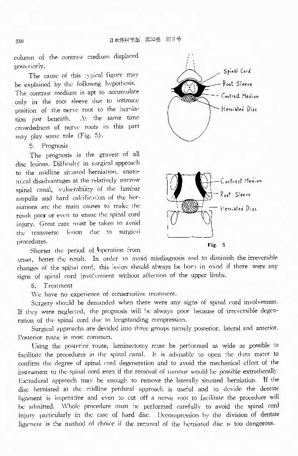
column of the contrast medium displaced
postげ iorly.
The cause of this tvpical fi只uremay
be explained by the followng hvpothesis.
The contrast medium is apt to accumulate
only in the root sleeve due to intimate
position of the nerve root to the hernia-
tion just beneath. えtthe same time
crowdedness of nerve roots in this part
may play some role (Fig. 5).
5. Prognosis
The prognosis is the gravest of all
disc lesions. Difficulty in surgical approach
to the rnidline situated herniation, anato-
mica¥ disadvantages at the relatively narrow
spinal canal, vulnerability of the lumbar
ampulla and hard calcific川 ionof the her-
niations are the main causes to make the
result poor or even to ensue the spinal cord
injury. Great care must be taken to avoid
the transverse lesion due to surgic:1 I
procedures.
Shorter the period of ‘bperation from
,、’l
Hem; ~teJ D:st
Contrut Me」;川
Root Sleeve
Hern : ~fed D ; ~ι
Fig. 5
onset, better the result. In order to avoid misdiagnosis and to diminish the irreversible
changes of the spinal cord, this l引 ionshould always be born in mind if there were any
signs of spinal cord involvement without affection of the upper limbs.
6. Treatment
明、 haveno experience of conservative treatment.
Surgery should be demanded when there were any signs of spinal cord involvement.
If they were neglected, the prognosis will be always poor because of irreversible degen-
ration of the spinal cord due to longstanding compression.
Surgical approachs are devided into three groups namely posterior, lateral and anterior.
Posterior route is most common.
Using the posterior route, laminectomy must be performed as wide as possible to
facilitate the procedures in the spinal canal. It is advisable to open the dura mater to
confirm the degree of spinal cord degeneration and to avoid the mechanical effect of the
instrument to the spinal cord even if the removal of tumour would be possible extrathecallv.
Extradural approach may be enough to remove the laterally situated herniation. If the
disc herniated at the midline perdural approach is useful and to devide the dentate
ligament is imperative and even to cut off a nerve root to facilitate the procedure will
he admitted. Whole procedure must he performed carefully to avoid the spinal cord
injury particularly in the case of hard disc. Decompression h’ the division of dentate
ligament is the method of choice if the removal of the herniated disc is too dangerous.

(JN THE THOR主cuしUMBARDISC HERNIATION 381
HULME in 1960 advocated a lateral approach through intervertebral foramen, a varn1・
tion of costotransversectomy and obtained a good result, but we have no experience in
this operation.
Recent progress in anaesthesiology and chest surgery made it possible to use the
anterior approach through thoracotomy with the fusion of vertebral bodies.
But these methods have common disadvantages not to visualize the spinal cord, their
estimation must be convinced by further experiences.
SUMMARY
Three cases of thoracolumbar disc herniation were reported and some aspects of their
myelogram were discussed.
Statistical analyses of herniated thoracic discs were made and clinical features, roent-
genograms, treatment and prognosis of thoracolumbar disc herniations were described in
some details.
The authors are very grateful to Prof. T. lwahara for his warm encouragement and kind guidance.
REFERENCES
1 J Andrae. R. : Uber Knorpelkni:itchen am hinteren Ende der Wirbelbandscheiben im Bereich des Spinalka-nals. Beitr. z. path. Anat., 82. 461, 1929.
2) Arseni, C. and Nash, F. : Thoracic Intervertebral Di>-l・ Protrusion. l. Neurosurg., 17. 418, 1960.
3) Abbot, K. H.λnd Retter, R. H. : Protrusion of thoracic Intervertebral Disks. Neurology, 6, 1, 1956.
4) Bradford, F. K. and Spurling, R.じ.: The Intervertebral Di,c, Charles C. Thomas, Springfield, 1941.
5) Elsberg, C. A. : The Extradural Vεntral Chondromas (Ecchondn•"、 I Bull. Neural. !1トt New York,
1, 350, 1931.
6) Epstein, J. A. : The Syndrome of Herniation of the L《川町 Thoraciclnten・ぞrtebralDiscs with Nerve
Root and Spinal Cord Compre日 ion.J.トJeurosurg.11, 525, 1954.
7) Haley, J. C. and Perry, J. H. : Protrusion of Intervertebral Discs. Amer. J. Surg., 80, 394, 1950.
8) Hawk, W. A. : Spinal Compression Caused by Ecchondrosis of the Intervertebral Fibr口cartilage.Brain,
59, 204, 1936.
9) Hulme, A. : 1、i】eSurgical 主I》pr••巴
Psychiat.’23, 133, 1960. 10) Kondo, E. : A C日明 of Herniated Intervertebral Disc between the 11th and 12th Do庁間lVertebra. The
Central Japan J. Orthopaed. & Traumat. Surg., 2, 166, 1959.
11) Kite, W. C. et al. : The Herniated Intervertebral Disc以ndrome.J. Neurosurg., 14, 61, 1957.
12) Kroll, F. W. und Reiss, E. : Der thorakale Bandscheibenprolaps. Dt可 h.med. ¥Vschr・'76. 600, 1951
13) Logue, V. : Thoracic Intervertebral Disc Prolapse "ith Spinal Cir.cl Compression. J. Neuml. ;¥eurosurg.
Psychiat., 15, 227, 1952.
14) Love, J. G. : and Kiefer, E. J. : Root Pain and Paraplegia due to Protrusions of Thoracic Interverte-
bral Disks. J. Neurosurg・, 7, 62, 1950.
15) Morita, S. and Matsushima, H. : A Ca問。fHerniated Thoracic Disc. Orthopaed.メmg.,12, 994, 1961.
16) Mixter, W. J. and Barr, J. S. : Rupture of the Intervertebral Disc with Involvement of the メpinal
Canal. Ne" Engl. J. Med., 211, 210, 1934
17) Muller, R. ・ Protrusion of Thoracic Inten-ertebral Disks with Compr引 sionof the Spinal Cord. ..¥eta
Med. Scand., 139, 99, 1951.
18) Nagaya, I. and Takagi, G. : A Case of Herniated Thortacic ll川、 J.Japan. Orth•>paed. A"'""・ 34
899. 1960.
19) Peck, F. C.:人 CalcifiedThoracic Intervertebral Disk with Herniation and只pmalCord Compression in
a Child. J. Neurosurg., 14, 19:5, 1957.
20) Sakamaki, H. and T,;uji, A. : /¥. Caぜ ofHerniated Thoracic Disc. J. Japan. Orthop .. .¥,,oc., 34, 1036, 1960.
382 日本外刈宝函第32鶏第3号
21)たhmorl,G. : Uber Kn<>rp,..!knoten an der Hinterflache der ¥¥' irbelhand~heiben. Fけrt'-clir.a. cl. Geh1et.
R, >t'ntgenstrahl., 40, 629, J 929.
22〕 VanLandingham, J. H. : Herniation of Thoracic Intervertιhral Di"・' with Spud Cordれ mpre括ionin
Kypho-," Dor回!isJuvenilis. J. :'-Jeurosurg .. 11, 327, 1951. 23) Young, J. H.:ぐervicaland Thoracic Intervertebral Disk Di川 1ぜ :vJcd. J. :¥IIバパ 19462, 833.
和文抄録
胸腰移行部椎間板ヘルニアとくにその
ミエログラムについて
慶応義塾大学医学部整形外科ザ教室 f主任:岩原寅猪教授)
泉田重 雄・池川 1杉
ljiqJ4必Hrf』1:椎f}j]板ヘルニアの 3症例を報告し,特に
その ミエログラムにつき述べる.
本町,l占Ii梯で白験全椎間板ヘルニア手術例 232例の
1.3 %にすぎない.
臨床像は慢性進行性裁:·~,で脊髄麗疹に類似するが一
層経過が永く p 時に弛張を示す.初発症状として背腰
痛p 下肢脱力\~. . l•ir i L等を訴え歩行障害を来して来院
する. !!;f:lt l こ神経痛といれk,脚~~(,内臓疾患等と誤
診されているものが多い.
髄液に笹君事所見を呈したものが 1例ある.
レ 似! ·. :~1u2 附甘rnだの狭縮, 限局性変形性変化即ち線
形成,機体辺縁隆起,みf話相'(本J)模代及び魚権状変形,
脊住者号曲異常,シユモール紡節などをみる. 2例にお
いて少年性亀背の合併をみたことは害i_c11去がある.
ミエログラムは特徴的で,それはパナナの房状或は
八の字状の定型像である.沃度油は穣患椎間板直上
てv八の字状のつfて拡がりの像を拾いて停留し,両側に
明らかな HL笈~ を摘出する.両側の下縁は比較的境界
明瞭であるが中央は薄ボケている.側面像では後方に
圧排されている.八の字型定型像の成因としては多数
の神科m;;;密集しているところへ,胸髄神経根起始部
直下に腫癌が存在するため,沃度f由が根褒:'.j;1:のみ停
留するためであると理解され;:,.
他部のものに比して予後は惑い.それはヘルニアが
脊髄前方正中線上にあることが多く, ( i 1.;:く沈着,骨提
等になっていることもあ りp 腫癌占在部位が腰膨大部
に相当し,手術的侵麹の悶郊なことに帰せられる.
治療に枇づ切除』重癒易日出術を理想、とするが,粗暴な
操作により脊髄煩傷の危倶なしとしないので別出には
慎重を要する.Hulmeは肋骨横突起切除に より椎間孔
から別出を行ない良好な結果を得ており p また,閲胸
により前方から侵襲し,脊佳固定を併用する術式も考
えられるが,これらの批判は今後の経験に侯たねばな
らなし\


![r n a f S o u pi J ne Suetsuna et al., Spine 214, 3:1 ...€¦ · The rate of traumatic cervical disc herniation accompanied by spinal injury using myelography and CT was low[8,9]](https://img.pdfslide.tips/doc/110x75/600653f2549eb807296d20df/r-n-a-f-s-o-u-pi-j-ne-suetsuna-et-al-spine-214-31-the-rate-of-traumatic.jpg)