Embed Size (px)
Citation preview

Title Regulation of type 1 iodothyronine deiodinase by LXRα(Dissertation_全文 )
Author(s) Sakane, Yoriko
Citation Kyoto University (京都大学)
Issue Date 2017-09-25
URL https://doi.org/10.14989/doctor.k20670
Right Final publication is available athttps://doi.org/10.1371/journal.pone.0179213
Type Thesis or Dissertation
Textversion ETD
Kyoto University

RESEARCH ARTICLE
Regulation of type 1 iodothyronine deiodinase
by LXRαYoriko Sakane1¤a, Naotetsu Kanamoto1¤b*, Ichiro Yamauchi1, Tetsuya Tagami2,
Yusuke Morita3, Masako Miura1¤c, Masakatsu Sone1, Akihiro Yasoda1, Takeshi Kimura3,
Kazuwa Nakao4, Nobuya Inagaki1
1 Department of Diabetes, Endocrinology and Nutrition, Kyoto University Graduate School of Medicine,
Kyoto, Japan, 2 Division of Endocrinology and Metabolism, Clinical Research Institute, National Hospital
Organization Kyoto Medical Center, Kyoto, Japan, 3 Department of Cardiovascular Medicine, Kyoto
University Graduate School of Medicine, Kyoto, Japan, 4 Medical Innovation Center, Kyoto University
Graduate School of Medicine, Kyoto, Japan
¤a Current address: Preemptive Medicine and Lifestyle Disease Research Center, Kyoto University Hospital,
Kyoto, Japan
¤b Current address: Department of Endocrinology, Osaka City General Hospital, Osaka, Japan
¤c Current address: Department of Endocrinology, Rakuwakai Otowa Hospital, Kyoto, Japan
Abstract
The iodothyronine deiodinases are selenoenzymes that regulate the activity of thyroid hor-
mone via specific inner- or outer-ring deiodination. In humans, type 1 deiodinase (D1) is
highly expressed in the liver, but the mechanism by which its gene expression is regulated
remains to be elucidated. Liver X receptor α (LXRα), a transcription factor of the nuclear
receptor superfamily, is highly expressed in the liver, where it functions as a sensor for
excess intracellular oxysterols. LXRα interacts with other nuclear receptors on promoters of
genes that contain a binding core sequence for nuclear receptors. In addition, it is reported
that the promoter of the gene encoding human D1 (hDIO1) contains the core sequence for
one of nuclear receptors, thyroid hormone receptor (TR). We investigated the involvement
of LXRα in the regulation of hDIO1, in the liver. We performed hDIO1 promoter–reporter
assays using a synthetic LXR agonist, T0901317, and compared promoter activity between
a human liver carcinoma cell line, HepG2, and a clone of human embryonic kidney cells,
TSA201. We defined the region between nucleotides −131 and −114, especially nucleotides
−126 and −125, of the hDIO1 promoter as critical for basal and LXRα-mediated specific tran-
scriptional activation in HepG2 cells. An increase in hDIO1 expression was observed in
LXRα-stimulated cells, but absent in cycloheximide-treated cells, indicating that new protein
synthesis is required for LXRα-mediated regulation of hDIO1. On the other hand, electro-
phoretic mobility shift assays revealed that LXRα and RXRα bound to the hDIO1 promoter.
We also demonstrated that LXRα and TRβ compete with each other on this specific region
of the promoter. In conclusion, our results indicated that LXRα plays a specific and important
role in activation of TH by regulating D1, and that LXRα binds to and regulates the hDIO1
promoter, competing with TRβ on specific sequences within the promoter.
PLOS ONE | https://doi.org/10.1371/journal.pone.0179213 June 15, 2017 1 / 18
a1111111111
a1111111111
a1111111111
a1111111111
a1111111111
OPENACCESS
Citation: Sakane Y, Kanamoto N, Yamauchi I,
Tagami T, Morita Y, Miura M, et al. (2017)
Regulation of type 1 iodothyronine deiodinase by
LXRα. PLoS ONE 12(6): e0179213. https://doi.org/
10.1371/journal.pone.0179213
Editor: Makoto Makishima, Nihon University
School of Medicine, JAPAN
Received: December 4, 2016
Accepted: May 25, 2017
Published: June 15, 2017
Copyright: © 2017 Sakane et al. This is an open
access article distributed under the terms of the
Creative Commons Attribution License, which
permits unrestricted use, distribution, and
reproduction in any medium, provided the original
author and source are credited.
Data Availability Statement: All relevant data are
within the paper.
Funding: This work was supported by JSPS
KAKENHI (Grant Number 16H06902), MEXT
KAKENHI (Grant Numbers 21229013, 20790659,
18790635, and 19591075; https://www.jsps.go.jp/
j-grantsinaid/), and Takeda Science Foundation
(http://www.takeda-sci.or.jp). The funders had no
role in study design, data collection and analysis,
decision to publish, or preparation of the
manuscript.

Introduction
Iodothyronine deiodinases type 1, 2, and 3 (D1, D2, and D3, respectively) are selenoenzymes
that regulate the activity of thyroid hormone (TH) via specific inner- or outer-ring deiodina-
tion [1]. Biologically active intracellular triiodothyronine (T3) is supplied from the serum and
via activation of thyroxine (T4) by D1 and D2 [1]. In human, D1 is expressed at high levels in
the liver, kidney, and thyroid, whereas D2 is expressed in the brain, pituitary, and brown adi-
pose tissue. The local T3 concentration in hepatic cells depends on serum TH concentration,
T3 generated from T4 by D1, and clearance of T3 from hepatic cells. The expression and activ-
ity of D1 are modulated by multiple factors including T3, cAMP, retinoic acid, and TSH [2–4].
In a previous report, we demonstrated that expression of the human D1 gene (hDIO1) is liver-
specifically and cooperatively regulated by forkhead box A1 (FOXA1), FOXA2, and upstream
stimulatory factor (USF) via specific regions of the hDIO1 promoter [5]. However, much
remains unknown regarding the mechanisms of regulation of hDIO1 and its physiological role
in humans.
Liver X receptors (LXRs) are ligand-activated transcription factors of the nuclear receptor
superfamily that bind oxidized cholesterol and function as sensors for excess intracellular oxy-
sterols [6]. LXRs exist in two isoforms, LXRα and LXRβ. LXRβ is expressed ubiquitously,
whereas LXRα is highly expressed in the liver, and at lower levels in adipose tissue, adrenal
glands, intestines, lungs, kidneys, and cells of myeloid origin [7]. LXRs form heterodimers
with the retinoid X receptor (RXR) and bind to the LXR-responsive element (LXRE) in pro-
moters of target genes that contain direct repeats (DRs) of the core sequence AGGTCA sepa-
rated by four nucleotides (DR4) [8].
LXRs bind to and compete with another subgroup of nuclear receptor, the thyroid hormone
receptors (TRs), at the same sites in the 5´-region of promoters, e.g., those of the genes encod-
ing acetyl-CoA carboxylase-α, cholesterol 7-alpha-hydroxylase, and ATP-binding cassette
transporter A1 [9–11]. Toyoda et al. previously reported that the hDIO1 promoter contains
two functional TH response elements (TREs), each of which also consists of two DRs with the
core sequence, AGGTCA [3]. Although LXRs and TRs belong to two distinct receptor sub-
groups with respect to ligand-binding affinity [12], the two receptor systems have similar
molecular mechanisms, i.e., both form heterodimers with RXRs and to bind to DR4 with iden-
tical geometry and polarity [13,14]. To date, it remains unknown whether LXRs regulate the
expression of hDIO1. Because LXRα is expressed at very high levels in the liver, and the hDIO1promoter contains TREs, we hypothesized that LXRα is involved in regulation of hDIO1 in a
liver-specific manner by unknown mechanisms.
Thus, in this study, we investigated the involvement of LXRα in the regulation of hDIO1, in
the liver using a human liver carcinoma cell line HepG2.
Materials and methods
Ligand
A synthetic LXR agonist, T0901317 (TO), was purchased from Cayman Chemical (Ann Arbor,
MN, USA), and a synthetic LXR antagonist, GSK2033 (GSK), was purchased from Axon Med-
chem BV (Groningen, The Netherlands). TO and GSK were dissolved in and diluted with
dimethyl sulfoxide at various concentrations.
Cell culture
A human liver carcinoma cell line, HepG2 [5], and a clone of human embryonic kidney 293
cells, TSA201 [15], were cultured as described previously.
Regulation of type 1 iodothyronine deiodinase by LXRα
PLOS ONE | https://doi.org/10.1371/journal.pone.0179213 June 15, 2017 2 / 18
Competing interests: The authors have declared
that no competing interests exist.
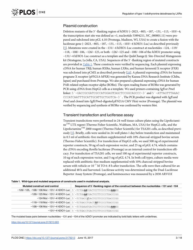
Plasmid construction
Deletion mutants of the 5´-flanking region of hDIO1 (−2023, −803, −187, −131, −113, −103/−4;
the transcription start site was defined as +1, nucleotide 53894211, NC_000001.11) were pre-
pared and subcloned into pGL 4.10 (Promega, Madison, WI, USA) to create a fusion with the
luciferase gene (−2023, −803, −187, −131, −113, −103/−4 hDIO1-Luc) as described previously
[5]. Mutations were created in the −131/−4 hDIO1-Luc construct at nucleotides −124, −119/
−118, −108/−106, −126/−125, or both −126/−125 and −108/−106 of the hDIO1 promoter using
−131/−4 hDIO1-Luc construct as a template and the QuikChange1 Site-Directed Mutagenesis
kit (Stratagene, La Jolla, CA, USA). Sequences of the 5´-flanking region of mutated constructs
are provided in Table 1. These constructs were verified by sequencing. Each plasmid expressing
cDNA for human TRβ, human RXRα, human LXRα, and human farnesoid X receptor (FXR)
was subcloned into pCMX as described previously [16]. A plasmid expressing cDNA for human
pregnane X receptor (pFN21A-hPXR) was generated by Kazusa DNA Research Institute (Chiba,
Japan) and purchased from Promega. We also prepared a plasmid expressing cDNA for human
PAR-related orphan receptor alpha (RORα). The open reading frame of RORαwas generated by
PCR using cDNA from HepG2 cells as a template. We used primers containing SgfI or PmeI
linker: 5´-GACCGCGATCGCCATGGAGTCAGCTCCGGCAGCCC-3´and 5´-ATTAGTTTAAACCCCATCAATTTGCATTGCATTGCTGGTCA-3´.The PCR product was digested with SgfI and
PmeI and cloned into SgfI/PmeI-digested pFN21A CMV Flexi vector (Promega). The plasmid was
verified by sequencing and synthesis of RORαwas confirmed by western blot.
Transient transfection and luciferase assay
Transient transfections were performed in 24-well tissue culture plates using the Lipofectami-
neTM LTX regent (Thermo Fisher Scientific, Waltham, MA, USA) for HepG2 cells, and the
LipofectamineTM 2000 reagent (Thermo Fisher Scientific) for TSA201 cells, as described previ-
ously [5]. Briefly, cells were seeded in 24-well plates 1 day before transfection and maintained
in 0.5 ml of antibiotic-free medium supplemented with 10% charcoal-stripped bovine serum
(Thermo Fisher Scientific). For transfection of HepG2 cells, we used 500 ng of experimental
reporter constructs, 50 ng of each expression vector, and 25 ng of pGL 4.74, which contains
the cDNA encoding Renilla luciferase (Promega) as an internal control for transfection effi-
cacy. For transfection of TSA201 cells, we used 100 ng of experimental reporter constructs,
10 ng of each expression vector, and 5 ng of pGL 4.74. In both cell types, culture media were
replaced with antibiotic-free medium supplemented with 10% charcoal-stripped bovine
serum with vehicle or 10−7 M TO 6–8 h after transfection. The cells were then cultured for an
additional 48 h and harvested. Luciferase activity was determined using the Dual-Luciferase
Reporter Assay System (Promega), and luminescence was measured by a 2030 ARVOX
Table 1. Wild-type and mutated sequence of constructs used in mutational analysis.
Mutated construct and control Sequence of 5´-flanking region of the construct between the nucleotides −131 and −104
−126/−125, −108/−106 Mut −131/−4 hDIO1-Luc 5´-TCTGAAATGACTCCTTCCCCTGAAAAGG
−126/−125 Mut −131/−4 hDIO1-Luc 5´-TCTGAAATGACTCCTTCCCCTGACCCGG
−124 Mut −131/−4 hDIO1-Luc 5´-TCTGACCCGACTCCTTCCCCTGACCCGG
−119/−118 Mut −131/−4 hDIO1-Luc 5´-TCTGACCTGACTTGTTCCCCTGACCCGG
−108/−106 Mut −131/−4 hDIO1-Luc 5´-TCTGAAATGACTCCTTCCCCTGAAAAGG
−131/−4 hDIO1-Luc 5´-TCTGACCTGACTCCTTCCCCTGACCCGG
The mutated base pairs between nucleotides −131 and −104 of the hDIO1 promoter are indicated by bold italic letters with underlines.
https://doi.org/10.1371/journal.pone.0179213.t001
Regulation of type 1 iodothyronine deiodinase by LXRα
PLOS ONE | https://doi.org/10.1371/journal.pone.0179213 June 15, 2017 3 / 18

multilabel reader (PerkinElmer, Waltham, MA, USA). Firefly luciferase activity was normal-
ized against Renilla luciferase activity in each well to control for transfection efficacy.
RNA isolation, reverse transcription, and quantitative PCR
HepG2 cells were cultured in antibiotic-free medium supplemented with 10% charcoal-
stripped bovine serum (Thermo Fisher Scientific) and treated with TO at various concentra-
tions and 10−6 M GSK, or pre-treated with cycloheximide for 30 min before treatment with
10−7 M TO for 24 h. Then, total RNA was extracted from HepG2 cells using the RNeasy1
Plus Mini Kit (QIAGEN, Valencia, CA, USA). One microgram of total RNA was reverse-tran-
scribed with random hexamers using First-strand cDNA Synthesis Kit (GE Healthcare UK
Ltd. Buckinghamshire, UK). The resultant cDNA was diluted 1:10 by addition of TE buffer.
Quantitative PCR was performed, recorded, and analyzed using TaqMan1 Gene Expression
Assays with the StepOnePlus™ Real-time PCR System (Thermo Fisher Scientific). The probe/
primer sets were Hs00174944_m1 (hDIO1), Hs00230861_m1 (THRB), and Human PPIA (Cyclo-
philin A) Endogenous Control, purchased from Thermo Fisher Scientific. Diluted cDNA was
amplified as described previously [5]. Expression levels of each gene were normalized against the
corresponding mRNA levels of cyclophilin A to compensate for variations in input RNA.
Preparation of electrophoresis mobility shift assay (EMSA)
Nuclear extracts were prepared from HepG2 cells and TSA201 cells using the Nuclear Extract
Kit (Active Motif, Carlsbad, CA, USA). HepG2 cells were treated with vehicle or TO, with
or without co-transfection of expression vectors for LXRα and RXRα (LXRα/ RXRα). One
microgram of each expression vector was transfected into HepG2 cells in 60-mm dishes using
the LipofectamineTM LTX regent (Thermo Fisher Scientific). Vehicle or TO was added to the
culture media 24 h after transfection. The cells were then cultured for an additional 24 h and
harvested. EMSAs and supershift assays were conducted using the LightShiftTM Chemilumi-
nescent EMSA kit (Thermo Fisher Scientific) as directed by the manufacturer with slight mod-
ifications, as described previously [5]. The antibodies used in the supershift assays were as
follows: anti–human LXRα mouse monoclonal antibody (PP-PPZ0412-00, Perseus Proteo-
mics, Tokyo, Japan), anti–human RXRα mouse monoclonal antibody (PP-K8508-00, Perseus
Proteomics), anti–human TRβ1 mouse monoclonal antibody (sc-738X, Santa Cruz Biotech-
nology, Dallas, TX, USA), and normal mouse IgG (sc-2025, Santa Cruz Biotechnology). The
sequences of oligonucleotides used for EMSA are provided in Table 2.
Table 2. Sequences of double-stranded oligonucleotides used in EMSA.
Oligonucleotides Sequences
Wt1 5´-GCAAACATCTTCTGACCTGACTCCTTCCCC-3´
−124 mut1 5´-GCAAACATCTTCTGACCCGACTCCTTCCCC-3´
−126/−125 mut1 5´-GCAAACATCTTCTGAAATGACTCCTTCCCC-3´
Wt2 5´-TCTGACCTGACTCCTTCCCCTGACCCGG-3´
−126/−125 mut2 5´-TCTGAAATGACTCCTTCCCCTGACCCGG-3´
LXRE 5´-GATCTTAGTTCACTCAAGTTCAAGGATC-3´
Wt1, oligonucleotide containing the wild-type sequence of the region between nucleotides −141 and −112 of the hDIO1 promoter; −124 mut1,
oligonucleotide containing a mutation at nucleotide −124 of the Wt1 oligonucleotide; −126/−125 mut1, oligonucleotide containing mutations at nucleotides
−126 and −125 of the Wt1 oligonucleotide; Wt2, oligonucleotide containing the sequence of the region between nucleotides −131 and −104 of the hDIO1
promoter [3]; −126/−125 mut2, oligonucleotide containing mutations at nucleotides −126 and −125 of the Wt2 oligonucleotide; LXRE, oligonucleotide
containing the consensus sequence of the LXR response element [17]. The oligonucleotide sequence between nucleotides −131 and −114 of the hDIO1
promoter is indicated by bold letters, and mutated base pairs are indicated by bold italic letters with underlines.
https://doi.org/10.1371/journal.pone.0179213.t002
Regulation of type 1 iodothyronine deiodinase by LXRα
PLOS ONE | https://doi.org/10.1371/journal.pone.0179213 June 15, 2017 4 / 18

Transfection of short interfering RNA (siRNA)
An aliquot of 2.5 pmol siRNA specific for THRB (s14121, Silencer Select1 siRNA, Thermo
Fisher Scientific) or a negative control siRNA (Negative Control #2, Silencer Select1 siRNA,
Thermo Fisher Scientific) was introduced into HepG2 cells using 24-well plates and the
Lipofectamine RNAiMAX reagent (Thermo Fisher Scientific) by reverse transfection. Trans-
fections of siRNA were performed 24 h before transfections of expression vectors for the lucif-
erase assay. To determine the knockdown efficacy of siRNA, mRNA was extracted 72 h after
siRNA transfection and analyzed by quantitative PCR as described above. Changes in levels of
TRβ protein (coded by THRB) following siRNA transfections were verified by western blot.
See S1 File for detailed information.
Statistical analysis
Data are expressed as means ± standard error of the mean (SEM) obtained from at least three
separate experiments. Significance of differences was evaluated using analysis of variance
(ANOVA) followed by the Tukey–Kramer method, unless otherwise specified. P values < 0.05
were considered to be statistically significant.
Results
Identification of the specific region of the hDIO1 promoter required for its
basal activity and LXRα-mediated in HepG2 cells
To identify the specific region of the hDIO1 promoter important for its activation by the syn-
thetic LXR agonist, TO, we transiently transfected a series of 5´-deletion constructs into
HepG2 and TSA201 cells, along with or without expression vectors for LXRα and RXRα(LXRα/RXRα). The cells were then cultured with or without TO (Fig 1 and S2 File). In both
cell lines, basal luciferase activity was significantly decreased by deletion of nucleotides −131 to
−114 (P< 0.01) (Fig 1A and Fig A in S2 File). Luciferase activity following addition of TO was
significantly increased (1.48-fold) without expression vectors for LXRα/RXRα (Fig B in S2
File") and further increased (1.93-fold) with those vectors in HepG2 cells transfected with the
−131/−4 hDIO1-Luc construct (P< 0.01) (Fig 1B). These increases in response to TO were
abolished by deletion of nucleotides −131 to −114. On the other hand, in TSA201 cells trans-
fected with the −131/−4 hDIO1-Luc construct, no significant increase in luciferase activity was
observed in response to TO (Fig 1B). These results indicated the importance of the specific
region between nucleotides −131 and −114 for both basal activity and LXRα-mediated activa-
tion of the hDIO1 promoter in HepG2 cells.
Regulation of hDIO1 expression by LXRαTo confirm the change in hDIO1 mRNA levels, we performed quantitative PCR analysis. The
hDIO1 mRNA level was increased by TO in a dose-dependent manner (Fig 2A). Furthermore,
the increase in hDIO1 mRNA following addition of TO was diminished by co-treatment with
a LXR antagonist GSK (Fig 2B).
TO is a ligand for FXR, PXR, and RORα as well as LXRα [18,19]. To verify that the effect of
TO on the hDIO1 promoter depends on LXRα, we performed luciferase assays using expres-
sion vectors for these receptors in HepG2 cells (S3 File). Exclusively in cells co-transfected
with expression vectors for LXRα/RXRα, luciferase activity in response to TO was significantly
higher than in cells transfected with empty vectors. Overexpression of FXR and RORα did not
alter luciferase activity, and PXR instead decreased, in comparison with the results of each
empty vector. These results indicated that LXRα specifically activates the hDIO1 promoter.
Regulation of type 1 iodothyronine deiodinase by LXRα
PLOS ONE | https://doi.org/10.1371/journal.pone.0179213 June 15, 2017 5 / 18

To determine whether LXRα needs de novo protein synthesis to stimulate hDIO1 transcrip-
tion, we performed the TO stimulation experiment using HepG2 cells pre-treated with cyclo-
heximide. In this case, the mRNA level of hDIO1 was not increased by TO (Fig 2C).
Investigation of the key nucleotides within the region between
nucleotides −131 and −114 of the hDIO1 promoter
To narrow down the specific site required for the basal activity and LXRα-mediated activation
of the hDIO1 promoter, we performed luciferase assays using HepG2 cells transfected with
wild-type or mutant hDIO1 promoter–reporter constructs. From the previous reports of
Toyoda et al. [3] and a computational analysis [20], the region between nucleotides −131 and
−114 of the hDIO1 promoter contains a half-site of TRE, whose binding site consists of two
octamer half-site motifs (YYRGGTCA) separated by 10 bp [3], as well as an activator protein 1
Fig 1. HepG2-specific regulation of the hDIO1 promoter by T0901317 (TO). A series of 5´-deletion constructs of the hDIO1 promoter were
transiently transfected into HepG2 and TSA201 cells along with expression vectors for human LXRα and human RXRα (LXRα/RXRα) with and without
10−7 M TO. Promoter activity was normalized against Renilla luciferase activity, and the normalized value is expressed relative to that of promoterless
pGL 4.10 in the absence of TO. Results are expressed as means ± SEM. *, P < 0.05; **, P < 0.01. A. Basal luciferase activity of each construct.
Statistical analysis was performed on pairwise comparisons of constructs, and significant pairs are presented. B. Luciferase activities of each construct
with and without 10−7 M TO. TO induction indicates ratio of promoter activity with TO to the activity without TO. Statistical analysis was performed to
compare TO induction of each construct with that of promoterless pGL 4.10, and significant differences are presented.
https://doi.org/10.1371/journal.pone.0179213.g001
Regulation of type 1 iodothyronine deiodinase by LXRα
PLOS ONE | https://doi.org/10.1371/journal.pone.0179213 June 15, 2017 6 / 18

(AP-1) site, whose consensus sequence contains TGA(C/G) TCA [21–23], as shown in Fig 3A.
Thus, mutations were introduced to disrupt the sequences constituting the TRE [3] and puta-
tive AP-1 site (Fig 3A). Basal luciferase activity was significantly reduced when nucleotides
−126/−125 or nucleotide −124 was mutated, in comparison to the construct lacking any muta-
tions (Fig 3B); in the former case, the reduction was larger, reaching the baseline level (i.e., the
level in cells transfected with promoterless pGL 4.10). On the other hand, basal luciferase activ-
ity increased when nucleotides −108/−106 were mutated, but did not change when nucleotides
−119/−118 were mutated (Fig 3B). The increase in luciferase activity of the hDIO1 promoter
by TO was abolished when nucleotides −126/−125 or a nucleotide −124 was mutated, but not
when nucleotides −108/−106 or −119/−118 were mutated (Fig 3C).
Fig 2. Analysis of relative hDIO1 mRNA levels in HepG2 cells. hDIO1 mRNA levels were normalized against the
corresponding levels of cyclophilin A mRNA, and the value in HepG2/vehicle cells was defined as 1. Values are
expressed as means ± SEM. *, P < 0.05; **, P < 0.01; N.S., not significant. A. HepG2 cells were treated at different
concentrations of TO for 24 h. B. HepG2 cells were treated with vehicle, 10−6 M TO, or 10−6 M TO and 10−6 M
GSK2033 (GSK) for 24 h. C. HepG2 cells were exposed to cycloheximide for 30 min before being treated with TO (10−7
M) for 24 h.
https://doi.org/10.1371/journal.pone.0179213.g002
Regulation of type 1 iodothyronine deiodinase by LXRα
PLOS ONE | https://doi.org/10.1371/journal.pone.0179213 June 15, 2017 7 / 18

Identification of nuclear proteins that bind to nucleotides −131 to −114 of
the hDIO1 promoter in HepG2 and TSA201 cells
To confirm the interaction between DNA and proteins within nucleotides −131 to −114 of
the hDIO1 promoter, we performed EMSA using oligonucleotides containing the wild-type
sequence of the region between nucleotides −141 and −112 of the hDIO1 promoter (Wt1) (Fig
4A). Incubation of nuclear proteins from HepG2 cells or TSA201 cells with biotin-labeled Wt1
oligonucleotides led to the formation of several DNA/protein complexes, as shown in lanes 2
and 4, respectively. In both cell lines, specific formation of these complexes was inhibited by
incubation with excess unlabeled Wt1 oligonucleotides (lanes 3 and 5).
Furthermore, we performed EMSA using nuclear extracts from HepG2 cells and mutated
unlabeled nucleotides (−124 mut1 or −126/−125 mut1) (Fig 4B). The same DNA/protein com-
plexes observed in previous experiments (lane 2) were abolished when the nuclear proteins
were incubated with excess unlabeled Wt1 oligonucleotides (lane 3), but not with excess unla-
beled −126/−125 mut1 oligonucleotides (lane 5), supporting the idea that the specific DNA/
protein complexes formed within the region between nucleotides −131 and −114 of the hDIO1promoter. However, both DNA/protein complexes were abolished when nuclear extracts were
incubated with excess unlabeled −124 mut1 oligonucleotides (lane 4).
Binding of LXRα and RXRα on the hDIO1 promoter
The results of EMSA using nuclear extracts from vehicle-treated, TO-treated, and TO-treated
and LXRα/RXRα-overexpressing HepG2 cells were shown in Fig 5A. The DNA/protein com-
plexes were observed at the same positions and appeared slightly stronger using nuclear
extracts from TO-treated cells (lane 3) and much stronger using nuclear extracts from TO-
treated and LXRα/RXRα-overexpressing cells (lane 4), in comparison with those using nuclear
extracts from vehicle-treated cells (lane 2).
To identify the protein that binds this region, we performed supershift assays using anti-
bodies against LXRα, TRβ, and RXRα with nuclear extracts from TO-treated and LXRα/
RXRα-overexpressing HepG2 cells (Fig 5A and 5B). The results of validation of the antibodies
used in supershift assays are shown in S4 File. The supershift of the complexes was not ob-
served with antibodies against LXRα (Fig 5A, lane 7) or TRβ (Fig 5B, lane 5), but was observed
with an antibody against RXRα (Fig 5B, lane 6). These results indicated that the DNA/protein
complexes formed within nucleotides −131 to −114 of the hDIO1 promoter contained RXRα.
Considering that RXRα forms a heterodimer with other nuclear receptors on the 3´ side of
RXRα binding position, such as LXRα and TRβ [24], and that nucleotides −131 to −114 con-
tain a half-site of TRE while nucleotides −113 to −104 contain another half-site, we performed
EMSA using an oligonucleotide containing the sequence of the region between nucleotides
−131 and −104 (Wt2) [3] with nuclear extracts from TO-treated and LXRα/RXRα-overexpres-
sing HepG2 cells (Fig 5C). As a result, a DNA/protein complex was formed (lane 2), but this
complex abolished when the nuclear extracts were incubated with excess unlabeled Wt2 oligo-
nucleotides (lane 3) or excess unlabeled −126/−125 mut2 oligonucleotides (lane 4). In a super-
shift assay, the formed complex was supershifted with antibodies against both LXRα and
RXRα (lanes 6 and 8), but not TRβ (lane 7). These results indicated that LXRα, as well as
RXRα, can bind to the region between nucleotides −131 and −104 of the hDIO1 promoter. In
addition, unlike RXRα binding, LXRα binding also required the region between nucleotides
−113 and −104.
Furthermore, to determine whether LXRα binds to the hDIO1 promoter under physiologi-
cal conditions, we performed ChIP assays using an antibody against LXRα. We described
materials and methods in S1 File. Using both primer sets, approximately 6-fold to 8-fold
Regulation of type 1 iodothyronine deiodinase by LXRα
PLOS ONE | https://doi.org/10.1371/journal.pone.0179213 June 15, 2017 8 / 18
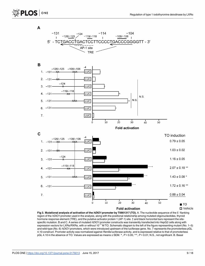
Fig 3. Mutational analysis of activation of the hDIO1 promoter by T0901317 (TO). A. The nucleotide sequence of the 5´-flanking
region of the hDIO1 promoter used in the analysis, along with the positional relationship among mutated oligonucleotides, thyroid
hormone response element (TRE), and the putative activator protein 1 (AP-1) site. † and black horizontal bars represent the site-
specific mutation. B and C. A series of mutated hDIO1 promoter constructs was transiently transfected into HepG2 cells along with
expression vectors for LXRα/RXRα, with or without 10−7 M TO. Schematic diagram to the left of the figure representing mutant (No. 1–5)
and wild-type (No. 6) hDIO1 promoters, which were introduced upstream of the luciferase gene. No. 7 represents the promoterless pGL
4.10 construct. Promoter activity was normalized against Renilla luciferase activity, and is expressed relative to that of promoterless
pGL 4.10 in the absence of TO. Values are expressed as means ± SEM. *, P < 0.05; **, P < 0.01; N.S., not significant. B. Basal
Regulation of type 1 iodothyronine deiodinase by LXRα
PLOS ONE | https://doi.org/10.1371/journal.pone.0179213 June 15, 2017 9 / 18

enrichment of ChIP-DNA following LXRα immunoprecipitation was observed in comparison
with the negative control (S5 File), indicating that LXRα could physiologically bind to the
hDIO1 promoter.
Interaction of LXRα/RXRα and TRβ on a specific region of the hDIO1
promoter
TRs bind to or compete with LXRs on the same regions in the promoters of some genes [9–
11], and basal suppression of gene promoters by un-liganded TRs has been reported [25,26].
Therefore, we examined the interaction of LXRα, RXRα, and TRβ on a specific region of the
hDIO1 promoter using the −131/−4 hDIO1-Luc construct (Fig 6). Basal luciferase activity,
which was significantly decreased by overexpression of TRβ, was not abolished when expres-
sion vector for either LXRα or RXRα was transfected alone (Fig 6A and 6B), but was abolished
when expression vectors for LXRα/RXRα were co-transfected (Fig 6C). Similarly, overexpres-
sion of TRβ decreased TO induction, except when expression vectors for LXRα/RXRα were
co-transfected (Fig 6C).
Next, we examined TO induction in TRβ-knockdown conditions (Fig 6D). The siRNA spe-
cific for TRβ efficiently knocked down the THRB mRNA and the encoded protein (S6 File).
Basal luciferase activity was significantly increased in TRβ-knockdown cells co-transfected
with expression vectors for LXRα/RXRα. Furthermore, in TRβ-knockdown cells, TO
luciferase activity of each construct. Statistical analysis was performed on comparisons between all constructs. Because most pairs
exhibited significance, only non-significant pairs are presented. C. Luciferase activities of each construct with and without 10−7 M TO.
TO induction indicates ratios of promoter activity with TO to the activity without TO. Statistical analysis was performed on comparisons
of TO induction of each construct with that of promoterless pGL 4.10, and significant differences are presented.
https://doi.org/10.1371/journal.pone.0179213.g003
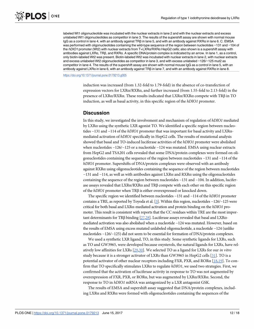
Fig 4. Specific binding of transcription factors to the region between nucleotides −131 and −114 of the hDIO1
promoter. A. EMSA with oligonucleotide containing the wild-type sequence of the region between nucleotides −141
and −112 of the hDIO1 promoter (Wt1) and nuclear extracts from un-treated HepG2 cells and TSA201 cells. In lane 1,
as a control, only the biotin-labeled Wt1 oligonucleotide was present. Biotin-labeled Wt1 oligonucleotide was incubated
with nuclear extracts from HepG2 or TSA201 cells without competitors in lanes 2 and 4, respectively, and with 25-fold
molar excesses of unlabeled Wt1 oligonucleotides in lanes 3 and 5, respectively. The specific DNA/protein complexes
formed are indicated by arrows. B. EMSA with mutant oligonucleotides and nuclear extracts from HepG2 cells. Biotin-
labeled Wt1 oligonucleotide was incubated with nuclear extracts from HepG2 cells without competitors in lane 2, and
with 25-fold molar excesses of unlabeled Wt1, −124mut1, and −126/−125mut1 oligonucleotides in lanes 3, 4, and 5,
respectively. The specific DNA/protein complexes formed are indicated by arrows.
https://doi.org/10.1371/journal.pone.0179213.g004
Regulation of type 1 iodothyronine deiodinase by LXRα
PLOS ONE | https://doi.org/10.1371/journal.pone.0179213 June 15, 2017 10 / 18

Fig 5. Binding of LXRα on the hDIO1 promoter. A. EMSA with oligonucleotide containing the wild-type sequence of the
region between nucleotides −141 and −112 of the hDIO1 promoter (Wt1) with nuclear extracts from vehicle-treated (V),
T0901317 (TO)-treated (T), or TO-treated and LXRα/RXRα-overexpressing (T+LXRα/RXRα) HepG2 cells; also shown is a
supershift assay with an antibody against LXRα. Specific DNA/protein complexes are indicated by arrows. In lane 1, as a
control, only biotin-labeled Wt1 oligonucleotide was present. In lanes 2, 3, and 4, biotin-labeled Wt1 oligonucleotide was
incubated with nuclear extracts from V, T, and T+LXRα/RXRαHepG2 cells without competitor, respectively. In lane 5,
biotin-labeled Wt1 oligonucleotide was incubated with nuclear extracts from T+LXRα/RXRαHepG2 cells with excess
unlabeled Wt1 oligonucleotides as competitor. The results of the supershift assay are shown with normal mouse IgG as a
control in lane 6 and with an antibody against LXRα in lane 7. B. EMSA with Wt1 oligonucleotides with nuclear extracts from
T+LXRα/RXRαHepG2 cells and a supershift assay with antibodies against TRβ and RXRα. Specific DNA/protein
complexes are indicated by arrows. In lane 1, as a control, only biotin-labeled Wt1 oligonucleotide was present. Biotin-
Regulation of type 1 iodothyronine deiodinase by LXRα
PLOS ONE | https://doi.org/10.1371/journal.pone.0179213 June 15, 2017 11 / 18
induction was increased (from 1.32-fold to 1.79-fold) in the absence of co-transfection of
expression vectors for LXRα/RXRα, and further increased (from 1.55-fold to 2.13-fold) in the
presence of LXRα/RXRα. These results indicated that LXRα/RXRα compete with TRβ in TO
induction, as well as basal activity, in this specific region of the hDIO1 promoter.
Discussion
In this study, we investigated the involvement and mechanism of regulation of hDIO1 mediated
by LXRα using the synthetic LXR agonist TO. We identified a specific region between nucleo-
tides −131 and −114 of the hDIO1 promoter that was important for basal activity and LXRα-
mediated activation of hDIO1 specifically in HepG2 cells. The results of mutational analysis
showed that basal and TO-induced luciferase activities of the hDIO1 promoter were abolished
when nucleotides −126/−125 or a nucleotide −124 was mutated. EMSA using nuclear extracts
from HepG2 and TSA201 cells revealed that some DNA/protein complexes were formed on oli-
gonucleotides containing the sequence of the region between nucleotides −131 and −114 of the
hDIO1 promoter. Supershifts of DNA/protein complexes were observed with an antibody
against RXRα using oligonucleotides containing the sequence of the region between nucleotides
−131 and −114, as well as with antibodies against LXRα and RXRα using the oligonucleotides
containing the sequence of the region between nucleotides −131 and −104. In addition, lucifer-
ase assays revealed that LXRα/RXRα and TRβ compete with each other on this specific region
of the hDIO1 promoter when TRβ is either overexpressed or knocked down.
The specific region we identified between nucleotides −131 and −114 of the hDIO1 promoter
contains a TRE, as reported by Toyoda et al. [3]. Within this region, nucleotides −126/−125 were
critical for both basal and LXRα-mediated activation and protein binding on the hDIO1 pro-
moter. This result is consistent with reports that the CC residues within TRE are the most impor-
tant determinants for TRβ binding [27,28]. Luciferase assays revealed that basal and LXRα-
mediated activation was also abolished when a nucleotide −124 was mutated. However, based on
the results of EMSA using excess mutated unlabeled oligonucleotide, a nucleotide −124 (unlike
nucleotides −126/−125) did not seem to be essential for formation of DNA/protein complexes.
We used a synthetic LXR ligand, TO, in this study. Some synthetic ligands for LXRs, such
as TO and GW3965, were developed because oxysterols, the natural ligands for LXRs, have rel-
atively low affinities for LXRs [29,30]. We selected TO as a ligand for LXRs for our in vitrostudy because it is a stronger activator of LXRs than GW3965 in HepG2 cells [31]. TO is a
potential activator of other nuclear receptors including FXR, PXR, and RORα [18,19]. To con-
firm that TO specifically stimulates LXRα to regulate hDIO1, we used two strategies. First, we
confirmed that the activation of luciferase activity in response to TO was not augmented by
overexpression of FXR, PXR, or RORα, but was augmented by LXRα/RXRα. Second, the
response to TO in hDIO1 mRNA was antagonized by a LXR antagonist GSK.
The results of EMSA and supershift assay suggested that DNA/protein complexes, includ-
ing LXRα and RXRα were formed with oligonucleotides containing the sequences of the
labeled Wt1 oligonucleotide was incubated with the nuclear extracts in lane 2 and with the nuclear extracts and excess
unlabeled Wt1 oligonucleotides as competitor in lane 3. The results of the supershift assay are shown with normal mouse
IgG as a control in lane 4, with an antibody against TRβ in lane 5, and with an antibody against RXRα in lane 6. C. EMSA
was performed with oligonucleotides containing the wild-type sequence of the region between nucleotides −131 and −104 of
the hDIO1 promoter (Wt2) with nuclear extracts from T+LXRα/RXRαHepG2 cells; also shown is a supershift assay with
antibodies against LXRα, TRβ, and RXRα. A specific DNA/protein complex is indicated by an arrow. In lane 1, as a control,
only biotin-labeled Wt2 was present. Biotin-labeled Wt2 was incubated with nuclear extracts in lane 2, with nuclear extracts
and excess unlabeled Wt2 oligonucleotides as competitor in lane 3, and with excess unlabeled −126/−125 mut2 as
competitor in lane 4. The results of the supershift assay are shown with normal mouse IgG as a control in lane 5, with an
antibody against LXRα in lane 6, with an antibody against TRβ in lane 7, and with an antibody against RXRα in lane 8.
https://doi.org/10.1371/journal.pone.0179213.g005
Regulation of type 1 iodothyronine deiodinase by LXRα
PLOS ONE | https://doi.org/10.1371/journal.pone.0179213 June 15, 2017 12 / 18

region between the nucleotides −131 and −104 of the hDIO1 promoter. The supershift with
an antibody against RXRα was observed not only with an oligonucleotide containing the
sequences of the region between nucleotides −141 and −112 of the hDIO1 promoter (contain-
ing the sequences of the 5´ single half-site of TRE), but also with an oligonucleotide containing
Fig 6. Interaction of LXRα/RXRα and TRβ on the activity of the hDIO1 promoter. HepG2 cells were transfected with or without expression vectors for
LXRα, RXRα, or LXRα/RXRα, with or without TRβ or siRNA specific for THRB. We performed luciferase assay using a −131/−4 hDIO1-Luc construct. All
upper figures show only basal luciferase activities, whereas lower figures show both basal activities and activities with or without 10−7 M TO. TO induction
indicates ratios of promoter activity with TO to the activity without TO. Promoter activity was normalized against Renilla luciferase activity, and the
normalized value is expressed relative to that of promoterless pGL 4.10 in the absence of TO. Results are expressed as means ±SEM. Statistical analysis
was performed on comparisons among all conditions; the results of basal activity are shown in the upper figure, and those of TO induction are shown in the
lower figure; *, P < 0.05; **, P < 0.01. A. Comparison with and without overexpression of LXRα or TRβ. B. Comparison with and without overexpression of
RXRα or TRβ. C. Comparison with and without overexpression of LXRα/RXRα or TRβ. D. Comparison with and without overexpression of LXRα/RXRα and
TRβ knockdown using siRNA. si, siRNA specific for THRB; N/C, negative control siRNA.
https://doi.org/10.1371/journal.pone.0179213.g006
Regulation of type 1 iodothyronine deiodinase by LXRα
PLOS ONE | https://doi.org/10.1371/journal.pone.0179213 June 15, 2017 13 / 18

the sequences of the region between nucleotides −131 and −104 (containing the sequences of
complete TRE). On the other hand, the supershift with an antibody against LXRα was ob-
served only with the oligonucleotide containing the sequences of the region between nucleo-
tides −131 and −104. These results suggest that LXRα has effects on the region between
nucleotides −111 and −104 of the hDIO1 promoter, where the 3´ half-site of TRE is located.
RXR forms heterodimers with numerous other nuclear receptors, including retinoic acid
receptor, vitamin D receptor, TRs, and LXRs [8,32,33]. The consensus sequence of RXR,
5´-AGGTCA-3´ [34], is located between nucleotides −129 and −124 of the hDIO1 promoter,
supporting our EMSA results showing that supershift with an antibody against RXRα was
observed with oligonucleotides containing the sequences of the regions between nucleotides
−131 and −114 and nucleotides −131 and −104. In addition, interactions mediated by dimer-
ization of RXRα with other receptors occur only when RXR binds to the 5´ half-site of their
binding positions [35]. The importance of the polarity of RXRα was consistent with the results
of the supershift assay with an antibody against LXRα. We observed that the DNA/protein
complexes were more prominent when nuclear extracts from TO-treated (and especially TO-
treated and LXRα/RXRα-overexpressing) HepG2 cells were used. This suggests that the DNA/
protein complexes might contain some other proteins induced by TO or overexpression of
LXRα/RXRα. Cycloheximide treatment abolished the increase in mRNA levels of hDIO1 by
TO, suggesting that some newly synthesized proteins are involved in stimulation of hDIO1transcription by LXRα.
Interactions between TR and LXR have been reported on promoters of some genes [9–
11,36]. We confirmed that TR and LXR interact on the hDIO1 promoter; TO induction was
significantly decreased by overexpression of TRβ, but this effect was abolished by co-transfec-
tion of expression vectors for LXRα/RXRα. Interestingly, the change in TO induction by TRβwas not affected by transfection of expression vector for either LXRα or RXRα alone. This
highlights the relevance of the EMSA/supershift results showing that complexes including
both LXRα and RXRα bound to the specific region between nucleotides −131 and −104 of the
hDIO1 promoter. By contrast, in TRβ-knockdown cells, TO induction was significantly
increased with or without overexpression of LXRα/RXRα, indicating that deterioration of TRβfunction augments the effects of LXRα/RXRα on the hDIO1 promoter.
Our observation that stimulation of LXRα by TO increased the expression of hDIO1 in
HepG2 cells was inconsistent with previous in vivo observations that TO decreases hepatic
expression of Dio1 in rat and mouse models [37,38]. Importantly, there is a large difference in
the sequences of this gene’s promoter regions between human and rodent; TREs have not
been identified in the promoter regions of rat or mouse Dio1, whereas two TREs are present in
the promoter region of human DIO1 [3]. Therefore, we performed in vitro examinations using
human cell lines, and obtained meaningful data on human DIO1 gene.
In conclusion, we demonstrated that a specific region of the hDIO1 promoter, which con-
tains a TRE, was important for the basal activity and LXRα-mediated activation of this gene.
Our results provide the additional insights that LXRα plays a specific and important role in
activation of TH by regulating D1. Furthermore, LXRα binds to and regulates the hDIO1 pro-
moter, and competes with TRβ on a specific region of the hDIO1 promoter.
Supporting information
S1 File. Supplemental materials and methods.
(DOCX)
S2 File. A series of 5’-deletion analysis of the hDIO1 promoter without LXRα/RXRα over-
expression. A. A series of 5’-deletion constructs of the hDIO1 promoter were transiently
Regulation of type 1 iodothyronine deiodinase by LXRα
PLOS ONE | https://doi.org/10.1371/journal.pone.0179213 June 15, 2017 14 / 18
transfected into HepG2 and TSA201 cells in the absence of expression vectors for human
LXRα and human RXRα (LXRα/RXRα), with or without 10−7 M TO. Promoter activity was
normalized against Renilla luciferase activity, and the normalized value is expressed relative to
that of promoterless pGL 4.10 in the absence of TO. Results are expressed as means ± SEM. ��,
P< 0.01. A. Basal luciferase activity of each construct. Statistical analysis was performed for
pairwise combinations of constructs, and significant pairs are presented. B. Luciferase activi-
ties of each construct with and without 10−7 M TO. TO induction indicates ratios of promoter
activity with TO vs. without TO. Statistical analysis was performed between TO induction of
each construct and that of promoterless pGL 4.10, and significant differences are presented.
(TIF)
S3 File. Specificity of T0901317 (TO) in the regulation of hDIO1 by LXRα. HepG2 cells
were treated with and without 10−7 M TO and transfected using expression vectors as follows.
pCMX-LXRα, co-transfection with pCMX-LXRα and pCMX-RXRα; pCMX-FXR, co-trans-
fection with pCMX-FXR and pCMX-RXRα; pCMX-empty, transfection of pCMX-empty vec-
tor alone; pFN21A-RORα, co-transfection of pFN21A-RORα and pCMX-empty vector;
pFN21A-PXR, co-transfection of pFN21A-PXR and pCMX-RXRα; and pFN21A-empty, co-
transfection of pCMX-empty vector and pFN21A-empty vector. Promoter activity was nor-
malized against Renilla luciferase activity, and the normalized value is expressed relative to
that of promoterless pGL 4.10 with pCMX-empty or pFN21A-empty in the absence of TO. TO
induction indicates ratios of promoter activity with TO vs. without TO. Statistical analysis was
performed to compare each group with the respective empty vector group. Results are
expressed as means ± SEM. �, P< 0.05; ��, P< 0.01; N.S., not significant.
(TIF)
S4 File. Positive control for antibodies against TRβ, RXRα, and LXRα. A. EMSA with oligo-
nucleotide containing the wild-type sequence of the region between nucleotides −131 and
−104 of the hDIO1 promoter (Wt2) with nuclear extracts from HepG2 cells overexpressing
TRβ and RXRα, and supershift assays with antibodies against TRβ and RXRα. Specific DNA/
protein complexes are indicated by black arrows and supershifted bands are by a gray arrow.
In lane 1, as a control, only biotin-labeled Wt2 oligonucleotide was present. Biotin-labeled
Wt2 oligonucleotide was incubated with nuclear extracts in lane 2 and with nuclear extracts
and excess unlabeled Wt2 oligonucleotides as competitor in lane 3. The results of supershift
assays are shown with normal mouse IgG as a control in lane 4, with an antibody against TRβin lane 5, and with an antibody against RXRα in lane 6. B. EMSA with oligonucleotide contain-
ing consensus sequence of LXR response element (LXRE) with nuclear extracts from HepG2
cells overexpressing LXRα and RXRα, and supershift assays with an antibody against LXRα.
Specific DNA/protein complexes are indicated by black arrows and a supershifted band is by a
gray arrow. In lane 1, as a control, only biotin-labeled LXRE oligonucleotide was present. Bio-
tin-labeled LXRE oligonucleotide was incubated with nuclear extracts in lane 2 and with
nuclear extracts and excess unlabeled LXRE oligonucleotides as competitor in lane 3. The
results of supershift assays are shown with normal mouse IgG as a control in lane 4 and with
an antibody against LXRα in lane 5.
(TIF)
S5 File. Binding of LXRα on the hDIO1 promoter. We purified chromatin fragments follow-
ing immunoprecipitation from HepG2 cells using an antibody against LXRα. Normal mouse
IgG was used as a negative control for an antibody against LXRα. The presence of specific
DNA fragments binding to LXRα was determined by quantitative PCR using primer set 1 and
2, which covers nucleotides −128/+41 and −188/−25 of the genomic hDIO1 promoter,
Regulation of type 1 iodothyronine deiodinase by LXRα
PLOS ONE | https://doi.org/10.1371/journal.pone.0179213 June 15, 2017 15 / 18

respectively. Enrichment of LXRα-bound DNA and that of normal mouse IgG-bound DNA
were normalized against 10% of the corresponding input DNA, and the results of enrichment
of LXRα-bound DNA were expressed as the fold change relative to normal mouse IgG-bound
DNA. Data were compared by unpaired Student’s t test. Values are expressed as means ± SEM.��, P < 0.01.
(TIF)
S6 File. Efficiency of siRNA-mediated knockdown of TRβ. A. The results of quantitative
PCR to detect THRB mRNA. We compared HepG2 cells transfected with negative control
siRNA (N/C) or siRNA against THRB (si-TRβ). THRB mRNA levels were normalized against
the corresponding levels of cyclophilin A mRNA, and the value in N/C was defined as 1. Val-
ues are expressed as means ± SEM. ��, P < 0.01. B. Protein levels of TRβ after siRNA transfec-
tion were evaluated by western blot analysis. β-actin was used as a standard.
(TIF)
Acknowledgments
This work was supported by JSPS KAKENHI (Grant Number 16H06902), MEXT KAKENHI
(Grant Numbers 21229013, 20790659, 18790635, and 19591075), and the Takeda Science
Foundation. We gratefully acknowledge the work of past and present members of our labora-
tory and our discussions with them, which have helped to shape many of the ideas presented
here.
Author Contributions
Conceptualization: NK.
Data curation: YS.
Formal analysis: YS.
Funding acquisition: NK AY KN NI.
Investigation: YS IY YM.
Methodology: NK AY.
Project administration: NK.
Resources: NK TT MM AY.
Software: NK.
Supervision: NK TT MM MS AY TK KN NI.
Validation: YS IY.
Visualization: YS.
Writing – original draft: YS.
Writing – review & editing: NK AY NI.
References1. Bianco AC, Salvatore D, Gereben B, Berry MJ, Larsen PR (2002) Biochemistry, cellular and molecular
biology, and physiological roles of the iodothyronine selenodeiodinases. Endocr Rev 23: 38–89. https://
doi.org/10.1210/edrv.23.1.0455 PMID: 11844744
Regulation of type 1 iodothyronine deiodinase by LXRα
PLOS ONE | https://doi.org/10.1371/journal.pone.0179213 June 15, 2017 16 / 18
2. Koenig RJ (2005) Regulation of type 1 iodothyronine deiodinase in health and disease. Thyroid 15:
835–840. https://doi.org/10.1089/thy.2005.15.835 PMID: 16131326
3. Toyoda N, Zavacki AM, Maia AL, Harney JW, Larsen PR (1995) A novel retinoid X receptor-indepen-
dent thyroid hormone response element is present in the human type 1 deiodinase gene. Mol Cell Biol
15: 5100–5112. PMID: 7651427
4. Schreck R, Schnieders F, Schmutzler C, Kohrle J (1994) Retinoids stimulate type I iodothyronine 5’-
deiodinase activity in human follicular thyroid carcinoma cell lines. J Clin Endocrinol Metab 79: 791–
798. https://doi.org/10.1210/jcem.79.3.8077363 PMID: 8077363
5. Kanamoto N, Tagami T, Ueda-Sakane Y, Sone M, Miura M, Yasoda A, et al. (2012) Forkhead box A1
(FOXA1) and A2 (FOXA2) oppositely regulate human type 1 iodothyronine deiodinase gene in liver.
Endocrinology 153: 492–500. https://doi.org/10.1210/en.2011-1310 PMID: 22067325
6. Janowski BA, Willy PJ, Devi TR, Falck JR, Mangelsdorf DJ (1996) An oxysterol signalling pathway
mediated by the nuclear receptor LXR alpha. Nature 383: 728–731. https://doi.org/10.1038/383728a0
PMID: 8878485
7. Repa JJ, Mangelsdorf DJ (2000) The role of orphan nuclear receptors in the regulation of cholesterol
homeostasis. Annu Rev Cell Dev Biol 16: 459–481. https://doi.org/10.1146/annurev.cellbio.16.1.459
PMID: 11031244
8. Chawla A, Repa JJ, Evans RM, Mangelsdorf DJ (2001) Nuclear receptors and lipid physiology: opening
the X-files. Science 294: 1866–1870. https://doi.org/10.1126/science.294.5548.1866 PMID: 11729302
9. Zhang Y, Yin L, Hillgartner FB (2001) Thyroid hormone stimulates acetyl-coA carboxylase-alpha tran-
scription in hepatocytes by modulating the composition of nuclear receptor complexes bound to a thy-
roid hormone response element. J Biol Chem 276: 974–983. https://doi.org/10.1074/jbc.M005894200
PMID: 11027684
10. Hashimoto K, Cohen RN, Yamada M, Markan KR, Monden T, Satoh T, et al. (2006) Cross-talk between
thyroid hormone receptor and liver X receptor regulatory pathways is revealed in a thyroid hormone
resistance mouse model. J Biol Chem 281: 295–302. https://doi.org/10.1074/jbc.M507877200 PMID:
16260782
11. Huuskonen J, Vishnu M, Pullinger CR, Fielding PE, Fielding CJ (2004) Regulation of ATP-binding cas-
sette transporter A1 transcription by thyroid hormone receptor. Biochemistry 43: 1626–1632. https://
doi.org/10.1021/bi0301643 PMID: 14769039
12. Enmark E, Gustafsson JA (2001) Comparing nuclear receptors in worms, flies and humans. Trends
Pharmacol Sci 22: 611–615. PMID: 11730970
13. Apfel R, Benbrook D, Lernhardt E, Ortiz MA, Salbert G, Pfahl M (1994) A novel orphan receptor specific
for a subset of thyroid hormone-responsive elements and its interaction with the retinoid/thyroid hor-
mone receptor subfamily. Mol Cell Biol 14: 7025–7035. PMID: 7935418
14. Berkenstam A, Farnegardh M, Gustafsson JA (2004) Convergence of lipid homeostasis through liver X
and thyroid hormone receptors. Mech Ageing Dev 125: 707–717. https://doi.org/10.1016/j.mad.2004.
05.005 PMID: 15541766
15. Moriyama K, Tagami T, Akamizu T, Usui T, Saijo M, Kanamoto N, et al. (2002) Thyroid hormone action
is disrupted by bisphenol A as an antagonist. J Clin Endocrinol Metab 87: 5185–5190. https://doi.org/
10.1210/jc.2002-020209 PMID: 12414890
16. Umesono K, Murakami KK, Thompson CC, Evans RM (1991) Direct repeats as selective response ele-
ments for the thyroid hormone, retinoic acid, and vitamin D3 receptors. Cell 65: 1255–1266. PMID:
1648450
17. Zhao LF, Iwasaki Y, Nishiyama M, Taguchi T, Tsugita M, Okazaki M, et al. (2012) Liver X receptor
alpha is involved in the transcriptional regulation of the 6-phosphofructo-2-kinase/fructose-2,6-bispho-
sphatase gene. Diabetes 61: 1062–1071. https://doi.org/10.2337/db11-1255 PMID: 22415873
18. Mitro N, Vargas L, Romeo R, Koder A, Saez E (2007) T0901317 is a potent PXR ligand: implications for
the biology ascribed to LXR. FEBS Lett 581: 1721–1726. https://doi.org/10.1016/j.febslet.2007.03.047
PMID: 17418145
19. Houck KA, Borchert KM, Hepler CD, Thomas JS, Bramlett KS, Michael LF, et al. (2004) T0901317 is a
dual LXR/FXR agonist. Mol Genet Metab 83: 184–187. https://doi.org/10.1016/j.ymgme.2004.07.007
PMID: 15464433
20. Heinemeyer T, Wingender E, Reuter I, Hermjakob H, Kel AE, Kel OV, et al. (1998) Databases on tran-
scriptional regulation: TRANSFAC, TRRD and COMPEL. Nucleic Acids Res 26: 362–367. PMID:
9399875
21. Ahmed AH, Gordon RD, Sukor N, Pimenta E, Stowasser M (2011) Quality of life in patients with bilateral
primary aldosteronism before and during treatment with spironolactone and/or amiloride, including a
Regulation of type 1 iodothyronine deiodinase by LXRα
PLOS ONE | https://doi.org/10.1371/journal.pone.0179213 June 15, 2017 17 / 18

comparison with our previously published results in those with unilateral disease treated surgically. J
Clin Endocrinol Metab 96: 2904–2911. https://doi.org/10.1210/jc.2011-0138 PMID: 21778218
22. Lee W, Mitchell P, Tjian R (1987) Purified transcription factor AP-1 interacts with TPA-inducible
enhancer elements. Cell 49: 741–752. PMID: 3034433
23. Angel P, Imagawa M, Chiu R, Stein B, Imbra RJ, Rahmsdorf HJ, et al. (1987) Phorbol ester-inducible
genes contain a common cis element recognized by a TPA-modulated trans-acting factor. Cell 49:
729–739. PMID: 3034432
24. Orlov I, Rochel N, Moras D, Klaholz BP (2012) Structure of the full human RXR/VDR nuclear receptor
heterodimer complex with its DR3 target DNA. EMBO J 31: 291–300. https://doi.org/10.1038/emboj.
2011.445 PMID: 22179700
25. Damm K, Thompson CC, Evans RM (1989) Protein encoded by v-erbA functions as a thyroid-hormone
receptor antagonist. Nature 339: 593–597. https://doi.org/10.1038/339593a0 PMID: 2733791
26. Graupner G, Wills KN, Tzukerman M, Zhang XK, Pfahl M (1989) Dual regulatory role for thyroid-hor-
mone receptors allows control of retinoic-acid receptor activity. Nature 340: 653–656. https://doi.org/
10.1038/340653a0 PMID: 2549424
27. Ayers S, Switnicki MP, Angajala A, Lammel J, Arumanayagam AS, Webb P (2014) Genome-wide bind-
ing patterns of thyroid hormone receptor beta. PLoS One 9: e81186. https://doi.org/10.1371/journal.
pone.0081186 PMID: 24558356
28. Katz RW, Koenig RJ (1994) Specificity and mechanism of thyroid hormone induction from an octamer
response element. J Biol Chem 269: 18915–18920. PMID: 8034648
29. Schultz JR, Tu H, Luk A, Repa JJ, Medina JC, Li L, et al. (2000) Role of LXRs in control of lipogenesis.
Genes Dev 14: 2831–2838. PMID: 11090131
30. Collins JL, Fivush AM, Watson MA, Galardi CM, Lewis MC, Moore LB, et al. (2002) Identification of a
nonsteroidal liver X receptor agonist through parallel array synthesis of tertiary amines. J Med Chem
45: 1963–1966. PMID: 11985463
31. Albers M, Blume B, Schlueter T, Wright MB, Kober I, Kremoser C, et al. (2006) A novel principle for par-
tial agonism of liver X receptor ligands. Competitive recruitment of activators and repressors. J Biol
Chem 281: 4920–4930. https://doi.org/10.1074/jbc.M510101200 PMID: 16354658
32. Bugge TH, Pohl J, Lonnoy O, Stunnenberg HG (1992) RXR alpha, a promiscuous partner of retinoic
acid and thyroid hormone receptors. EMBO J 11: 1409–1418. PMID: 1314167
33. Yu VC, Delsert C, Andersen B, Holloway JM, Devary OV, Naar AM, et al. (1991) RXR beta: a coregula-
tor that enhances binding of retinoic acid, thyroid hormone, and vitamin D receptors to their cognate
response elements. Cell 67: 1251–1266. PMID: 1662118
34. Lee MS, Kliewer SA, Provencal J, Wright PE, Evans RM (1993) Structure of the retinoid X receptor
alpha DNA binding domain: a helix required for homodimeric DNA binding. Science 260: 1117–1121.
PMID: 8388124
35. Mader S, Chen JY, Chen Z, White J, Chambon P, Gronemeyer H (1993) The patterns of binding of
RAR, RXR and TR homo- and heterodimers to direct repeats are dictated by the binding specificites of
the DNA binding domains. EMBO J 12: 5029–5041. PMID: 8262045
36. Ishida E, Hashimoto K, Okada S, Satoh T, Yamada M, Mori M (2013) Crosstalk between thyroid hor-
mone receptor and liver X receptor in the regulation of selective Alzheimer’s disease indicator-1 gene
expression. PLoS One 8: e54901. https://doi.org/10.1371/journal.pone.0054901 PMID: 23359226
37. Davies JS, Kotokorpi P, Lindahl U, Oscarsson J, Wells T, Mode A (2008) Effects of the synthetic liver X
receptor agonist T0901317 on the growth hormone and thyroid hormone axes in male rats. Endocrine
33: 196–204. https://doi.org/10.1007/s12020-008-9067-9 PMID: 18473193
38. Stulnig TM, Steffensen KR, Gao H, Reimers M, Dahlman-Wright K, Schuster GU, et al. (2002) Novel
roles of liver X receptors exposed by gene expression profiling in liver and adipose tissue. Mol Pharma-
col 62: 1299–1305. PMID: 12435796
Regulation of type 1 iodothyronine deiodinase by LXRα
PLOS ONE | https://doi.org/10.1371/journal.pone.0179213 June 15, 2017 18 / 18