Embed Size (px)
Citation preview
RESEARCH Open Access
Transiently proliferating perivascularmicroglia harbor M1 type and precedecerebrovascular changes in a chronichypertension modelTakashi Koizumi1, Katsutoshi Taguchi2, Ikuko Mizuta1, Hiroe Toba3, Makoto Ohigashi3, Okihiro Onishi4,Kazuya Ikoma4, Seiji Miyata5, Tetsuo Nakata3, Masaki Tanaka2, Sébastien Foulquier6, Harry W. M. Steinbusch7 andToshiki Mizuno1*
Abstract
Background: Microglia play crucial roles in the maintenance of brain homeostasis. Activated microglia show abiphasic influence, promoting beneficial repair and causing harmful damage via M2 and M1 microglia, respectively.It is well-known that microglia are initially activated to the M2 state and subsequently switch to the M1 state, calledM2-to-M1 class switching in acute ischemic models. However, the activation process of microglia in chronic andsporadic hypertension remains poorly understood. We aimed to clarify the process using a chronic hypertensionmodel, the deoxycorticosterone acetate (DOCA)-salt-treated Wistar rats.
Methods: After unilateral nephrectomy, the rats were randomly divided into DOCA-salt, placebo, and controlgroups. DOCA-salt rats received a weekly subcutaneous injection of DOCA (40 mg/kg) and were continuouslyprovided with 1% NaCl in drinking water. Placebo rats received a weekly subcutaneous injection of vehicle andwere provided with tap water. Control rats received no administration of DOCA or NaCl. To investigate thetemporal expression profiles of M1- and M2-specific markers for microglia, the animals were subjected to theimmunohistochemical and biochemical studies after 2, 3, or 4 weeks DOCA-salt treatment.
Results: Hypertension occurred after 2 weeks of DOCA and salt administration, when round-shaped microglia withslightly shortened processes were observed juxtaposed to the vessels, although the histopathological findings werenormal. After 3 weeks of DOCA and salt administration, M1-state perivascular and parenchyma microgliasignificantly increased, when local histopathological findings began to be observed but cerebrovascular destructiondid not occur. On the other hand, M2-state microglia were never observed around the vessels at this period.Interestingly, prior to M1 activation, about 55% of perivascular microglia transiently expressed Ki-67, one of the cellproliferation markers.
(Continued on next page)
© The Author(s). 2019 Open Access This article is distributed under the terms of the Creative Commons Attribution 4.0International License (http://creativecommons.org/licenses/by/4.0/), which permits unrestricted use, distribution, andreproduction in any medium, provided you give appropriate credit to the original author(s) and the source, provide a link tothe Creative Commons license, and indicate if changes were made. The Creative Commons Public Domain Dedication waiver(http://creativecommons.org/publicdomain/zero/1.0/) applies to the data made available in this article, unless otherwise stated.
* Correspondence: [email protected] of Neurology, Graduate School of Medical Science, KyotoPrefectural University of Medicine, 465 Kajii-cho Kamigyo-ku, Kyoto 602-8566,JapanFull list of author information is available at the end of the article
Koizumi et al. Journal of Neuroinflammation (2019) 16:79 https://doi.org/10.1186/s12974-019-1467-7
(Continued from previous page)
Conclusions: We concluded that the resting perivascular microglia directly switched to the pro-inflammatory M1state via a transient proliferative state in DOCA-salt rats. Our results suggest that the activation machinery ofmicroglia in chronic hypertension differs from acute ischemic models. Proliferative microglia are possible initial keyplayers in the development of hypertension-induced cerebral vessel damage. Fine-tuning of microglia proliferationand activation could constitute an innovative therapeutic strategy to prevent its development.
Keywords: Neuroinflammation, Cerebral small vessel disease, Chronic hypertension, Perivascular microglia,Proliferation
BackgroundMicroglia are the resident immune cells in the brainand play pivotal roles in environmental surveillanceto maintain brain homeostasis. Inflammation or cellu-lar damage can stimulate microglia to increase the ac-tivity of immune functions [1]. In vivo two-photonmicroscopy studies showed that activated microgliarapidly migrate to and accumulate at sites of patho-logical lesions, such as ischemic lesions [2] or newlyformed amyloid-β plaques [3].The activated microglia show a biphasic influence,
promoting beneficial repair and causing harmful dam-age. Those responsible for the former are sometimes re-ferred to as anti-inflammatory M2 microglia and thelatter as pro-inflammatory M1 microglia [4]. These dif-ferent types of activated microglia can be distinguishedbased on the expression of specific markers. These acti-vated microglia are involved in various neurological dis-orders [1, 5] and also influence the function andintegrity of the blood-brain barrier (BBB) [6, 7]. In thepresent study, we focused on the interaction betweenmicroglia dynamics and cerebrovascular disease.According to previous studies, microglia are activated in
the acute phase of ischemic stroke as shown in animalmodels of transient middle cerebral artery occlusion(tMCAO) [8–10]. In the tMCAO model, numbers ofM2-state microglia rapidly increase around vessels in thepenumbra after infarction, and, in a few days, M1-statemicroglia dominantly increase. This process is called“M2-to-M1 phenotype-switching” or “shift in the M2-to-M1 phenotypes” [11, 12]. In addition to microglia activa-tion, various molecules are involved in the acute phase ofischemic strokes, such as free radicals [13], damage-associ-ated molecular patterns (DAMPs) [14], and T cells [15].DAMPs include heat shock protein, high-mobility groupbox 1 (HMGB1), and peroxiredoxin [14]. For example,HMGB1 is produced by ischemic neuronal cells about 2–4h after an ischemic event and peaks at around 6 h. HMGB1affects vascular endothelial cells and induces BBB destruc-tion, as well as microglia activation [16].In contrast, in cerebral small vessel disease (CSVD), only
a limited number of reports referred to M1 and M2 micro-glia phenotyping and their molecular mechanisms. Chronic
hypertension being the major risk factor of CSVD [17, 18],we focused on a hypertensive CSVD model.The aim of this study is to clarify microglia involve-
ment in CSVD caused by chronic hypertension using de-oxycorticosterone acetate (DOCA)-salt-treated Wistarrat (DOCA-salt rat) as a model mimicking sporadic andchronic hypertension.
MethodsAnimalsAdult male Wistar rats (150–180 g) were purchasedfrom SHIMIZU Laboratory Supplies Co, Ltd. (Kyoto,Japan). Protocols were approved by Animal Care andUse Committees of Kyoto Prefectural University ofMedicine and Kyoto Pharmaceutical University. Westudied Wistar rats fed standard chow and water ad libi-tum. Care and use of rodents met the standards set bythe National Institutes of Health for experimental ani-mals. The rats were housed under specific pathogen-freeconditions and fed standard laboratory chow and waterad libitum before entering the study. They were main-tained on a 12-h light/day cycle at 20–22 °C and 40–50%humidity.
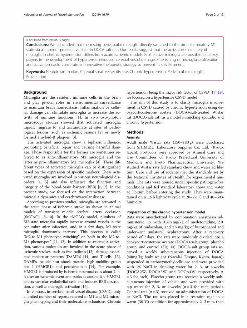
Preparation of the chronic hypertension modelRats were anesthetized by combination anesthesia ad-ministered i.p. with 0.375 mg/kg of medetomidine, 2.0mg/kg of midazolam, and 2.5 mg/kg of butorphanol andunderwent unilateral nephrectomy. After a recoveryperiod of 7 days, the rats were randomly divided into adeoxycorticosterone acetate (DOCA)-salt group, placebogroup, and control (Fig. 1a). DOCA-salt group rats re-ceived a weekly subcutaneous injection of DOCA(40mg/kg body weight (Nacalai Tesque, Kyoto, Japan))suspended in carboxymethylcellulose and were providedwith 1% NaCl in drinking water for 2, 3, or 4 weeks(DOCA2W, DOCA3W, and DOCA4W, respectively, n= 3 for each). Placebo group rats received a weekly sub-cutaneous injection of vehicle and were provided withtap water for 2, 3, or 4 weeks (n = 3 for each period).Control rats (n = 3) received no administration of DOCAor NaCl. The rat was placed in a restraint cage in awarm (38 °C) condition for approximately 2–3 min, then
Koizumi et al. Journal of Neuroinflammation (2019) 16:79 Page 2 of 13
systolic blood pressure and heart rate in a consciousstate were measured by the tail-cuff method (BP-98A-L,Softron, Tokyo, Japan). The values were measuredthree times for each rat and the average value wascalculated. After resting overnight under a lightshield, the rats underwent magnetic resonanceimaging (MRI) under anesthesia with the inhalationof isoflurane. Immediately after MRI, the rats were
perfused transcardially with 4% paraformaldehyde inphosphate buffer, and their brains were removed. Forimmuno- or pathological staining, brains werepost-fixed in the same fixative overnight at 4 °C andfurther cryoprotected sequentially in 5, 10, 15, and25% sucrose. Brains embedded in optimal cuttingtemperature compounds were stored at − 20 °C untilexamination. Frozen sections of a brain were cut into
0
120
160
200
240b
c
Sys
tolic
blo
od p
ress
ure
(mm
Hg)
**
*
)W0( lortno
CW2
AC
OD
W3A
CO
DW4
AC
OD
hematoxylin and eosin Klüver-Barrera
White matterCortex
a Hemi-nephrectomy
Control 0W
DOCA2W 3W 4W
Placebo2W 3W 4W
One week recovery period
Control(0W)
2W 3W 4W
DOCAplacebo
1
2
3
4
5
6
7
8
9
10
11
12
13
16
15
14
Fig. 1 Progression of hypertension and histological damage in deoxycorticosterone acetate (DOCA)-salt rats. a Experimental grouping forhistological analysis of the model animals. b Systolic blood pressure in DOCA2W, DOCA3W, and DOCA4W was compared with that in the control.Values are expressed as the means ± SEM (n = 3 in each group, *p < 0.05). c Hematoxylin and eosin or Klüver-Barrera staining of brain tissues. InDOCA3W, focal vascular remodeling including the perivascular space enlargements (open arrowheads) and the formation of vacuoles in thewhite matter (closed arrowheads) appeared. In DOCA4W, cerebral hemorrhage (dotted area) and myelinoclasis lesions were found. Scale bars500 μm (1–4), 20 μm (5–12), and 40 μm (13–16)
Koizumi et al. Journal of Neuroinflammation (2019) 16:79 Page 3 of 13
20-μm-thick slices with a cryostat (CM1850, Leica,Germany).For biochemical analysis, fresh brain tissues rapidly
frozen in liquid nitrogen were prepared from the con-trol, DOCA2W, and DOCA3W (n = 4, in each group)rats under anesthesia.
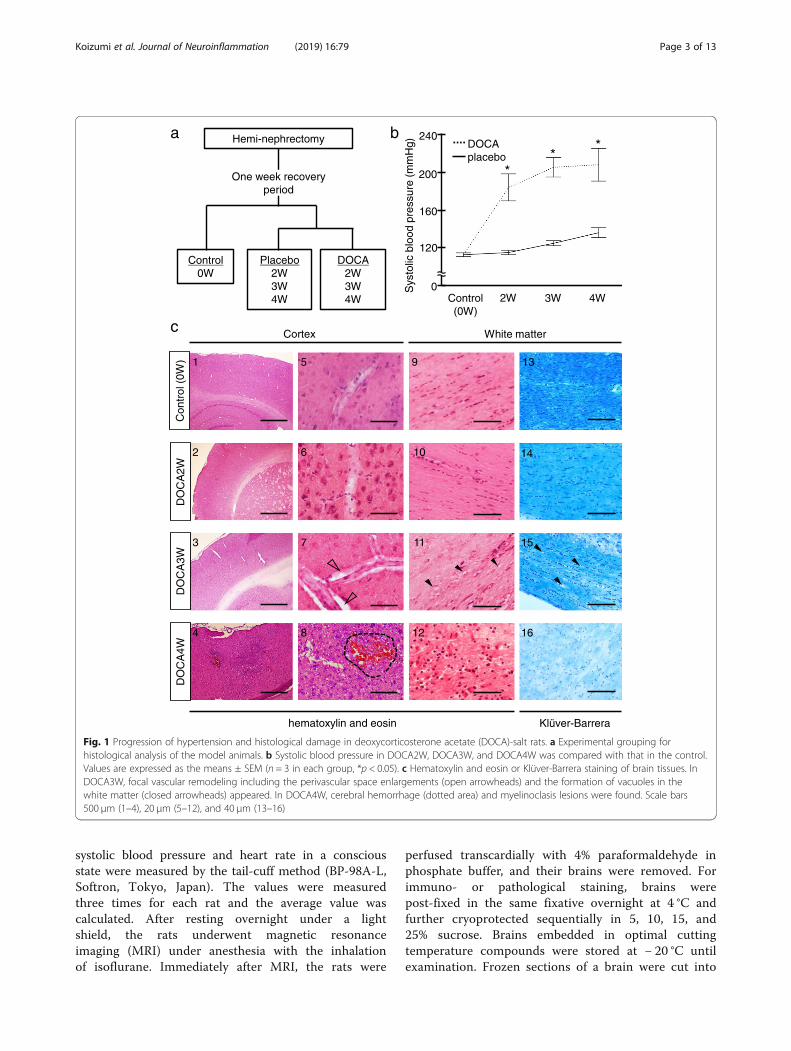
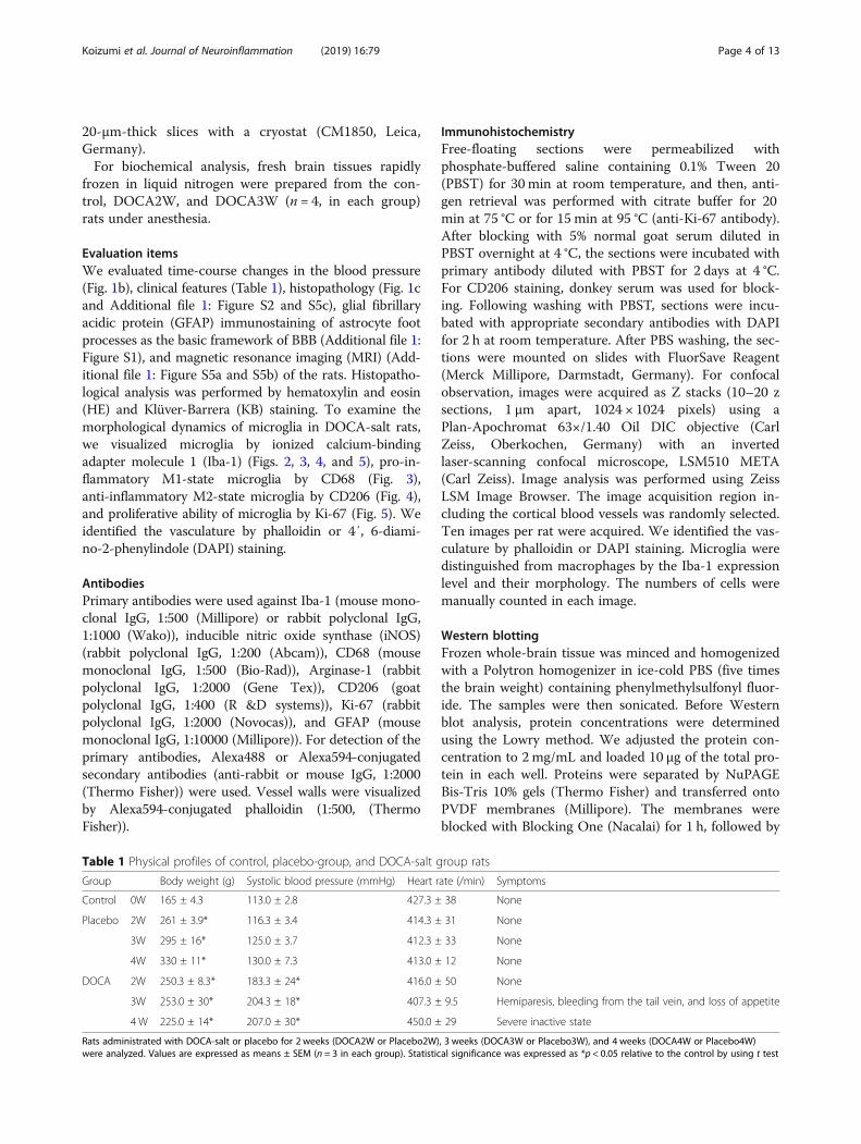
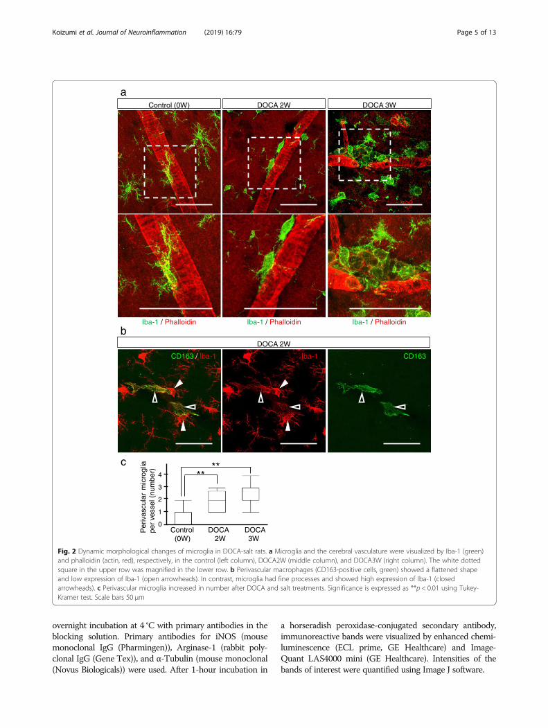
Evaluation itemsWe evaluated time-course changes in the blood pressure(Fig. 1b), clinical features (Table 1), histopathology (Fig. 1cand Additional file 1: Figure S2 and S5c), glial fibrillaryacidic protein (GFAP) immunostaining of astrocyte footprocesses as the basic framework of BBB (Additional file 1:Figure S1), and magnetic resonance imaging (MRI) (Add-itional file 1: Figure S5a and S5b) of the rats. Histopatho-logical analysis was performed by hematoxylin and eosin(HE) and Klüver-Barrera (KB) staining. To examine themorphological dynamics of microglia in DOCA-salt rats,we visualized microglia by ionized calcium-bindingadapter molecule 1 (Iba-1) (Figs. 2, 3, 4, and 5), pro-in-flammatory M1-state microglia by CD68 (Fig. 3),anti-inflammatory M2-state microglia by CD206 (Fig. 4),and proliferative ability of microglia by Ki-67 (Fig. 5). Weidentified the vasculature by phalloidin or 4′, 6-diami-no-2-phenylindole (DAPI) staining.
AntibodiesPrimary antibodies were used against Iba-1 (mouse mono-clonal IgG, 1:500 (Millipore) or rabbit polyclonal IgG,1:1000 (Wako)), inducible nitric oxide synthase (iNOS)(rabbit polyclonal IgG, 1:200 (Abcam)), CD68 (mousemonoclonal IgG, 1:500 (Bio-Rad)), Arginase-1 (rabbitpolyclonal IgG, 1:2000 (Gene Tex)), CD206 (goatpolyclonal IgG, 1:400 (R &D systems)), Ki-67 (rabbitpolyclonal IgG, 1:2000 (Novocas)), and GFAP (mousemonoclonal IgG, 1:10000 (Millipore)). For detection of theprimary antibodies, Alexa488 or Alexa594-conjugatedsecondary antibodies (anti-rabbit or mouse IgG, 1:2000(Thermo Fisher)) were used. Vessel walls were visualizedby Alexa594-conjugated phalloidin (1:500, (ThermoFisher)).
ImmunohistochemistryFree-floating sections were permeabilized withphosphate-buffered saline containing 0.1% Tween 20(PBST) for 30 min at room temperature, and then, anti-gen retrieval was performed with citrate buffer for 20min at 75 °C or for 15 min at 95 °C (anti-Ki-67 antibody).After blocking with 5% normal goat serum diluted inPBST overnight at 4 °C, the sections were incubated withprimary antibody diluted with PBST for 2 days at 4 °C.For CD206 staining, donkey serum was used for block-ing. Following washing with PBST, sections were incu-bated with appropriate secondary antibodies with DAPIfor 2 h at room temperature. After PBS washing, the sec-tions were mounted on slides with FluorSave Reagent(Merck Millipore, Darmstadt, Germany). For confocalobservation, images were acquired as Z stacks (10–20 zsections, 1 μm apart, 1024 × 1024 pixels) using aPlan-Apochromat 63×/1.40 Oil DIC objective (CarlZeiss, Oberkochen, Germany) with an invertedlaser-scanning confocal microscope, LSM510 META(Carl Zeiss). Image analysis was performed using ZeissLSM Image Browser. The image acquisition region in-cluding the cortical blood vessels was randomly selected.Ten images per rat were acquired. We identified the vas-culature by phalloidin or DAPI staining. Microglia weredistinguished from macrophages by the Iba-1 expressionlevel and their morphology. The numbers of cells weremanually counted in each image.
Western blottingFrozen whole-brain tissue was minced and homogenizedwith a Polytron homogenizer in ice-cold PBS (five timesthe brain weight) containing phenylmethylsulfonyl fluor-ide. The samples were then sonicated. Before Westernblot analysis, protein concentrations were determinedusing the Lowry method. We adjusted the protein con-centration to 2 mg/mL and loaded 10 μg of the total pro-tein in each well. Proteins were separated by NuPAGEBis-Tris 10% gels (Thermo Fisher) and transferred ontoPVDF membranes (Millipore). The membranes wereblocked with Blocking One (Nacalai) for 1 h, followed by
Table 1 Physical profiles of control, placebo-group, and DOCA-salt group rats
Group Body weight (g) Systolic blood pressure (mmHg) Heart rate (/min) Symptoms
Control 0W 165 ± 4.3 113.0 ± 2.8 427.3 ± 38 None
Placebo 2W 261 ± 3.9* 116.3 ± 3.4 414.3 ± 31 None
3W 295 ± 16* 125.0 ± 3.7 412.3 ± 33 None
4W 330 ± 11* 130.0 ± 7.3 413.0 ± 12 None
DOCA 2W 250.3 ± 8.3* 183.3 ± 24* 416.0 ± 50 None
3W 253.0 ± 30* 204.3 ± 18* 407.3 ± 9.5 Hemiparesis, bleeding from the tail vein, and loss of appetite
4 W 225.0 ± 14* 207.0 ± 30* 450.0 ± 29 Severe inactive state
Rats administrated with DOCA-salt or placebo for 2 weeks (DOCA2W or Placebo2W), 3 weeks (DOCA3W or Placebo3W), and 4 weeks (DOCA4W or Placebo4W)were analyzed. Values are expressed as means ± SEM (n = 3 in each group). Statistical significance was expressed as *p < 0.05 relative to the control by using t test
Koizumi et al. Journal of Neuroinflammation (2019) 16:79 Page 4 of 13
overnight incubation at 4 °C with primary antibodies in theblocking solution. Primary antibodies for iNOS (mousemonoclonal IgG (Pharmingen)), Arginase-1 (rabbit poly-clonal IgG (Gene Tex)), and α-Tubulin (mouse monoclonal(Novus Biologicals)) were used. After 1-hour incubation in
a horseradish peroxidase-conjugated secondary antibody,immunoreactive bands were visualized by enhanced chemi-luminescence (ECL prime, GE Healthcare) and Image-Quant LAS4000 mini (GE Healthcare). Intensities of thebands of interest were quantified using Image J software.
DOCA 2W
ralucsavireP
ailgorcim
per
vess
el (
num
ber)
Iba-1
Control (0W) DOCA 2W DOCA 3W
Iba-1 / Phalloidin Iba-1 / Phalloidin Iba-1 / Phalloidin
CD163 / Iba-1 CD163
4
3
2
1
0
****
Control DOCA DOCA(0W) 2W 3W
a
b
c
Fig. 2 Dynamic morphological changes of microglia in DOCA-salt rats. a Microglia and the cerebral vasculature were visualized by Iba-1 (green)and phalloidin (actin, red), respectively, in the control (left column), DOCA2W (middle column), and DOCA3W (right column). The white dottedsquare in the upper row was magnified in the lower row. b Perivascular macrophages (CD163-positive cells, green) showed a flattened shapeand low expression of Iba-1 (open arrowheads). In contrast, microglia had fine processes and showed high expression of Iba-1 (closedarrowheads). c Perivascular microglia increased in number after DOCA and salt treatments. Significance is expressed as **p < 0.01 using Tukey-Kramer test. Scale bars 50 μm
Koizumi et al. Journal of Neuroinflammation (2019) 16:79 Page 5 of 13
MRIIsoflurane-anesthetized rats underwent MRI in a proneposition. The head was kept in a fixed position duringthe scanning. The breathing rate was monitoredthroughout the experiment. MRI was performed using a
7.04 Tesla (Agilent Technologies, Palo Alto, CA, USA).T2-weighted contrast images were obtained using thefollowing parameters: echo time = 50ms, repetition time= 2000 ms, field of view = 2.5 × 2.5 cm2, matrix = 512 ×512, and slice thickness = 1 mm. To select the imaging
86D
C-
ailgorcim evitisop
)%( ailgorci
m latot ni
aControl (0W) DOCA 2W DOCA 3W
b
1 2 3 4 1 2 3 4 1 2 3 4
iNOS
α-Tubulin
Control(0W)
DOCA-salt
2W 3W
c
iNO
S /
α-T
ubul
in
(nor
mal
ized
to th
e co
ntro
l )
Control 2W 3W(0W)
DOCA-salt
*d
5
4
3
2
1
0
100
80
60
40
20
0
Control 2W 3W 4W
(0W) DOCA-salt
**
*#
CD68 / Iba-1CD68 / Iba-1CD68 / Iba-1
CD68 / Iba-1 / DAPICD68 / Iba-1 / DAPICD68 / Iba-1 / DAPI
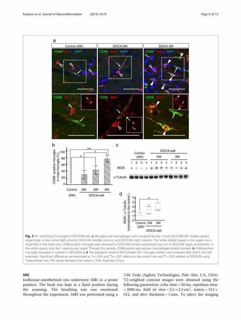
Fig. 3 M1 switching of microglia in DOCA3W-rats. a Microglia and macrophages were visualized by Iba-1 (red) and CD68 (M1 marker, green),respectively, in the control (left column), DOCA2W (middle column), and DOCA3W (right column). The white dotted square in the upper row ismagnified in the lower row. CD68-positive microglia were observed in DOCA3W (closed arrowheads) but not in DOCA2W (open arrowheads). Inthe white square, only Iba-1 staining was noted. Through the periods, CD68-positive perivascular macrophages existed (arrows). b CD68-positivemicroglia increased in number in DOCA3W. c, d The expression level of iNOS protein (M1 microglia marker) was increased after DOCA and salttreatment. Significant differences are expressed as *p < 0.05 and **p < 0.01 relative to the control rats and #P < 0.05 relative to DOCA2W usingTukey-Kramer test. The values represent the means ± SEM. Scale bars 50 μm
Koizumi et al. Journal of Neuroinflammation (2019) 16:79 Page 6 of 13
position, proton density-weighted images were obtainedusing the following parameters: echo time = 11ms, repe-tition time = 2000 ms, field of view = 2.5 × 2.5 cm2,matrix = 512 × 512, and slice thickness = 1 mm.
Hematoxylin and eosin (HE) or Klüver-Barrera (KB)stainingHE staining was performed to observe the tissue andvascular changes according to the standard procedure.Briefly, sections were stained with Mayer’s hematoxylinfor 3 min and then washed in running tap water for 10min. Thereafter, the sections were stained with eosin for90 s. These sections were subsequently dehydrated andcleared using alcohol and xylene, respectively. The vas-cular remodeling structure in HE staining was observedusing microscopy (IX73, Olympus, Tokyo, Japan). We
identified vascular remodeling as perivascular enlarge-ment and vessel wall thickening [19]. The image acquisi-tion region was randomly selected so that a corticalblood vessel was always included in the image. Ten ves-sels per rat, that is, 30 vessels per group, were acquired.All images within an experiment were acquired underthe same microscope settings.KB staining was performed to observe the demyelin-
ation according to the standard procedure. Briefly,sections were stained with Luxol Fast Blue solution in a56 °C oven overnight and then washed in 95% alcoholand distilled water. Thereafter, the sections were stainedwith lithium carbonate solution for 30 s. Then, sectionscontinued to undergo differentiation in 70% alcoholuntil the gray matter was clear and the white mattersharply defined. Next, they were counterstained with
aControl (0W) DOCA 2W DOCA 3W
b c
1 2 3 4 1 2 3 4 1 2 3 4
α-Tubulin
Arginase-1
Control(0W)
DOCA-salt
2W 3W
Arg
inas
e-1
/ α-T
ubul
in
(nor
mal
ized
to th
e co
ntro
l )
2
1
0Control 2W 3W
(0W)DOCA-salt
CD206 / Iba-1 CD206 / Iba-1
CD206 / Iba-1
CD206 / Iba-1 /DAPI
CD206 / Iba-1 / DAPICD206 / Iba-1 / DAPI
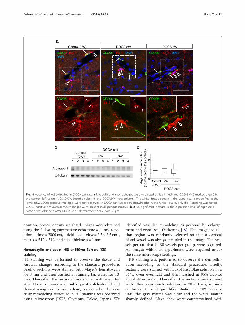
Fig. 4 Absence of M2 switching in DOCA-salt rats. a Microglia and macrophages were visualized by Iba-1 (red) and CD206 (M2 marker, green) inthe control (left column), DOCA2W (middle column), and DOCA3W (right column). The white dotted square in the upper row is magnified in thelower row. CD206-positive microglia were not observed in DOCA-salt rats (open arrowheads). In the white square, only Iba-1 staining was noted.CD206-positive perivascular macrophages were present in all periods (arrows). b, c No significant increase in the expression level of arginase-1protein was observed after DOCA and salt treatment. Scale bars 50 μm
Koizumi et al. Journal of Neuroinflammation (2019) 16:79 Page 7 of 13
cresyl violet acetate. These sections were subsequentlydehydrated and cleared using alcohol and xylene, re-spectively. The severity of the white matter lesions wasgraded as reported previously [20]. We also confirmed
the presence of a white matter lesion as the formation ofmarked vacuoles or disappearance of myelinated fibers.We observed images of every slide by microscopy (IX73,Olympus, Tokyo, Japan).
Control 2W 3W 4W
(0W) DOCA-salt
Control (0W) DOCA2W DOCA3W DOCA4W
**
a
b
f
***
*100
80
60
40
20
0Ki-6
7-po
sitiv
e m
icro
glia
in
)%( ailgorci
m latot
100
80
60
40
20
0
Ki-6
7-po
sitiv
e pe
rivas
cula
r m
icro
glia
(%
) c d
Ki-67 / Iba-1
Ki-67 / CD68 Ki-67 / CD206
DOCA3W
Ki-67 / CD163
Control 2W 3W 4W
(0W) DOCA-salt
*
Control 2W 3W 4W
(0W) DOCA-salt
##
†
††
##100
80
60
40
20
0
Per
cent
age
of K
i-67
posi
tive
M
1-st
ate
mic
rogl
ia (
%)
††
Control (0W) DOCA2W DOCA3W DOCA4W
e
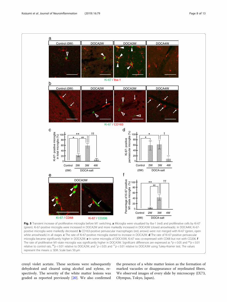
Fig. 5 Transient increase of proliferative microglia before M1 switching. a Microglia were visualized by Iba-1 (red) and proliferative cells by Ki-67(green). Ki-67-positive microglia were increased in DOCA2W and more markedly increased in DOCA3W (closed arrowheads). In DOCA4W, Ki-67-positive microglia were markedly decreased. b CD163-positive perivascular macrophages (red, arrows) were not merged with Ki-67 (green, openwhite arrowheads) in all stages. c The rate of Ki-67-positive microglia started to increase in DOCA2W. d The rate of Ki-67-positive perivascularmicroglia became significantly higher in DOCA2W. e In some microglia of DOCA3W, Ki-67 was co-expressed with CD68 but not with CD206. fThe rate of proliferative M1-state microglia was significantly higher in DOCA3W. Significant differences are expressed as *p < 0.05 and **p < 0.01relative to control rats, ##p < 0.01 relative to DOCA2W, and †p < 0.05 and ††p < 0.01 relative to DOCA3W using Tukey-Kramer test. The valuesrepresent the means ± SEM. Scale bars 50 μm
Koizumi et al. Journal of Neuroinflammation (2019) 16:79 Page 8 of 13
Statistical analysisAll statistical analyses were performed with JMP12 (SASInstitute, Cary, NC, USA). We used Student’s t test,Dunnett’s test, or Tukey-Kramer test. Error bars repre-sent the means ± SEM in all figures. A p value of < 0.05was considered significant.
ResultsDOCA-salt-mediated hypertension induces abnormalparenchymal and cerebrovascular morphologiesCompared with control, placebo groups remainednormotensive (Fig. 1b) and did not show any abnormalfindings in histology or MR images (Additional file 1:Figure S2c and S5). DOCA2W showed a marked eleva-tion of the blood pressure in the absence of any clinical,histopathological and MRI features of CSVD (Fig. 1b, c,Additional file 1: Figure S5 and Table 1). DOCA3Wdemonstrated several changes in addition to the sus-tained blood pressure elevation. First, DOCA3W showedclinical symptoms including hemiparesis, a decreasedfood intake, and bleeding from the tail vein (Table 1).Second, vascular remodeling in the cortex and the for-mation of vacuoles in the white matter were apparent(Fig. 1c). Third, focal high- and low-intensity areas wereseen on T2-weighted on MR images (Additional file 1:Figure S5a and S5b). In DOCA4W, we observed shrink-age of the astrocyte foot processes (Additional file 1: Fig-ure S1), a marked decrease in movement, myelindegeneration, and diffuse high-intensity areas on MRimages, in addition to hypertension.
Morphological changes of microglia precede theappearance of histopathological abnormalitiesIn the control and placebo groups, resting microglia, mor-phologically characterized by their fine processes, wereobserved sparsely in the cerebral parenchyma (Fig. 2a, leftcolumn). In DOCA2W, round-shaped microglia withshortened processes were observed juxtaposing vessels inthe cerebral parenchyma (Fig. 2a, middle column), in theabsence of histopathological abnormalities (Fig. 1c).Thereafter, morphological changes of microglia furtherprogressed. In DOCA3W, more amoeboid microglia accu-mulated around structurally altered vessels (Fig. 2a, rightcolumn). In DOCA4W, amoeboid microglia were wide-spread across the cortex and white matter (Additional file 1:Figure S3a). The number of microglia juxtaposing vesselsin the cortex was significantly increased in DOCA2W andDOCA3W (Fig. 2c).We distinguished microglia from perivascular macro-
phages (PVM) by their Iba-1 intensity and morphology,based on a previous report that the expression of Iba-1 isweak in macrophages [21]. We confirmed this using amacrophage-specific marker, CD163 [22]. CD163-negativemicroglia showed intense Iba-1 immunoreactivity and
processes. On the other hand, CD163-positive PVMshowed faint Iba-1 immunoreactivity and a flattened shape(Fig. 2b). Moreover, the distribution of microglia was dif-ferent from that of the PVM. CD163-positive macro-phages were never observed to be accumulated in theinflammatory lesion (Additional file 1: Figure S3a).
Activated perivascular microglia express a pro-inflammatory patternFor characterization of the morphologically activated peri-vascular microglia, we first studied the expression of CD68as a pro-inflammatory M1 marker. In the control group,microglia were CD68-negative (Fig. 3a, left column). Whilemost of the microglia were CD68-negative in DOCA2W(Fig. 3a, middle column), CD68-positive, Iba1-positivemicroglia significantly increased around vessel walls andparenchyma in DOCA3W (Fig. 3a, right column). Quanti-tative analysis showed that the percentage of CD68-positivemicroglia increased significantly in DOCA3W comparedwith the control (Fig. 3b). As for the PVM, a sparse distri-bution pattern of CD68-positive PVMs was similar amongthe control, placebo, and DOCA groups (Fig. 3a). Biochem-ical analysis indicated that the expression level of induciblenitric oxide synthase (iNOS) as another pro-inflammatoryM1 marker was increased in DOCA3W (Fig. 3c and d).Next, we studied the expression of CD206 as ananti-inflammatory M2 marker. Perivascular microglia didnot express CD206 in rat brains of any groups (Fig. 4a), ex-cept for a few CD206-positive microglia around hemorrhage sites (Additional file 1: Figure S3b). As for the PVM,a sparse distribution pattern of CD206-positive PVMs wassimilar among in the control, placebo, and DOCA groups(Fig. 4a). Biochemical analysis also indicated that the ex-pression level of arginase-1, another anti-inflammatory M2marker, was not changed in the rat brains of any groups(Fig. 4b and c). Taken together, direct M1 activation, butnot the M2 state, was identified in our DOCA-salt model.
Activated microglia transiently expressed a cellproliferation marker, Ki-67, prior to M1 switchingThe total number of microglia did not change betweenthe control and placebo groups, whereas it significantlyincreased in the DOCA group (Additional file 1: FigureS4a). In the control and placebo groups, no microgliaexpressed Ki-67 (Fig. 5a, c and Additional file 1: FigureS4c). The rate of Ki-67-positive microglia significantlyincreased to 22% of the total microglia in DOCA2W,peaked to 54% in DOCA3W, and then decreased to thebaseline level in DOCA4W (Fig. 5a and c). A similar in-crease in the number of Ki-67-positive microglia wasalso observed (Additional file 1: Figure S4b). The rate ofKi-67-positive perivascular microglia peaked to 55% ofthe total perivascular microglia in DOCA2W and remainat the same level in DOCA3W (Fig. 5d). Rates of both
Koizumi et al. Journal of Neuroinflammation (2019) 16:79 Page 9 of 13
Ki-67-positive and M1-state microglia were highest inDOCA3W (Fig. 5e and f). In DOCA4W, proliferativeM1-state microglia markedly decreased (Fig. 5c and d). Incontrast, PVM did not express Ki-67 in any group (Fig. 5b).
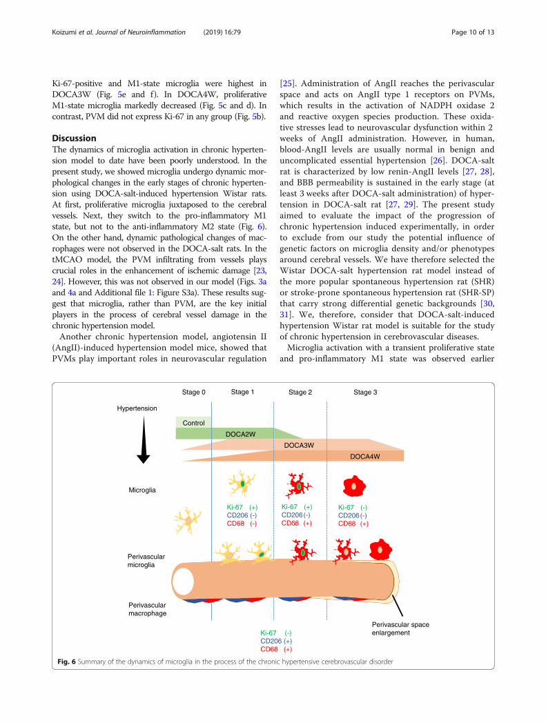
DiscussionThe dynamics of microglia activation in chronic hyperten-sion model to date have been poorly understood. In thepresent study, we showed microglia undergo dynamic mor-phological changes in the early stages of chronic hyperten-sion using DOCA-salt-induced hypertension Wistar rats.At first, proliferative microglia juxtaposed to the cerebralvessels. Next, they switch to the pro-inflammatory M1state, but not to the anti-inflammatory M2 state (Fig. 6).On the other hand, dynamic pathological changes of mac-rophages were not observed in the DOCA-salt rats. In thetMCAO model, the PVM infiltrating from vessels playscrucial roles in the enhancement of ischemic damage [23,24]. However, this was not observed in our model (Figs. 3aand 4a and Additional file 1: Figure S3a). These results sug-gest that microglia, rather than PVM, are the key initialplayers in the process of cerebral vessel damage in thechronic hypertension model.Another chronic hypertension model, angiotensin II
(AngII)-induced hypertension model mice, showed thatPVMs play important roles in neurovascular regulation
[25]. Administration of AngII reaches the perivascularspace and acts on AngII type 1 receptors on PVMs,which results in the activation of NADPH oxidase 2and reactive oxygen species production. These oxida-tive stresses lead to neurovascular dysfunction within 2weeks of AngII administration. However, in human,blood-AngII levels are usually normal in benign anduncomplicated essential hypertension [26]. DOCA-saltrat is characterized by low renin-AngII levels [27, 28],and BBB permeability is sustained in the early stage (atleast 3 weeks after DOCA-salt administration) of hyper-tension in DOCA-salt rat [27, 29]. The present studyaimed to evaluate the impact of the progression ofchronic hypertension induced experimentally, in orderto exclude from our study the potential influence ofgenetic factors on microglia density and/or phenotypesaround cerebral vessels. We have therefore selected theWistar DOCA-salt hypertension rat model instead ofthe more popular spontaneous hypertension rat (SHR)or stroke-prone spontaneous hypertension rat (SHR-SP)that carry strong differential genetic backgrounds [30,31]. We, therefore, consider that DOCA-salt-inducedhypertension Wistar rat model is suitable for the studyof chronic hypertension in cerebrovascular diseases.Microglia activation with a transient proliferative state
and pro-inflammatory M1 state was observed earlier
Ki-67 (+) CD206 (-)CD68 (-)
Hypertension
Control (0W) DOCA2W
DOCA3W
DOCA4W
Ki-67 (+) CD206(-)CD68 (+)
Ki-67 (-) CD206(-)CD68 (+)
Microglia
Perivascularmacrophage
Ki-67 (-) CD206 (+)CD68 (+)
Stage 0 Stage 1 Stage 2 Stage 3
Perivascular spaceenlargement
Perivascularmicroglia
Fig. 6 Summary of the dynamics of microglia in the process of the chronic hypertensive cerebrovascular disorder
Koizumi et al. Journal of Neuroinflammation (2019) 16:79 Page 10 of 13
than cerebral vessel damage in our model. Regarding ab-normal findings of the cerebral vasculature, perivascularspace enlargement was observed in DOCA3W, and ap-parent BBB breakdown identified by astrocyte footshrinkage was noted in DOCA4W (Additional file 1: Fig-ure S1). These results are consistent with a previousstudy indicating that BBB permeability remained un-changed in DOCA3W mice using the Evans Blue ex-travasation method [27].Regarding microglia dynamics, our findings differed from
those of previous reports using acute cerebrovascular dis-ease models, such as tMCAO. In the tMCAO model,microglia were activated to an anti-inflammatory M2 statefollowed by a transition to a pro-inflammatory M1 state[11]. The variation in microglia dynamics between chronichypertension and acute ischemic models may be due to thedifference of microglia-activating factors. In the tMCAOmodel, various factors were produced in the ischemic brainseveral hours or days after reperfusion. Interferon regula-tory factor 4, known as an M2-switching factor, was rapidlyupregulated within 2 h after ischemic insult [32]. HMGB1,one of the DAMPs, was produced several hours after ische-mic events, and HMGB1 was able to activate microglia[16]. ATP or excessive glutamate was immediately releasedfrom necrotizing neuronal cells and could activate micro-glia [33]. In a chronic hypertension model, other than theDOCA-model, a previous study using partial renal arteryocclusion model showed that microglia were activated toan inflammatory state within 5 weeks after the operation[34]. In this model, chronic hypertension increased the ex-pression of adhesion molecules such as JAM-1, ICAM-1,and VCAM-1 on the cerebral endothelium and this led todeposition of platelets. Deposited platelets producedCD40L, which mediated the activation of pro-inflammatorymicroglia and activated NFκB and mitogen-activated pro-tein kinase signaling in microglia.A previous report showed that extracellular signal-regu-
lated kinase (ERK)-activated microglia acquired a prolifera-tive ability and produced mainly pro-inflammatorycytokines, which cause synaptic and neuronal losses in thebrain and result in lethal neurodegenerative disease in adultmice [35]. This suggests that microglia proliferate prior toactivation of the M1 state. We, therefore, analyzed the ex-pression of Ki-67, as a cell proliferation marker, in microgliaby applying immunostaining to our model. In our model,proliferative microglia had a close relationship withM1-state activation. Interestingly, such relationship was re-cently demonstrated using transgenic mice harboring som-atic BRAF mutation, p.V600E. Activation of the MEK-ERKpathway induces microglia proliferation and is associatedwith an upregulation of pro-inflammatory cytokines fromthe proliferative microglia [35]. The rate of Ki-67-positivemicroglia significantly increased in DOCA2W, peaked inDOCA3W, and then decreased to the baseline in
DOCA4W (Fig. 5a and c). On the other hand, the rate ofKi-67-positive perivascular microglia peaked in DOCA2Wand DOCA3W (Fig. 5d) and similarly decreased to thebaseline in DOCA4W. These results indicate that restingmicroglia were preferentially activated around the vessels,and those perivascular microglia acquired the ability to pro-liferate earlier than other microglia apart from the vessels.Our results suggest that specific cytokines released fromthe vessels induced perivascular microglia to enter a prolif-erative state. Further studies are required to clarify the mo-lecular signaling involved in this phenomenon.According to our immunohistochemical study, we
concluded that perivascular microglia were from resi-dential microglia. We distinguished microglia from mac-rophages by Iba-1 intensity and cell morphology. In thepresent study, we did not employ flow cytometry,lineage-specific markers, or other reporter methods.Flow cytometry succeeded to detect infiltration of mac-rophages in the ischemic brain by using tMCAO mice inwhich ischemic changes were observed globally inre-perfusion area [15]. However, in DOCA-salt rats,localization of vascular damages was sparse, and hence,we performed immunohistochemical analysis rather thanflow cytometry analysis of brain homogenate. Accordingto a previous study, TMEM119 antigen is expressed spe-cifically in residential microglia in the brain but not inmacrophages [36]. Unfortunately, we failed to detectTMEM119 in rat brain using antibody raised againstmouse TMEM119 [37]. This may be due to the differ-ence of amino acid sequence in the epitope region be-tween mouse and rat. Finally, reporter method will be apowerful strategy and may be possible if the DOCA-saltmodel is prepared by transgenic mice expressing re-porter gene such as EGFP under TMEM promoter. Welike to leave this strategy for our future plan to explorethe process of microglia activation under hypertension.
ConclusionsThe present study demonstrates that perivascular micro-glia proliferate transiently and subsequently underwentdirect M1 switching, prior to cerebral vessel destruction.Our findings raise the intriguing possibility of a link be-tween perivascular microglia activation and initiation ofcerebrovascular diseases induced by chronic hyperten-sion, and that both anti-hypertensive therapy and thefine-tuning of microglia proliferation might generate asynergistic effect.
Additional file
Additional file 1: Figure S1. Morphological changes of astrocyte footprocesses making up the small vessel wall in DOCA-salt rats. Figure S2.Quantitative analysis of vessel wall thickening and perivascular space en-largement. Figure S3. Distribution of perivascular macrophages and
Koizumi et al. Journal of Neuroinflammation (2019) 16:79 Page 11 of 13
microglia in DOCA4W, and presence of CD206-positive M2-state microgliaaround a site of hemorrhage. Figure S4. Quantitative analysis of micro-glia. Figure S5. Sequential MRI analysis of rat brains. (PPTX 8857 kb)
AbbreviationsAngII: Angiotensin II; BBB: Blood-brain barrier; CSVD: Cerebral small vesseldisease; DAMPs: Damage-associated molecular patterns; DAPI: 4′, 6-Diamino-2-phenylindole; DOCA: Deoxycorticosterone acetate; ERK: Extracellular signal-regulated kinase; GFAP: Glial fibrillary acidic protein; HE: Hematoxylin andeosin; HMGB1: High-mobility group box 1; Iba-1: Ionized calcium-bindingadapter molecule 1; iNOS: Inducible nitric oxide synthase; KB: Klüver Barrera;MRI: Magnetic resonance imaging; PBST: Phosphate-buffered salinecontaining 0.1% Tween 20; PVMs: Perivascular macrophages;tMCAO: Transient middle cerebral artery occlusion
AcknowledgementsWe thank Hiromi Yasuike (Kyoto Prefectural University of Medicine) fortechnical support.
FundingThis work was supported by the construction and application of a databasefor CADASIL, a hereditary small-vessel disease, by the Japan Agency for Med-ical Research and Development (AMED), and by a grant-in-aid for Researchon Intractable Disease “Research group on medical infrastructure for adult-onset leukoencephalopathy” from the Japanese Ministry of Health, Labour,and Welfare, Japan.
Availability of data and materialsThe datasets used and/or analyzed during the current study are availablefrom the corresponding author on reasonable request.
Authors’ contributionsTK, KT, TN, MT, and MT designed research; TK, KT, HT, MO, OO, and KIperformed research; SM contributed new reagents/analysis tools; TK and KTanalyzed the data; TK, KT, IM, SF, HS, and TM wrote the paper. All authorsread and approved the final manuscript.
Ethics approval and consent to participateProtocols were approved by Animal Care and Use Committees of KyotoPrefectural University of Medicine and Kyoto Pharmaceutical University. Careand use of rodents met the standards set by the National Institutes of Healthfor experimental animals.
Consent for publicationNot applicable.
Competing interestsThe authors declare that they have no competing interests.
Publisher’s NoteSpringer Nature remains neutral with regard to jurisdictional claims inpublished maps and institutional affiliations.
Author details1Department of Neurology, Graduate School of Medical Science, KyotoPrefectural University of Medicine, 465 Kajii-cho Kamigyo-ku, Kyoto 602-8566,Japan. 2Department of Anatomy and Neurobiology, Graduate School ofMedical Science, Kyoto Prefectural University of Medicine, Kyoto, Japan.3Department of Clinical Pharmacology, Division of Pathological Sciences,Kyoto Pharmaceutical University, Kyoto, Japan. 4Department of Orthopaedics,Graduate School of Medical Science, Kyoto Prefectural University of Medicine,Kyoto, Japan. 5Department of Applied Biology, Kyoto Institute of Technology,Kyoto, Japan. 6Department of Pharmacology and Toxicology, School forMental Health and Neuroscience, Maastricht University, Maastricht, theNetherlands. 7Department of Neuroscience, School for Mental Health andNeuroscience, Maastricht University Medical Center +, Maastricht, TheNetherlands.
Received: 19 July 2018 Accepted: 26 March 2019
References1. Salter MW, Stevens B. Microglia emerge as central players in brain disease.
Nat Med. 2017;23:1018–27.2. Wake H, Moorhouse AJ, Jinno S, Kohsaka S, Nabekura J. Resting microglia
directly monitor the functional state of synapses in vivo and determine thefate of ischemic terminals. J Neurosci. 2009;29:3974–80.
3. Meyer-Luehmann M, Spires-Jones TL, Prada C, Garcia-Alloza M, de CalignonA, Rozkalne A, Koenigsknecht-Talboo J, Holtzman DM, Bacskai BJ, Hyman BT.Rapid appearance and local toxicity of amyloid-beta plaques in a mousemodel of Alzheimer's disease. Nature. 2008;451:720–4.
4. Franco R, Fernandez-Suarez D. Alternatively activated microglia andmacrophages in the central nervous system. Prog Neurobiol. 2015;131:65–86.
5. H EH, Noristani HN, Perrin FE. Microglia responses in acute and chronicneurological diseases: what microglia-specific transcriptomic studies taught(and did not teach) us. Front Aging Neurosci. 2017;9:227.
6. Huber JD, Campos CR, Mark KS, Davis TP. Alterations in blood-brain barrierICAM-1 expression and brain microglial activation after lambda-carrageenan-induced inflammatory pain. Am J Physiol Heart Circ Physiol.2006;290:H732–40.
7. Erdő F, Denes L, de Lange E. Age-associated physiological and pathologicalchanges at the blood–brain barrier: a review. J Cereb Blood Flow Metab.2017;37:4–24.
8. Erdo F, Trapp T, Mies G, Hossmann KA. Immunohistochemical analysis ofprotein expression after middle cerebral artery occlusion in mice. ActaNeuropathol. 2004;107:127–36.
9. Perego C, Fumagalli S, De Simoni MG. Temporal pattern of expression andcolocalization of microglia/macrophage phenotype markers following brainischemic injury in mice. J Neuroinflammation. 2011;8:174.
10. Jolivel V, Bicker F, Biname F, Ploen R, Keller S, Gollan R, Jurek B, BirkenstockJ, Poisa-Beiro L, Bruttger J, et al. Perivascular microglia promote blood vesseldisintegration in the ischemic penumbra. Acta Neuropathol. 2015;129:279–95.
11. Hu X, Li P, Guo Y, Wang H, Leak RK, Chen S, Gao Y, Chen J. Microglia/macrophage polarization dynamics reveal novel mechanism of injuryexpansion after focal cerebral ischemia. Stroke. 2012;43:3063–70.
12. Hu X, Leak RK, Shi Y, Suenaga J, Gao Y, Zheng P, Chen J. Microglial andmacrophage polarization[mdash]new prospects for brain repair. Nat RevNeurol. 2015;11:56–64.
13. Rodrigo R, Fernandez-Gajardo R, Gutierrez R, Matamala JM, Carrasco R,Miranda-Merchak A, Feuerhake W. Oxidative stress and pathophysiology ofischemic stroke: novel therapeutic opportunities. CNS Neurol Disord DrugTargets. 2013;12:698–714.
14. Shichita T, Ito M, Yoshimura A. Post-ischemic inflammation regulates neuraldamage and protection. Front Cell Neurosci. 2014;8:319.
15. Shichita T, Sugiyama Y, Ooboshi H, Sugimori H, Nakagawa R, Takada I, IwakiT, Okada Y, Iida M, Cua DJ, et al. Pivotal role of cerebral interleukin-17-producing gammadeltaT cells in the delayed phase of ischemic brain injury.Nat Med. 2009;15:946–50.
16. Liu K, Mori S, Takahashi HK, Tomono Y, Wake H, Kanke T, Sato Y, Hiraga N,Adachi N, Yoshino T, Nishibori M. Anti-high mobility group box 1monoclonal antibody ameliorates brain infarction induced by transientischemia in rats. FASEB J. 2007;21:3904–16.
17. Pantoni L. Cerebral small vessel disease: from pathogenesis and clinicalcharacteristics to therapeutic challenges. Lancet Neurol. 2010;9:689–701.
18. Pires PW, Dams Ramos CM, Matin N, Dorrance AM. The effects ofhypertension on the cerebral circulation. Am J Physiol Heart Circ Physiol.2013;304:H1598–614.
19. Schreiber S, Bueche CZ, Garz C, Braun H. Blood brain barrier breakdown asthe starting point of cerebral small vessel disease? - New insights from a ratmodel. Exp Transl Stroke Med. 2013;5:4.
20. Lin JX, Tomimoto H, Akiguchi I, Wakita H, Shibasaki H, Horie R. White matterlesions and alteration of vascular cell composition in the brain ofspontaneously hypertensive rats. Neuroreport. 2001;12:1835–9.
21. Zarruk JG, Greenhalgh AD, David S. Microglia and macrophages differ intheir inflammatory profile after permanent brain ischemia. Exp Neurol. 2018;301:120–32.
Koizumi et al. Journal of Neuroinflammation (2019) 16:79 Page 12 of 13
22. Faraco G, Park L, Anrather J, Iadecola C. Brain perivascular macrophages:characterization and functional roles in health and disease. J Mol Med (Berl).2017;95:1143–52.
23. Shichita T, Sakaguchi R, Suzuki M, Yoshimura A. Post-ischemic inflammationin the brain. Front Immunol. 2012;3:132.
24. Shichita T, Hasegawa E, Kimura A, Morita R, Sakaguchi R, Takada I, Sekiya T,Ooboshi H, Kitazono T, Yanagawa T, et al. Peroxiredoxin family proteins are keyinitiators of post-ischemic inflammation in the brain. Nat Med. 2012;18:911–7.
25. Faraco G, Sugiyama Y, Lane D, Garcia-Bonilla L, Chang H, Santisteban MM,Racchumi G, Murphy M, Van Rooijen N, Anrather J, Iadecola C. Perivascularmacrophages mediate the neurovascular and cognitive dysfunctionassociated with hypertension. J Clin Invest. 2016;126:4674–89.
26. Catt KJ, Cran E, Zimmet PZ, Best JB, Cain MD, Coghlan JP. Angiotensin IIblood-levels in human hypertension. Lancet. 1971;1:459–64.
27. Rodrigues SF, Granger DN. Cerebral microvascular inflammation in DOCAsalt-induced hypertension: role of angiotensin II and mitochondrialsuperoxide. J Cereb Blood Flow Metab. 2012;32:368–75.
28. Chamorro V, Wangensteen R, Sainz J, Duarte J, O'Valle F, Osuna A, Vargas F.Protective effects of the angiotensin II type 1 (AT1) receptor blockade inlow-renin deoxycorticosterone acetate (DOCA)-treated spontaneouslyhypertensive rats. Clin Sci (Lond). 2004;106:251–9.
29. Werber AH, Fitch-Burke MC. Effect of chronic hypertension on acutehypertensive disruption of the blood-brain barrier in rats. Hypertension.1988;12:549–55.
30. Waki H, Liu B, Miyake M, Katahira K, Murphy D, Kasparov S, Paton JF.Junctional adhesion molecule-1 is upregulated in spontaneouslyhypertensive rats: evidence for a prohypertensive role within the brainstem. Hypertension. 2007;49:1321–7.
31. Waki H, Hendy EB, Hindmarch CC, Gouraud S, Toward M, Kasparov S,Murphy D, Paton JF. Excessive leukotriene B4 in nucleus tractus solitarii isprohypertensive in spontaneously hypertensive rats. Hypertension. 2013;61:194–201.
32. Guo S, Li Z, Jiang D, Lu YY, Liu Y, Gao L, Zhang S, Lei H, Zhu L, Zhang X, etal. IRF4 is a novel mediator for neuronal survival in ischaemic stroke. CellDeath Differ. 2014;21:888–903.
33. Barakat R, Redzic Z. The role of activated microglia and residentmacrophages in the neurovascular unit during cerebral ischemia: is the jurystill out? Med Princ Pract. 2016;25(Suppl 1):3–14.
34. Bhat SA, Goel R, Shukla R, Hanif K. Platelet CD40L induces activation ofastrocytes and microglia in hypertension. Brain Behav Immun. 2017;59:173–89.
35. Mass E, Jacome-Galarza CE, Blank T, Lazarov T, Durham BH, Ozkaya N,Pastore A, Schwabenland M, Chung YR, Rosenblum MK, et al. A somaticmutation in erythro-myeloid progenitors causes neurodegenerative disease.Nature. 2017;549:389–93.
36. Li Q, Barres BA. Microglia and macrophages in brain homeostasis anddisease. Nat Rev Immunol. 2018;18:225–42.
37. Furube E, Kawai S, Inagaki H, Takagi S, Miyata S. Brain region-dependentheterogeneity and dose-dependent difference in transient microgliapopulation increase during lipopolysaccharide-induced inflammation. SciRep. 2018;8:2203.
Koizumi et al. Journal of Neuroinflammation (2019) 16:79 Page 13 of 13


![From Stroke to Dementia: a Comprehensive Review Exposing ... · after both hemorrhagic and ischemic stroke, as observed in rodents and non-human primates [17, 18]. Abnormal perivascular](https://img.pdfslide.tips/doc/110x75/5e47cc033fa49928c25efa78/from-stroke-to-dementia-a-comprehensive-review-exposing-after-both-hemorrhagic.jpg)