Embed Size (px)
Citation preview

Urinary incontinence
Jianhong Zhou

INTRODUCTION
• Urinary incontinence (UI) affects well over 13 million people in USA
• Estimated costs in excess of $15 billion annually
• Most women do not seek help for incontinence– Because of social embarrassment– Be unaware that help is available

INTRODUCTION
• Incontinence is part of the “normal” aging process is no longer acceptable
• The advances in modern medicine during the last 80 years have increased the life expectancy of women well into the eighth and ninth decades
• We are caring for patients longer and better than ever, enabling women to enjoy longer and more productive lifetimes
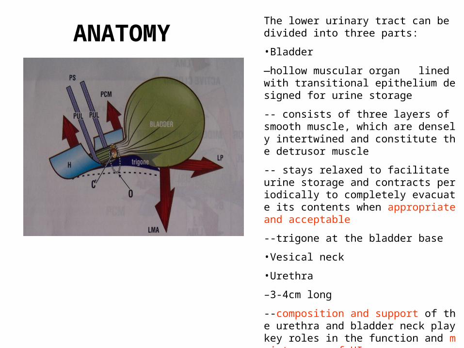
ANATOMYThe lower urinary tract can be divided into three parts:
•Bladder
—hollow muscular organ lined with transitional epithelium designed for urine storage
-- consists of three layers of smooth muscle, which are densely intertwined and constitute the detrusor muscle
-- stays relaxed to facilitate urine storage and contracts periodically to completely evacuate its contents when appropriate and acceptable
--trigone at the bladder base
•Vesical neck
•Urethra
–3-4cm long
--composition and support of the urethra and bladder neck play key roles in the function and maintenance of UI
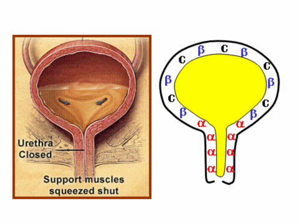

NEUROANATOMY•Neuronal innervation of the lower urinary tract is considered part of the autonomic and somatic nervous systems
•The autonomic system receives visceral sensation and regulates smooth muscle activity during conscious and involuntary LUTF
•Voluntary control of micturition is controlled by the central nervous system
•Receiving both sensory afferent and modulating motor efferent nerves, the net effect is that the brain provides tonic inhibition of detrusor contraction
•Lesions in the frontal lobe chiefly cause loss of voluntary control of micturiton and thus loss of suppression of the detrusor reflex, resulting in uncontrolled voiding or urge urinary incontinence
•A reflex activation in the central brainstem and peripheral spinal cord mediate a coordinated series of events, consisting of relaxation of the striated urethral musculature and detrusor contraction that result in opening of the bladder neck and urethra

URINARY INCONTINENCE
• Types and Definition
• Evaluation
• Treatment

Types and Definition
• Stress urinary incontinence (SUI)
• Urge incontinence
• Mixed incontinence
• Overflow incontinence
• Extraurethral sources of urine

Types and Definition
SUI
• Loss of urine that occurs with increased abdominal pressure, such as coughing or straining
• Result of loss of anatomic support of the urethrovesical junction or urethra
• It most commonly occurs following pelvic floor muscle and nerve damage that resulted from childbearing

Types and Definition
Urge incontinence
– is defined by the symptom of urine loss t
hat occurs when the patient experiences
urgency, or a strong desire to void
– is often accompanied by symptoms of ur
inary frequency, urgency, and nocturia

Types and Definition
Mixed incontinence
• Occurs when both stress and detrusor ins
tability occur simultaneously
• Patients may present with symptoms of bo
th types of incontinence

Types and Definition
Overflow incontinence
• Occurs because of underactivity of the det
rusor muscle
• Be associated with retention of urine
• The bladder does not empty completely, a
nd “dribbling” of urine occurs

Types and Definition
Extraurethral sources of urine
• Include genitourinary fistulas
• Be congenital or follow pelvic surgery or ra
diation
• These typically cause continuous leaking o
f urine

Evaluation
• History
• Physical examination
• Diagnostic tests
• Cystoscopy

EvaluationA detailed history is essential and should include: a. Urinary symptoms, including the presence of voiding freq
uency, nocturia, urgency, precipitating events, and frequency of loss. A voiding diary allows the patient to document voiding frequency and incontinence episodes during a specific period
b. Previous urologic surgeryc. Obstetric history, including parity, birth weights, and mod
e of deliveryd. CNS or spinal cord disorderse. Use of medications, including diuretics, antihypertensive
s, caffeine, alcohol, anticholinergics, decongestants, nicotine, and psychotropics
f. Presence of other medical disorders (e.g., hypertension or hematuria)

Evaluation
Physical examination may detect:
a. Exacerbating conditions, such as chronic
obstructive pulmonary disease, obesity, or
intra-abdominal mass
b. Hypermobility of the urethra
c. POP
d. Neurologic disorder
Evaluation
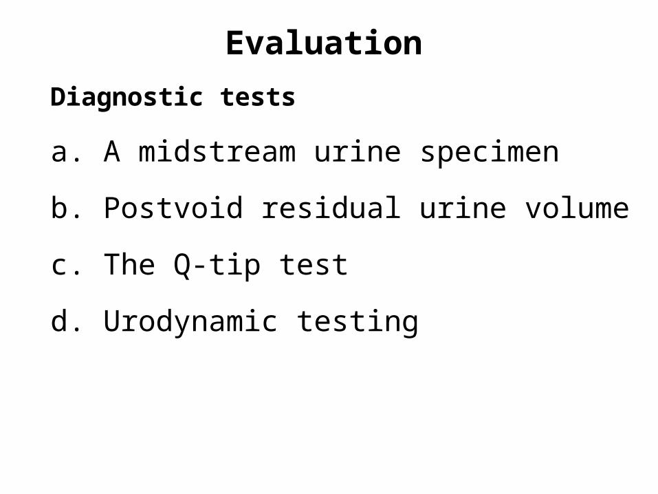
Diagnostic tests
a. A midstream urine specimen
b. Postvoid residual urine volume
c. The Q-tip test
d. Urodynamic testing

Evaluation
Diagnostic tests -- midstream urine specimen
• Be collected for urinalysis or culture and s
ensitivity
• Infection may aggravate urinary incontinen
ce

Evaluation
Diagnostic tests -- Postvoid residual urine volume
• should be measured (by ultrasound or cat
heterization) after the patient has voided
• Typically, the postvoid residual urine volu
me is less than 50 to 100 ml

Evaluation
Diagnostic tests -- The Q-tip test
• is an indirect measure of the urethral axis
• A Q-tip is inserted into the urethra with the
patient in the lithotomy position
• If the Q-tip moves more than 30 degrees fr
om the horizontal with straining, urethra hy
permobility is present

Bladder at restBladder at rest
Bladder with strainingBladder with straining
Q-TIP TEST At rest the Q-tip is in a horizontal position,but with straining & coughing it shows a positive deflectionowing to inadequate support at the urethrovesical junction.

Bladder with strainingBladder with straining
Bladder at restBladder at rest
Q-TIP TEST At rest the Q-tip is in a horizontal position,but with straining & coughing it shows a positive deflectionowing to inadequate support at the urethrovesical junction.

EvaluationDiagnostic tests -- Urodynamic testing
• including a cystometrogram and voiding studies, may be useful for demonstrating the type of incontinence present
• These tests measure pressures within the bladder and abdomen during bladder filling and emptying
• Urodynamic testing is indicated for complex cases of urinary incontinence such as mixed incontinence or in patients with incontinence and retention of urine

Evaluation
Cystoscopy
• is performed in some patients
• to examine the bladder and urethral muco
sa for abnormalities such as diverticula or
neoplasms

CystoscopyCystoscopy

What can irritate bladder?
Urinalysis: WBC, RBC, BacteriaCulture: Positive

Typical case
• 51 years old woman complaining urine loss almost all time on daytime, especially after micturition
• TOT for her ,but she cannot pass the urine
• Open the urethra with pressure, she can pass the urine with frequency, urgency
• Postvoid residual urine volume—450ml

Treatment
Therapy depends on the underlying diagnosis.
• Treatment of exacerbating factors
• Pelvic muscle rehabilitation
• Pessaries are useful conservative therapies for SUI
• Drug therapy
• Surgery

Treatment
Treatment of exacerbating factors may improve SUI
• excess weight
• chronic cough
• constipation

Treatment
Pelvic muscle rehabilitation -- be helpful for both SUI and DO
a. Kegel exercises
b. Vaginal cones
c. Biofeedback
d. Electrical stimulation

TreatmentDrug therapy is the mainstay of treatment for DO b
ut is of limited value in treating SUI. a. Antispasmodic agents (tolterodine) are highly eff
ective and are the most commonly prescribed treatments for DO
b. α-Adrenergic stimulating agents increase smooth muscle contraction in the urethral sphincter and may decrease SUI symptoms
c. Estrogens improve irritative bladder symptoms such as urgency and dysuria in postmenopausal women but do not significantly improve urinary leakage. HRT does not reduce the incidence of urinary symptoms in postmenopausal women

Treatment
Surgery is extremely effective in the treatment of SUI. It is rarely helpful for DO
a. Injection of bulking agents around the uret
hra
b. Retropubic urethropexy
c. Transvaginal needle procedures
d. Suburethral sling procedures

Treatment
Surgery --Injection of bulking agents around the urethra
• is a minimally invasive procedure to treat SUI resulting from intrinsic urethral sphincteric deficiency
• Collagen, the bulking agent currently used most commonly, provides a temporary (3 to 12 months) cure or improvement rates ranging from 50% to 70%
• They are generally indicated for patients unable to tolerate major surgery

TreatmentSurgery --Retropubic urethropexy
• elevates the urethra and bladder neck by fixing the paraurethral connective tissues to the pubis
• The most common type is the Burch procedure, which suspends the vaginal fascia lateral to the iliopectineal line (Cooper ligament)
• Burch procedure are most successful in patients who have SUI with urethral hypermobility, resulting in long-term cure rates of 75% to 90%
• Postoperative complications are uncommon but may include urinary retention and new DO

Elevation ofElevation ofUrethrovesicle JunctionUrethrovesicle Junction
Brubaker, p.167, Fig 19-1
Burch procedure/ MMKBurch procedure/ MMK

Treatment
Surgery -- Suburethral sling procedures
• place biologic and synthetic materials under the urethra• appear to affect treatment by partially obstructing the ur
ethra during times of increased intra-abdominal pressure• differ according to the type of material and the sling fixati
on points used; however, they all have high cure rates (80% to 90%)
• are more effective than retropubic operations in patients with intrinsic urethral sphincteric deficiency
• Complications may include infection, ulceration and urinary retention

Tension-freeTension-freeVaginal TapeVaginal Tape
ProcedureProcedure

THANK YOU FOR YOUR ATTENTION