Embed Size (px)
Citation preview

Vascular and Inflammatory Diseases of the Intestines
Harvard-MIT Division of Health Sciences and Technology HST.121: Gastroenterology, Fall 2005 Instructors: Dr. Jonathan Glickman

Overview• Vascular disorders
– Vascular “malformations” – Vasculitis – Ischemic disease
• Inflammatory disorders of specific etiology – Infetious enterocolitis – “Immune-mediated” enteropathy – Diverticular disease
• Idiopathic inflammatory bowel disease – Crohn’s disease – Ulcerative colitis

Sporadic Vascular Ectasia (Telangiectasia)
• Clusters of tortuous thin-walled small vessels lacking muscle or adventitia located in the mucosa and the submucosa
• The most common type occurs in cecum or ascending colon of individuals over the age of 50 and is commonly known as “angiodysplasia”
• Angiodysplasias account for 40% of all colonic vascular lesions and are the most common cause of lower GI bleeding in individuals over the age of 60

Angiodysplasia

Hereditary Vascular Ectasia
• Hereditary Hemorrhagic Telangiectasia (HHT) or Osler-Webber-Rendu disease
• Systematic disease primarily involving skin and mucous membranes, and often the GI tract
• Autosomal dominant disease with positive family history in 80% of cases
• After epistaxis which occurs in 80% of individuals, GI bleed is the most frequent presentation and occurs in 10-40% of cases

Arteriovenous Malformations (AVM’s)• Irregular meshwork of
structurally abnormal medium to large ectatic vessels
• Unlike small vessel ectasias, AVM’s can be distributed in all layers of the bowel wall
• AVM’s may present anywhere at any age, although some are thought to be congenital

VasculitisAorta Artery Arteriole Capillary Venule Vein
Goodpasture
Microscopic PolyangiitisSLE
Polyarteritis Nodosa,
Giant-Cell Arteritis, Takayasu Arteritis
, Wegener’s, Churg-Strauss Syndrome,
Henoch-Schonlein Purpura Kawasaki Disease

Vasculitis (PAN)
Vascular Insufficiency
• Vaso-occlusive Diseases – Mesenteric arterial occlusion
(embolism/thrombosis)– Mesenteric vein thrombosis – Bowel strangulation (volvulus, hernia)
• Non-Occlusive Vascular Insufficiency – Systemic hemodynamic disturbances – Local hemodynamic disturbances

Ischemic colitis

Ischemic Colitis
Infectious Enteritis• The most common GI problem worldwide
• Most symptomatic infections produce diarrhea and some produce malabsorption
• Diagnosis is most often by stool culture or O&P
• Organisms rarely produce a pathognomonic pattern of injury

Mechanism of Injury: Toxin Production
• V. cholera
• E. coli
• “Food poisoning”
– Staphylococcus
– Clostridium

Mechanism of Injury: Invasion
• Bacteria – Salmonella, shigella, campylobacter, E. coli,
yersinia, mycobacteria • Protozoa
– Cryptosporidia, isospora, microsporidia • Viruses
– Rotaviruses, adenovirus, CMV, HSV• Fungi
– Histoplasma, candida

Cryptosporidiosis

The Lumen Dwellers
Ascaris lumbricoides Enterobius vermicularis

Antibiotic-Associated Colitis
• Antibiotic-associated pseudomembraneous colitis (PMC) is an acute colitis characterized by the formation of an inflammatory exudate
• PMC is a toxin-mediated colitis usually caused by C. difficile or less commonly by E. Coli
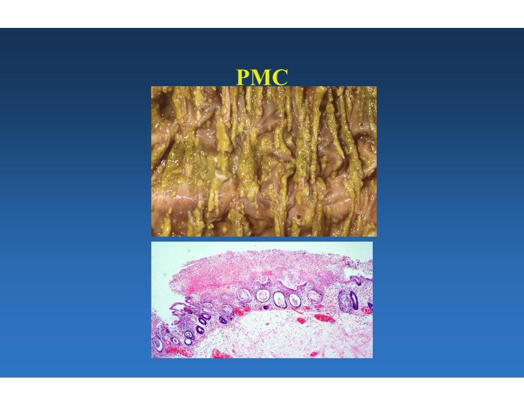
PMC

“Immune” Enteropathy: Celiac sprue
• Gluten-sensitive enteropathy, Celiac disease, Non-tropical sprue
• Chronic inflammatory disease of the proximal small intestine with generalized malabsorption
• Most common in the Irish, British, and other northern European populations
• Immune mediated injury to enterocytes accompanied by serum antibodies to gliadin, a component of gluten

Celiac disease-Immunologic mechanism of damage
Figure removed due to copyright reasons.

Celiac Sprue• Symptoms:
– Steatorrhea, abdominal distention, flatulence, fatigue,and weight loss
• Complications: – Iron and vitamin deficiency– Risk of lymphoma (T-cell type)
• Extraintestinal manifestation: – Dermatitis herpetiformis (a pruritic papulovesicular
rash with IgA deposits at the dermoepidermaljunction)

CD

Malabsorption- other causes
• Immune conditions • Hypersensitivity/allergy/eosinophilic gastroenteritis • Infection- Whipple’s dis., tropical sprue, bacterial overgrowth • Nutritional deficiencies• Inherited- Microvillous inclusion dis.,lymphangiectasia • Infiltrative disorders- amyloidosis, lymphoma • Systemic disorders- lipid storage • Other- short bowel
Whipple’s disease
Images removed due to copyright reasons. Please See:
Rosai, Juan, and Lauren Ackerman. Rosai and Ackerman's Surgical Pathology. 9th ed. New York, NY: Mosby, 2004. ISBN: 0323013422.

Collagenous, lymphocytic colitis
• Collectively, “microscopic colitis” • Middle aged to elderly adults • Chronic watery diarrhea • Endoscopically normal mucosa

Diverticulosis Coli• Acquired colonic diverticula are present in nearly half of the
population over the age of 50
• Diverticula are associated with low-fiber, low-residue diets
• Etiology is most likely high intraluminal pressure required for propulsion of hard, small stools
• Complications include hemorrhage, acute diverticulitis, perforation, fistula formation

Idiopathic Inflammatory Bowel Disease (IBD)
• Chronic, relapsing, idiopathic inflamamtory disease of the GI tract
• Crohn’s Disease
– Transmural granulomatous disease affecting any portion of the GI tract
• Ulcerative Colitis
– Superficial, non-granulomatous inflammatory disease restricted to the colon

Ulcerative Colitis
• Bloody mucoid diarrhea, rarely toxic megacolon
• Can begin at any age, peaks at 20-25 years
• Annual incidence of ~10 per 100,000 in US
• Negligible risk of cancer in the first 10 years, but 1% per year risk of cancer thereafter
• Good response to total colectomy if medical therapy fails

Ulcerative colitis- pseudopolyps
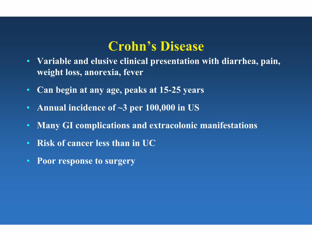
Crohn’s Disease• Variable and elusive clinical presentation with diarrhea, pain,
weight loss, anorexia, fever
• Can begin at any age, peaks at 15-25 years
• Annual incidence of ~3 per 100,000 in US
• Many GI complications and extracolonic manifestations
• Risk of cancer less than in UC
• Poor response to surgery

Crohn’s disease- gross appearance
Images removed due to copyright reasons. Please See:
Rosai, Juan, and Lauren Ackerman. Rosai and Ackerman's Surgical Pathology. 9th ed. New York, NY: Mosby, 2004. ISBN: 0323013422.
Aphthous ulcer “Cobblestoning”
Wall thickening
Crohn’s disease- stricture
Images removed due to copyright reasons. Please See:
Rosai, Juan, and Lauren Ackerman. Rosai and Ackerman's Surgical Pathology. 9th ed. New York, NY: Mosby, 2004. ISBN: 0323013422.

Crohn’s disease- microscopic
Images removed due to copyright reasons. Please See:
Rosai, Juan, and Lauren Ackerman. Rosai and Ackerman's Surgical Pathology. 9th ed. New York, NY: Mosby, 2004. ISBN: 0323013422.
Transmural inflammation Granuloma formation

Macroscopic Features of CD vs UC
NoYesCreeping fat
YesNoDilatation
ThinThickWall
NoYesStrictures
NoYesSkip areas
ColonSI, colonRegion
UCCrohn’sFeature

Microscopic Features of CD vs UC
MinimalMarkedFibrosis
NoYesFistulas
NoYesSinuses
MucosalTransmuralInflammation
SuperficialFissuringUlcers
NoYesGranulomas
UCCrohn’sFeature