Embed Size (px)
Citation preview

YU ISSN 0042-8450
VOJNOSANITETSKI PREGLED^asopis lekara i farmaceuta Vojske Srbije
Military Medical and Pharmaceutical Journal of Serbia
Vojnosanitetski pregledVojnosanit Pregl 2014; June Vol. 71 (No. 6): p. 527-614.
YU ISSN 0042-8450 vol. 71, br. 6, 2014.
Štampa Vojna štamparija, Beograd, Resavska 40 b.
VOJNOSANITETSKI PREGLEDPrvi broj Vojnosanitetskog pregleda izašao je septembra meseca 1944. godine
asopis nastavlja tradiciju Vojno-sanitetskog glasnika, koji je izlazio od 1930. do 1941. godine
IZDAVAUprava za vojno zdravstvo MO Srbije
IZDAVA KI SAVET
prof. dr sc. med. Boris Ajdinoviprof. dr sc. pharm. Mirjana Antunovi
prof. dr sc. med. Dragan Din i , puk.prof. dr sc. med. Miodrag Jevti , general potpukovnik
prof. dr sc. med. Nebojša Jovi , puk.prof. dr sc. med. oko Maksi , puk.
prof. dr sc. med. Marijan Novakovi , brigadni generalprof. dr sc. med. Zoran Popovi , brigadni general (predsednik)
prof. dr Sonja Radakoviprof. dr sc. med. Zoran Šegrt, puk.
ME UNARODNI URE IVA KI ODBOR
Assoc. Prof. Kiyoshi Ameno (Japan)Prof. Jovan Antonovi (Sweden)
Prof. Rocco Bellantone (Italy)Prof. Thorsten Gehrke (Germany)
Prof. Hanoch Hod (Israel)Prof. Thomas John (USA)
Prof. Abu-Elmagd Kareem (USA)Prof. Hiroshi Kinoshita (Japan)
Prof. Celestino Pio Lombardi (Italy)Prof. Philippe Morel (Switzerland)
Prof. Kiyotaka Okuno (Japan)Prof. Mirjana Pavlovi (USA)Prof. Hitoshi Shiozaki (Japan)
Prof. H. Ralph Schumacher (USA)Prof. Sadber Lale Tokgozoglu, (Turkey)
Assist. Prof. Tibor Tot (Sweden)
URE IVA KI ODBOR
Glavni i odgovorni urednikprof. dr sc. pharm. Silva Dobri
Urednici:
prof. dr sc. med. Bela Balintprof. dr sc. stom. Zlata Brkiakademik Miodrag oli , brigadni generalakademik Radoje oloviprof. dr sc. med. Gordana Dediprof. dr sc. med. Aleksandar urovi , puk.prof. dr sc. med. Tihomir Ili , ppuk.prof. dr sc. med. Borisav Jankoviprof. dr sc. med. Lidija Kandolf-Sekuloviakademik Vladimir Kanjuhakademik Vladimir Kostiakademik Zoran Krivokapidoc. dr sc. med. Sr an Lazi , puk.prof. dr sc. med. Zvonko Magiprof. dr sc. med. Dragan Miki , puk.prof. dr sc. med. Darko Mirkoviprof. dr sc. med. Branka Nikoliprof. dr sc. med. Slobodan Obradovi , ppuk.akademik Miodrag Ostojiakademik Predrag Peško, FACSakademik or e Radakprof. dr sc. med. Slavica Ra enprof. dr sc. med. Leposava Sekuloviprof. dr sc. med. Slobodan Slavkoviprof. dr sc. med. Dušan Stefanovi , puk.prof. dr sc. med. Dino Tarabar, puk.prof. dr sc. stom. Ljubomir Todoroviprof. dr sc. med. Maja Šurbatoviprof. dr sc. med. Slavica Vu iniprof. dr sc. med. Slavica Ušaj-Kneževi
Tehni ki sekretari Ure iva kog odbora:dr sc. Aleksandra Gogi , dr Snežana Jankovi
REDAKCIJAGlavni menadžer asopisa:dr sc. Aleksandra GogiStru ni redaktori:mr sc. med. dr Sonja Andri -Krivoku a, dr Maja Markovi ,Prim. dr Snežana R. JankoviRedaktor za srpski i engleski jezik:Dragana Mu ibabi , prof.
Tehni ki urednik: Milan PerovanoviKorektori: Ljiljana Milenovi , Brana SaviKompjutersko-grafi ka obrada:Vesna Toti , Jelena Vasilj, Snežana uji
Adresa redakcije: Vojnomedicinska akademija, Institut za nau ne informacije, Crnotravska 17, poštanski fah 33–55, 11040 Beograd, Srbija. Telefoni:glavni i odgovorni urednik 3609 311, glavni menadžer asopisa 3609 479, pretplata 3608 997. Faks 2669 689. E-mail (redakcija): [email protected] objavljene u „Vojnosanitetskom pregledu“ indeksiraju: Science Citation Index Expanded (SCIE), Journal CitationReports/Science Edition, Index Medicus (Medline), Excerpta Medica (EMBASE), EBSCO, Biomedicina Serbica. Sadržajeobjavljuju Giornale di Medicine Militare i Revista de Medicina Militara. Prikaze originalnih radova i izvoda iz sadržajaobjavljuje International Review of the Armed Forces Medical Services.
asopis izlazi dvanaest puta godišnje. Pretplate: Žiro ra un br. 840-314849-70 MO – Sredstva objedinjene naplate – VMA (za Vojnosanitetskipregled), poziv na broj 12274231295521415. Za pretplatu iz inostranstva obratiti se službi pretplate na tel. 3608 997. Godišnja pretplata: 5 000dinara za gra ane Srbije, 10 000 dinara za ustanove iz Srbije i 150 € (u dinarskoj protivvrednosti na dan uplate) za pretplatnike iz inostranstva.Kopiju uplatnice dostaviti na gornju adresu.

YU ISSN 0042-8450 vol. 71 No. 6, 2014
Printed by: Vojna štamparija, Beograd, Resavska 40 b.
VOJNOSANITETSKI PREGLEDThe first issue of Vojnosanitetski pregled was published in September 1944
The Journal continues the tradition of Vojno-sanitetski glasnik which was published between 1930 and 1941
PUBLISHERMilitary Health Department, Ministry of Defence, Serbia
PUBLISHER’S ADVISORY BOARD
Prof. Boris Ajdinovi , MD, PhDAssoc. Prof. Mirjana Antunovi , BPharm, PhD
Col. Assoc. Prof. Dragan Din i , MD, PhDLt. Gen. Prof. Miodrag Jevti , MD, PhD
Col. Prof. Nebojša Jovi , MD, PhDžCol. Assoc. Prof. oko Maksi , MD, PhD
Brigadier General Prof. Marijan Novakovi , MD, PhDBrigadier General Prof. Zoran Popovi , MD, PhD (Chairman)
Prof. Sonja Radakovi , MD, PhDCol. Assoc. Prof. Zoran Šegrt, MD, PhD
INTERNATIONAL EDITORIAL BOARD
Assoc. Prof. Kiyoshi Ameno (Japan)Prof. Jovan Antonovi (Sweden)
Prof. Rocco Bellantone (Italy)Prof. Thorsten Gehrke (Germany)
Prof. Hanoch Hod (Israel)Prof. Abu-Elmagd Kareem (USA)
Prof. Thomas John (USA)Prof. Hiroshi Kinoshita (Japan)
Prof. Celestino Pio Lombardi (Italy)Prof. Philippe Morel (Switzerland)
Prof. Kiyotaka Okuno (Japan)Prof. Mirjana Pavlovi (USA)Prof. Hitoshi Shiozaki (Japan)
Prof. H. Ralph Schumacher (USA)Prof. Sadber Lale Tokgozoglu (Turkey)
Assist. Prof. Tibor Tot (Sweden)
EDITORIAL BOARDEditor-in-chiefProf. Silva Dobri , BPharm, PhD
Co-editors:Prof. Bela Balint, MD, PhDAssoc. Prof. Zlata Brki , DDM, PhDProf. Gordana Dedi , MD, PhDBrigadier General Prof. Miodrag oli , MD, PhD, MSAASProf. Radoje olovi , MD, PhD, MSAASCol. Assoc. Prof. Aleksandar urovi , MD, PhDLt. Col. Prof. Tihomir Ili , MD, PhDProf. Borisav Jankovi , MD, PhDAssoc. Prof. Lidija Kandolf-Sekulovi , MD, PhDProf. Vladimir Kanjuh, MD, PhD, MSAASProf. Vladimir Kosti , MD, PhD, MSAASProf. Zoran Krivokapi , MD, PhD, MSAASCol. Assist. Prof. Sr an Lazi , MD, PhDProf. Zvonko Magi , MD, PhDCol. Assoc. Prof. Dragan Miki , MD, PhDProf. Darko Mirkovi , MD, PhDProf. Branka Nikoli , MD, PhDLt. Col. Assoc. Prof. Slobodan Obradovi , MD, PhDProf. Miodrag Ostoji , MD, PhD, MSAASProf. Predrag Peško, MD, PhD, MSAAS, FACSProf. or e Radak, MD, PhD, MSAASAssoc. Prof. Slavica Radjen, MD, PhDAssist. Prof. Leposava Sekulovi , MD, PhDCol. Prof. Dušan Stefanovi , MD, PhDProf. Slobodan Slavkovi , MD, PhDProf. Slavica Vu ini , MD, PhDProf. Maja Šurbatovi , MD, PhDCol. Prof. Dino Tarabar, MD, PhDProf. Ljubomir Todorovi , DDM, PhDProf. Slavica Ušaj-Kneževi , MD, PhD
Technical secretaryAleksandra Gogi , PhD, Snežana Jankovi , MD
EDITORIAL OFFICEMain Journal ManagerAleksandra Gogi , PhDEditorial staffSonja Andri -Krivoku a, MD, MSc; Snežana Jankovi , MD;Maja Markovi , MD; Dragana Mu ibabi , BATechnical editorMilan PerovanoviProofreadingLjiljana Milenovi , Brana SaviTechnical editingVesna Toti , Jelena Vasilj, Snežana uji
Editorial Office: Military Medical Academy, INI; Crnotravska 17, PO Box 33–55, 11040 Belgrade, Serbia. Phone:Editor-in-chief +381 11 3609 311; Main Journal Manager +381 11 3609 479; Fax: +381 11 2669 689; E-mail: [email protected] published in the Vojnosanitetski pregled are indexed in: Science Citation Index Expanded (SCIE), Journal CitationReports/Science Edition, Index Medicus (Medline), Excerpta Medica (EMBASE), EBSCO, Biomedicina Serbica. Contents arepublished in Giornale di Medicine Militare and Revista de Medicina Militara. Reviews of original papers and abstracts ofcontents are published in International Review of the Armed Forces Medical Services.The Journal is published monthly. Subscription: Giro Account No. 840-314849-70 Ministry of Defence – Total means ofpayment – VMA (for the Vojnosanitetski pregled), refer to number 12274231295521415. To subscribe from abroad phone to+381 11 3608 997. Subscription prices per year: individuals 5,000.00 RSD, institutions 10,000.00 RSD, and foreign subscribers150 €.

Volumen 71, Broj 6 VOJNOSANITETSKI PREGLED Strana DXXIX
CONTENTS / SADRŽAJ
SHORT COMMUNICATION / KRATKO SAOPŠTENJE
Miroslav Pavlovi , Janko Pejovi , Jovan Mladenovi , Radovan ekanac, Dalibor Jovanovi , RadovanKarkali , Danijela Randjelovi , Slaviša DjurdjeviEjection experience in Serbian Air Force, 1990–2010Napuštanje aviona izbacivim sedištem: analiza katapultiranja pilota Vojske Srbije u periodu od 1990.do 2010. godine ........................................................................................................................................... 531
ORIGINAL ARTICLES / ORIGINALNI LANCITatjana utovi , Nebojša Jovi , Ljiljana Stojanovi , Julija Radoji i , Irena Mladenovi , StevoMatijevi , Ružica KozomaraA cephalometric analysis of the cranial base and frontal part of the face in patients withmandibular prognathismKefalometrijska analiza kranijalne baze i prednjeg dela lica kod osoba sa mandibularnimprognatizmom.............................................................................................................................................. 534Damir Jašarovi , Dragoš Stojanovi , Nebojša Mitrovi , Dejan StevanoviResection or radiofrequency ablation of colorectal liver metastasisResekcija ili radiofrekventna ablacija metastaze kolorektalnog karcinoma u jetri ........................................ 542Mikica Lalkovi , Jefta Kozarski, Ljubomir Panajotovi , Milan Višnji , Dragan Djurdjevi , BobanDjordjevi , Goran Šijan, Milomir Ga evi , Saša Mili evi , Vladimir Stojiljkovi , Igor MaljkoviSurface enlargement of a new arterialised venous flap by the surgical delay methodPove anje površine novog arterijalizovanog venskog režnja metodom hirurškog odlaganja ..................... 547Dragana J. Daruši, Danka M. Radulovi , Ivana D. RadovanoviCerebral edema in drug addictsEdem mozga kod zavisnika od droge .......................................................................................................... 554Danijela Radojkovi , Milica Peši , Tatjana RistiBone turnover markers in medicamentous and physiological hyperprolactinemia in female ratsMarkeri koštanog metabolizma u medikamentnoj i fiziološkoj hiperprolaktinemiji kod ženki pacova...... 559Milica Pejovi Milovan evi , Lazar Tenjovi , Veronika Išpanovi , Marija Mitkovi , JelenaRadosavljev Kir anski, Teodora Min i , Vladimir Mileti , Smiljka Popovi Deuši , Dušica Le iToševskiPsychopathology and resilience in relation to abuse in childhood among youth first referred to thepsychiatristPovezanost psihopatologije i rezilijentnosti na zlostavljanje u detinjstvu kod mladih upu enih na prvipsihijatrijski pregled .................................................................................................................................... 565Jelena Roganovi , Nina Petrovi , Ljiljana DjukiEffect of neuropeptide Y on norepinephrine-induced constriction in the rabbit facial artery aftercarotid artery occlusionEfekat neuropeptida Y na konstrikciju facijalne arterije kuni a izazvane norepinefrinom posle okluzijekarotidne arterije.......................................................................................................................................... 571Aleksandra Tubi -Pavlovi , Dragana Radovi -Janoševi , Aleksandra Petri , Milan StefanoviAre there any association between polycistic ovary syndrome and congenital abnormalities ofMüllerian ductsDa li postoji udruženost sindroma policisti nih ovarijuma i uro enih anomalija Milerovih kanala........... 576

Strana DXXX VOJNOSANITETSKI PREGLED Volumen 71, Broj 6
GENERAL REVIEW / OPŠTI PREGLED
Marija Kutleši , Ranko Kutleši , Goran Kora eviSignificance, aetiology and prevention of venous thromboembolism in pregnancy andpuerperiumZna aj, etiologija i prevencija venskog tromboembolizma u trudno i i puerperijumu ............................... 580
CURRENT TOPIC / AKTUELNA TEMADragica Živojinovi , Nina Planojevi , Božidar BanoviTerms of clinical research consent’s validityUslovi za punovažnost pristanka na klini ka ispitivanja............................................................................. 588
CASE REPORTS / KAZUISTIKABrankica Terzi , Djoko Maksi , Vesna Škuleti , Dejan Pil evi , Mirjana Mijuškovi , Zoran uki ,Katarina Obren evi , Marijana Petrovi , Jelena Tadi -Pil evi , Milica PetroviMyeloma multiplex with pulmonary disseminationMultipli mijelom sa pulmonalnom diseminacijom...................................................................................... 596Filip Vukmirovi , Mihailo Vukmirovi , Irena Tomaševi VukmiroviPapillary fibroelastoma of the aortic valvePapilarni fibroelastom aortnog zaliska ........................................................................................................ 600Zvezdana Stojanovi , Željko ŠpiriAcute psychosis followed by fever – Malignant neuroleptic syndrome or viral encephalitis?Akutna psihoza pra ena febrilnoš u – Maligni neurolepti ki sindrom ili virusni encefalitis?.................... 603
HISTORY OF MEDICINE / ISTORIJA MEDICINESlobodan M. MitroviTerminology, diagnostics and therapy of laryngopharyngeal reflux– A glimpse into the pastTerminologija, dijagnostika i terapija laringofarinksnog refluksa – pogled u prošlost ............................... 608
INSTRUCTIONS TO THE AUTHORS / UPUTSTVO AUTORIMA....................................................... 611
May 2014 will forever be remembered in the history of Serbia by thecatastrophic floods that struck Serbia and neighboring countries, Bos-nia and Herzegovina and Croatia, leaving behind numerous humancasualties, demolished homes, roads, destroyed crops ... EditoralBoard of the Vojnosanitetski Pregled invites all its readers and col-laborators from the country and abroad to, within their capabilities,provide all necessary medical and humanitarian assistance to disastervictims.
Maj 2014. osta e upam en u istoriji stanovništva Srbije po katastrofal-nim poplavama koje su zadesila Srbiju i susedne zemlje, Bosnu i Her-cegovinu i Hrvatsku, ostavivši za sobom brojne ljudske žrtve, razru-šene domove i puteve, uništenu letinu... Uredništvo „Vojnosanitetskogpregleda“ poziva sve svoje itaoce i saradnike iz zemlje i inostranstvada, u okviru svojih mogu nosti, pruže svu neophodnu medicinsku ihumanitarnu pomo postradalom stanovništvu.

Vojnosanit Pregl 2014; 71(6): 531–533. VOJNOSANITETSKI PREGLED Strana 531
Correspondence to: Miroslav Pavlovi , Institute of Aviation Medicine, Serbian Air Force, Military Medical Academy, Crnotravska 17,Belgrade, Serbia. E-mail: [email protected]
S H O R T C O M M U N I C A T I O N UDC: 616-001::[358.4:61DOI: 10.2298/VSP130517044P
Ejection experience in Serbian Air Force, 1990–2010Napuštanje aviona izbacivim sedištem: analiza katapultiranja pilota Vojske
Srbije u periodu od 1990. do 2010. godine
Miroslav Pavlovi *†, Janko Pejovi †‡, Jovan Mladenovi §, Radovan ekanac†§,Dalibor Jovanovi ¶, Radovan Karkali ||, Danijela Randjelovi *, Slaviša Djurdjevi *
*Institut of Aviation Medicine, Serbian Air Force, Military Medical Academy, Belgrade,Serbia; †Faculty of Medicine of the Military Medical Academy, University of Defence,
Belgrade, Serbia; ‡Institute of Medical Biochemistry, §Institute of Epidemiology,Military Medical Academy, Belgrade, Serbia; ¶Military Technical Testing Center,
Belgrade, Serbia, ||Military Academy, University of Defence, Belgrade, Serbia
Abstract
Background/Aim. Ejection injuries are the problem for airforces. The present risk for injuries is still too high, approxi-mately 30–50%. The aim of this study was to determine fac-tors responsible for and contributing to injuries in the SerbianAir Force (SAF) in the last two decades. Methods. All ejec-tion cases in the SAF between 1990 and 2010 were analyzed.The collected data were: aircraft type, ejection seat generation,pilots´ age and experience, causes of ejection, aeronautical pa-rameters, the condition of aircraft control and types of inju-ries. For ease of comparison the US Air Force Safety Regula-tions were used to define major injuries: hospitalization for 5days or more, loss of consciousness for over 5 min, bonefracture, joint dislocation, injury to any internal organ, anythird-degree burn, or second–degree burn over 5% of thebody surface area. Results. There were 52 ejections (51 pilots
and 1 mechanic) on 44 airplanes. The ejected persons werefrom 22 to 46 years, average 32 years. Major injuries werepresent in 25.49% cases. Of all the ejected pilots 9.61% hadfractures of the thoracic spine, 11.53% fractures of the legs,3.48% fractures of the arms. Of all major injuries, fractures ofthe thoracic spine were 38.46%. None of the pilots had expe-rienced ejection previously. Conclusion. Our results suggestthat taking preventive measures is obligatory. Namely, mag-netic resonance imaging (MRI) scan must be included in thestandard pilot selection procedure and procedure after ejec-tion, physical conditioning of pilots has to be improved,training on ejection trainer has to be accomplished, too.
Key words:aerospace medicine; military personnel; occupationalexposure; accidents aviation; wounds and injuries;serbia.
Apstrakt
Uvod/Cilj. Povrede nastale katapultiranjem predstavljajuproblem za ratno vazduhoplovstvo. Rizik od nastajanja pov-reda još uvek je visok i kre e se od 30% do 50%. Cilj ove stu-dije bio je da se odrede faktori koji doprinose povredama uvazduhoplovstvu (V) i protivvazdušnoj odbrani (PVO) Voj-ske Srbije u poslednje dve dekade. Metode. Analizirani su svislu ajevi katapultiranja u V i PVO Vojske Srbije u periodu1990–2010. Prikupljeni podaci odnosili su se na: tip vazduho-plova, generaciju (tip) izbacivog sedišta, starost pilota, iskus-tvo sa katapultiranjem, uzrok katapultiranja, aerodinami keparametre koji prethode katapultiranju (vazdušna brzina, visi-na, položaj vazduhoplova), stanje upravljivosti aviona, vremeiskakanja, težina povreda (teške telesne povrede – TTP; laketelesne povrede – LTP; bez povreda). Zbog mogu nosti lak-šeg pore enja sa drugim zemljama, koriš ena je klasifikacijaAmeri kog ratnog vazduhoplovstva za teške telesne povredekoja podrazumeva: bolni ko le enje preko pet dana, gubitaksvesti preko 5 minuta, prelome kostiju, iš ašenje zglobova,
povrede unutrašnjih organa, sve opekotine III stepena, sveopekotine II stepena koje zahvataju preko 5% površine tela.Rezultati. U navedenom periodu bilo je 52 katapultiranja (51pilot i jedan mehani ar leta ), na ukupno 44 aviona. Starostpilota bila je u rasponu od 22 do 46 godina, prose no 32 go-dine. Teške telesne povrede bile su zastupljene kod 25,49%pilota. Od svih katapultiranih pilota 9,61% imalo je prelometorakalne ki me, 11,53% prelome nogu, 3,48% prelome ruku.Od svih TTP prelom torakalne ki me bio je zastupljen kod38,46% katapultiranih pilota. Niko od pilota nije imao pret-hodno iskustvo sa katapultiranjem. Zaklju ak. Naši rezultatiukazuju da je neophodno sprovo enje mera prevencije. Mag-netna rezonanca mora biti uklju ena u standardnu proceduruselekcije pilota, kao i u proceduru nakon katapultiranja. Pot-rebno je podi i nivo fizi ke kondicije. Tako e, potrebno jevršiti obuku na trenažeru izbacivog sedišta.
Klju ne re i:medicina, vazduhoplovna; kadar, vojni; profesionalnaizloženost; udesi, vazduhoplovni; rane i povrede; srbija.
Strana 532 VOJNOSANITETSKI PREGLED Volumen 71, Broj 6
Pavlovi M, et al. Vojnosanit Pregl 2014; 71(6): 531–533.
Introduction
Emergency escape from aircraft has been of utmost im-portance to air force since its inception. Safety and survivalof crewmembers have been a major thrust of the entire safetyprogram.
Although survival rates, nature of injuries, and reasonsfor ejection have been investigated for various air forces andshow different characteristics, ejection injuries are still theproblem for air forces.
The present risk of injuries is too high, approximately30–50%. The aim of this study was to determine factors res-ponsible for and contributing to injuries in the Serbian AirForce (SAF) in the last two decades.
Methods
All ejection cases in the SAF between 1990 and 2010were analyzed. The collected data were: type of aircraft,generation of ejection seat, pilots´ age, pilots´ experience,causes of ejection, aeronautical parameters, the condition ofaircraft control types of injuries (major, minor, non-injury).For ease of comparison, the US Air Force Safety Regulati-ons were used to define major injuries: hospitalization for 5days or more, loss of consciousness for over 5 min, fractureof bone, dislocation of joint, injury to any internal organ,any third-degree burn, or second–degree burn over 5% ofbody surface area.
Results
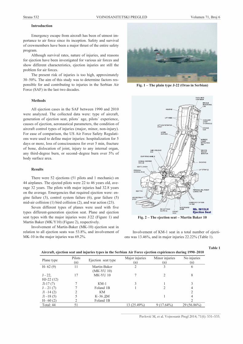
There were 52 ejections (51 pilots and 1 mechanic) on44 airplanes. The ejected pilots were 22 to 46 years old, ave-rage 32 years. The pilots with major injuries had 32.8 yearson the average. Emergencies that required ejection were: en-gine failure (3), control system failure (6), gear failure (5)mid-air collision (1) bird collision (2), and war action (23).
Seven different types of planes were used with fivetypes different-generation ejection seat. Plane and ejectionseat types with the major injuries were J/22 (Figure 1) andMartin Baker (MK/Y10) (Figure 2), respectively.
Involvement of Martin-Baker (MK-10) ejection seat inrelation to all ejection seats was 53.8%, and involvement ofMK-10 in the major injuries was 69.2%.
Fig. 1 – The plain type J-22 (Orao in Serbian)
Fig. 2 – The ejection seat – Martin Baker 10
Involvement of KM-1 seat in a total number of ejecti-ons was 13.46%, and in major injuries 22.22% (Table 1).
Table 1Aircraft, ejection seat and injuries types in the Serbian Air Force ejection expiriences during 1990–2010
Plane type Pilots(n) Ejection seat type Major injuries
(n)Minor injuries
(n)No injuries
(n)- 62 (9) 11 Martin-Baker
( -YU 10)2 3 6
J - 22,J-22 (12)
17 -YU 10 7 2 8
-17 (7) 7 -1 3 1 3J – 21 (7) 7 Foland 1B 1 2 4
–14 (2) 2 2 –18 (5) 5 –36 1 4– 60 (2) 2 Foland 1B 2
Total: 44 51 13 (25.49%) 9 (17.64%) 29 (56.86%)

Volumen 71, Broj 6 VOJNOSANITETSKI PREGLED Strana 533
Pavlovi M, et al. Vojnosanit Pregl 2014; 71(6): 531–533.
It is obvious that major injuries were present in 25.49%cases suggesting that every fourth pilot had experiencedmajor injury. Major injuries in war action were only 2,namely15.3% of all major injuries. It should be noted thatthere was no major injuries with K-36 , in spite of ejecti-ons in war actions.
A list of injuries included: fractura fibulae l.sin.; frac.mal. lat. cruris. sin.; fractura oss nasale; fractura Th – IV;frac. subcapitis ossis methacarp. II manus dex. aperta; frac.Th – VIII; frac. tibiae; frac. costae X l. sin; frac. compresi-va Th – VIII; frac. Th - X – XII, cum fractura cruris sin. gr.I aperta; frac.Th - IX , spondilodesis Th -VII –XI.
Of all the ejected pilots, 5 (9.61%) had fractures of thethoracic spine, 6 (11.53%) fractures of the legs, and 2(3.48%) fractures of the arms. Two of them had united frac-tures.
Of all the major injuries, 38.46%, were related to fracturesof thoracic spine, and 60% of them were inflicted on the plain J-22 (MK-10).
Minor injuries that should be mentioned were lacerationof the face and burns of the arms.
Obesity (adipositas) was presented in 23.07% of thepilots with major injuries.
None of the pilots had previously experienced ejection.
Discussion
Analysis done by foreign air forces for long periods oftime, with different types of planes and generations of seats,revealed different degrees of fatal injuries during ejection.The highest degree of fatal injuries was recorded in the Japa-nese Air Force, 22.9% of mortal outcomes in a study for aperiod 1956–2004 1. The main reason was the delay in ma-king decision for ejection.
In the study on accidents from 1973 to 1985 US AirForce (USAF) presented a survival rate of 86% 2. Swedes, intheir study for a period 1967–1987 claimed 83 successfulejections and 9 fatal outcomes 3. Finns, in the study from1958 to 1991 quoted survival rate higher than 80% 4. En-glish, in the study of 232 cases of ejection, for a period1973–2002, quoted the survival rate of 89.2% 5. In our studythere was no case of ejection with fatal outcome.
Compression a fracture of the spine is a commonconsequence of ejection. Finns quoted 18% of such cases inthe total number of all major injuries, Swedes 25%, Italians15%, USAF 6%, Japanese 63% and English 29.4% of allaircrew. Germans quoted 17.6% of spine fractures in their Air
Force, for a period from 1981 to 1997 6. Americans quoted 6spine fractures from 18 ejections in the “Desert Storm” 6.
In our survey spine fractures were presented in 9.61%of all ejections. The incidence of spine injures was 38.46%of the major injuries and 60% of all spine fractures was onplane J-22(MK-10).
In their study, English quoted that 44% of minor spinalcompression fractures and injuries of spinal ligaments couldnot be diagnosed with classic Roentgen recording, but onlywith magnetic resonance 5. This emphasizes the importanceof examination with magnetic resonance of all aircrews afterejections.
Irregular seating position during ejection was accusedto be the main reason for spinal fractures and a combinationof accomplished highest acceleration and rate of onset. Theinjuries appeared in the moment of discharge, and accelerati-on upward. It was established that every reduction of accele-ration in the moment of discharge reduces forces acting onthe spine and the degree of spine injures. It was concludedthat acceleration reduction from 24 m/s to 18 m/s reducesrate of injuries 5. The highest rate of injuries was on a planeTornado, with Martin-Baker seats Mk-10A, with the speed of20.7 m/s, compared with 19.5 m/s for other types of planes 5.
In our case, the estimated speed for MK-YU10(MK-10)was 19.8 m/s.
It should be pointed out that a connection between spinefractures and anthropometric measures of pilots could not beestablished 5.
In war action the rate of major injuries during ejectionwas lower than it could be expected. A possible reason wasthe participation of most experienced aircrews.
In their study Swedes quoted that two third ofsuccessfully ejected pilots returned to job after 1 week, ot-hers were absent for one year, and only 3.5% finished theirflying career 3.
Conclusion
Risk of injuries during ejection still remains too high,approximately 30–50%, in our survey 25.49%. There were noejections with fatal outcome in our study. The main reason forspine injuries was irregular position of the spine in the seat anda combination of the peak of acceleration and the rate of onset.Preventive measures must be promoted: MRI scan should beincluded in the standard selection procedure and procedureafter ejection physical conditioning has to be improved, trai-ning on ejection trainer has to be accomplished, too.
R E F E R E N C E S
1. Nakamura A. Ejection experience 1956-2004 in Japan: an epi-demiological study. Aviat Space Environ Med 2007; 78(1): 54 8.
2. McCarthy GW. USAF take-off and landing ejections, 1973-85.Aviat Space Environ Med 1988; 59(4): 359 62.
3. Sandstedt P. Experiences of rocket seat ejections in the SwedishAir Force:1967-1987. Aviat Space Environ Med 1989; 60(4):367 73.
4. Visuri T, Aho J. Injuries associated with the use of ejection se-ats in Finnish pilots. Aviat Space Environ Med. 1992; 63(8):727 30.
5. Lewis ME. Survivability and injuries from use of rocket-assisted ejection seats: analysis of 232 cases. Aviat Space Envi-ron Med 2006; 77(9): 936 43.
6. Damon AM, Lessley DJ, Salzar RS, Bass CR, Shen FH, Paskoff GR,et al. Kinematic response of the spine during simulated aircraftejections. Aviat Space Environ Med 2010; 81(5): 453 9.
Received on May 17, 2013.Revised on July 2, 2013.
Accepted on August 2, 2013.OnLine-First October, 2013.

Strana 534 VOJNOSANITETSKI PREGLED Vojnosanit Pregl 2014; 71(6): 534–541.
Correspondence to: Tatjana utovi , Department of Orthodontics, Military Medical Academy, Crnotravska 17, 11 000 Belgade, Serbia.Phone: +381 11 3608 138. E-mail: [email protected]
O R I G I N A L A R T I C L E UDC: 616.314-089.23:616.716.1/.4-007]:[611.714:572.087DOI: 10.2298/VSP121212011C
A cephalometric analysis of the cranial base and frontal part of theface in patients with mandibular prognathism
Kefalometrijska analiza kranijalne baze i prednjeg dela lica kod osoba samandibularnim prognatizmom
Tatjana utovi *†, Nebojša Jovi †‡, Ljiljana Stojanovi §,Julija Radoji i ¶, Irena Mladenovi , Stevo Matijevi †**, Ružica Kozomara†‡
*Department of Orthodontics, ‡Department of Maxillofacial Surgery, **Department ofOral Surgery, Military Medical Academy, Belgrade, Serbia; †Faculty of Medicine of the
Military Medical Academy, University of Defence, Belgrade, Serbia; §Department ofOrthodontics, Faculty of Dentistry, University of Belgrade, Belgrade, Serbia;
¶Department of Orthodontics, Faculty of Medicine, University of Niš, Niš, Serbia;Department of Prosthodontics, Faculty of Medicine, University of East Sarajevo, Fo a,
Bosnia and Herzegovina
Abstract
Bacground/Aim. The literature suggests different viewson the correlation between the cranial base morphologyand size and saggital intermaxillary relationships. The aimof this study was to investigate the cranial base morphol-ogy, including the frontal facial part in patients with man-dibular prognathism, to clarify a certain ambiguities, inopposing viewspoints in the literature. Methods. Cepha-lometric radiographies of 60 patients were analyzed at theDental Clinic of the Military Medical Academy, Belgrade,Serbia. All the patients were male, aged 18–35 years, withno previous orthodontic treatment. On the basis of dentaland sceletal relations of jaws and teeth, the patients weredivided into two groups: the group P (patients with man-dibular prognathism) and the group E (the control groupor eugnathic patients). A total of 15 cephalometricparametres related to the cranial base, frontal part of theface and sagittal intermaxillary relationships were meas-ured and analyzed. Results. The results show that cranialbase dimensions and the angle do not play a significantrole in the development of mandibular prognathism. Inter-
relationship analysis indicated a statistically significantnegative correlation between the cranial base angle(NSAr) and the angles of maxillary (SNA) and mandibular(SNB) prognathism, as well as a positive correlation be-tween the angle of inclination of the ramus to the cranialbase (GoArNS) and the angle of sagittal intermaxillary re-lationships (ANB). Sella turcica dimensions, its width anddepth, as well as the nasal bone length were significantlyincreased in the patients with mandibular prognathism,while the other analyzed frontal part dimensions of theface were not changed by the malocclusion in comparisonwith the eugnathic patients. Conclusion. This studyshows that the impact of the cranial base and the frontalpart of the face on the development of profile in patientswith mandibular prognathism is much smaller, but cer-tainly more complex, so that morphogenetic tests of themaxillomandibular complex should be included in furtherassessment of this impact.
Key words:mandible; prognathism; cephalometry; skull; facialbones; sella turcica.
Apstrakt
Uvod/Cij. U litetaruri postoje razli iti stavovi o povezano-sti morfologije i veli ine kranijalne baze i sagitalnih me uvi-li nih odnosa. Cilj ovog rada bio je da se ispita morfologijakranijalne baze, uklju uju i i prednji deo lica, kod ispitanikasa mandibularnim prognatizmom da bi se razjasnile nedou-mice donekle suprotnih stavova u literaturi. Metode. Anali-zirani su rendgenkefalometrijski snimci ukupno 60 bolesni-ka Klinike za stomatologiju VMA. Svi bolesnici bili su muš-
kog pola, starosti od 18 do 35 godina koji ranije nisu biliortodontski le eni. Bolesnici su prema dentoskeletnim od-nosima vilica i zuba bili svrstani u dve grupe: grupu P (bole-snici sa mandibularnim prognatizmom) i grupu E (kontrol-na grupa ili grupa eugnatih bolesnika). Izmereno je i analizi-rano 15 kefalometrijskih parametara koji su se odnosili nakranijalnu bazu, frontalni deo lica kao i sagitalne me uvili -ne odnose. Rezultati. Dobijeni rezultati ukazuju da ni di-menzije kranijalne baze, ni njen ugao ne igraju zna ajnu ulo-gu u nastanku mandibularnog prognatizma. Analizom me-

Volumen 71, Broj 6 VOJNOSANITETSKI PREGLED Strana 535
utovi T, et al. Vojnosanit Pregl 2014; 71(6): 534–541.
uzavisnosti ustanovljeno je da postoji statisti ki zna ajnanegativna korelacija izme u ugla kranijalne baze (NSAr) iuglova maksilarnog (SNA) i mandibularnog (SNB) progna-tizma, kao i pozitivna korelacija izme u ugla nagiba ramusaprema kranijalnoj bazi (GoArNS) i ugla sagitalnih me uvili-nih odnosa (ANB). Dimenzije sedlaste jamice (sella turcica),
njena širina i dubina, kao i dužina nosne kosti statisti ki suzna ajno pove ane kod bolesnika sa mandibularnim prog-natizmom, dok ostale analizirane dimenzije prednjeg dela li-ca nisu bile izmenjene kod ove malokluzije u odnosu na eu-
gnate bolesnike. Zaklju ak. Pokazalo se da je uticaj krani-jalne baze i prednjeg dela lica na ispoljavanje profila kodbolesnika sa mandibularnim prognatizmom mnogo manji alisvakako složeniji, pa bi u dalja istraživanja trebalo uklju itimorfogenetska ispitivanja maksilomandibularnog kompleksakod ocenjivanja ovog uticaja.
Klju ne re i:mandibula; prognatizam; kefalometrija; lobanja; lice,kosti; sela turcika.
Introduction
The cranial base plays an important role in the devel-opment of face, especially in achieving sagittal and verticalintermaxillary relationships, primarily because of the differ-ent ways of ossification of its synchondrosis. It also repre-sents a central skeletal axis which achieves its final size veryearly – long before the face.
All the bones that form the cranial base (apart fromtemporal) are of cartilaginous origin and created by encho-dral ossification which already begins prenatally and ends inearly childhood (especially the growth of sphenoethmoidaland sphenofrontal synchondroses ends early), following thegrowth of sphenooccipital synchondroses which is completedapproximately at the age of 12–16, so that the length of thefrontal cranial base becomes defined in a very early pe-riod 1, 2. In postnatal period, especially in puberty, the frontalsinus enlargement and remodelling of its frontal surface oc-cur, which also influence the nasal bone 3.
The opinions, that the growth, dimensions and shapeof the cranial base influence the middle face growth havebeen accepted. Apposition and remodelling of the cranialbase sutures until the age of 5 affect the growth and posi-tion of the maxilla, thus forming the maxillary sagittal po-sition to the cranial base very early. Afterwards, when thegrowth of the cranial base sutures stops, it is replaced bythe growth of the sutures connecting the maxilla with thecranial base, thus moving the maxilla forward and down-wards. According to another theory, the growth of the en-tire cranial complex gradually decreases from the age of 3–7 years, when remains active only in the mandibular con-dyle, so that the mandible grows smoothly, changing par-tially its sagittal position to the cranial base until generalsomatic growth is completed 4.
Anatomically speaking, the middle face is set in such away that the maxilla is attached to the anterior cranial baseby its sutures, whereas the mandible is connected to the pos-terior cranial base by the temporomandibular joint. Due tothe anatomy, any change in dimensions or the angle of thecranial base, results in changes of the maxilla or mandibleposition as well as their interrelationship.
Many cephalometric studies have confirmed that theshape and dimensions of the cranial base vary in patientswith different sagittal intermaxillary relationships 4–10. Man-dibular prognathism (MP) is a genetic, complex cranio-dento-facial developmental disorder, where disordered in-
termaxillary sagittal and vertical relationships dominate,primarely as a result of overdevelopment of the mandible.There are still dilemmas, whether the cranial base reallyplays such a decisive role in etiology, as the authors haveoften reported. In mandibular prognathism, the cranial baseangle is sharper and the cranial base is shorter in comparisonwith skeletal Class I patients, while the case of skeletal ClassII patients is completely opposite 3, 5, 6, 10.
Some authors believe that the temporomandibular jointis placed in more anterior position precisely because of thereduction of the cranial base angle, which results in a pro-gnathic facial profile. However, Singh et al. 6, 7, 9 and Proff etal. 5 have demonstrated in their extensive studies that thebiological basis of anterior positioning of the temporoman-dibular joint lies in the posterior cranial base. The sameauthors suggest that the reason could be the premature ces-sation of the growth of petro-spheno-occipital complex, inother words, that a premature synostosis is responsible fordeficient orthocephalization (horizontalization) of the cranialbase angle in Class III malocclusion. Therefore, the reducedposterior part of the cranial base can be a primary factor inskeletal Class III etiology.
Consequently, the shape of cranial base could deter-mine facial profiles and represent the key factor in develop-ing skeletal class malocclusions. Is this really true since thatMP is a developmental disorder, which reaches its full mani-festation until after puberty, and the cranial base ossificationoccurs in early childhood?
In almost all cephalometric analyses of the neurocraniumand the viscerocranium, sella turcica (ST) takes a central place.More precisely, the cental point (sella point – S) is a part ofmany reference planes by which other structures are oriented.Thus, ST shape, dimensions and position in relation to the sur-ronding structures are of great importance. For a long time,authors have had a tendency to determine ST dimensions as pre-cise as possible, primarily because of its close relationship witha pituitary gland. Today, however, it is well-known that ST en-largement does not imply that the pituitary gland is also en-hanced, and vice versa 1, 11–13.
It has been found that changes in ST shape and dimen-sions are caused by many congenital anomalities: cleft lipand palate 14, lumbosacral myelomeningocele 15, Seckelsyndrome 16, Rieger’s syndrome 17, congenital craniofacialdeviations 18–20, even by congenital dental anomalies, such asa palatal position of the upper fangs and hypodontia of man-dibular second premolars 21. A sella turcica bridge in pa-

Strana 536 VOJNOSANITETSKI PREGLED Volumen 71, Broj 6
utovi T, et al. Vojnosanit Pregl 2014; 71(6): 534–541.
tients with various craniofacial deviations treated by surgi-cal-orthodontic means to correct the existing deformities wasinvestigated by Becktor et al. 18, Jones et al. 19 and Alko-fide 22. All the authors found significant differences betweenstudy groups and general population, emphasizing that themajority of patients with craniofacial deviations were latertreated by surgery on mandible.
utovi et al. 20, analyzing sella turcica dimensions inpatients with mandibular prognathism, found that all thethree ST measured dimensions (surface, width and depth)were significantly higher in patients with mandibular prog-nathism than in eugnathic subjects, but the degree of themanifested anomaly did not have any influence on the size ofchanges in the abovementioned dimensions.
The anterior cranial base, whose growth ends veryearly, has a weak influence on positioning the frontal facialparts, that is, only the orbital part directly depends on it.Since the floor of eye socket is also the roof of maxilla, En-low 4 assumed that the dimensions and the position of the or-bital cavity should be correlated with the position of maxilla.However, Holly et al. 23 tested this hypothesis on 32 primatesand found that the correlation was too weak.
The growth of the frontal facial parts later in puberty ismostly seen in the increase of the frontal sinuses volumechanging the shape of supraorbital ridge and also indirectly af-fecting the nasal bone 3. Singh et al. 9 found that the elongationof the anterior cranial base, particularly around the age of 9,directly influenced the enlargement of the frontal sinus, sig-nificantly affecting the morphological changes of supraorbitaland nasal structures. In the literature available to us, we foundthat the cephalometric changes of supraorbital ridge and fron-tal sinus had been recorded only by Dostalova et al. 24, whoinvestigated a number of cephalometric abnormalities in pa-tients with acromegaly, and among other things, came to aconclusion that the frontal sinus was increased and the supra-orbital ridge pronounced in these patients in comparison withthe control group. The changes were more prominent in menthan women and did not depend on the growth hormone, buton the duration of the illness.
The nasal bone consists of two bones, forming theskeleton of nose and is located between the frontal exten-sions of the maxilla and frontal bone.
Dostálová et al. 24 measured the nasal bone length andinclination to the cranial base in healthy subjects and patientswith acromegaly and found that the dimensions of nasal bonewere not changed in patients with acromegaly.
Singh et al. 9 found a negative correlation between thefrontonasal angle and the cranial base angle. In patients withskeletal Class III, the cranial base angle is normally reduced,so that the frontonasal angle is increased, resulting in a flatmidface profile, which is a common feature of this dentofa-cial deformity.
The aim of this study was to conduct a cephalometricanalysis of morphological characteristics of the cranial base,including sella turcica and frontal facial part (supraorbitalridge, frontal sinus, nasal bone) in patients with mandibularprognathism, as well as of their correlation with the indica-tors of sagittal intermaxillary relationships.
Methods
Lateral cephalometric images of 60 orthodontic pa-tients, were taken and analyzed before their treatment at theDental Clinic, Military Medical Academy.
Using the findings from the literature on gender differ-ences and growth changes dynamics 25, 26, we decided tostudy male subjects, aged 18–30 years.
The group P consisted of 30 patients with mandibularprognathism, diagnosed on the basis of the following criteria:the angle of mandibular prognathism (SNB) 80°; the angleof sagittal intermaxillary relationship (ANB) 0°; the angleB 30°; Bjork 396°; reverse overlap of the frontal teethand relationship of the first permanent molars in Class III.
The control group, the group E, consisted of 30 patientswith normal intermaxillary relationships (skeletal Class I,eugnathic subjects): SNB 80°; ANB = 0 - 5°; normaloverlap of the frontal teeth and relationship of the first per-manent molars in Class I.
All the patients from the group P were planned for andlater treated with orthodontic-surgical therapy, which wasperformed by the same team.
A cephalometric analysis
Lateral cephalometric images of the head were takenfor each patient under standard conditions. The head wasfixed in a cephalostat, and recording conducted at the dis-tance of 1.5 m. Analysis of lateral cephalogram images waspreceded by drawing the corresponding structures on a trac-ing paper fixed on a film. Afterwards, numerous points andplanes were marked for analyzing certain angular and linearparameters taken from the analyses of Steiner, Jacobson,Ricketts, Downs and Bjork. Measurements were performedtwice by the same examiner, on different days, with the accu-racy of 0.5 mm or 0.5°. Statistically significant differencesdid not appear between these two measurements.
Analysis of the following cephalometric parameters wascarried out between the patients with mandibular prognathismand the control group of eugnathic patients: SN – the anteriorcranial base length; SAr – the posterior cranial base length;NAr – the total cranial base length; NSAr – the cranial baseangle; SG – supraorbital ridge; F1F2 – the frontal sinus range;SGN – the angle of protrusion of the supraorbital ridge; Ss –the width of sella turcica (the largest anteroposterior diame-ter); Ds – the depth of sella turcica (from the line connectingclinoid extensions to the lowest point of the floor); NR – thenasal bone length; SNR – the angle of the inclination of thenasal bone; SNA – the angle of maxillary prognathism; SNB –the angle of mandibular prognathism; ANB – the angle ofsagittal intermaxillary relationships; GoArNS – the angle ofinclination of the ramus to the cranial base (Figure 1).
According to the data collected by lateral cephalometricanalysis for each patient and each feature, the database wasformed in the SPSS12 Program for Windows and the fol-lowing statistical methods were used in the statistical analy-sis: tables and graphical presentations, descriptive statisticsmethods, the Bonferroni test for detecting intergroup differ-ences and the linear correlation method.

Volumen 71, Broj 6 VOJNOSANITETSKI PREGLED Strana 537
utovi T, et al. Vojnosanit Pregl 2014; 71(6): 534–541.
Fig. 1 – Angular and linear measurements:1. SNA – maxillary prognathism angle; 2. SNB – mandibular prognathismangle; 3. ANB – angle of sagittal intermaxillary relationships; 4. GoArNS –angle of inclination of the ramus to the cranial base; 5. NSAr – cranial base
angle; 6. SGN – protrusion angle of the supraorbiatal ridge; 7. F1F2 –frontalsinus range; 8. SG – supraorbital ridge; 9. NR – nasal bone length.
Results
Tables 1, 2 and 3 show the statistical results of analyz-ing the following parameters of the cranial base, the frontalfacial part and sagittal intermaxillary relationships: the ante-rior cranial base (SN), the posterior cranial base length(SAr), the total cranial base length (NAr), the cranial baseangle (NSAr), supraorbital ridge (SG), the frontal sinus range(F1F2), the angle of protrusion of the supraorbital ridge(SGN), the width of sella turcica (Ss), the depth of sella tur-cica (DS), the nasal bone length (NR), the angle of inclina-tion of the nasal bone (SNR), the angle of maxillary progna-thism (SNA), the angle of mandibular prognathism (SNB),the angle of the sagittal intermaxillary relationships (ANB),the angle of inclination of the ramus to the cranial base(GoArNS).
Sella turcica (Ss) width showed higher values in thepatients with mandibular prognathism than in the eugnathicsubjects. The average value of the sella turcica width in theeugnathic subjects is 9.53 mm ± 1.34, whereas it was 11.07
Table 1Analyzed parameters values for the cranial base in the eugnathic subjects (E) and the patients
with mandibular prognathism (P) (descriptive statistical indicators)Analyzed parameters n SD Min MaxS-N E 30 77.90 4.20 65.00 84.50
P 30 76.07 4.07 69.00 83.00Total 60 77.40 4.20 65.00 88.00
S-Ar E 30 39.30 3.82 31.00 47.00P 30 37.37 3.62 29.00 45.50Total 60 38.51 3.94 29.00 47.00
NAr E 30 104.08 7.03 88.00 117.00P 30 100.13 6.79 90.00 116.00Total 60 101.66 11.57 11.50 117.00
NSAr E 30 120.55 5.59 110.00 133.00P 30 118.90 7.81 101.00 135.00Total 60 119.32 6.58 101.00 135.00
Ss E 30 9.53 1.34 6.50 12.00P 30 11.07 1.45 7.50 15.00Total 60 10.49 1.61 6.00 15.00
Ds E 30 7.55 1.75 3.00 11.00P 30 9.33 1.66 6.00 13.00Total 60 8.38 1.73 3.00 13.00
SN – anterior cranial base length; SAr – posterior cranial base length; NAr – total cranial base length;NSAr – cranial base angle; Ss – width of sella turcica (the largest anteroposterior diameter); Ds – depth of sellaturcica (from the line connecting clinoid extensions to the lowest point of the floor).
Table 2Analyzed values parameters of the frontal facial part in the eugnathic subjects (E)and patients with mandibular prognathism (P) (descriptive statistical indicators)
Analyzed parameters n SD Min MaxF1-F2 E 30 14.43 4.50 5.00 25.00
P 30 14.07 2.69 8.00 19.00Total 60 14.53 3.86 5.00 25.00
S-G E 30 84.27 4.48 71.00 91.50P 30 82.85 4.89 74.00 90.00Total 60 84.26 4.58 71.00 96.00
SGN E 30 57.20 5.49 38.50 66.00P 30 54.73 6.49 41.00 66.00Total 60 55.95 5.90 38.50 72.00
NR E 30 24.70 3.54 17.00 33.00P 30 27.58 3.65 20.00 35.00Total 60 26.40 3.57 17.00 35.00
SNR E 30 118.10 9.34 104.00 135.00P 30 117.82 6.92 105.00 133.00Total 60 118.98 7.40 104.00 135.00
F1F2 – frontal sinus range; SG – supraorbital ridge; SGN – angle of protrusion of the supraorbital ridge;NR – nasal bone length; SNR – inclination angle of the nasal bone.

Strana 538 VOJNOSANITETSKI PREGLED Volumen 71, Broj 6
utovi T, et al. Vojnosanit Pregl 2014; 71(6): 534–541.
mm ± 1.45 in the patients with mandibular prognathism. Ta-ble 4 shows a statistically significant difference in the Ssvalues between the group E and the group P (p < 0.001).
Sella turcica depth (Ds) shows higher values in the pa-tients with mandibular prognathism than in the eugnathicsubjects. The average value of the depth of sella turcica inthe eugnathic subjects is 7.55 mm ± 1.75, whereas it is 9.33mm ± 1.66 in the patients with mandibular prognathism. Ta-ble 4 shows a statistically significant difference in the Dsvalues between the group E and the group P (p < 0.001).
Nasal bone length (NR) showed higher values in thepatients with mandibular prognathism than in the eugnathicsubjects. The average value of the nasal bone length in theeugnathic subjects is 24.70 mm ± 3.54, whereas it was 27.58mm ± 3.65 in the patients with mandibular prognathism. Ta-ble 4 shows a statistically significant difference in the NRvalues between the group E and the group P (p < 0.001).
Maxillary prognathism angle (SNA) shows higher val-ues in the eugnathic subjects than in the patients with man-dibular prognathism. The average value of the SNA in theeugnathic subjects is 82.38 ± 4.05, whereas it is 77.67 ± 4.29
in the patients with mandibular prognathism. Table 4 showsa statistically significant difference in the SNA values be-tween the group E and the group P (p < 0.001).
Mandibular prognathism angle (SNB) shows highervalues in the patients with mandibular progmathism than inthe eugnathic subjects. The average value of the SNB in the
eugnathic subjects is 79.30 ± 4.18, whereas it was 83.92 ±2.74 in the patients with mandibular prognathism. Table 4shows a statistically significant difference in the SNB valuesbetween the group E and the group P (p < 0.001).
Sagittal intermaxillary relationships angle (ANB) showshigher values in the eugnathic subjects than in the patientswith mandibular prognathism. The average value of this an-gle in the eugnathic subjects is 3.15 ± 1.70, whereas it is -6.22 ± 3.13 in the patients with mandibular prognathism. Ta-ble 4 shows a statistically significant difference in the ANBvalues between the group E and the group P (p < 0.001).
The angle of ramus inclination to the cranial base(GoArNS) shows higher values in the eugnathic subjectsthan in the patients with mandibular prognathism, but theyare statistically insignificant. The average value of this anglein the eugnathic subjects is 82.58 ± 5.34, whereas it is 80.18± 5.13 in the patients with mandibular prognathism.
For the remaining cranial base parameters (SN, SAr,NAr, NSAr, SG, F1F2, SGN, SNR), the Bonferroni test didnot show any statistically significant difference between thetwo groups of examinees (Table 4).
By analyzing interrelationships between 5 parameters inmandibular prognathism: (NSAr, SNA, SNB, ANB, andGoArNS, given in Table 5, a statistically significant andhighly negative correlation was found between the cranialbase angle and NSAr and SNA (p = -0.567) and SNB (p = -0.676) angles. The GoArNS showed a statistically significant
Table 3Analyzed parameters values of the sagittal intermaxillary relationships in the eugnathic subjects (E)
and the patients with mandibular prognathism (P)Analyzed parameters n SD Min MaxSNA E 30 82.38 4.05 73.00 89.00
P 30 77.67 4.29 71.00 86.50Total 60 79.94 4.28 71.00 89.00
SNB E 30 79.30 4.18 72.00 87.50P 30 83.92 2.74 77.00 90.50Total 60 83.54 4.73 72.00 93.00
ANB E 30 3.15 1.70 0.50 7.00P 30 -6.22 3.13 -12.00 -0.50Total 60 -3.54 5.50 -15.00 7.00
GoArNS E 30 82.58 5.34 71.00 94.00P 30 80.18 5.13 72.00 99.00Total 60 79.73 5.65 68.00 99.00
SNA – angle of maxillary prognathism; SNB – angle of mandibular prognathism; ANB – angle of sagittalintermaxillary relationships; GoArNS – angle of inclination of the ramus to the cranial base.
Table 4Boniferri test results, examining intergroup differences (eugnatics subjects vs
patients with mandibular prognatism) by using all the cranial base, frontal facialpart and sagittal intermaxillary relationships analyzed parameters
Parameters Differences in average values pSs -1.53333 0.000Ds -1.78333 0.000NR -2.88333 0.004SNA 4.72 0.000SNB -4.62 0.000ANB 9.37 0.000
Ss – sella turcica width; Ds – sella turcica depth; NR – nasal bone length; SNA – angle of maxillaryprognathism; SNB – angle of mandibular prognathism; ANB – the angle of sagittal intermaxillaryrelationships.
Volumen 71, Broj 6 VOJNOSANITETSKI PREGLED Strana 539
utovi T, et al. Vojnosanit Pregl 2014; 71(6): 534–541.
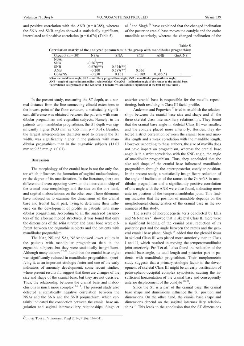
and positive correlation with the ANB (p = 0.385), whereasthe SNA and SNB angles showed a statistically significant,interrelated and positive correlation (p = 0.674) (Table 5).
In the present study, measuring the ST depth, as a nor-mal distance from the line connecting clinoid extensions tothe lowest point of the floor contours, a statistically signifi-cant difference was obtained between the patients with man-dibular prognathism and eugnathic subjects. Namely, in thepatients with mandibular prognathism, the ST depth was sig-nificantly higher (9.33 mm vs 7.55 mm, p < 0.01). Besides,the largest anteroposterior diameter used to present the STwidth, was significantly higher in the patients with man-dibular prognathism than in the eugnathic subjects (11.07mm vs 9.53 mm, p < 0.01).
Discussion
The morphology of the cranial base is not the only fac-tor which influences the formation of sagittal malocclusions,or the degree of its manifestation. In the literature, there aredifferent and even opposing views on the interrelationship ofthe cranial base morphology and the size on the one hand,and sagittal malocclusions on the other one. These dilemmashave induced us to examine the dimensions of the cranialbase and frontal facial part, trying to determine their influ-ence on the development of profile in patients with man-dibular prognathism. According to all the analyzed parame-ters of the aforementioned structures, it was found that onlythe dimensions of the sella turcica and nasal bone were dif-ferent between the eugnathic subjects and the patients withmandibular prognathism.
The NAr, NS and SAr, NSAr showed lower values inthe patients with mandibular prognathism than in theeugnathic subjects, but they were statistically insignificant.Although many authors 27–29 found that the cranial base anglewas significantly reduced in mandibular prognathism, speci-fying it, as an important etiologic factor and one of the earlyindicators of anomaly development, some recent studies,where present results fit, suggest that there are changes of thesize and shape of the cranial base, but they are not decisive.Thus, the relationship between the cranial base and maloc-clusions is much more complex 3, 5, 8. The present study alsodetected a statistically negative correlation between theNSAr and the SNA and the SNB prognathism, which cer-tainly indicated the connection between the cranial base an-gulation and sagittal intermaxillary relationships. Singh et
al. 9 and Singh 28 have explained that the changed inclinationof the posterior cranial base moves the condyle and the entiremandible anteriorly, whereas the changed inclination of the
anterior cranial base is responsible for the maxilla reposi-tioning, both resulting in Class III facial profile.
Anderson and Popovich 10 tried to establish the relation-ships between the cranial base size and shape and all thethree skeletal class intermaxillary relationships. They foundthat the cranial base angle in skeletal Class III was smaller,and the condyle placed more anteriorly. Besides, they de-tected a strict correlation between the cranial base and max-illa length and a weak correlation with the mandible length.However, according to these authors, the size of maxilla doesnot have impact on prognathism, whereas the cranial baseangle is in a strict correlation with the SNB angle, the angleof mandibular prognathism. Thus, they concluded that thesize and shape of the cranial base influenced mandibularprognathism through the anteroposterior condylar position.In the present study, a statistically insignificant reduction ofthe angle of inclination of the ramus to the GoArNS in man-dibular prognathism and a significantly positive correlationof this angle with the ANB were also found, indicating moreanterior position of the temporomandibular joint. This find-ing indicates that the position of mandible depends on themorphological characteristics of the cranial base in the ex-aminees of this study.
The results of morphogenetic tests conducted by Ellisand McNamara 27 showed that in skeletal Class III there werea significant bending of the cranial base, reduction of itsposterior part and the angle between the ramus and the gen-eral cranial base plane. Singh 28 added that the glenoid fossain skeletal Class III was placed more anteriorly than in ClassI and II, which resulted in moving the temporomandibularjoint anteriorly. Proff et al. 5 also found the reduction of thecranial base angle, its total length and posterior part in pa-tients with mandibular prognathism. Their morphometricstudy suggests that a primary etiologic factor in the devel-opment of skeletal Class III might be an early ossification ofpetro-spheno-occipital complex synostosis, causing the in-sufficient horizontation of the cranial base and consequentlyanterior displacement of the condyle 30, 31.
Since the ST is a part of the cranial base, the cranialbase shape and dimensions influence the ST position anddimensions. On the other hand, the cranial base shape anddimensions depend on the sagittal intermaxillary relation-ships 3. This leads to the conclusion that the ST dimensions
Table 5Correlation matrix of the analyzed parameters in the group with mandibular prognathism
Group P (n = 30) NSAr SNA SNB ANB GoArNSNSAr 1SNA -0.567(**) 1SNB -0.676(**) 0.674(**) 1ANB -0.200 0.787(**) 0.076 1GoArNS -0.230 0.161 -0.189 0.385(*) 1
NSAr – cranial base angle; SNA – maxillary prognathism angle; SNB – mandibular prognathism angle;ANB – angle of sagittal intermaxillary relationships; GoArNS – inclination angle of the ramus to the cranial base.*Correlation is significant at the 0.05 level (2-tailed); **Correlation is significant at the 0.01 level (2-tailed).

Strana 540 VOJNOSANITETSKI PREGLED Volumen 71, Broj 6
utovi T, et al. Vojnosanit Pregl 2014; 71(6): 534–541.
and position also depend on the sagittal intermaxillary rela-tionships. The clinical picture of mandibular prognathismactually shows abnormal intermaxillary relationships as itsdominant symptom.
Examining the sella turcica dimensions in the patientswith dentofacial deformities, several autors 18–20 found thatall the three measured dimensions of sella turcica (surface,width and depth) were statistically much higher in the pa-tients with deformities than in the eugnathic subjects, but thedegree of the anomaly manifestation did not influence thesize of changes in the aforementioned dimensions. Investi-gating correlations, they found that the depth of sella turcicahad a positive correlation with the ST surface. This could berelated to the fact that the ST floor, anterior and posteriorwall are most susceptible to changes 14, 17, 24. Alkofide 22
found that the largest anteroposterior diameter in patientswith skeletal Class III was significally higher than in patientswith other analyzed classes.
Having in mind that several studies have recently provedthe increase in sella turcica dimensions in mandibular prog-nathism, it can be expected that future studies will find thecause and relationship between these phenomena 18–22.
The patients with mandibular prognathism also showeda significant increase in the nasal bone length. The nasalbone length and inclination to the cranial base in healthysubjects and patients with acromegaly were measured byDostálová et al. 24, and they found that the nasal bone did notchange its dimensions in the patients with acromegaly. Inaddition, these results showed that the average value of thenasal bone length in healthy women and men was similar ap-proximately 23 mm, (in our study, the average value was24.7 in the eugnathic subjects, whereas it was 27.58 mm inthe patients with mandibular prognathism). The angle be-tween the nasal bone and cranial base was 115° in theeugnathic subjects (in present study, the average value was118.10° in the eugnathic subjects and there were no statisti-cally significant differences between the analyzed groups).
Singh et al. 9 found a negative correlation between thefrontonasal angle and the cranial base angle. In patients withskeletal Class III, the cranial base angle is actually reduced,so that the frontonasal angle is increased, resulting in a flatmid-face profile, which is a frequent characteristic of thisdentofacial deformity. The angle of protrusion of the supra-orbital ridge, which we measured, although reduced in man-dibular prognathism, did not show any statistically signifi-cant differences between the groups.
As mentioned at the beginning, in both mandibularprognathism and developmental malocclusion, a significantincrease in the mandible and change in mandibular shape oc-cur during rapid growth at puberty. This change is primarilycaused by the opening of the gonial angle, particularly char-
acteristic for a hyperdivergent facial profile. It certainly re-sults in changing the inclination of the ramus to the cranialbase. The condylar cartilage is still active, therefore it is alsovery likely to have the remodeling growth of condyle andglenoid fossa changing their position and shape in this typeof malocclusion 1, 2. Thus, the inclination of the ramus to thecranial base causing the anterior mandibular positioning,does not strictly depend on the length of the posterior cranialbase and its angulation, but most likely on other growth pro-cesses, such as the opening of gonial angle which occursmuch later.
When discussing skeletal Class III, one should alsothink about mandibular prognathism and its developmentalnature, which often camouflages by compensatory mecha-nisms (in rare cases potentiates) some important indicators incertain life phases. Therefore, the results of many studies arecontradictory. It should not be forgotten that many growthand developmental studies have found that the mandiblegrows more intensively and longer through all life phases,even a year longer after the completion of general somaticgrowth 25, 26, 29. Since the cranial base growth ends early, itcan be considered as one of the etiologic factors of man-dibular prognathism, but certainly not the decisive one, andalthough existing, the cranial base correlation with man-dibular prognathism is not as simple as previously thought.
Conclusion
The results of this study show that the cranial base di-mensions and angle do not play a significant role in the de-velopment of mandibular prognathism.
An interrelationship analysis indicated a statisticallysignificant negative correlation between the NSAr and theSNA and the SNB prognathism, as well as a positive corre-lation between the GoArNS and the ANB.
Sella turcica dimensions, width and depth, as well asthe nasal bone length were significantly increased in patientswith mandibular prognathism, while the other analyzed di-mensions of frontal part of the face were not changed by themalocclusion in comparison with eugnathic patients.
The impact of the cranial base and frontal part of theface on facial profile in patients with mandibular progna-thism is much smaller, but certainly more complex than pre-viously thought, and therefore it suggests, that morphoge-netic tests of the maxillomandibular complex should be in-cluded in further assessment of this impact.
Acknowledgments
This study was a part of the project of the Ministry ofEducation, Science and Technological Development of theRepublic of Serbia (III 41017).
R E F E R E N C E S
1. Bishara SE. Textbook of orthodontics. Philadelphia: Saunders2001.
2. Proffit WR, Fields HW, Sarver DM. Orthodontics. Jastrebarsko:Naklada Slap; 2009. (Croatian)
3. Dhopatkar A, Bhatia S, Rock P. An investigation into the rela-tionship between the cranial base angle and malocclusion. An-gle Orthod 2002; 72(5): 456 63.
4. Enlow DH. Facial growth. 3rd ed. Philadelphia: Saunders; 1990.

Volumen 71, Broj 6 VOJNOSANITETSKI PREGLED Strana 541
utovi T, et al. Vojnosanit Pregl 2014; 71(6): 534–541.
5. Proff P, Will F, Bokan I, Fanghänel J, Gedrange T. Cranial basefeatures in skeletal Class III patients. Angle Orthod 2008;78(3): 433 9.
6. Singh GD, Mcnamara JA, Lozanoff S. Finite element analysis ofthe cranial base in subjects with Class III malocclusion. Br JOrthod 1997; 24(2): 103 12.
7. Singh GD, McNamara JA, Lozanoff S. Thin-plate spline analysisof the cranial base in subjects with Class III malocclusion. EurJ Orthod 1997; 19(4): 341 53.
8. Hayashi I. Morphological relationship between the cranial baseand dentofacial complex obtained by reconstructive computertomographic images. Eur J Orthod 2003; 25(4): 385 91.
9. Singh GD, McNamara JA, Lozanoff S. Morphometry of the cra-nial base in subjects with Class III malocclusion. J Dent Res1997; 76(2): 694 703.
10. Anderson D, Popovich F. Relation of cranial base flexure to cra-nial form and mandibular position. Am J Phys Anthropol1983; 61(2): 181 7.
11. Choi WJ, Hwang EH, Lee SE. The study of shape and size ofnormal sella turcica in cephalometric radiographs. Korean JOral Maxillofac Radiol 2001; 31: 43 9.
12. Axelsson S, Storhaug K, Kjaer I. Post-natal size and morphologyof the sella turcica: Longitudinal cephalometric standards forNorwegians between 6 and 21 years of age. Eur J Orthod2004; 26(6): 597 604.
13. Weisberg LA. Asymptomatic enlargement of the sella turcica.Arch Neurol 1975; 32(7): 483 5.
14. Nielsen BW, Mølsted K, Kjaer I. Maxillary and sella turcica mor-phology in newborns with cleft lip and palate. Cleft PalateCraniofac J 2005; 42(6): 610 7.
15. Kjaer I, Wagner A, Madsen P, Blichfeldt S, Rasmussen K, Russell B.The sella turcica in children with lumbosacral myelomeningo-cele. Eur J Orthod 1998; 20(4): 443 8.
16. Kjaer I, Hansen N, Becktor KB, Birkebaek N, Balslev T. Craniofa-cial morphology, dentition, and skeletal maturity in four sib-lings with Seckel syndrome. Cleft Palate Craniofac J 2001;38(6): 645 51.
17. Koshino T, Konno T, Ohzeki T. Bone and joint manifestations ofRieger's syndrome: a report of a family. J Pediatr Orthop 1989;9(2): 224 30.
18. Becktor JP, Einersen S, Kjaer I. A sella turcica bridge in subjectswith severe craniofacial deviations. Eur J Orthod 2000; 22(1):69 74.
19. Jones RM, Faqir A, Millett DT, Moos KF, McHugh S. Bridgingand dimensions of sella turcica in subjects treated by surgical-orthodontic means or orthodontics only. Angle Orthod 2005;75(5): 714 8.
20. utovi T, Pavlovi J, Kozomara R. Analysis of dimensions of sellaturcica in patients with mandibular prognathism. VojnosanitPregl 2008; 65(6): 456 61. (Serbian)
21. Leonardi R, Barbato E, Vichi M, Caltabiano M. A sella turcicabridge in subjects with dental anomalies. Eur J Orthod2006;28(6):580-585.
22. Alkofide EA. The shape and size of the sella turcica in skeletalClass I, Class II, and Class III Saudi subjects. Eur J Orthod2007; 29(5): 457 63.
23. Holly SB, Crummett TL, Brandt KL. Ages of eruption of primateteeth: A compendium for aging individuals and comparing lifehistories. Am J Phys Anthropol 1994; 37(S19): 177 231.
24. Dostálová S, Sonka K, Smahel Z, Weiss V, Marek J. Cephalomet-ric assessment of cranial abnormalities in patients with acro-megaly. J Craniomaxillofac Surg 2003; 31(2): 80 7.
25. Reyes BC, Baccetti T, McNamara JA. An estimate of craniofacialgrowth in Class III malocclusion. Angle Orthod 2006; 76(4):577 84.
26. Baccetti T, Reyes BC, McNamara JA. Craniofacial changes inClass III malocclusion as related to skeletal and dental matura-tion. Am J Orthod Dentofacial Orthop 2007; 132(2): 171 8.
27. Ellis E, McNamara JA. Components of adult Class III maloc-clusion. J Oral Maxillofac Surg 1984; 42(5): 295 305.
28. Singh GD. Morphologic determinants in the etiology of classIII malocclusions: A review. Clin Anat 1999; 12(5): 382 405.
29. Chang JZ, Chen Y, Chang FH, Yao JC, Liu P, Chang C, et al.Morphometric analysis of mandibular growth in skeletal ClassIII malocclusion. J Formos Med Assoc 2006; 105(4): 318 28.
30. Hoyte DAN. The cranial base in normal and abnormal skullgrowth. Neurosurg Clin North Am 1991; 2: 515 37.
31. Baccetti T, Franchi L, McNamara JA. Cephalometric variablespredicting the long-term success or failure of combined rapidmaxillary expansion and facial mask therapy. Am J OrthodDentofacial Orthop 2004; 126(1): 16 22.
Received on December 12, 2012.Revised on January 10, 2013.
Accepted on January 11, 2013.OnLine-First February 2014.
Strana 542 VOJNOSANITETSKI PREGLED Vojnosanit Pregl 2014; 71(6): 542–546.
Correspondence to: Damir Jašarovi , Clinical Hospital Center Zemun, 11000 Belgrade, Serbia. E-mail: [email protected]
O R I G I N A L A R T I C L E UDC: 616.348/.351-006-033.1::616.36-033.2-089DOI: 10.2298/VSP1406542J
Resection or radiofrequency ablation of colorectal liver metastasisResekcija ili radiofrekventna ablacija metastaze kolorektalnog karcinoma u jetri
Damir Jašarovi , Dragoš Stojanovi , Nebojša Mitrovi , Dejan Stevanovi
Clinical Hospital Center Zemun, Belgrade, Serbia
Abstract
Background/Aim. Liver resection is the treatment ofchoice for solitary colorectal liver metastases in suitablecandidates. Recently, radiofrequency ablation (RFA) has be-come a very popular procedure in the treatment of livermetastases. The aim of this study was to compare outcomesin patients with solitary colorectal liver metastasis who hadbeen subjected to resection or ablation. Methods. In thisretrospective study we analyzed and compared patients withsolitary colorectal liver metastases treated by resection orablation in the University Hospital Centre “Dr Dragiša Mi-šovi ” in Belgrade from January 2002 until December 2009.Results. In this study 94 (67.1%) patients underwent resec-tion whereas 46 (32.9%) patients underwent RFA. Most ofthe resected patients (59.6%) required major hepatectomy.The median follow-up time was 28.4 months. Tumor abla-
tion was a significant predictor of the overall survival (p =0.002; OR 3.75; 95% CI 1.696–8.284). Our study demon-strated longer disease free-survival in the group of resectedpatients compared to the RFA group (37.6 vs 22.3 months, p= 0.073). The median overall survival was 56.3 months forpatients who underwent resection vs 25.1 months for thosein the RFA group (p = 0.005). Conclusion. This studyshows that the patients with solitary hepatic colorectal can-cer metastases should be considered for hepatic resectionwhenever it is feasible, because this procedure provides su-perior long-term survival as compared to radiofrequencyablation.
Key words:colorectal neoplasms; digestive system surgicalprocedures; liver neoplasms; neoplasm metastasis;catheter ablation, treatment outcome.
Apstrakt
Uvod/Cilj. Hirurška resekcija jetre predstavlja metod iz-bora u le enju pojedina nih metastaza kolorektalnog kar-cinoma u jetri kod odgovaraju ih bolesnika. Radiofrek-ventna ablacija postaje sve popularnija metoda za le enjemetastaza u jetri. Cilj ove studije bio je da uporedi ishodebolesti kod bolesnika sa pojedina nom metastazom kolo-rektalnog karcinoma u jetri koji su le eni hirurškom resek-cijom u odnosu na bolesnike koji su le eni radiofrekvent-nom ablacijom (RFA). Metode. U ovoj retrospektivnojstudiji analizirani su bolesnici sa pojedina nom metasta-zom kolorektalnog karcinoma u jetri koji su le eni u KBC„Dr Dragiša Mišovi “ u Beogradu u periodu od januara2002. do decembra 2009. godine. Pore eni su ishodi bole-sti nakon hirurške resekcije jetre i nakon RFA metastaza ujetri. Rezultati. Studijom je bilo obuhva eno 94 (67,1%)bolesnika podvrgnutih resekciji jetre, dok je 46 (32,9%)bolesnika le eno radiofrekventnom ablacijom. Kod ve ine
bolesnika (59,6%) podvrgnutih hirurškoj resekciji u injenaje major hepatektomija. Prose na dužina pra enja bolesnikabila je 28,4 meseca. Utvr eno je da RFA tumora predstav-lja zna ajni prediktor dužine ukupnog preživljavanja (p =0,002, OR 3,75, 95% CI 1,696–8,284), te da je duže preži-vljavanje bez tegoba bilo u grupi bolesnika sa resekcijom upore enju sa RFA grupom (37,6 vs 22,3 meseca, p =0,073). Prose no ukupno preživljavanje iznosilo je 56,3meseca u grupi bolesnika sa hirurškom resekcijom naspram25,1 mesec u RFA grupi (p = 0,005). Zaklju ak. Kod od-govaraju ih bolesnika sa pojedina nom metastazom kolore-ktalnog karcinoma u jetri trebalo bi razmotriti hiruršku rese-kciju kad god je to izvodljivo, jer pruža duži period preživ-ljavanja nego le enje radiofrekventnom ablacijom.
Klju ne re i:kolorektalne neoplazme; hirurgija digestivnog sistema,procedure; jetra, neoplazme; neoplazme, metastaze;ablacija preko katetera; le enje, ishod.

Volumen 71, Broj 6 VOJNOSANITETSKI PREGLED Strana 543
Jašarovi D, et al. Vojnosanit Pregl 2014; 71(6): 542–546.
Introduction
Colorectal cancer (CRC) remains one of the leadingcauses of mortality caused by malignancy. Approximately25% of all colorectal cancer patients at the time of initial di-agnosis already have liver metastases, and additionally 50%will develop distant metastases in the next 5 years 1. Treat-ment of colorectal cancer patients with metastases on theliver is a therapeutic challenge and requires multidisciplinarytreatment. Nevertheless, surgery is the treatment of choicefor these patients. Survival data shows that with modern,multidisciplinary treatment, 25–60% of patients with liverresection to treat CRC metastases survive more than 5years 2–7. The goal of operation is to remove all metastatictumor tissue with acceptable resection margins. Some studiesshow that narrow margins do not have influence on survival,and that complete removal of metastases with minimal mar-gins can be acceptable when it is not possible technically toobtain wider margins 8–10. Due to the importance of liver dis-ease reduction in cases with metastases that cannot be re-sected, new methods of local treatment of metastases are de-veloped, among which is radiofrequency ablation (RFA).RFA uses thermal energy produced by radiofrequency gen-erator to destroy tumor and a small part of surroundinghealthy tissue 1, 3, 4, 11. The five-year survival rate after RFAin different studies ranges from 14% up to 27% 1, 3, 5, 11–13.
Methods
The study is a retrospective analysis of patients withsolitary CRC liver metastases treated with RFA or surgicalresection, in the University Hospital Centre “Dr Dragiša Mi-šovi ” in Belgrade, from January 2002 until December 2009.Metastases are considered resectable when it is possible toremove the tumor with negative resection margins, leavingfunctionally sufficient liver tissue. The patients with extrahe-patic metastases are excluded from this study. RFA was per-formed with open approach after laparotomy to all patients inthis group, and the criteria for RFA were unresectability ofmetastases and comorbidity (accompanying diseases andconditions), which significantly increased the risk of liver re-
section. Data about chemotherapy were not known for all thepatients, and for most were not reliable, so these were notconsidered in this study. The patients treated with RFA werecompared with the patients treated by liver resection by us-ing t-test, 2-test and Fisher’s exact test where appropriate.Statistical analysis was performed by using JMP 4.0 and SPSSversion 16 software. Continuous variables were compared us-ing Student’s t-test, and categorical variables were comparedby using 2-test. The survival was plotted by Kaplan-Meiermethod, and compared using the log-rank test. A value p <0.05 was considered significant. The overall survival was cal-culated from the moment of diagnosis until death. Cox regres-sion method was used in order to establish independent pre-dictors of disease outcome. Multivariate analysis was per-formed with the Cox's proportional hazards model.
Results
A total of 140 patients with solitary CRC liver metasta-ses were indentified from the database of operated patients inthe University Hospital Centre “Dr Dragiša Mišovi ” in Bel-grade within the period from January 2002 until December2009. The median follow-up time was 28.4 months. The me-dian age of the patients was 62.9 years, among which were74 (52.9%) male and 66 (47.1%) female patients.
Primary tumor localization was mostly on the left colonand rectum, and most often localization was the sigmoid colonwith 32.9%, then cecum and ascending colon with 31.4%, therectum with 21.4%. In 10% of the patients, primary localiza-tion was unknown. Most of the patients had locally advancedprimary tumor, 72.9% with T3 stage, and 60% of the patientshad regional lymph nodes metastasis during the initial opera-tion of the colon. Synchronous metastases in the liver wereseen in 66 (47.1%) of the patients (Table 1).
Liver metastases were resected in 94 (67.1%) of the pa-tients, while in 46 (32.9%) of the patients RFA was per-formed. The majority of resected patients (59.6%) underwentmajor hepatectomy. The most often anatomic resection wasright hepatectomy (29.8%) then left hepatectomy (12.8%)and extended right hepatectomy (12.8%). Extra-anatomic“wedge” resections were represented with 12.8% (Table 2).
Table 1Primary and metastatic tumors characteristics
Tumor characteristics n %Depth of primary tumor invasion
T1 2 1.4 T2 10 7.1 T3 102 72.9 T4 12 8.6 unknown 14 10.0
Primary tumor localization cecum and ascending colon 44 31.4 transverse colon 4 2.9 descending colon 2 1.4 sigmoid colon 46 32.9 rectum 30 21.4 unknown 14 10.0
Lymph node involvement (N1) 84 60.0Synchronous primary tumor and metastasis 66 47.1

Strana 544 VOJNOSANITETSKI PREGLED Volumen 71, Broj 6
Jašarovi D, et al. Vojnosanit Pregl 2014; 71(6): 542–546.
Most of the patients (89.4%) had R0 resection, with the me-dian resection margin of 1.8 cm.
Table 2Type of liver resection
Type of resection n % Right hepatectomy 28 29.8 Left hepatectomy 12 12.8 Extended right hepatectomy 12 12.8 Extended left hepatectomy 4 4.3 Central liver resection 2 2.1 Left lateral segmentectomy 10 10.6 Right posterior segmentectomy 6 6.4 Resection of one segment 8 8.5 Wedge resection 12 12.8
Comparing the patients with RFA and those with resec-tion showed no significant difference regarding sex (p =0.632), age (p = 0.992) and lymph node involvement (p =0.368) in these two groups. The patients in the resection grouphad significantly larger metastases in the liver (5.5 cm in rela-tion to 3.85 cm in the RFA group, p = 0.004) (Table 3).
The median disease free survival was 37.6 months forthe group of resected patients, and for the RFA group itwas 22.3 months (p = 0.073). The median overall survivalwas 56.3 months for the resected patients, while for theRFA group it was 25.1 months (p = 0.005). There were nosignificant predictors of recurrence using univariate analy-sis. Age, T-stage, N-stage, resection margins, size of me-tastasis and RFA individually did not affect the recurrencerate (Table 4).
Table 4Local recurrence predictors
Factor Univariate p-valueRFA vs resection 0.07Metastasis size 0.092Lymph node involvement (N1) 0.20Age 0.557Gender 0.544Tumor invasion (T-stage) 0.663Primary tumor localization 0.910Resection margin 0.569
RFA – radiofrequent ablation.
The significant difference regarding a shorter overallsurvival period in the RFA group was established using mul-tivariate analysis (p = 0.002; OR 3.75; 95% CI 1.696–8.284).Age, T-stage, N-stage, resectional margin and size of metas-tasis did not individually affect the overall survival period(Table 5).
From the RFA group 16 (34.8%) patients developed lo-cal intrahepatic recurrence. In 6 (13.0%) of the patients, re-currence was on the ablation site or its margin. In the groupof resected patients, 12 (12.8%) patients developed local in-trahepatic recurrence (p = 0.026). One of them had recur-rence on the resection area. There were no significant differ-ences in the occurrence of extrahepatic metastases betweenthe RFA and the resection group (21.7% and 18.9%, respec-tively; p = 0.2).
There were no significant differences in the postopera-tive complications rate (p = 0.35) between the two groups. Inthe group of resected patients, 23 (48.9%) had complications
Table 3Demographics and tumor characteristics of the patients
Characteristics RFA Liver resection p-valueGender (male), (%) 52.2 51.1 0.632Age (years) 62.2 60.8 0.992Size of liver metastases (cm) 3.8 5.5 0.004Lymph node involvement (N1), (%) 52.17 63.83 0.368Depth of primary tumor invasion(T-stage) 0.11
T1 0 2 T2 2 8 T3 34 68 T4 4 8unknown 6 8
Synchronous metastasis (%) 43.48 48.94 0.627RFA – radiofrequent ablation.
Table 5Overall survival predictors
Factor Univariate p value Hazard ratio 95% CIRFA vs resection 0.006 2.5 1.3–4.8Age 0.407Gender 0.558T stage 0.995Primary tumor localization 0.946Resection margin 0.330Size of metastasis 0.975Lymph node involvement (N1) 0.842
RFA – radiofrequent ablation; CI – Confidence interval.

Volumen 71, Broj 6 VOJNOSANITETSKI PREGLED Strana 545
Jašarovi D, et al. Vojnosanit Pregl 2014; 71(6): 542–546.
of any kind, while in the RFA group, 18 (39.1%) of the pa-tients had complications. Nevertheless, complications in theRFA group were minor, mostly prolonged fever, while in thegroup of resected patients major complications were regis-tered. There was one (1.1%) postoperative death in the groupof resected patients. A patient with metastasis size 6 × 7 cm,located between the right and the middle hepatic vein, un-derwent extended right hepatectomy. Because of liverbleeding, the patient was reoperated after 18 h, and adequatehemostasis was accomplished. On the fourth postoperativeday the patient died from massive myocardial infarction. Inthe RFA group, there was no postoperative mortality.
Discussion
Surgical resection of CRC liver metastases is not possi-ble for many patients despite indisputable improvement ofsurgical technique 14, 15. The main limiting factors are anat-omic localization of metastases, functionally insufficientremnant liver, extensive comorbidity or the presence of ex-trahepatic metastases 16. Resection could not be performed incases of metastases invading the portal vein, hepatic artery orthe cava vein, or the portal vein thrombosis 17. Several stud-ies show that patients with unoperated liver metastases havea low 5-year survival rate 12, 15. Patients with CRC liver me-tastases treated only with chemotherapy have poor prognosiswith the approximate median survival of 21 months, despiteits enormous improvement 2. The most usual cause of deathis the progression of liver illness, leading to liver insuffi-ciency.
Some patients are not ideal candidates for the liversolitary metastases resection, even though that is the methodof choice. Furthermore, the incidence and severity of com-plications after RFA are more acceptable to surgeons thanthose after resection of the liver. These factors have, amongothers, increased popularity of RFA in treatment of livermetastases. Despite its attractiveness, RFA can give worseresults than resection to patients that could be operated 3.
We tried to establish if there is a significant differencein the outcome for patients with solitary CRC liver metasta-ses, treated by resection or RFA. Our data show that resec-tion of the liver is superior to RFA. Therefore, we believethat resection of CRC liver metastases remains the method ofchoice for solitary lesions on the liver. Due to patients whoare not suitable candidates for hepatectomy, new “less inva-sive” methods for treatment of liver metastases are pro-moted. One of the most popular methods is thermal ablationof metastases using radiofrequency power. During previousfew years, RFA was suggested as alternative for surgical re-section for patients with CRC metastases on the liver. In2003, Oshowo et al. 4 published that the 3-year survival was55% for patients with solitary metastases in the liver thatwere resected, while in the group where solitary metastaseswere treated by RFA, survival was 53%. In that study, RFAwas used only for patients considered unsuitable for surgicalresection. This selection was made in our study as well, be-cause surgical opinion in our hospital is that CRC liver me-tastasis resection has to be done whenever it is possible, and
RFA is reserved for patients with non-resectable metastases,or have other severe accompanying diseases that signifi-cantly increase the risk concerning extensiveness of liversurgery.
Our study showed a longer disease free survival in thegroup of resected patients comparing to the RFA group (37.6vs 22.3 months, respectively; p = 0.073). Many factors arerelated with higher risk of recurrence after treatment of CRCmetastases in the liver. Most commonly described factor isthe size of metastases. In our study, the size of metastaseswas significantly larger in the group of resected patients thanin the RFA group. A recent study has shown that the inci-dence of local recurrence increases by 33% after RFA ofmetastases larger than 3 cm 2. Aloia et al. 5 have analyzedpatients with solitary colorectal metastases in the liver andcompared patterns of local recurrence after hepatectomy andRFA. They have established that RFA was associated withthe high (37%) incidence of local recurrence, unrelated to thesize of metastases, and with shorter disease-free survival andsurvival period. Our study showed a significantly higher in-cidence of local recurrence in the group of patients with RFA(34.8%), compared to the resection group (12.8%). A recur-rence rate after RFA in our study was similar to those pub-lished in recent studies 5, 11. Unfortunately, repeated treatmentof local recurrence with RFA is often impossible or unsuc-cessful 12, 18. That is the reason why we believe that resectionof the liver should be offered to all patients with resectableCRC liver metastases.
We found no significant differences between the twogroups of patients in characteristics of primary tumor, in-cluding depth of tumor invasion (T-stage), involvement oflymph nodes (N-stage) or synchronicity of metastases. De-spite similarities between the two groups in traditional pre-dictors of survival, liver resection showed significantly betterresults. The only statistically significant predictor of theoverall survival was the type of treatment (resection orRFA). The differences in the disease-free survival and theoverall survival between the resected and RFA patients can-not be explained by differences in demographic characteris-tics, characteristics of primary tumor, characteristics of me-tastases or other perioperative factors. That proves oncologi-cal superiority of resection comparing to RFA.
Abdalla et al. 3 have published a retrospective analysisof patients with CRC liver metastases submitted to resec-tion, RFA or a combination of these two methods. Theyhave shown a 4-year survival of 65% in the group of re-sected patients, while in the RFA group it was 22%. Inter-esting fact is that this percent in the group of patients sub-mitted to both resection and RFA was only 36%. Thesedata show that RFA individually, or in combination withresection, does not provide the length of survival compara-ble with that after resection of the liver in treatment ofCRC liver metastases. RFA was used in treatment of soli-tary metastases localized on the places impossible to leavea negative resection margin. In their study, there is a statis-tically significant difference in survival between patientssubmitted to resection and those submitted to RFA (p =0.025) 3. These data support our results that the median

Strana 546 VOJNOSANITETSKI PREGLED Volumen 71, Broj 6
Jašarovi D, et al. Vojnosanit Pregl 2014; 71(6): 542–546.
survival of the patients in the resection group is signifi-cantly longer than in group treated with RFA.
There are certain limitations of our study. Firstly, thisis a retrospective study with some unknown data. Secondly,resection was done to our patients wherever it was possible,leaving RFA for unresectable metastases or for those withseriously damaged health by comorbidities. It is possible thatthose factors contributed to the difference in the duration ofsurvival. Finally, not all the patients in this study weretreated by chemotherapy, and those who were did not receiveidentical protocols, and we did not have data about chemo-therapy of a certain number of patients. Therefore, the con-
clusions of this study should be interpreted with some cau-tion.
Conclusion
Patients with solitary CRC liver metastases should beconsidered for surgical liver resection whenever it is feasible,because it provides a long-term survival compared to treat-ment with radiofrequency ablation. This study promotes ag-gressive resection of solitary liver metastases, because RFAis associated with a shorter disease free-survival and ashorter overall survival.
R E F E R E N C E S
1. Siperstein AE, Berber E, Ballem N, Parikh RT. Survival after ra-diofrequency ablation of colorectal liver metastases: 10-yearexperience. Ann Surg 2007; 246(4): 559 65; discussion 565 7.
2. Biasco G, Derenzini E, Grazi G, Ercolani G, Ravaioli M, PantaleoMA, et al. Treatment of hepatic metastases from colorectalcancer: many doubts, some certainties. Cancer Treat Rev 2006;32(3): 214 28.
3. Abdalla EK, Vauthey JN, Ellis LM, Ellis V, Pollock R, Broglio KR,et al. Recurrence and outcomes following hepatic resection,radiofrequency ablation, and combined resection/ablation forcolorectal liver metastases. Ann Surg 2004; 239(6): 818 25;discussion 825 7.
4. Oshowo A, Gillams A, Harrison E, Lees WR, Taylor I. Comparisonof resection and radiofrequency ablation for treatment of solitarycolorectal liver metastases. Br J Surg 2003; 90(10): 1240 3.
5. Aloia TA, Vauthey JN, Loyer EM, Ribero D, Pawlik TM, Wei SH,et al. Solitary colorectal liver metastasis: resection determinesoutcome. Arch Surg 2006; 141(5): 460 6; discussion 466 7.
6. Fong Y, Fortner J, Sun RL, Brennan MF, Blumgart LH. Clinicalscore for predicting recurrence after hepatic resection formetastatic colorectal cancer: analysis of 1001 consecutivecases. Ann Surg 1999; 230(3): 309 18; discussion 318 21.
7. Cummings LC, Payes JD, Cooper GS. Survival after hepatic resec-tion in metastatic colorectal cancer: a population-based study.Cancer 2007; 109(4): 718 26.
8. Minagawa M, Makuuchi M, Torzilli G, Takayama T, Kawasaki S,Kosuge T, et al. Extension of the frontiers of surgical indica-tions in the treatment of liver metastases from colorectal can-cer: long-term results. Ann Surg 2000; 231(4): 487 99.
9. Kokudo N, Miki Y, Sugai S, Yanagisawa A, Kato Y, Sakamoto Y,et al. Genetic and histological assessment of surgical marginsin resected liver metastases from colorectal carcinoma: mini-mum surgical margins for successful resection. Arch Surg2002; 137(7): 833 40.
10. Ohlsson B, Stenram U, Tranberg KG. Resection of colorectal livermetastases: 25-year experience. World J Surg 1998; 22(3):268 76; discussion 276 7.
11. Siperstein A, Garland A, Engle K, Rogers S, Berber E, Foroutani A,et al. Local recurrence after laparoscopic radiofrequency ther-mal ablation of hepatic tumors. Ann Surg Oncol 2000; 7(2):106 13.
12. McKay A, Fradette K, Lipschitz J. Long-term outcomes followinghepatic resection and radiofrequency ablation of colorectalliver metastases. HPB Surg 2009; 2009: 346863..
13. Hur H, Ko YT, Min BS, Kim KS, Choi JS, Sohn SK, et al. Com-parative study of resection and radiofrequency ablation in thetreatment of solitary colorectal liver metastases. Am J Surg2009; 197(6): 728 36.
14. McKay A, Dixon E, Taylor M. Current role of radiofrequencyablation for the treatment of colorectal liver metastases. Br JSurg 2006; 93(10): 1192 201.
15. Wood TF, Rose DM, Chung M, Allegra DP, Foshag LJ, Bilchik AJ.Radiofrequency ablation of 231 unresectable hepatic tumors:indications, limitations, and complications. Ann Surg Oncol2000; 7(8): 593 600.
16. Feliberti EC, Wagman LD. Radiofrequency ablation of livermetastases from colorectal carcinoma. Cancer Control 2006;13(1): 48 51.
17. Penna C, Nordlinger B. Colorectal metastasis (liver and lung).Surg Clin North Am 2002; 82(5): 1075 90, x xi.
18. Mulier S, Ni Y, Jamart J, Michel L, Marchal G, Ruers T. Radiofre-quency ablation versus resection for resectable colorectal livermetastases: time for a randomized trial? Ann Surg Oncol 2008;15(1): 144 57.
Received on June 28, 2012.Revised on September 8, 2012.
Accepted on December 5, 2013.

Vojnosanit Pregl 2014; 71(6): 547–553. VOJNOSANITETSKI PREGLED Strana 547
Correspondence to: Lalkovi Mikica, Clinic for Plastic Surgery and Burns, Military Medical Academy, Crnotravska 17, 11 000 Belgrade,Serbia. Phone/fax: +381 65 3432004. E-mail: [email protected]
O R I G I N A L A R T I C L E UDC: 617-089.844:617-089.166-092.9DOI: 10.2298/VSP120418006L
Surface enlargement of a new arterialised venous flapby the surgical delay method
Pove anje površine novog arterijalizovanog venskog režnjametodom hirurškog odlaganja
Mikica Lalkovi *†, Jefta Kozarski*†, Ljubomir Panajotovi ‡, Milan Višnji §,Dragan Djurdjevi †||, Boban Djordjevi *†, Goran Šijan*, Milomir Ga evi *†,
Saša Mili evi *, Vladimir Stojiljkovi *, Igor Maljkovi ¶
*Clinic for Plastic Surgery and Burns, ||Institute for Medical Research, Military MedicalAcademy, Belgrade, Serbia; †Faculty of Medicine of the Military Medical Academy,
University of Belgrade, Belgrade, Serbia; ‡High Medical College of Vocationas Studies“Milutin Milankovi ”, Belgrade, Serbia; §Faculty of Medicine, University of Niš, Niš,
Serbia; ¶Department of Surgery, Medical Center Bor, Bor, Serbia
Abstract
Background/Aim. The delay method is a surgical, pharma-cological and combined method that includes two or moretime separated phases, which gives bigger flap surface. In ourresearch we explored the possibility of flap surface enlarge-ment in a new arterialised venous flap (AVF) on an experi-mental rabbit ear model by the delay surgical method. The aimof this research was to establish vitality surface of our AVFand to maintain the difference in flap vital surface betweenAVF flaps, with or without performing the delay surgerymethod. Methods. We used both ears of “Big Chinchilla”rabbits in 10 experimental male animals, divided into twogroups, average weight 3–3.5 kg, and average age 8–10months. In the first (experimental) group, a venous flap wasarterialised by our method. In the second (control) group, thevenous flap was arterialised 14 days after the delay surgicalmethod. AVF surface was measured on the 1 and 14 days bythe method of trapezoid rule. Results. Vital surface on ourAVF experimental model was bigger than 87% of elevatedflap surface after the delay surgical method. Vital surface onAVF without delay on our experimental model was biggerthan 30% of elevated flap surface (p < 0.001). Conclusion.Analysis of previous experimental models on the rabbit ear,non-delayed and delayed (to enlarge flap surface) led us toconclusion that previously created experimental models ofnon-delayed AVF are hemodynamically negative. Our experi-mental non-delay AVF model is hemodynamically more posi-tive than previously created models of non-delay AVF andprovides better conditions for AVF survival and enlargementof vital flap surface of elevated flap. On the other hand, surgi-cal delay method significantly enlarges vital surface of AFF.
Key words:surgical flaps; reconstructive surgical procedures;methods; rabbits.
Apstrakt
Uvod/Cilj. Metoda odlaganja je hirurška, farmakološka ikombinovana metoda koja obuhvata dve ili više vremenskirazdvojenih faza, što daje ve u površinu režnju. U istraživanjuje ispitivano pove anje površine novog arterijalizovanog ven-skog režnja (AVR) na eksperimentalnom modelu uha kuni ametodom hirurškog odlaganja. Cilj rada bio je da se utvrdivitalna površina našeg AVR i utvrdi da li postoji razlika u ve-li ini vitalne površine AVR sa ili bez primene metode hirur-škog odlaganja. Metode. U eksperimentalnom istraživanjuupotrebljena su oba uha kuni a rase Big Chinchila u 10 ekspe-rimentalnih životinja muškog pola, težine od 3–3,5 kg, staro-sti 8–12 meseci podeljenih u dve grupe. U prvoj (eksperi-mentalnoj) grupi, arterijalizovan je venski režanj po našojmetodi, a u drugoj (kontrolnoj) grupi arterijalizovan je venskirežanj nakon 14 dana od metode hirurškog odlaganja. Površi-na AVR odre ivana je 1. i 14. dana metodom trapezoidnogpravila. Rezultati. Vitalna površina AVR na našem ekperi-mentalnom modelu nakon metode hirurškog odlaganja bila jeve a od 87% površine odignutog režnja. Vitalna površinaAVR koja nije odložena na našem eksperimentalnom modeluiznosila je više od 30% površine odignutog režnja. (p <0,001). Zaklju ak. Analizom dosadašnjih eksperimentalnihmodela AVR na uhu kuni a, neodloženih i onih kod kojih jeradi pove anja vitalne površine primenjena metoda odlaganjarežnja, utvrdili smo da su dosadašnji kreirani modeli neodlo-ženih AVR hemodinamski nepovoljniji. Naš eksperimentalnimodel neodloženog AVR je hemodinamski povoljniji odprethodno kreiranih modela neodloženog AVR i omogu avapovoljnije uslove za preživljavanje AVR i pove anje vitalnepovršine odignutog režnja. S druge strane, metodahirurškogodlaganja zna ajno pove ava vitalnu površinu AVR
Klju ne re i:režnjevi, hirurški; hirurgija, rekonstruktivna,procedure; metodi; ze evi.

Strana 548 VOJNOSANITETSKI PREGLED Volumen 71, Broj 6
Lalkovi M, et al. Vojnosanit Pregl 2014; 71(6): 547–553.
Introduction
Delay flap methods have been used for many centuriesin various models, but remained not completely pathophisi-ologically explained until today. The term “delay phenome-non” was introduced by Blair 1 in 1912 for decryption ofpreliminary stages of flap elevation, and is used until today.In the 16th century Gaspare Tagliacozzi delayed brachialflap, making parallel incisions in skin and subcutaneous tis-sue for nose reconstruction 2.
The delay method is a surgical, pharmacological andcombined method that includes two or more time separatedphases, which gives bigger flap surface.
In the first phase, the skin and subcutaneous tissue arecut via marked lines of a planed flap. The aim of delay in thisphase is to interrupt peripheral circulation, for making inde-pendent orientation of blood vessels in the part of connectedtissue which was a flap basis.
In the second phase, after 10 to 14 days, the flap iscompletely divided (except flap basis) from undersurface andmoved into defect. This is an optimal period in the surgicaldelay flap method, confirmed by Cheng et al. 3, Kelly et al. 4,Kushima et al. 5 and used in this research, too.
Arterialized vein flap (AVF) delay was demonstratedfor the first time by Nakayama et al. 6 in 1981, in epigastricskin flap in rats. The results were confirmed in a similarstudy by Voukidis 7.
Another researches were directed to arterialisations ofvenous flap without previous delay (so-called acute arteriali-zation of venous vessel space) 8. Nichter and Haines 9 devel-oped experimental AVF model on rabbit ear, making arterio-venous communication side-to-side, between the artery andvein, preserving both marginal vein for drainage.
Usage of central vein path and preservation of symmet-rical marginal drainage veins increases survival rate of anAVF 10–12.
To clear up some of dilemmas, we studied that situa-tion, discussing the delay surgical method in AVF. The aimof this study was to explore the possibility of surface en-largement in an AVF using the delay surgical method of flap.
In this work, we introduced an original experimentalAVF model hemodynamically better than the previouslydemonstrated delay surgical method of flap 11.
In the first (experimental group) of study animals, AVFwas applied by the surgical method.
In the second (control group) of experimental animals,AVF was applied by the delay surgical method.
We measured and compared AVF in both groups ofanimals, and, according to the results, we established the dif-ference in size between surfaces of AVF performing one oranother method.
Methods
In this experimental research we used both ears of “BigChinchilla” male rabbits, weight 3.2–4.5 kg, age 12–18months, from the experimental animals farm of MilitaryMedical Academy in Belgrade (Figure 1).
Fig. 1 – Experimental model.
Before the experiment, animals were kept in the vivar-ium of the Institute for Medical Research of Military Medi-cal Academy, Belgrade, for the period of adaptation. Ani-mals were nourished and watered “ad libidum”.
For intravenous anesthesia, we used ketamine chloridein the dosage of 35 mg/kg of body mass, acepromazine ma-leate 1 mg/kg of body mass in 500 mL 0.9% NaCl and 100IU heparine per kg of body mass. An intravenous cannulawith 0.5 mm diameter was placed in the anterior marginalvein, and with infusion, drop by drop, anesthesia was main-tained for approximately 4 h (Figure 2).
Fig. 2 – Intravenous cannula at site.
First, incision on the skin and subcutaneous tissue wasdone under the communicant vein, and access made to thevascular pedicle, thus preparing blood vessels. Emptinesstest of prepared vessels was used to determine anatomic po-sition of the central artery and vein. Central nerve and bloodvessels were cut. A distal part of central vein and proximalpart of central artery were tied. After establishing end-to-endanastomosis, we used Acland test to estimate transience, andthen the ear was cut at all levels, with preservation of ante-rior marginal vein in which was intravenous cannula placed.Wound was sutured on anatomical layers. In this way, arte-rial blood supplied through venous vessel net a block of tis-sues, consisting of skin, subcutaneous tissue and cartilage.Preserved anterior marginal vein represented outflow vesselof AVGF. Flap vitality was estimated on the days 1 and 14,recording all parameters in experimental protocol for eachanimal (Figure 3). All animals were treated with heparine in

Volumen 71, Broj 6 VOJNOSANITETSKI PREGLED Strana 549
Lalkovi M, et al. Vojnosanit Pregl 2014; 71(6): 547–553.
the dosage of 100 IU per kg of body mass during 14 postop-erative days. To prevent infection, all animals were treatedwith 100 mg oxitetracycline in a single daily dosage.
Fig. 3 – Arterialised vein flap formation.
Five experimental animals were used and an AVF wasapplied to both rabbit ears (10 samples).
The ear was cut at all levels, preserving the anteriormarginal vein (Figure 4). The microsurgical technique was
performed under 20 times of magnification, and the centralartery and the central vein were anastomosed by singlestitches (Figure 5). For the microsurgical end-to-end anasto-
mosis technique we used ethilon 10.0 monofilament poly-amide black suture material, with a nontraumatic rounded
curved needle 3.75 mm long, and 75 in diameter, manu-factured by Ethicon Ltd. Blood vessels were tied with surgi-cal thread monofilament polypropylene prolene blue 6–0,and wound was sutured with prolene blue 3–0 by singlestitches, manufactured by Ethicon Ltd.
The same number of experimental animals (5) was usedand before vein flap arterialisation the surgical delay methodwas performed on both rabbit ears also, so we had the samenumber of samples (10 samples).
Ear skin was cut only (Figure 6). Subcutaneous bloodvessels and the central nerve were identified and all cut (Fig-ure 7) except the anterior marginal vein, central artery andcentral vein, which still nourished rabbit ear. After 14 daysfrom performing the surgical delay method (Figure 8), arteri-alisation of a venous flap was done by microsurgical end-to-end anastomosis technique, using the previously explainedprocedure.
Fig. 6 – Ear skin incision.
Fig. 7 – Cut of the nerve.
Fig. 8 – Delayed arterialised vein flap.
Fig. 4 – Arterialised vein flap preparation.
Fig. 5 – End-to-end arterial-vein anastomosis.

Strana 550 VOJNOSANITETSKI PREGLED Volumen 71, Broj 6
Lalkovi M, et al. Vojnosanit Pregl 2014; 71(6): 547–553.
For surgery we used a binocular operational microscopeOPTION I, with limited magnification of 5 to 25 times, with200 mm focus optical distance. Standard microsurgical in-struments and the above mentioned suture material wereused.
The experimental animals were numbered previouslyand marked by experimental groups in experimental proto-col.
After arterialisation of the venous flap, clinical estima-tion of microanastomosis transience was done. Early tran-sience was evaluated by inspection and Acland test, immedi-ately after microanastomosis was completed, exactly after 20minutes. Microanastomosis transience was evaluated after 24h by palpation and enlightening technique of the rabbit earusing neon light.
Palpable pulsations in regular time interval and fast ful-fillness of blood vessels after vessel emptiness, were treatedas a previous sign of clinical anastomosis. Previous anasto-moses were included in further investigation.
AVF surface size in the experimental groups, on thedays 1 and 14 were defined as: flap length in cm, measuredby the longest axis; flap wideness in cm, measured by thewidest axis; flap surface in cm2.
Flap surface was measured after previously marked flapborders on a transparent foil with regions of flap necrosis(Figure 9). The marked surfaces were measured by computerprogram with integrals and trapezoid rules, as well as statis-tical program set. After the described process, transparentfoils were put in the experimental animal evidence docu-mentation.
Fig. 9 – Marking flap surface borderson transparent foil.
Necrosis surface was defined as a region of full thick-ness of the rabbit ear without bleeding (sterile needle test)(Figure 10), as well as surfaces which did not epithelise andwere covered by crusts. Measurements were performed onthe days 1 and 14 after venous flap arterialisation in bothgroups.
Fig. 10 – Sterile needle test.
Statistical methods
For statistical evaluation of the differences betweensome characteristics of experimental groups, the examinedparameters were presented as mediana, minimal and maxi-mal values, standard deviation (SD) and coefficient of varia-tion (CV).
A statistical significance between some characteristicsof experimental groups was defined by the Student t-test forindependent samples.
The 3 levels of statistical significance were formed: p <0.05, p < 0.01 and p < 0.001.
The commercial statistical software for PC (Stat forWindows, R.4.5, Stat Soft, Inc., SAD, 1993) was used.
All procedures were done according to the ethic princi-pals of scientific research work on experimental animals inMilitary Medical Academy, Int. No. 282-12 from December20, 2002.
Results
The final results of both groups of experimental animalsin which venous flap arterialisation was done without thesurgical delay method (group I) or with the surgical delaymethod (group II), are presented in Table 1.
Table 2 shows the surface of both sides of AVF in cm2,surface of necrosis in cm2, and a total percentage of necrosisand survival tissue AVF. Measurements were done on thedays 1 and 14 after vein system arterialisation.
Discussion
According to the experimental research, the delaymethod in axial flap can be performed by one of the men-tioned ways: artery, vein and nerve cutting; nerve cuttingwith arterial and vein adventitia cut; artery and vein cuttingwith adventitia preservation; cutting only artery with the big-gest preservation of adventitia; cutting only vein (adventitiapreserved).
According to the traditional Krough method, oxygentransportation is done through terminal arterioles. Duringblood circulation through the lungs, carbon dioxide diffusesinto alveoli. That decreases carbon dioxide partial pressure in

Volumen 71, Broj 6 VOJNOSANITETSKI PREGLED Strana 551
Lalkovi M, et al. Vojnosanit Pregl 2014; 71(6): 547–553.
blood, as well as hydrogen ions with decreases of carbonacid concentration. Both factors move oxyhemoglobin disso-ciation curve to the left. That increases oxygen amount,which bonds to hemoglobin on that partial pressure level,and increases oxygen amount for tissues. When that bloodreaches the tissue capillary net, a completely different phe-nomenon appears. Carbon dioxide leaving tissue moves dis-sociation curve to the right, facilitating oxygen release fromoxyhemoglobin, which improves tissue supply with moreoxygen compare with situation in oxygen release during thatpartial pressure in blood. This phenomenon is known asBohr`s effect and is the key for understanding non-conventional circulation 12. Noreldin et al. 13 experimentedon rats inferior epigastic flap and discussed about smallnumber of capillaries in areolar tissue that surround veinduring flap vein elevation. They think that this periareolar
tissue can give enough arterial blood for survival. They con-cluded that monopedicle vein flap survive primary on smallarterial inflow from arterial plexus that surrounds vein pedi-cle. There is an opinion that any peri-vein vessel that is in-cluded in nutritional vein pedicle (arteriole, capillary loop,small vein) can bring enough oxygen to flap, if oxygen par-tial pressure in blood is higher than oxygen partial pressurein flap. According to the ischemic state of flap at the begin-ning, it is expected that periareolar blood vessels provide gasdiffusion in flap until arterial neovascularisation. On theAVF example, this survival mechanism is not adequate. Veinmono- and bipedicle flap is completely separated from sur-roundings, and with vein separation completely transferredas free, and with microanastomosis between the recipientartery and flap vein, circulation in a flap is “reconstructed”.So, periareolar blood vessels are initially cut.
Table 1Characteristics of arterialsed venous flaps in the group without (I) and with (II) the surgical delay method
Days after surgery1 14Studied
group Parameters ± SD min–max CV (%) ± SD min–max CV (%)
Left ear viable surface (cm2) 111.42 ± 11.32 99.29 –125.35 10.16 65.59 ± 12.44 52.13–80.27 18.96
necrotising sur-face (cm2) 45.83 ± 6.06 38.92–52.45 13.08
necrosis (%) 41.13 ± 6.76 31.54– 52.38 16.16Right ear viable surface (cm2) 102.43 ± 9.14 90.28–112.57 8.92 63.41 ± 8.46 51.07–73.12 13.34
necrotising sur-face (cm2) 39.01 ± 1.58 36.29–40.18 4.05
necrosis (%) 38.17 ± 6.35 26.77–48.99 16.63Total viable surface (cm2) 106.92 ± 10.80 90.28 ± 125.35 10.10 64.50 ± 10.10 51.07–80.27 15.65
necrotising sur-face (cm2) 42.66 ± 5.68 36.26–52.45 13.31
I
necrosis (%)
Left ear viable surface (cm2) 106.13 ± 11.52 95.54–120.11 10.85 96.85 ± 10.72 86.01–111.73 11.06
necrotising sur-face (cm2) 9.28 ±2.09 6.52–12.24 22.52
necrosis (%) 8.58 ± 3.31 4.49–13.38 38.57Right ear viable surface (cm2) 99.98 ± 7.98 91.18–109.43 7.98 91.23 ±7.95 79.53–99.01 8.71
necrotising sur-face (cm2) 8.75 ± 2.63 6.07–11.64 30.05
necrosis (%) 8.76 ± 3.80 4.08–15.45 43.37Total viable surface (cm2) 103.06 ± 9.89 91.18–120.11 9.59 94.04 ± 9.37 79.53–111.73 9.96
necrotising sur-face (cm2) 9.01 ± 2.25 6.07–12.24 24.97
II
necrosis (%) 8.67 ± 3.47 4.08–15.45 40.02 – average flap surface; SD – standard deviation; min – minimal; max – maximal; CV – coefficient of variation.
Table 2Tissue survival on the days 1 and 14 after arterialisation of venous flaps on both rabbit ears surfaces
Flap surface1st day 14th dayParameters
anterior posterior anterior posteriorSurface (cm2), ± SD 55.50 ± 5.38 51.42 ± 5.77 22.02 ± 3.66 20.64 ± 4.11Necrotising surface (cm2),
± SD [%]33.48 ± 4.85
[39.89 ± 6.98]29.78 ± 4.62
[40.11 ± 6.70]Survival tissue (%), ± SD 60.11 ± 6.97 59.89 ± 6.97
– average flap surface; SD-standard deviation.

Strana 552 VOJNOSANITETSKI PREGLED Volumen 71, Broj 6
Lalkovi M, et al. Vojnosanit Pregl 2014; 71(6): 547–553.
At the beginning of ischemic state, the flap is nour-ished by plasmatic imbibition’s mechanism, using diffusionof oxygen from a recipient bed, with higher oxygen partialpressure, as in the case of free tissue transfer. Thesemechanisms provide gas diffusion and oxygen usage onprecapillary level, until conventional neovasularisation isestablished 14.
The most of today's theories about survival mechanismof nonconventional flaps, consider that gas transportation onthe capillary bed is essential for flap vitality until neovascu-larisation happen. In explaining blood flow through capillarysystem, the “rolling” mechanism of blood is considered, inregard to turns of blood flow forming. In total vein and AVF,blood enters capillary bed with the help of the already exist-ing arteriovenous anastomosis.
Harris et al. 15 suggested that because of the parallelarrangement of bigger arterioles and venules in skeletalmuscles, where arteriovenous anastomoses are less devel-oped, mutual oxygen exchange appeared. They supportedthat diffusion between arterioles and venules can decreaseoxygen partial pressure in tissue and increase in venulas.This mutual exchange is considered harmful for oxygendelivery. According to that theory, a part of oxygen in arte-rial blood diffuses in vein blood, making diffusion gradientin shunt. In the presence of Borh`s effect, mutual exchangeincreases oxygen partial pressure in tissues, according tomathematical models. This effect is especially emphasizedin hypoxia, which is intensified by lactic acidosis. Duringhyperoxaemia, Borh`s effect does not have a big influenceon oxygen partial pressure, because oxihemoglobin is com-pletely saturated. Because of that, mutual oxygen exchangebehaved as oxygen diffusion shunt, decreasing oxygen par-tial pressure and preventing toxic influence of hyperoxae-mia. Mutual gas exchange, according to the previously ex-plained diffusion gas transport, can be the another mecha-nism which helps in surviving of nonconventional miocu-taneous flaps in the initial ischemic state without relying onthe exchange between oxygen and carbon oxide on thecapillary level 16.
Clinical appearance of AVF at the beginning is charac-terized with edema and congestion which withdraw slowly,between the postoperative days 5 and 10. Congestion is un-derstandably the result of neovascularisation and the vascularnet adaptation to the flap. Blood vessels neovascularisation isinducted by hypoxia, and adaptation by arteriovenous shuntsopening and pressure increase in vein vascular net. Oscil-lated veins play a big role in these processes 17.
On our experimental model this phenomenon was alsoseen. The vein system is not anatomically adapted to highblood pressure. In newly developed conditions, filtrationpressure on the ending part of venous net is increased. Exu-dation of albumins and other proteins through capillary fen-esters in interstitial happens. Protein fractions moved elec-trolytes and water, which makes edemas. Lymphatic systemin normal conditions takes albumins, electrolytes and water,and brings them again into the vein drainage system. In thoseconditions the lymphatic net is not capable to take overtransported role, because a muscle pump is missing. Edema
brings cascade reaction of hypoxia and tissue adaptation innew conditions. On our experimental model, in both groupsduring arterialisation of the vein system, the nerve was cut.Denervation in this case widens the venous vascular net anddecreases vascular wall tonus. It is considered that openingof arteriovenous shunts and precapillary sphincter are moreexpressed when denervation action is done 18.
The experimental rabbit model with demonstrationAVF by Byun et al. 19 carries out “shorted” circle of perfu-sion in the rabbit ear with anastomosis of afferent central ar-tery and anterior branch of central vein without the delaymethod. Arterial blood perfusion in venous vascular spacehas no possibilities for perfusion in distal ear parts. Total flapnecrosis occurs in all AVF, despite the previous T-T microanastomosis. A high percentage of AVF necrosis is similar tothat reported in 1997 by Cho et al. 20, in 2003 by Morham-mer et al. 21 and in 2005 by Baser et al. 22, on pedicle venousflap on rats.
According to this, it was established that denervationand ischemia improve surface enlargement of a flap with thedelay method performed 23–25. There are other explanations,based on the fact that surface enlargement of flap by the de-lay method is directly connected to the level of denervation.Since denervation in the elevated flap is bigger, the surfaceand level of necrosis is smaller, and the opposite way round.Delay of AVF was for the first time demonstrated by Naka-yama et al. 6 in 1981, on the epigastic skin flap model in rats.The results were confirmed in the similar study of Vou-kidis 7. Despite these promising results, the performed ex-perimental model was not appropriate for two separate mi-crosurgical procedures; venous flap arterialisation and delaymethod, with significant artery manipulation on the donorsite with possible morbidity.
Clinical usage of big AVFs, was not achieved due tocomplex tissue. The first successful AVF in 1981 had thesurface of only a few cm2. Enlargement of the surface ofthis flap by the delay method had a limited success. In 1995Byun et al. 19 examined the delay method of AVF on therabbit ear. Both ears in 25 New Zealand white rabbits (n =50) were randomized in three experimental groups. In thefirst one, AVFs were performed. In the second and the thirdone surgical delay method was performed, marked as lim-ited and extensive. Two weeks after arterialisation of ve-nous flap was done as in the first group, by end-to-end an-astomosis of central blood vessels. The central vein abovecommunicant branch with anterior marginal vein was ex-cluded from blood flow by ligation. After two weeks flapsurface was compared in all groups after arterialisation ofvenous system. In the non delay group, first one, all flapsnecrotized completely (100%). Tissue vitality was 0%. Inthe second group the surface of AVF was 67.9% and in thethird 94%, so the surface size of AVF was significantly in-creased by the delay method. During analysis of previousexperimental models of AVF on the rabbit ear, in which thedelayed method was performed to enlarge flap surface, weconcluded that designed AVF models were hemodynamicallyinviolable. The mentioned author excluded some parts of thevenous system of the rabbit ear from circulation (in our

Volumen 71, Broj 6 VOJNOSANITETSKI PREGLED Strana 553
Lalkovi M, et al. Vojnosanit Pregl 2014; 71(6): 547–553.
opinion) making better conditions for AVF survival (delayedmethod performed).
After a preliminary research we designed AVF simplyfor experimental work using the surgical delay method. Inthis way it was more realistic to view surface enlargementof AVF after surgical delay method. AVF survival is opti-mized by using smaller-caliber veins for inflow and re-serving larger-caliber veins for outflow. This regulates in-flow and eliminates high blood pressure, and AVF behavesas physiologic flaps do, by not relying on neovascularisa-tion for survival 26.
Conclusion
In the first experimental animal group a venous flapwas arterialised with no surgical delay method. The averageflap surface was 64.50 cm2, and the average necrosis surface42.66 cm2. In the second experimental animal group a flapwas arterialised with the surgical delay method. The averageflap surface was 94.04 cm2, and the average necrosis surface9.01 cm2. This clearly points out that surgical delay methodusage significantly improves AVF circulation and vitality, aswell as AVF surface.
R E F E R E N C E S
1. Blair VP. The delayed transfer of long pedicle flaps in plasticsurgery (face). Surg Gynecol Obstet 1921; 33: 261.
2. Lamberty BG, Cormack GC. Progress in flap surgery: greateranatomical understanding and increased sophistication inapplication. World J Surg 1990; 14(6): 776 85.
3. Cheng TJ, Chen YS, Tang YB. Use of a sequential two-in-onefree arterialised venous flap for the simultaneous reconstruc-tion of two separate defects on the foot. Br J Plast Surg 2004;57(7): 685 8.
4. Kelly JL, Eadie PA, Orr D, Al-Rawi M, O'Donnell M, Lawlor D.Prospective evaluation of outcome measures in free-flap sur-gery. J Reconstr Microsurg 2004; 20(6): 435 8; discussion 439.
5. Kushima H, Iwasawa M, Maruyama Y. Recovery of sensitivity inthe hand after reconstruction with arterialised venous flaps.Scand J Plast Reconstr Surg Hand Surg 2002; 36(6): 362 7.
6. Nakayama Y, Soeda S, Kasai Y. Flaps nourished by arterial in-flow through the venous system: an experimental investiga-tion. Plast Reconstr Surg 1981; 67(3): 328 34.
7. Voukudis T. An axial-pattern flap based on the arterialised ve-nous network: an experimental study in rats. Br J Plast Surg1982; 35(4): 524 9.
8. Alexander G. Multistage type III venous flap or 'pre-arterialisation of an arterialised venous flap'. Br J Plast Surg2001; 54(8): 734.
9. Nichter LS, Haines PC. Arterialized venous perfusion ofcomposite tissue. Am J Surg 1985; 150(2): 191 6.
10. Krishnan KG. The venous flaps: an experimental study of themicrovascular architecture, the area of perfusion and their cor-relation. Br J Plast Surg 2002; 55(4): 340 50.
11. Lalkovic M, Kozarski J, Panajotovic Lj, Šijan G, Djurdjevic D, Mik-ovi N, et al. The new experimental design arterialized venousflap on the rabbit ear model. Acta Vet 2010; 60(5 6): 633 40.
12. Nichter LS, Jazayeri MA. The physiologic basis for nonconven-tional vascular perfusion. Plast Reconstr Surg 1995; 95(2):406 12.
13. Noreldin AA, Fukuta K, Jackson IT. Role of perivenous areolartissue in the viability of venous flaps: an experimental study onthe inferior epigastric venous flap of the rat. Br J Plast Surg.1992; 45(1):18–22.
14. Woo SH, Kim SE, Lee TH, Jeong JH, Seul JH. Effects of bloodflow and venous network on the survival of the arterializedvenous flap. Plast Reconstr Surg 1998; 101(5): 1280 9.
15. Harris JR, Seikaly H, Calhoun K, Daugherty E. Effect of diameterof microvascular interposition vein grafts on vessel patencyand free flap survival in the rat model. J Otolaryngol. 1999;28(3): 152–7.
16. O'Toole G, MacKenzie D, Poole M. Therapeutic angiogenesis. Br JPlast Surg 2004; 57(2): 179.
17. Krishnan KG, Schackert G, Steinmeier R. The role of near-infraredangiography in the assessment of post-operative venous con-gestion in random pattern, pedicled island and free flaps. Br JPlast Surg 2005; 58(3): 330 8.
18. Adanali G, Seyhan T, Turegun M, Senen D, Sensoz O. Effects ofdifferent vascular patterns and the delay phenomenon on ratventral island flap viability. Ann Plast Surg 2002; 48(6): 660 4.
19. Byun JS, Constantinescu MA, Lee WP, May JW Jr. Effects of delayprocedures on vasculature and survival of arterialized venousflaps: an experimental study in rabbits. Plast Reconstr Surg1995; 96(7): 1650 9.
20. Cho BC, Lee MS, Lee JH, Byun JS, Baik BS. The effects of surgi-cal and chemical delay procedures on the survival of arterial-ized venous flaps in rabbits. Plast Reconstr Surg 1998; 102(4):1134 43.
21. Moshammer HE, Schwarzl FX, Haas FM, Maechler H, Pierer G,Wiltgen M, et al. Retrograde arterialized venous flap: an ex-perimental study. Microsurgery 2003; 23(2): 130 4.
22. Baser NT, Silistreli OK, Sisman N, Oztan Y. Effects of surgical orchemical delaying procedures on the survival of proximal pre-dicled venous island flaps: an experimental study in rats. ScandJ Plast Reconstr Surg Hand Surg 2005; 39(4): 197 203.
23. Cutting C, Bardach J, Rosewall D. Skin flap delay procedures:proximal delay versus distal delay. Ann Plast Surg 1980; 4(4):293 6.
24. Sano K, Hallock GG, Rice DC.Venous "supercharging" aug-ments survival of the delayed rat TRAM flap. Ann Plast Surg2003; 51(4): 398 402.
25. Daniel RK, Kerrigan CL. Principles and Physiology of Skin FlapSurgery. In: McCarthy JG, editor. Plastic Surgery. Philadel-phia: WB Saunders Company; 1990. p. 1314 8.
26. Pittet B, Quinodoz P, Alizadeh N, Schlaudraff KU, Mahajan AL.Optimizing the arterialized venous flap. Plast Reconstr Surg2008; 122(6): 1681 9.
Received on April 18, 2012.Revised on April 11, 2013.
Accepted on April 12, 2013.

Strana 554 VOJNOSANITETSKI PREGLED Vojnosanit Pregl 2014; 71(6): 554–558.
Correspondence to: Dragana Daruši, Private General Practice, SO- VIL, Radni ka 14, 21 000 Novi Sad, Serbia. Phone: +381 21 66 19332. E-mail: [email protected]
O R I G I N A L A R T I C L E UDC: 616.89-008.441.3:616.831-005.98DOI: 10.2298/VSP1406554D
Cerebral edema in drug addictsEdem mozga kod zavisnika od droge
Dragana J. Daruši*, Danka M. Radulovi †, Ivana D. Radovanovi ‡
*Private General Practice, SO-VIL, Novi Sad, Serbia; †Faculty for Special Educationand Rehabilitation, University of Belgrade, Belgrade, Serbia; ‡Criminology and Police
Academy, Belgrade, Serbia
Abstract
Background/Aim. The effect of drugs leaves permanentconsequences on the brain, organic in type, followed bynumerous manifestations, and it significantly affects the de-velopment of mental dysfunctions. The clinicians are oftengiven a task to estimate a patient’s personality during treat-ment or during experts estimate of a drug addict. The aimof this research was to determine the differences, if any, incharacteristics of addicts experience and personality traits indrug addicts with or without cerebral edema. Methods. Theresearch was conducted on a sample of 252 male drug ad-dicts, the average age of 23.3 (SD = 4.3) years. Cerebraledema was confirmed on magnetic resonance (MR) imagesof the brain performed during the treatment of the addicts.The participants were tested by the psychologists usingMinnesota Multiphasic Personality Inventory (MMPI-201)test, and the data were processed using canonical discrimi-nate analysis within the SPSS program. The dependent vari-able in the study was cerebral edema. A block of independ-ent variables, designed for the requirements of this study,consisted of two subgroups. The first one consisted of 12
variables describing the relevant characteristics of drugabuse. The second subgroup consisted of 8 psychopatho-logical tendencies in the personality defined by the men-tioned test. Results. Cerebral edema was confirmed in 52(20.63%) of the drug addicts. The differences between thegroups of drug addicts with and without cerebral edemawere determined in the following: the time span of takingdrugs (0.301), use of alcohol parallel with drugs (0.466), andtreatment for addiction (0.603). In the drug addicts with acerebral edema, MMPI-201 confirmed the increase in thescales for hypochondria, psychopathic deviations and psy-chastenia, and the decrease in the scales for schizophreniaand depression. Conclusion. Our study confirmed a possi-ble connection between cerebral edema and personalitytraits in a number of the examined drug addicts. Consider-ing the fact that practice often requires personality estima-tion, regardless whether it is about treatment or expert’s es-timate, it is necessary to further research in this direction.
Key words:brain edema; substance-related disorders; personalitydisorders.
Apstrakt
Uvod/Cilj. Dejstvo droge ostavlja trajne posledice organ-skog tipa, na mozak, pra ene brojnim neurološkim mani-festacijama, i bitno uti e na razvoj psihi kih poreme aja.Klini arima se esto postavlja zadatak da procene li nost to-kom le enja ili vešta enja zavisnika od droge. Cilj istraživanjabio je utvr ivanje karakteristika narkomanskog staža i osobinali nosti kod zavisnika od droge sa i bez edema mozga. Me-tode. Istraživanje je sprovedeno u grupi od 252 zavisnika oddroge muškog pola, prose ne starosti 23,3 (SD = 4,3) godine.Za vreme le enja zavisnika od droge ra ena je magnetna re-zonanca mozga, a za procenu psihopatoloških tendencija li -nosti koriš en je Minnesota Multiphasic Personality Inventory(MMPI-201) test. Za obradu podataka koriš ena je kanoni kadiskriminativna analiza u sklopu SPSS programa. Zavisnavarijabla u istraživanju bio je edem mozga. Blok nezavisnihvarijabli, sa injen za potrebe ovog istraživanja, sastojao se oddve podgrupe. Prvu je inilo 12 varijabli kojima se opisuju
relevantna obeležja zavisnosti od droga. Drugu je inilo 8 psi-hopatoloških tendencija li nosti definisanih pomenutim tes-tom. Rezultati. Edem mozga utvr en je kod 52 (20,63%) za-visnika od droge. Razlike izme u grupa zavisnika od droge sai bez edema mozga utvr ene su u dužini narkomanskog staža(0,301), koriš enju alkohola paralelno sa drogama (0,466) i le-enju zavisnosti (0,603). Kod zavisnika od droge sa cerebral-
nim edemom na MMPI-201 testu utvr ene su povišene skalehipohondrije, psihopatske devijacije i psihastenije, a sniženjeskala šizofrenije i depresije. Zaklju ak. Kod jednog brojaispitivanih zavisnika od droge potvr ena je sumnja u mogu -nost povezanosti edema mozga i osobina li nosti. S obziromna to da praksa esto iziskuje potrebu za procenom li nosti,bilo da se radi o le enju ili vešta enju, neophodno je nastavitiistraživanja u ovom pravcu.
Klju ne re i:mozak, edem; poreme aji izazvani supstancama;li nost, poreme aji.

Volumen 71, Broj 6 VOJNOSANITETSKI PREGLED Strana 555
Daruši ID, et al. Vojnosanit Pregl 2014; 71(6): 554–558.
Introduction
Without doubt, drug addiction is accompanied by nu-merous social, mental and medical disorders. Nowadays, it isalmost certainly known that the use of drugs is closely associ-ated with dysfunctions in the functioning of the central nerv-ous system (CNS) and the nervous system in general. Medicalpractice recognizes many conditions and diseases that could beinitiated by or manifested among drug addicts. Most fre-quently, these manifestations include the crises of conscious-ness, seizures, ischemic changes in the brain, cerebral edema,polyneuropathy, various forms of metabolopathies, etc 1 .
Recently, a significant increase in the number of cere-brovascular diseases by the type of ischemia has been no-ticed among younger population. According to the authori-ties in this field, uncontrolled and excessive intake of certainmedicines and drugs affect this CNS disease. Cerebraledema is a frequent comorbidity of CNS diseases. In thiscontext, the most frequently mentioned drugs are ampheta-mines, cocaine, phenicyclidine, as well as natural and syn-thetic medications that have pharmacological effects similarto morphine – the so-called opioids 2. Data on possible re-search, closely related to this work could not be found in theliterature available to us.
Although cerebral edema is primarily a medical phe-nomenon, it is not absolutely independent on the individualphysiological features. Sufficient proof of this is the fact thatdrug abuse has an important place in the etiology and mecha-nism of its formation. Long-term experience in working withdrug addicts motivated the authors to research possible influ-ence of cerebral edema on personality traits. The reason forthis is that drug addicts often suffer from various neurologicaldisorders, and that their treatment requires team work andmore often than not the expertise of the committed felony.Consequently, the nature of the research calls for setting upone general and two specific hypotheses, in accordance withthe groups of independent variables. The general hypothesisrelates to the joint role of variables of the drug addicts’ experi-ence and personality traits as follows: There is a significant,but limited contribution of situational and personal factors todifferentiating between the incidences of cerebral edema indrug addicts. The first particular hypothesis concerns the vari-ables in drug addicts’ experience: Variables that are typical ofdrug addicts significantly affect the differentiation of drug ad-dicts with or without edema. The other particular hypothesisthat refers to personality traits is as follows: The personalitytraits that bear the character of pathological tendencies have noeffect on the distinguishing addicts with or without cerebraledema. Due to the mentioned, the aim of this study was to de-termine the existence of differences in the characteristics ofaddicts’ experience and personality traits among drug addictswith or without cerebral edema.
Methods
The investigation was conducted from 2007 to 2012 on252 male drug addicts, aged between 19 and 25, average23.3 (SD = 4.3) years of age. Male addicts were selected for
the purposes of the research, because, according to the expe-rience of the authors, they outnumber female addicts in bothdrug abuse and committing felony, and are as such moreavailable for the research. The patients had been hospital-ized, semi-hospitalized and treated in out-patient clinics in anumber of health institutions: Clinic for Psychiatry andMental Health in Novi Sad, private institutions for treatmentof addiction such as “Dr Vorobljev” Hospital in Zemun,“Lorijan” Hospital in Belgrade; “Vita” Hospital in Novi Sad;SO-VIL General Practice (GP) Office, and at the extendedhome treatment in Dispensaries “Novi Sad” in Novi Sad.The data were processed only for the purposes of this re-search in the SO-VIL GP Office in Novi Sad. During thetreatment of addicts whose clinical manifestations were thebasis for a reasonable doubt that they suffer from CNS ail-ment, magnetic resonance imaging (MRI) of the brain wasrequired. Most frequent issues addicts complained aboutwere a diffuse headache, varying in intensity and duration,feeling of tension in the head, occasional vertigo, nausea,tinnitus, and photophobia. The neurological examination waswithin the physiological limits. MRI confirmed the cerebraledema, while other CNS diseases (head injuries, anomaliesand brain tumors) and similar were not confirmed. Cerebraledema was diagnosed in 52 (20.63%) of the cases.
The psychologists who professionally estimated ad-dicts’ personality used the Minnesota Multiphasic Personal-ity Inventory (MMPI-201). The test has 11 scales of whichthe first 8 measure classical psychopathic personality dimen-sions, while the remaining 3 scales are control scales and aredesigned to assess the validity of the obtained materials ofthe test material 3, 4. The data about the addicts’ experiencewere collected in a specially designed questionnaire, thoughtup based on the experience of the researchers, and adapted tothe needs of this research. The dependent variable is a bi-nary-encoded data about cerebral edema. It is a categoryvariable and that is why the analysis of the difference amongsubjects who are and are not diagnosed with cerebral edemais done using a canonical discriminate analysis. The inde-pendent variables comprise two groups of dimensions con-tained in the previously mentioned questionnaire. The firstgroup that describes the addict’s experience is marked withfactors that define the shape of the addiction of the given in-dividual. They are: age when first using drugs; length of druguse (in months); frequency of drug use; the amount of drugsused daily; using alcohol along with the drugs; using tabletsalong with drugs; using cocaine; venous drug application;whether he or she was treated for addiction; length of ab-staining periods; using blockers of opiate receptors; usingmethadone. The first 4 variables are numerical and the othersare categorical. Each category is especially binary coded(and thus became a new variable) to be available for theforeseen model of data analysis 5.
The second group is related to the personality traits ofpsychopathological connate space. The instrument that wasused in the process of psychological expertise (MMPI-201)is approximated by the following 8 properties: hypochondria,depression, hysteria, psychopathic deviation, paranoia, psy-chastenia, schizophrenia, hypomania, while the function of
Strana 556 VOJNOSANITETSKI PREGLED Volumen 71, Broj 6
Daruši ID, et al. Vojnosanit Pregl 2014; 71(6): 554–558.
the last 3 dimensions: defense mechanism, rigidity and con-fused thinking is to check the validity of the protocol.
All the properties have the form of interval scales. Theresult of the subjects at subtests that measure these dimen-sions is expressed as a total score at each of the subscales.
For the research into the difference among the forms ofcerebral edema of drug addicts within the variables of ad-dicts’ experience and personality traits, the canonic discrimi-nate analysis was used within the SPSS programmed for sta-tistical processing of data.
Results
The analysis of differences in the addicts experienceand personality traits between the drug addicts who have ordo not have a diagnosed cerebral edema begins with a matrixof univariate tests of equality of arithmetic means of thegroup of addicts with or without cerebral edema at each in-dependent variable as shown in Table 1.
The matrix univariate tests of equality contains a sum-marized one-way analysis of variance for each variable. Uni-variate tests are not identical to the actual F-tests, becausethey are derived from the existing discriminatory solutionand do not have a particular significance because of this andbecause derivativeness of univariate quality, apart from indi-cating that with some variables there were significant differ-ences in the average values between the groups and that
these differences suggest that there will be a total discrimi-nation between the two groups.
According to the results obtained in this paper for theexistence of differences in the diagnose of cerebral edema inthe drug addicts, in the space that is defined by some vari-ables of their experience and pathological tendencies in theirpersonality, one canonical dimension is responsible ( =0.269, Wilks lambda 0.788, 2 = 56,810, p < 0.000), the ca-nonical correlation which was 0.460. This dimension couldexplain 21.16% of the variance of the existing differences.
The meaning of that discriminate function is determinedby standardized canonical discriminate coefficients of inde-pendent variables, and the orthogonal projections of thesevariables on the discriminate function. In the first case, it isabout the so-called regression ponders, and in the other aboutthe correlations of the variables with the function, i.e. aboutthe structure of the discriminate function. These results aregiven in Table 3.
Judging from the coefficients obtained the substantialcontribution to the function of separating addicts with andwithout cerebral edema are the variables that define the ad-dicts experience, while the results obtained through vari-ables analysis which describe psychopathological tenden-cies of personality have significantly lesser predictivepower apart from hypochondria, depression, psychastenia(Table 3).
Table 1Characteristics of experience among the addicts with/without cerebral edema[0]
Caracteristics of addicts Wilks’ lambda F SignificanceAge of the first using drugs 0.978 5.519 0.020Length of drug use 0.929 19.195 0.000Frequency of drug use 0.983 4.359 0.038The amount of drugs used daily 0.956 11.384 0.001Using alcohol along with the drugs 0.975 6.504 0.011Using tablets along with drugs 1.000 0.077 0.781Using cocaine 1.000 0.068 0.795Intravenous drug administration 0.962 9.950 0.002Treatment for addiction 0.999 0.127 0.722Length of abstinence 0.975 6.479 0.012Using blockers 0.992 2.046 0.154Using methadone 0.994 1.481 0.225
Table 2 Characteristics of personality traits among addicts with/without cerebral edema[0]
Characteristics of personality traits Wilks’lambda F Significance
Hypochondria 0.995 1.999 0.275Depression 0.995 1.265 0.262Hysteria 1.000 0.001 0.977Psychopathic deviation 0.992 2.043 0.154Paranoia 0.999 0.226 0.635Psychastenia 1.000 0.071 0.790Schizophrenia 0.989 2.880 0.091Hypomania 0.999 0.249 0.618Defense mechanism 1.000 0.003 0.953Rigidity 0.999 0.300 0.584Confused thinking 0.994 1.465 0.227

Volumen 71, Broj 6 VOJNOSANITETSKI PREGLED Strana 557
Daruši ID, et al. Vojnosanit Pregl 2014; 71(6): 554–558.
Based on the prognostic power of an isolated function,centroids of the groups on the discriminate function and theresults of the classification of addicts into the two groupsspeak of the nature and the role of influences of the drug ad-dicts experience variables and pathological tendencies in thepersonality on differentiating the addicts with and withoutcerebral edema. The data on the group centroids show thatthere is a clear difference in the experience of addicts andpathological tendencies in personality between addictsgroups with cerebral edema (1.013) and without cerebraledema (-0.263). The predicted affiliation was confirmed with74.6% of successfully grouped drug addicts. As far as the re-sults of the classification made on the basis of discriminatefunctions, there is a satisfactory level of agreement betweenboth procedures. The success of this method lies in the factthat, based on the isolated latent dimension and utilized sam-ple of independent variables, one can recognize the realsituation and place the vast majority of addicts in groups,where they belong, based on the objective diagnosis.
Discussion
The results unambiguously confirm that certain vari-ables of the addicts experience and pathological tendenciesof personality have significant canonical discriminate coeffi-cients with a function responsible for the differentiation ofdrug users with and without a diagnosed cerebral edema.
In the case of variables used to analyze the drug experi-ence, we confirmed that the use of drugs has an impact onthe occurrence of edema due to biochemical processes duringintoxication of the nervous system. It is therefore logicallythat the drug as an external stimulus appears as an outer riskfactor. This study suggests that it involves a small part of thatvariance, not only because the canonical correlation of the
discriminate function is relatively low, but also because thedrugs are only one of the conditions that are to be met for theoccurrence of edema.
What is surprising is the direction of the impact of someof these variables on the creation of these differences. Judg-ing by the signs of canonical coefficients (and correlations),and bearing in mind that the input data are numerated, theseunexpected results indicate that the cerebral edema will bemore frequently found in drug addicts who started usingdrugs later (aged 18–20) rather than earlier in life (aged 13–18), and who have spent less time in drug use and takesmaller amounts of drugs on a daily basis. A question arisesas to whether a complete maturation of the brain after 18years of age, is also a better ground for the development ofcerebral edema, rather than a younger age, when thedevelopment of the brain is not yet complete. What is alsosurprising is that using pills (sedatives, psychostimulants,analgesics, antipsychotics, etc.) along with drugs played norole in the differences of the variables formation.Considering the particularity of the relations that weanalyzed, in quotes from literature available to us and in thejournals that deal with this issue, we found no data that couldbe compared with our results. The direction of the impact ofother variables of the addicts experience is expected and it iseasy to understand: the use of alcohol along with drugs,cocaine use, lack of treatment and lack of abstinence increasethe likelihood of cerebral edema and, consequently, its effecton the characteristics and behavior of addicts 6–8.
When the pathological tendencies in the person areconcerned, the direction of defining the functions (discrimi-nate coefficients) shows that high hypochondria, psycho-pathic deviations and psychastenia, and low depression andschizophrenia confirm the differences in the existence ofcerebral edema. Hysteria, paranoia, hipomania, rigidity and
Table 3Canonical discriminate function of characteristics in the group of addicts with/without cerebral
edema[0], their experience and personality traits Variables Discrim. coefficient Structure of functionAge of the first using drugs -0.277 -0.286Length of drug use (in months) 0.301 0.534Frequency of drug use -0.042 0.255The amount of drugs used daily 0.241 0.411Using alcohol along with the drugs 0.466 0.311Using tablets along with drugs -0.087 -0.034Using cocaine 0.152 0.385Intravenous drug administration -0.269 0.032Treatment for addiction 0.603 -0.044Length of abstinence 0.297 0.310Using blockers -0.698 -0.175Using methadone -0.394 -0.148Hypochondria -0.493 -0.134Depression 0.455 0.137Hysteria 0.093 -0.004Psychopathic deviation -0.223 0.174Paranoia -0.025 0.058Psychastenia -0.359 -0.033Schizophrenia 0.542 0.207Hypomania -0.075 -0.061Defense Mechanism 0.203 0.007Rigidity -0.024 -0.067Confused Thinking 0.019 0.148

Strana 558 VOJNOSANITETSKI PREGLED Volumen 71, Broj 6
Daruši ID, et al. Vojnosanit Pregl 2014; 71(6): 554–558.
lenience to confused thinking had zero impact on those dif-ferences. The mentioned claims point to the existence of in-terconnectedness of the examined relation. Given the clinicalimportance and the nature of the independent variables, thescope of 21.16% variance difference, is under no circum-stances to be underestimated. For these reasons, the value ofthe MMPI test scales could contribute to the quality of theexperts findings.
Some psychologists do not even recommend the use ofthe MMPI test (and no other, for that matter) in the case ofsuspected or proven organicism of any origin even with ad-diction to drugs. A number of psychologists do not supportthose claims and in case of need for psychological process-ing they regularly use this test. The possibility that these re-sults are related to the existence of cerebral edema is certain,because the addict is tested during the treatment or withinpre-existing clinical images. Nevertheless, we are confidentthat the scores were not significantly influenced by thesechanges and that they reflect the real picture of a personality.For this, there is some important empirical evidence. Firstly,the correlations of some of these properties to a variable'cerebral edema' are not statistically significant or are verylow (paranoid 0.35, psychastenia 0.34, confusing thinking0.32).* If these properties depended on the cerebral edemathere would be correlations with all of them even at the low-est substantial level. Secondly, there are significant differ-ences in arithmetic means of each of these traits among addictswith and without cerebral edema. These differences wouldhave to have existed if cerebral edema had impact on theseproperties. Thirdly, there are no significant differences in thearithmetic means in any characteristics between the subjectswho were addicted to drugs and those who are not 4, 6 This re-sult refutes even the argument that addiction itself changes thestructure of personality traits and affect the test results. Un-fortunately, we found no results of this relation in the literatureavailable to us, and we could not make comparison.
With this in mind we can claim with certainty that theresults of the trend in the personality, which the MMPI cov-ers, are valid. This means that to a significant degree high
hypochondria, psychopathic deviation, psychastenia, and lowdepression and schizophrenia allow predicting of the differ-ences in the presence of cerebral edema between the twogroups of drug users. Variables of addicts experience havethe impact to the same degree. We realize that psychopathictendencies in personalities are predispositions of an individ-ual and characteristics of their personality set, and that thesymptoms of organicism are due to the impact of drugs onthe brain 1. The importance of the results is not only of prac-tical but is also of theoretical importance. It refers to the ob-servation that based on personality traits one can predict theexistence of changes in the central nervous system. Thisstudy shows that there are relationships between mental real-ity, and its physiological basis, which are mathematicallyprovable, outside laboratories in simple paper-pencil situa-tions.
Cerebral edema was diagnosed in 52 (26.63%) of thecases, which corresponds to the obtained value of the pre-dicted affiliation of 74.6% of the total of the studied drug ad-dicts. The obtained value of the canonical discriminativeanalysis points to the fact that one canonical dimension,whose canonical correlation of 0.460 is responsible for theexistence of the tested relation. This dimension can explain21.16% of variance in the existing differences.
Conclusion
The direction towards which function defining (of dis-criminative coefficients) of the observed psychopathologicalpersonality tendencies is going shows that high hypochon-dria, psychopatic deviation and psychastenia, and low de-pression and schizophrenia confirm the differences regardingthe existence of cerebral edema. The results obtained in thisresearch point to a high importance of the examined relationand also to the fact that Minnesota Multiphasic PersonalityInventory test should find an important place in diagnosingand treating drug addicts as well as in experts estimationwork. For these reasons, it is important to continue researchin this direction.
R E F E R E N C E S
1. Vu kovi N. Droga i medicina. Novi Sad: Medicinski fakultet;2009.
2. Jovi evi M, Divjak I, Jovanovi A, Žarkov M, Rabi-Žiki T, Ruži kaS. Faktori rizika ishemi nog moždanog udara kod mladih ljudi.Aktuelnosti iz neurologije, psihijatrije i grani nih podru ja2003; 11(2): 32 8.
3. Greene LR. Stability of MMPI scale scores within four codetypes across forty years. J Person Assessment 1990; 55:497 512.
4. Biro M, Berger J. Praktikum za primenu i interpretaciju MMPI.Beograd: Društvo psihologa Srbije - Centar za primenjenu psi-hologiju; 2002.
5. Fajgelj S. Psihometrija metod i teorija psihološkog merenja.Beograd: Društvo psihologa Srbije - Centar za primenjenu psi-hologiju; 2009.
6. Substance Abuse and Mental Health Services Administration. Resultsfrom the 2006 National Survey on Drug Use and Health: Na-tional Findings. Rockville, MD: Office of Applied Studies;2007.
7. Johnston LD, O'Malley PM, Bachman JG, Schulenberg JE. Moni-toring the Future national survey results on drug use, 1975-2006. Volume I: Secondary school students. Bethesda, MD:National Institute on Drug Abuse; 2007.
8. Jovanovi -Neši K. Uticaj droge na mozak, imunost i ponašanje.Beograd: Akademska misao; 2004.
Received on September 6, 2012.Revised on January 13, 2013.
Accepted on January 21, 2013.———
*Two of these three features are irrelevant, since only psycasteniasolely has influence on a discriminant function.

Vojnosanit Pregl 2014; 71(6): 559–564. VOJNOSANITETSKI PREGLED Strana 559
Correspondence to: Danijela Radojkovi , Clinic of Endocrinology, Clinical Center Niš, Sestre Bakovi 14/28, 18 000 Niš, Serbia.Phone: +381 4 519 737. E-mail: [email protected]
O R I G I N A L A R T I C L E UDC: 616.71-092.9:[612.43:615.03DOI: 10.2298/VSP1406559R
Bone turnover markers in medicamentous and physiologicalhyperprolactinemia in female rats
Markeri koštanog metabolizma u medikamentnoj i fiziološkojhiperprolaktinemiji kod ženki pacova
Danijela Radojkovi *, Milica Peši *, Tatjana Risti †
*Clinic of Endocrinology, †Center for Medical Biochemistry, Clinic Center Niš, Niš,Serbia
Abstract
Background/Aim. There is a lack of data on the effectsof prolactin on calcium metabolism and bone turnover inhyperprolactinemia of various origins. The aim of thisstudy was to compare the influence of medicamentous andphysiological hyperprolactinemia on bone turnover in fe-male rats. Methods. Experimental animals (18 weeks old,Wistar female rats) were divided as follows: the group P –9 rats, 3 weeks pregnant; the group M3–10 rats that wereintramuscularly administrated sulpirid (10 mg/kg) twicedaily for 3 weeks, the group M6 – 10 rats that were intra-muscularly administrated with sulpirid (10 mg/kg) twicedaily for 6 weeks, and age matched nulliparous rats as thecontrol group: 10 rats, 18-week-old (C1) and 7 rats, 24weeks old (C2). Laboratory investigations included serumionized calcium and phosphorus, urinary calcium andphosphorous excretion, osteocalcin and serum procollagentype 1 N-terminal propeptide (P1NP). Results. Experi-mental animals in the group P compared to the controlgroup, displayed lower mean serum ionized calcium(0.5 ± 0.2 vs 1.12 ± 0.04 mmol/L; p < 0.001); higher meanserum phosphorus (2.42 ± 0.46 vs 2.05 ± 0.2 mmol/L;
p < 0.05); increased urinary calcium (3.90 ± 0.46 vs3.05 ± 0.58; p < 0.01) and significantly increased P1NP(489,22 ± 46,77 vs 361.9 ± 53,01 pg/mL; p < 0.001). Ex-perimental animals in the group M3 had significantly de-creased P1NP, compared to the contol group. Prolongatedmedicamentous hyperprolactinemia (the group M6) in-duced increased serum ionized calcium (1.21 ± 0.03 vs1.15 ± 0.02 mmol/L; p < 0.001); decreased serum phos-phorus (1.70 ± 0.13 vs 1.89 ± 0.32 mmol/L; p < 0.001);decreased osteocalcin and P1NP. Conclusions. Physio-logical hyperprolactinemia does not have such harmful ef-fect on bone metabolism as medicamentous hyperprolac-tinemia. Chronic medicamentous hyperprolactinemia pro-duces lower serum levels of bone formation markers. As-sessment of bone turnover markers in prolongatedmedicamentous hyperprolactinemia provides an opportu-nity for earlier diagnosis of bone metabolism disturbancesand should be considered as mandatory.
Key words:hyperprolactinemia; pregnancy; sulpiride; rats;osteogenesis; biological markers; calcium; phosphorus;osteocalcin.
Apstrakt
Uvod/Cilj. Nema dovoljno podataka o efektima prolaktinana metabolizam kalcijuma i koštani promet kod hiperprola-ktinemije razli itog porekla. Cilj ovog rada bio je da uporediuticaj medikamentne i fiziološke hiperprolaktinemije na ko-štani metabolizam kod ženki pacova. Metode. Eksperi-mentalne životinje (ženke pacova soja Wistar, stare 18 ne-delja) podeljene su na slede e grupe: grupa P – devet pacovau tre oj nedelji trudno e; grupa M3 – 10 pacova, kojima jetokom tri nedelje, dva puta dnevno, davan intramuskularnosulpirid (10 mg/kg); grupa M6 – 10 pacova kojima je tokomšest nedelja, davan sulpirid (10 mg/kg) intramuskularno, dvaputa dnevno; starosno odgovaraju e nulipare ženke pacovakao kontrolne grupe: C1 – 10 pacova, starih 18 nedelja i C2– sedam pacova, starih 24 nedelje. Odre ivana je koncentra-
cija jonizovanog kalcijuma i fosfora u serumu, 24- asovnoizlu ivanje kalcijuma i fosfora urinom, osteokalcin i serum-ski amino terminalni propeptid prokolagena tipa I (P1NP).Rezultati. U pore enju sa kontrolnom grupom eksperi-mentalne životinje u grupi P imale su snižen jonizovan kal-cijum u serumu (0,5 ± 0,2 vs 1,12 ± 0,04 mmol/L;p < 0,001), povišene fosfate u serumu (2,42 ± 0,46 vs2,05 ± 0,2mmol/L; p < 0,05), pove ano 24- asovno izlu i-vanje kalcijuma urinom (3,90 ± 0,46 vs 3,05 ± 0,58mmol/24 h; p < 0,01) i zna ajno povišen P1NP(489,22 ± 46,77 vs 361,9 ± 53,01 pg/mL; p < 0,001). Zna-ajan pad P1NP zabeležen je u eksperimentalnoj grupi M3 u
pore enju sa kontrolnom grupom. Prolongirana medika-mentna hiperprolaktinemija u grupi M6 dovela je do porastajonizovanog kalcijuma u serumu (1,21 ± 0,03 vs1,15 ± 0,02mmol/L; p < 0,001), pada fosfata (1,70 ± 0,13 vs

Strana 560 VOJNOSANITETSKI PREGLED Volumen 71, Broj 6
Radojkovi D, et al. Vojnosanit Pregl 2014; 71(6): 559–564.
1,89 ± 0,32 mmol/L; p < 0,001) i sniženja koncentracijeosteokalcina i P1NP. Zaklju ak. Fiziološka hiperprolakti-nemija u manjoj meri uti e na koštani metabolizam negomedikamentna hiperprolaktinemija. Hroni na medikament-na hiperprolaktinemija dovodi do pada koncentracije P1NP,što je odraz snižene koštane formacije. Rutinsko odre iva-nje biohemijskih markera koštanog metabolizma u prolongi-
ranoj medikamentnoj hiperprolaktinemiji pruža mogu nostranije dijagnoze poreme aja koštanog metabolizma.
Klju ne re i:hiperprolaktinemije; trudno a; sulpirid; pacovi;osteogeneza; biološki pokazatelji; kalcijum; fosfor;osteokalcin.
Introduction
Hyperprolactinemia (HP) is a common hypothalamic-pituitary axis disorder. This “abnormal laboratory value” maybe caused by any process interfering with dopamine synthesis,its transport to the pituitary gland or its action on lactotroph do-pamine receptors 1. Considering the complexity of various eti-ologies, HP could be divided into 4 categories: physiological,pathological, medicamentous and functional HP.
The most frequent clinical symptoms of HP, regardlessof its origin, are galactorrhea, oligo- or amenorrhea and ste-rility in women and impotence, libido loss and gynecomastiain men. In last few decades, there is growing evidence of de-creased bone mineral density (BMD) and increased activityof bone turnover markers caused by HP. Prolactin (PRL)-secreting pituitary tumors in people 2–6 and rats 7 are associ-ated with osteopenia. Antipsychotic-induced HP can causeosteoporosis and increased risk of hip fracture 8–10. Preg-nancy and prolonged lactation, conditions with physiologicalHP, can lead to a significant bone loss 11–13. Although rapidmineralizing neonatal skeleton (during pregnancy) andhigher calcium demand for milk production (during lacta-tion) places significant stress on maternal calcium homeosta-sis, bone loss is usually recovered after weaning 14–16. Lon-gidutinal studies have shown that there is no detrimental ef-fect of parity and prolonged breast-feeding on long-termbone health 17, 18.
Although physiological and medicamentous HP havedifferent final effects on skeletal system, there is a lack ofdata regarding the effects of PRL on calcium metabolism andbone turnover in HP of various origins.
The aim of this experimental study was: 1) to determineif there was a difference in calcium metabolism during preg-nancy (physiological HP) and in sulpirid-induced HP(medicamentous HP); 2) to compare the influence ofmedicamentous and physiological HP on bone turnovermarkers; 3) and to reveal a possible effect of prolongedmedicamentous HP on calcium metabolism and bone turn-over markers.
Methods
Animals
Pregnant and age matched nulliparous Wistar femalerats (18 weeks old) were obtained from the Animal Labora-tory Centre Torlak, Institute for Medical Research, MilitaryMedical Academy, Belgrade, Serbia. Experimental studywas conducted in Biomedical Research Center, Medical Fac-ulty, University of Niš, Serbia. The weight of experimentalanimals ranged 290–340 g. They were housed under a
12 : 12 h light-dark cycle (lights on at 06 h) and fed standardchow and water. Room temperature was 23–25°C with aver-age humidity of 50–60%. The study was approved by theEthical Committee of the Medical Faculty, University of Niš,Serbia.
Experimental design
Experimental animals were divided into the followinggroups: 9 rats, 3 week pregnant (P – physiological HP duringpregnancy; gestation period in Wistar rats 19–22 days); 10rats with intramuscularly administrated sulpirid (10 mg/kg)twice daily for 3 weeks (M3 – medicamentous HP); 10 ratswith intramuscularly administrated sulpirid (10 mg/kg) twicedaily for 6 weeks (M6 – medicamentous HP). Since bonegrowth and calcium accretion are normally age dependent,we used age matched nulliparous rats as control groups: 10rats, 18 weeks old (C1), and 7 rats, 24 weeks old (C2). Allrats in each group (pregnant, medicamentous treated andcontrols) were sacrificed on the same day.
Laboratory investigations
In order to confirm HP serum PRL levels were meas-ured in all experimental groups and compared with controls.PRL concentration was measured using enzyme-linked im-munosorbent assay kit for PRL. The kit is a sandwich en-zyme immunoassay for in vitro quantitative measurement ofPRL in rat serum, plasma and other biological fluids (manu-factured by Uscnk, Life Science Inc.).
All experimental animals were analyzed for serum ion-ized calcium and urinary calcium, inorganic phosphorus andurinary phosphate. All rats, in each group, were kept in sin-gle rat metabolic cages, 24 h before they were sacrificed, inorder to collect 24 h urine for calciuresis and phosphorus di-uresis. Rats were anesthetized with intramuscular injection of10% ketamine hydrochloride (0.3 mL per animal). Bloodsamples untill exsanguination were taken by punction of leftmyocardial ventricle through midline thoracoabdominal inci-sion. Mineral assays were done by the following methods:serum ionized calcium by potentiometric method; urine cal-cium by photometric colour test (Beckman Coulter, OLYM-PUS analyzer); serum and urine phosphate concentration byphotometric UV test (Beckman Coulter, OLYMPUS ana-lyzer); The bone turnover markers studied were: osteocalcin(OC) and serum procollagen type 1 N-terminal propeptide(P1NP). The methods used for bone turnover markers were:OC by electrochemiluminescence immunoassay (N-MIDOsteocalcin, Cobas, Roche) and P1NP was measured usingenzyme-linked immunosorbent assay kit for P1NP (Uscnk,Life Science Inc.).

Volumen 71, Broj 6 VOJNOSANITETSKI PREGLED Strana 561
Radojkovi D, et al. Vojnosanit Pregl 2014; 71(6): 559–564.
Statistical analysis
Data were analyzed using SPSS (version 15.0). Con-tinuous (measurable) parameters were presented with meanvalues ( ) and standard deviation (SD), median (md), maxi-mum (max) and minimum (min) values. The Shapiro-Wilk testwas used to determine normality of parameters distribution.Differences were tested by Student's t-test for independentsamples if the distribution of parameters was normal andMann-Whitney U-test was used if parameters distribution wasdeviated. We used Student's t-test for dependent samples(normal distribution) and Wilcoxon test (deviated distribution)to test statistical significance between continuous parametervalues at the beginning and the end of the study.
Results
Changes in prolactin concentration, calciummetabolism and bone turnover markers inphysiological hyperprolactinemia
PRL concentrations were significantly higher during thethird week of pregnancy (P), compared with C1 (181.80 ± 29.65vs 105.38 ± 28.34 pg/mL; p < 0.001) (Table 1).
OC concentrations were significantly lower duringpregnancy compared to C1 (9.01 ± 1.09 ng/mL vs17.50 ± 2.76 ng/mL, p < 0.001) (Table 1).
P1NP concentrations were significantly higher inphysiological HP (P) in comparison to that in the C1 group(489.22 ± 46.77 pg/mL vs 361 ± 53.01 pg/mL, p < 0.001)(Table 1).
Serum ionized calcium concentrations were signifi-cantly decreased in pregnancy (0.5 ± 0.2 mmol/L vs1.12 ± 0.04 mmol/L, p < 0.001) followed by significantly in-
creased phosphorus compared to C1 (2.42 ± 0.46 mmol/L vs2.05 ± 0.19 mmol/L; p < 0.05) (Table 2).
Urinary calcium and phosphorus excretion (measuredas daily total calcium and daily phosphorus excretion) sig-nificantly increased during pregnancy compared to the con-trol group (urinary calcium 3.90 ± 0.46 mmoL/24 h vs3.05 ± 0.58 mmoL/24 h; p < 0.01; urinary phosphorus141.15 ± 20.65 mmoL/24 h vs 45.54 ± 7.99 mmoL/24 h,p < 0.001) (Table 2).
Changes in prolactin concentration, calciummetabolism and bone turnover markers inmedicamentous hyperprolactinemia
Significantly increased PRL levels in sulpirid-treatedrats, compared to age matched controls, confirmed the stateof medicamentous HP (M3: 182.03 ± 57.80 vs105.38 ± 28.34 pg/mL; p < 0.001; M6: 148.92 ± 20.46 vs112.01 ± 11.92 pg/mL, p < 0.001). Even though lower PRLconcentration were verified in M6 in comparison with M3,decrease was not significant (148.92 ± 20.46 vs 182.03 ± 57.80pg/mL, p > 0.05).
In medicamentous HP (M3), serum calcium levels werehigher, with no significant difference compared to C1(1.15 0.04 vs 1.12 0.04 mmol/L) but serum calcium levels
in physiological HP were significantly decreased in comparisonwith M3 (0.5 ± 0.2 vs 1.15 0.04 mmol/L, p < 0.001). Phospho-rus concentrations were not significantly changed during 3weeks of medicamentous HP compared to C1 (2.14 ± 0.48 vs2.05 ± 0.19 mmol/L, p > 0.05). Even though lower phosphorusconcentration were verified in comparison with pregnant rats, adecrease was not significant (2.14 ± 0.48 vs 2.42 ± 0.46
Table 1Concentration of prolactin, osteocalcin and P1NP in experimental groups
Prolactin (pg/mL) Osteocalcin (ng/mL) P1NPExperimental groups ± SD ± SD ± SDC1 105.38 ± 28.34 17.5 ± 2.76 361.90 ± 53.01P 181.8 ± 29.65 9.01 ± 1.09 489.22 ± 46.77M3 182.03 ± 57.8 15.28 ± 2.51 309.60 ± 36.74M6 148.92 ± 20.46 13.55 ± 3.42 291.70 ± 71.03C2 112.01 ± 11.92 16.18 ± 2.0 314.86 ± 50.99
< 0.001 (P : C1) < 0.001 (P : C1)< 0.001 (M3 : C1) < 0.05 (M3 : C1)p< 0.001 (M6 : C2) < 0.001 (M3 : P)
Experimental groups: P – physiological hyperprolactinemia (HP) during pregnancy; M3 – medicamentous HP with a 3-week duration; M6 – medicamentousHP with a 6-week duration; C1 – control group age matched with P and M3; C2 – control group age matched with M6; ± SD – mean value; SD – standard de-viation.
Table 2Calcium and posphorous serum concentrations and 24-h urine exeretion values
C1 P M3 M6 C2Parameter (n = 10) (n = 9) (n = 10) (n = 10) (n = 7)Serum ionized calcium (mmol/L), ± SD 1.12 ± 0.04 0.50 ± 0.20‡ 1.15 ± 0.04 1.21 ± 0.03‡ 1.15 ± 0.02Serum phosphorous (mmol/L), ± SD 2.05 ± 0.19 2.42 ± 0.46* 2.14 ± 0.48 1.70 ± 0.13 1.89 ± 0.32Urine calcium excretion (mmol/24 h), ± SD 3.05 ± 0.58 3.90 ± 0.46† 4.31 ± 1.11† 2.88 ± 0.60 3.37 ± 0.87Urine phosphorous (mmol/24 h) excretion ( ± SD) 45.54 ± 7.99 141.15 ± 20.65‡ 50.58 ± 9.77 53.93 ± 14.05 55.03 ± 20.37*p < 0.05 (P vs C1); †p < 0.01 (P vs C1; M3 vs C1, M6); ‡p < 0.001P, M3, M6, C1, C2 (for explantation see under Table 1).

Strana 562 VOJNOSANITETSKI PREGLED Volumen 71, Broj 6
Radojkovi D, et al. Vojnosanit Pregl 2014; 71(6): 559–564.
mmol/L). With prolongation of medicamentous HP (M6) cal-cium concentrations continued to rise and serum phosphoruslevels to fall. After 6 weeks of sulpirid provoked HP, serum cal-cium concentrations were significantly increased in comparisonwith C2 (1.21 0.03 mmol/L vs 1.15 0.02 mmol/L,p < 0.001). There was no significant difference in serum phos-phorus concentrations with a prolongation of sulpirid treatment(1.70 ± 0.13 mmol/L vs 1.89 ± 0.32 mmol/L, p > 0.05).
In M3 rats calciuresis was significantly increased com-pared to the control group (4.31 ± 1.11 mmoL/24 h vs3.05 ± 0.58 mmoL/24 h; p < 0.01) while phosphorus diuresiswas not significantly changed (50.58 ± 9.77 mmoL/24 h vs45.54 ± 7.99 mmoL/24 h, p > 0.05).
With longer duration of medicamentous HP (M6) therewere no significant changes in urinary calcium and phospho-rus excretion compared to C2 (calciuresis: 2.88 ± 0.6mmoL/24 h vs 3.37 ± 0.87 mmoL/24 h; phosphorus diuresis:53.93 ± 14.05 mmoL/24 h vs 55.03 ± 20.37 mmoL/24 h).
OC concentrations were decreased in sulpirid-treated ratscompared to age matched control groups (M3: 15.28 ± 2.51ng/mL vs 17.50 ± 2.76 ng/mL; M6: 13.55 ± 3.42 ng/mL vs16.18 ± 2.0 ng/mL) but without statistical significancy.
P1NP concentration in M3 compared to C1 was signifi-cantly decreased (309.60 pg/mL ± 36.74 vs 361.90 ± 53.01pg/mL, p < 0.05) and even more in comparison with physio-logical HP (309.60 ± 36.74 pg/mL vs 489.22 ± 46.77 pg/mL;p < 0.001). Although the tendency of further P1NP decreasewas noticed with longer duration of medicamentous HP, nosignificant difference was verified (291.70 ± 71.03 pg/mL, vs314.86 ± 50.99 pg/mL, p > 0.05).
Discussion
Our study results confirmed the expected PRL increaseduring pregnancy. The results of mineral analyses and boneturnover markers conducted in this experimental group couldbe considered as representative for physiological HP.
Calcium homeostasis during pregnancy is changed dueto elevated fetus demand for calcium and maternal adapta-tions. Adaptation mechanisms include increase in intestinalcalcium absorption, decrease in urinary calcium excretion ormobilization of maternal bone mineral.
A decrease in total serum calcium concentration duringpregnancy has already been reported 19, 20 and usually con-sidered as a consequence of hemodilution and decreased se-rum albumin 19, 21, 22. In order to avoid low calcium concen-tration due to dilutional effect, we measured serum ionizedcalcium level. Our study results confirmed a significant de-crease in ionized calcium, which is in the contrast with pre-viously reported results of unchanged ionized calciumthroughout gestation 23, 24 and more consistent with data fromseveral animal models, reporting fall of ionized calcium inlate pregnancy 25, 26. Rapid fetus growing in late pregnancymay exceed the maternal capacity to maintain a normal se-rum calcium level and result in decreased ionized calcium.
Inorganic phosphorus is very often considered to be a pas-sive companion of calcium fluxes. Studies which evaluate phos-phate balance during physiological HP (as pregnancy) are less
common than calcium studies. Serum phosphate levels are usu-ally reported as normal throughout pregnancy in humans andanimals 19, 21. Our study results showed significantly increasedserum phosphorus during pregnancy. It is consistent with de-creased serum ionized calcium in our experimental study, in-creased parathyroid hormone during rat pregnancy, reported inprevious animal models 21 and a fact that dietary phosphorus isabsorbed almost twice as efficiently as dietary calcium 27.
The increase of urinary calcium excretion during preg-nancy is consistent with previous reports 19, 20. It is consid-ered as a consequence of increased calcium absorption andelevation in glomerular filtration rate (GFR) during preg-nancy, which together exceed the reabsorptive capacity ofthe kidney 19–21.
Changes in urinary phosphorus excretion, during preg-nancy, could be also due to increased dietary intake in latepregnancy, increased absorption and increased GFR duringpregnancy.
OC fulfils all three of the following criteria for reliablebone turnover marker: it is osteoblast-produced protein, itsincrease correlates with increased bone formation, and it hasfast response to changes in skeletal homeostasis. A decline inserum OC during pregnancy, in this study is consistent withthe findings of previous reports 19, 20, 28, 29. Decreased OC inpregnancy may be related to hemodilution, fetal contribu-tion 20, increased renal degradation secondary to increasedGFR 29 or lacking of normal values during pregnancy 22.
P1NP together with carboxy terminal propeptide(P1CP) are a part of the process in which type I procollagenis transformed into type I collagen. Since type I collagenconstitutes 90% of bone proteins, it may be considered asvery valuable and precise marker of bone formation. Ourstudy results are consistent with limited, previously reporteddata, showing low P1CP and P1NP concentration in the firsttrimester 28, 30 with the tendency to rise above normal in thelate pregnancy 30, 31. Higher osteoblastic activity, in physio-logical HP, could explain faster recovery of bone loss afterpregnancy and lactation.
Increased PRL levels in sulpirid-treated rats were con-firmed in our study. Therefore, the results of mineral analy-ses and bone turnover markers, conducted in these experi-mental groups, could be considered as representative formedicamentous HP. With sulpirid treatment prolongationslightly decreased PRL concentrations were verified. Datafrom the literature usually cover the issue of different sul-piride effects, according to the low or high dosages 32. Lowerconcentration of dopamine antagonist (sulpiride) can blockpresynaptic dopamine (D) 2 receptors, leading to decreaseddopamine synthesis and release. Lactotrophs are released ofdopamine inhibition and hyperprolactinemia occurs. Highersulpiride concentrations are needed to block postsynaptic D2receptors 33. There are no literature data showing different ef-fect of sulpiride with longer treatment duration. A possibleexplanation for decreased PRL concentration with prolonga-tion of sulpiride treatment could be up-regulation of D2 re-ceptors in lactotrophs after longer blocking sulpiride effect orpostreceptors downstream of cAMP/calcium signallingwhich is necessary for PRL release 34.

Volumen 71, Broj 6 VOJNOSANITETSKI PREGLED Strana 563
Radojkovi D, et al. Vojnosanit Pregl 2014; 71(6): 559–564.
Studies conducted to reveal a connection betweenmedicamentous HP and skeletal system are often based on pa-rameters of bone mineral density and biochemical turnovermarkers. There are less available data about HP influence oncalcium and phosphorus levels. Our study results, showing nosignificant changes in serum ionized calcium and phosphorus,during a 3-week sulpirid-provoked HP, are consistent withlimited previously reported data 35–37. Even though there aregrowing evidences that prolonged medicamentous HP can leadto decreased bone mineral density 8–10, 36, there are still missingdata about calcium and phosphorus changes during those con-ditions. Our study results revealed a significant calcium in-crease and phosphorus decrease during longer medicamentousHP. Hypercalcemia in prolonged medicamentous HP could bea result of increased calcium absorption in upper intestine (ab-sorptive hypercalcemia), increased net bone resorption (re-modelling hypercalcemia) or increased tubular calcium reab-sorption (tubular reabsorptive hypercalcemia). In last decade,many experimental studies confirmed very important and di-rect PRL role in regulating intestinal calcium absorption 38–41.All of these studies are based on physiological HP. There areno literature data showing that medicamentous HP also leadsto increased intestinal calcium absorption. The findings ofPRL receptor mRNA expression in osteosarcoma cell lines 42,cultured calvaria osteoblasts 43 and in tibia, femur and verte-brae in normal adult rats 44 suggested bones as possible directtargets of PRL. It is still uncertain whether hypercalcemia inprolonged medicamentous HP could be considered as a conse-quence of direct PRL influence on bones.
Renal tubular dysfunction resulting in excess calciumloss, caused by sulpirid is not so far reported. A significantfall of urinary calcium excretion in prolonged medicamen-tous HP was not previously reported, to our knowledge, andcould be a result of some still unknown mechanisms,switched on to prevent further calcium loss.
Different studies conducted in women with major de-pressive disorder, with or without borderline personality dis-order, before psychotropic medication, or treated with anti-depressant, found increased OC 35, 45, 46. Our study results,
presenting lower OC, but still in normal referent rang, aremore consistent with data provided in schizophrenic patientswith antipsychotic treatment 36, 47.
There are no previously reported data, to our knowl-edge, about changes in P1NP in medicamentous HP. Our re-sults, for the first time show statistical significant decrease ofthis osteoblastic marker in sulpirid-induced HP. Prolongedmedicamentous HP leads to further fall of P1NP, reflectingpoor osteoblastic activity.
Even though OC and P1NP are bone formation markerstheir serum levels reflect different aspects of osteoblastic ac-tivity. Osteocalcin is mostly produced during the mineraliza-tion phase, while procollagen peptides are mostly producedby proliferating osteoblasts 47.
Limitation of this study is a lack of biochemical mark-ers of bone resorption. Comparison of bone resorp-tion/formation rates could also be very important data aboutbone remodelling in physiological and medicamentous hy-perprolactinemia.
Conclusion
We demonstrated herein that ionized calcium concen-trations were significantly different in physiological andmedicamentous hyperprolactinemia (decreased in late preg-nancy and increased in sulpirid-induced hyperprolactinemia).Quite opposite influence of physiological and medicamen-tous hyperprolactinemia on bone formation marker procolla-gen type 1 N-terminal propeptide, revealed an increased os-teoblastic activity in pregnancy and decreased bone forma-tion in sulpirid-provoked hyperprolactinemia. These resultsprovide a possible explanation why pregnancy does not de-termine such harmful effect on bone metabolism, whilemedicamentous hyperprolactinemia leads to decreased bonemineral density. The present experimental data of furtherprocollagen type 1 N-terminal propeptide decrease in pro-longed medicamentous hyperprolactinemia, provide infor-mation on dynamic, time-dependent and origin-dependentosteoregulatory roles of prolactin.
R E F E R E N C E S
1. Mancini T, Casanueva FF, Giustina A. Hyperprolactinemia andprolactinomas. Endocrinol Metab Clin North Am 2008; 37(1):67 99.
2. Greenspan SL, Neer RM, Ridgway EC, Klibanski A. Osteoporosisin men with hyperprolactinemic hypogonadism. Ann InternMed 1986; 104(6): 777 82.
3. Schlechte J, Khoury G, Kathol M, Walkner L. Forearm and verte-bral bone mineral in treated and untreated hyperprolactine-mic amenorrhea. J Clin Endocrinol Metab 1987; 64(5):1021 6.
4. Biller BM, Baum HB, Rosenthal DI, Saxe VC, Charpie PM, Kliban-ski A. Progressive trabecular osteopenia in women with hy-perprolactinemic amenorrhea. J Clin Endocrinol Metab 1992;75(3): 692 7.
5. Vestergaard P, Jorgensen JOL, Hagen C, Hoeck HC, Laurberg P,Rejnmark L, et al. Fracture risk is increased in patients with GHdeficiency or untreated prolactinomas - a case-control study.Clin Endocrinol (Oxf) 2002; 56(2): 159 67.
6. Zadrozna-Sliwka B, Bolanowski M, Ka uzny M, Syrycka J. Bonemineral density and bone turnover in hyperprolactinaemia ofvarious origins. Endokrynol Pol 2007; 58(2): 116 22.
7. Adler RA, Farrell ME, Deiss WP, Krieg RJ, MacLeod RM. Hyper-calciuria in a new rat model of hyperprolactinemia. Metab ClinExp 1991; 40(3): 292 6.
8. Haddad PM, Wieck A. Antipsychotic-induced hyperprolac-tinaemia: mechanisms, clinical features and management.Drugs 2004; 64(20): 2291 314.
9. Howard L, Kirkwood G, Leese M. Risk of hip fracture in patientswith a history of schizophrenia. Br J Psychiatry 2007; 190:129 34.
10. Muench J, Hamer A. Adverse effects of antipsychotic medica-tions. Am Fam Physician 2010; 81(5): 617 22.
11. Lotinun S, Limlomwongse L, Krishnamra N. The study of aphysiological significance of prolactin in the regulation of cal-cium metabolism during pregnancy and lactation in rats. Can JPhysiol Pharmacol 1998; 76(2): 218 28.

Strana 564 VOJNOSANITETSKI PREGLED Volumen 71, Broj 6
Radojkovi D, et al. Vojnosanit Pregl 2014; 71(6): 559–564.
12. Kovacs CS. Calcium and bone metabolism in pregnancy andlactation. J Clin Endocrinol Metab 2001; 86(6): 2344 8.
13. Lotinun S, Limlomwongse L, Sirikulchayanonta V, Krishnamra N.Bone calcium turnover, formation, and resorption in bromo-criptine- and prolactin-treated lactating rats. Endocrine 2003;20(1 2): 163 70.
14. Polatti F, Capuzzo E, Viazzo F, Colleoni R, Klersy C. Bone min-eral changes during and after lactation. Obstet Gynecol 1999;94(1): 52–6.
15. Karlsson C, Obrant KJ, Karlsson M. Pregnancy and lactation con-fer reversible bone loss in humans. Osteoporos Int 2001;12(10): 828 34.
16. Bezerra FF, Mendonça LM, Lobato EC, Brien KO, Donangelo CM.Bone mass is recovered from lactation to postweaning in ado-lescent mothers with low calcium intakes. Am J Clin Nutr2004; 80(5): 1322 6.
17. Streeten EA, Ryan KA, McBride DJ, Pollin TI, Shuldiner AR, MitchellBD. The relationship between parity and bone mineral density inwomen characterized by a homogeneous lifestyle and high par-ity. J Clin Endocrinol Metab 2005; 90(8): 4536 41.
18. Lenora J, Lekamwasam S, Karlsson MK. Effects of multiparityand prolonged breast-feeding on maternal bone mineral den-sity: a community-based cross-sectional study. BMC WomensHealth 2009; 9: 19.
19. Cross NA, Hillman LS, Allen SH, Krause GF, Vieira NE. Cal-cium homeostasis and bone metabolism during pregnancy,lactation, and postweaning: a longitudinal study. Am J ClinNutr 1995; 61(3): 514 23.
20. Ritchie LD, Fung EB, Halloran BP, Turnlund JR, Van LM, CannCE, et al. A longitudinal study of calcium homeostasis duringhuman pregnancy and lactation and after resumption of men-ses. Am J Clin Nutr 1998; 67(4): 693 701.
21. Kovacs CS, Kronenberg HM. Maternal-fetal calcium and bonemetabolism during pregnancy, puerperium, and lactation. En-docr Rev 1997; 18(6): 832 72.
22. Mahadevan S, Kumaravel V, Bharath R. Calcium and bone disordersin pregnancy. Indian J Endocrinol Metab 2012; 16(3): 358 63.
23. Rasmussen N, Frolich A, Hornnes PJ, Hegedus L. Serum ionizedcalcium and intact parathyroid hormone levels during preg-nancy and postpartum. Br J Obestet Gynecol 1990; 97(9):857 62.
24. Dahlman T, Sjöberg HE, Bucht E. Calcium homeostasis in nor-mal pregnancy and puerperium. A longitudinal study. ActaObstet Gynecol Scand 1994; 73(5): 393 8.
25. Garner SC, Peng TC, Toverud SU. Modulation od serum parathy-roid hormone and ionized calcium concentrations during re-production in rats fed a low calcium diet. J Bone Miner Res1988; 3(3): 319 23.
26. Boass A, Garner SC, Schultz VL, Toverud SU. Regulation of Se-rum Calcitriol by Serum Ionized Calcium in Rats During Preg-nancy and Lactation. J Bone Miner Res 1997; 12(6): 909 14.
27. Peacock M. Calcium Metabolism in Health and Disease. Clin JAm Soc Nephrol 2010; 5(Supplement 1): 23 30.
28. Gallacher SJ, Fraser WD, Owens OJ, Dryburgh FJ, Logue FC, Jen-kins A, et al. Changes in calciotrophic hormones and bio-chemical markers of bone turnover in normal human preg-nancy. Eur J Endocrinol 1994; 131(4): 369 74.
29. Cole DE, Gundberg CM, Stirk LJ, Atkinson SA, Hanley DA, AyerLM, et al. Changing osteocalcin concentrations during preg-nancy and lactation: implications for maternal mineral me-tabolism. J Clin Endocrinol Metab 1987; 65(2): 290 4.
30. Cross NA, Hillman LS, Allen SH, Krause GF. Changes in bonemineral density and markers of bone remodeling during lacta-tion and postweaning in women consuming high amounts ofcalcium. J Bone Miner Res 1995; 10(9): 1312 20.
31. Hellmeyer L, Ziller V, Anderer G, Ossendorf A, Schmidt S, Hadji P.Biochemical markers of bone turnover during pregnancy: a
longitudinal study. Exp Clin Endocrinol Diabetes 2006;114(9): 506 10.
32. Rosenzweig P, Canal M, Patat A, Bergougnan L, Zieleniuk I, Bi-anchetti G. A review of the pharmacokinetics, tolerability andpharmacodynamics of amisulpride in healthy volunteers. HumPsychopharmacol 2002; 17(1): 1 13.
33. Beaulieu J, Gainetdinov RR. The physiology, signaling, and phar-macology of dopamine receptors. Pharmacol Rev 2011; 63(1):182 217.
34. Stojilkovic SS, Murano T, Gonzalez-Iglesias AE, Andric SA, PopovicMA, Van GF, et al. Multiple roles of Gi/o protein-coupled re-ceptors in control of action potential secretion coupling in pi-tuitary lactotrophs. Ann NY Acad Sci 2009; 1152: 174 86.
35. Aydin H, Mutlu N, Akbas NB. Treatment of a major depres-sion episode suppresses markers of bone turnover in pre-menopausal women. J Psychiatr Res 2011; 45(10): 1316 20.
36. Yang J, Joe S, Lee M, Ko Y, Jung I, Kim S. Effects of long-termcombination treatment with valproate and atypical antipsy-chotics on bone mineral density and bone metabolism in pre-menopausal patients with bipolar disorder: a preliminary study.Psychiatry Investig 2011; 8(3): 256 61.
37. Wyszogrodzka-Kucharska A, Rabe-Jab o ska J. Calcium balanceand regulation in schizophrenic patients treated with secondgeneration antipsychotics. Psychiatr Pol 2005; 39(6): 1157 71.
38. Tanrattana C, Charoenphandhu N, Limlomwongse L, Krishnamra N.Prolactin directly stimulated the solvent drag-induced calciumtransport in the duodenum of female rats. Biochim BiophysActa 2004; 1665(1 2): 81 91.
39. Charoenphandhu N, Limlomwongse L, Krishnamra N. Prolactin di-rectly enhanced Na+/K+- and Ca2+-ATPase activities in theduodenum of female rats. Can J Physiol Pharmacol 2006;84(5): 555 63.
40. Jantarajit W, Thongon N, Pandaranandaka J, Teerapornpuntakit J,Krishnamra N, Charoenphandhu N. Prolactin-stimulated transe-pithelial calcium transport in duodenum and Caco-2 mono-layer are mediated by the phosphoinositide 3-kinase pathway.Am J Physiol Endocrinol Metab 2007; 293(1): 372 84.
41. Charoenphandhu N, Krishnamara N. Prolactin is an importantregulator of intestinal calcium transport. Can J Physiol Phar-macol 2007; 85(6): 569 81.
42. Bataille-Simoneau N, Gerland K, Chappard D, Basle MF, Mercier L.Expression of prolactin receptors in human osteosarcomacells. Biochem Biophys Res Commun 1996; 229(1): 323 8.
43. Clément-Lacroix P, Ormandy C, Lepescheux L, Ammann P, DamotteD, Goffin V, et al. Osteoblasts are a new target for prolactin:analysis of bone formation in prolactin receptor knockoutmice. Endocrinology 1999; 140(1): 96 105.
44. Charoenphandhu N, Tudpor K, Thongchote K, Saengamnart W, Pun-theeranurak S, Krishnamra N. High-calcium diet modulates ef-fects of long term prolactin exposure on the cortical content inovariectomized rats. Am J Physiol Endocrinol Metab 2007;292(2): E443 52.
45. Herrán A, Amado JA, García-Unzueta MT, Vázquez-Barquero JL,Perera L, González-Macías J. Increased bone remodeling in first-episode major depressive disorder. Psychosom Med 2000;62(6): 779 82.
46. Kahl KG, Greggersen W, Rudolf S, Stoeckelhuber BM, Bergmann-Koester CU, Dibbelt L, et al. U. Bone mineral density, boneturnover, and osteoprotegerin in depressed women with andwithout borderline personality disorder. Psychosom Med 2006;60(6): 669 74.
47. Lee T, Chung M, Chung H, Choi J, Kim T, So H. Bone density inchronic schizophrenia with long-term antipsychotic treatment:preliminary study. Psychiatry Investig 2010; 7(4): 278 84.
Received on August 26, 2012.Revised on November 27, 2012.Accepted on February 18, 2013.

Vojnosanit Pregl 2014; 71(6): 565–570. VOJNOSANITETSKI PREGLED Strana 565
Correspondence to: Milica Pejovi Milovan evi , Institute of Mental Health, Faculty of Medicine, University of Belgrade, Belgrade, Ser-bia. E-mail: [email protected]
O R I G I N A L A R T I C L E UDC: 616.89-053.6DOI: 10.2298/VSP1406565P
Psychopathology and resilience in relation to abuse in childhoodamong youth first referred to the psychiatrist
Povezanost psihopatologije i rezilijentnosti sa zlostavljanjem u detinjstvu kodmladih upu enih na prvi psihijatrijski pregled
Milica Pejovi Milovan evi *†, Lazar Tenjovi ‡, Veronika Išpanovi §,Marija Mitkovi †, Jelena Radosavljev Kir anski†§, Teodora Min i †, Vladimir
Mileti ||, Saveta Dragani Gaji *†, Dušica Le i Toševski*†
*Faculty of Medicine, University of Belgrade, Belgrade, Serbia; †Institute of MentalHealth, Belgrade, Serbia; ‡Department of Psychology, Faculty of Philosophy, Universityof Belgrade, Belgrade, Serbia; §Faculty of Media and Communication, Department ofPsychology, University Singidunum, Belgrade, Serbia; ||Association for Mental Health
Promotion, Belgrade, Serbia
Abstract
Background/Aim. Child abuse may be related to adversepsychological outcomes in adult life. However, little isknown about specific clinical, family and resilience profilesof adolescents that have experienced child abuse. The aim ofthis study was to investigate clinical symptoms, family func-tioning and resilience characteristics of adolescents with theexperience of abuse, first referred to psychiatrists. Methods.The study included 84 young participants (mean age 14.90 ±3.10, ranging from 11 to 18 years) as consecutive first refer-rals to the Clinic for Children and Youth of the Institute ofMental Health, Belgrade, Serbia. The sample consisted oftwo groups, based on the Child Abuse Matrices of Risks.The first group included adolescents with the experience ofabuse in childhood (n = 38, 13 males, 25 females), whereasthe second, control group, comprised of non-abused adoles-cents (n = 47, 20 males, 27 females). The presence of abusewas evaluated by the Child Abuse Matrices of Risks. Thestudy used the following questionnaires: Youth Self-Report
(YSR), Adolescent Resilience Attitudes Scale (ARAS), andSelf-Report Family Inventory (SFI). Results. Significant dif-ferences were found only among females. According to YSR,the abused girls had significantly higher scores on the Delin-quent Behavior scale and marginally higher scores on Anx-ious/Depressed and Social Problems scales. Analyses of theSFI showed significantly lower family functioning among thegirls with the child abuse history for all scales except for theDirective Leadership. The abused girls also showed signifi-cantly lower scores on the Insight scale, and marginally lowerInitiative scores at the ARAS. Conclusions. These findingsmay have practical application in the creation of specific pre-ventive and treatment strategies, particularly focused on de-linquent tendencies, as well as on enhancing resiliencethrough providing positive environments within families,schools and communities.
Key words:psychopathology; resilience, psychological; childabuse; adolescent psyhiatry.
Apstrakt
Uvod/Cilj. Zlostavljanje u detinjstvu može biti uzrok razli-itih psiholoških problema kod odraslih osoba. Malo se,
me utim, zna o specifi nim klini kim i porodi nim profili-ma, kao i karakteristikama rezilijentnosti adolescenata kojisu doživeli zlostavljanje u detinjstvu. Cilj našeg rada bio jeispitivanje simptoma, porodi nog funkcionisanja i rezilijent-nosti adolescenata sa iskustvom zlostavljanja u detinjstvuupu enih na psihijatrijski pregled. Metode. Uzorak se sas-tojao od 84 konsekutivno regrutovana mlada ispitanika(prose ne starosti 14,90 ± 3,10, u rasponu od 11 do 18 go-dina) upu ena na prvi pregled u Kliniku za decu i omladinu
Instituta za mentalno zdravlje u Beogradu, koji su na osno-vu Matrice rizika za zlostavljanje i zanemarivanje dece bilipodeljeni u dve grupe. Prvu grupu inili su adolescenti sa is-kustvom zlostavljanja u detinjstvu (n = 38, 13 de aka, 25devoj ica), a drugu, kontrolnu grupu, adolescenti bez isku-stva zlostavljanja u detinjstvu (n = 47, 20 de aka, 27 devoj-ica). U istraživanju su koriš eni slede i upitnici: Upitnik za
samoprocenu adolescenata (Youth Self-Report – YSR), Skalaadolescentnih rezilijentnih stavova (Adolescent Resilience Atti-tudes Scale – ARAS) i Upitnik za porodicu, (Self-Report FamilyInventory – SFI). Rezultati. Zna ajne razlike prona ene sukod adolescentkinja. Na upitniku YSR, zlostavljane adoles-centkinje imale su zna ajno više skorove delinkventnog po-

Strana 566 VOJNOSANITETSKI PREGLED Volumen 71, Broj 6
Pejovi Milovan evi M, et al. Vojnosanit Pregl 2014; 71(6): 565–570.
našanja i marginalno ve e skorove anksioznosti/depresivnostii socijalnih problema. Analize upitnika SFI pokazale su zna-ajno lošije funkcionisanje kod zlostavljanih adolescentkinja
u svim podskalama osim na podskali direktivnog vo stva.Na upitniku ARAS, zlostavljane adolescentkinje imale suzna ajno niže skorove na podskali uvida i marginalno nižeskorove na podskali za inicijativu. Zaklju ak. Navedeni re-zultati mogli bi imati prakti nu primenu pri planiranju speci-
fi nih preventivnih strategija i tretmana koji se posebno fo-kusiraju na delinkventne tendencije kao i na ja anje rezili-jentnosti obezbe ivanjem pozitivnog okruženja u okviruporodice, škole i zajednice.
Klju ne re i:psihopatologija; rezilijentnost, psihološka; zlostavljanjedece; psihijatrija, adolescentna.
Introduction
Adolescent victims of any form of previous childhoodabuse are at greater risk for developing mental health prob-lems in comparison to young persons who have not beenabused 1–6. As a concept, resilience was introduced in thefield of child abuse and neglect in order to encourage inves-tigators to think in terms of protective, rather than risk fac-tors. It has been suggested that a resilient person has the ca-pacity to withstand, overcome or recover from a seriousthreat 7. Resilience is also conceptualized as the strength ofprosocial skills and emotional regulation 8. According to Bis-coe and Harris 9, being more resilient means having better in-sight or understanding of the events, independence from oth-ers, capacity for forming relationships, initiative to solveproblems, more frequent use of humor, creativity and a finersense of morality. Factors recognized as protective belong topersonal, familial and social domains, and the effects of thesefactors depend on risk constellations and environmental con-ditions 10, implying the possibility for resilience to be a plasticphenomenon through developmental age, modeled through theinteraction of an individual with various environmental expe-riences 11–13. Therefore, it may be hypothesized that resiliencecould have a bidirectional relationship with adverse childhood,and that resilience disturbances in abused adolescents may bedifferent from the decreased resilience in adolescents withnon-abuse related psychopathology.
Resilience depends on the supportive family system 14,helping adolescents to successfully adapt to adversity. Over-coming adversities and being resilient are different depend-ing on the presence or absence of consistent, loving, caringmentoring adults who are helping the adolescent to overcomethis troublesome period of life. Stable warm relationshipwith an adult person was found to be a protective factor indevelopment of dissociative pathology and other deleteriouseffects of childhood abuse such as transgenerational abusecycle 15.
Previous studies that have investigated the effects ofchild abuse have shown that victims are in more risk for laterpsychopathology, including conduct disorder, antisocial per-sonality disorder, aggression, poor self-esteem, cognitiveproblems, poor academic achievements, anxiety and depres-sion, and suicidal behaviors, compared to non-abused indi-viduals from general population 1, 5, 6, 16. Other studies com-pared abused persons with psychological disturbances withmore resilient, abused adolescents that did not develop psy-chiatric symptoms 17. However, there are insufficient dataabout specific abuse-related clinical features in adolescent
population of first-time psychiatric patients. Furthermore,there are not enough data on resilience and family factorsamong abused clinical adolescents compared to adolescentpsychiatric patients with no abuse history. Such findingswould be helpful in differentiating specific effects of childabuse from a wide range of general adolescent non-psychoticpsychopathology unrelated to abuse, and give directions forplanning specific preventive and therapeutic strategies.Therefore, our study was aimed at investigating clinicalsymptoms, resilience factors and family functioning in ado-lescents with the experience of abuse, at their first referral topsychiatric services.
Methods
The study sample consisted of 84 adolescents (33males, 52 females, mean age 14.90 ± 3.10, ranging from 11to 18 years) recruited as consecutive first referrals to theClinic for Children and Youth of the Institute of MentalHealth, Belgrade, Serbia, in the period 2006–2010. The firstgroup of participants included adolescents with the experi-ence of abuse in childhood (n = 38, 13 males, 25 females),whereas the second, control, group consisted of non-abusedadolescents (n = 47, 20 males, 27 females).
The first, the group of abused adolescents was selectedfrom the Unit for Mental Healthcare of Abused and Ne-glected Children and Adolescents. Abuse was confirmed ac-cording to the Child Abuse Matrices of Risks used in theNational Child Abuse Protection Protocol 18. Most referralscame from regional centers for social work, pediatric units,from the non-abusive family member or adolescents them-selves. In the group of abused boys, 5 of them had been ex-posed to physical abuse, 3 to emotional abuse and 5 boys toboth physical and emotional abuse. Six girls, from the totalof 25, had been sexually abused, 10 emotionally abused, 3physically abused and 6 girls had suffered both physical andemotional abuse. In almost all cases of physical and emo-tional abuse the perpetrators were the victim's father (mostfrequently in the cases of physical abuse) or mother (mostfrequently in the cases of emotional abuse). In fewer cases(almost exclusively cases of sexual abuse) perpetrators werethe victim's brother or sister (in one case of physical abuseand in two cases of sexual abuse) or the victim's grandfather(one case of sexual abuse), cousin (one case of sexual abuse)or peers (two cases of sexual abuse). The second, non-abusedgroup of adolescents included consecutive first referrals atthe Outpatient Department for Children and Adolescents.About 42% of adolescents in the outpatient group were diag-

Volumen 71, Broj 6 VOJNOSANITETSKI PREGLED Strana 567
Pejovi Milovan evi M, et al. Vojnosanit Pregl 2014; 71(6): 565–570.
nosed as having mixed emotional and conduct disorder, 13%with conduct disorder, 35% with depression, and 10% withadjustment disorders. Exluded from the study were adoles-cents with schizophrenia, schizoaffective and affective psy-chosis, mental retardation and pervasive developmental disor-ders. Adolescents from the second group had no experience ofabuse according to the Child Abuse Matrices of Risks used inthe National Child Abuse Protection Protocol 18.
All study assessment was conducted during psychia-trists evaluation through clinical interviews with adolescents,as well as with the parents.
The two groups were not different in gender or age (p >0.05).
The study was approved by the Ethical Committee ofthe Institute of Mental Health.
The participating adolescents were further assessed byself-report instruments that were previously adjusted for Ser-
bian population by bidirectional translations and semantic,technical and conceptual analysis: 1) Youth Self–Report(YSR) is a measure of various behavioral and emotionalproblems in adolescents aged 11–18 years 19. The question-naire consists of 112 items and results in 8 syndrome scales(withdrawal, somatic complaints, anxiety/depression, socialproblems, thought problems, attention problems, delinquentbehavior, aggressive behavior, and self-destructive/identityproblems), as well as the overall externalizing and internal-izing score. Items and scores are gender specific (for exam-ple, there is a self-destructive/identity problems scale in YSRonly for boys); 2) Adolescent Resilience Attitudes Scale(ARAS) is a self report instrument intended to measure theresilience of adolescents 9. This 67 items questionnaire in-cludes seven resilience factors: Independence, Insight, Rela-tionships, Initiative, Creativity, Humor and Morality as wellas General Resilience defined as persistence in overcomingtroubles and belief that troubles can be resolved; 3) Self-Report Family Inventory (SFI) is a 36 items questionnaireintended to assess family functioning 20. It includes the fol-lowing factors: Family Health, Conflict, Cohesion, Directive
Leadership, Expressiveness. Lower scores represent greatercompetence on all SFI scales.
Data were separately statistically examined for genders,according to the considerable gender differences in YSRscale definitions. Descriptive data were presented throughmeans and standard deviations for both study groups. Differ-ences were analyzed by the means of multivariate analysis ofvariance for all the scales of the explored variables, and fur-ther on by univariate analysis of variance, if the differenceswere significant.
Results
The means and standard deviations of the YSR subscalescores for the abused and non-abused clinical groups of boy-and girls are presented in Table 1.
The findings of separate multivariate analyses of vari-ances for boys and girls showed statistically significant dif-
ferences between the abused and non-abused girls (but notbetween the abused and non-abused boys) in the clinicalpopulation with respect to their mean scores on the YouthSelf-Report scales (for girls: Wilks' = 0.69, F (8; 42) =2.42, p = 0.03, multivariate 2 = 0.32; for boys: Wilks' =0.84, F < 1). Therefore, the univariate analyses of variancesfor each scale of YSR were conducted as follow-up testsonly for girls. These analyses showed a significant differencebetween the means of the abused and non-abused girls on theDelinquent Behavior scale (F (1; 49) = 4.46, MSe = 16.01, p= 0.04, 2 = 0.08), with higher values for the abused group,whereas the differences on the Anxious/Depressed and So-cial Problems scales were marginally significant (Anx-ious/Depressed scale: F (1; 49) = 3.82, MSe = 59.23, p =0.06, 2 = 0.07; Social Problems scale: F(1;49) = 3.23, MSe= 6.84, p = 0.07, 2 = 0.06), with higher scores for theabused females (differences marked in Table 1).
The means and standard deviations of the ARAS sub-scale scores for the abused and non-abused clinical groups ofboys and girls are presented in Table 2. The findings of mul-tivariate analyses of variances with the subscales of Adoles-
Table 1Means (and standard deviations) of the Youth Self-Report (YSR) syndrome subscale scores for the abused and non-abused
boys and girls, with the difference significance presented for girlsBoys Girls
Scale abused(n = 13)
non-abusedclinical (n = 20)
abused(n = 24)
non-abusedclinical (n = 27)
UAV sig.:
Withdrawn 4.00 (2.65) 3.80 (2.50) 5.08 (3.51) 5.00 (2.60) /Somatic complains 4.15 (4.00) 3.20 (2.98) 4.71 (3.20) 4.81 (3.01) /Anxious/ Depressed 10.15 (8.01) 8.70 (6.04) 14.63 (9.06) 10.41 (6.25) ‡Social problems 4.15 (2.27) 3.58 (2.49) 4.88 (2.61) 3.56 (2.62) ‡Thought problems 2.15 (2.44) 2.00 (2.08) 3.79 (3.41) 2.93 (3.16) /Attention problems 6.92 (5.04) 7.00 (3.08) 8.71 (4.54) 7.30 (3.78) /Delinquent behavior 4.31 (4.52) 5.00 (4.26) 6.00 (4.40) 3.63 (3.61) *Aggressive behavior 10.00 (6.90) 9.20 (6.91) 11.29 (6.91) 9.89 (5.98) /Self-destructive/Identityproblems†
4.62 (4.54) 4.00 (4.53) /
Internalizing 17.62 (12.61) 15.20 (9.55) 23.25 (13.60) 19.37 (8.69) /Externalizing 14.31 (10.93) 14.20 (10.28) 17.29 (10.77) 15.29 (10.00) /
UAV sig. – Difference significance after univariate analysis of variance (conducted only for girls because the preceding multivariate analysis of variance wasnon-significant in boys); * – Statistically significant (p 0.05); ‡ – Marginally significant (p 0.07); / – Not significant (p 0.07). †Note: There is a self-destructive/identity problems scale in YSR only for boys.

Strana 568 VOJNOSANITETSKI PREGLED Volumen 71, Broj 6
Pejovi Milovan evi M, et al. Vojnosanit Pregl 2014; 71(6): 565–570.
cent Resilience Attitudes Scale as dependent variables, con-ducted separately for boys and girls, showed significant dif-ferences on these subscales between the abused and non-abused adolescents but only in the group of girls (for girls:Wilks' = 0.73, F(7;43) = 2.32, p = 0.04, multivariate 2 =0.27; for boys: Wilks' = 0.81, F < 1).
Follow-up tests (univariate analyses of variances foreach subscale) were conducted only for the girls. The
findings of univariate analyses of variances showed a sig-nificant mean difference between the abused and non-abused girls on the Insight subscale of the ARAS, withlower scores among the abused girls, and a respective dif-ference on the Initiative subscale (lower in abused girls)approaching the significance (Insight subscale: F (1; 49) =7.92, MSe = 138.16, p = 0.007, 2 = 0.14; Initiative sub-scale: F(1;49) = 3.45, MSe = 78.35, p = 0.07, 2 = 0.07)(differences marked in Table 2).
The means and standard deviations of the SFI subscalescores for the clinical groups of the abused and non-abusedgirls are shown in Table 3.
As in previously presented analyses the mean differencesbetween the abused and non-abused boys with respect to theresults on the Self-Report Family Inventory did not reach sig-nificance (Wilks' = 0.82, F (5; 24) = 1.06, p = 0.40). How-ever, there were significant differences between the abusedand non-abused females with respect to their mean scores onthe Self-Report Family Inventory (Wilks' = 0.76, F (5; 44) =2.74, p = 0.03, multivariate 2 = 0.24). Follow-up univariateanalyses of variances showed that abused and non-abused girlswere significantly different with respect to the mean scores on
all of the subscales of SFI except for the Directive Leadership(Family Health: F (1; 48) = 9.04, MSe = 249.86, p = 0.004, 2= 0.16; Conflict: F (1; 48) = 7.15, MSe = 94.12, p = 0.01, 2 =0.13; Cohesion : F (1;48) = 5.20, MSe = 20.77, p = 0.03, 2 =0.10; Expressiveness: F(1;48) = 11.88, MSe = 35.49, p =0.001, 2 = 0.20), with higher scores in abused girls (differ-ences marked in Table 3).
Discussion
Child and adolescent abuse is a major risk factor for avariety of behavior problems and psychiatric disorders inyouth 1 as well as for detrimental physical and psychologicalproblems in adulthood 21. It is shown that abused personshave a variety of psychopathological symptoms compared tonon-abused persons from the general population 16. In ourstudy, the abused adolescents had clinical specificities incomparison to non-abused ones only among females, re-porting significantly more frequent delinquent behavior, andmarginally more symptoms of anxiety, depression and socialproblems. These specificities are in accordance with otherfindings associating child abuse with depressive symptoms,anxiety, and antisocial behavior 16. This can be explained bythe fact that adolescents with a history of early abuse interactwith their friends in a less intimate fashion compared to non-abused adolescents 1, 22, and are more likely to exhibit delin-quent behavior 23. One of the proposed mechanisms is identi-fication with the aggressor 24 that could explain why victimsare more prone to aggressive behavior 25 and at higher riskfor intergenerational transmission of abuse 26. The girls
Table 2Means (and standard deviations) of the Adolescent Resilience Attitudes Scale (ARAS) subscale scores for the abused and
non-abused boys and girls, with the difference significance presented for girlsBoys Girls
Scale abused(n = 12)
non-abusedclinical (n = 20)
abused(n = 26)
non-abusedclinical (n = 26)
UAV sig.:
Insight 61.19 (13.48) 64.00 (13.02) 59.09 (10.96) 68.35 (12.47) *Independence 66.48 (12.44) 70.78 (12.43) 57.42 (12.00) 62.39 (9.02) /Relationships 69.50 (4.98) 66.50 (10.15) 67.20 (8.93) 67.85 (11.22) /Initiative 64.00 (10.23) 67.90 (13.74) 62.32 (6.87) 66.92 (10.40) ‡Creativity and humor 60.00 (7.72) 60.70 (10.12) 59.68 (9.79) 60.54 (10.50) /Morality 67.78 (11.20) 67.08 (8.29) 67.67 (7.58) 68.27 (9.75) /General resilience 72.22 (9.92) 73.33 (14.13) 66.49 (13.74) 70.51 (13.84) /
UAV sig. – Difference significance after univariate analysis of variance (conducted only for girls because the preceding multivariate analysis of variancewas non-significant in boys) ; * – Statistically significant (p 0.05); ‡ - Marginally significant (p 0.07); / – Not significant (p 0.07).
Table 3Means (and standard deviations) of the Self-Report Family Inventory (SFI) subscale scores for the abused and non-abused
boys and girls, with the difference significance presented for girlsBoys Girls
Scale abused(n = 12)
non-abusedclinical (n = 18)
abused(n = 23)
non-abusedclinical (n = 27)
UAV:
Family health 55.25 (14.67) 45.67 (13.73) 63.26 (15.87) 49.78 (15.75) *Conflict 33.75 (6.20) 27.11 (8.82) 36.43 (10.97) 29.07 (8.48) *Cohesion 14.83 (4.43) 12.83 (4.74) 16.91 (4.75) 13.96 (4.39) *Directive leadership 8.25 (2.99) 8.06 (2.46) 7.74 (3.02) 8.93 (2.79) /Expressiveness 15.33 (4.46) 11.11 (5.54) 17.83 (6.65) 12.00 (5.30) *
UAV sig. – Difference significance after univariate analysis of variance (conducted only for girls because the preceding multivariate analysis of vari-ance was non-significant in boys); * – Statistically significant (p 0.05); / – Not significant (p 0.07).

Volumen 71, Broj 6 VOJNOSANITETSKI PREGLED Strana 569
Pejovi Milovan evi M, et al. Vojnosanit Pregl 2014; 71(6): 565–570.
growing up in abusive families develop a kind of “self-preservational” behavior as an act of escape from abusivehome-life (delinquency and truancy). They engage in vio-lence in response to their own victimization whereas boysengage in aggressive acts because of other reasons (such aspeer pressure) 27. Our findings might contribute to under-standing of the general relationship between abuse and anti-social features, emphasizing it as a potentially pathogno-monic dimension of abuse-related psychopathology, not onlyin comparison to the general population, but also in compari-son to non-abused adolescents with psychological distur-bances.
The clinical differences found in females in our studymay be closely related to the abuse and with the generalfamily dysfunction found among girls. The ones with the ex-perience of abuse had predictably lower family functioningin terms of being less competent (healthy) with more severeconflicts, lower cohesion and with less emotional expres-siveness. These findings support previous findings thatabused children experience their families as more conflictedand less cohesive. Poor social support may lead to juveniledelinquency 28 and adolescents with high resilient capabilitieshave more cohesive families, they rely more on immediatefamily support and have more positive concepts of them-selves and their families 29. Some authors agree that poorparenting skills, parental stress, poor interaction betweenparents and adolescents, poverty, young parents, parentalcriminal behaviour or mental health problems and low pa-rental education are connected with more psychological dis-turbances in their children or adolescents 30. Similar factorssuch as young motherhood, lack of positive involvement,low empathy, unstable home environment have been relatedto abused adolescents 31. On the other hand, family factorssuch as stable environment and supportive relationshipsamong family members appear to be linked with resil-ience 29.
Despite the severe risks, factors of resilience help ado-lescents thrive and have the ability to successfully adapt toadversity 32, 33. Regarding the fact that synapses are con-stantly remodeled following significant experience in a per-manently renewed manner 34, resilience factors may beclosely, bidirectionally related to the child abuse. Therefore,we hypothesized that resilience disturbances in abused ado-lescents may be different from the disturbed resilience inadolescents with psychopathology unrelated to abuse. Ourfindings support this assumption, showing that abused girlshad significantly lower insight – the ability to sense, knowand understand, and marginally lower initiative, the capacityfor problem solving with goal directed behavior. This is inaccordance with the assumption that insight and initiativemay relate closely to the phenomenon of personal control,previously hypothesized as the key factor of well-being andresilience following childhood abuse 21. There may be a reci-procity of the level of insight to the tendency to dissociationthat is found to be an important consequence of childabuse 35, with the role of protecting the ego-function by de-creasing experience of active involvement in the adversesituation. Thus, lower insight may be patognomonic of
abuse-related vulnerability in comparison with the vulner-ability of non-abused adolescents.
Among male adolescents, we found no differences inany of the examined variables. This may be related to thesmaller number of the abused boys which may underestimatethe significance of differences that, on some scales, werefound to be similar to the female subsample but without sta-tistical confirmation. Another explanation could be related todifferences in male and female vulnerability to psychopa-thology. For various biological and social reasons, males aremore prone to disturbances before birth, to accidents or vio-lence victimization, and have a shorter average lifespan thanfemales 36. They are also more likely than females to havepervasive developmental disorders 37. This specific gendervulnerability may result in males having stronger adverse re-action to different kinds of negative stimuli that producepsychopathology, related or not related to child abuse.
There are some limitations of this study. All types ofabuse were aggregated in analyses, because of the small fre-quencies of various abuse forms. Furthermore, analyses in asmaller sample of males may have resulted in significanceunderestimation. Even though age of study participants maybe of particular relevance when it comes to resilience and ef-fects of abuse and neglect, due to the very small sizes of spe-cific age groups in this study it was not possible to differen-tiate the effects of abuse and/or neglect between them anddetermine if any statistically significant differences exist. Inaddition, part of the assessment was based on psychiatrists’evaluation through clinical interviews with adolescents, aswell as with the parents who, in cases of child abuse couldhave reported less reliable information and cooperation, em-phasizing the need for multi-informant reports about theadolescent behavior in different settings.
Future research should include multi-informant studieswith larger sample for both genders and for different types ofchild abuse, which could give the possibility to examinethese factors as covariates in multivariate analyses. Further-more, future studies could bypass the age limitation by usinglarger samples or by focusing on specific age groups. Also,analyses could engage additional factors, such as interestsand enjoyment in school, including the out-of-family rela-tions with peers and other important persons, as well as non-abusive traumatic events.
Conclusion
Our results show a specific clinical, family and resil-ience profile for abused adolescent females at their first re-ferral to psychiatric service, compared to their non-abused,first referred peers of the same gender, whereas such speci-ficity was not found among males. These findings may havepractical implications in terms of greater focusing on delin-quent tendencies among young victims of child abuse (espe-cially females), while the resilience could be enhanced byencouraging the creation of positive environments withinfamilies, schools and communities.
The assessment of risk, protection and resilience mayhelp in planning early intervention strategies aimed at pre-
Strana 570 VOJNOSANITETSKI PREGLED Volumen 71, Broj 6
Pejovi Milovan evi M, et al. Vojnosanit Pregl 2014; 71(6): 565–570.
venting abuse and neglect and its adverse outcomes such asbehavioral and emotional problems. Early intervention pro-grams that successfully target a number of specific risk and
protective factors may contribute to prevention of multipleproblems, increasing the chance for better outcomes forevery adolescent victim of abuse and neglect.
R E F E R E N C E S
1. Egeland B, Yates T, Appleyard K, van Dulmen M. The long-termconsequences of maltreatment in the early years: a develop-mental pathway model to antisocial behavior. Children's Serv-ices 2002; 5(4): 249 60.
2. Kendall-Tackett KA, Williams LM, Finkelhor D. Impact of sexualabuse on children: A review and synthesis of recent empiricalstudies. Psycholl Bull 1993; 113(1): 164 80.
3. Forbey DJ, Ben-Porath SY, Davis LD. A comparison of sexuallyabused and non-sexually abused adolescents in a clinical treat-ment facility using MMPI-A. Child Abuse Negl 2000; 24(4):557 68.
4. Dodge KA, Bates JE, Pettit GS. Mechanisms in the cycle of vio-lence. Science 1990; 250(4988): 1678 83.
5. Flisher AJ, Kramer RA, Hoven CW, Greenwald S, Alegria M, BirdHR, et al. Psychosocial characteristics of physically abusedchildren and adolescents. J Am Acad Adolesc Psychiatry 1997;36(1): 123 31.
6. Vandivere S, Gallagher M, Moore KA. Changes in children's wellbeing and family environments: Snapshots of America's fami-lies III. Washington, DC: The Urban Institute; 2004.
7. Masten AS. Ordinary magic: Resilience process in develop-ment. Am Psychol 2001; 56(3): 227 38.
8. Howell KH, Graham-Sandra A, Czyz E, Lilly M. Assessing resil-ience in preschool children exposed to intimate partner vio-lence. Violence Vict 2010; 25(2): 150 64.
9. Biscoe B, Harris B. R.A.S. Resilience Attitudes Scale Manuel(adolescent version). Oklahoma City: Eagle Ridge Institute;1994.
10. Lyssenko L, Rottmann N, Bengel J. Research on psychological re-silience. Relevance for prevention and health promotion. Bun-desgesundheitsblatt Gesundheitsforschung Gesundheitsschutz2010; 53(10): 1067 72. (German)
11. Strand JA, Peacock TD. Nurturing Resilience and School Suc-cess in American Indian and Alska Native Students. ERIC Di-gest. Champaign, IL: ERIC Clearinghouse on Rural Educationand Small Schools Charleston WV; 1999. (ERIC DocumentReproduction Service No. ED-99-CO-0027).
12. Rutter M. Resilience: Some conceptual considerations. J Ado-lesc Health 1993; 14(8): 626 31, 690 6.
13. Grotberg E. Promoting resilience in children: A new approach.Birmingham: University of Alabama, The Civitan Center;1994.
14. Hunter AJ, Chandler GE. Adolescent Resilience. J Nurs Schol-arsh 1999; 31(3): 243 7.
15. Rutter M. Psychosocial resilience and protective mechanisms.Am J Ortopsychiatry 1987; 57(3): 316 31.
16. McCrory E, De Brito SA, Viding E. The link between childabuse and psychopathology: a review of neurobiological andgenetic research. J R Soc Med 2012; 105(4): 151 6.
17. Williams J, Nelson-Gardell D. Predicting resilience in sexuallyabused adolescents. Child Abuse Negl 2012; 36(1): 53 63.
18. Pejovi Milovan evi M, Min i T, Kalanj D (Ed.). Manual for theapplication of the Special Healthcare Protocol for Protectionof Children from Abuse and Neglect. Belgrade: Institute ofMental Health; 2012. (Serbian)
19. Achenbach TM. Manual for the Youth Self-Report and 1991Profile. Burlington, VT: Department of Psychiatry, Universityof Vermont; 1991.
20. Beavers WR, Hampson RB. Successful families, assessment andintervention. New York, London: WW Norton & Company;1990.
21. Pitzer LM, Fingerman KL. Psychosocial resources and associa-tions between childhood physical abuse and adult well-being. JGerontol B Psychol Sci Soc Sci 2010; 65(4): 425 33.
22. Parker JG, Herrera C. Interpersonal processes in friendship: Acomparison of maltreated and non-maltreated children's expe-riences. Dev Psychol 1996; 32: 1025 38.
23. Widom CS. Childhood victimization: Early adversity, later psy-chopathology. Washington, DC: National Institute of JusticeJournal; 2000.
24. Papazian B. Brief analytic essay on unconscious forces facili-tating transgenerational repetition of physical or sexual abuse.Psychiatr Enfant 1994; 37(2): 353 60.
25. Lee V, Hoaken PN. Cognition, emotion, and neurobiologicaldevelopment: mediating the relation between maltreatmentand aggression. Child Maltreat 2007; 12(3): 281 98.
26. Boulet M, Ethier LS, Couture G. Life events and trauma inchronic negligent mothers. Sante Ment Qué 2004; 29(1):221 42.
27. Peters SR, Peters SD. Violent adolescent females. CorrectionsToday 1998; 60(3): 28 9.
28. Ghazarian SR, Roche KM. Social support and low-income, urbanmothers: longitudinal associations with adolescent delin-quency. J Youth Adolesc 2010; 39(9): 1097 108.
29. Afifi TO, Macmillan HL. Resilience following child maltreat-ment: a review of protective factors. Can J Psychiatry 2011;56(5): 266 72.
30. Hawkins JD, Herrenkohl T, Farrington DP, Brewer D, Catalano RF,Harachi T. A review of predictors of youth violence. In: LoeberR, Farington D , editors. Serious and violent juvenile offenders:Risk factors and successful interventions. Thousand Oaks, CA:Sage; 1998. p. 106 46.
31. Miller BV, Fox BR, Garcia-Beckwith L. Intervening in severephysical child abuse cases: mental health, legal and social serv-ices. Child Abuse Negl 1999; 23(9): 905 14.
32. Rousseau C, Drapeau A. Are Refugee Children an At-RiskGroup? A Longitudinal Study of Cambodian Adolescents. JRefug Stud 2003; 16(1): 67 81.
33. Luthar SS, Zigler E. Vulnerability and competence: A review ofresearch on resilience in childhood. Am J Ortopsychiatry 1991;61(1): 6 22.
34. Magistretti PJ, Ansermet F. Neuronal plasticity: a new paradigmfor resilience. Schweiz Arch Neurol Psychiatr 2008; 159(8):475 9.
35. Egeland B, Susman A. Dissociation as a mediator of child abuseacross generations. Child Abuse Negl 1996; 20(11): 1123- 2.
36. Baron-Cohen S. The extreme-male-brain theory of autism. In:Tager-Flusberg H, editor. Neurodevelopmental disorders. Cam-bridge, MA: The MIT Press; 1999. p. 401 30.
37. Lai D, Tseng Y, Hou Y, Guo H. Gender and geographic differ-ences in the prevalence of autism spectrum disorders in chil-dren: analysis of data from the national disability registry ofTaiwan. Res Dev Disabil 2012; 33(3): 909 15.
Received on August 6, 2012.Revised on December 14, 2012.
Accepted on January 17, 2013.

Vojnosanit Pregl 2014; 71(6): 571–575. VOJNOSANITETSKI PREGLED Strana 571
Correspondence to: Jelena Roganovi , Department of Pharmacology in Dentistry, University of Belgrade, Dr Suboti a 8, Belgrade 11000, Serbia. Phone: +381 11 268 2373. E-mail: [email protected]
O R I G I N A L A R T I C L E UDC: 615.03::616.313-007.272-092.9DOI: 10.2298/VSP1406571R
Effect of neuropeptide Y on norepinephrine-induced constriction inthe rabbit facial artery after carotid artery occlusion
Efekat neuropeptida Y na konstrikciju facijalne arterije kuni a izazvanenorepinefrinom posle okluzije karotidne arterije
Jelena Roganovi *, Nina Petrovi †, Ljiljana Djuki *
*Department of Pharmacology in Dentistry, Faculty of Dental Medicine, University ofBelgrade, Serbia; †Department of Radiobiology and Molecular Genetics, Institute of
Nuclear Sciences “Vin a”, University of Belgrade, Belgrade, Serbia
Abstract
Background/Aim. Atherosclerotic-occlusive changescould be observed in orofacial branches of the external ca-rotid artery. Atherosclerosis-induced ischemia caused al-teration in production and release of endothelial factors.The aim of this study was to investigate the influence ofcarotid artery occlusion (10, 30 and 60 min) on vasculareffects of norepinephrine (NOR) and neuropeptide Y(NPY) in the isolated glandular branch of the rabbit facialartery, the main feeding artery for the submandibulargland. Method. Changes in isometric tension were re-corded in organ bath studies with arterial rings, before andafter carotid artery occlusion. Results. Concentration-dependent vasocontractile effect of NOR was significantlyaugmented after 30 and 60 min of carotid occlusion, butonly in the rings with intact endothelium. Given alone,NPY showed no effect in isolated glandular branch of therabbit facial artery, but enhanced NOR vasoconstriction inall the investigated rings. NOR vasocontractile effect en-hancement in the presence of NPY was attenuated after30 and 60 min of carotid occlusion. Also, enhancement ofNOR vasoconstriction by NPY was significantly higher inendothelium-intact rings compared to endothelium-denuded rings obtained after 30 and 60 min of carotid oc-clusion. Conclusion. The present investigation providesresults of increased vasocontractile effect of NOR and de-creased enhancing effect of NPY on NOR vasoconstric-tion in the rabbit facial artery after carotid occlusion that isrelated to altered endothelium function.
Key words:neuropeptides; norepinephrine; carotid arteries; carotidstenosis; rabbits; vasoconstriction.
Apstrakt
Uvod/Cilj. Pokazano je da se okluzivne promene ateros-kleroti ne prirode mogu opaziti na orofacijalnim granamaspoljne karotidne arterije. Ishemija izazvana aterosklero-zom, dovodi do poreme aja stvaranja i osloba anja faktoraporeklom iz endotela. Cilj ovog istraživanja bio je da se is-pita uticaj okluzije karotidne arterije (10, 30 i 60 min) navazokontraktilne efekte norepinefrina (NOR) i neuropep-tida Y (NPY) na izolovanoj žlezdanoj grani facijalne arte-rije kuni a, glavne dovodne arterije za submandibularnužlezdu. Metode. U kupatilu za izolovane krvne sudove is-pitivane su izometrijske promene tonusa arterijskih prepa-rata, pre i posle okluzije karotidne arterije. Rezultati.Koncentracijski-zavisan vazokontraktilni efekat NOR bioje zna ajno ve i posle 30 i 60 min karotidne okluzije, alisamo na preparatima sa o uvanim endotelom. Primenjen urastu im koncentracijama, NPY nije imao efekta na tonusizolovane žlezdane grane facijalne aterije kuni a, ali je po-ve ao vazokontraktilni efekat NOR na svim ispitivanimpreparatima. Pove anje vazokontraktilnog efekta NOR uprisustvu NPY bilo je zna ajno manje posle 30 i 60 minokluzije. Tako e, efekat NPY posle karotidne okluzije bioje zna ajno ve i na preparatima sa o uvanim endotelom uodnosu na preparate sa uklonjenim endotelom. Zaklju-ak. Ovo istraživanje pokazalo je pove anje vazokontrak-
tilnog efekta NOR i smanjenje potenciraju eg efekta NPYna vazokonstrikciju izazvanu NOR na facijalnoj arterijikuni a posle okluzije karotidne arterije koje su povezanesa promenjenom funkcijom endotela.
Klju ne re i:neuropeptidi; noradrenalin; aa. carotis; okluzija;ze evi; vazokonstrikcija.

Strana 572 VOJNOSANITETSKI PREGLED Volumen 71, Broj 6
Roganovi J, et al. Vojnosanit Pregl 2014; 71(6): 571–575.
Introduction
Salivary gland blood flow, mainly controlled by theparasympathetic and sympathetic nervous system, signifi-cantly contributes to salivary secretion. Cholinergic neuro-transmitter, acetylcholine (ACh) and adrenergic, norepineph-rine (NOR), are mainly responsible for vasodilatory andvasoconstrictory responses in salivary glands, but also for theimportant role in vascular regulation has non-adrenergic non-cholinergic system involving: vasodilators such as vasoactiveintestinal polypeptide (VIP) and nitric oxide (NO), and vaso-constrictors such as neuropeptide Y (NPY) and adenosinetriphosphate (ATP) 1–3. Immunohistochemistry revealed NPYto be present around blood vessels and secretory parts in ratand human submandibular gland, colocalized with NOR insympathetic nerves 4, 5. Previous studies showed that NPYacts as vasoconstrictor and enhances the response to variousconstrictor substances in a number of animal and human ar-terial beds 6–8 . Moreover, some studies emphasize the sig-nificance of NPY as a mediator strongly activated underconditions of oxygen deprivation such as in ischemic coro-nary artery disease 9, 10 . It is important to note that in nonis-chemic tissues, vascular effect of NPY is mainly a result ofactivation of NPY Y1 receptors, but under ischemia therewas an induction and upregulation of NPY Y2 receptors,mainly involved in non-vasocontractile and angiogenic ac-tivities of NPY 3, 11 .
Cellular function depends upon adequate oxygen sup-ply. Oxygen deprivation as the result of ischemia of thehuman vascular tissue plays the critical role in developmentand progression of ischemic disorders 12. It is interesting tonote that atherosclerotic-occlusive changes could be ob-served in human orofacial arteries which maintain localblood flow in salivary glands , such as facial, maxillary andlingual arteries, branches of external carotid artery. Underexperimental conditions, Vág et al. 14 have shown that ca-rotid artery occlusion is followed by a decrease of subman-dibular blood flow in rat, and associated with decreased NOsynthesis/release in intraglandular blood vessels. Ischemiaaffects vascular function in terms of impairment of endo-thelial function and alteration in production and release ofendothelium-derived vasodilators and vasoconstrictors, infavor of the last one 15–18. A previous study in isolatedglandular branch of rabbit facial artery (feeding artery forsubmandibular gland), after acute carotid occlusion, re-vealed a decreased responsiveness to both, ACh-endothelium-dependent and VIP-endothelium-independentvasorelaxation after carotid occlusion as a result of im-pairment of transduction signals including NO, prostaglan-dins and cAMP 19.
Having in mind the impact of ischemia on endothelialcell function and impairment of vasodilatory responses toACh and VIP in rabbit facial artery, we hypothesized thatacute ischemia would change sympathetic control of vasculartone causing potentiation of vasocontractile responses ofNOR and NPY in isolated glandular branch of rabbit facialartery. To test this hypothesis, we investigated the impact of10, 30 and 60 min of carotid occlusion on vasocontractile ef-
fects of NOR and NPY, as well as involvement of endothe-lium in these effects.
Methods
Animals and organ bath studies
The study was approved by the Ethical Committee ofthe Faculty of Dental Medicine at the University of Belgrade.The experiments were conducted on Chincilla rabbits (17males and 5 females), aged 3 months, weighing 2.5–3.0 kg.The animals were anesthetized using urethane (1g/kg iv).Experimental ischemia was induced by left or right commoncarotid artery occlusion by cords for 10, 30 or 60 min. Afterthe mentioned periods, the segments of occluded glandularbranch of facial artery and contralateral, nonoccluded (con-trols) were dissected out and placed in Krebs-Ringer bicar-bonate solution (37°C, pH = 7.4 gassed with 95% O2 and 5%CO2). The endothelium was removed in some rings by wire.After 60 min of equilibration, 1.0 g tension was applied andsegments stabilized for further 30 min. A Hugo Sachs modelMC 6621 recorder was used for isometric tension changesregistration.
Experimental protocol
Endothelium removal and functional integrity of arterialsegments were confirmed by the inability of segments to re-lax (< 70%) to ACh (10 M) and by the ability to contract topotassium-rich Krebs-Ringer solution (KCl = 60 mM). Con-centration-response curves to NOR (0.1–10 M), alone, and30 min after incubation with NPY (0.1 M ), were obtainedin endothelium-intact and endothelium-denuded arterialsegments. In another group of experiments, vascular effect ofcumulative concentrations of NPY (0.01–0.3 M) was in-vestigated.
Drugs
All the compounds were obtained from Sigma–Aldrich,St. Louis, USA. All the drugs were dissolved in distilledwater and prepared as the final concentrations for the 150 Lof bath solution.
Statistics
The vasoconstriction induced by each concentration ofNOR and NOR+NPY was expressed as a percent constric-tion of Krebs-Ring solution (KCl = 60mM)-induced maximalconstriction. The maximal effect (Emax) and the concentrationof the agonist which produced half of Emax (pEC50 = -log EC50) for each concentration-response curve were ob-tained in nonlinear regression analysis (GraphPad Prismsoftware). The results were expressed as ± S.E.M.; n refersto the number of experiments. The results of comparison ofvascular effects of NPY, in the control and the occludedrings, were calculated as differences of area under the con-centration–response curves (AUC) for NOR, obtained in thepresence or absence of NPY, in control and experimentalsituation. In this way, we were able to integrate effects ofNPY and carotid occlusion, both affecting vasoconstriction.The AUC was calculated from each cumulative concentra-

Volumen 71, Broj 6 VOJNOSANITETSKI PREGLED Strana 573
Roganovi J, et al. Vojnosanit Pregl 2014; 71(6): 571–575.
tion-response curve before, and after 10, 30 and 60 min ofcarotid occlusion (GraphPad Prism software). The resultswere analyzed using Student's t-test for paired and unpairedobservations and analyses of variance (one way ANOVAfollowed by a Dunnet's post hoc test or two way ANOVAfollowed by a Bonferroni's correction). The significance wasconsidered from a value of p < 0.05.
Results
NOR (0.1–10 M) induced concentration-dependentconstriction in the rabbit facial arterial rings with and with-out endothelium is shown in Figure 1. The maximal vaso-
contractile effect of NOR (10 M) was significantly aug-mented in endothelium-denuded compared to endothelium-intact rings but with no change in pEC50 (100.5 ± 5.5%;pEC50 = 6.07 ± 0.07 compared to 76.0 ± 4.2%; pEC50 =5.91 ± 0.02, respectively). After 30 and 60 min of carotid oc-clusion, significant augmentation of maximal vasocontractileeffect (Emax) to NOR was observed in endothelium-intact butnot in denuded rings, while pEC50 remain unchanged (Tables1 and 2). The values of AUC for NOR-induced vasocon-striction in endothelium-intact rings were augmented also inrings after 30 and 60 min, but not after 10 min of occlusion(Figure 2).
Norepinephrine (0.1-30 M)
AU
C (%
M
)
0
50
100
150
200
250
300
before occlusion
after 10 min of occlusion
after 30 min of occlusion
after 60 min of occlusion
*
**
Fig. 2 – The area under curve (AUC) values for norepi-nepheine (NOR) on endothelium-intact rabbit facial arterialrings before and after 10, 30 and 60 min of carotid occlusion.Each column represents the ± S.E.M. from 5 experiments.
*p < 0.05; **p < 0.01 compared to the AUC for NORvasoconstriction obtained in rings before carotid occlusion
(one-way ANOVA with Dunnet's post hoc test).
Given in increasing concentrations, NPY (0.01–0.3 M)showed no effect on the resting tone of the isolated glandularbranch of the rabbit facial artery, before and after carotid ar-tery occlusion, regardless the endothelium presence.
Fig. 1 – Concentration-response curves for norepinephrine(NOR) in the rabbit facial arterial rings with the intact ( )and denuded endothelium ( ). Each point represents the ± S.E.M from 5 experiments. The responses are expressedas percent contraction of maximal contraction induced by60 mM KCl. *p < 0.05; **p < 0.01 endothelium-denuded incomparison with endothelium-intact rings (Student's t test
for unpaired observations).
Table 1Maximal effect (Emax) of norepinephrine in endothelium-intact and -denuded rings
before and after 10, 30 and 60 min of carotid artery occlusionEmax (%), ± S.E.M.Carotid artery occlusion (min) endothelium-intact rings endothelium-denuded rings
0 76.0 ± 4.2 100.5 ± 5.5†10 82.6 ± 4.5 108.7 ± 6.3†30 131.5± 5.6†* 90.4± 4.560 145.5 ± 4.5†** 107.4 ± 6.5
*p < 0.05 ; **p < 0.01 (rings before vs rings after carotid occlusion); †p < 0.05 (endothelium-intact vs -denuded rings).
Table 2pEC50 values of norepinephrine in endothelium-intact and -denuded rings
before and after 10, 30 and 60 min of carotid artery occlusionpEC50Carotid artery occlusion (min) endothelium-intact rings endothelium-denuded rings
0 5.91 ± 0.02 6.07 ± 0.0710 5.90 ± 0.05 6.00 ± 0.0330 6.00 ± 0.04 5.86 ± 0.0760 6.09 ± 0.03 5.95 ± 0.03
EC50 – concentration producing half of maximal effect (Emax)

Strana 574 VOJNOSANITETSKI PREGLED Volumen 71, Broj 6
Roganovi J, et al. Vojnosanit Pregl 2014; 71(6): 571–575.
In the presence of NPY (0.1 M), vasocontractile effectto NOR was significantly enhanced in all the investigatedrings obtained before and after 10, 30 and 60 min of carotidocclusion (data not shown). The comparison of dAUC valuesshowed that enhancement of NOR vasocontractile effect inthe presence of NPY was attenuated after 30 and 60 min ofocclusion, in endothelium-denuded rings, as well as after 60min of occlusion in endothelium-intact rings. Also, enhanc-ing effect of NPY was significantly higher in endothelium-intact compared to endothelium-denuded rings obtained after30 and 60 min but not after 10 min of carotid occlusion (Fig-ure 3).
0 min 10 min 30 min 60 minEnha
ncem
ent o
f NO
R v
asoc
onst
rictio
n
by
NPY
d(
AUC
) (%
)
0
20
40
60
80
Endothelium denuded Endothelium intact
*
**
**
Fig. 3 – Potentiation of norepinepheine (NOR)-mediated va-soconstriction in rabbit facial arterial rings [calculated as
the percentual differences of the area curve (AUC) for NOR]in the presence of neuropeptide Y (NPY), before and after10, 30 and 60 min of carotid artery occlusion. Each column
represents the ± S.E.M. from 6 experiments.*p < 0.05, **p < 0.01 compared to d (AUC) (%) obtained in
the rings before carotid artery occlusion;p < 0.05 endothelium-denuded in comparison with endot-
helium-intact rings(two-way ANOVA with Bonferroni correction).
Discussion
The results of the present functional study add new in-sights into the impact of different time of carotid artery oc-clusion duration as well as of endothelium on vascular ef-fects to NOR and NPY in the glandular branch of the rabbitfacial artery. Our study showed that NOR-induced concen-tration-dependent vasoconstriction in the glandular branch ofthe isolated rabbit facial artery was significantly enhanced inendothelium-denuded compared to endothelium-intact arte-rial rings. These results suggest that under non-ischemicconditions in this artery, the presence of endothelium influ-ences the effect of NOR and partly masks it, most probablyas a result of endothelium release of vasodilator substances,as seen in other arterial beds 20, 21. Carotid artery occlusionaffects the vasocontractile effect of NOR only in endothe-lium-intact rings and time-dependently: 10 min was withouteffect while it was most pronounced after 60 min of carotidocclusion. In the endothelium-intact rings, but not in the de-nuded, after 30 and 60 min of carotid occlusion vasocontrac-tile effect of NOR was found to be significantly enhanced
compared to the matched rings obtained before carotid oc-clusion. Furthermore, the present results show that carotidocclusion affected responsiveness, measured by Emax, but nosensitivity, measured by pEC50, to NOR. It implies that is-chemic enhancement of the effect of NOR is rather a resultof altered signalling in dysfunctional endothelium than analteration at the receptor level suggesting the significant roleof ischemia-altered endothelium in vasoconstriction inducedby NOR in rabbit facial arterial rings after carotid occlusion.Moreover, the fact that in endothelium-denuded rings the ef-fect of carotid artery occlusion on NOR vasoconstriction wasnot observed, implies that vascular smooth muscle-derivedvasoconstrictors are not involved in the observed ischemicalteration of the effect of NOR.
It is well-known that, under physiological conditions, theresting arterial tone is under the control of endothelium andbalanced release of vasodilators and vasoconstrictors derivedfrom endothelium, while under conditions of ischemia, thisbalance is altered in favor of vasoconstrictors due to endothe-lial dysfunction 22. Recently, in the glandular branch of therabbit facial artery, the results showed that 30 and 60 min ofcarotid occlusion resulted in the impairment of endotheliumfunction and decreased ACh-endothelium-dependent vasore-laxation 19. The suggested underlying mechanism included adecrease in endothelial NO production/release and a con-comitant increased involvement of cyclooxygenase (COX) 19.
Having in mind the increased function of NPY underischemic conditions 9 we investigated the impact of ex-perimentally-induced ischemia on NPY effect in the glan-dular branch of the rabbit facial artery. In the present in-vestigation, NPY, given alone in increasing doses, had noeffect in the glandular branch of facial artery neither beforenor after carotid occlusion. However, NPY significantly in-creased NOR effect in this artery, under ischemic and non-ischemic conditions, regardless of endothelium presence.Thus, NPY alone is not a potent vasoconstrictor in the rab-bit facial artery, but requires a tone to contract, similarly tosome other arterial beds such as mesenteric artery of ratand ear artery of rabbit 23, 24. Comparison of dAUC valuesshowed that under ischemia, enhancing effects of NPY onNOR vasoconstriction decreased in the isolated rabbit fa-cial artery. One possible reason for this could be lying inthe fact that under ischemia, expression and activity ofNPY Y2 receptors in vascular smooth muscle are increased11 and that, contrary to activation of NPY Y1, activation ofthese receptors opposes constriction 25. Although NPY-mediated enhancement of NOR vasocontractile effectshows no difference in rabbit endothelium-intact comparedto denuded facial rings before carotid occlusion, after 30and 60 min of occlusion, enhancement of NOR vasocon-striction by NPY is higher in endothelium intact comparedto denuded rings. This could be due to increased endothe-lium-release of vasoconstrictors which could contribute toNPY effect , but some studies also point out the fact thatendothelium per se could synthesize NPY or internalizesympathetically-derived NPY and release it under somecircumstances, such as artery occlusion 26, 27.

Volumen 71, Broj 6 VOJNOSANITETSKI PREGLED Strana 575
Roganovi J, et al. Vojnosanit Pregl 2014; 71(6): 571–575.
Conclusion
Having in mind the significance of sympathetically-regulated salivary gland blood flow for salivary gland func-tion, our results provide some new findings concerning NORand NPY vascular effects on the main feeding artery forsubmandibular gland under ischemic conditions. This couldbe important for clarifying mechanisms of endothelial dys-
function underlying salivary gland diseases related to ische-mic circulatory disorders.
Acknowledgements
This study was supported by the Serbian Ministry ofEducation, Science and Technological Development, GrantNo. 175021.
R E F E R E N C E S
1. Edwards AV. Autonomic control of salivary blood flow. In:Garrett JR, Ekström J, Anderson LC, editors. Glandular mecha-nisms of salivary secretion. Frontiers of oral biology. Basel:Karger; 1998. p. 110 7.
2. Lohman AW, Billaud M, Isakson BE. Mechanisms of ATP re-lease and signalling in the blood vessel wall. Cardiovasc Res2012; 95(3): 269 80.
3. Hirsch D, Zukowska Z. NPY and stress 30 years later: the pe-ripheral view. Cell Mol Neurobiol 2012; 32(5): 645 59.
4. Ekström J, Ekman R, Luts A, Sundler F, Tobin G. NeuropeptideY in salivary glands of the rat: origin, release and secretory ef-fects. Regul Peptides 1996; 61(2): 125 34.
5. Kusakabe T, Matsuda H, Kawakami T, Syoui N, Kurihara K, Tsu-kuda M, et al. Distribution of neuropeptide-containing nervefibers in the human submandibular gland, with special refer-ence to the difference between serous and mucous acini. CellTissue Res 1997; 288(1): 25 31.
6. Edvinsson L, Ekblad E, Håkanson R, Wahlestedt C. NeuropeptideY potentiates the effect of various vasoconstrictor agents onrabbit blood vessels. Br J Pharmacol 1984; 83(2): 519 25.
7. Kwan YW, Wadsworth RM, Kane KA. Effects of neuropeptide Yand calcitonin gene-related peptide on sheep coronary arteryrings under oxygenated, hypoxic and simulated myocardial is-chaemic conditions. Br J Pharmacol 1990;99(4):774-8.
8. Hunter LW, Tyce GM, Rorie DK. Neuropeptide Y release andcontractile properties: differences between canine veins andarteries. Eur J Pharmacol 1996; 313(1 2): 79 87.
9. Gullestad L, Pernow J, Bjurö T, Aaberge L, Skårdal R, Kjekshus E,et al. Differential effects of metoprolol and atenolol to neuro-peptide Y blockade in coronary artery disease. Scand Cardio-vasc J 2012; 46(1): 23 31.
10. Gullestad L, Jorgensen B, Bjuro T, Pernow J, Lundberg JM, Dota CD,et al. Postexercise ischemia is associated with increased neuro-peptide Y in patients with coronary artery disease. Circulation2000; 102(9): 987 93.
11. Pankajakshan D, Jia G, Pipinos I, Tyndall SH, Agrawal DK. Neu-ropeptide Y receptors in carotid plaques of symptomatic andasymptomatic patients: effect of inflammatory cytokines. ExpMol Pathol 2011; 90(3): 280 6.
12. Michiels C. Physiological and pathological responses to hypoxia.Am J Pathol 2004; 164(6): 1875 82.
13. Suarez-Cunqueiro MM, Duker J, Liebehenschel N, Schön R, Schmel-zeisen R. Calcification of the branches of the external carotidartery detected by panoramic radiography: a case report. OralSurg Oral Med Oral Pathol Oral Radiol Endod 2002; 94(5):636 40.
14. Vág J, Hably C, Kerémi B, Kovács E, Bartha J, Fazekas A. Role ofnitric oxide in the regulation of blood flow in the rat subman-dibular gland during carotid artery occlusion. Arch Oral Biol2001; 46(3): 261 7.
15. Widlansky ME, Gokce N, Keaney JF, Vita JA. The clinical impli-cations of endothelial dysfunction. J Am Coll Cardiol 2003;42(7): 1149 60.
16. Ruijtenbeek K, Kessels CG, Villamor E, Blanco CE, De Mey JG. Di-rect effects of acute hypoxia on the reactivity of peripheral ar-teries of the chicken embryo. Am J Physiol Regul Integr CompPhysiol 2002; 283(2): R331 8.
17. Juránek I, Bauer V, Donnerer J, Lembeck F, Peskar BA. Severe hy-poxia inhibits prostaglandin I(2) biosynthesis and vasodilatoryresponses induced by ionophore A23187 in the isolated rabbitear. Pharmacology 2002; 66(4): 199 205.
18. Sobey CG. Potassium channel function in vascular disease. Ar-terioscler Thromb Vasc Biol 2001; 21(1): 28 38.
19. Roganovi J, Radenkovi M, Stoji D. ACh- and VIP-induced vaso-relaxation in rabbit facial artery after carotid artery occlusion.Arch Oral Biol 2010; 55(5): 333 42.
20. Pagán RM, Martínez AC, Hernández M, Martínez MP, García-Sacristán A, Correa C, et al. Endothelial and neural factorsfunctionally involved in the modulation of noradrenergicvasoconstriction in healthy pig internal mammary artery. Bio-chem Pharmacol 2012; 83(7): 882 92.
21. Xavier FE, Blanco-Rivero J, Avendaño MS, Sastre E, Yela R, Veláz-quez K, et al. Aldosterone alters the participation of endothelialfactors in noradrenaline vasoconstriction differently in resis-tance arteries from normotensive and hypertensive rats. Eur JPharmacol 2011; 654(3): 280 8.
22. Durand MJ, Gutterman DD. Diversity in mechanisms of endo-thelium-dependent vasodilation in health and disease. Micro-circulation 2013; 20(3): 239 47.
23. Cortés V, Donoso MV, Brown N, Fanjul R, López C, Fournier A, etal. Synergism between neuropeptide Y and norepinephrinehighlights sympathetic cotransmission: studies in rat arterialmesenteric bed with neuropeptide Y, analogs, and BIBP 3226.J Pharmacol Exp Ther 1999; 289(3): 1313 22.
24. Hieble JP, Duesler JG, Daly RN. Effects of neuropeptide Y onthe response of isolated blood vessels to norepinephrine andsympathetic field stimulation. J Pharmacol Exp Ther 1989;250(2): 523 8.
25. Lewis CJ, Evans RJ, Neild TO. Inhibition of vasoconstrictionand Ca2+ currents mediated by neuropeptide Y Y2 receptors.J Smooth Muscle Res 1999; 35(5 6): 147 56.
26. Abdel-Samad D, Jacques D, Perreault C, Provost C. NPY regulateshuman endocardial endothelial cell function. Peptides 2007;28(2): 281 7.
27. Zukowska-Grojec Z, Karwatowska-Prokopczuk E, Rose W, Rone J,Movafagh S, Ji H, et al. Neuropeptide Y: a novel angiogenicfactor from the sympathetic nerves and endothelium. Circ Res1998; 83(2): 187 95.
Received on March 13, 2013.Revised on April 10, 2013.
Accepted on April 11, 2013.

Strana 576 VOJNOSANITETSKI PREGLED Vojnosanit Pregl 2014; 71(6): 576–579.
Correspondence to: Aleksandra Tubi -Pavlovi , Clinic of Ginecology and Obstetrics, Clinical Center Niš, Bulevar dr Zorana Djindji a48, 18 000 Niš, Serbia. E-mail: [email protected]
O R I G I N A L A R T I C L E UDC: 618.177:[618.1-056.7DOI: 10.2298/VSP1406576T
Are there any association between polycistic ovary syndrome andcongenital abnormalities of Müllerian ducts
Da li postoji udruženost sindroma policisti nih ovarijuma i uro enih anomalijaMilerovih kanala
Aleksandra Tubi -Pavlovi , Dragana Radovi -Janoševi , Aleksandra Petri ,Milan Stefanovi
Clinic of Gynecology and Obstetrics, Clinical Center Niš, Niš, Serbia
Abstract
Background/Aim. There are many specificities of meritalinfertility and sometimes surprising connections betweensome thinks with no connections at first sight. Examina-tions of these patients imply diagnostic actions such as theblood basal hormone sample, doing hysterosalpingogra-phy, ultrahysterosonography, ultrasound examinations,and sometimes laparoscopy and hysteroscopy if there arenecessary. The aim of the study was to determine thecharacteristics of the connection between policystic ovary(PCO) syndrome (Sy) and congenital Müllerian ducts ab-normalities. Methods. This study included 356 patientstreated in the period from January 1, to December 31,2009, in the Department of Infertility of the Clinic forObstetrics and Gynecology in Niš, Serbia. Exclusion crite-ria were no myoma, ovary cysts, tubal and male factors ofinfertility. Results. A total of 180 patients were dividedinto 3 groups: the group I with PCO sy, the group II withuterine congenital malformation and the group III with a
combination of these disorders. The middle age of pa-tients was 29.6 ± 4.8, body mass index (BMI) was 26.1 ±4,8 kg/m2 the middle thicknes of endometrium was 5.2 +2.7 mm, and there were no significant differences betweenthe examined groups. There were no significant among ina number of miscarriages in the examined groups. Wefound that PCO Sy and congenital abnormalities of Mülle-rian ducts were conjoint in 30% of examined patients.Conclusion. Conjoined PCO Sy and congenital abnor-malities of Müllerian ducts do not result in a higher num-ber of misscarriages than only either PCO Sy or abnor-malities of Müllerian ducts. It is important to check BMI,basal level of follicle stimulating hormone and number ofantral follicles because the induction protocol and con-centracion of inductors depends on these characteristics,thus, the succsessful cycles and pregnancy.
Key words:polycystic ovary syndrome; uterus; congenitalabnormalities; comorbidity; risk assessment.
Apstrakt
Uvod/Cilj. Postoje mnoge specifi nosti u ispitivanju bra -nog steriliteta koje ponekad mogu da nas iznenade svojompojavom, povezanoš u ili mogu om zajedni kom genskomekspresijom. Tako, mogu a je i povezanost sindroma polici-sti nih ovarijuma (PCOS) i kongenitalnih malformacija Mi-lerovih kanala. Cilj rada bio je da se utvrde karakteristikepovezanosti PCOS i kongenitalnih malformacija Milerovihkanala. Metode. Studijom je bilo obuhva eno 356 infertil-nih žena le enih u periodu od 1. januara 2008. do 31. de-cembra 2009. u Ginekološko-akušerskoj klinici u Nišu. Pa-cijentkinje sa miomima, cistama jajnika, tubarnim sterilite-tom, kao i infertilni muškarci nisu uklju eni u studiju. Re-zultati. Ukupno 180 pacijentkinja bilo je podeljeno u trigrupe: grupa sa kongenitalnim malformacijama Milerovogkanala, grupa sa PCOS i grupa sa udružene obe pojave. Pro-
se na starost pacijentkinja iznosila je 29,6 ± 3,7 g, srednjavrednost indeksa telesne mase (BMI) 26,1 ± 4,8 kg/m2, asrednja vrednost debljine endometrijuma 5,2 ± 2,7 mm.Nije utvr ena statisti ki zna ajna razlika me u grupama zanavedene parametre izuzev za BMI. Nije bilo zna ajne razli-ke u broju poba aja me u ispitivanim grupama. Kombina-cija PCOS i kongenitalnih malformacija Milerovih kanalautvr ena je kod 30% ispitivanih pacijentkinja. Zaklju ak.Udruženost ispitivanih pojava nema uticaja na pove anje in-cidencije spontanih poba aja, ali treba obratiti pažnju naprate e faktore kao što je BMI, bazalni uzorak folikulosti-muliraju eg hormona i broj antralnih folikula jer oni, tako-
e, govore o kvalitetu jajnih elija koje sazrevaju.
Klju ne re i:jajnik, policisti ni, sindrom; materica; anomalije;komorbititet; rizik, procena.

Volumen 71, Broj 6 VOJNOSANITETSKI PREGLED Strana 577
Tubi -Pavlovi A, et al. Vojnosanit Pregl 2014; 71(6): 576–579.
Introduction
In everyday work sometimes some specific and un-common thinks at first sight can be found. Examinationssuch as blood basal hormone samples, hysterosalpingogra-phy, ultrahysterosonography, ultrasound examinations andsometimes laparoscopy and hysteroscopy are perfomedsometimes. But there are some investigations with the aim tofind the connections between polycystic ovary syndrome(PCO sy) 1 and congenital abnormalities of Müllerian ducts 2
as well as different genes expressions. In that case there is nodifference between mild or other forms of congenital Mülle-rian abnormalities. Genes which are maybe involved areWNT genes probably WNT 4. WNT4 gene has influence ongrowth factor for development of kidney, adrenal glands,mammary glands, pituitary glands and female reproductivetracts. With late development of that gene locus, masculasa-tions happens in female fetus as well as stronger steroido-genesis 3. WNT5, WNT7, HOXA 10 and HOXA 11 geneslocus have a very important part in regulation uterus stromaas well as production estrogens and progesterons. WNT andHOXA genes are both important in developing anteroposte-rior axis of reproductive tract. It means that in special condi-tions their expression influences the different development ofMüllerian ducts and ovaries.
The aim of the study was to determine if the connec-tions between PCO sy and congenital Müllerian ducts ab-normalities exist.
Methods
This study included 356 patients treated in the periodfrom January 1, 2008 to December 31, 2009, in the Depart-ment of Infertility in Clinic for Obstetrics and Gynecology in
Niš. All the patients with myoma, ovary cists, tubular andmale factors of infertility were excluded. A total of 180 pa-tient left, so they were divided into 3 groups: the group Iwith PCO sy, the group II with uterine congenital malforma-tions and the group III with a combination of these disorders.
The patients were examined using the protocols: basalblood hormone sample, hysterosalpingography or ultrahis-terosonography. If the diagnosis of PCO sy was made oralinsulin glucose test with the level of insulin, ultrasound ex-amination, and endoscopic procedures, if necessary, werealso performed.
Also, we investigated variables like body mass index(BMI), age, as well as the number of miscarriages.
All the results were statistically analyzed and shown asmean values standard deviation. The difference was statisti-cally significant when p < 0.05.
Results and discussion
The percentage of association between PCO sy andMüllerian duct congenital abnormalities was about 30 but aswe cannot find the similar datas in the literature, it is hard tocompare. The clinical and epidemiological features of thegroup of patients with PCO sy and the group with the combi-nation of PCO sy and congenital Millerian abnormalities areshown in Table 1. There is no significant difference betweenthe examined groups in any variables except BMI (Table 2).Also, there were no significant differences in a number ofmisscarrigies among the groups (Table 3). If we compare con-genital Müllerian abnormalities in our study (Table 4) and theliterature data the percentage of them is similar, except uterusbicornis and arcuatus which differs a little; there were morebicornis than other abnormalities. Usually, the biggest preva-lence had uterus arcuatus (Table 5). It could be the result of
Table 1Clinical and epidemiological characteristics of the patients
CharacteristicsPCO sy and con-genital Müllerian
abnormalitiesPCO sy Total
Patients (n) 60 60 120Age (years) x ± SD 29.7 ± 3.5 30.1 ± 3.6 29.6 ± 3.7BMI (kg/m2), x ± SD 25 ± 0.46 27.1 ± 4.5 26.1 ± 4.6Duration of infertility (years), x ± SD 3.4 ± 2.4 3.6 ± 2.3 3.5 ± 2.4
Endomerial thickness 2–4 days (mm), x ± SD 4.8 ± 2.2 5.8 ± 3.2 5.2 ± 2.7
FSH 2–4.days (miV/mL),x ± SD 6.1 ± 2.0 5.9 ± 1.9 5.2 ± 2.7
The antral follicle count (n), x ± SD 18.3 ± 14.2 21.5 ± 16.2 20.2 ± 15.7
PCO sy – policystic ovary syndrome; BMI – body mass index; FSH – follicle stimulating hormone.
Table 2Body mass index (BMI) in the studied groups of patients
BMI (kg/m2)Groups of patients < 20 20–29 20PCO Sy, n 6* 49 5Mixed, n 0 55 5
PCO sy – policystic ovary syndrome; Mixed – PCO sy and congenitalMüllerian abnormalities; *PCO sy vs mixed p < 0.05 (statistically significant).

Strana 578 VOJNOSANITETSKI PREGLED Volumen 71, Broj 6
Tubi -Pavlovi A, et al. Vojnosanit Pregl 2014; 71(6): 576–579.
different diagnosis procedures such as hysterosalpingography,hysteroscopy, ultrasound, and it is also possible that the inves-tigator can have his/her personal opinion.
There are no significance differences between miscar-riages in the group with PCO sy and the group PCOsy andMüllerian congenital abnormalities, so it means that perinataloutcome is not worse if we have there two problems con-joined. It means that if we make good diagnostic procedureswe can treat PCO sy as well as congenital Müllerian abnor-malities and have good results. Literature data have reportedsuccessful results between 10% and 65%, our were about11%.
Some studies prove a significant difference betweenthose with uterine malformations and those with normaluterus in the abortion rate, and preterm deliveries. It is ex-pected, but we did not have a group with the normal uterusbecause it was not the aim of our study. The patients withdidelphys and unicornuate uterus have similar effect on re-production, because we can considered that didelphys issymmetrical duplication of unicornuate uterus. Patientswith bicornuate uterus also have a poor pregnancy out-come, so patients with partial bicornis have a better preg-nancy outcome than patients with a complete one. Even ifthere are no differences between the number of miscar-riages, the highest number is in the group with PCO sy withone miscarriage, two had only one, and more than twonone. It means that patients after one miscarriage go toconcluding and other diagnostic procedures (ultrasound orhysteroscopy) as well as resection of septum, metroplastyor medicaments for PCO sy.
It seems that the patients with uterine malformationshave high abortion and delivery rates as well as low deliv-
ery rates from their first pregnancy to the every next preg-nancy.
It is very interesting, if there are any differences be-tween BMI in different groups, what are the values of basalblood hormone samples, and the measurements of endome-trium on the day 4 following ultrasound. Our results demon-strated that the middle age of patients was 29.6 ± 3.7, BMI26.1 ± 4.8 kg/m2 and the middle thicknes of endometrium 5.2± 2.7 mm with no significant differences among the groups.
The number of miscarriages was equal in the groupsand similar to the literature data (from 10% to 60%), and ifsimilar multicentric investigatations are performed, we canhave good results, for instance higher number of ovulationscan improve perinatal outcome. It means that the improvedquality of ovarian cells will result in good ovulation andpregnancy 4. Some studies have examined the pregnancyoutcome in patients with untreated uterine malfomationsand their pregnancy rate is exactly the same in their firstand later pregnancies as the abortion, and the preterm deliv-ery rates 5–7.
It has been very imoprant to identify all endocrinologi-cal, clinical and ultrasound disorders which can influnceovulations.
We do not speak about the level of basal follicle stimu-lating hormone (FSH), which is also very important for theprotocol of induction of ovulations, as well as the types ofdrugs and their effects.
BMI, and age are also important. Women with higherBMI could have more antral follicles, but lower intermediateand higher, as we can see in many multicentric studies. Itshould be kept in mind and, if necessery, to use differentprotocol of ovulation.
Table 3The number of miscarriages in the studied groups of patients
MiscarriagesGroups of patients One Two ThreeMixed, n (%) 3 (5) 2 (3.3) 2 (3.3)PCO Sy, n (%) 6 (10) 1 (1.7) 0 (0)
PCO sy – policystic ovary syndrome; Mixed – Müllerian ducts abnormalities conjoined with policystic ovary syndrome.
Table 4Congenital anomalies of Müllerian ducts in the different groups
AnomaliesGroup of patient Arcuatus Bicornis Unicornus Duplex Subseptus SeptusMixed, n (%) 24 (40) 20 (30.3) 2 (3.3) 2 (3.3) 10 (16.5) 2 (3.3)Müllerian ductsabnormalities, n (%)
24(40) 24 (40) 4 (6.6) 4 (6.6) 4 (6.6) 4 (6.6)
Mixed – Müllerian ducts abnormalities conjoined with policystic ovary syndrome.
Table 5The number of miscarriages depending on the type of anomaly
Miscarriages (n)Anomalies Mixed group Müllerian abnormalities groupArcuatus 2 2Bicornus 2 1Unicornus 0 0Duplex 0 2Subseptus 1 1Septus 0 1
Mixed – Müllerian ducts abnormalities conjoined with policystic ovary syndrome.

Volumen 71, Broj 6 VOJNOSANITETSKI PREGLED Strana 579
Tubi -Pavlovi A, et al. Vojnosanit Pregl 2014; 71(6): 576–579.
Conclusion
Conjoined PCO sy and congenital abnormalities ofMüllerian ducts do not result in the higher number of miscar-
riages. It is important to check BMI, basal level of FSH andnumber of antral follicles because the induction protocol andconcentration of inductors depend on these characteristics,and consequently the successesful cycles and pregnancy.
R E F E R E N C E S
1. Kobayashi A, Behringer RR. Developmental genetics of the fe-male reproductive tract in mammals. Nat Rev Genet 2003;4(12): 969 80.
2. Rey R. Anti-Müllerian hormone in disorders of sex determina-tion and differentiation. Arq Bras Endocrinol Metabol 2005;49(1): 26 36.
3. Taylor HS. The role of HOX genes in the development andfunction of the female reproductive tract. Semin Reprod Med2000; 18(1): 81 9.
4. Cermik D, Karaca M, Taylor HS. HOXA10 expression is re-pressed by progesterone in the myometrium: differential tis-sue-specific regulation of HOX gene expression in the repro-ductive tract. J Clin Endocrinol Metab 2001; 86(7): 3387 92.
5. Laven JS, Imani B, Eijkemans MJ, Fauser BC. New approach topolycystic ovary syndrome and other forms of anovulatory in-fertility. Obstet Gynecol Surv 2002; 57(11): 755 67.
6. Thessaloniki ESHRE/ASRM-Sponsored PCOS ConsensusWorkshop Group. Consensus on infertility treatment relatedto polycystic ovary syndrome. Hum Reprod 2008; 23(3):462 77.
7. Després JP, Lemieux I, Prud'homme D. Treatment of obesity: needto focus on high risk abdominally obese patients. BMJ 2001;322(7288): 716 20.
Received on November 3, 2012.Accepted on May 7, 2013.

Strana 580 VOJNOSANITETSKI PREGLED Vojnosanit Pregl 2014; 71(6): 580–587.
Correspondence to: Kutleši Marija, Romanijska 2/11, 18 000 Niš, Serbia. Phone: +381 64 23 02324. E-mail: [email protected]
G E N E R A L R E V I E W UDC: 616.14-005.6/.7::[618.2/.37618.6/.7DOI: 10.2298/VSP1406580K
Significance, aetiology and prevention of venous thromboembolism inpregnancy and puerperium
Zna aj, etiologija i prevencija venskog tromboembolizma u trudno i ipuerperijumu
Marija Kutleši *, Ranko Kutleši †, Goran Kora evi ‡
*Center of Anesthesiology, †Obstetrics and Gynecology Clinic, ‡Cardiology Clinic,Clinical Center Niš, Niš, Serbia
Key words:venous thromboembolism; pregnancy; postpartumperiod; risk factors; heparin, low-molecular weight.
Klju ne re i:tromboembolija; vene; trudno a; puerperijum; faktoririzika; heparin, niskomolekulski.
Introduction
Venous thromboembolism (VTE) complicates only0.05 0.2% of all pregnancies, yet it is a leading cause ofmaternal morbidity in the western world, while pulmonaryembolism (PE) is the cause of about 10% of all maternaldeaths during pregnancy and puerperium 1–11.
Deep vein thrombosis (DVT) and PE are distinct butrelated aspects of the disease process called VTE 6, 10. This isa dynamic process which can be presented as acute form,with all clinical symptoms and signs, and as a silent, sub-clinical, chronic form, with the recurrence rate of 17.5%during the first two years, and 30.3% within eight years afterthe first episode 12. More than 60% of women developchronic venous insufficiency due to postthrombotic syn-drome, with leg swelling, pain, ulcerations and varices 11, 13,
14. The most significant consequence of VTE is massive PE,which may end by sudden death in 20% of cases, leaving notime for any medical intervention 10. Discovering such a statecould be a diagnostic problem, but it was proven to be worththe effort, because appropriate diagnosis, prevention andtreatment could reduce mortality rate from 20% to 0.7% 15.
The goal of this article was to analyse the risk factorsfor VTE in pregnancy and puerperium, and medicational op-tions and protocols for thromboprophylaxis.
Characteristics of venous thromboembolism inpregnancy and puerperium
During pregnancy approximately 80% of VTE areDVT and 20% are PE 6, 8, 9, 16. Two thirds of pregnancy-related DVTs occur before, and one third after the deliv-
ery 1, 4, 17. Considering the fact that postpartal period lastsmuch shorter than antepartal (6 : 40 weeks respectively),we conclude that the risk of developing DVT is approxi-matelly 3 times higher in the postpartum period. The sameholds for PE 43 60% develops in the first 4 6 weeks afterthe delivery 8, 9, 13, 18.
Pregnancy itself is an independent risk factor for VTE(4 5 times higher risk than in non-pregnant women ofsimilar age) and in puerperium the risk is even higher (from15 up to 60 times) 1, 6, 8, 9, 11, 19–21. Pregnancy is associatedwith prothrombotic state, which is one of physiologicchanges meant to protect pregnant women from peripartalhaemorrhage, which is still the main cause of maternalmortality in the developing world 9, 16, 21. Beginning earlyin pregnancy, there is an increase of procoagulant factorsand a decrease in the level of protein S, and a resistance toactivated protein C 8, 9, 11, 13. Two other elements of Vir-chow's triad are also present. Venous stasis is a conse-quence of mechanical obstruction of blood flow by graviduterus, and of hormonally induced decrease in venous tone,resulting in slower blood flow in lower limbs. Endothelialdamage is a result of tearing and distension during deliv-ery 8, 9, 11, 18.
One of the characteristics of pregnancy-related DVT is80 90% occurence in the left leg (compared to 55% in non-pregnant patients), which is a consequence of the anatomy ofthe left common iliac vein 7, 9, 13, 17, 22. More than 70% of ges-tational DVTs are located in the ileofemoral region (9% innon-pregnant patients), again predominantly on the left side;they are more likely to embolise (in 40 50% of cases), andare associated with low abdominal pain, mild fewer and leu-kocytosis 23, 24.

Volumen 71, Broj 6 VOJNOSANITETSKI PREGLED Strana 581
Kutleši M, et al. Vojnosanit Pregl 2014; 71(6): 580–587.
Risk factors for venous thromboembolism
It is believed today that VTE is a convergence of un-derlying genetic predisposition thrombophilia, and of ac-quired precipitating causes 12.
Inherited thrombophilias
Factor V Leiden (FVL) is thrombophilia with highprevalence in general population; in 20 46% of pregnantwomen with VTE the heterozygot for FVL is found 11, 13.FVL is a single base pair mutation on a factor V gene thatmakes it resistant to inactivation by the activated protein Ccomplex. This results in a loss of one of the normal protec-tive antithrombotic mechanisms 25 and increases the risk ofVTE 41 times in homozygous and 2 16 times in heterozy-gous patients 8, 11. In the non-pregnant state, FVL deficiencyequally leads to DVT and PE; in pregnancy DVT dominates,probably because FVL is here associated with a more adher-ent and stable thrombus, due to increased local thrombingeneration, which reduces the probability of embolisation 11.
Mutation on a 20210 nucleotide on a prothrombin gene(G20210A) increases plasma prothrombin concentration,with consequent increased risk of myocardial infarct, cere-bral venous thrombosis and VTE 25. In pregnancy it in-creases the risk of VTE 3 10 times, and it is found in 20% ofgestational VTE 8.
Hyperhomocysteinemia is rare in pregnancy, because ofpregnancy-related physiological reduction in homocysteine 13, 26.
Protein S, protein C and antithrombin (AT III) defi-ciency are less common. The prevalence of AT III deficiencyin women with pregnancy-associated VTE is 1 19%, theprevalence of protein C deficency 2–14%, and for protein Sdeficiency 1–12% 11. The risk of pregnancy-associated VTEin cases of AT III deficiency type I (quantitative) was foundto be 1 : 2.8, and for type II (qualitative) 1 : 42 patients withno previous history of VTE. This leads to more aggressivethromboprophyllaxis in pregnancy for AT III deficient pa-tients 27. AT III deficiency mainly leads to DVT, often in un-common regions: kidney, retina, mesenterium, upper limbs,vena cava. In cases of protein C and protein S deficiency therisk of VTE is increased 2 4 and 3 fold respectively 8. Oneshould keep in mind that there are numerous conditions thatinfluence thrombophilia screen: liver disease, as well as
pregnancy itself, lower protein C and S levels; severe infec-tion, nephrotic syndrome, massive thrombosis lower AT IIIlevel, so laboratory results should be carefully interpreted 28.
4G/5G sequence polymorphism in PAI 1 (plasminogenactivator inhibitor 1) gene promoter: PAI 1 is produced byendothelial, smooth muscle, liver cells and platelets and rep-resents a principal inhibitor of fibrinolysis. Its plasma levelsteadily increases during pregnancy and at term it is 3 foldhigher than in the non-pregnancy state; there is also PAI 2,produced by placenta 29, 30. They both contribute to hyperco-agulable state seen in every pregnancy. DVT risk could beaugmented by the presence of mutation changes on 4G/5Gbase-pair region on PAI 1 gene, that modulate PAI 1 synthe-sis. 4G allele is too small to bind gene transcriptional re-pressors, so 4G/4G allele homozygosity leeds to 3 5 foldhigher level of circulating PAI 1, and was associated withmetabolic syndrome and a greater risk for cardiovascular andthrombotic disease 30–35. Sartori et al. 36 report a greater riskof thrombosis both in sympthomatic thrombophilic patients(OR 2.85, 95% CI 1.26 4.46) and idiopatic DVT patients(OR 3.1, 95% CI 1.26 7.59) with 4G/4G phenotype. Inpregnancy, this disorder is frequently associated with FVLmutation, or antiphospholipid syndrome, or protein S defi-ciency, further predisposing these women to thrombosis aswell as implantation failure 30, 32, 35, 37, 38.
It should be mentioned that the inherited thrombophiliasare involved in development of other obstetrics complica-tions associated with insufficient fetomaternal circulationand failure of implantation, such as early and late abortion,preeclampsia, placental abruption, fetal intrauterine growthrestriction (IUGR) 25, 39–41.
Inherited thrombophilias are proven to be the cause ofgestational VTE in 20 50% 25 and represent a danger thatshould not be underestimated 2, 3, 7, 16. On the other hand,thromboprophylaxis itself could be risky for both fetus andmother; so one should be rational, meaning that one shouldestimate the risk and identify patients who really needthromboprophylaxis. Estimated risk of pregnancy-associatedVTE in thrombophylic women without prior VTE (odds ra-tio) is 34.4 for FVL homozygosity, 8.32 for heterozygosity;26.6 for homozygous prothrombin gene mutation 6.8 for het-erozygous; 4.76 for antithrombin, 4.76 for protein C and 2.19for protein S deficiency 9, 20, 26, 42, 43 (Table 1).
Table 1Risk of pregnancy-associated venous thromboembolism (VTE) in women with thrombophilia and without previous VTE
Type of thrombophilia Relative VTE riskOR (95% CI)
Estimated absolute VTE risk(per 1,000 patients)
AT III deficiency 4.7 (1.3–17.0) 4/1000Protein C deficiency 4.8 (2.2–10.6) 4/1000Protein S deficiency 3.2 (1.5–6.9) 3/1000Factor V Leiden homozygous heterozygous
34.4 (9.9–121)8.3 (5.4–12.7)
34/10008/1000
Prothrombin G20210A homozygous heterozygous
26.4 (1.2–559.2)6.8 (2.5–18.8)
26/10006/1000
MTHFR C677T(homozygous)
0.74 (0.22–2.48) 1/1000
OR – odds ratio; CI – confidence interval; AT III – antithrombin III; MTHFR – methylene tetrahydrofolate reductase.

Strana 582 VOJNOSANITETSKI PREGLED Volumen 71, Broj 6
Kutleši M, et al. Vojnosanit Pregl 2014; 71(6): 580–587.
Based on these estimates, in cases of thrombophiliawith no prior VTE the American College of Chest Physicians(ACCP) recommends antepartal low-molecular weight hepa-rin (LMWH) prophylaxis only for homozygous women withFVL or prothrombin gene mutations, who have a positivefamily history for VTE 26, 43, 44.
At the moment there are no proofs in the literature thatjustify universal screening for thrombophilias in pregnancy.It is recommended that screening should be selective, con-ducted in patients with a history of VTE, with first and sec-ond degree relatives who had VTE, as well as those withcomplications in previous pregnancies 45.
Acquired risk factors
The most important among acquired risk factors is theprevious VTE 3, 4, 18, 21. Pregnancy itself amplifies the riskof VTE recurrence 3.5 fold 7, 9, 11, 12, 20. In their prospectivestudy on 125 pregnant women with prepregnancy VTEBrill-Edvards et al. 46 using only postpartal VTE prophy-laxis, have shown that the absolute recurrence rate was2.4%. No recurrence was observed in 44 women who hadno evidence of thrombophilia, and who had experiencedVTE related to a temporary risk factor. This study chal-lenged the assumption that all women with the history ofVTE should receive antepartal thromboprophylaxis. Intheir retrospective cohort study Roeters Van Lennep et al.47 tried to evaluate effectiveness of low doses of LMWHprophylaxis in women with intermediate risk for VTE(during six weeks postpartum) and with high risk (duringentire pregnancy and six weeks postpartum). They had5.5% VTE recurrence, all in the high risk group and con-cluded that a low dose LMWH might not be sufficient inthe high risk pregnant patients. Stratta et al. 48 emphasizethat it is important to estimate the risk properly and deter-mine the appropriate LMWH dose and dosing regimen(once/twice daily) for each risk level. Thus, althoughavailable data from clinical trials are not completely uni-form, it can be concluded that even though pregnancy in-creases the risk of VTE recurrence, antepartal prophylaxisis not routinely recommended. It should be applied in pa-tients with idiopathic first VTE episode, in the presence ofan underlying thrombophilia, in women who used oralcontraceptives 45. Puerperium is by all means a period thatdemands prophylaxis and, in cases of higher risk, evenaugmentation of the usual doses. Phabinger et al. 49 studiedthe risk of VTE recurrence in pregnancy without antepartalthromboprophylaxis and found that antepartal risk was6.2%, and postpartal 6.5% 41.
Antiphospholipid-antibody syndrome (APLA) leads torecurrent artherial and venous thrombosis, as well as otherpregnancy complications (spontaneous abortion, preeclamp-sia, IUGR) 21. APLA presence affects almost all haemostaticfactors, provoking thrombotic diathesis, complement activa-tion, inflammation, disbalance of angiogenic factors anddisturbance of normal fetoplacental development. Anticom-plemental and vasomodulatory action of heparin make thisdrug irreplaceable in prevention of APLA complications inpregnancy 50. ACCP recommends antepartum administration
of heparin combined with a low dose aspirin (75 100mg/day) 26.
After Caesarean section the prevalence of clinicallysignificant VTE is 0.9%. We should consider thrombopro-phylaxis in women older than 35 years, with body mass over90 kg, with present infection, varicous veins, gestational hy-pertension, multiparity, VTE in anamnesis, emergency Cae-sarean section or hysterectomy after Caesarean section 12.
Assisted reproductive treatment (ART) also representsa risk factor for VTE, especially when associated withovarian hyperstimulation syndrome (OHSS). It is believedthat this is a consequence of 10 fold increased estradiollevels by hormonal stimulation, increased coagulation fac-tors concentration and decreased fibrinolysis. Thrombosistypically occurs between the 7th and 10th week of gesta-tion, located in about 60% in upper limbs, neck and headveins 7, 12. In cases of pregnancies after ART routinethromboprophylaxis is not recommended, but in cases ofOHSS, LMWH prophylaxis is recommended after a reso-lution of the syndrome 26.
Preeclampsia was found to increase three fold the riskof VTE in the third trimester and postpartum. Preeclampsiais a consequence of maternal immunologic maladaptation tofetal and placental tissue, resulting in generalised endothelialinsufficiency, disturbed eikosanoid metabolism, lipid peroxi-dation, inflammation, activation of complement system andcoagulation cascade. There are numerous studies that con-nect inherited thrombophilias and preeclampsia 51–55. Hepa-rin and low-dose aspirin are used to treat procoagulant andinflammatory disorders in such situations 51, 55, 56.
Among other risk factors (advanced age, parity, ma-ternal comorbidity etc) maternal obesity should not be for-gotten, because it is a global epidemic nowadays 57. Hyper-coagulability, venous stasis and endothelial disfunction inpregnancy are exacerbated by obesity. Body mass index(BMI) over 30 kg/m2 increases the risk for VTE 1.5 5.3fold 5, 7, so it is recommended that all women with morbidobesity (BMI over 40 kg/m2) should receive seven dayspostpartal LMWH prophylaxis 4, 45, 57. In cases of increasedbody mass higher LMWH doses may be needed, but doseestimation according to actual body weight could lead tooverdosing. In such cases it would be wiser to use leanbody weight for appropriate dosing, with plasma anti-factorXa level monitoring 57.
We thought it would be interesting to mention a studyof Jakobsen et al. 58, who investigated ante- and postpartalfactors for development of VTE. To antepartal risk factorsalready mentioned in the literature (age older than 35 years,multiple pregnancy, blood group A, obesity, smoking), theyadded pregnancy after assisted reproductive treatment, ges-tational diabetes, nulliparity, weight gain in pregnancy lessthan 7 kg. The authors found that besides the age over 35years, operative delivery, hypertension, blood group A, post-partal risk factors were emergency Caesarean section, haem-orrage or infection, preeclampsia, IUGR, assisted reproduc-tive treatment, smoking. Immobilisation, especially in com-bination with higher BMI, represents an important risk factorante- and postpartaly 4, 58.

Volumen 71, Broj 6 VOJNOSANITETSKI PREGLED Strana 583
Kutleši M, et al. Vojnosanit Pregl 2014; 71(6): 580–587.
Medication options for thromboprophylaxis inpregnancy and puerperium
The goal of thromboprophylaxis is to provide VTEprotection with minimal side effects for the mother and noeffects on the fetus. Although today we have a huge choiceof anticoagulant and antiplatelet agents, heparin is still theanticoagulant of choice for VTE prophylaxis and treatmentin pregnancy 6, 16, 26, 45, 59.
Heparin, neither unfractionated (UFH) nor LMVH,crosses the placenta; it is not secreted in breast milk, it isnot teratogenic and there is no evidence of risk of fetalhaemorrhage. Currently, LMWHs have replaced UFH asthe first choice anticoagulant 26, 27, 45, 59–63. LMWHs are atleast as effective as UFH, but produce more predictableanticoagulant response due to better bioavailability(90 100% after subcutaneous administration), longer halflife (4 6 h), dose-independent renal clearance, decreasedaffinity for heparin-binding proteins, endothelial cells andmacrophages 6, 7, 48. Effective anticoagulation can beachieved by subcutaneous application of LMWH oncedaily, with no need for routine laboratory monitoring ofanti-factor Xa activity in plasma.
Nevertheless, in certain situations LMWH dose adjust-ment is necessary and that demands anti-Xa level assess-ment. Pregnancy changes LMWH pharmacokinetics. To-gether with the increase of cardiac output and plasma vol-ume, glomerular filtration rate progressively increases fromthe first trimester; at term it is 50 60% higher than in thenon-pregnant state, with increased volume of distribution anddrug clearance 48. This could result in subprophylactic anti-Xa level in 26% of patients 6, 7, 48. Anti-Xa activity is in-versely related to body weight, so LMWH doses should bemodified in cases of maternal obesity 14, 57. On the otherhand, in patients with renal disease and creatinine clearancebelow 30 mL/min, standard LMWH doses may lead to ac-cumulation of heparin, higher anti-Xa level and increasedrisk of bleeding. In such cases LMWH dose should be re-duced (based on anti-Xa level) or UFH used instead 18, 26, 64.
According to dose regimen, LMWH doses used forVTE prophylaxis in pregnancy could be prophylactic (forexample dalteparin 5000 units or enoxaparine 40 mg, sub-cutaneously every 24), intermediate (dalteparin 5000 units orenoxaparine 40 mg, subcutaneously every 12) or adjusted(dalteparin 200 units/kg once daily or 100 units/kg/12 h;enoxaparin 1 mg/kg/12 h) 26, 43, 45 (Table 2).
More favorable anti-Xa/IIa ratio of LMWHs comparing toUFH (2 4 : 1), significantly lowers the risk of haemorrhage(1 2%, and mostly related to obstetric causes) 1, 26, 63. Prolongeduse of UFH increases risk of osteoporosis, with 2 3% incidenceof osteoporotic fractures and significant reduction in bone den-sity in up to 30% of patients 26, 64. After LMWH therapy bonemass reduction equals physiological bone mass reduction inpregnancy 1, 13, 22, 26, 65–68. Heparin induced thrombocytopeniawith or without thrombosis (HIT/HITT), carries 1 3% risk withUFH and is 10 fold less frequent after LMWH than after UFHtherapy 26, 66. The risk is indeed very low in obstetric patients re-ceiving LMWH, so routine platelet monitoring is not recom-mended 7, 68–71. Cutaneous allergic reactions (delayed type 4 hy-persensitivity reaction) occur more frequently in pregnancy(1.8 29%) than in general population, but they are seldom se-vere 26. This problem may be resolved by switching to anotherLMWH preparation or danaparoid or fondaparinux 1, 72. Onefurther advantage of LMWH over UFH is significantly less pro-coagulant “rebound effect” after withdrawal of the therapy 73, 74.
At the time of delivery LMWH advantages may becamedisadvantages: anticoagulant effect can persist more than 12h after the last dose and protamin sulfate cannot neutralise itsaction completely in case of need (anti-Xa effect remains), soperipartal haemorrhage can occur 14, 64, 75–78. LMWH discon-tinuation is recommended 24 h before induction of labour orplanned Caesarean section 7, 26, 78. High risk patients can beconverted to intravenous UFH (aPTT monitoring required),which can than be stopped 4–6 h before the induction of la-bour 1, 6, 26, 76. If haemostasis is adequate, thromboprophylaxiscould be continued 6 12 h after vaginal and 12 24 h afterCaesarean delivery 1, 4, 7, 9, 26, 45, 76, 78.
With the increased use of neuraxial anesthesia in la-bor, we should be aware of its possible complication –spinal haemathoma with subsequent paraplegia 79. In or-der to reduce the risk of such event, epidural catheter canbe safely inserted 12 h after prophylactic and 24 h aftertherapeutic LMWH dose; it can be remowed 12 h after thelast dose 4, 7, 16, 77. Anticoagulation can be restarted 4–24 hafter catheter removal 8, 9, 20, 27, 45, 64, 77, 80.
Warfarin crosses the placenta and has proven terato-genity 6, 26, 45. Coumarin embriopathy is dose-dependent(more than 5 mg/24 h) and occurs in 5% of cases exposedbetween the 6th and 12th gestational weeks, so in this periodwarfarin is contraindicated 45. The first trimester warfarinexposure leads to abortion; later exposure may be associatedwith fetal haemorrhage, CNS abnormalities, child's mental
Table 2Thromboprophylactic low-molecular weight heparin (LMWH) dose regimen in pregnancy/puerperium
Subcutaneous doses (for women weighting 50–90 kg)LMWH prophylactic
(low)intermediate(moderate)
weight adjusted(high dose)
Enoxaparin 40 mg/24 h 40 mg/12 h 1 mg/kg/12 h or1.5 mg/kg/24 h
Dalteparin 5000 U/24 h 5000 U/12 h 100 U/kg/12 h or200 U/kg/24 h
Tinazaparin 4500 U/24 h 4500 U/12 h 175 U/kg/24 h

Strana 584 VOJNOSANITETSKI PREGLED Volumen 71, Broj 6
Kutleši M, et al. Vojnosanit Pregl 2014; 71(6): 580–587.
retardation and increased risk of placental abruption andpostpartum haemorrhage 9, 13, 26. The use of warfarin in preg-nancy can be justified in case of women with mechanicalheart valves, where benefit overweighs the risk 6, 26. In all theother cases it should be replaced by heparin as soon as pa-tient finds that she is pregnant 26. Since it is not secreted inbreast milk, warfarin is safe to use during breastfeeding, al-though it requires close monitoring 6, 13, 26, 45.
Dextran is today practically abandoned in obstetrics,because of the risk of anaphyllaxis associated with uterinehypertonus, profound fetal distress and even death 45.
Danaparoid – experience is limited with its use; cross-ing the placenta and secretion in breast milk were not found,but still its only indication in pregnancy for the time being iscomplication of heparin therapy 14, 26, 45. In the newest ACCPrecommendations danaparoid is suggested over lepirudin orfondaparinux for treatment of HIT during pregnancy 26, 81.
Fondaparinux during its use 10% of anti-Xa activityin mother's plasma was found in umbilical blood; althoughthere were no fetal complications, it is still early to concludethat fondaparinux is safe for use in pregnancy 9, 26, 45, 72. It hasbeen used in pregnant patients with HIT 71.
Graduated elastic compression stockings (GCS) is rec-ommended when LMWH is contraindicated, or in combina-tion with LMWH after Caesarean section in the presence ofseveral risk factors, or in pregnant women travelling by airlonger than 4 h 59, 82. Using GCS also reduces the risk ofpostthrombotic syndrome 2.
During lactation warfarin, LMWH, UFH, danaparoidand hirudin are allowed; pentasacharides (fondaparinx) arenot recommended 26.
Recommendations for venous thromboembolismprophylaxis in pregnancy and puerperium
In order to diminish the risk of VTE in preg-nancy/puerperium all women should undergo assessment ofthe risk factors for VTE in early pregnancy or, ideally, beforepregnancy 6, 18, 45, 83. This assessment should be repeatedduring pregnancy, before and after the labor. Based on thisassessment an thromboprophylactic plan should be madewith haemathologist or other experts if needed 2, 4, 26, 45.
Patients with very high VTE risk are those with previousVTE on the long term warfarin/AT III deficiency/APLA withprevious VTE. For them, antepartal high dose LMWH and atleast 6 weeks postpatral LMWH or warfarin are recommended 45.
Women with previous recurrent or unprovoked or es-trogen provoked VTE, previous VTE with thrombophilia orwith family history of VTE and those with asymptomaticthrombophilia (homozygous for FVL or prothrombin genemutation) with positive VTE family history, are in high VTErisk. For them antepartal and 6 weeks postpartal LMWHprophylaxis is recommended 45.
Women with intermediate VTE risk are those with sin-gle previous VTE provoked by transient risk factor no longerpresent, without thrombophilia, family history, or other riskfactors and those with mild asymptomatic thrombophilia orwith medical comorbidities, or BMI over 40 kg/m2. In thisgroup consider antepartal LMWH prophylaxis and apply 7days 6 weeks postpartal prophylaxis 45.
Lower-risk group consists of women older than 35 years,with BMI higher than 30 kg/m2, or systemic infection, or OHSS,preeclampsia, ART, immobility, varicous veins, multiple preg-nancies, operative delivery, postpartal blood loss more than1,000 mL. If there is a combination of more than three of thoserisk factors antepartum and more than two postpartum, LMWHprophylaxis is given antepartum and at least 7 days postpar-tum 45. If there is less than three factors ante- and two postpar-tum, early mobilization and rehidratation are sufficient 45.
Recommendaions for thromboprophylaxis in preg-nancy/puerperium according to risk assessment are summa-rised in the Table 3.
We should emphasize that those recommendations arenot meant to dictate the definite course of management; theyshould, of course, be adjusted to individual patient.
Conclusion
Venous thromboembolism although relatively rare inpregnancy, is a serious problem with hard consequences, soit deserves and demands medical attention. The fact that rec-ognizing risk factors and performing adequate prophylaxissignificantly reduces the incidence of venous thromboembo-lism, obligates us to an active relation towards this problem.
Table 3Thromboprophylaxis in pregnancy/puerperium according to risk assessment
Risk assessmentThromboprophylaxisRisk degree Risk factors antepartal postpartal
Very high AT III deficiency - recurrent VTE + or APLA
high doseLMWH
at least6 weekshigh doseLMWH/warfarin
High documented thrombophilia - previous VTE + or (unprovoked positive family or recurrent history of VTE or idiopathic or or estrogen-provoked) other risk factors
- asymptomatic + positive family thrombophilia history of VTE(homozygous for FVL or homozygous forprothtrombin gene mutation or combined defects)
prophylacticor intermedi-ate doseLMWH
6 weeksprophylactic dose orintermediate doseLMWH

Volumen 71, Broj 6 VOJNOSANITETSKI PREGLED Strana 585
Kutleši M, et al. Vojnosanit Pregl 2014; 71(6): 580–587.
Intermediate - previous VTE(provoked by a transitient risk factor no longer pres-ent, without other risk factors)- asymptomatic thrombophilia(other than those mentioned above)- medical comorbidities(heart/lung /sickle cell/inflamatory disease/SLE /cancer/nephrotic sy/surgical procedure/BMI > 40kg/m2)
considerprophylacticdoseLMWH(not routinely)
7 days to6 weeksprophylactic doseLMWH
Lower risk - age > 35 years - BMI > 30 kg/m2
- parity > 3 - gross varicose veins - immobility - preeclampsia - dehydratation/hyperemesis - OHSS / ART - multiple pregnancy - smoker - current infection - postnatal: - Caesarean section, - prolonged labour - postpartal haemorrhage > 1 L or blood transfusion
- with more than 3 risk factors: consider LMWH prophylaxis dose
- with lessthan 3 riskfactors: mobi-lization andrehydratation
- with 2 or more riskfactors:at least 7 days pro-phylactic doseLMWH
- with less than 2risk factors:mobilization and re-hydratation
VTE venous thromboembolism; AT III – antithrombin III; APLA – antiphospholipid-antibody syndrome; LMWH – low molecular weight heparin;FVL Factor V Leiden; SLE – systemic lupus erythematosus; BMI – body mass index; OHSS – ovarian hyperstimulation syndrome;ART – assisted reproductive treatment.
R E F E R E N C E S
1. Middeldorp S. How i treat pregnancy-related venous thrombo-embolism. Blood 2011; 118(20): 5394 400.
2. Fiengo L, Bucci F, Patrizi G, Giannoti D, Redler A. Postpartmdeep vein thrombosis and pulmonary embolism in term preg-nancy: understanding of clinical symptoms leading to massivecomplications. Thromb J 2013; 11(1): 4.
3. James AH, Konkle BA, Bauer KA. Prevention and treatment ofvenous thromboembolism in pregnancy in patients with he-reditary antithrombin deficiency. Int J Womens Health 2013;5: 233 41.
4. McLintock C, Brighton T, Chunilal S, Dekker G, Mc Donnell N,McRae S, et al. Recommendations for the prevention of preg-nancy-associated venous thromboembolism. Aust N Z J Ob-stet Gynaecol 2012; 52(1): 3 13.
5. Sultan AA, Tata LJ, West J, Fiaschi L, Fleming KM, Nelson-PiercyC, et al. Risk factors for first venous thromboembolism in andaround pregnancy; a population-based cohort study from theUnited Kingdom. Blood 2013; 121(19): 3953 61.
6. Bagaria SJ, Bagaria VB. Strategies for diagnosis and preventionof venous thromboembolism during pregnancy. J Pregnancy2011; 2011: 206858.
7. Arya R. How I manage venous thromboembolism in preg-nancy. Br J Hematol 2011; 153(6): 698 708.
8. Weitz JI. Prevention and treatment of venous thromboembo-lism during pregnancy. Catheter Cardio Intery 2009; 74(S1):S22 6.
9. James AH. Venous thromboembolism in pregnancy. Arterio-scler Thromb Vasc Biol 2009; 29(3): 326 31.
10. Kroegel C, Reissig A. Principle mechanisms underlying venousthromboembolism: epidemiology, risk factors, pathophysio-logy and pathogenesis. Respiration 2003; 70(1): 7 30.
11. McLeod AG, Ellis C. Prevention and treatment of venousthromboembolism in high risk situations in pregnancy. FetalMaternal Med Rev 2005; 16(1): 51 70.
12. Schafer AI, Levine MI, Konkle BA. Thrombotic disorders: diag-nosis and treatment. Hematology Am Soc Hematol Educ Pro-gram 2003: 520 39.
13. Greer IA. Prevention of venous thromboembolism in preg-nancy. Best Practice Res Clin Haematol 2003; 16(2): 261 78.
14. Schreiber D. Deep venous thrombosis and thrombophlebitis.Emergency medicine [homepage on the Internet]. Availablefrom: http://www.emedicine.medscape.com/. [updated 2010Jun 10; cited 2010 Dec 11].
15. Martin SR, Foley MR. Intensive care in obstetrics: an evidence-based review. Am J Obstet Gynecol. 2006; 195(3): 673 89.
16. James A. Committee on Practice Bulletins-Obstetrics. Practice bulletinno. 123: thromboembolism in pregnancy. Obstet Gynecol2011; 118(3): 718 29.
17. Bates SM, Jaeschke R, Stevens SM, Goodacre S, Wells PS, StevensonMD, et al. Diagnosis of DVT.Antithrombotic therapy and pre-vention of thrombosis. 9th ed. Amedican College of ChestPhysicians. Evidence-Based Clinical Practice Guidelines.CHEST 2012; 141(2 Suppl): e381S e418S.
18. McLintock C. Venous thromboembolism and pregnancy. O&GMagazine 2013; 15(1): 39 40
19. Simpson EL, Lawrenson RA, Nightingale AL, Farmer RD. Venousthromboembolism in pregnancy and the puerperium: inci-dence and aditional risk factors from a London perinatal database. RCOG 2001. BJOG 2001; 108(1): 56 60.
20. Bates SM. Management of pregnant women with thrombo-philia or a history of venous thromboembolism. HematologyAm Soc Hematol Educ Program 2007: 143 50.
21. Previtali E, Bucciarelli P, Pessamonti SM, Martinelli I. Risk factorsfor venous and artherial thrombosis. Blood Transfus 2011;9(2): 120 38.
22. Suneja A, Rashmi, Arora M, Agarwal N. Deep vein thrombosisin pregnancy. J Ind Acad Clin Med 2001; 2(4): 260 9.
23. Chan WS, Spenser FA, Ginsbergm JS. Anatomic distribution ofdeep vein thrombosis in pregnancy. CMAJ 2010; 182(7):657 60.
24. Kaaja R. Is deep vein thrombosis different during pregnancy?(Commentary). CMAJ 2010; 182(7): 649 50.
25. Kupferminc MJ, Eldor A, Steinman N, Many A, Bar-Am A, JaffaA, et al. Increased frequency of genetic thrombophillia in

Strana 586 VOJNOSANITETSKI PREGLED Volumen 71, Broj 6
Kutleši M, et al. Vojnosanit Pregl 2014; 71(6): 580–587.
women with complications of pregnancy. N Engl J Med 1999;34(5): 384 9.
26. Bates SM, Greer IA, Middeldorp S, Veenstra DL, Prabulos AM,Vandvik PO. VTE, thrombophilia, antithrombotic therapy andpregnancy. Antithrombotic therapy and prevention of throm-bosis. 9th ed. American College of Chest Physicians Evidene-Based Clinical Practice Guidelines. Chest 2012; 141(2 Suppl):e691S e736S.
27. Dresang LT, Fontaine P, Leeman L, King VJ. Venous thrombo-embolism during pregnancy. Am Fam Physician 2008; 77(12):1709 16.
28. Maclean PS, Tait C. Hereditary and acquired antithrombin defi-ciency; epidemiology, pathogenesis and treatment options.Drugs 2007; 67(10): 1429 40.
29. Dawood F. Pregnancy and thrombophilia. J Blood DisordersTransf; 4: 164.
30. Lockwood CJ. Inherited thrombophilias in pregnant patients:detecdion and treatment paradigm. Obstet Gynecol 2002;99(2): 333 41.
31. Djordjevic V, Pruner I, Radojkovic D. Molecular basis of throm-bophilia. J Med Biochem 2014; 33: 22 7.
32. Ivanov P, Tomov S, Tsvyatkovska T, Konova E, Komsa-Penkova R.Thrombophilia in assisted reproductive technology - place andneeds for thromboprophylaxis. In: Ivanov P, editor. Pregnancythrombophilia - the unsuspected risk. Rijeka, Croatia: InTech;2013. p. 129.
33. Al-Hamodi ZH, Saif-Ali R, Ismail IS,Ahmed KA, Muniandy S.Plasminogen activator inhibitor-1 4G/5G polymorphism is as-sociated with metabolis syndrome parameters in Malaysiansubjects. J Clin Boichem Nutr 2012; 50(3): 184 9.
34. Lane D, Grant P. Role of hemostatic gene polymorphisms invenous and arterial thrombotic disease. Blood 2000; 95(5):1517 32.
35. Tsantes AE, Nikolopoulos GK, Bagos PG, Rapti E, Mantzios G,Kapsimali V, et al. Association between the plasminogen acti-vator inhibitor-1 4G/5G polymorphism and venous thrombo-sis. A meta-analysis. Thromb Haemost 2007; 97(6): 907 13.
36. Sartori MT, Danesin C, Saggiorato G, Tormene D, Simioni P, SpieziaL et al. The PAI-1 gene 4G/5G polymorphism and deep veinthrombosis in patients with inherited thrombophilia. Clin ApplThromb Hemost 2003; 9(4): 299 307.
37. Singh NK, Gupta A, Dibyi R, Dash D. Elevated plasminogen ac-tivator inhibitor type - 1 (PAI-1) as contributing factor inpathogenesis of hypercoagulable state in antiphospholipidsyndrome. Rheumatol Int 2013; 33(9): 2331-2336.
38. Glueck CJ, Kupferminc MJ, Fontaine RN, Wang P, Weksler BB, El-dor A. Genetic hypofibrinolysis in complicated pregnancies.Obstet Gynecol 2001; 97(1): 44 8.
39. Tempfer CB, Riener EK, Hefler LA, Keck C. Genetic thrombo-philia has pleiotropic effects in pregnancy. Personalized Med2004; 1(1): 105 14.
40. Brenner B. Clinical management of thrombophilia-relatedplacental vascular complications. Blood 2004; 103(11):4003 9.
41. De Stefano V, Martinelli I, Rossi E, Battagiloli T, Za T, MannuccioP, et al. The risc of reccurent venous thromboembolism inpregnancy and puerperium without antithrombotic prophy-laxis. Br J Haematol 2006; 135(3): 386 91.
42. Robertson L, Wu O, Langhorne P, Twaddle S, Clark P, Lowe GD, etal. Thrombophilia in pregnancy: a systematic review. Br JHaematol 2006; 132(2): 171 96.
43. Bates SM. Preventing thrombophilia-related complications ofpregnancy: an update. Expert Rev Hematol 2013; 6(3):287 300.
44. Barini R, Annichino-Bizzache J, Couto E, Nomura ML, Soligo AG,Machado IN. Main Types of Clinical Appearance of Thrombo-philic States During Pregnancy – Target Groups for Throm-
bophilia Testing. In: Ivanov P, editor. Pregnancy thrombophilia- the unsuspected risk. Rijeka, Croatia: InTech; 2013. p. 39.
45. Royal College of Obstetricians and Gynaecologists. Reducing the riskof thrombosis and embolism during pregnancy and the puer-perium. Green-top Guideline No 37. London: Royal Collegeof Obstetricians and Gynaecologists; 2009.
46. Brill-Edvards P, Ginsberg JS, Gent M, Hirsch J, Burrows R, KearonC, et al. Safety of witholding heparin in pregnant women witha history of venous thromboembolism. Recurrence of clot inthis pregnancy study group. N Eng J Med 2000; 343(20):1439–44.
47. Roeters VanLennep JE, Meijer E, Klumper FJ, Middeldorp JM, Bloe-menkamp KW, Middeldorp S. Prophylaxis with low-dose lowmolecular weight heparin during pregnancy and puerperium: isit effective? J Thromb Haemost 2011; 9(3): 473 80.
48. Stratta P, Canavese C, Cena T, Quaglia M, Pergolini P, Bellomo G, etal. Low molecular weight heparin and pregnancy, when thedose does it: a nephrologists s opinion:a rebuttal. J ThrombHaemost 2011; 9(10): 2127 9.
49. Phabinger I, Grafenhofer H, Kaider A, Kyrle A, Quehenberger P,Mannhalters C, et al. Risk of pregnancy-associated reccurent ve-nous thromboembolism in women with a history of venousthrombosis. J Thromb Haemost 2005; 3(5): 949 54.
50. Salmon JE, deGroot PG. Pathogenic role of antiphospholipidantibodies. Lupus 2008; 17(5): 405 11.
51. Feinberg BB. Preeclampsia: the death of Goliath. Am J ReprodImmunol 2006; 55(2): 84 98.
52. Mello G, Parreti E, Marozio L, Pizzi C, Lojacono A, Frusca T, et al.Thrombophilia is significatly associated with severe pre-eclampsia. Hypertension 2005; 46: 1270 4.
53. Phabinger I, Grafenhofer H, Kaider A, Ilic A, Eichinger S, Quechen-berger P, et al. Preeclampsia and fetal loss in women with ahistory of venous thromboembolism. Arterioscler ThrombVasc Biol 2001; 21(5): 874 9.
54. Omar SZ, Qvist R, Khaing SL, Muniandy S, Bhalla S. Thrombo-philic mutations in preeclampsia and pregnancy induced hy-pertension. J Obstet Gynaecol Res 2008; 34(2): 174 8.
55. Alguel G, Vormittag R, Simanek R, Kyrle PA, Quehenberger P,Mannhalter C, et al. Preeclampsia and pregnancy loss in womenwith a history of venous thromboembolism and prophylacticlow molecular weight heparin during pregnancy. ThrombHaemost 2006; 96(3): 285 9.
56. Mello G, Paretti E, Fatini C, Riviello C, Gensini F, Marchionni M, etal. Low molecular weight heparin lowers the recurrence rate ofpreeclampsia and restores the physiological vascular changes inangiotensin converting enzyme DD women. Hypertension2005; 45(1): 86 91.
57. Morgan ES, Wilson E, Watkins T, Gao F, Hunt BJ. Maternal obe-sity and venous thromboembolism. Int J Obstet Anesth 2012;21(3): 253 63.
58. Jakobsen AF, Skjeldestad FE, Sandset PM. Ante- and postnatalrisk factors of venous thrombosis: a hospital-based case-control study. J Thromb Haemost 2008; 6(6): 905 12.
59. Adriance SM, Murphy CV.Prophylaxis and treatment of venousthromboembolism in the critically ill. Int J Crit Illn Inj Sci2013; 3(2): 143 51.
60. Kora evi G, Pavlovi M, Andrejevi S, Burazor M, Todorovi L, Sta-nojkovi Z. New recommendation for heparin use in acutecoronary syndromes critical review. Acta Medica Medianae2000; 39(3): 53 9. (Serbian)
61. Koracevic G. Six almost unknown reasons why LMWH is betterthan unfractionated heparin in therapy of patients with presentor threating heart failure. In: Braissant O, Wakamatsu H, editors.Recent Research in Modern Medicine 2011: Proceedings ofthe 2nd International Conference of the World Scientific andEngineering Academy and Society; 2011 Feb 23 – 25; Cam-bridge UK: WSEAS Press; 2011. p. 137– 41.
Volumen 71, Broj 6 VOJNOSANITETSKI PREGLED Strana 587
Kutleši M, et al. Vojnosanit Pregl 2014; 71(6): 580–587.
62. Kora evi G, Andrejevi S, Saka D, Stanojevi Z, Stefanovi S, Anto-vi J, et al. Heparin rebound phenomenon in acute coronarysyndromes: advantage of low molecular weight heparins. FactaUniv Ser Med Biol 2000; 7(1): 62 9.
63. Koracevic G. LMWHs have an additional advantage overunfractioned heparin: no volume load. Swiss Med Wkly 2009;139 (43 44): 642.
64. Alquwairani M, Buckley L, Adams C, Fanikos J. Anticoagulants:A review of the pharmacology, dosing and complications.Cuurr Emerg Hosp Med Rep 2013; 1(2): 83 97.
65. Rawat A, Huynh TT, Peden EK, Kougias P, Lin PH. Primary pro-phylaxis of venous thromboembolism in surgical patients.Vasc Endovascr Surg 2008; 42(3): 205 16.
66. Chakrabarti R, Das SK. Advances in antithrombotic agents.Cardiovasc Hematol Agents Med Chem 2007; 5(3): 175 85.
67. Gutt CN, Oniu T, Wolkener F, Mehrabi A, Mistry S, Buchler MW.Prophylaxis and treatment of deep vein thrombosis in generalsurgery. Am J Surg 2005; 189(1): 14 22.
68. Baglin T, Barrowcliffe TW, Cohen A, Greaves M. Guidelines on theuse and monitoring of heparin. Br J Haematol 2006; 133(1):19 34.
69. Daneschvar HL, Daw H. Heparin- induced thrombocytopenia(an overview). Int J Clin Pract 2007; 61(1): 130 7.
70. Keeling D, Davidson S, Watson H. The management of heparin-induced thrombocytopenia. Br J Haematol 2006; 133(3):259 69.
71. Ekbatani A, Azaro LR, Malinow AM. Anticoagulation withagratoban in a patient with heparin-indced thrombocytopenia.Int J Obstet Anesth 2010; 19(1): 82 7.
72. Knol HM, Schultinge L, Erwich M, Meijer K. Fondaparinux as analternative anticoagulant therapy during pregnancy. J ThrombHaemost 2010; 8(8): 1876 79.
73. Koracevic G. What is heparin rebound? Exploring the parallelmeanings that have existed for over 45 years. Cardiol Rew(USA) 2008; 25(5): 17 20.
74. Koracevic G. ‘Heparin rebound’ means the opposite in cardiacsurgery (bleeding) and in cardiology (thrombosis). Blood Co-agul Fibrinolysis 2010; 21(2): 198 9.
75. Hammerstingl C. Monitoring therapeutic anticoagulation withlow molecular weight heparins: is it useful or misleading? Car-diovasc Hematol Agent Med Chem 2008; 6(4): 282 6.
76. Lambert JR, Austin SK, Peebles D, Cohen H. Audit of the peri-delivery use of unfractionated heparin in women on therapeu-tic low-molecular weight heparin. Br Haematol 2008; 142(3):453 6.
77. Horlocker TT, Wedel DJ, Rowlingson JC, Enneking FK, Kopp SI,Benzon HT, et al. Regional anesthesia in the patient receivingantithrombotic or thrombolytic therapy; American Society ofRegional Anesthesia and Pain Medicine evidence-based guide-lines (Thrd Ed). Reg Anesth Pain Med 2010; 35(1): 64 101.
78. Roshani S, Cohn DM, Stehouwer AC, Wolf H, van der Post JA, Bul-ler HR, et al. Incidence of postpartum haemorrhage in womenreceiving therapeutic doses of low-molecular-weight heparin:result of a retrospective cohort study. BMJ Open 2011; 1(2):e000257.
79. Butwick AJ, Carvalho B. Neuraxial anesthesia in obstetrics pa-tients receiving anticoagulant and antithrombotic drugs. Int JObstet Anesth 2010; 19(2):193 201.
80. Nicolaides AN, Fared J, Kakkar AK, Breddin HK, Goldhaber SZ,Hull R, et al. Prevention and treatment of venous thrombo-embolism. International consensus statement (Guidelines ac-cording to scientific evidence). Int Angiol 2006; 25(2): 101 61.
81. Stratta P, Canavese C, Cena T, Quaglia M, Pergolini P, Bellomo G,Maget al. Low molecular weight heparin and pregnancy, whenthe dose does it: a nephrologists s opinion:a rebuttal. JThromb Haemost 2011; 9(10): 2127 9.
82. Jamieson R, Calderwood CJ, Greer IA. The effect of graduatedcompression stockins on blood velocity in the deep venoussystem of the lower limb in the postnatal period. BJOG 2007;114(10): 1292 4.
83. Lindquist PG, Hellgren M. Is obstetric thromboprophylaxis withlow-molecular weight heparin effective? Yes, if administredproperly. J Thromb Haemost 2011; 9(8): 1669 70.
Received on December 7, 2012.Revised on October 10, 2013.
Accepted on January 14, 2014.

Strana 588 VOJNOSANITETSKI PREGLED Vojnosanit Pregl 2014; 71(6): 588–595.
Correspondence to: Nina Planojevi , Faculty of Law, University of Kragujevac, Jovana Cviji a 1, 34 000 Kragujevac, Serbia.Phone: + 381 11 288 50 28. E-mail: [email protected]
C U R R E N T T O P I C UDC: 340.68:[174:61DOI: 10.2298/VSP1406588Z
Terms of clinical research consent’s validityUslovi za punovažnost pristanka na klini ka ispitivanja
Dragica Živojinovi , Nina Planojevi , Božidar Banovi
Faculty of Law, University of Kragujevac, Kragujevac, Serbia
Key words:biomedical research; legislation; jurisprudence;informed consent; ethics, medical.
Klju ne re i:istraživanja, klini ka; zakonodavstvo; pravna nauka;informisani pristanak; etika, medicinska.
Introduction
Clinical researches of new medicaments and medicalproducts on animals and humans are preceding and un-avoidable stage before its application. While from the legalstandpoint animals are objects and, therefore, can be con-sidered as things within a clinical research, a human be-comes legal subject in all areas of his actions by the mo-ment of birth, including clinical research performed over it.Although underlining this fact might be considered super-fluous, World War II and Nazi camps should not be for-gotten 1 for the cruelest medical experiments performedagainst humans without their consent, treating them asmere objects. Modern history also gives examples of hu-mans' abuse for these purposes 2, due to which commencedlegal stipulation of the position of people participating inclinical research, as well as terms under which this is pos-sible. Although general agreement exists that clinical trialscannot be denied regardless the risks they carry againsthealth of its subjects – there is also a general agreementthat we have to renounce trials performed disrespectingsubjectivity of those who are subjects of it 3.
To respect subjectivity of persons who are the subjectsof clinical research means their consent to submit themselvesto research 4. We can, thus, name consent the border linebetween a human as a subject and an object of the research.Realizing its importance, experts from the area of medicine,ethics and law defined terms for consent to be fulfilled in or-der to consider it valid 5. Therefore, many international andnational regulations set rules for that. However, those regu-lations are often not harmonized, and the same issue isstipulated in different manners and no clear guidelines aregiven to researchers how to act, although they are often per-sonally tasked to get subjects' consent. Researchers also donot have all the rules in one document which if to be appliedguarantees getting valid subjects’ consent and protection of
their subjectivity, but also protection from potential re-searcher's accountability for getting invalid consent.
Having all the mentioned in mind, we collected andanalyzed terms that have to be fulfilled in order to considervalidity of subject’s consent for medical research, startingfrom solutions in the Serbian law, as well as in internationallegal and ethics-related documents. In that spirit, we stress asthe most important that consent originates from legally com-petent person or legally authorized representative of legallyincompetent person or person incapable of giving consent; ithas to be given voluntarily, i.e. must not have shortcomingsin a form of the so-called “defects of will”; and must havethe attribute of an informed one. A precise definition ofterms for validity of research subject's consent in Serbia,however, is not an easy task both for a lawyer and for a phy-sician, since regulations regarding this are given in severallegal texts: the Health Care Act 6, the Medicines and MedicalDevices Act 7, the Family Act 8, the Obligations Act 9, aswell as in Guidelines for Good Practice in Clinical Research(Guidelines) 10, and in international conventions ratified bySerbia. The number of these regulations that have to be keptin mind, can be a problem for a researcher, therefore, objec-tive of our paper was to facilitate researchers in their focuson medical aspects by analysis of legal aspects of consent toclinical researches. Finally, in the last part of the paper, wewill indicate also the form of possible accountability of re-searchers for performing clinical trials with no valid subjects'consent.
Legal competence of the subject
Minimal quality of each consent marking it as “abso-lutely essential”, the term for any experiment against hu-mans, ever since the Nuremberg Code 11 till today, is to bevoluntary. Since consent to participate in clinical research inlegal terms represents expression of will, it can be considered
Volumen 71, Broj 6 VOJNOSANITETSKI PREGLED Strana 589
Živojinovi D, et al. Vojnosanit Pregl 2014; 71(6): 588–595.
voluntary only if it originates from a person that can formu-late legally relevant will, i.e. legally competent person. Legalcompetency of a person by the rule means its reasoning ca-pability 12–14. Legally competent (research) subject can betemporarily incapable of reasoning (under influence of alco-hol, drugs, etc.), but that is obvious for a researcher andtherefore consent getting in such shape can be avoided.However, in some states reasoning capability in this contextis considered more relevant than legal competency 15; thus, itis taken that each person capable of reasoning can give validconsent. Although this position is not illogical, we think thata subject in Serbia has to be legally competent in order togive valid consent (all until corresponding regulations do notforesee differently), and that a researcher has to take carealso of the existence of subjects’ capability to reason and toinform a competent authority if it recognizes that this lackstemporarily or permanently for some reason.
1. Legal competency is gained by the majority that indifferent states means different ages – most often 18 (alsorelevant for Serbia), 21 or 25, with the tendency to lower thislimit. Exclusively, legal competency can be acquired evenearlier by the so-called public emancipation, if that is ac-knowledged to a certain person by the decision of competentauthority (unlike private emancipation, given by parents, thatis not possible in Serbia). Reasons to acknowledge legalcompetency before the majority are different, and in Serbiathat is possible for persons married by the permission of thecourt or who became parents independently of being marriedeven if in the age of 16 but mature, capable to independentlytake care of him(her)self and personal rights and interests(Article 11 and 23 of the Family Act). These persons keeptheir legal competency even if the marriage is terminated.For a researcher it is relevant to obtain legally relevantstatement of will from every legally competent person, andtherefore also valid consent – independently of how andwhen the subject gained that legal competency.
Legally incompetent minor, therefore, cannot give dulyvalid consent to participate in clinical researches, but most ofinternational and national regulations provide an option forthat – to be done by their legal representatives instead (or be-side them). Serbian Medicines and Medical Devices Act(Article 63) went, however, one step further, prescribing theban of participation in clinical researches for this category ofpersons. Exclusively, they can be research subjects with theconsent of their legally authorized representatives only if suf-fering of illness or are in the stages of illness for which clini-cally tested medicine is intended to (if that is necessary andunder special precautions measures); or they are healthy andtheir participation in the medical experiment is for their in-terest. Thus, no difference is made between therapeutic andnon-therapeutic experiments, as it is done in some Europeancountries 16.
This Serbian regulation, however, does not deal withlegally competent or legally incompetent persons, but intro-duces this ban for persons under 18. Since some minors, asmentioned, can gain legal competency even before the ma-jority, the question is raised are they capable of giving dulyvalid consent? We could solve this dilemma starting with the
question: is getting age of 18 in life legally or medicallyrelevant? The answer is simple: turning 18 is legally relevantbecause it leads to legal ages and legal competency. Fromthe medical viewpoint, this fact is not relevant, since it hap-pens that exact participation of these persons in research isneeded. It is also the fact that different states link legal com-petency to different ages; and from this viewpoint it is clearthat a Serbian legislator obviously had in mind legal compe-tency – not ages as such – when prescribing ban of partici-pation in research for persons under 18. The law linked banto ages most probably because the number of situations whenminors are also legally competent is negligible small. There-fore, it was more correct to use the term “legal competency”in this law instead of indicating ages, as done in for ex. theHealth Protection Act (Article 38) regulating validity of theconsent to medical measure. We consider that persons under18 in Serbia can also give duly valid consent to clinical re-searches if they are legally competent (legal competencemeans also capability of reasoning, since it is granted by thecourt. In any case, in order to remove any possible perplexi-ties, the Medicines and Medical Devices Act could also fore-see, as by the model of Serbian Organs TransplantationAct 17, that person giving its consent to participate in clinicaltrails – exactly as person giving consent to donate its or-gans 18 has to be both legally competent and capable of rea-soning, as well of legal age (Article 42 etc.). Although, bythe general rule, mentioned characteristics of one personexit's simultaneously (an adult is both legally competent andcapable of reasoning) such regulation would solve dilemmasin explicit situations where that is not the case.
The question that is raised in relation to legally author-ized representative in this context is: should we make a dif-ference there between persons under 14 and those between14 and 18 (junior and senior minors), as by general rules ofSerbian law contained in the Family Act or not? Accordingto these rules, the difference between these categories of per-sons are that senior minors are limited in their legal compe-tency and they can independently give duly valid statementsof will, and thus also consents to researches – if obtain pre-vious or latter consent of the legally authorized representa-tive. Junior minors cannot independently give statements ofwill (except for jobs of smaller significance or jobs not cre-ating particular obligations for them), but representativesgive consent on their behalf (Articles 64, 72, etc. of theFamily Act) – something researcher has to take care of. It isan opinion of ours that these general rules should be validalso in the context of consents to participate clinical re-searches, because consent is a statement of will as any other.
2. Researchers have to have in mind that not all adultsare legally competent. Some of them – incapable of reason-ing or to take care of themselves and protection of theirrights and interests, or they directly endanger their or othersinterests (ill persons, person with difficulties in psycho-physical development, etc. - Articles 146–147 of the FamilyAct), can be denied of legal competency totally or partly bythe decision of competent authority. These persons also can-not give duly valid consent to clinical research; therefore, aresearcher has to inquire do they also have legal competency,

Strana 590 VOJNOSANITETSKI PREGLED Volumen 71, Broj 6
Živojinovi D, et al. Vojnosanit Pregl 2014; 71(6): 588–595.
except of being over 18, as requested by the legislator. Itwould be good to foresee this by regulations, i.e. have thisquestion as a mandatory content of the interview between re-searcher and potential subject. This is also an argument sup-porting the position that existence of legal competency isimportant both for persons over 18 – not only ages. Even ifsubjects are adults lacking legal competency, their consentgiven in the so-called lucida intervalla of a mental illness,cannot be considered duly valid for legal certainty reasons –until legal competency is acknowledged back by the decisionof a competent authority.
According to the majority of international and nationalregulations, as well as according to the Serbian Medicinesand Medical Devices Act (Article 61) both legally incompe-tent and adults with limited legal competency can be in-volved in the research, if consent is given by their legallyauthorized representatives (also including some other termsfulfilled, but not elaborated here since those are not the topicof our paper). By this Act, however, a consent of a legal rep-resentative is necessary also for legally competent adults in-capable of giving the consent (ex. state of unconsciousness)– under the condition they did not reject to give consent toparticipate the study before their incompetence begun (Arti-cle 66). Mentioned expansion of the group of adults for whoconsent is given by legal representatives is acceptable, be-cause those persons at the given moment are obviously inca-pable to give valid consent although formally legal compe-tence exists; however, the question is raised: Who are theirlegally authorized representatives 19? Since the mentionedAct does not define this, representative of the legally com-petent person in such state cannot be nominated by the re-searcher – the only solution left is that this should be done bythe guardianship body. But, it can perform such action justafter it denies legal competency of such person by the proce-dure prescribed by the Family Act, and this requires time. Onthe other hand, the question is will then the consent of(newly) appointed guardian be of any relevance, particularlyif subject incompetence was of temporary character andclinical research referred exactly to the state in which subjectperson was (ex. state of unconsciousness). Therefore, weconsider that this Act should foresee options of analogue en-forcement of regulations from the Health Care Act (Article34) by which such consent to a medical measure can be sub-stituted by the conclusion of a consilium; however, this op-tion, to our opinion, should be limited only to experimentshaving therapeutic character.
3. As the following question we should consider: on thebasis of which criteria can legally authorized representativesgive consent on behalf of the subject they represent 20, 21? Dothey give consent starting from their personal position; i.e.would they give it if they were in the position of the repre-sented person; or they should give it independently of theirbeliefs (religious and others), but taking care exclusively ofinterests of the represented person 22? The second approachis obviously more correct, but what guarantees we may havethat representative will act that way? Therefore, it is ouropinion that each legally authorized representative or at leastguardian as legal representative should obtain consent of the
guardianship body, before it gives consent to participation ofthe represented person in a medical experiment, particularlyif it is of nontherapeutic character. In the Family Act of Ser-bia there already is a provision (Article 137) by which aguardian can give consent to a medical treatment over a pro-tégé only with the consent of guardianship authority; and theHealth Care Act (Article 35) foresees the obligations of ahealth worker to inform a guardian authority if he/she consid-ers that consent of a legal representative (both guardian andparents) to a medical measure is not in the best interest of therepresented patient. Since medical experiments are not of thesame relevance for the subject, as medical treatment for thepurpose of curing, even more important would be to oblige thelegally authorized representative to obtain consent of theguardianship authority before giving any further consent.
Hence, the fact is that neither international regulationsnor the Medicines and Medical Devices Act. of Serbia whichin details regulates clinical research, foresee this – we con-sider this an omission to be corrected. The literature containsthe position by which request to engage guardianshipauthority needs to be rejected, since it can lead to prolonga-tions of the research commence. Our opinion, however, isthat if one subject decides of the submission of other subjectto testing which are not medically necessary with certainrisks against the subject (which always exist) – then the re-search commence has to wait and an opportunity has to begiven to the guardianship authority to examine the wholesituation. To the objection that a guardianship authority ismedically incompetent and that ethical committees are suffi-cient, we have to respond that analogue to that legallyauthorized representatives are incompetent in this sense, too,as well as subjects, but again asked for their consent. Finally,ethical committees assess permissibility of the clinical re-search wider, from the aspect of numerous ethical and medi-cal principles; therefore, those cannot sufficiently focus onissues if a legally authorized representative gives a consentjustifiably or unjustifiably on behalf of the represented per-son. Their starting point is a consent as already given andthey consider it in other context – was it given on the basis ofcorrectly composed questionnaire, were the subject personand his representative sufficiently informed, etc. (Articles 64and 73 of the Medicines and Medical Devices Act). On theother hand, guardianship authority has different assessmentmethods and criteria, other composition and experience inwork, as well findings on the relation between the repre-sented person and the representative; therefore, their previ-ous assessment in this domain is considered important, be-sides the assessment done by ethical committees in a certainlatter stage. Finally, if participation in researches can, by theopinion of experts, positively reflect upon the health of le-gally incompetent subject or it is in his/her interest for otherreasons, it should not be suspected that position of hisguardianship authority will be positive. The position of theguardianship authority will not be as such only if participa-tion in researches is really problematic from the aspect ofrepresented person and its representative for some reasonsintends to give its consent, particularly when the representedperson is opposing to that.

Volumen 71, Broj 6 VOJNOSANITETSKI PREGLED Strana 591
Živojinovi D, et al. Vojnosanit Pregl 2014; 71(6): 588–595.
The following question imposes this: can a legallyauthorized representative give consent for participation of arepresented person in a clinical research if such person is op-posing to it, even after the assessment that it is in his inter-est? Reasons for this can be plain ones – ex. fear of a childfrom “people in white”, etc. The Medicines and Medical De-vices Act gives an answer to this question: consent is accept-able only if it is an expression of assumed will of legally in-competent persons (Article 64 and 66). Therefore, argu-mentum a contrario, their discontent cannot lead to dulyvalid consent of the legal representative. Regulations of cer-tain states 23, as well some international regulations 24, 25, ex-plicitly foresee that besides the consent of a legal representa-tive, consent of a minor is also necessary; and that the rejec-tion of participation in researches by such subject has to berespected. Exception is allowed if out of research no therapyexists for minor’s illness, or minor’s therapeutic benefit is inprospect. If elder children are opposing to research, the re-searcher needs to obtain a license for continuation of an ex-periment from scientific or ethical committees. Finally, bythe Serbian Family Act (Article 62) and the Health Care Act(Article 35), a minor who turned 15 (and capable of reason-ing) can individually give consent to a medical measure, andother legally incompetent persons should be involved in theprocess of decision making related to its undertaking – there-fore, there is no reason not to take into consideration theopinion of those persons also in relation to participation inclinical researches.
Voluntary consent
Ban to subject someone to a clinical research or ex-periment without its free-will consent is foreseen by interna-tional 26–28, as well as national legal acts (commonly, by con-stitutions) and means respect of individual's autonomy 29 andright of every human being to self-determination 30. TheConstitution of Serbia 31 guarantees that “no one can be …..subjected to medical or scientific experiments without hisconsent given by free will” (Article 25). This means that ex-pression of will, representing the consent of the subject, mustnot have any shortcomings, i.e. there has to be harmony be-tween internal and expressed will of the subject. Causes ofthe disharmony can be numerous.
1. Consent of the subject or his legal representativecan be a consequence of fraud, threat or coercion by a re-searcher or a third person, and therefore it is neither freewill nor valid. That originates both from the Guideline,forbidding coercion or other inappropriate impact on thesubject (point 4.8.3.); also, from the analogue enforcementof the Serbian Obligations Act referring to expression ofwill in contracts (Ariticle 60–65, 112 and 117). Such ex-pression of will can be made void, since it is not in har-mony with the real expression of subject's will, while per-son obtaining consent in this way shall also be responsiblefor that. Hence, it is important to stress that this is right,but not also an obligation of the subject, and therefore itsimplementation is not obligatory (Article 112 of the Obli-gations Act).
2. Threat against the subject, however, may not alwaysbe an explicit one. It happens that some subjects who are in adependent relationship with the researcher give their consentunder the impact of fear from negative consequences of re-jection – although not directly threatened by the researcher.Those, for example, can be medicine students or clinical staffto whom the researcher is a superior, as well as personswhose physician he/she is. In order to prevent this situation,it would be good to exclude the researcher from the processof all or at least such subjects' recruitment; and, this taskshould be given to an appropriately qualified individual in-dependent on this relationship. Since this is not an easily fea-sible requirement in countries having low research resources,the other option is to eliminate the mentioned subjects fromthe list of possible subjects. The Serbian Medicines andMedical Devices Act (Article 63) selected the second option,foreseeing that persons whose free-will consent to participatein clinical research can be influenced by coercion or someother way of impact – cannot be participants of those. TheGuideline (point 1.61.) classify this category of subjects asvulnerable subjects, i.e. those for who it is presumed thathave diminished ability to protect one's interests manifestedby a compromised capability to give voluntary consent toparticipation in an experiment 32. Beside them, it is possiblealso that consent of other categories of vulnerable subjects isnot essentially free will due to fear of negative consequencesof rejection, and that should be taken care of 33.
3. The right to annul consent belongs also to the subjectgiving it in deception, where he got under no influence ofothers, personal or others guilt. Although convinced that heunderstood well all in relation to forthcoming research, itmay happen that the subject got incorrect idea of the realsituation and as such gave the consent which for this is notduly valid. This and the next quality of the consent areclosely related, since one of the aims of subject's informingbefore the consent is to be given is exactly to remove anypossible deceptions.
4. Finally, consent has to be given seriously, not in ajoke, and this is guaranteed also by the requirement to have itformally, i.e. in written, signed and dated, i.e. given before awitness if the subject is not able to read (points 4.8.8. and4.8.9. of the Guideline and Article 61 of the Medicines andMedical Devices Act).
Informing the subject before giving the consent
The next term of consent fulfillment validity which isrequired by all international and national regulations, is that asubject has to be informed about what he/she actually ac-cepts prior to the clinical trial participation. This consent isin short named “informed consent”. Some regulations, suchare international conventions dealing with the problem ofhuman rights, stay at the stage of proclamation – not enteringinto the details when it is actually considered that this term isfulfilled [Convention on Human Rights and Biomedicine(Article 16); Universal Declaration on Bioethics and HumanRights UNESCO (Article 6), etc.]. International and nationalregulations of clinical research involving human subjects,

Strana 592 VOJNOSANITETSKI PREGLED Volumen 71, Broj 6
Živojinovi D, et al. Vojnosanit Pregl 2014; 71(6): 588–595.
however, elaborate this issue in details. On the basis of theanalysis, we conclude that a subject can be considered in-formed if two conditions are cumulatively fulfilled: one ofobjective and the other one of subjective character. Objectivecondition is fulfilled if the subject is provided with all infor-mation necessary to overview the situation where he will getinto and by this make a decision of consent. Subjective con-dition means that the obtained information is well-understood by the subject.
Objective condition
It is very obvious in international regulations related toclinical research involving human subjects that differentmanners that regulate what the subject has to be informedabout and in what scope in order to consider his/her consentinformed. Some of those determine the scope and the objectof informing in an abstract manner, while others concretelylist types of information the researcher has to present to thesubject 34.
By the Nuremberg Code, the subject can give a consentwhich is considered informed if he/she sufficiently knows of“the nature, duration and purpose of the experiment, themethod and means by which it is to be conducted; all incon-veniences and hazards reasonable to be expected; and the ef-fects upon his/her health or person which may possibly comefrom his/her participation in the experiment” and if he/sheunderstood provided information.
The Declaration of Helsinki stresses the need for sub-ject's adequate informing, necessity of provided informationunderstanding and provided full freedom when consent isbeing given (Article 24). The object of informing is set a bitwider in this Declaration than in the Nuremberg Code andcan purport duty to inform the subject about: “aims, meth-ods, sources of funding, any possible conflicts of interest, in-stitutional affiliations of the researcher, the anticipated bene-fits and potential risks of the study and the discomfort it mayentail, and any other relevant aspects of the study”.
The Directive 20/2001 of the European Parliament andthe Council from 2001 35 underlines that the requirement forinformed consent is fulfilled if the subject was duly informedon the “nature, significance, implications and risks of thestudy” [Article 2, par. 1, point ( )].
According to the International Ethical Guidelines forBiomedical Research Involving Human Subjects, informedconsent exists if it was given by the competent subject whoadequately understood necessary information which wasprovided (Guideline 4). This document differs from the pre-viously mentioned because it in details regulates what is nec-essary information that a researcher has to present to a po-tential subject, giving list of 26 points.
The guidelines for Good Clinical Practice 36 do not de-fine subject's informing as a short-term act, but as “a processby which a subject voluntarily confirms his or her willing-ness to participate in a particular trial, after having been in-formed of all aspects of the trial that are relevant to the sub-ject's decision to participate.” As the previously mentioneddocument, the Guidelines also do not just abstractly deter-mine the scope and the content of the right to be informed
(point 4.8.10.). Those elaborate the subject of informing alsothrough indication of a wide list of 20 information to be pre-sented to the subject.
On the basis of the presented we could, above all, con-clude that there is a disharmony in respect of terminologyused in international regulations to determine scope and ob-ject of the obligation to inform the subject, as well as thatquite a few regulations define this obligation more con-cretely. The obligation to inform is mostly defined abstractlyand requires more precise definition; this is obviously left toa person obtaining the consent. At the end, this can lead toselective and by subjective criteria chosen scope and objectof subject's informing. The space left for assessment in eachindividual case with how much information the level of“adequate”, “necessary” or “sufficient” was reached can en-danger reaching the quality for the informed one and by thisduly valid subject's consent reflecting negatively in respectof his rights. On the other side, such regulations neither dosufficiently protect interest of researchers who can be treatedas responsible for infringement of the obligation to informthe subject.
Under the Serbian law, the scope and object of subject'sinforming were determined within the Health Care Act – if itis about medical experiments (Article 38), and the Medicinesand Medical Devices Act – if it is about clinical trials ofmedicines (Article 2). The first mentioned foresees that a pa-tient over who a medical experiment is performed has to be“sufficiently informed about the sense, goal, procedures, ex-pected results, possible risks, as well as inconvenient accom-panying circumstances of the experiment”; the other men-tioned defines informed consent as written statement of thesubject “which is given voluntarily after duly informing onthe nature, significance, consequences and risk to health”.The Medicines and Medical Devices Act in its Article 59 re-fers also to implementation of already analyzed Guidelines.The mentioned indicates that the Health Care Act determinesthe scope of an obligation to inform the subject slightly nar-rower than the Medicines and Medical Devices Act (suffi-ciently informed is less than duly informed), while the scopeof information which are object of informing are determinedslightly wider.
Having in mind recognized differences, the question israised: which legal text the researcher should follow per-forming the duty to inform the subject, as clinical trails are acertain form of medical experiments? Since in the area ofclinical researches the Health Care Act has a character ofgeneral one (lex generalis), and the Medicines and MedicalDevices Act of special one (lex specialis), the obligation ofinforming for the purpose of clinical research should be har-monized with the second mentioned which refers to imple-mentation of the Guideline; therefore, a researcher in Serbiahas the obligation of duly informing of the subject about allconcrete information foreseen in the Guideline. In this way,respect of autonomy of potential participant in clinical re-search is fully ensured, as well as credibility of the re-searcher; therefore, we could conclude that the scope andobject of the obligation to inform the subject is adequatelyand widely set in the Serbian law.
Volumen 71, Broj 6 VOJNOSANITETSKI PREGLED Strana 593
Živojinovi D, et al. Vojnosanit Pregl 2014; 71(6): 588–595.
Subjective condition
Fulfillment of just analyzed, objective condition of theinformed consent, i.e. providing necessary information to asubject – is not enough, if the subject did not understand it.Therefore, most of the referred regulations particularly un-derline the necessity to fulfill subjective condition, too.Some researches with this topic show, however, that inpractice exactly fulfillment of this condition lacks 37–39, andproviding consent to access the study is made equal to ful-fillment of its objective, but the formal side of its validity –by communication of necessary information with signing ofproper form. Obtaining essentially informed consent, how-ever, has to have features of a process comprised of theprocedure of information repetition, its additional clarifica-tion, responding to questions, etc. – all to the end of cogni-tion that those are clear to the subject (point 4.8.7. of theGuideline). Important way to achieve this goal is to usewords and expressions understandable both for the medi-cally “illiterate” subject (point 4.8.6. of the Guideline).Some studies 40 showed that use of certain methods in orderto improve understanding of provided information, likemultimedia presentations or extension of the form intendedto inform the subject, did not give the expected results. Onthe other hand, extended discussion on the relation re-searcher-subject proved as the most efficient way to im-prove understanding, as well as the use test method, i.e.asking for a feedback in order to check was the providedinformation understood well. Therefore, with the aim toreach legally and ethically acceptable and essentially dulyvalid consent to include people in clinical research, wesuggest implementation of these methods in the procedureof potential subject selection in Serbia.
Responsibility for conducting clinical researcheswith invalid consent
If the researcher approaches clinical research with noconsent of the subject or with his/her consent not fulfillingthe mentioned conditions to be considered duly valid – theissue of researcher’s responsibility will be raised, and it canbe civil, criminal or both 41.
a) Civil responsibility means obligation of the re-searcher to compensate material and non-material damagesuffered by the subject for being subjected to the clinical re-search without any or duly valid consent, and it is a fault ofthe researcher. It is an infringement of personality right. Theresearcher can't insure him(her)self against the responsibilityfor this kind of damage, for reasonable causes. Mandatoryinsurance of the subject refers to possible damages he/shecould suffer as a consequence of participation in researchperformed by law (Article 72 of the Medicines and MedicalDevices Act and Article 38 of the Health Care Act), but notfrom the damage suffered due to its involvement in the re-search with invalid consent.
b) The second form of researchers' responsibility in thissituation is a criminal one, that will be raised if his/her actionhas features of a criminal act under the name “Illegal Conduct
of Medical Experiments and Testing of Medicaments”, fore-seen in many countries by the Penal Code, as in Serbia (Article252) 42, in the group of criminal offences against humanhealth. A person shall be responsible for it, if: 1) against regu-lations performs medical and other similar experiment or clini-cal testing of drugs against people; and 2) clones people orperforms experiments with that objective. While ban to clon-ing has an absolute character, medical and other similar re-search involving human subjects or clinical researches ofdrugs can be performed and responsibility of the researchershall be raised only if done against regulations. The Guideline(point 4.1.3.) explicitly foresee that researcher has to know andrespect regulations applicable in this field. Since the funda-mental condition foreseen by Serbian and international regula-tions for medical experiments is the subject consent (whichhas to have all the features mentioned in this paper to be con-sidered duly valid) – for this criminal offence shall be respon-sible the researcher who commits this intentionally withoutsuch consent. By the law, criminal offence is committed by thefinal performance of the described action (a research with noduly valid consent, in our case), and it is not necessary to es-tablish did any other consequence appeared 43.
Beside the mentioned criminal offence, a person whoprovided the consent of a subject (or his legally authorizedrepresentative) to do something, not to do something or suf-fer in clinical research, by use of coercion, force or threat,shall also be responsible for the criminal offence of coercion(Article 135 of the Penal Code).
Conclusion
On the basis of everything presented in the paper wewould like to make several conclusions.
Firstly, it is of tremendous importance for researchers toprecisely know which terms have to be fulfilled in order toconsider consent to clinical researches duly valid: both forpossible serious consequences to the autonomy and subjec-tivity of the subject and its right to self-determination in re-gard to its body, as well as for prevention of personal respon-sibility, and, at the bottom line, for successful finalization ofplanned researches – without these there is no progress inmedicine and no survival of the humankind.
Secondly, it is necessary to have clearly and completelydefined terms within the national legislation in order to makeresearchers aware what are the conditions; those also have tobe formulated in cooperation with experts from areas ofmedicine, ethics and law, taking into the account standardsset in international regulations in this field.
Thirdly, researchers shall fulfil terms for getting validconsent in the easiest way if these terms are stipulated withinone legal document.
Having all in mind, we could note that at least one of theindicated prerequisites was surely fulfilled in Serbia: obviouslycooperation with physicians, ethicists and lawyers existedwhen terms for subject’s valid consent were defined; require-ments contained in international legal and ethic documentswere also taken into account. This is proved by the fact thatSerbian medical law today requires fulfilment of wide span of
Strana 594 VOJNOSANITETSKI PREGLED Volumen 71, Broj 6
Živojinovi D, et al. Vojnosanit Pregl 2014; 71(6): 588–595.
terms for duly valid consent of the subject when participatingclinical research, and content of some of those terms is well-defined both quantitatively and qualitatively (ex. number andsort of information with which subject in Serbia has to be in-formed). Serbian regulations, however, have two shortcom-ings, resulting primarily from the omission of lawyers.
First, some of the terms are not fully defined and somehave no sufficiently precise definition – this is underlined bythe paper and its authors gave also concrete suggestions toamend and make subject regulations more precise (ex. sug-gestions regarding relation of legal competency, legal ageand subjects reasoning capability; regarding the need to getconsent of guardianship authorities in certain situations; re-garding actions of persons unable to give consent and havingno legal representative, etc.).
The second weakness of Serbian regulations related toclinical researches stems from the first one: in order to re-move its incompleteness and impreciseness, it is necessary toconsult numerous legal texts – this exceeds capacities of anyphysician–researcher. Therefore, we consider importantchanges and amendments of the basic law in this field – theMedicines and Medical Devices Act in ways suggested in thepaper, in order to gather all rules in Serbia regarding the pro-vision of the subject’s consent in one place and by this en-able undisturbed performance of researchers.
Acknowledgements
This work was supported by the Ministry of Education,Science and Technological Development of the Republic ofSerbia, through the Contract No. 179012.
R E F E R E N C E S
1. Shuster E. Fifty years later: the significance of the NurembergCode. N Engl J Med 1997; 337(20): 1436 40.
2. Živojinovi D. The historical development and internationalregulations of clinical research involving human subjects. In:Mi ovi M , editor. The collection of papers from the Interna-tional scientists’ gathering under the topic Law and Service;Kragujevac: Faculty of Law; 2012. p. 677 97.
3. Radiši J. Professional responsibility of medical staff. Belgrade:Institute for Social Sciences; 1986. (Serbian)
4. Ivovi N. Saglasnost za eksperimentalne medicinske tretmane.Pravni život 2004; 53(9): 339 51. (Serbian)
5. Živojinovi D. Requirements of ethical justification of clinical re-search. Novi Sad: Collection of papers of the Law Faculty;2012; 46(1): 331 47. (Serbian)
6. Official Gazette of RS 107/05, 72/09, 88/10, 99/10, 57/11.(Serbian)
7. Official Gazette of RS 30/10, 107/2012. (Serbian)8. Official Gazette of RS 18/05, 72/11. (Serbian)9. „Official Gazette of SFRY“ 29/78, 39/85, 45/89; „Official
Gazette of SRY“ 31/93; „Official Gazette of SM“ 1/03. (Ser-bian)
10. Official Gazette of RS 2008 28/08. (Serbian)11. The Nuremberg Code. Trials of War Criminals Before the
Nuremberg Military Tribunals Under Control. Washington,DC: US Government Printing Office. 1949.
12. Vodineli V. Civil law - Introduction to the civil law and itsgeneral part. Belgrade: Union University - Law School, PublicCompany Official Gazette; 2012. (Serbian)
13. Stojanovi D, Anti O. Introduction to the Civil Law. Belgrade:Belgrade; Faculty of Law; 2004.
14. Appelbaum PS, Grisso T. Assessing patients' capacities to con-sent to treatment. N Engl J Med 1988; 319(25): 1635 8.
15. Radiši J. Medical Law. Belgrade: Nomos; 2004.16. Tati -Klajn V. Children as subjects of biomedical researches or
experiments from the perspective of of medical, ethical and le-gal considerations. Pravni život 2009; 529(9): 837 54.
17. Official Gazette of RS 2009 72/09.18. Planojevi N. The property law in practice. Kragujevac: Faculty
of Law, Institute for Legal and Social Sciences; 2012.19. Coots TJ, Shakur H. Consent in emergency research: new regu-
lations. Emerg Med J 2005; 22(10): 683 5.20. Simar R, Johnson VA. Pediatric informed consent. Challenges
for investigators. Appl Clin Trial 2002; 7: 3 10.21. Wirgley A. Proxy consent: moral authority misconceived. J
Med Ethics 2007;33: 527 31.
22. Mason S, Barrow H, Phillips A, Eddison G, Nelson A, Cullum N, etal. Brief report on the experience of using proxy consent forincapacitated adults. J Med Ethics 2006; 32:61 2.
23. A Guides of the British Medical Association (BMA). OnlineApplication System. Available from: http://www.bma.org.uk/
24. World Medical Association. World Medical Association Declara-tion of Helsinki: ethical principles for medical research in-volving human subjects. J Postgrad Med 2002; 48(3): 206 8.
25. Council for International Organizations of Medical Sciences. Interna-tional ethical guidelines for biomedical research involving hu-man subjects. Geneva, Switzerland: World Health Organiza-tion; 2002.
26. UN General Assembly. International Covenant on Civil andPolitical Rights; 16 December 1966, United Nations, TreatySeries. Available from:http://www.unhcr.org/refworld/docid/3ae6b3aa0.html [ac-cessed 20 February 2013].
27. Council of Europe. Convention for the Protection of HumanRights and Dignity of the Human Being with Regard to theApplication of Biology and Medicine: Convention on humanrights and biomedicine. Oviedo: Council of Europe; 1997.
28. General Conference of United Nations Educational, Scientificand Cultural Organizations. Universal Declaration onBioethics and Human Rights. Paris: UNESCO; 2005.
29. Childress JF. The Place of Autonomy in Bioethics. HastingsCenter Report 1990; 20(1): 12 7.
30. Živojinovi D. Consent on clinical research: notion and groundsfor the consent, and content of subject intended information.Kragujevac: Faculty of Low; 2012. (Serbian)
31. Official Gazette of RS 2006 98/06.32. Grady C. Vulnerability in Research: Individuals with Limited
Financial and/or Social Resources. J Law Med Ethics 2009;37(1): 19 27.
33. Živojinovi D. Ethical dilemma of globalization of clinical re-search. Pravni život 2012; 555(9): 537 52. (Serbian)
34. Del CMG. Informed Consent for Medical Treatment and Re-search: A Review. Oncologist 2005; 10(8): 636 41.
35. Directive 2001/20/EC of the European Parliament and of theCouncil. Luxembourg: Official Jurnal of the European Com-munities; 1.5.2001; 1996.
36. European Medicines Agency. ICH Topic E 6 (R1). Guidelinesfor Good Clinical Practice, CPMP/ICH/135/95. London:European Medicines Agency; 2002.
37. Joffe S, Cook E, Cleary PD, Clark JW, Weeks JC. Quality of in-formed consent in cancer clinical trials: a cross-sectional sur-vey. Lancet 2001; 358(9295): 1772 7.

Volumen 71, Broj 6 VOJNOSANITETSKI PREGLED Strana 595
Živojinovi D, et al. Vojnosanit Pregl 2014; 71(6): 588–595.
38. Lynöe N, Sandlund M, Dahlqvist G, Jacobsson L. Informed con-sent: study of quality of information given to participants in aclinical trial. BMJ 1991; 303(6803): 610 3.
39. Zhang D, Yin P, Freemantle N, Jordan R, Zhong N, Cheng KK. Anassessment of the quality of randomised controlled trials con-ducted in China. Trials 2008; 9: 22.
40. Flory J. Interventions to Improve Research Participants' Un-derstanding in Informed Consent for Research. J Am Med As-soc 2004; 292(13): 1593 1601.
41. Mujovi -Zorni H, Petrovi Z. Responsibility of Health Institu-tions for Harms as Consequences of Treatment. VojnosanitPregl 2012; 69(8): 692 9. (Serbian)
42. Official Gazette of RS 85/05, 88/05, 107/05, 72/09, 111/09.43. Stojanovi Z. Comment of the Penal Code. Belgrade: Savremena
administracija; 2009. (Serbian)
Received on January 15, 2013.Revised on February 6, 2013.
Accepted on February 8, 2013.

Strana 596 VOJNOSANITETSKI PREGLED Vojnosanit Pregl 2014; 71(6): 596–599.
Correspondence to: Brankica Terzi , Clinic of Nephrology, Military Medical Academy, Crnotravska 17, 11 000 Belgrade, Serbia. Phone:+381 1 3608 622. E-mail: [email protected]
C A S E R E P O R T S UDC: 616.15: 616-006.44-033.1:616.24DOI: 10.2298/VSP1406596T
Myeloma multiplex with pulmonary disseminationMultipli mijelom sa pulmonalnom diseminacijom
Brankica Terzi *, Djoko Maksi *†, Vesna Škuleti ‡, Dejan Pil evi *, MirjanaMijuškovi *, Zoran uki *, Katarina Obren evi *, Marijana Petrovi *, Jelena
Tadi -Pil evi *, Milica Petrovi *
*Clinic of Nephrology, ‡Insitute of Pathology, Military Medical Academy,Belgrade, Serbia; †Faculty of Medicine of the Military Medical Academy, University of
Defence, Belgrade, Serbia
Abstract
Introduction. Multiple myeloma is a hemathological malig-nancy characterized by the clonal proliferation of plasmacells in the bone the marrow. Extramedullary disseminationof multiple myeloma is uncommon. In several cases only,the multiple myeloma malignant plasma cells had disemi-nated to the lung parenchyma. Case report. We presented acase of multiple myeloma with lung plasmacytoma, in a 79-year-old patient, hospitalized for febrility and infiltrativemass in the right lung. Two months before the patient wasadmitted, because of developing terminal renal failure, he-modialysis treatment had started three times a week. Sincethen, the patient was oliguric, but because of febrility andhemoptysis that appeared, at first he was treated with dualantibiotic therapy which resulted in temporary improvementof his general condition, but pleural effusion remained. Af-ter thoracocentesis, followed by myelogram, the multiplemyeloma diagnosis was established. Conclusion. In pa-tients of middle and older age, with general weakness, ex-haustion, loss of weight, renal failure which progresses tothe end stage rapidly, if symptoms of respiratory tract occur,consider this uncommon disease – extramedullary dissemi-nation of multiple myeloma.
Key words:multiple myeloma; neoplasm metastasis; lung; plasmacells.
Apstrakt
Uvod. Multipli mijelom je hematološko maligno oboljenjekoje se odlikuje klonalnom proliferacijom plazma elija ukoštanu srž. Ekstramedularna diseminacija multiplog mije-loma izuzetno je retka. U samo nekoliko slu ajeva opisana jediseminacija multiplog mijeloma u plu a. Prikaz bolesnika.Prikazali smo bolesnika sa multiplim mijelomom plu ne lo-kalizacije, starog 79 godina, koji je hospitalizovan zbog feb-rilnosti i infiltrativne promene u desnom plu u. Dva mesecapre prijema, zbog razvoja terminalne bubrežne slabosti, za-po eto je le enje hemodijalizom, tri puta nedeljno. Od togperioda bolesnik je bio oliguri an, a zbog pojave febrilnosti ihemoptizija le en je najpre dvojnom antibiotskom terapi-jom, na iju primenu je došlo do prolaznog poboljšanja op-šteg stanja, ali bez povla enja pleuranog izliva. Nakon u i-njene torakocenteze, a potom i mijelograma, postavljena jedijagnoza multiplog mijeloma. Zaklju ak. Kod bolesnikasrednjeg i starijeg životnog doba uz opštu slabost, malaksa-lost, gubitak telesne mase, bubrežnu insuficijenciju koja br-zo progredira do terminalne, ukoliko se pojave simptomi urespiratornom traktu, diferencijalno dijagnosti ki treba raz-mišljati i o ekstramedularnoj, plu noj diseminaciji multiplogmijeloma.
Klju ne re i:multipli mijelom; neoplazme, diseminacija; plu a;plazma elije.
Introduction
Multiple myeloma (MM) is a plasmaproliferative dis-ease that is most often characterized with uncontrolledmonoclonal proliferation of plasma cells in the bone marrow.As a consequence of tumor activity and its products, thereare osteolytic lesions, osteopenia with pathologic fractures,followed with hypercalcemia, renal failure and hyperviscoussyndrome 1, 2. It appears in adults, more often male, the aver-
age age around 65 years and constitutes approximately 1% ofall malignant diseases and slightly more than 10% of all he-matologic malignancies 3–5. The annual incidence in Americais approximately 4 5 in 100,000 people and the similar trendis recorded in Europe, too 5–7.
Extramedullary plasmacytoma (EMP) represents ap-proximately 3% of plasma cell neoplasms. EMP are uncom-mon and typically manifested like solitary plasmocytoma,and about 80% is in the upper respiratory tract, and less than

Volumen 71, Broj 6 VOJNOSANITETSKI PREGLED Strana 597
Terzi B, et al. Vojnosanit Pregl 2014; 71(6): 596–599.
5% of all extramedullary plasmocitomas is localized in-trapulmonary 8. Having in mind rates of occurrences of theabovesaid diseases, MM with pulmonary localization is con-sidered as very uncommon.
Renal failure appears in this disease in approximately50% of patients and is manifested as chronic, but rarely asacute renal failure. Whereas, it is a well-known fact thatmyeloma spreading to kidneys is an adverse prognosticsign 9, 10.
Chejfec et al. 11 defined even in 1983 the term “mye-loma lung” with diffuse infiltrative plasma cells in pleuralpunctate or tissue samples obtained with needle biopsy andin rarely described cases, with MM spreading to lungs. Thediagnosis is often established with biopsy, intraoperatively orby autopsy 11, 12.
Case report
A 79-year old male patient was admitted to our depart-ment as a dialysis patient with temporary vascular access(CVC-right jugular vein), pronounced anemic syndrome,febrility and infiltrative mass in the right lung. Regular he-modialysis, three times a week lasting 4 hours, had started 2months before he was hospitalized in our hospital, but addi-tional difficulties had occurred 4 weeks before he was ad-mitted with body temperature of up to 40°C. He was hospi-talized in a regional hospital on suspicion of pleuropneumo-nia, and dual antibiotic therapy was introduced. This therapyled to lowering body temperature, but the increased levels ofacute infection phase reactants remained with sedimentationrate 84 mm/h and C-reactive protein 103 mg/L. According toobjective findings, the patient was markedly pale, with visi-ble mucosa and with weakened respiratory murmur in theright lung, with a mass of inspirium crackles and inaudiblebreathing on the basis, left lung. The radiography of thelungs showed the infiltrative mass in the right lung with thepresence of pleural effusion in both, more in the left (Figure1), while the bronchoscopic findings were normal. The ultra-sound of abdomen was almost normal, except the left kidneywhich was with a larger number of cysts, and the largest onewas around 45 mm.
Fig. 1 Infiltrative mass in the right lung, and the presenceof pleural effusion in the left one.
Computed tomography (CT) of the chest confirmed theexisting pleural effusion in both lungs and in the right, thezones that would most likely correspond to pneumonitis.Additionally, at the level of the thoracic vertebral body Th9-Th10, there was a hyperdense, solid mass with the diameterof 32 19.5 mm, while the other was with the diameter of upto 18 mm at the level of transversal L4 (Figure 2). Osteolyticchanges were not observed.
Fig. 2 Magnetic resonance imaging – a paravertebraltumor mass.
Magnetic resonance imaging (MRI) of the thoracic andlumbar spine confirmed the existence of paravertebral tumormass on both sides, without any sign of bone destruction.
Laboratory analyses showed anemic syndrome whichrequired substitution (red blood cells 2.87 3.33 10¹²,hemoglobin 83 101 g/L, hematocrit 0.26 0.29), while thedifferential blood test found monocytosis (16 17.3% Mo)with white blood cells 14.9 9.31 109. The values of ureaand serum creatinine were such that the patient was onregular hemodialysis program three times a week (creatin-ine around 700 μmol/L and urea 20 mmol/L; creatinineclearance rate was of around 6 mL/min). The patient wasanuric. The values of alkaline phosphatase ranged from317 838 407 IU/L, lactate dehydrogenase 914 843 IU/Land once serum hypercalcemia was recorded to be up to2.71 mg/dL, while the values of total proteins were from75 87 g/L with normal and slightly lower albumins (35 31g/L). The completed serum protein electrophoresis indi-cated M-peak in gamma globulins (36.2%), albumins44.1%, 1 globulins 4.9%, 2 globulins 7.8%, 1 globulins2.4%, 2 globulins 4.6%. Immunoglobulin (Ig) lightchains were 11.8%, and light chains 2.49%. The ratio /was 0.21. Because of the aforementioned pleural effusion,thoracocentesis was done, but the cytological findings ofpleural punctuate corroborated plasmacytic infiltration(Figure 3), while in myelogram done subsequently, all the 3lineages of hematopoiesis were suppressed with 65% byplasma cell infiltration (Figure).

Strana 598 VOJNOSANITETSKI PREGLED Volumen 71, Broj 6
Terzi B, et al. Vojnosanit Pregl 2014; 71(6): 596–599.
Fig. 3 Centrifuged deposit of pleural fluid showed plentyof plasmacytoid cells, including binucleated and
multinucleated ones admixed with a few reactive mesothelialcells and macrophages (MGG, 200).
Therefore the final diagnosis was multiple myelomawith myelomatous involvment of pleura.
Flat bones radiography, except degenerative changes topelvis, found no other pathologic changes.
On the account of technical reasons, it was not possibleto begin chemotherapy in accordance with the protocol forplasmaproliferative disease so the patient was referred to thecompetent regional hospital for further treatment. Three daysafter the patient was transferred, his general condition wors-ened in terms of his state of consciousness, which was up tothe level of somnolence, so that the prescribed therapy wasnot administered. On the seventh day after his transfer, res-piratory arrest appeared which resulted in lethal outcome.
Discussion
In multiple myeloma, the bone marrow is infiltratedwith abnormal plasma cells leading to multifocal destructivebone lesions. Clinical presentation of MM is seen in the ap-pearance of general weakness, exhaustion, loss of weight,pain in lumbar spine area, various degrees of renal failure,
anemic syndrome, and if also spread to the respiratory sys-tem, there are symptoms in the upper part of the respiratorytract, too 1–9.
Myeloma cells found at extramedullary site may be be-cause of EMP or due to extramedullar dissemination of mul-tiple myeloma. EMP is an uncommon variant of MM, end itmanifests as solitary plasmocytoma. Solitary plasmocytomasoccur most commonly in the nasal cavities, paranasal si-nuses, nasopharngs lymph nodes, lung, intestinal tract, with-out bonemarrow involvement. This tipe of plasmocytoma isresponsive to local irradiation and has very good prognosis.
Intrapulmonary plasmocytoma is uncommon repre-senting less than 5% of all EMP 8. In the MM with pulmo-nary localization there is plasma cells infiltration of the bonemarrow and myeloma plasma cells in the lung mass 9. Sev-eral cases of extramedulary plasmocytoma with the involve-ment of lung parenchyma were only described.
In the presented patient, renal failure was the first mani-festation of MM, and later pulmonary simptomatology oc-curred. Because of the infiltrative mass in the lung paren-chyma, pulmonary examination was initiated. Computer to-mography of the chest showed pleural effusion, but also thetumorous mass paravertebrally bilaterally, while the con-ducted MRI of the thoracolumbar spine showed no signifi-cant changes on bone structures 13–15. In the course of furtherexamination in order to do diagnostic puncture of pleural ef-fusion, thoracocentesis was done, while cytological findingsof pleural punctuate showed numerous plasma cells. Concur-rently, laboratory analysis of serum protein electrophoresisshowed M-peak in the gamma region 12, 14, 16. The MM diag-nosis was confirmed with biopsy of the bone marrow and theresults of more than 65% of plasma cells in myelogram.
Renal failure developed at the very beginning of thedisease, while the need for continuous hemodialysis proce-dures as a sign of irreversible renal failure also corroboratedthe gravity of the disease and shorter median survival ofthese patients (on the average 4 months from the first symp-toms of the disease).
Fig. 4 – Bone marrow examination revealed hypercellular marrow with depression of erythropoiesis and leukopoiesis andadequate megakaryopoiesis, and a large number of plasma cells, above 65% of nucleated cell population, including binu-
cleate and multinucleate forms (MGG: left 200; right 400).

Volumen 71, Broj 6 VOJNOSANITETSKI PREGLED Strana 599
Terzi B, et al. Vojnosanit Pregl 2014; 71(6): 596–599.
Dissemination of EMP in the lung is exceptionally rare,and shows up in about 3% of a total number of patients diag-nosed with MM 11–13. Malignant pleural effusion combinedwith pleural infiltration represents one of the late complica-tions of the disease 17. The appearance of pleural effusion isan adverse prognostic indicator as well as the resistance tothe applied therapy, but also there are great chances for therelapse of the disease despite conducting the radio-and/orpolychemotherapy. In some cases, regardless of the therapy,it is also necessary to perform pleurodesis so as to improvethe general condition of the patient.
The literature lists the facts that very often, after the ap-pearance of pleural effusion, even with the applied chemo-therapy, there is a fatal outcome in the period of less than 4months.
The presented patient had had the first symptoms of therespiratory tract 3 months before the diagnosis of MM withpulmonary localization was established. In that time, there
was a sudden worsening of respiratory symptomatology andprogression of pleural effusion so that chemotherapy couldnot be applied, but the condition resulted in lethal outcome.
Conclusion
Extramedullary dissemination of multiple myeloma inthe lung is very uncommon and the prognosis of patientswith it is very poor, oppositely to patients with primary pul-monary plasmocytoma to long survival rates. Multiple mye-loma is a disease of aged population, with its severe clinicalprognosis, heterogeneous symptoms, and the diagnosis is dif-ficult.
Because of that, in patients of middle and older age,with general weakness, exhaustion, loss of weight, anemia,and renal failure, if symptoms of respiratory tract occur, con-sider this uncommon disease – multiple myeloma with lunginvolvement.
R E F E R E N C E S
1. Kariyawasan CC, Hughes DA, Jayatillake MM, Mehta AB.Multiple myeloma: causes and consequences of delay in diag-nosis.QJM 2007; 100(10): 635 40.
2. Dimopoulos M, Terpos E, Comenzo RL, Tosi P, Beksac M, Sezer O,et al. International myeloma working group consensus state-ment and guidelines regarding the current role of imagingtechniques in the diagnosis and monitoring of multiple mye-loma. Leukemia 2009; 23(9): 1545 56.
3. Kyle RA, Gertz MA, Witzig TE, Lust JA, Lacy MQ, DispenzieriA, et al. Review of 1027 patients with newly diagnosed multi-ple myeloma. Mayo Clin Proc 2003; 78(1): 21 33.
4. Phekoo KJ, Schey SA, Richards MA, Bevan DH, Bell S, Gillett D, etal. A population study to define the incidence and survival ofmultiple myeloma in a National Health Service Region in UK.Br J Haematol 2004; 127(3): 299 304.
5. Jemal A, Siegel R, Ward E, Hao Y, Xu J, Thun MJ. Cancer statis-tics, 2009. CA Cancer J Clin 2009; 59(4): 225 49
6. Turesson I, Velez R, Kristinsson SY, Landgren O, Patterns of mul-tiple myeloma during the past 5 decades: Stable incidence ratesfor all age groups in the population but rapidly changing agedistribution in the Clinic Mayo Clin Proc 2010; 85(3): 225–30.
7. Swerdlow SH, Campo E, Harris NL, Jaffe ES, Pileri SA, Stein H,et al. WHO Classification of Tumours of Haematopoietic andLymphoid Tissues. Lyon, France: IARC Press; 2008.
8. Alexiou C, Kau RJ, Dietzfelbinger H, Kremer M, Spiess JC,Schratzenstaller B, et al. Extramedullary plasmacytoma: tumouroccurrence and therapeutic concepts. Cancer 1999; 85(11):2305–14.
9. Haubitz M, Peest D. Myeloma-new approaches to combinednephrological-haematological management. Nephrol DialTransplant 2006; 21(3): 582 90.
10. Dispenzieri A, Kyle RA, Katzmann JA, Therneau TM, Larson D,Benson J, et al. Immunoglobulin free light chain ratio is an in-dependent risk factor for progression of smoldering (asymp-tomatic) multiple myeloma. Blood 2008; 111(2): 785 9.
11. Chejfec G, Natarelli J, Gould VE. "Myeloma lung"-a previouslyunreported complication of multiple myeloma. Hum Pathol1983; 14(6): 558 61.
12. Marisavljevic D, Markovic O, Cemerkic-Martinovic V, Ponomarev D.Plasmacytoma of the lung: an indolent disease resistant toconventional myeloma treatment: report of a case. Med Oncol2005; 22(2): 207 10.
13. Dispenzieri A, Kyle R, Merlini G, Miguel JS, Ludwig H, Hajek R, etal. International Myeloma Working Group guidelines for se-rum-free light chain analysis in multiple myeloma and relateddisorders. Leukemia 2009; 23(2): 215 24.
14. Wong PW, Stefanec T, Brown K, White DA. Role of fine-needleaspirates of focal lung lesions in patients with hematologicmalignancies. Chest 2002; 121(2): 527 32.
15. Walker R, Barlogie B, Haessler J, Tricot G, Anaissie E, ShaughnessyJD Jr, et al. Magnetic resonance imaging in multiple myeloma:diagnostic and clinical implications. J Clin Oncol 2007; 25(9):1121 8.
16. Alexandrakis MG, Passam FH, Kyriakou DS, Bouros D. Pleuraleffusions in hematologic malignancies. Chest 2004; 125(4):1546 55.
17. Maachi M, Fellahi S, Diop ME, Francois T, Capeau J, Bastard JP.Pleural effusion as a first sign of Ig D lambda multiple mye-loma. Ann Med Interne (Paris) 2003; 154(1): 70 2. (French)
Received on April 12, 2012.Revised on July 26, 2012.
Accepted on November 20, 2012.
Strana 600 VOJNOSANITETSKI PREGLED Vojnosanit Pregl 2014; 71(6): 600–602.
Correspondence to: Filip Vukmirovi , Clinical Center of Montenegro, 81 000 Podgorica, Montenegro. +00382 20 241 976.E-mail: [email protected]
C A S E R E P O R T UDC: 616.12-006.03DOI: 10.2298/VSP1406600V
Papillary fibroelastoma of the aortic valvePapilarni fibroelastom aortnog zaliska
Filip Vukmirovi *, Mihailo Vukmirovi †, Irena Tomaševi Vukmirovi ‡
*Department of Pathology, †Department of Cardiology, ‡Department of Radiology,Clinical Center of Montenegro, Podgorica, Montenegro
Abstract
Introduction. Primary tumors of the heart are rare, usuallybenign and occur mostly in adults, and usually originate fromthe endocardium, followed by the myocardium and rarest ofthe pericardium. Papillary fibroelastoma accounts for less than10% of all cardiac tumors, but they are most common valvulartumors. The clinical presentation of papillary fibroelastomavaries from asymptomatic cases to cases which have severeclinical presentation that is most likely due to embolic compli-cations. Tumor can usually be discovered by echocardiographyor during autopsy. Case report. We reported a case of 53-year-old man submitted to routine echocardiographic exami-nation. The patient had the history of hypertension for fiveyears, without any other symptoms. Echocardiography founda round tumor attached to the noncoronary cuspis of the aor-tic valve. The tumor was surgically removed and pathohis-tological examination confirmed diagnosis of papillary fibro-elastoma. After surgery the patient fully recovered withouttumor recurrence or aortic regurgitation. Conclusion. His-tologically, papilary fibroelastoma is benign tumor of the heart.As demonstrated in this case, a papillary fibroelastoma can bean incidental finding discovered during echocardiography inpatients with the history of hypertension.
Key words:heart neoplasms; fibroma; aortic valve; diagnosis;echocardiography; histological techniques.
Apstrakt
Uvod. Primarni tumori srca su rijetki, uglavnom benignii javljaju se najš e kod odraslih. Naj eš e ovi tumori po-ti u od endokarda, re e od miokarda, a najre e od peri-karda. Papilarni fibroelastom ini manje od 10% svih tu-mora srca, ali ovaj tumor predstavlja naj eš i tumor kojise javlja na sr anim valvulama. Klini ki, ovaj tumor možebiti asimptomatski, ali može i dati teške klini ke simpto-me koji se uglavnom javljaju zbog embolijskih komplika-cija. Ovaj tumor se naj eš e otkriva ultrazvukom ili prili-kom autopsije. Prikaz bolesnika. Prikazan je bolesnik,star 53 godine, koji se lije io pet godina od hipertenzije ikod koga je na rutinskom ultrazvuku otkriven okruglasttumor na nekoronarnom kuspisu aortnog zaliska. Tumorje hirurški odstranjen, a patohistološki je imao strukturupapilarnog fibroelastoma. Nakon hirurškog odstranjenjatumora bolesnik se u potpunosti oporavio i nije imaoznakove aortne regurgitacije. Zaklju ak. Papilarni fibro-elastom histološki je benigni tumor srca. Ovaj prikaz po-kazuje da se papilarni fibroelastom može slu ajno otkritiu toku ultrazvu nog pregleda srca kod bolesnika sa hi-pertenzijom.
Klju ne re i:srce, neoplazme; fibromi; zalistak, aortni; dijagnoza;ehokardiografija; histološke tehnike.
Introduction
Primary tumors of the heart are rare, usually benign,and occur mostly in adults. The prevalence of these tumorsranges from 0.002 to 0.3% in autopsy series. Usually, thesetumors originate from the endocardium, followed by themyocardium and the rarest by the pericardium 1–4. The ma-jority of primary tumors of the heart are benign 4, 5. Papillaryfibroelastoma (PFE) accounts for less than 10% of all cardiactumors, but it is the most common valvular tumor 6–9. Theclinical presentation of papillary fibroelastoma varies fromasymptomatic cases to cases with severe clinical presentation
most likely due to embolic complications. Most PFEs are notdiscovered during echocardiography or autopsy. We pre-sented a case of PFE of aortic valve accidentally discoveredduring echocardiography.
Case report
A 53-year-old man underwent routine echocardio-graphic examination without any other symptoms. He hadbeen treated for hypertension five years. Echocardiographyfound a round tumor with the diameter of 13 mm attachedwith 6 mm length pedicle to the noncoronary cuspis of the

Volumen 71, Broj 6 VOJNOSANITETSKI PREGLED Strana 601
Vukmirovi F, et al. Vojnosanit Pregl 2014; 71(6): 600–602.
aortic valve. The tumor was mobile, round, with echo denseand well-demarcated borders (Figure 1). The tumor did notcause any aortic insufficiency.
Fig. 1 – Echocardiography (parasternal long axis view)demonstrates a pedunculates mass of 13 mm in size attached
to the aortic valve (black arrow).
The patient immediately underwent surgery because ofthe high risk of embolic complication. The removed tumorwas round, soft, grayish, with the diametar of 13 mm. Histo-pathologic examination showed that the tumor was of papil-lary configuration. The papillary cord, being an elastic fiber,was surrounded by a mucous layer without blood vessels andcovered by a layer of flattened cells. Based on these charac-teristics, the diagnosis of PFE was made (Figures 2 and 3).
The patient fully recovered after surgery, and echocar-diogram 2 and 4 weeks after the surgery demonstrated notumor recurrence and aortic regurgitation.
Discussion
Tumors of the heart are rare with the prevalence forprimary tumors from 0.002% to 0.3% discovered on
autopsy 10. The three quarters of hearth tumors are benign.The most common primary benign cardiac tumor ismixoma, while PFEs account for less than 10% of thesetumors. The most frequent sites of their occurence are theaortic or mitral valves 11. In some cases PFE was found inother parts of the heart such as the left ventricle or the atrialseptum 12–14.
PFE occurs occasionally, usually in middle-aged andolder patients, with a slightly higher incidence in males 2, 15.Xu et al. 16 described a case of PFE of the tricuspid valve in a1-mounth-old child. At the time of diagnosis PFE is usually asmall tumor with papillary structures of collagen deposits,elastic material and proteoglycans.
To date it remains unclear whether this tumor is trueneoplasm, hamartomatous proliferation or organized thromb.Kurup et al. 17 reported that PFE could be associated with ir-radiation of chest and cardiac surgery. Composition of PFEfavors the hypothesis that this tumor represents organizedthrombus. Based on the presence of dendritic cells and cyto-megalovirus in some patients some authors 18 propose thatPFE can be associated with viral endocarditis. De Feo etal. 19 reported a posibility of exposure to environmental pol-lution with the development of PFE.
Most patients with PFE have no symptoms and thetumors in this patients were incidentally diagnosed onechocardiography, catheterization, cardiac surgery orautopsy 2, 9, 20, 21. PFE is benign tumor but in some patientstorn apart parts of the tumor may lead to embolic compli-cations. Stroke, transient ischemic attack, angina, myo-cardial infarction or sudden death were the most serious
complications described, embolism with parts of the tu-mor, being main reason for the early diagnosis of PFE tobe of major interest 6, 15, 22, 23. Interestingly, valve disfunc-tion is rarely described, although the tumor is usually lo-cated on it.
Due to the risk of treatment complication, usually sur-gical excision of the tumor, it is done in either symptomaticand asymptomatic patients. Patients with tumor size more
Fig. 2 – The tumor of long, thin papillary structures(HE, 40).
Fig. 3 – The papillary tumor with hyalinized collagenousmatrix encountered by uniform single-layer cells without
any blood vessel (HE, 100).

Strana 602 VOJNOSANITETSKI PREGLED Volumen 71, Broj 6
Vukmirovi F, et al. Vojnosanit Pregl 2014; 71(6): 600–602.
than 1 cm, as in the presented case, should be surgicallytreated by tumor excision due to increased risk of emboliza-tion and sudden cardiac death 24, 25. Asymptomatic patientswith nonmobile tumor smaller than 1 cm can be closely fol-lowed-up with echocardiography until symptoms develop ortumors enlarges and becomes mobile, and after that tumorcould be surgically removed 15, 22. Patients who from otherreasons are not candidates for surgical treatment should betreated with long-term anticoagulation therapy.
Conclusion
Papillary fibroelastomas are histologically rare benigncardiac tumors. Although benign due to their structure andlocalization these tumors can cause serious embolic compli-cations and sudden cardiac death. Surgical removal of tu-mors is the best way to prevent possible complications. Asdemonstrated in this case, papillary fibroelastoma can be anincidental finding in echocardiography.
R E F E R E N C E S
1. Grande AM, Ragni T, Viganò M. Primary cardiac tumors. A clinicalexperience of 12 years. Tex Heart Inst J 1993; 20(3): 223 30.
2. Boodhwani M, Veinot JP, Hendry PJ. Surgical approach to cardiacpapillary fibroelastomas. Can J Cardiol 2007; 23(4): 301 2.
3. Burke A, Jeudy J, Virmani R. Cardiac tumours: an update: car-diac tumours. Heart 2008; 94(1): 117 23.
4. Rafajlovski S. Tumors of the heart. Belgrade: Military MedicalAcademy; 2010. (Serbian)
5. Peters PJ, Reinhardt S. The echocardiographic evaluation of in-tracardiac masses: a review. J Am Soc Echocardiogr 2006;19(2): 230 40.
6. Gopaldas RR, Atluri PV, Blaustein AS, Bakaeen FG, Huh J, ChuD. Papillary fibroelastoma of the aortic valve: operative ap-proaches upon incidental discovery. Tex Heart Inst J 2009;36(2): 160 3.
7. Klarich KW, Enriquez-Sarano M, Gura GM, Edwards WD, TajikAJ, Seward JB. Papillary fibroelastoma: echocardiographic char-acteristics for diagnosis and pathologic correlation. J Am CollCardiol 1997; 30(3): 784 90.
8. Bossert T, Gummert JF, Battellini R, Richter M, Barten M, WaltherT, et al. Surgical experience with 77 primary cardiac tumors.Interact Cardiovasc Thorac Surg 2005; 4(4): 311 5.
9. Parthenakis F, Nyktari E, Patrianakos A, Pitsis A, Asimaki A,Vardas P. Asymptomatic papillary fibroelastoma of the aorticvalve in a young woman – a case report. Cardiovasc Ultra-sound 2009; 7: 43.
10. Yamaguchi M, Yagi K, Ikeya E, Fujimura T, Taguchi J, Shibuya M,et al. A case report of papillary fibroelastoma attached tochorda tendineae of mitral valve. Tokai J Exp Clin Med 2006;31(2): 56 9.
11. Grinda JM, Couetil JP, Chauvaud S, Attellis DN, Berrebi A, FabianiJN, et al. Cardiac valve papillary fibroelastoma: surgical exci-sion for revealed or potential embolization. J. Thorac Cardio-vasc Surg 1999; 117(1): 106 10.
12. Sato Y, Yokoyama H, Satokawa H, Takase S, Maruyama Y. A re-port of a surgical case of papillary fibroelastoma in the leftventricular outflow tract. Ann Thorac Cardiovasc Surg 2003;9(4): 270 3.
13. Gowda RM, Khan IA, Mehta NJ, Gowda MR, Gropen TI, DoganOM, et al. Cardiac papillary fibroelastoma originating frompulmonary vein-a case report. Angiology 2002; 53(6): 745 8.
14. Matsumura Y, Sasaki T, Hashimoto K, Hachiya T, Onoguchi K, Ta-kakura H. Left ventricular papillary fibroelastoma associated
with chief complaints of palpitation, general fatigue, and fe-verish. Kyobu Geka 2003; 56(9): 793 6. (Japanese)
15. Edwards FH, Hale D, Cohen A, Thompson L, Pezzella AT, Vir-mani R. Primary cardiac valve tumors. Ann Thorac Surg 1991;52(5): 1127 31.
16. Xu BJ, Xie XY, Tan LH, Shu Q. Papillary Fibroelastoma of theTricuspid Valve in a 1-mo-Old Child. Indian J Pediatr. 2013;80 (4): 353–4.
17. Kurup AN, Tazelaar HD, Edwards WD, Burke AP, Virmani R,Klarich KW, et al. Iatrogenic cardiac papillary fibroelastoma: astudy of 12 cases (1990 to 2000). Hum Pathol 2002; 33(12):1165 9.
18. Gowda RM, Khan IA, Nair CK, Mehta NJ, Vasavada BC, SacchiTJ. Cardiac papillary fibroelastoma: a comprehensive analysisof 725 cases. Am Heart J 2003; 146(3): 404 10.
19. de Feo M, Dialetto G, Oria VD, Pepino P, Giordano S, Cotrufo M, etal. Environmental pollution as a cause of papillary fibroelas-toma: hints for a new etiological hypothesis. Heart Surg Forum2011; 14(4): E269 70.
20. Mendes LC, Melo NJ, Souza JB, Correia EB, Zamorano M, SilvaLM. Papillary fibroelastoma: report of seven cases. Arg BrasCardiol; 2012; 98(3): e59 61.
21. Haron H, Yusof MR, Maskon O, Ooi J, Rahman MR. Tricuspidvalve papillary fibroelastoma: a rare tumor with a diagnosticdilemma. Heart Surg Forum 2012; 15(1): E59 61.
22. Albuquerque LC, Trindade VD. Heart valve papillary fibroelas-toma associated with cardioembolic cerebral events. Rev BrasCir Cardiovasc 2011; 26(4): 670 2.
23. Bicer M, Cikirikcioglu M, Pektok E, Müller H, Dettwiler S, KalangosA. Papillary fibroelastoma of the left atrial wall: a case report. JCardiothorac Surg 2009; 4: 28.
24. Grandmougin D, Fayad G, Moukassa D, Decoene C, Abolmaali K,Bodart JC, et al. Cardiac valve papillary fibroelastomas: clinical,histological and immunohistochemical studies and aphysiopathogenic hypothesis. J Heart Valve Dis 2000; 9(6):832 41.
25. Anastacio MM, Moon MR, Damiano RJ, Pasque MK, Maniar HS,Lawton JS. Surgical experience with cardiac papillary fibroelas-toma over a 15-year period. Ann Thorac Surg 2012; 94(2):537 41.
Received on August 10, 2012.Revised on December 7, 2012.
Accepted on December 21, 2012.

Vojnosanit Pregl 2014; 71(6): 603–607. VOJNOSANITETSKI PREGLED Strana 603
Correspondence to: Zvezdana Stojanovi , Clinic of Psychiatry, Military Medical Academy, Crnotravska 17, 11000 Belgrade Serbia.Phone: +381 11 3608 386 +381 65 8206 802. E-mail: [email protected]
C A S E R E P O R T UDC:616.895-085-06-079.4DOI: 10.2298/VSP1406603S
Acute psychosis followed by fever – Malignant neuroleptic syndromeor viral encephalitis?
Akutna psihoza pra ena febrilnoš u – Maligni neurolepti ki sindromili virusni encefalitis?
Zvezdana Stojanovi *, Željko Špiri *†
*Clinic of Psychiatry, Military Medical Academy, Belgrade Serbia, †Faculty of Medicineof the Military Medical Academy, University of Defence, Belgrade, Serbia
Abstract
Introduction. Neuroleptic malignant syndrome is rare, butpotentially fatal idiosyncratic reaction to antipsychotic medica-tions. It is sometimes difficult to diagnose some clinical casesas neuroleptic malignant syndrome and differentiate it fromthe acute viral encephalitis. Case report. We reported a pa-tient diagnosed with acute psychotic reaction which appearedfor the first time. The treatment started with typical antipsy-chotic, which led to febrility. The clinical presentation of thepatient was characterised by the signs and symptoms thatmight have indicated the neuroleptic malignant syndrome aswell as central nervous system viral disease. In order to make adetailed diagnosis additional procedures were performed:electroencephalogram, magnetic resonance imaging of thehead, lumbar puncture and a serological test of the cerebro-spinal fluid. Considering that after the tests viral encephalitiswas ruled out and the diagnosis of neuroleptic malignant syn-drome made, antipsychotic therapy was immediately stopped.The patient was initially treated with symptomatic therapy andafter that with atypical antipsychotic and electroconvulsivetherapy, which led to complete recovery. Conclusion. Wepresent the difficulties of early diagnosis at the first episode ofacute psychotic disorder associated with acute febrile condi-tion. Concerning the differential diagnosis it is necessary toconsider both neuroleptic malignant syndrome and viral en-cephalitis, i.e. it is necessary to make the neuroradiological di-agnosis and conduct cerebrospinal fluid analysis and bloodtest. In neuroleptic malignant syndrome treatment a combineduse of electroconvulsive therapy and low doses of atypical an-tipsychotic are confirmed to be successful.
Key words:neuroleptic malignant syndrome; encephalitis, viral;diagnosis; diagnosis, differential; treatment outcome.
Apstrakt
Uvod. Maligni neurolepti ki sindrom je retka, potencijalnofatalna, idiosinkratska reakcija na antispihoti nu terapiju. Unekim klini kim slu ajevima je teško postaviti diferencijal-nu dijagnozu izme u malignog neurolepti nog sindroma iakutnog virusnog encefalitisa. Prikaz bolesnika. U ovomradu prikazan je bolesnik kod koga je prvi put dijagnosti-kovana akutna psihoti na reakcija i zapo eto le enje tipi -nim antipsihotikom, nakon ega je došlo do razvoja febril-nosti. Klini ka slika odlikovala se znakovima i simptomi-ma koji su mogli ukazivati i na maligni neurolepti ni sin-drom i na virusno oboljenje centralnog nervnog sistema. Ucilju detaljnog dijagnostikovanja ura ene su dodatne pro-cedure: elektroencefalografija, magnetna rezonanca glave,lumbalna punkcija i serološke analize likvora. S obziromna to da je nakon toga isklju en virusni encefalitis i posta-vljena dijagnoza malignog neurolepti kog sindroma, bole-sniku je odmah isklju ena antipsihoti na terapija. Le en jeprvo simptomatskom terapijom, a potom atipi nim antip-sihotikom i elektrokonvulzivnom terapijom, nakon ega jedošlo do potpunog oporavka. Zaklju ak. Prikazane su te-ško e rane dijagnoze kod prve epizode akutnog psihoti -nog poreme aja udruženog sa akutno nastalim febrilnimstanjem. Diferencijalnodijagnosti ki je neophodno razmot-riti pojavu malignog neurolepti kog sindroma i virusnogencefalitisa, odnosno potrebno je uraditi neuroradiološkudijagnostiku, i analizu likvora i krvi. U terapiji malignogneurolepti kog sindroma potvr ena je uspešnost kombi-novane primene elektrokonvulzivne terapije i niskih dozaatipi nog antipsihotika.
Klju ne re i:neurolepti ki maligni sindrom; encefalitis, virusni;dijagnoza; dijagnoza, diferencijalna; le enje, ishod.
Introduction
In the psychiatric clinical practice, it is sometimes verydifficult to differentiate an organic acute psychotic reaction
from neuroleptic malignant syndrome (NMS) 1, 2. NMS maymask an infectious disease of the central nervous system(CNS), while, on the other hand, CNS infection itself may bea risk factor of faster and easier onset of NMS as a response

Strana 604 VOJNOSANITETSKI PREGLED Volumen 71, Broj 6
Stojanovi Z, Špiri Ž. Vojnosanit Pregl 2014; 71(6): 603–607.
to the applied antipsychotic drug therapy 3, 4. However, acuteviral encephalitis, whose clinical picture primarily includespronounced psychiatric phenomenology, constitutes a par-ticular problem.
Acute viral encephalitis is a non-purulent inflamatorydisease of the brain, most commonly caused by Herpes sim-plex viruses – type 1 (HSV-1) and type 2 (HSV-2), enterovi-ruses, or arboviruses 4. The onset is abrupt, acute and fol-lowed by high temperature and headache. Disorders of con-sciousness, personality and mood (particularly in cases ofHSV encephalitis), epileptic seizures, hemiparesis, or me-ningeal signs and symptoms are also common occurances.The most accurate diagnostic tool is brain biopsy, but it isnon-applicable. The other reliable parameter is the polymer-ase chain reaction (PCR) test, with the sensitivity of 95%,and specificity of 100% for Herpes simplex viruses. How-ever, the other laboratory values are not considered to be areliable aid in the diagnosis of viral encephalitis, since theserological test of the cerebrospinal fluid (CSF) very oftenremains within the normal range.
NMS is a rare (0.02–3.22%), idiosyncratic adverse re-action to antipsychotic drug therapy, and it is not directlyassociated with antipsychotic drugs dosage 1, 5. Such idio-syncratic adverse reaction to antipsychotic drug therapy oc-curs as a result of the polymorphism of dopamine receptorsand it is associated with individual genetic vulnerability toantipsychotic medications. All antipsychotics, both typicaland atypical, might cause NMS, but high-potency antipsy-chotic medications (haloperidol, fluphenazine) are consid-ered a more common cause of this syndrome 6. The etiol-ogy of NMS has not been fully elucidated yet; it is thoughtthat blockade of the dopamine transmission in the ni-grostriatum causes muscle rigidity, tremor and rhabdomy-olysis, while the dopamine blockade in the hypothalamusleads to the changes in thermoregulation and hyperthermia2. Death occurs as a result of respiratory depression, car-diovascular collapse, renal dysfunction, rhabdomyolysis,disseminated intravascular coagulation (DIC) or pulmonaryembolism. In recent years reduction in mortality caused byNMS has been registered, i.e. until 1970 mortality rate wasup to 76%, but since 1980 it has been gradually reducingand now it is 10–20% 7. Some 90% of patiens the devel-oped NMS recover within 2 to 14 days. This period wasprolonged up to 35 days in patients receiving a depotpreparation of antipsychotic drug 7.
In all the patients who had high temperature accompa-nied by the change in mental status, the lumbar puncture wasindicated in order to exclude a possible CNS infection. Lum-bar puncture and the electroencephalogram (EEG) are essen-tial diagnostic procedures for elucidating and determiningthe etiology of the altered mental status 8, 9. EEG is an earlyand sensitive indicator, so it can be used to help in the differ-ential diagnosis of some cases. During the acute phase of thedisease focal abnormalities are often recorded on EEG. Theseverity of EEG abnormalities does not usually correlatewith the severity of the disease (encephalitis), but their im-provement predicts good prognosis 10, 11.
Case report
A 22-year-old soldier prior to joining the Army func-tioned adequatelly in all the spheres of his life. The patientwas treated in the regional military hospital, diagnosed withacute psychotic reaction. As soon as his general conditiondeteriorated (after 13 days), he was referred to the Clinic forthe Internal and Emergency Medicine of the Military Medi-cal Academy (MMA), where he was kept for 3 days, andthen transferred to the MMA’s Psychiatry Clinic for furthertreatment which lasted 39 days. So, his treatment lasted twomonths altogether.
Based on the heteroanamnestic data (the mother) andthe available medical documentation, it was found out that10 days prior to his admission to the regional military hos-pital his mood altered: he was nervous, tense, inclined toconflict situations, physically agressive, complaining of se-vere headaches and caugh. On admission he was psycho-motorically agitated, with paranoid and bizarre sexual delu-sions. The patient was initially treated with high doses ofanxiolytics – diazepam 50 mg/day and on the 11th daysince the start of the treatment he was prescribed haloperi-dol, parenterally, at a total daily dose of 30 mg. As a reac-tion to this therapy a sudden change in his mood occured(he became negativistic and mutistic). A day after the an-typsychotic therapy was administered, his body tempera-ture elevated up to 38oC, and some laboratory values in-creased: creatine phosphokinase (CPK) – 1706 U/L (normalvalues 26–200 U/L), leucocytes – 15,000 109/L (normal4,000–10,000 109/L), ALT – 135 U/L (normal 10–49),AST – 73 U/L (normal 0–37 U/L). As for the neurologicalstatus rigidity and tremor were registered in all the ex-tremities. As the differential diagnosis NMS and viral en-cephalitis were considered. Antipsychotic therapy was im-mediately stopped. Because of the suspected encephalitis(headache, cough, febrility, altered state of consciousness),computed tomography (CT) of the head and lumbar punc-ture were performed. CT was normal. The lumbar punctureshowed 8 lymphocytes/mm3 (normal values – lymphocytes
5 mm3). The serological tests of the CSF were normal.Due to the poor somatic state the patient was transferred
(on the day 13) to the Clinic for Internal and EmergencyMedicine of the Military Medical Academy. On admissionhe was conscious, disoriented, poorly communicative, hy-pertensive (200/120 mmHg), febrile (38.6ºC), with rigidityand tremor reported in his neurological status. The antibiotic(ceftriaxone 2 g/day), antihypertensive and anticholinergic(biperiden 10 mg/day) therapies were immediately started.After the second lumbar puncture analysis of the cerebrospi-nal fluid showed 5 elements/mm3 (normal – lymphocytes
5 mm3), glucose 5.7 mmol/L (normal values 2.2–4.4mmol/L); other findings from cerebrospinal fluid (CSF) (se-rological analysis, proteins) were normal. On the day 14 afterthe initiation of the treatment EEG reading indicated thetadysfunction in the right frontocentral region.
Since the differential diagnosis ruled out viral encepha-litis, on the day 15 from the start of the tretament the patientwas transferred to the Psychiatry Clinic. Upon the admission

Volumen 71, Broj 6 VOJNOSANITETSKI PREGLED Strana 605
Stojanovi Z, Špiri Ž. Vojnosanit Pregl 2014; 71(6): 603–607.
to the Clinic he was febrile (38.6ºC) and very agitated. Theblood values obtained by the repeated laboratory testsshowed the leucocyte count of 20,000 109/L, and the eryth-rocyte sedimentation rate (SE) of 30 mm/1h (normal values< 25 mm/1 h). His blood pressure was elevated (160/110mmHg). The rigidity in muscles accompanied by the pro-nounced tremor and excessive sweating was still present inhis neurological status. The slowness of movement andspeech, disorientation in time and space and confusion wereidentified by psychiatric observation. In the course of histreatment daily oscillations from somnolency to the state ofagitation with paranoid delusions were registered. At thatpoint the patient was treated only with symptomatic therapy(intravenous rehydration polyvitamin, diazepam in highdoses – 50 mg/day, antibiotics – ceftriaxone 2 g/day, antihy-pertensives).
On the day 22 after the start of the treatment the pa-tient's laboratory values and blood pressure were normalized,so his antihypertensive and antibiotics therapy was with-drawn. The rigidity and tremor also disappeared and duringthe examination of the muscle tone an active muscle resis-tance was noted. With the improvement of his somatic con-dition the clinical picture of confusion, delirium syndromewas becoming more pronounced. Therefore, an atypical an-typsychotic drug (clozapine 75 mg/day) and an affective sta-bilizer (valproate 1500 mg/day) were introduced into histherapy on the day 23 from the start of the treatment. Sincethe above-described clinical picture persisted even after theadministered antipsychotic drug therapy, electroconvulsivetherapy (ECT) was added to his therapy on the day 28, withthe informed consent of the patient’s close relative. Thecourse of ECT consisted of 4 applications administered inthe 2 following weeks.
EEG was performed 3 times (on the days 14, 21 and 34)and each time theta dysfunction was recorded in the rightfrontal region. The patient also underwent magnetic reso-nance imaging (MRI) of the head with contrast – the findingswere normal. One month after the beginning of the treatmentthe patient was clinically stable, with no symptoms of confu-sion, delirium syndrome, but with persistent paranoid delu-sions. The therapy was not changed and on the day 48 thefourth EEG was performed, which, in comparison to the pre-vious findings, displayed the phase of recovery. The fifthEEG, carried out at the completion of his hospital treatment(on the day 75), was normal. Medical check-ups in an outpa-tient setting (the first after 2 weeks and later repeatedly atone-month intervals), showed that the patient was in stableremission, with complete distancing from paranoid delu-sions. He underwent antipsychotic drug therapy with cloza-pine and an affective mood stabilizer (valproate) given at theremission-maintaining doses of 50 mg and 1000 mg per dayrespectively. Six months upon the completion of his hospitaltreatment full recovery with normal clinical psychiatric andneurological findings was recorded. The laboratory values ofbiochemical and hematological values fell within the normalranges. The EEG readings were normal. As the patient’smedical condition and recovery remained stable, the therapywas withdrawn.
Discussion
NMS most often occurs during the first administrationof antipsychotic drug therapy, or after the increase in thedose of antipsychotic. Risk factors associated with the devel-opment of neuroleptic malignant syndrome range from theambient temperature rise, dehidration, agitation or catatonia,the use of high-potency neuroleptic drugs or depot prepara-tions, to an organic brain damage and the concomitant use oflithium or anticholingerics.
To diagnose NMS it is necessary to take a thoroughmedical history, perform somatic and neurological examina-tion and monitor laboratory parameters 12. For now, there areno generally accepted diagnostic criteria for NMS, but themost commonly used criteria are DSM IV 13 and Levensoncriteria which require the existence of all the 3 major symp-toms (fever (> 38oC), rigidity, increased CPK), or 2 majorand 4 out of 7 minor symptoms (tachycardia, abnormal bloodpressure, tachypnea, leukocytosis (10,000–40,000 109/L),altered consciousness, sweating) 7, 14.
The presented patient developed a clinical picture ofNMS within 24 h after the administration of the typical anti-psychotic, haloperidol, in the daily dose of 30 mg. Informa-tion about the occurence of a strong headache and cough inearly stages of the disease required a detailed examination ofthe patient, because the differential diagnose raised the ques-tion whether NMS masked the primary CNS infectious dis-ease 3, 4, 14. For this reason and also because of the lympho-cytic pleocytosis (8 limphocytes/mm3 – on the day 10 fromthe start of the treatment), the patient was thoroughly neuro-logically and infectologically examined (lumbar puncture,serological analysis CSF, CT, MRI, EEG).
EEG was performed on several occasions (on the days14, 21, 34, 48 and 75). Theta dysfunction was registered inthe right frontocentral region, corresponding with the pe-riod when confusiondelirium syndrome was most promi-nent (on the days 14, 21 and 34). As the patient's clinicalfindings improved, the EEG results improved as well (onthe day 48 and 75).
Such changes in EEG were also mentioned by otherauthors who were describing NMS 15. Very few studies so farperformed have established that it is most likely that dopa-mine blockade leads to changes in neural pathways, causingabnormal EEG 16.
The diagnostic criteria for encephalitis 10, 11 are: generalsigns of inflammation, including changes on the skin, mu-cous membrane and lymph nodes; neurological signs (hemi-paresis, aphasia, ataxia, pyramidal signs deficits, autonomicdysfunction, etc.); changes in laboratory blood tests (lym-phocytosis is characteristic of viral encephalitis, while leuco-penia and thrombocytopenia are characteristic of viral hem-orrhagic fevers); changes in X-ray of the lungs; changes inEEG readings, most often temporofrontally slow rhytm orrecurrent complex 17; changes in CT or MRI of the head(with contrast), such as hyperintensity, edemas, hemorrhage,or inflamation; temporal lobe hyperperfusion registered onSingle Photon Emission Computed Tomography (SPECT);positive findings obtained by lumbar puncture: lymphocytic
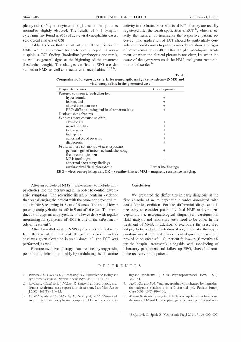
Strana 606 VOJNOSANITETSKI PREGLED Volumen 71, Broj 6
Stojanovi Z, Špiri Ž. Vojnosanit Pregl 2014; 71(6): 603–607.
pleocytosis (> 5 lymphocytes/mm3), glucose normal, proteinsnormal/or slightly elevated. The results of > 5 lympho-cytes/mm3 are found in 95% of acute viral encephalitis cases;serological analysis of CSF.
Table 1 shows that the patient met all the criteria forNMS, while the evidence for acute viral encephalitis was asuspicious CSF finding (borderline lymphocytes per mm3),as well as general signs at the biginning of the treatment(headache, cough). The changes verified in EEG are de-scribed in NMS, as well as in acute viral encephalitis 10, 15, 16.
After an episode of NMS it is necessary to include anti-psychotics into the therapy again, in order to control psychi-atric symptoms. The scientific literature contains evidencethat rechallenging the patient with the same antipsychotic re-sults in NMS recurring in 5 out of 6 cases. The use of lowerpotency antipsychotics is safe in 9 out of 10 cases. The intro-duction of atypical antipsychotic in a lower dose with regularmonitoring for symptoms of NMS is one of the safest meth-ods of treatment 2.
After the withdrawal of NMS symptoms (on the day 23from the start of the treatment) the patient presented in thiscase was given clozapine in small doses 2, 18 and ECT wasperformed, as well.
Electroconvulsive therapy can reduce hyperpyrexia,perspiration, delirium, probably by modulating the dopamine
activity in the brain. First effects of ECT therapy are usuallyregistered after the fourth application of ECT 14, which is ex-actly the number of treatments the respective patient re-ceived. The application of ECT should be particularly con-sidered when it comes to patients who do not show any signsof improvement even 48 h after the pharmacological treat-ment, or when the clinical picture is not clear, i.e. when thecause of the symptoms could be NMS, malignant catatonia,or mood disorder 14.
Conclusion
We presented the difficulties in early diagnosis at thefirst episode of acute psychotic disorder associated withacute febrile condition. For the differential diagnose it isnecessary to consider potentially fatal NMS and viral en-cephalitis, i.e. neuroradiological diagnostics, cerebrospinalfluid analysis and laboratory tests need to be done. In thetreatment of NMS, in addition to excluding the prescribedantipsychotic and administration of a symptomatic therapy, acombination of ECT and low doses of atypical antipsychoticproved to be successful. Outpatient follow-up (6 months af-ter the hospital treatment), alongside with monitoring oflaboratory parameters and follow-up EEG, showed a com-plete recovery of the patient.
R E F E R E N C E S
1. Pelonero AL, Levenson JL, Pandurangi AK. Neuroleptic malignantsyndrome: a review. Psychiatr Serv 1998; 49(9): 1163 72.
2. Geethan J, Chandran GJ, Mikler JR, Keegan DL. Neuroleptic ma-lignant syndrome: case report and discussion. Can Med AssocJ 2003; 169(5): 439 42.
3. Caroff SN, Mann SC, McCarthy M, Naser J, Rynn M, Morrison M.Acute infectious encephalitis complicated by neuroleptic ma-
lignant syndrome. J Clin Psychopharmacol 1998; 18(4):349 51.
4. Hillis RE, Lee DA. Viral encephalitis complicated by neurolep-tic malignant syndrome in a 7-year-old girl. Pediatr EmergCare 2003; 19(2): 99 100.
5. Mihara K, Kondo T, Suzuki A. Relationship between functionaldopamine D2 and D3 receptors gene polymorphisms and neu-
Table 1Comparison of diagnostic criteria for neuroleptic malignant syndrome (NMS) and
viral encephalitis in the presented caseDiagnostic criteria Criteria presentFeatures common to both disorders
hyperthermia +leukocytosis +altered consciousness +EEG: diffuse slowing and focal abnormalities +
Distinguishing featuresFeatures more common to NMS
elevated CK +muscle rigidity +tachycardia +tachypnea +abnormal blood pressure +diaphoresis +
Features more common to viral encephalitisgeneral signs of infection, headache, cough +focal neurologic signs -MRI: focal signs -abnormal chest x-ray findings -cerebrospinal fluid: pleocytosis Borderline findings
EEG electroencephalogram; CK creatine kinase; MRI magnetic resonance imaging.

Volumen 71, Broj 6 VOJNOSANITETSKI PREGLED Strana 607
Stojanovi Z, Špiri Ž. Vojnosanit Pregl 2014; 71(6): 603–607.
roleptic malignant syndrome. Am J Med Genet B Neuropsy-chiatr Genet 2003; 117B(1): 57 60.
6. Bourgeois JA, Babine S, Meyerovich M, Doyle JA. A Case of Neu-roleptic Malignant Syndrome With Quetiapine. J Neuropsy-chiatry Clin Neurosci 2002; 14(1): 87.
7. Adnet P, Lestavel P, Krivosic-Horber R. Neuroleptic malignantsyndrome. Br J Anaesth 2000; 85(1): 129 35.
8. Stevens JR, Livermore A. Telemetered EEG in schizophrenia:spectral analysis during abnormal behaviour episodes. J NeurolNeurosurg Psychiatry 1982; 45(5): 385 95.
9. Gattaz WF, Mayer S, Ziegler P, Platz M, Gasser T. Hypofrontalityon topographic EEG in schizophrenia. Correlations with neu-ropsychological and psychopathological parameters. Eur ArchPsychiatry Clin Neurosci 1992; 241(6): 328 32.
10. Steiner I, Budka H, Chaudhuri A, Koskiniemi M, Sainio K, SalonenO, et al. Viral encephalitis: a review of diagnostic methods andguidelines for management. Eur J Neurol 2005; 12(5): 331 43.
11. Chaudhuri A, Kennedy PG. Diagnosis and treatment of viral en-cephalitis. Postgrad Med J 2002; 78(924): 575 83.
12. Hermesh H, Manor I, Shiloh R, Aizenberg D, Benjamini Y, MunitzH, et al. High serum creatinine kinase level: possible risk factorfor neuroleptic malignant syndrome. J Clin Psychopharmacol2002; 22(3): 252 6.
13. American Psychiatric Association. Diagnostic and Statistical Man-ual of Mental Disorders. 4th ed. Text Revision. Washington,DC: American Psychiatric Publishing; 2000.
14. Guzofski S, Peralta R. Neuroleptic Malignant Syndrome, withAttention to Its Occurrence with Atypical AntipsychoticMedication: A Review. Jefferson J Psychiatry 2006; 20(1):53 62.
15. Kemperman CJ, van den Hoofdakker RH. Neuroleptic malignantsyndrome (NMS): challenge with zuclopenthixol and follow-up: a case report. Eur Neuropsychopharmacol 1990; 1(1):67 9.
16. Benzer TI, Tarabar A. Neuroleptic Malignant Syndrome inEmergency Medicine. Medscape 2012. Available from: emedi-cine.medscape.com/article/816018-overview [cited 2012 June7].
17. Kennedy PG. Viral encephalitis: causes, diferential diagnosis, andmanagement. J Neurol Neurosurg Psychiatry 2004; 75(90001):10 5.
18. Huang TL. Neuroleptic malignant syndrome associated withlong-term clozapine treatment: report of a case and results of aclozapine rechallenge. Chang Gung Med J 2001; 24(8): 522 5.
Received on January 17, 2013.Revised on April 18, 2013.
Accepted on April 26, 2013.

Strana 608 VOJNOSANITETSKI PREGLED Vojnosanit Pregl 2014; 71(6): 608–610.
Correspondence to: Slobodan M. Mitrovi , Ear, Nose and Throat University Clinic, Clinical Center of Vojvodina, Hajduk Veljkova 1–3,21 000 Novi Sad, Serbia. Phone: +381 21 484 3 670. E-mail: [email protected]
H I S T O R Y O F M E D I C I N E UDC: 61:[616.22-02:616.34-008.1(091)DOI: 10.2298/VSP1406608M
Terminology, diagnostics and therapy of laryngopharyngeal reflux– A glimpse into the past
Terminologija, dijagnostika i terapija laringofarinksnog refluksa – pogled uprošlost
Slobodan M. Mitrovi
Phoniatric Department, Ear, Nose and Throat University Clinic, Clinical Center ofVojvodina, Faculty of Medicine, University of Novi Sad, Novi Sad, Serbia
Key words:larynx; gastroesophageal reflux; diagnosis, differential;therapeutics; hydrochloric acid; pepsin a.
Klju ne re i:larinks; gastroezofagusni refluks; dijagnoza,diferencijalna; le enje; hlorovodoni na kiselina;pepsin.
Introduction
Laryngopharyngeal reflux (LPR) is the presence of re-fluxed gastric contents in the laryngopharyngeal space,where it is in close contact with the tissues of upper aerodi-gestive tract. Some authors consider it an entity, while othersas extraesophageal manifestation of gastroesophageal reflux.However, it is considered that its pathophysiology andsymptoms are different from gastroesophageal reflux. Mainsymptoms are: throat clearing, hoarseness, chronic cough,postnasal drip, halitosis, dysphagia, sense of foreign body inpharynx. As a confirmation of suspected LPR, indirect lar-yngoscopy shows: mucous edema, redness, ventricular oblit-eration, pseudoulcer, hypertrophy of posterior laryngeal re-gion and laryngeal granulomas. It is believed that the pres-ence of gastric contents in larynx and hypopharynx plays arole in the development of cancer lesions of this region. To-day, an influence of pepsin and hydrochlorid acid on manyother disorders in otorhinolaryngological region are beingconsidered 1.
At the level of assumptions
Although in the middle of XIX century the only possi-ble way to visualize larynx was post mortem, negative im-pact of numerous factors on different laryngeal functions,predominantly phonation, were known. Singing teacher inParis, Manuel Garcia 2, wrote “Studies about human voice”in 1840, where he stated: “Gourmet dishes, oils from certainherbs, spirits, it all has negative impact on our apparatus.”Thus, he pointed out a possible influence of reflux on larynx,
without actually being able to prove it. Guided by a wish toshow larynx, in 1854 Garcia performed the first “autolaryn-goscopy”, and managed to visualize this organ for the firsttime. By the end of the XIX century, changes of posteriorpart of larynx were confirmed histopathologically, whichcaused a series of researches on gastric contents reflux as anetiology factor in many laryngeal disorders in the XX cen-tury.
In 1880, Rudolf Virchow wrote a descriptive term“pachydermia verrucosa laryngis“ for changes on arytenoidmucosa, which he associated with vocal abuse. In his origi-nal paper, Virchow described it as a state different from la-ryngeal leucoplacia and keratosis 3. In the same year, MorellMackenzie 4 identified the changes of arytenoid cartilagebased on the presence of ulceration. Journal of the Royal So-ciety of Medicine in 1908 published the abstract of W. Job-son Horne’s 5 presentation, in which he confirms that pachy-dermia is a consequence of hyperplasia due to persistent lar-yngitis, but he identified rather different causes. In the cutsof a 37-year old patient’s vocal cords, besides hyperplasia,he found excrescence with sulcus in front of and behind it.
In 1928, Chevalier Jackson 6 observed 217 patientsduring a 4-year period, and named changes he found on ary-tenoids “contact ulcer”. It was almost 50 years afterMackenzie’s description. Jackson 6 emphasized that this termrefers strictly to nontuberculous lesions, but that any of themcan turn into cancer. He associated these lesion to excessivevocal use, occurring frequently among salesman, doctorswho gave lectures, priests, with the significant influence ofalcohol and tobacco use. In his work about laryngeal kerato-sis in 1946, Clerf 7 also said that pachydermia occurred more

Volumen 71, Broj 6 VOJNOSANITETSKI PREGLED Strana 609
Mitrovi MS. Vojnosanit Pregl 2014; 71(6): 608–610.
often at alcoholics or excessive vocal use, as in street sellers,for example.
From posterior laryngitis to “silent“gastroesophageal reflux
The advance in understanding the posterior laryngealchanges happened in 1968, when first reports on posteriorlaryngitis caused by gastroesophageal reflux occurred. Thatyear Delahunty and Cherry 8 concluded on animal model thatdescribed changes on posterior larynx were caused by gas-troesophageal reflux, while Cherry and Margulies 9 treatedthree patients with posterior laryngitis, and changes disap-peared in six months. In 1970, Delahunty and Ardran 10 triedto explain globus hystericus as a possible manifestation ofreflux esophagitis. In 1972, Delahunty climbed one floorabove larynx, and in “Journal of Laryngology“ published thepaper titled “Acid laryngitis“ 11. In it, he stated as a postulatethat “patients with reflux esophagitis can regurgitate acidcontents even more, especially during the night, whichcauses inflammatory reaction of posterior larynx”.
Searching PubMed data base showed a lack of papersuntil 1984, when a research of otorhinolaryngologists fromLjubljana, Vinko Kambi and Zora Radšel 12, “Acid poste-rior laryngitis” was published in the same journal in whichDelahunty published the results of his researches. They pub-lished the histological results of biopsy of mucosa in 44 pa-tients with chronic posterior laryngitis caused by acid. Fromphoniatric point of view, Živko Majdevac 13, student ofCzech phoniatric school and founder of Phoniatric Depart-ment of Otorhinolaryngology Clinic in Novi Sad, was alsointerested in these changes. At the VIII Congress of UEP(Union of European Phoniatrics) in 1979 in Köszeg (Hun-gary), he presented his “Classification of dysphonia regard-ing primary etiological factor”, and in the first group of dis-orders – functional disorders, described “contact hyperplasticdysphonia” as a condition which occurs in men whose voiceis in range of baritone or bas, and more often at militarycommanders and teachers. During phonation, posterior thirdsof vocal cords are overcontracted, which causes the appear-ance of circumscriptive yellowish thickening. At the time, heassociated the mentioned changes on vocal processes of ary-tenoid cartilage with gastric hyperacidity, considering it asignificant cofactor.
In 1986, Benjamin 14 published a paper on extraesopha-geal complications of gastroesophageal reflux, stating thatgastric contents can get from esophagus into hypopharynxand lungs. In 1989, Wilson et al. 15 published a paper basedon a 23-hour long ambulatory measurement of pH and bi-opsy of posterior larynx in patients with hoarseness, throatburn and globus sensation.
In 1991, Olson 16 mentioned laryngopharyngeal mani-festations of gastroesophageal disease. In the same year,James A. Koufman 17 published a paper on otolaryngealmanifestations of gastroesophageal disease in 225 patients,which was the highest number of patients up to the time. Atthat moment, he did not refer to the disorder as LPR yet, butcalled it “occult (silent) gastroeophageal reflux disease”. A
paper on measuring pH on children with gastroesophagealreflux was published in 1992 by Contencin et al. 18.
Gastroesophageal vs laryngopharyngeal reflux
In 1996 in the Journal of Voice, Koufman, Sataloffand Toohil 19 published a report from consensus conferenceon LPR, and that was the first paper to refer to the term la-ryngopharyngeal reflux. In 1992, Kamel et al. 20 publishedthe results of prospective study on the use of omeprazole inthe therapy of reflux laryngitis, while Shaw et al. 21 in 1996published a paper with the special emphasis on laryngo-scopic findings before and after the therapy with omepra-zole. In 1997, Wo et al. 22 published a work on empiric useof high doses of omeprazole in patients with posterior lar-yngitis, while Habermann et al. 23 published a study on exiuvantibus therapy with pantoprazole in patients with poste-rior laryngitis in 1999. In 1999, Koufman 24 made a reportabout the treatment of LPR (for the first time with thisname) on I International Symposium about human pepsin,held at the University of York (England). The term of la-ryngeal reflux in children was first mentioned in 2000 byZalzal and Tran 25 in the paper titled “Pediatric gastro-esophageal reflux and laryngopharyngeal reflux”. The termLPR was not yet used in that year, but “laryngitis gastrica”was used instead.
In 2000, Koufman et al. 26 found that reflux is observedin 50% of their examined patients with laryngeal and vocalproblems. In June 2001, Belafsky, Postma and Koufman 27
published a paper in the Laryngoscope Journal, in which theyexplain their scoring system of reflux findings [Reflux Find-ing Score – (RFS)], questionnaire about the LPR symptomsnamed Reflux Simptom Index (RSI) and support the termLPR. In August of the same year, in the same journal, theyexplained the validity and usefulness of RFS in detail 28. Ayear later, the same team 29, (joined by Amin) made a de-tailed evaluation of RSI usefulness, while Koufman 30 de-finitively explained that LPR is different from classic gastro-esophageal reflux disease.
Nissen’s fundoplication as a method for the treatmentof extraesophageal manifestations of gastroesophageal refluxdisease was promoted in the study by Lindstrom et al. 31 in2002. In 2003 Siupsinskiene and Adamonis 32 published apaper on ex iuvantibus diagnosis by administration of omep-razole and monitoring the response. In the same year, Galli etal. 33 published a study based on the hypothesis that biliaryreflux, as well as gastric reflux, has an influence on inflam-matory, precancerous and neoplastic changes of larynx. Thefirst paper on the quality of life in patients with LPR waspublished in 2007 34.
In April 2011, during 132nd annual meeting of Ameri-can Laryngology Association (ALA), Jamie A. Koufman 35
was awarded for extraordinary contribution to the researchand medical references in laryngology. It was a reward for alifelong work on explaining etiology, pathophysiology, clini-cal findings, diagnostics, therapy and prevention of laryngo-pharyngeal reflux. In order to grasp the importance of hercontribution, it should be mentioned that among the scien-

Strana 610 VOJNOSANITETSKI PREGLED Volumen 71, Broj 6
Mitrovi MS. Vojnosanit Pregl 2014; 71(6): 608–610.
tists who got this award were Chevalier Jackson and JohnKirchner.
Conclusion
Researches on the influence of laryngopharyngeal re-flux on the organs of otorhinolaryngology region keep on,
especially after understanding that this disorder can be astand-alone factor or cofactor in etiology of secretory otitis,chronic rhinitis, nose-sinus polyposis and cancer of aerodi-gestive ways. However, without looking back in the past andpaying tribute to everyone who explained the term and na-ture of laryngopharyngeal reflux, no new researches wouldhave the full sense.
R E F E R E N C E S
1. Ford CN. Evaluation and management of laryngopharyngealreflux. JAMA 2005; 294(12): 1534 40.
2. Garcia M. The complete treatise on the art of singing. 1st
part. 1st ed. New York: Da Capo Press; 1975 1984. (French)3. Grossman AA, Mathews WH. Keratosis of the larynx with pro-
gression to malignancy. Can Med Assoc J 1952; 66(1): 39 41.4. MacKenzie M. Disease of the pharynx, larynx, and trachae. New
York: William Wood & Co; 1880. p. 347.5. Horne JW. Specimens showing the Pathogenesis of Pachyder-
mia laryngis verrucosa et diffusa. Proc R Soc Med. 1908;1(Laryngol Sect): 132–3.
6. Jackcon LC. Symposium on the larynx: II.—Etiology andtreatment of contact ulcer of the larynx. Laryngoscope 1933;43(9): 718–21.
7. Clerf HL. Keratosis of the larynx. JAMA 1946;132(14): 823 6.8. Delahunty JE, Cherry J. Experimentally produced vocal cord
granulomas. Laryngoscope 1968; 78(11): 1941 7.9. Cherry J, Margulies S. Contact ulcer of the larynx. Laryngoscope
1968; 78(11): 1937 40.10. Delahunty JE, Ardran GM. Globus hystericus-a manifestation of
reflux oesophagitis? J Laryngol Otol 1970; 84(10): 1049 5.11. Delahunty JE. Acid laryngitis. J Laryngol Otol 1972; 86(4): 335– 42.12. Kambi V, Radšel Z. Acid posterior laryngitis. Aetiology, histol-
ogy, diagnosis and treatment. J Laryngol Otol 1984; 98(12):1237 40.
13. Majdevac Ž. Heiserkeit. In: Frint T, Hirschberg J, editors. Haupt-referate und vorträge. VIII Kongres der Union foniater Eu-ropae; 1979. Köszeg: Ungarn; 1979. p. 97 118.
14. Benjamin SB. Extra-esophageal complications of gastroesopha-geal reflux. J Clin Gastroenterol 1986; 8(Suppl 1): 68 71.
15. Wilson JA, White A, von Haacke NP, Maran AG, Heading RC,Pryde A, et al. Gastroesophageal reflux and posterior laryngitis.Ann Otol Rhinol Laryngol 1989; 98(6): 405 10.
16. Olson NR. Laryngopharyngeal manifestations of gastroesophagealreflux disease. Otolaryngol Clin North Am 1991; 24(5): 1201 13.
17. Koufman JA. The otolaryngologic manifestations of gastro-esophageal reflux disease (GERD): a clinical investiogation of225 patients using ambulatory 24-hour pH monitoring and anexperimental investigation of the role of acid pepsin in the de-velopment of laryngeal injury. Laryngoscope 1991; 101(4 Pt 2Suppl 53): 1 78.
18. Contencin P, Adjoua P, Viala P, Erminy M, Narcy P. Long-termesophageal and oropharyngeal pH-metry in ORL manifesta-tions of gastroesophageal reflux in children. Ann OtolaryngolChir Cervicofac 1992; 109(3): 129 33. (French)
19. Koufman J, Sataloff RT, Toohill R. Laryngopharyngeal reflux:consensus conference report. J Voice 1996; 10(3): 215 6.
20. Kamel P, Kahrilas PJ, Hanson DG, McMahon J, Brenic S. Prospec-tive trial of omeprazole in the treatment of “reflux laryngitis”.Gastroenterology 1992; 102: A93.
21. Shaw GY, Searl JP, Young JL, Minner PB. Subjective, laryngo-scopic and acoustic measurements of laryngeal reflux, before
and after treatment with omeprazole. J Voice 1996; 10(4):410 8.
22. Wo JM, Grist WJ, Gussack G, Delgaudio JM, Waring JP. Empirictrial of high- dose omeprazole in patients with posterior laryn-gitis: a prospective study. Am J Gastroenterol 1997; 92(12):2160 5.
23. Habermann W, Eherer A, Lindbichler F, Raith J, Friedrich G. Exjuvantibus approach for chronic posterior laryngitis: results ofshort-term pantoprazole therapy. J Laryngol Otol 1999;113(8): 734 9.
24. Koufman JA. Treatment of laryngopharyngeal reflux (LPR).[Abstract]. Presented at the 1st International Symposium onHuman Pepsin; 1999 October 1-2; London: University ofYork; 1999.
25. Zalzal GH, Tran LP. Pediatric gastroesophageal reflux and la-ryngopharyngeal reflux. Otolaryngol Clin Nort Am 2000;33(1): 151 61.
26. Koufman JA, Amin MR, Panetti M. Prevalence of reflux in 113consecutive patients with laryngeal and voice disorders.Otolaryngol Head Neck Surg 2000; 123(4): 385 8.
27. Belafsky CP, Postma NG, Koufman JA. Laryngopharyngeal refluxsymptoms improve before changes in physical findings. La-ryngoscope 2001;111(6): 979 81.
28. Belafsky PC, Postma GN, Koufman JA. The validitiy and reliabil-ity of the reflux finding score (RFS). Laryngoscope 2001;111(8): 1313 7.
29. Belafsky PC, Postma GN, Amin MR, Koufman JA. Symptoms andfindings of laryngopharyngeal reflux. Ear Nose Troat J 2002;81(9 Suppl 2): 10 3.
30. Koufman JA. Laryngopharyngeal reflus is different from classicgastroesophageal reflux disease. Ear Nose Throat J 2002; 81(9Suppl 2): 7 9.
31. Lindstrom DR, Wallace J, Loehrl TA, Merati AL, Toohil RJ. Nis-sen fundoplication surgery for extraesophageal manifestationof gastroesophageal reflux (EER). Laryngoscope 2002;112(10): 1762 5.
32. Siupsinskiene N, Adamonis K. Diagnostic test with omeprazole inpatients with posterior laryngitis. Medicina (Kaunas) 2003;39(1): 47 55.
33. Galli J, Calo L, Agostino S, Cadoni G, Sergi B, Cianci R, et al. Bilereflux as possible risk factor in laryngopharyngeal inflamma-tory and neoplastic lesions. Acta Otorinolaryngol Ital 2003;23(5): 377 82.
34. Siupsinskiene N, Adamonis K, Toohil RJ. Quality of life in laryn-gopharyngeal reflux patients. Laryngoscope 2007; 117(3):480 4.
35. Kaufman JA. Biography and facts. Available from:http://www.whoislog.info/profile/charlie-kaufman.html
Received on November 3, 2012.Revised on February 28, 2013.
Accepted on February 12, 2013.OnLine-First February, 2014.

Volumen 71, Broj 6 VOJNOSANITETSKI PREGLED Strana 611
INSTRUCTIONS TO THE AUTHORS
Vojnosanitetski pregled (VSP) publishes only not previously pub-lished nor submitted papers in any other journals in the order deter-mined by the Editorial Board. The following should be enclosed withthe manuscript: a statement that the paper has not been submitted oraccepted for publication elsewhere, a statement specifiing the actualcontribution of each co-coautor, a consent signed by all the authorsthat the paper could be submitted; the name, exact address, phonenumber, and e-mail address of the first author and co-authors. VSP re-serves all copyrights.
From January 1, 2012 the Vojnosanitetski pregled has been editedusing the service e-Ur: Electronic Journal Editing.
All users of the system: authors, editors and reviewrs have to beregistrated users with only one e-mail address. Registration shouldbe made on the web-address:
http://scindeks-eur.ceon.rs/index.php/vsp
VSP publishes: editorials, original articles, short communications,reviews/meta-analyses, case reports, from the medical history (generalor military), personal views, invited comments, letters to the editor, reportsfrom scientific meetings, book reviews, extensive abstracts of interestingarticles from foreign language journals, and other contributions. Originalarticles, short communications, meta-analyses and case reports are pub-lished with abstracts in both English and Serbian.
General review papers will be accepted by the Editorial Board only ifthe authors prove themselves as the experts in the fields they write on byciting not less than 5 self-citations.
Papers should be written on IBM-compatible PC, using 12 pt font, anddouble spacing, with at least 4 cm left margin. Bold and italic lettersshould be avoided. Observational and experimental articles, reviews andmeta-analyses, should not exceed 16 pages (including tables and illus-trations); case reports – 6; short communications – 5; letters to the Edi-tor, reports on scientific meetings and book reviews – 2.
All measurements should be reported in the metric system interms of the International System of Units (SI). Standard, interna-tionally accepted terms should be used.
MS Word for Windows (97, 2000, XP, 2003) is recommended forword processing; other programs are to be used only exceptionally. Il-lustrations should be made using standard Windows programs. Avoidthe use of colors in graphs.
Papers are reviewed anonymously by at least two editors and/or in-vited reviewers. Remarks and suggestions are sent to the author for finalcomposition. Galley proofs are sent to the first author for correctionsthat should be returned within 3 days. Manuscripts accepted for publica-tion are not being returned.
Preparation of manuscriptParts of the manuscript are: Title page; Abstract with key words;
Text; References.1. Title pagea) The title should be concise but informative. Subheadings should be
avoided;b) Full name of each author;c) Name and place of department(s) and institution(s) of affiliation,
clearly marked by standard footnote signs.2. Abstract and key wordsThe second page should carry a structured abstract with the title for
original articles, metanalyses and case reports. The abstract should statethe purposes of the study or investigation, basic procedures (selection ofstudy subjects or laboratory animals; observational and analytical meth-ods), main findings (giving specific data and their statistical signifi-cance, if possible), and the principal conclusions. It should emphasizenew and important aspects of the study or observations. S t r u c -t u r e d abstract should contain typical subtitles: background/aim,methods, results and conclusion. The abstract for metaanalyses and obr-ginal papers should have up to 450 words, and up to 150 words for casereports (with subtitles background, case report, conclusion). Below theabstract authors should provide, and identify as such, 3–10 key words orshort phrases that will assist indexers in cross-indexing the article andwill be published with the abstract.
3. TextThe text of original articles is divided into sections with the headings:
Introduction, Methods, Results, and Discussion. Long articles mayneed subheadings within some sections to clarify their content.
In the Introduction repeat the title of the article, excluding the namesof authors. State the purpose of the article and summarize the rationalefor the study or observation. Give only strictly pertinent references anddo not include data or conclusions from the work being reported.
Methods. Describe your selection of the observational or experimen-tal subjects (patients or experimental animals, including controls)clearly. Identify the methods, apparatus (manufacturer's name and ad-dress in parentheses), and procedures in sufficient detail to allow otherworkers to reproduce the results. Give references to established meth-ods, including statistical methods. Identify precisely all drugs andchemicals used, with generic name(s), dose(s), and route(s) of admini-stration. State the approvement of the Ethnics Committe for the tests inhumans and enimals.
Results should be presented in logical sequence in the text, tables andillustrations. Emphasize or summarize only important observations.
Discussion is to emphasize the new and important aspects of the studyand the conclusions that result from them. Relate the observations toother relevant studies. Link the conclusions with the goals of the study,but avoid unqualified statements and conclusions not completely sup-ported by your data.
ReferencesReferences should be superscripted and numbered consecutively in the
order in which they are first mentioned in the text. The references mustbe verified by the author(s) against the original document. List allauthors, but if the number exceeds 6, give 6 followed by et al. Do notuse abstracts, secondary publications, oral communications, unpublishedpapers, official and classified documents. References to papers acceptedbut not yet published should be designated as ”in press“. Informationfrom manuscripts not yet accepted should be cited in the text as ”unpub-lished observations“. References are cited according to the InternationalCommittee of Medical Journal Editors. Uniform Requirements forManuscripts Submitted to Biomedical Journals. Ann Intern Med 1997;126: 36–47. Updated October 2001.
Examples of references:Jurhar-Pavlova M, Petlichkovski A, TrajkovD, Efinska-Mladenovska O,Arsov T, Strezova A, et al. Influence of the elevated ambient temperatureon immunoglobulin G and immunoglobulin G subclasses in sera ofWistar rats. Vojnosanit Pregl 2003; 60(6): 657–612.
DiMaio VJ. Forensic Pathology. 2nd ed. Boca Raton: CRC Press; 2001.Blinder MA. Anemia and Transfusion Therapy. In: Ahya NS, Flood K,Paranjothi S, editors. The Washington Manual of Medical Therapeutics, 30thedition. Boston: Lippincot, Williams and Wilkins; 2001. p. 413-28.Christensen S, Oppacher F. An analysis of Koza's computational effortstatistic for genetic programming. In: Foster JA, Lutton E, Miller J,Ryan C, Tettamanzi AG, editors. Genetic programming. EuroGP 2002:Proceedings of the 5th European Conference on Genetic Programming;2002 Apr 3-5; Kinsdale, Ireland. Berlin: Springer; 2002. p. 182-91.Abood S. Quality improvement initiative in nursing homes: the ANAacts in an advisory role. Am J Nurs [serial on the Internet]. 2002 Jun[cited 2002 Aug 12]; 102(6): [about 3 p.]. Available from:http://www.nursingworld.org/AJN/2002/june/Wawatch.htm
TablesEach table should typed double-spaced on a separate sheet, numbered
in the order of their first citation in the text in the upper right corner andsupplied with a brief title each. Explanatory notes are printed under a ta-ble, using the following symbols, in this sequence: *, †, ‡, §, ||, ¶, **, ††,... . Each table has to be mentioned in the text. If you use data from an-other source, acknowledge fully.
IllustrationsFigures are submitted as photos which should be sharp. Letters, num-
bers, and symbols should be clear and even throughout and of sufficientsize that when reduced for publication, each item will still be legible.Each figure should have a label on its back indicating the number of thefigure, author's name, and top of the figure. If a figure has been pub-lished, acknowledge the original source.
Legends for illustrations are typed on a separate page, with arabic nu-merals corresponding to the illustrations. Identify and explain each oneclearly in the legend symbols, arrows, numbers, or letters used to iden-tify parts of the illustrations. Explain the method of staining in photomi-crographs.
Abbreviations and symbolsUse only standard abbreviations. Avoid abbreviations in the title and
abstracts. The full term for which an abbreviation stands should precedeits first use in the text.
Detailed Instructions are available at the web site:www.vma.mod.gov.rs/vsp/download/instructions_to_authors.pdf.

Strana 612 VOJNOSANITETSKI PREGLED Volumen 71, Broj 6
UPUTSTVO AUTORIMA
Vojnosanitetski pregled (VSP) objavljuje radove koji ranije nisu nig-de publikovani, niti predati za publikovanje redosledom koji odre ujeure iva ki odbor. Prilikom prijave rada u sistem elektronskog ure iva-nja „Vojnosanitetskog pregleda“ neophodno je priložiti izjavu da su is-punjeni svi postavljeni tehni ki zahtevi uklju uju i i izjavu potpisanu odstrane svih autora da rad nije ranije ni u celini, niti delimi no objavljenniti prihva en za štampanje u drugom asopisu. Izjava o pojedina nomdoprinosu autora mora biti potpisana od strane svakog autora rada, ske-nirana i poslata uz rad kao dopunska datoteka. Tako e, autori su obave-zni da dostave i potpisanu izjavu o nepostojanju sukoba interesa. Timpostupkom svi autori postaju odgovorni za ispunjavanje svih postavlje-nih uslova, emu sledi odluka o prihvatanju za dalji ure iva ki postupak.Za objavljene radove VSP zadržava autorsko pravo. Primaju se radovinapisani samo na engleskom jeziku.
Od 1. januara 2012. godine Vojnosanitetski pregled prešao jena e-Ur: Elektronsko ure ivanje asopisa.
Svi korisnici sistema: autori, recezenti i urednici moraju biti regis-trovani jednozna nom e-mail adresom. Registraciju je mogu e izvršitina adresi:
http://aseestant.ceon.rs/index.phpU VSP-u se objavljuju uvodnici, originalni lanci, prethodna ili
kratka saopštenja, revijski radovi tipa opšteg pregleda (uz uslov daautori navo enjem najmanje 5 autocitata potvrde da su eksperti u oblastio kojoj pišu), aktuelne teme ili metaanalize, kazuistika, lanci iz isto-rije medicine, li ni stavovi, naru eni komentari, pisma uredništvu, izve-štaji sa nau nih i stru nih skupova, prikazi knjiga, referati iz nau ne istru ne literature i drugi prilozi. Radovi tipa originalnih lanaka, pretho-dnih ili kratkih saopštenja, metaanalize i kazuistike objavljuju se uz ap-strakte na srpskom i engleskom jeziku.
Rukopis se piše sa proredom 1,5 sa levom marginom od 4 cm. Koris-titi font veli ine 12, a na elno izbegavati upotrebu bold i italic slova, kojasu rezervisana za podnaslove. Originalni lanci, opšti pregledi i metaanali-ze ne smeju prelaziti 16 stranica (sa prilozima); aktuelne teme – osam, ka-zuistika – šest, prethodna saopštenja – pet, a pisma uredniku, izveštaji saskupova i prikazi knjiga – dve stranice.
U celom radu obavezno je koriš enje me unarodnog sistema mera(SI) i standardnih me unarodno prihva enih termina.
Za obradu teksta koristiti program Word for Windows verzije 97,2000, XP ili 2003. Za izradu grafi kih priloga koristiti standardne gra-fi ke programe za Windows, poželjno iz programskog paketa Micro-soft Office (Excel, Word Graph). Kod kompjuterske izrade grafikaizbegavati upotrebu boja i sen enja pozadine.
Prispeli radovi kao anonimni podležu ure iva koj obradi i recenzijinajmanje dva urednika/recenzenta. Primedbe i sugestije uredni-ka/recenzenata dostavljaju se autoru radi kona nog oblikovanja. Pre ob-jave, rad se upu uje koresponding autoru na kona nu saglasnost.
Priprema radaDelovi rada su: naslovna strana, apstrakt sa klju nim re ima,
tekst i literatura.1. Naslovna stranaa) Naslov treba da bude kratak, jasan i informativan i da odgovara
sadržaju rada. Podnaslove treba izbegavati.b) Ispisuju se puna imena i prezimena autora.c) Navode se puni nazivi ustanove i organizacijske jedinice u kojima
je rad obavljen i mesta u kojima se ustanove nalaze, sa jasnim obeleža-vanjem odakle je autor, koriste i standardne znake za fus-note.
2. Apstrakt i klju ne re iNa drugoj stranici nalazi se strukturisani apstrakt sa naslovom rada.
Kratkim re enicama na srpskom i engleskom jeziku iznosi se uvod icilj rada, osnovne procedure - metode (izbor ispitanika ili laboratorij-skih životinja; metode posmatranja i analize), glavni nalazi - rezultati(konkretni podaci i njihova statisti ka zna ajnost) i glavni zaklju ak.Naglasiti nove i zna ajne aspekte studije ili zapažanja. Strukturisaniapstrakt (250 re i) ima podnaslove: uvod/cilj, metode, rezultati i zak-lju ak. Za apstrakte na engleskom dozvoljeno je i do 450 re i. Struk-turisani apstrakt je obavezan za metaanalize (istog obima kao i za ori-ginalne lanke) i kazuistiku (do 150 re i, sa podnaslovima uvod, pri-kaz slu aja i zaklju ak). Ispod apstrakta, pod podnaslovom „Klju nere i“ predložiti 3–10 klju nih re i ili kratkih izraza koji oslikavaju sa-držinu lanka.
3. Tekst lankaTekst sadrži slede a poglavlja: uvod, metode, rezultate i diskusiju.
Zaklju ak može da bude posebno poglavlje ili se iznosi u poslednjempasusu diskusije. U uvodu ponovo napisati naslov rada, bez navo enja
autora. Navesti hipotezu (ukoliko je ima) i ciljeve rada. Ukratko iznetirazloge za studiju ili posmatranje. Navesti samo strogo relevantne po-datke iz literature i ne iznositi opširna razmatranja o predmetu rada, kaoni podatke ili zaklju ke iz rada o kome se izveštava.
Metode. Jasno opisati izbor metoda posmatranja ili eksperimentnihmetoda (ispitanici ili eksperimentne životinje, uklju uju i kontrolne).Identifikovati metode, aparaturu (ime i adresa proizvo a a u zagradi) iproceduru, dovoljno detaljno da se drugim autorima omogu i reproduk-cija rezultata. Navesti podatke iz literature za uhodane metode, uklju u-ju i i statisti ke. Ta no identifikovati sve primenjene lekove i hemika-lije, uklju uju i generi ko ime, doze i na ine davanja. Za ispitivanja naljudima i životinjama navesti saglasnost eti kog komiteta.
Rezultate prikazati logi kim redosledom u tekstu, tabelama i ilustra-cijama. U tekstu naglasiti ili sumirati samo zna ajna zapažanja.
U diskusiji naglasiti nove i zna ajne aspekte studije i izvedene zak-lju ke. Posmatranja dovesti u vezu sa drugim relevantnim studijama, una elu iz poslednje tri godine, a samo izuzetno i starijim. Povezati zak-lju ke sa ciljevima rada, ali izbegavati nesumnjive tvrdnje i one zaklju -ke koje podaci iz rada ne podržavaju u potpunosti.
LiteraturaLiteratura se u radu citira kao superskript, a popisuje rednim broje-
vima pod kojima se citat pojavljuje u tekstu. Navode se svi autori, aliako broj prelazi šest, n a v o d i s e p r v i h š e s t i dodaje etal. Svi podaci o citiranoj literaturi moraju biti t a n i . Literatura se ucelini citira na engleskom jeziku, a iza naslova se navodi jezik lanka uzagradi. Ne prihvata se citiranje apstrakata, sekundarnih publikacija,usmenih saopštenja, neobjavljenih radova, službenih i poverljivih do-kumenata. Radovi koji su prihva eni za štampu, ali još nisu objavljeni,navode se uz dodatak „u štampi“. Rukopisi koji su predati, ali još nisuprihva eni za štampu, u tekstu se citiraju kao „neobjavljeni podaci“ (uzagradi). Podaci sa Interneta citiraju se uz navo enje datuma.
Primeri referenci:urovi BM. Endothelial trauma in the surgery of cataract. Vojno-
sanit Pregl 2004; 61(5): 491–7. (Serbian)Balint B. From the haemotherapy to the haemomodulation. Beo-
grad: Zavod za udžbenike i nastavna sredstva; 2001. (Serbian)Mladenovi T, Kandolf L, Mijuškovi ŽP. Lasers in dermatology. In:
Karadagli , editor. Dermatology. Beograd: Vojnoizdava ki zavod &Verzal Press; 2000. p. 1437–49. (Serbian)
Christensen S, Oppacher F. An analysis of Koza's computational ef-fort statistic for genetic programming. In: Foster JA, Lutton E, Miller J,Ryan C, Tettamanzi AG, editors. Genetic programming. EuroGP 2002:Proceedings of the 5th European Conference on Genetic Programming;2002 Apr 3-5; Kinsdale, Ireland. Berlin: Springer; 2002. p. 182-91.
Abood S. Quality improvement initiative in nursing homes: theANA acts in an advisory role. Am J Nurs [serial on the Internet]. 2002Jun [cited 2002 Aug 12]; 102(6): [about 3 p.]. Available from:http://www.nursingworld.org/AJN/2002/june/Wawatch.htm
TabeleSve tabele pripremaju se sa proredom 1,5 na posebnom listu. Obele-
žavaju se arapskim brojevima, redosledom pojavljivanja, u desnom uglu(Tabela 1), a svakoj se daje kratak naslov. Objašnjenja se daju u fus-noti, ne u zaglavlju. Za fus-notu koristiti slede e simbole ovim redosle-dom: *, †, ‡, §, ||, ¶, **, ††, ... . Svaka tabela mora da se pomene u tek-stu. Ako se koriste tu i podaci, obavezno ih navesti kao i svaki drugi po-datak iz literature.
IlustracijeSlikama se zovu svi oblici grafi kih priloga i predaju se kao dopun-
ske datoteke u sistemu aseestant. Slova, brojevi i simboli treba da su ja-sni i ujedna eni, a dovoljne veli ine da prilikom umanjivanja budu it-ljivi. Slike treba da budu jasne i obeležene brojevima, onim redom kojimse navode u tekstu (Sl. 1; Sl. 2 itd.). Ukoliko je slika ve negde objav-ljena, obavezno citirati izvor.
Legende za ilustracije pisati na posebnom listu, koriste i arapskebrojeve. Ukoliko se koriste simboli, strelice, brojevi ili slova za ob-jašnjavanje pojedinog dela ilustracije, svaki pojedina no treba objasnitiu legendi. Za fotomikrografije navesti metod bojenja i podatak o uve a-nju.
Skra enice i simboliKoristiti samo standardne skra enice, izuzev u naslovu i apstraktu.
Pun naziv sa skra enicom u zagradi treba dati kod prvog pominjanja utekstu.
Detaljno uputstvo može se dobiti u redakciji ili na sajtu:www.vma.mod.gov.rs/vsp/download/uputstvo_za_autore.pdf.

VO
JNO
SAN
ITE
TSK
I P
RE
GL
ED
VO
JNO
ME
DIC
INSK
A A
KA
DE
MIJ
ACr
notra
vska
17,
110
40 B
eogr
ad, S
rbija
Tel/
Fax:
+38
1 11
266
9689
vsp@
vma.m
od.g
ov.rs
asop
is „V
ojno
sani
tets
ki p
regl
ed“
izlaz
i god
išnje
u 12
bro
jeva
.G
odišn
ja pr
etpl
ata
za 2
014.
god
inu
iznos
i: 5
000
dina
ra z
a gr
aan
e Sr
bije,
10 0
00 d
inar
a za
usta
nove
iz S
rbije
i 15
0 €
za st
rane
drž
avlja
ne i
usta
nove
. Pre
tplat
e:Ži
ro r
aun
br.
840-
3148
49-7
0 M
O –
Sre
dstv
a ob
jedin
jene
napl
ate
– V
MA
(za
Voj
nosa
nite
tski p
regl
ed),
poziv
na
broj
122
7423
1295
5214
15. U
plat
nicu
(do
kaz
oup
lati)
dosta
viti
lino
ili p
ošto
m (p
ismom
, fak
som
, -m
ail-o
m).
Za z
apos
lene
u M
O i
Voj
sci S
rbije
mog
ua
je i p
retp
lata
u 12
mes
eni
h ra
ta p
utem
tra
jnog
nalo
ga, t
j.„o
dbija
njem
od
plat
e“. P
opun
jen o
braz
ac p
oslat
i na a
dres
u V
SP-a
.
PR
IJA
VA
ZA
PR
ET
PL
AT
U N
A
ASO
PIS
„VO
JNO
SAN
ITE
TSK
I P
RE
GL
ED
“
Ime
i pre
zim
e ili
naz
iv u
stan
ove
Jedi
nstv
eni m
ati
ni b
roj g
raan
aPo
resk
i ide
ntifi
kaci
oni b
roj (
PIB)
za u
stan
ove
Mes
toU
lica
i bro
jTe
lefo
n /
tele
faks
Pret
plat
a na
as
opis
„Voj
nosa
nite
tski
pre
gled
“ (z
aokr
užiti
):1.
Lino
. Dok
az o
pre
tplat
i dos
tavl
jam u
z ov
u pr
ijavu
.2.
Za
prip
adni
ke M
O i
Voj
ske
Srbi
je:
Daje
m s
aglas
nost
da
sepr
iliko
m is
plat
e pl
ata
u Ra
unov
odst
veno
m c
entru
MO
iz m
ojih
prin
adle
žnos
ti ob
usta
vlja
izno
s mes
ene
rate
(pre
tplat
e).
3.V
irman
om p
o pr
ijem
u pr
ofak
ture
.
Dat
um _
____
____
____
___
Potp
is__
____
____
____
____
____
VO
JNO
SAN
ITE
TSK
I P
RE
GL
ED
VO
JNO
ME
DIC
INSK
A A
KA
DE
MIJ
ACr
notra
vska
17,
110
40 B
eogr
ad, S
rbija
Tel/
Fax:
+38
1 11
266
9689
vsp@
vma.m
od.g
ov.rs
asop
is „V
ojno
sani
tets
ki p
regl
ed“
izlaz
i god
išnje
u 12
bro
jeva
.G
odišn
ja pr
etpl
ata
za 2
014.
god
inu
iznos
i: 5
000
dina
ra z
a gr
aan
e Sr
bije,
10 0
00 d
inar
a za
usta
nove
iz S
rbije
i 15
0 €
za st
rane
drž
avlja
ne i
usta
nove
. Pre
tplat
e:žir
o ra
un b
r. 84
0-31
4849
-70
MO
– S
reds
tva
objed
injen
e na
plat
e –
VM
A (
zaV
ojno
sani
tetsk
i pre
gled
), po
ziv n
a br
oj 1
2274
2312
9552
1415
. Upl
atni
cu (
doka
z o
uplat
i) do
stavi
ti li
no il
i poš
tom
(pism
om, f
akso
m,
-om
). Za
zap
oslen
e u
MO
iV
ojsc
i Srb
ije m
ogu
a je
i pre
tplat
a u
12 m
ese
nih
rata
put
em t
rajn
og n
aloga
, tj.
„odb
ijanj
em o
d pl
ate“
. Pop
unjen
obr
azac
pos
lati n
a adr
esu
VSP
-a.
PR
IJA
VA
ZA
PR
ET
PL
AT
U N
A
ASO
PIS
„VO
JNO
SAN
ITE
TSK
I P
RE
GL
ED
“
Ime
i pre
zim
e ili
naz
iv u
stan
ove
Jedi
nstv
eni m
ati
ni b
roj g
raan
aPo
resk
i ide
ntifi
kaci
oni b
roj (
PIB)
za u
stan
ove
Mes
toU
lica
i bro
jTe
lefo
n /
tele
faks
Pret
plat
a na
as
opis
„Voj
nosa
nite
tski
pre
gled
“ (z
aokr
užiti
):1.
Lino
. Dok
az o
pre
tplat
i dos
tavl
jam u
z ov
u pr
ijavu
.2.
Za
prip
adni
ke M
O i
Voj
ske
Srbi
je:
Daje
m s
aglas
nost
da
sepr
iliko
m i
splat
e pl
ata
u Ra
unov
odst
veno
m c
entru
MO
iz
moj
ih p
rinad
ležno
sti o
bust
avlja
izno
s mes
ene
rate
(pre
tplat
e).
3.V
irman
om p
o pr
ijem
u pr
ofak
ture
.
Dat
um _
____
____
____
___
Potp
is__
____
____
____
____
____