7/23/2019 Rangkuman Week 1 (PBL) - Fracture
http://slidepdf.com/reader/full/rangkuman-week-1-pbl-fracture 1/25
Rangkuman Week 1 – Fracture
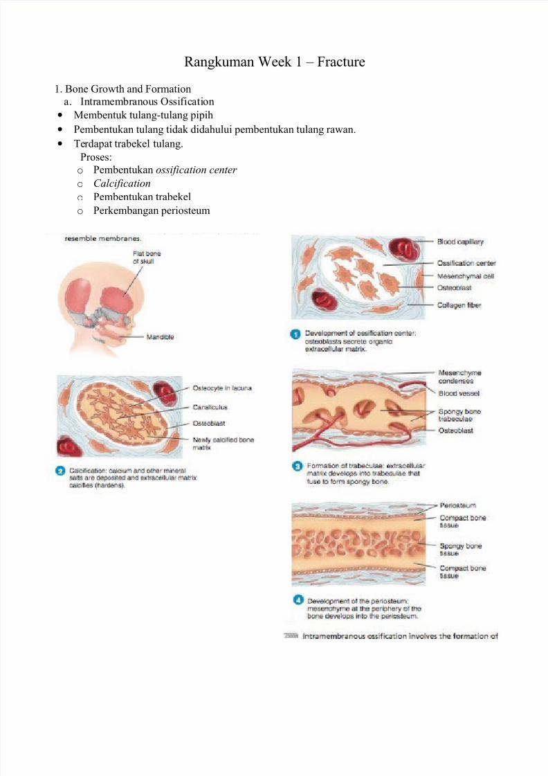
1. Bone Growth and Formation
a. Intramembranous Ossification
• Membentuk tulangtulang !i!ih
• "embentukan tulang tidak didahului !embentukan tulang rawan.
• #erda!at trabekel tulang.
"roses$
o "embentukan ossification center
o Calcification
o "embentukan trabekel
o "erkembangan !eriosteum
7/23/2019 Rangkuman Week 1 (PBL) - Fracture
http://slidepdf.com/reader/full/rangkuman-week-1-pbl-fracture 2/25
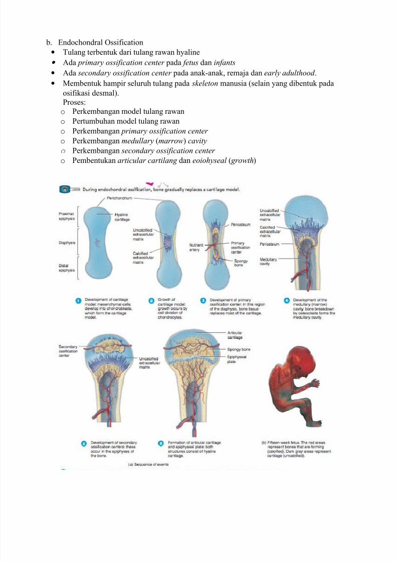
b. %ndochondral Ossification
• #ulang terbentuk dari tulang rawan h&aline
• 'da primary ossification center !ada fetus dan infants
• 'da secondary ossification center !ada anakanak( rema)a dan early adulthood .
• Membentuk ham!ir seluruh tulang !ada skeleton manusia *selain &ang dibentuk !ada
osifikasi desmal+.
"roses$
o "erkembangan model tulang rawan
o "ertumbuhan model tulang rawan
o "erkembangan primary ossification center
o "erkembangan medullary *marrow+ cavity
o "erkembangan secondary ossification center
o "embentukan articular cartilang dan eoiohyseal * growth+
7/23/2019 Rangkuman Week 1 (PBL) - Fracture
http://slidepdf.com/reader/full/rangkuman-week-1-pbl-fracture 3/25
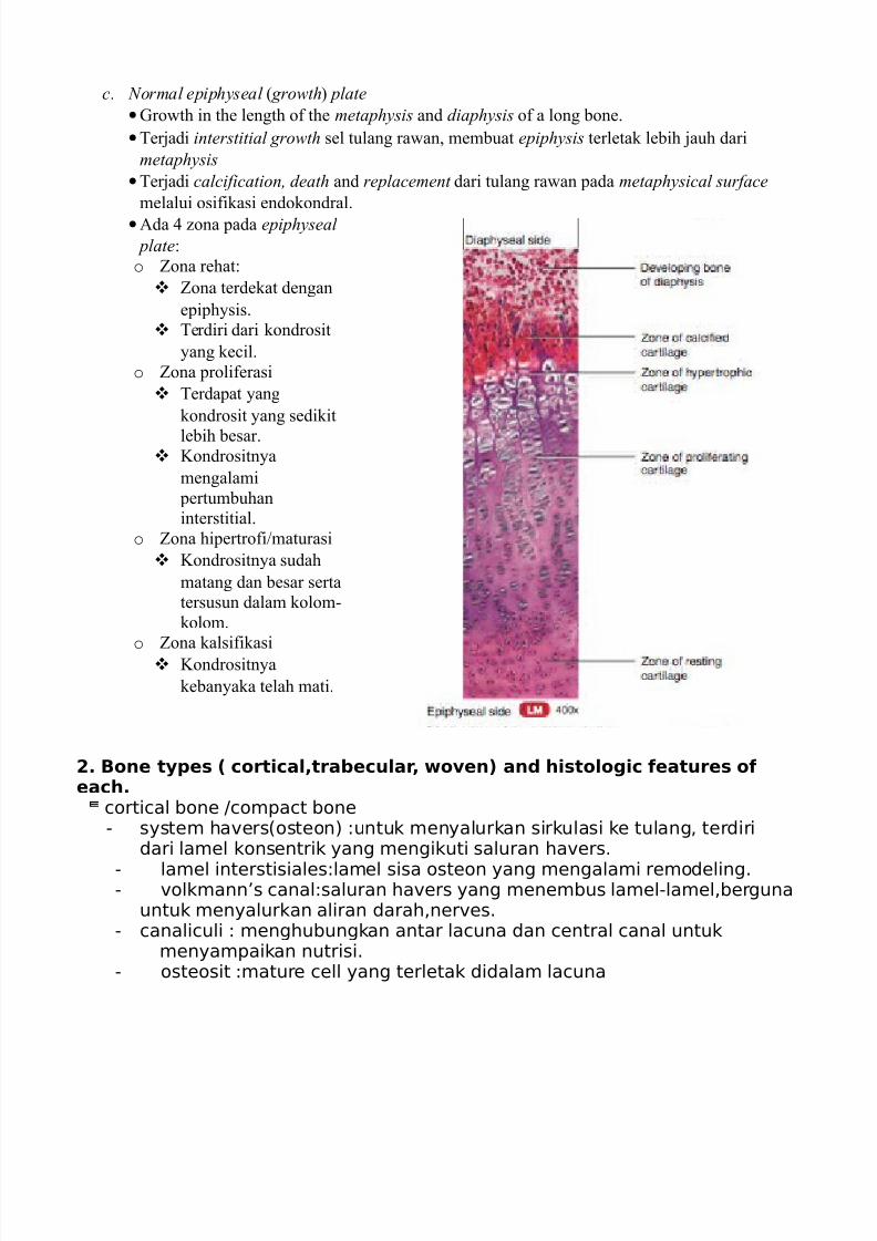
c. Normal epiphyseal * growth+ plate
•Growth in the length of the metaphysis and diaphysis of a long bone.
•#er)adi interstitial growth sel tulang rawan( membuat epiphysis terletak lebih )auh dari
metaphysis
•#er)adi calcification, death and replacement dari tulang rawan !ada metaphysical surface
melalui osifikasi endokondral.
•'da , -ona !ada epiphyseal
plate$
o ona rehat$
ona terdekat dengan
e!i!h&sis.
#erdiri dari kondrosit
&ang kecil.
o ona !roliferasi
#erda!at &ang
kondrosit &ang sedikit
lebih besar. /ondrositn&a
mengalami
!ertumbuhan
interstitial.
o ona hi!ertrofi0maturasi
/ondrositn&a sudah
matang dan besar serta
tersusun dalam kolom
kolom.
o ona kalsifikasi
/ondrositn&a
keban&aka telah mati.
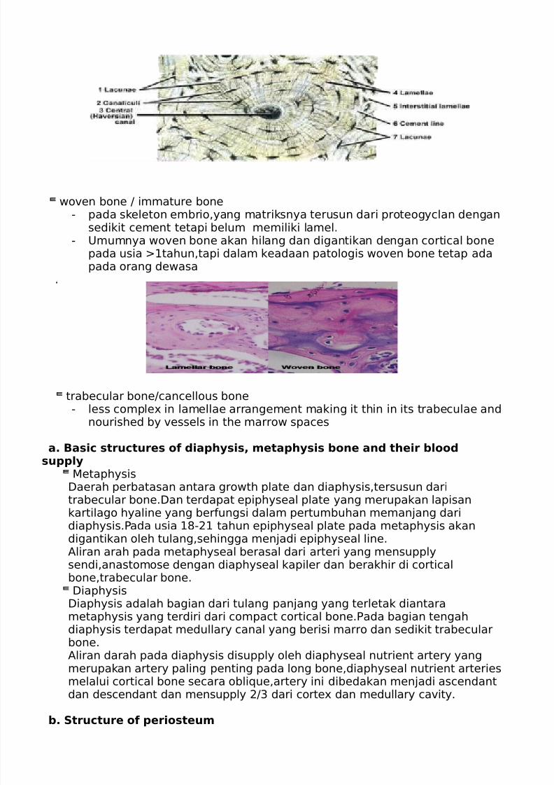
2. Bone types ( cortical,trabecular, woven) and histologic features ofeach.cortical bone /compact bone
- system havers(osteon) :untuk menyalurkan sirkulasi ke tulang, terdiridari lamel konsentrik yang mengikuti saluran havers.
- lamel interstisiales:lamel sisa osteon yang mengalami remodeling. - volkmann’s canal:saluran havers yang menembus lamel-lamel,berguna
untuk menyalurkan aliran darah,nerves. - canaliculi : menghubungkan antar lacuna dan central canal untuk
menyampaikan nutrisi. - osteosit :mature cell yang terletak didalam lacuna
7/23/2019 Rangkuman Week 1 (PBL) - Fracture
http://slidepdf.com/reader/full/rangkuman-week-1-pbl-fracture 4/25
woven bone / immature bone- pada skeleton embrio,yang matriksnya terusun dari proteogyclan dengan
sedikit cement tetapi belum memiliki lamel.- Umumnya woven bone akan hilang dan digantikan dengan cortical bone
pada usia !tahun,tapi dalam keadaan patologis woven bone tetap adapada orang dewasa
.
trabecular bone/cancellous bone
- less comple" in lamellae arrangement making it thin in its trabeculae andnourished by vessels in the marrow spaces
a. Basic structures of diaphysis, metaphysis bone and their bloodsupply#etaphysis$aerah perbatasan antara growth plate dan diaphysis,tersusun daritrabecular bone.$an terdapat epiphyseal plate yang merupakan lapisankartilago hyaline yang ber%ungsi dalam pertumbuhan meman&ang daridiaphysis.'ada usia !-! tahun epiphyseal plate pada metaphysis akandigantikan oleh tulang,sehingga men&adi epiphyseal line.
*liran arah pada metaphyseal berasal dari arteri yang mensupplysendi,anastomose dengan diaphyseal kapiler dan berakhir di corticalbone,trabecular bone.$iaphysis$iaphysis adalah bagian dari tulang pan&ang yang terletak diantarametaphysis yang terdiri dari compact cortical bone.'ada bagian tengahdiaphysis terdapat medullary canal yang berisi marro dan sedikit trabecularbone.*liran darah pada diaphysis disupply oleh diaphyseal nutrient artery yangmerupakan artery paling penting pada long bone,diaphyseal nutrient arteriesmelalui cortical bone secara obli+ue,artery ini dibedakan men&adi ascendantdan descendant dan mensupply / dari corte" dan medullary cavity.
b. Structure of periosteum
7/23/2019 Rangkuman Week 1 (PBL) - Fracture
http://slidepdf.com/reader/full/rangkuman-week-1-pbl-fracture 5/25
#erupakan åan double layer yang melapisi seluruh permukaan tulang
yang tidak dilapisi oleh articular cartilage.
'ada lapisan luar periosteum berhubungan langsung dengan pembuluh
darah
dan terdiri dari åan brosa,pada lapisan dalam terdiri dari lapisan
osteogenic dimana terdapat sel osteoprogenitor.'eriosteum berhubungan dengan tulang melalui serat kolagen yang kuat,
harpey’s bres.
ungsi periosteum:
-ebagai tempat menempelnya tendon dan ligament
-berperan penting dalam proses bone healing.
-memberi nutrisi pada sel-sel tulang melalui pembuluh darah yang terdapat
di periosteum
c. Major bone cells and their function0steoblast - #erupakan di%erensiasi dari sel mesenkim,ber%ungsi dalam proses osteogenisis atau osikasi. - 0steoblast mensintesis kolagen dan komponen organic lainnya yang
digunakan untuk membuat matri" e"tracellular agar dapat ter&adiproses
kalsikasi,&ika sudah tertutupi oleh matri" e"tracellular maka osteoblast berubah men&adi osteosit - el osteoblast yang belum mengalami kalsikasi disebut osteoid(prebone)
pada tahap ini tulang masih lunak. - 1idak mengalami mitosis dan tidak dikelilingi matri".0steosit - #ature bone cells dikelilingi matri" yang berasal dari osteoblast. - 2er%ungsi untuk mengatur metabolism tulang dalam pertukaran nutrisidan waste dengan aliran darah - 1idak mengalami mitosis.0steoclast
- 2er%ungsi sebagai bone resoption karna memiliki en"im lisosom dan acidyang berguna untuk melisis komponen protein dan mineral dalam
matri"e"tracellular tulang.
- 'eran penting dalam proses pertumbuhan,perkembangan dan bonerepair. - #engatur kadar kaslium dalam darah
- ecara mikrokopik berada diluar trabekel tulang dengan ukuran sel yang besar dan memiliki banyak inti.
0steoprogenitor bone stem cell yang berasal dari mesenkim dan mengalami
perkembanganmen&adi osteoblast. -terletak pada periosteum tulang.
7/23/2019 Rangkuman Week 1 (PBL) - Fracture
http://slidepdf.com/reader/full/rangkuman-week-1-pbl-fracture 6/25
d. !omponent of bone matri"2one matri" consist o%: - organic component - anorganic component-0rganic component(34)5
- osteoprogenitor cell - osteosit
- osteoblast - osteoclast - osteoid(sel osteoblast yang belum mengalami
kalsikasi)-6norganic component(734)5 - 8ydro"yapatite (mineral salts)
calcium phosphate 9a('0;) .(08) <(men&adikan tulang kuat)
-calcium carbonate, =uoride, magnesium.
e. !ommon type of collagenollagen ada &enis:collagen ! dan collagen .collagen type 6
- ma&or collagen pada kulit,tulang,brokartilago,meniskus- ber%ungsi untuk menguatkan tulang- ada &enis: 0>!*!
0>!*-collagen type ! terdiri dari rantai procollagen !?!men&adi 0>!*!
!rantai procollagen !?men&adi 0>!* &ika ter&adi mutasi pada kolagen type ! maka dapat menyebabkanosteogenesis imper%ect.collagen type
- banyak terdapat di kartilago,articular cartilage,vitreous(eyeball)- dalam ¨ah kecil ditemukan pada åan skeleton pada awal masapertumbuhan.
- 2er%ungsi untuk menguatkan connective tissue pada otot,sendi. @ika ter&adi mutasi makan dapat menyebabkan chondrodisplasia(dwarsm)
f. #escribe the minerali$ation of bone 1he mineraliAation o% so%t callus begins about ! week later, a%ter the
%ormation o% new so%t callus. 1he increased o"ygen tension leads to the production o%osteoid(visible on
radiographs) 1he presences o% osteoid provides rigidity within the callus id
dependent on the relative stability o% the%racture site, the larger callus is prevent this motion.
been
gained across the %racture site,thepatient may resume limited activity.
callus may take anything %rom ; to !7weeks and is a +uicker process in children and spongy bones.
7/23/2019 Rangkuman Week 1 (PBL) - Fracture
http://slidepdf.com/reader/full/rangkuman-week-1-pbl-fracture 7/25
. Bone Remodelling.
a. 2ortical *2om!act+ bone
- 2haracteri-ed b& the concentric arrangement dari lamella dan com!le3 formation dari sistem
ha4ers atau osteon.
b. 2ancellous *trabecular+ bone
- #he arrangement dari lamelan&a lebih sederhana dibandingkan dengan cortical bone karena
trabekeln&a thin dan da!at dinutrisi oleh !embuluh darah disekitarn&a dan marrow spaces.
c. Wolff’s Law
•Bone Reaction
o 'da , basic wa&s of bone reaction to abnormalities$
Local death$ keadaan sebuah area !ada tulang telah completely deprived of its blood
supply.
An alteration of bone deposition$
Increased de!osition$ !eningkatan !embentukan matri3 dengan kalsifikasi normal
5ecreased de!osition$ !engurangan !embentukan matri3 atau hypocalcification
An alteration of bone resorption$ increased reso!rtion dan decreased resor!tion
!echanical failure or fracture
o Reaksireaksi tulang lainn&a terhada! disorders and in"uries$
#steoporosis * !arble $ones+
Acromegaly
#steoporosis *#steopenia+
%ickets in children( #steomalacia in adults.
&egenerative #steoarthritis
'ractures
(nfection
#steosclerotic Neoplasms %heumatoid Arthritis
#steolytic Neoplasms
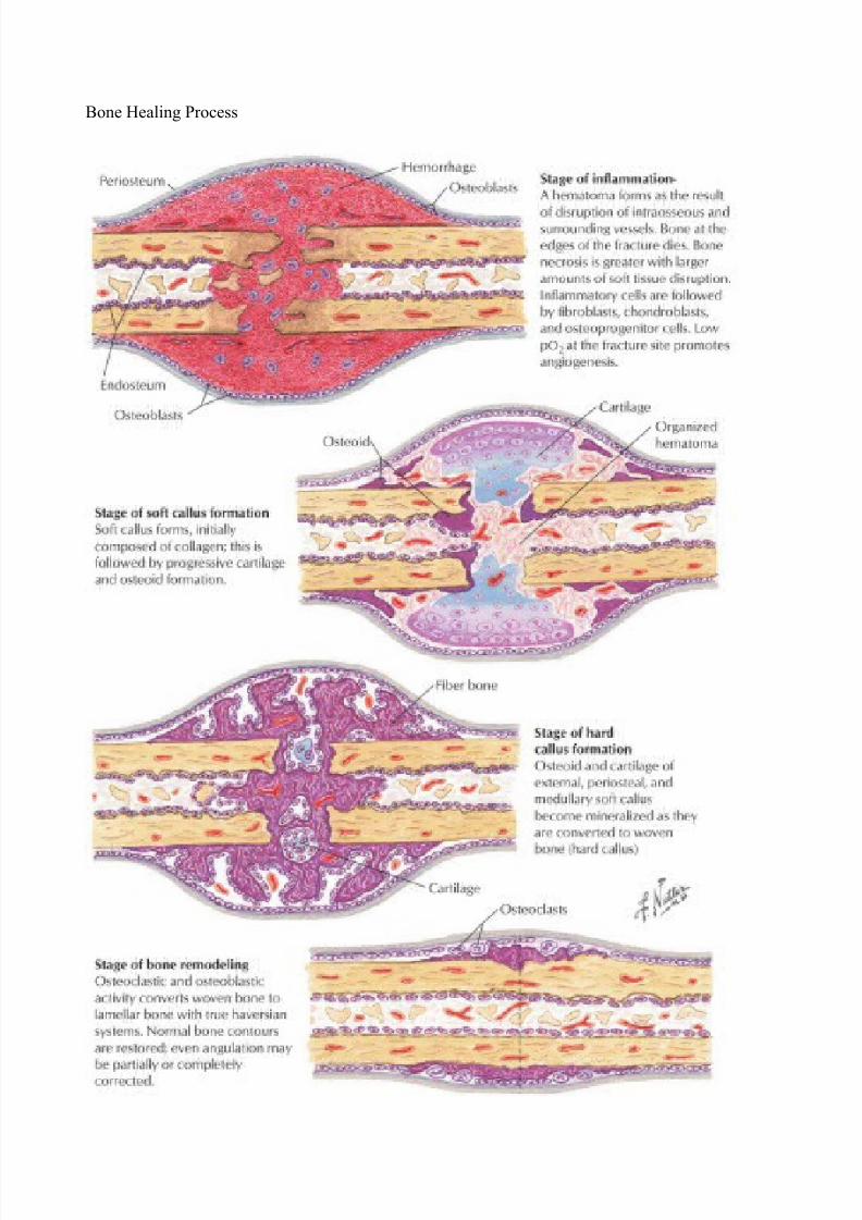
•Bone 6ealing
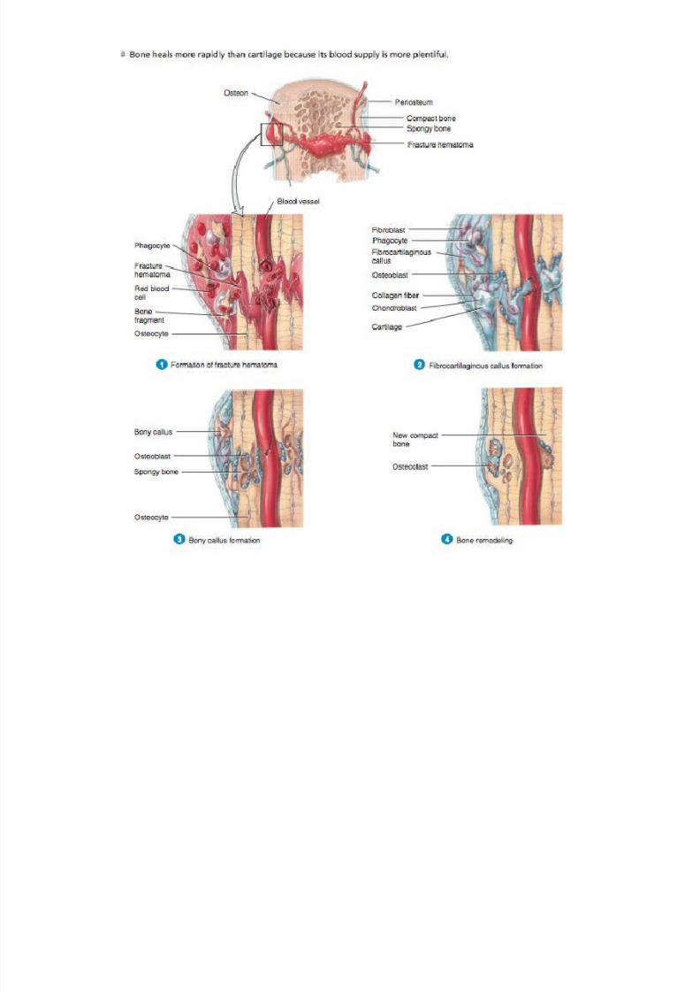
o Formation of fracture hematoma$ Blood 4essels crossing the fracture line are broken. 's
blood leaks from the torn ends of the 4essels( a mass of blood *usuall& clotted+ forms
around the site of the fracture. #his mass of blood( called a fracture hematoma, usuall&
forms 7 to 8 hours after the in)ur&. 9welling and inflammation occur in res!onse to dead
bone cells( !roducing additional cellular debris. "hagoc&tes *neutro!hils and
macro!hages+ and osteoclasts begin to remo4e the dead or damaged tissue in and around
the fracture hematoma.
o 'ibrocartilaginous callus formation: Fibroblasts from the !eriosteum in4ade the fracture
site and !roduce collagen fibers. In addition( cells from the !eriosteum de4elo! into
chondroblasts and begin to !roduce fibrocartilage. #hese e4ents lead to the de4elo!ment
of a fibrocartilaginous )soft* callus, a mass of re!air tissue consisting of collagen fibers
and cartilage that bridges the broken ends of the bone. Formation of the fibrocartilagi
nous callus takes about weeks.
o $ony callus formation$ osteogenic cells de4elo! into osteoblasts( which begin to !roduce
s!ong& bone trabeculae. #he fibrocartilage is con4erted to s!ong& bone( and the callus is
then referred to as a bony )hard* callus. #he bon& callus lasts about to , months.
o $one remodeling $
#he final !hase of fracture re!air is bone remodeling of the callus.5ead !ortions of the original fragments of broken bone are graduall& resorbed b&
osteoclasts. 2om!act bone re!laces s!ong& bone around the !eri!her& of the fracture.
7/23/2019 Rangkuman Week 1 (PBL) - Fracture
http://slidepdf.com/reader/full/rangkuman-week-1-pbl-fracture 8/25
7/23/2019 Rangkuman Week 1 (PBL) - Fracture
http://slidepdf.com/reader/full/rangkuman-week-1-pbl-fracture 9/25
• &iaphyseal +ealing
o 9tage of 6ealing from 9oft #issue
#steogenic cells &ang ber!roliferasi dari la!isan dalam !eriosteum untuk membentuk
eternal callus dan to a lesser etent from endosteum untuk membentuk internal callus.
o 9tage of 2linical :nion
#em!orar& eternal and internal callus mengelilingi fracture site dan membentuk
;biological glue< &ang mem!erkeras saat cartilaginous components dari callus digantikan dengan tulang melalui osifikasi endokondral.
o 9tage of Radiogra!hic :nion
9eiring waktu ber)alan( the temporary callus digantikan oleh mature lamellar bone. "ada
saat tulang &ang belum matang telah digantikan oleh mature lamellar bone dan tulang
sudah kembali ke diameter &ang ham!ir normal( fraktur itu sudah bisa dikatakan
radiographic union.
• !etaphyseal +ealing
5iawali dengan !embentukan internal atau endosteal callus( walau!un eternal atau
periosteal callus &ang mengelilingi thin shell of corte )uga memegang !eran !enting.
/arena ban&akn&a su!lai darah !ada thin trabeculae dari cancellous bone sehingga ter)adisedikit necrosis tulang. #he #steogenic repair cells ber!roliferasi membentuk primary
woven bonedi internal fracture hematoma. 9aat telah ter)adi :nion( fraktur sudah clinically
united . =alu( woven bone digantikan oleh lamellar bone saat fraktur telah radiographic
united .
• Growth "late Reaction
o 'da basic wa&s$
Increased Growth$
Generali-ed$
Arachnodactyly * +yperchondroplasia+ * !arfan’s -yndrome+
ituitary /igantism
=ocali-ed$
Chronic (nflammation
&isplaced fracture of the shaft long bone
Congenital Arteriovenous !alformations
5ecreased Growth
Generali-ed$
Achndroplasia
ituitary &warfism * Lorain type+
%ickets
=ocali-ed$
&isuse retardation
0hermal (n"ury
(schemia
(nfection
#orsional Growth
=ocali-ed$ when a growing long bone and its e!i!h&seal !late are sub)ected to
either continual or intermittent twisting forces. %3$ /nock /nees
•Bone 5eformit&
o =oss of 'lignment$
#er)adi !ada long bone. Bisa karena twisted in its long ais *torsional deformity+ atau
karena crooked *angulatory deformity+.
o 'bnormal =ength
7/23/2019 Rangkuman Week 1 (PBL) - Fracture
http://slidepdf.com/reader/full/rangkuman-week-1-pbl-fracture 10/25
Bisa abnormally !endek atau meman)ang. /alau han&a ter)adi !ada salah stu kaki atau
salah satu tangan disebut limb length discrepancy.
o Bon& Outgrowth$ ' lesion( se!erti #steochondroma.
•Bone >eo!lasm
o >eo!lasmlike =esions of Bone
Osteogenic
Osteoma *i4or& e3ostosis+
9ingle Ostechondroma *osteocartilaginous e3ostosis+
Multi!le Osteochondroma *multi!le hereditar& Osteoma+
Osteoid Osteoma
Benign Osteoblastoma
2hondrogenic
%chondroma
Multi!le %chondromata *Ollier?s 5&schondro!lasia+
Fibrogenic
9ub!eriosteal cortical defect *meta!h&seal fibrous defect+
>onosteogenic fibroma *nonossif&ing fibroma+
Monostotic fibrous d&s!lasia
"ol&ostotic fibrous d&s!lasia
Osteofibrous d&s!lasia *2am!anacci s&ndrome+
;Brown tumor< *h&!er!arath&roidism+
'ngiogenic
'ngioma of bone *hemangioma and l&m!hangioma+
'neur&smal bone c&st *'B2+
:ncertain origin
9im!le bone c&st *:nicameral bone c&st *:B2++
o #rue "rimar& >eo!lasm of Bone
Osteogenic
Osteosarcoma *Osteogenic 9arcoma+
9urface osteosarcoma *!eriosteal sarcoma+
2hondrogenic
Benign chondroblastoma
2hondrom&3oid fibroma
2hondrosarcoma
Fibrogenic
Fibrosarcoma of Bone
Malignant fibrous histioc&toma of bone
'ngiogenic
'ngiosarcoma of bone
M&elogenic
M&eloma of bone *Multi!le m&eloma+
%wing?s sarcoma *%wing?s tumor+
6odgkin?s l&m!homa of bone
>on6odgkin?s l&m!homa *reticulum cell sarcoma+
9keletal reticuloses *=angerhan?s cell histioc&toses+
=eukemia
:ncertain origin
Giant cell tumor of bone *Osteoclastoma+

7/23/2019 Rangkuman Week 1 (PBL) - Fracture
http://slidepdf.com/reader/full/rangkuman-week-1-pbl-fracture 11/25
7/23/2019 Rangkuman Week 1 (PBL) - Fracture
http://slidepdf.com/reader/full/rangkuman-week-1-pbl-fracture 12/25
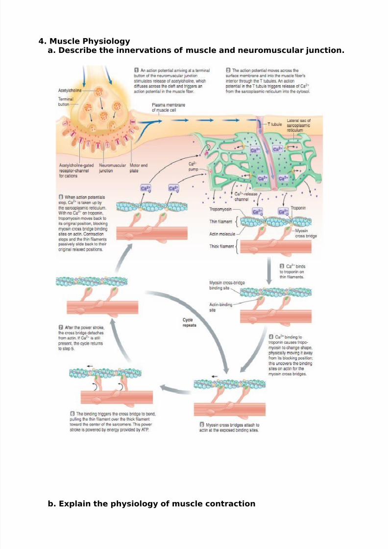
%. Muscle &hysiologya. #escribe the innervations of muscle and neuromuscular junction.
b. '"plain the physiology of muscle contraction
7/23/2019 Rangkuman Week 1 (PBL) - Fracture
http://slidepdf.com/reader/full/rangkuman-week-1-pbl-fracture 13/25
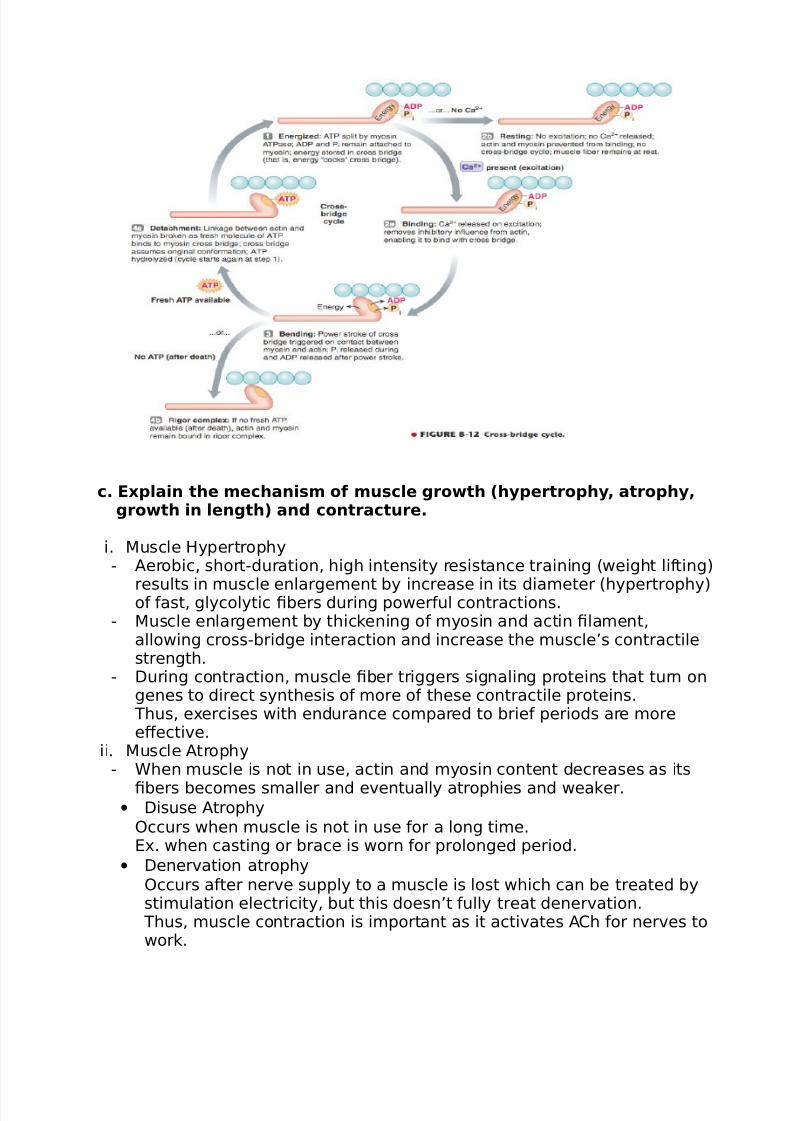
c. '"plain the mechanism of muscle growth (hypertrophy, atrophy,growth in length) and contracture.
i. #uscle 8ypertrophy- *erobic, short-duration, high intensity resistance training (weight li%ting)
results in muscle enlargement by increase in its diameter (hypertrophy)o% %ast, glycolytic bers during power%ul contractions.- #uscle enlargement by thickening o% myosin and actin lament,
allowing cross-bridge interaction and increase the muscle’s contractilestrength.
- $uring contraction, muscle ber triggers signaling proteins that turn ongenes to direct synthesis o% more o% these contractile proteins. 1hus, e"ercises with endurance compared to brie% periods are moreeBective.
ii. #uscle *trophy- Chen muscle is not in use, actin and myosin content decreases as its
bers becomes smaller and eventually atrophies and weaker.• $isuse *trophy
0ccurs when muscle is not in use %or a long time.D". when casting or brace is worn %or prolonged period.
• $enervation atrophy0ccurs a%ter nerve supply to a muscle is lost which can be treated bystimulation electricity, but this doesn’t %ully treat denervation. 1hus, muscle contraction is important as it activates *h %or nerves towork.
7/23/2019 Rangkuman Week 1 (PBL) - Fracture
http://slidepdf.com/reader/full/rangkuman-week-1-pbl-fracture 14/25
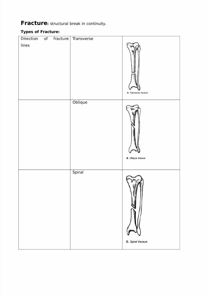
racture structural break in continuity.
*ypes of racture
$irection o% %racture
lines
1ransverse
0bli+ue
piral
7/23/2019 Rangkuman Week 1 (PBL) - Fracture
http://slidepdf.com/reader/full/rangkuman-week-1-pbl-fracture 15/25
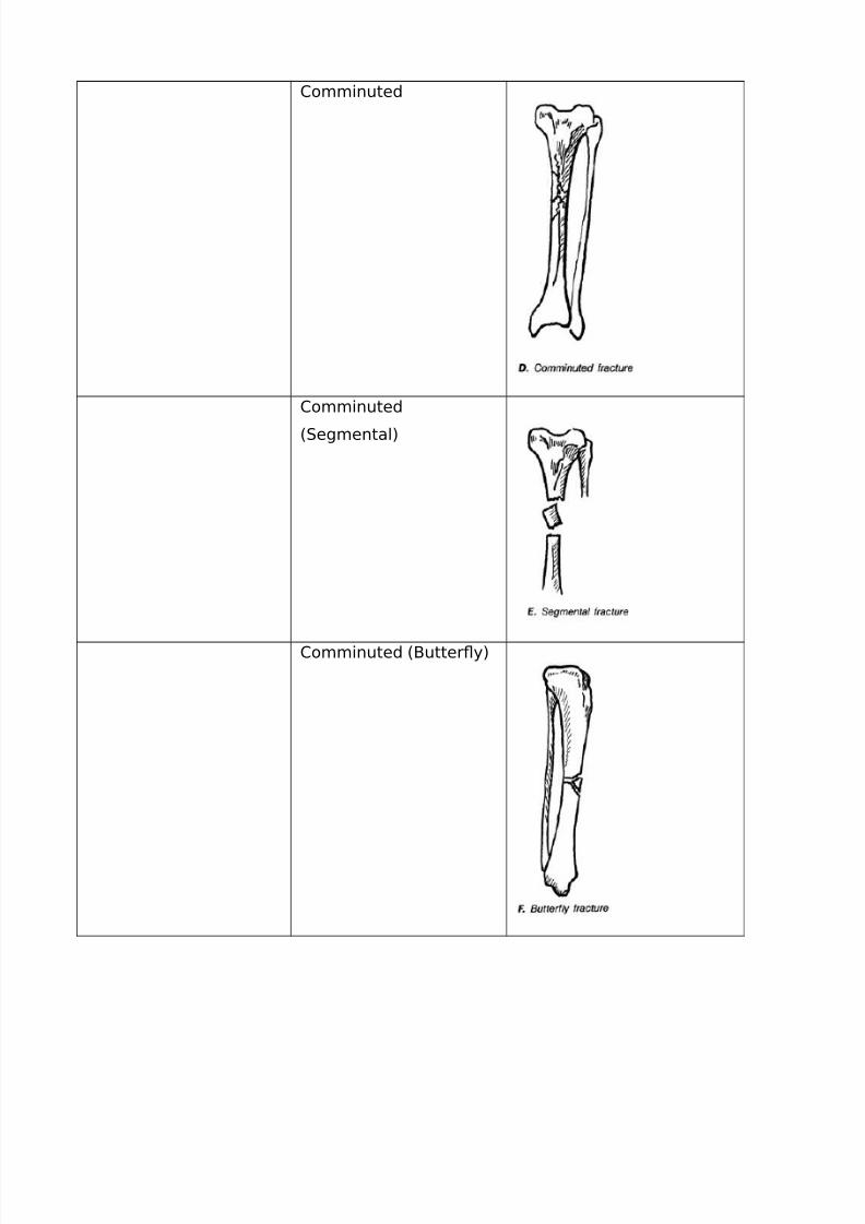
omminuted
omminuted
(egmental)
omminuted (2utter=y)
7/23/2019 Rangkuman Week 1 (PBL) - Fracture
http://slidepdf.com/reader/full/rangkuman-week-1-pbl-fracture 16/25
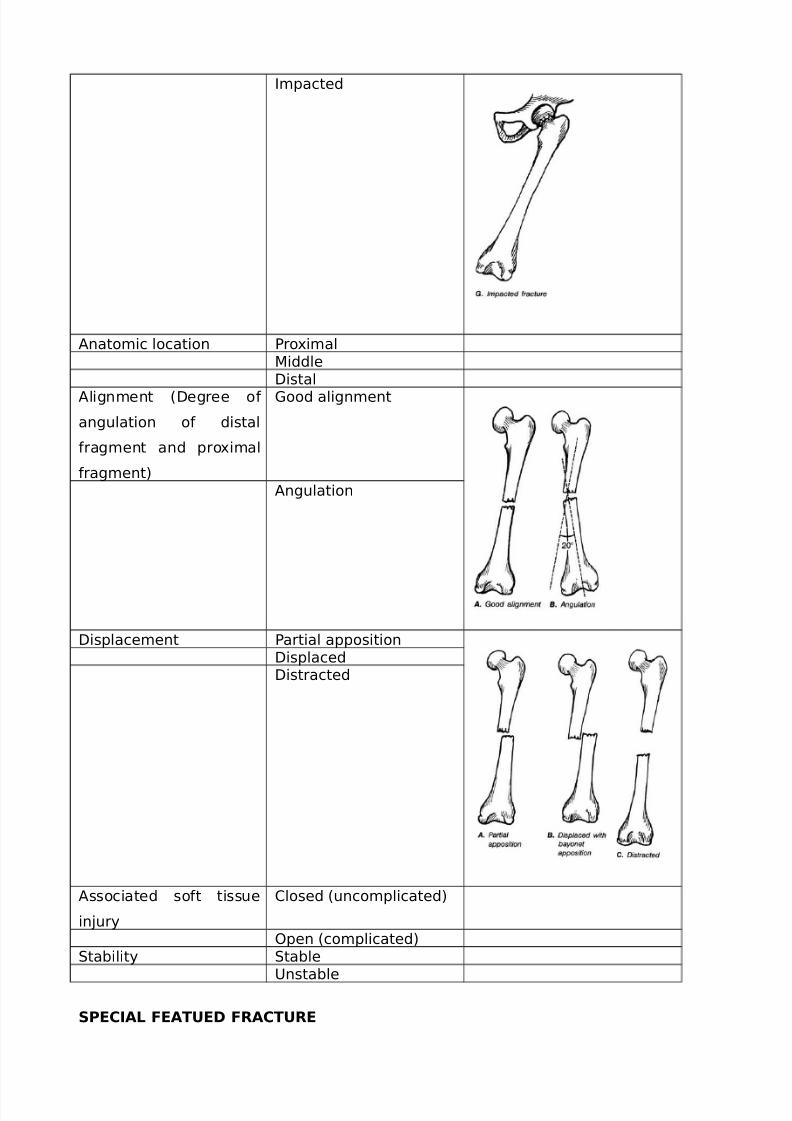
6mpacted
*natomic location 'ro"imal#iddle$istal
*lignment ($egree o%
angulation o% distal
%ragment and pro"imal
%ragment)
Eood alignment
*ngulation
$isplacement 'artial apposition$isplaced$istracted
*ssociated so%t tissue
in&ury
losed (uncomplicated)
0pen (complicated)tability table
Unstable
S&'!+- '*'# /!*/'
7/23/2019 Rangkuman Week 1 (PBL) - Fracture
http://slidepdf.com/reader/full/rangkuman-week-1-pbl-fracture 17/25
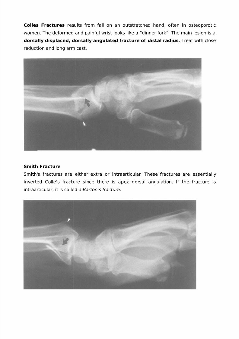
!olles ractures results %rom %all on an outstretched hand, o%ten in osteoporotic
women. 1he de%ormed and pain%ul wrist looks like a Fdinner %orkG. 1he main lesion is a
dorsally displaced, dorsally angulated fracture of distal radius. 1reat with close
reduction and long arm cast.
Smith racture
mithHs %ractures are either e"tra or intraarticular. 1hese %ractures are essentially
inverted olleHs %racture since there is ape" dorsal angulation. 6% the %racture is
intraarticular, it is called a Barton's fracture.
7/23/2019 Rangkuman Week 1 (PBL) - Fracture
http://slidepdf.com/reader/full/rangkuman-week-1-pbl-fracture 18/25
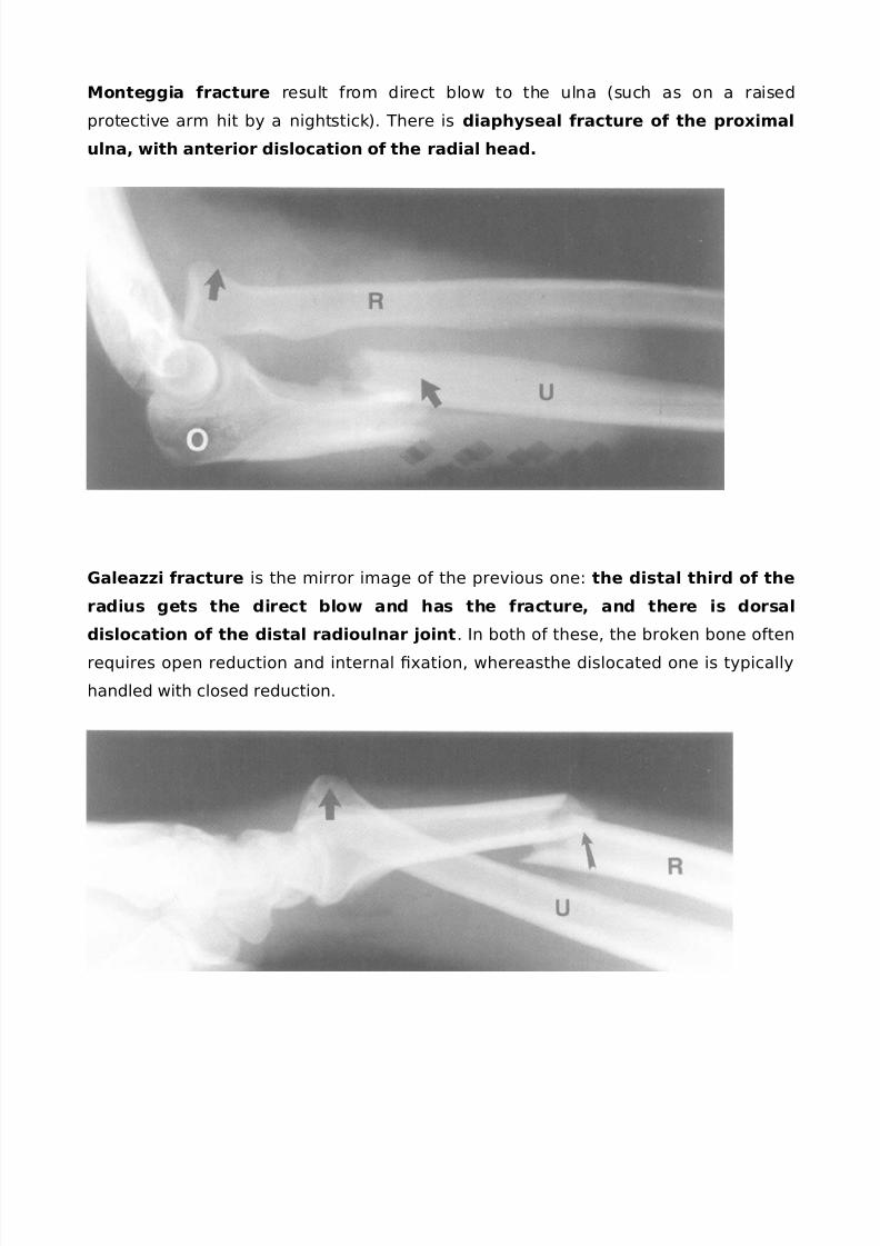
Monteggia fracture result %rom direct blow to the ulna (such as on a raised
protective arm hit by a nightstick). 1here is diaphyseal fracture of the pro"imal
ulna, with anterior dislocation of the radial head.
0alea$$i fracture is the mirror image o% the previous one: the distal third of the
radius gets the direct blow and has the fracture, and there is dorsal
dislocation of the distal radioulnar joint. 6n both o% these, the broken bone o%ten
re+uires open reduction and internal "ation, whereasthe dislocated one is typically
handled with closed reduction.
7/23/2019 Rangkuman Week 1 (PBL) - Fracture
http://slidepdf.com/reader/full/rangkuman-week-1-pbl-fracture 19/25
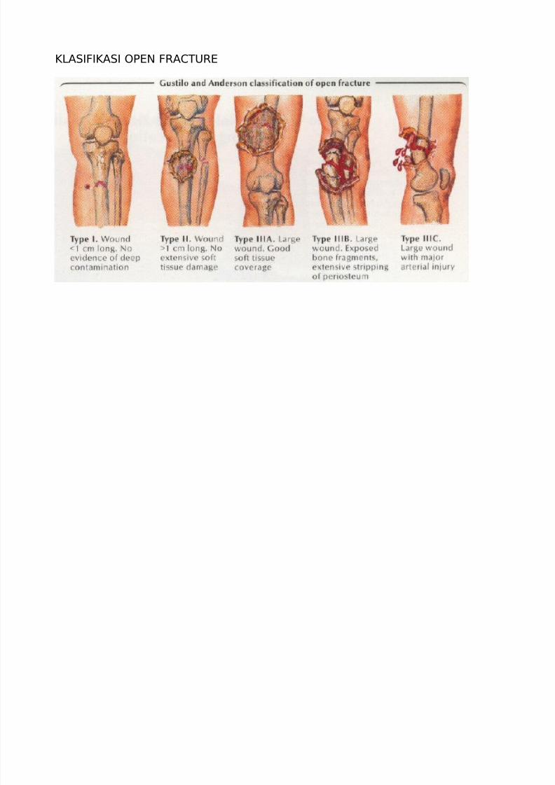
I>*66I*6 0'DJ K*1UKD
7/23/2019 Rangkuman Week 1 (PBL) - Fracture
http://slidepdf.com/reader/full/rangkuman-week-1-pbl-fracture 20/25

7/23/2019 Rangkuman Week 1 (PBL) - Fracture
http://slidepdf.com/reader/full/rangkuman-week-1-pbl-fracture 21/25
7/23/2019 Rangkuman Week 1 (PBL) - Fracture
http://slidepdf.com/reader/full/rangkuman-week-1-pbl-fracture 22/25
Bone 6ealing "rocess
7/23/2019 Rangkuman Week 1 (PBL) - Fracture
http://slidepdf.com/reader/full/rangkuman-week-1-pbl-fracture 23/25
C*I1U 'DJLD#2U8*J
'erkins rules
• ractures o% cancellous (metaphyseal) bone (e.g. those around &oints) will take 7
weeks to unite.
• ractures o% cortical (diaphyseal) bone (e.g. sha%ts o% long bones) will take !
weeks to unite.
• ractures o% the tibia (because o% poor blood supply), will take ; weeks to
unite.
• 1ime to union %or children e+uals the age o% the child in years plus one, e.g.
tibial %racture in a -year-old child will unite in weeks. ommon sense needs to
be applied when applying the rule to %ractures o% cancellous bone in older
children.
$6*EJ06
M-ray (rule o% )
- views
- &oints
- limbs
- in&uries
- occasions
1KD*1#DJ1
!. >0D$ K*1UKD
a. Keduce
i. losed Keduction
ii. 0pen reduction
b. 8old Keduction
i. ontinuous traction
!. kin traction
. keletal traction
. i"ed traction (1homas plint)
;. 2alanced traction
3. ombined traction
ii. ast plintage
iii. unctional 2racing
iv. 6nternal i"ation
!. 6nter%ragmentary screw
. Cires (trans"ing, cerclage and tension band)
7/23/2019 Rangkuman Week 1 (PBL) - Fracture
http://slidepdf.com/reader/full/rangkuman-week-1-pbl-fracture 24/25
. 'lates dan screw
v. D"ternal i"ation
. 0'DJ K*1UKD
a. $ebridement
b. Cound closure
c. tabiliAation o% %racture
d. *%tercare
e. e+uels to open %racture
0#'>6*160J 0 K*1UKD
!. D*K>L 0#'>6*160J
a. Nisceral in&uryb. Nascular in&ury
c. Jerve in&ury
d. ompartment syndrome
e. 8emarthrosis
%. 6n%ection
g. Eas gangrene
h. racture blister
i. 'laster sores and pressure sores
. >*1D 0#'>6*160J
a. $elayed Union: %ailure o% union to occur in !.3 times the normal time %or
%racture union.
b. Jon-Union: %ailure o% union to occur within times the normal time to
%racture union. 8owever, e"pect open %ractures to normally take times
the normal 'erkins rule. Jon-union can be broadly divided into:
i. hypertrophic: normally due to e"cess mobility, i.e. good healing
potentialO
ii. atrophic: normally due to poor blood supply, i.e. poor healing
potential.
c. #al-Union
d. *vascular Jecrosis
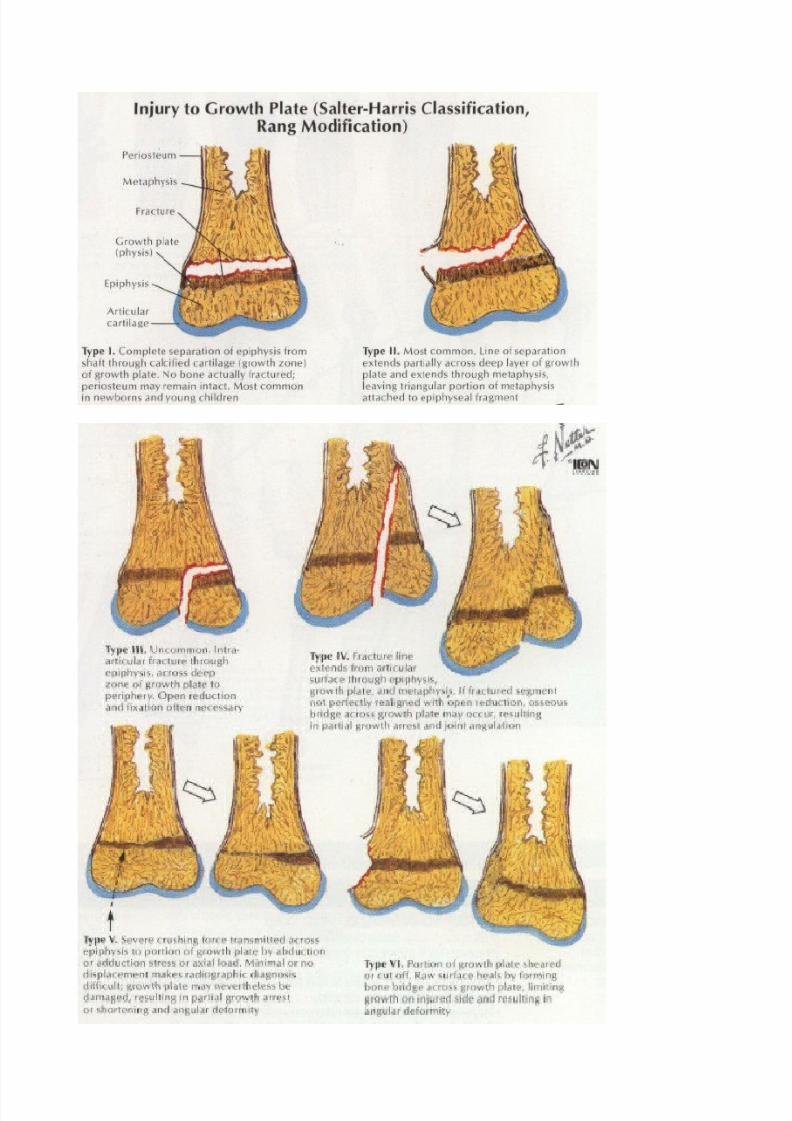
e. Erowth $isturbances epiphyseal %racture in children
%. 2ed ores elderly/paralyAed patients
g. #yositis ossicans heterotopic ossication in the muscle
h. 1endon lesion
i. Jerve compression
7/23/2019 Rangkuman Week 1 (PBL) - Fracture
http://slidepdf.com/reader/full/rangkuman-week-1-pbl-fracture 25/25
&. #uscle contracture
k. @oint instability
l. @oint stiBness
m. *lgodysthropy (omple" regional pain syndrome)
0steoarthrytis
Recommended