Embed Size (px)
Citation preview

1 Localized Osteoporosis

CLINICAL IMAGAGINGAN ATLAS OF DIFFERENTIAL DAIGNOSIS
EISENBERG
DR. Muhammad Bin Zulfiqar PGR-FCPS III SIMS/SHL

CLINICAL IMAGAGINGAN ATLAS OF DIFFERENTIAL DAIGNOSIS
EISENBERG
DR. Muhammad Bin Zulfiqar PGR-FCPS III SIMS/SHL
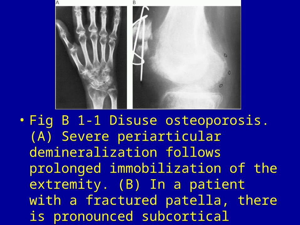
• Fig B 1-1 Disuse osteoporosis. (A) Severe periarticular demineralization follows prolonged immobilization of the extremity. (B) In a patient with a fractured patella, there is pronounced subcortical demineralization in the distal femur. The cortical margin (arrows) remains intact.
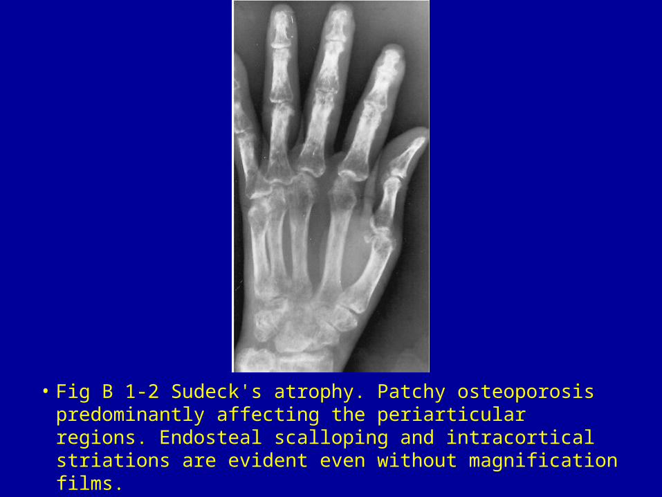
• Fig B 1-2 Sudeck's atrophy. Patchy osteoporosis predominantly affecting the periarticular regions. Endosteal scalloping and intracortical striations are evident even without magnification films.

• Fig B 1-3 Staphylococcal osteomyelitis. (A) Initial film of the first metatarsophalangeal joint shows soft-tissue swelling and periarticular demineralization due to hyperemia. (B) Several weeks later, there is severe bony destruction about the metatarsophalangeal joint.
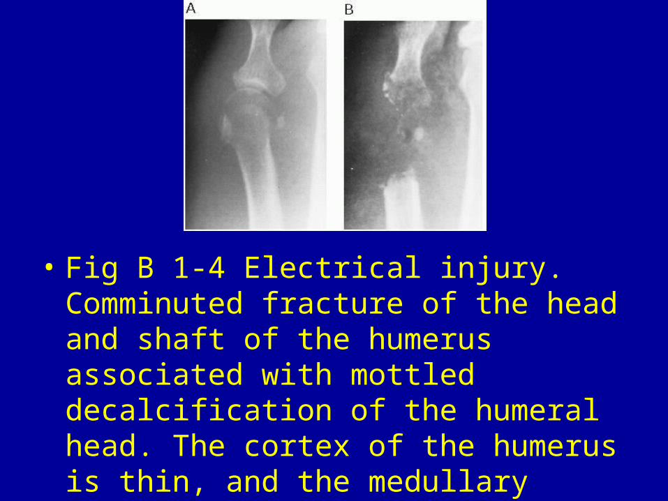
• Fig B 1-4 Electrical injury. Comminuted fracture of the head and shaft of the humerus associated with mottled decalcification of the humeral head. The cortex of the humerus is thin, and the medullary cavity is widened. Discrete areas of rarefaction can be seen in the shaft and distal metaphyseal region.1
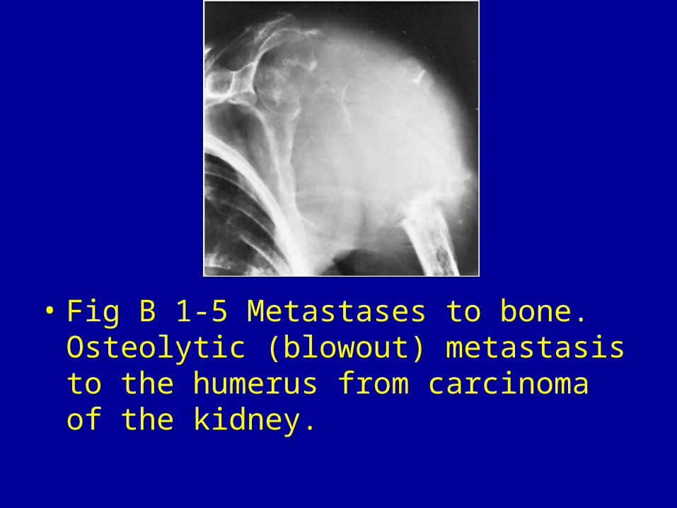
• Fig B 1-5 Metastases to bone. Osteolytic (blowout) metastasis to the humerus from carcinoma of the kidney.
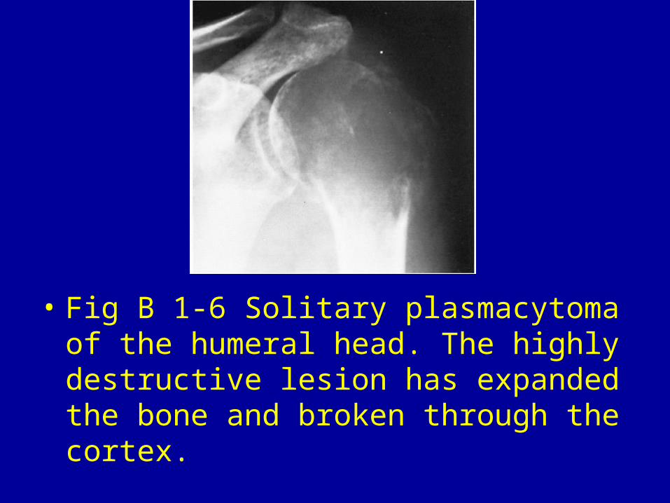
• Fig B 1-6 Solitary plasmacytoma of the humeral head. The highly destructive lesion has expanded the bone and broken through the cortex.
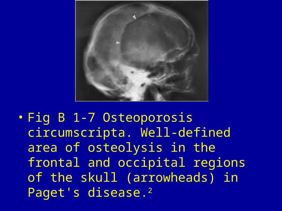
• Fig B 1-7 Osteoporosis circumscripta. Well-defined area of osteolysis in the frontal and occipital regions of the skull (arrowheads) in Paget's disease.2
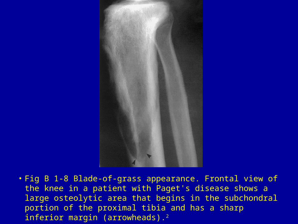
• Fig B 1-8 Blade-of-grass appearance. Frontal view of the knee in a patient with Paget's disease shows a large osteolytic area that begins in the subchondral portion of the proximal tibia and has a sharp inferior margin (arrowheads).2
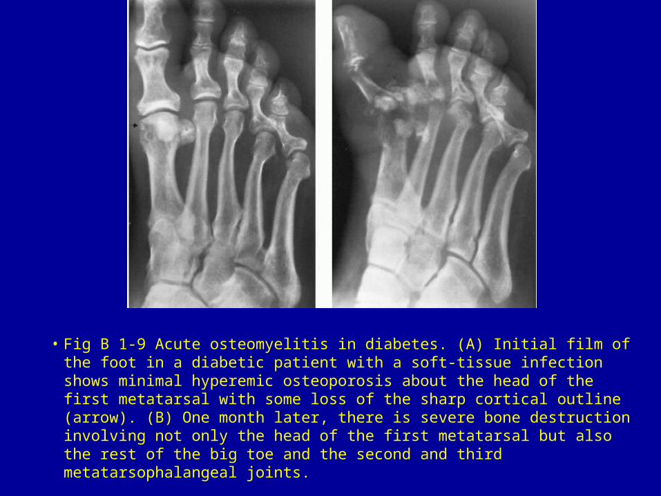
• Fig B 1-9 Acute osteomyelitis in diabetes. (A) Initial film of the foot in a diabetic patient with a soft-tissue infection shows minimal hyperemic osteoporosis about the head of the first metatarsal with some loss of the sharp cortical outline (arrow). (B) One month later, there is severe bone destruction involving not only the head of the first metatarsal but also the rest of the big toe and the second and third metatarsophalangeal joints.
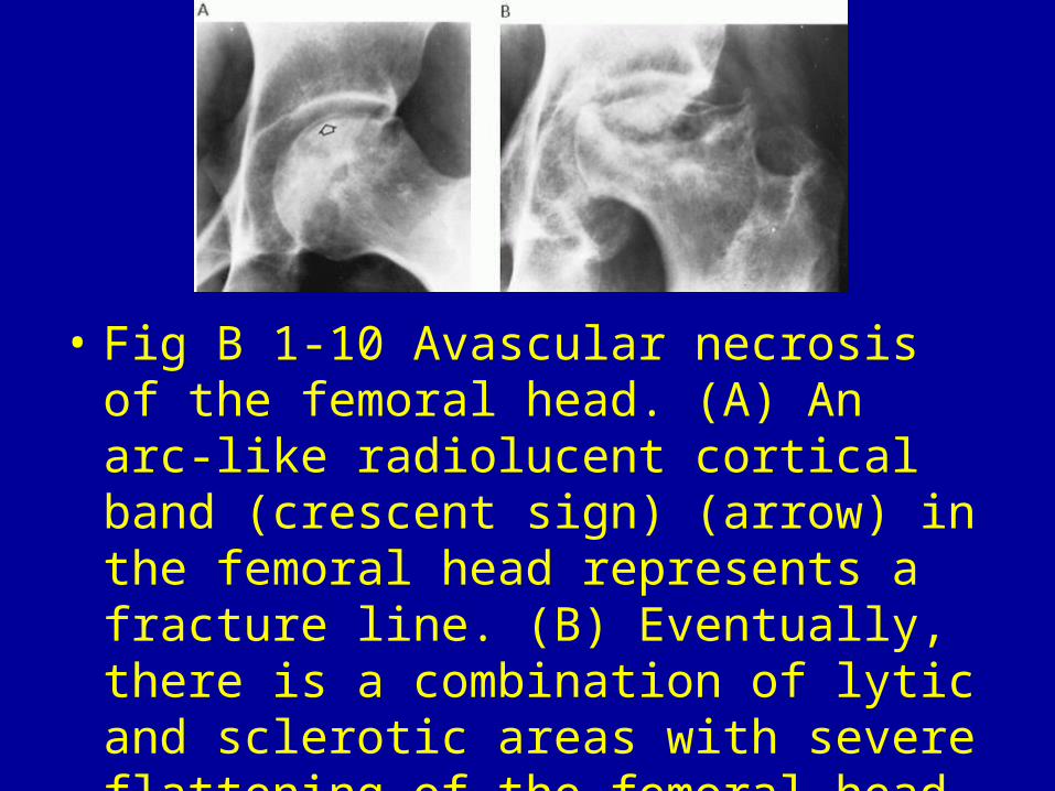
• Fig B 1-10 Avascular necrosis of the femoral head. (A) An arc-like radiolucent cortical band (crescent sign) (arrow) in the femoral head represents a fracture line. (B) Eventually, there is a combination of lytic and sclerotic areas with severe flattening of the femoral head.