Embed Size (px)
Citation preview
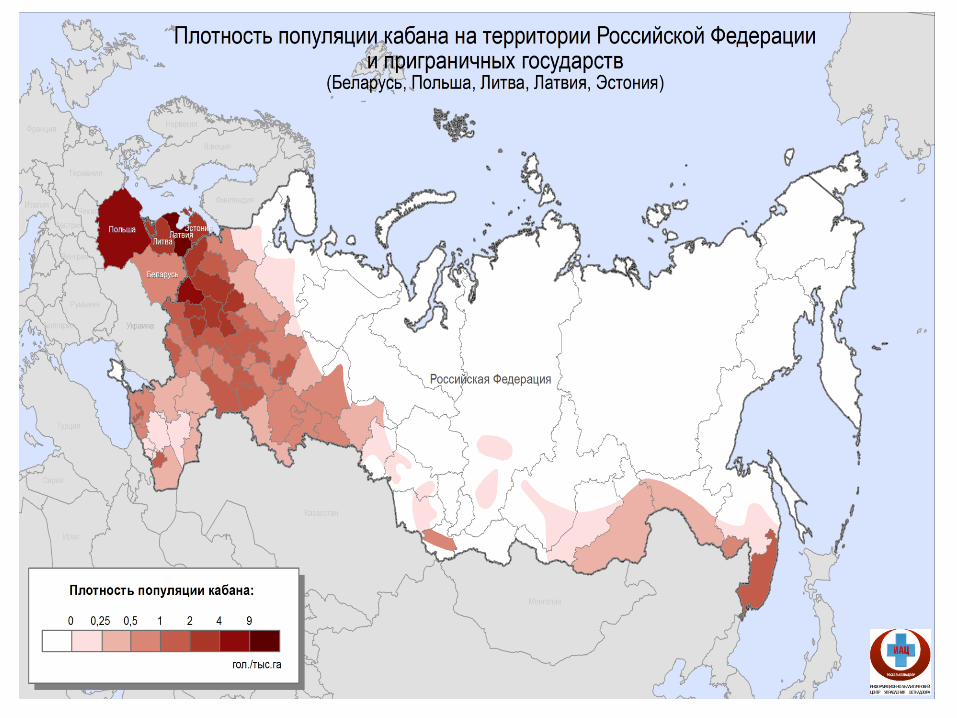
Specific characteristics
of ASF in wild boar
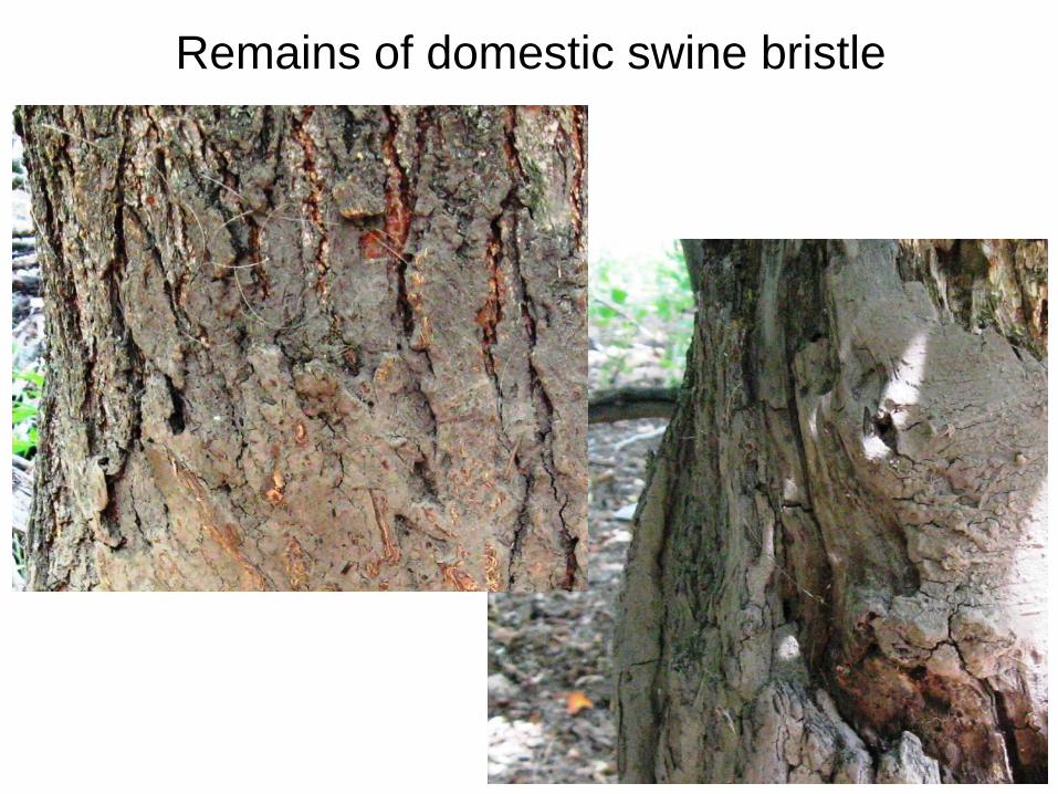
Remains of domestic swine bristle
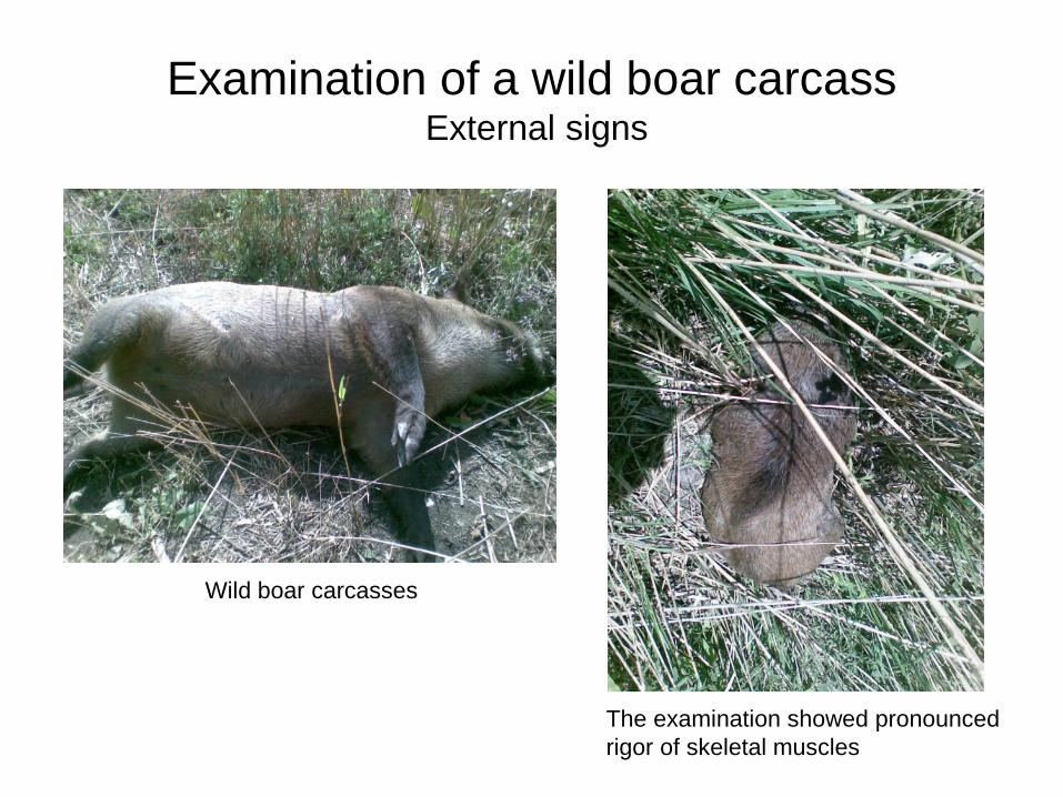
Examination of a wild boar carcass External signs
Wild boar carcasses
The examination showed pronounced
rigor of skeletal muscles
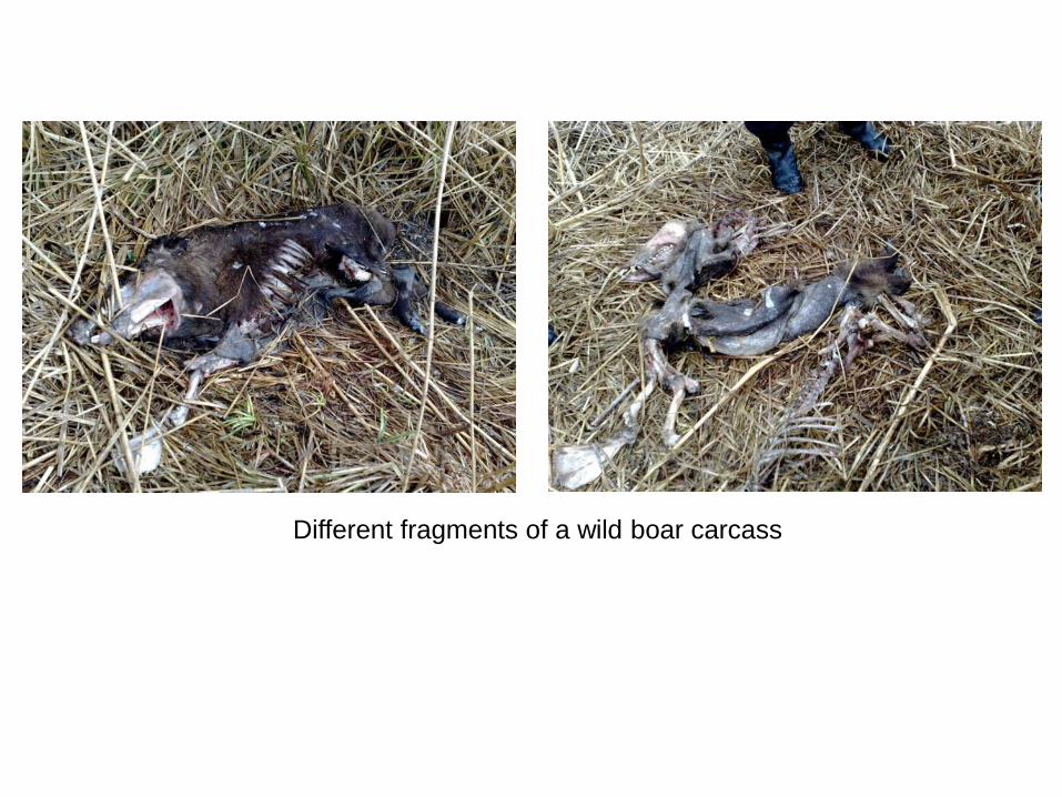
Different fragments of a wild boar carcass

Population welfare control
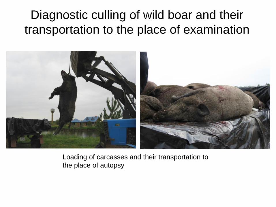
Diagnostic culling of wild boar and their
transportation to the place of examination
Loading of carcasses and their transportation to
the place of autopsy
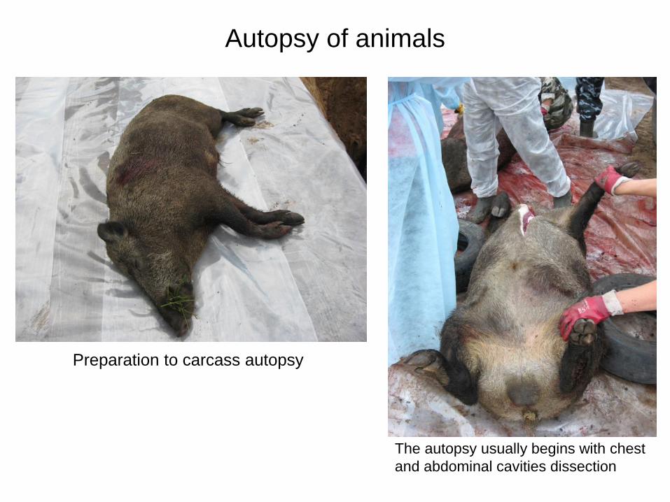
Autopsy of animals
Preparation to carcass autopsy
The autopsy usually begins with chest
and abdominal cavities dissection
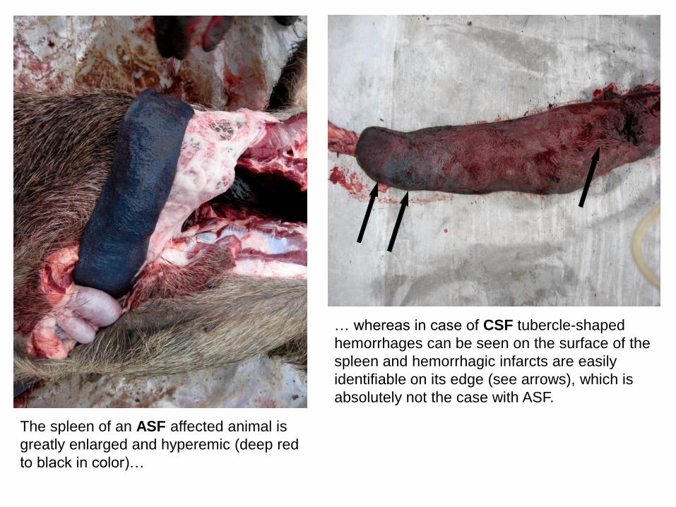
The spleen of an ASF affected animal is
greatly enlarged and hyperemic (deep red
to black in color)…
… whereas in case of CSF tubercle-shaped
hemorrhages can be seen on the surface of the
spleen and hemorrhagic infarcts are easily
identifiable on its edge (see arrows), which is
absolutely not the case with ASF.
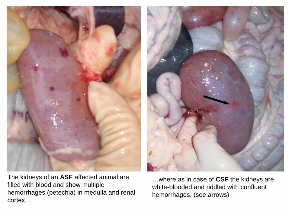
The kidneys of an ASF affected animal are
filled with blood and show multiple
hemorrhages (petechia) in medulla and renal
cortex…
…where as in case of CSF the kidneys are
white-blooded and riddled with confluent
hemorrhages. (see arrows)
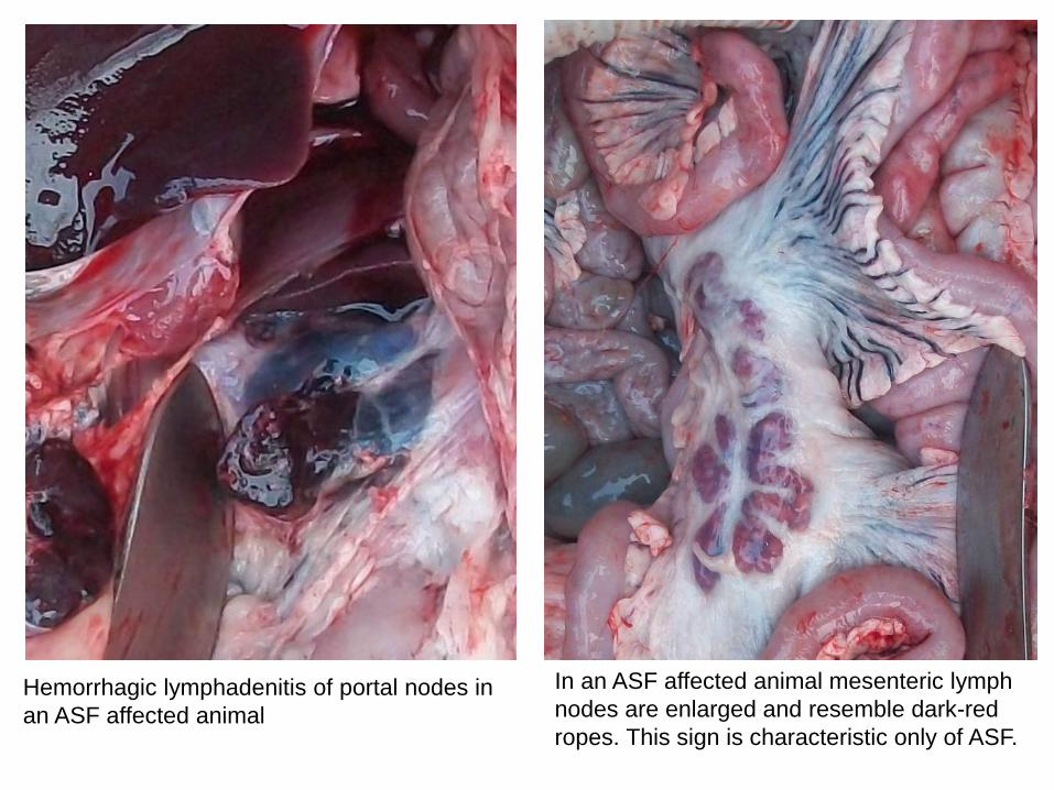
Hemorrhagic lymphadenitis of portal nodes in
an ASF affected animal
In an ASF affected animal mesenteric lymph
nodes are enlarged and resemble dark-red
ropes. This sign is characteristic only of ASF.
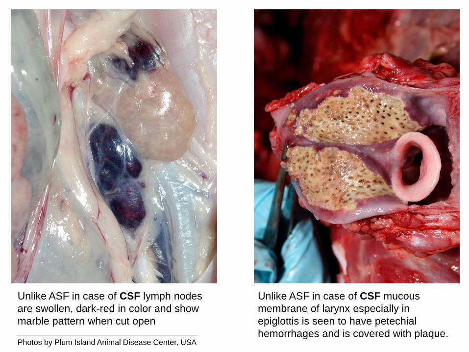
Unlike ASF in case of CSF lymph nodes
are swollen, dark-red in color and show
marble pattern when cut open
Unlike ASF in case of CSF mucous
membrane of larynx especially in
epiglottis is seen to have petechial
hemorrhages and is covered with plaque. Photos by Plum Island Animal Disease Center, USA
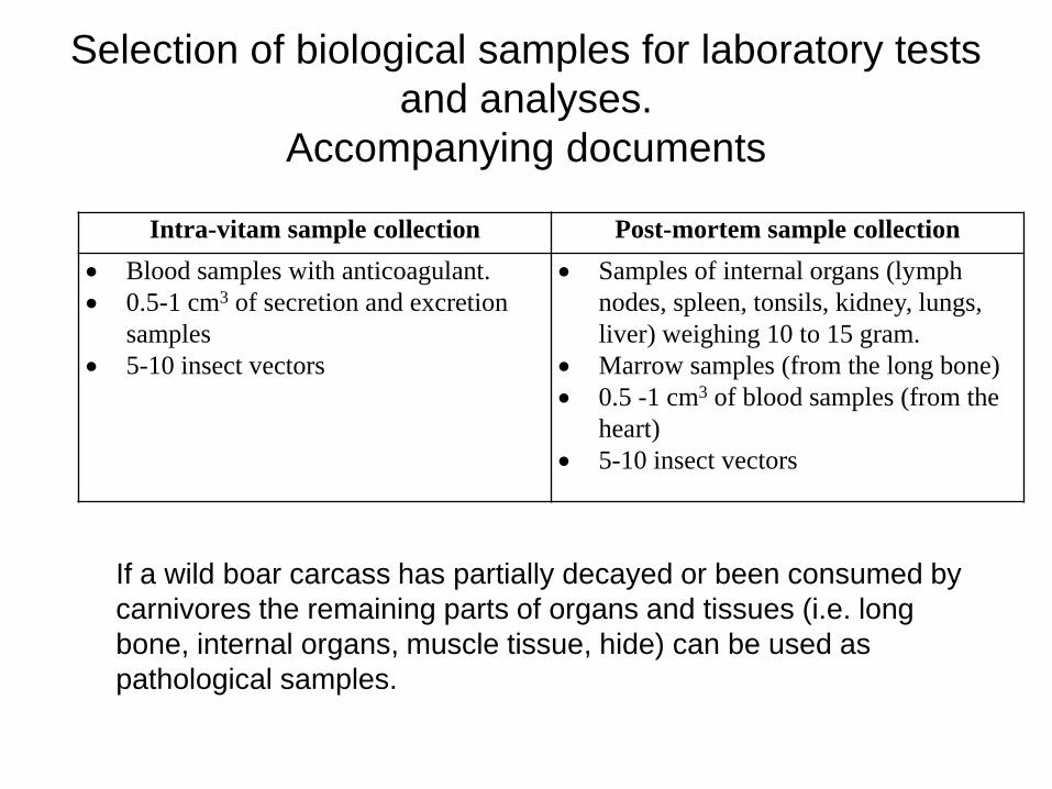
Selection of biological samples for laboratory tests
and analyses.
Accompanying documents
If a wild boar carcass has partially decayed or been consumed by
carnivores the remaining parts of organs and tissues (i.e. long
bone, internal organs, muscle tissue, hide) can be used as
pathological samples.
Intra-vitam sample collection Post-mortem sample collection
Blood samples with anticoagulant.
0.5-1 cm3 of secretion and excretion
samples
5-10 insect vectors
Samples of internal organs (lymph
nodes, spleen, tonsils, kidney, lungs,
liver) weighing 10 to 15 gram.
Marrow samples (from the long bone)
0.5 -1 cm3 of blood samples (from the
heart)
5-10 insect vectors
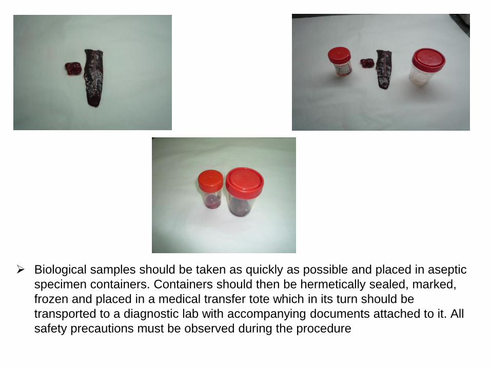
Biological samples should be taken as quickly as possible and placed in aseptic
specimen containers. Containers should then be hermetically sealed, marked,
frozen and placed in a medical transfer tote which in its turn should be
transported to a diagnostic lab with accompanying documents attached to it. All
safety precautions must be observed during the procedure
Measures to be taken if ASF is
suspected or during its eradication
in wild boar population
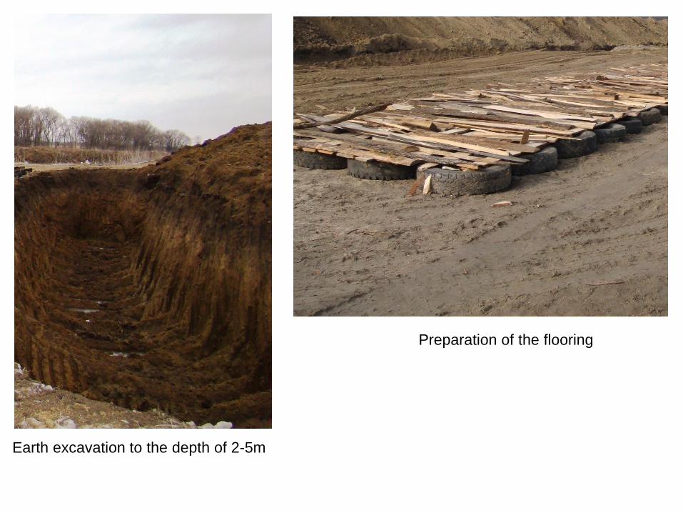
Earth excavation to the depth of 2-5m
Preparation of the flooring
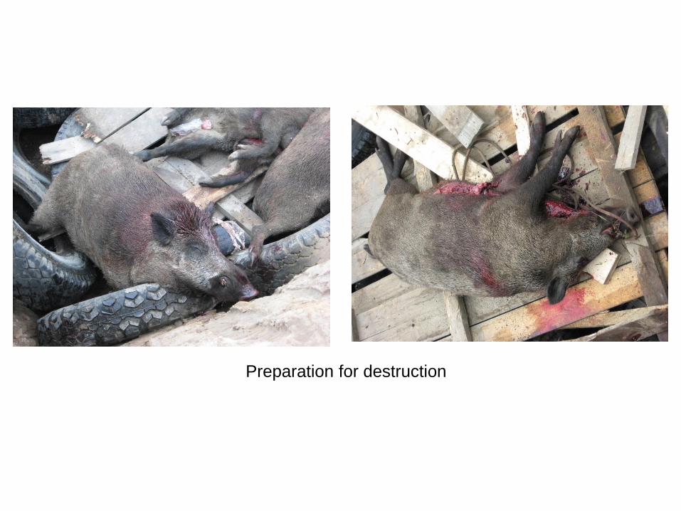
Preparation for destruction
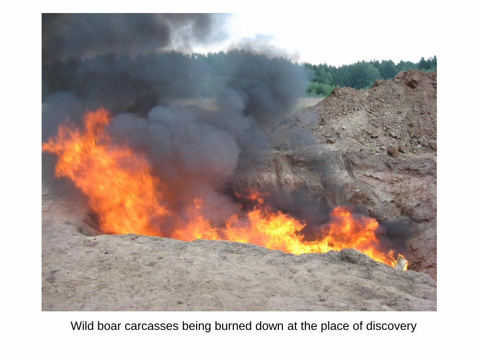
Wild boar carcasses being burned down at the place of discovery
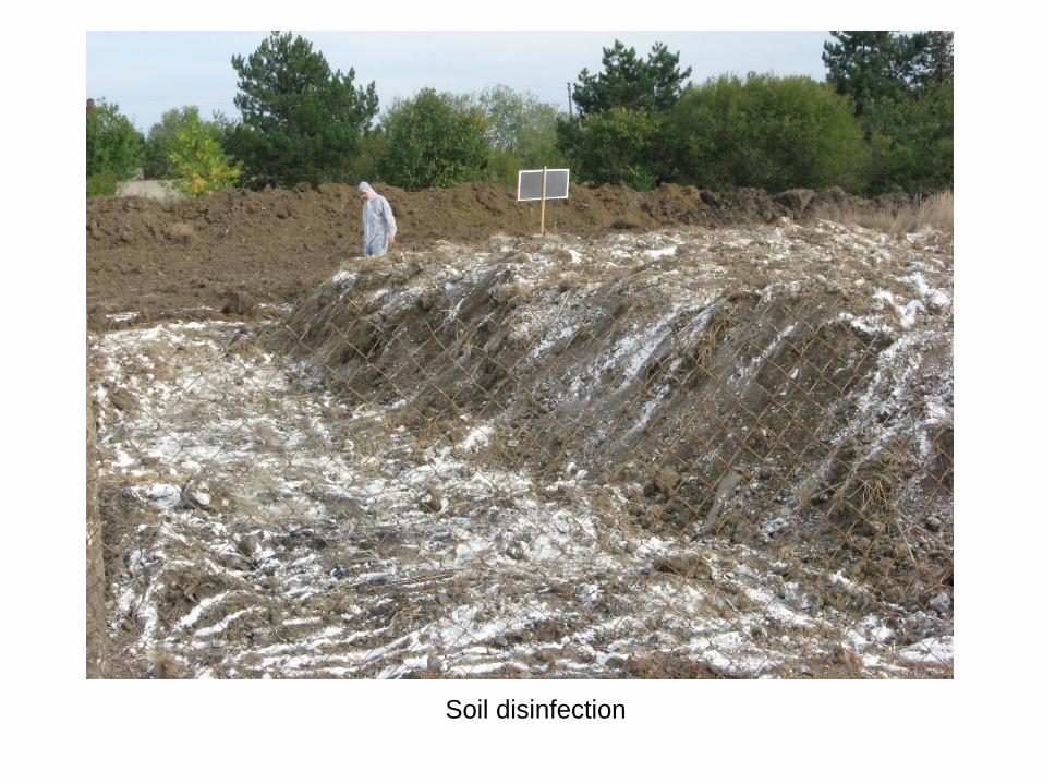
Soil disinfection