Embed Size (px)
Citation preview
Maxillofacial Trauma
Rathachai Kaewlai, MDDivision of Emergency Radiology, Ramathibodi Hospital, Bangkok, Thailand25 Sep 2016 10:40-11:00Society of Emergency Radiology (SER) India
Quick FactsMen two times more than womenConcomitant C-spine fracture 7%Concomitant skull base fracture 8%
Mundinger J Craniomaxillofac Surg 2014 (n=8127)
Role of ImagingDetection of soft tissue and bony injuriesCharacterization of soft tissue and bony injuriesSurgical planningCT preferred over radiography
Much more accurateEasier to perform in multi-trauma patientsMight be performed concurrently on other
body parts
Who Needs Facial Imaging?Wisconsin Criteria used for obtaining facial CTMulti-trauma patients with any 1 of 5 criteria: 98% sensitive for frx, 88% NPV
Bony stepoff or instabilityPeriorbital swelling or contusion
GCS <14Malocclusion
Tooth absenceSitzman et al. Plast Reconstr Surg 2011
CT Techniques
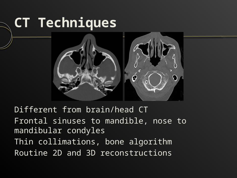
Different from brain/head CTFrontal sinuses to mandible, nose to mandibular condylesThin collimations, bone algorithmRoutine 2D and 3D reconstructions
First Thing FirstDo not get distracted by facial injuriesAre there intracranial or C-spine injuries?
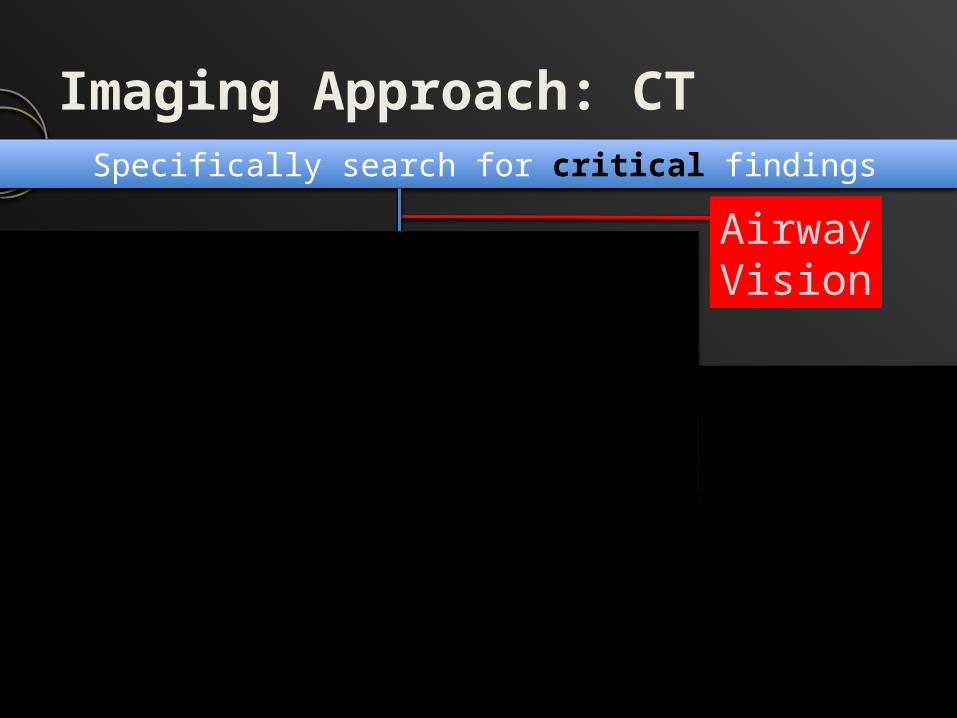
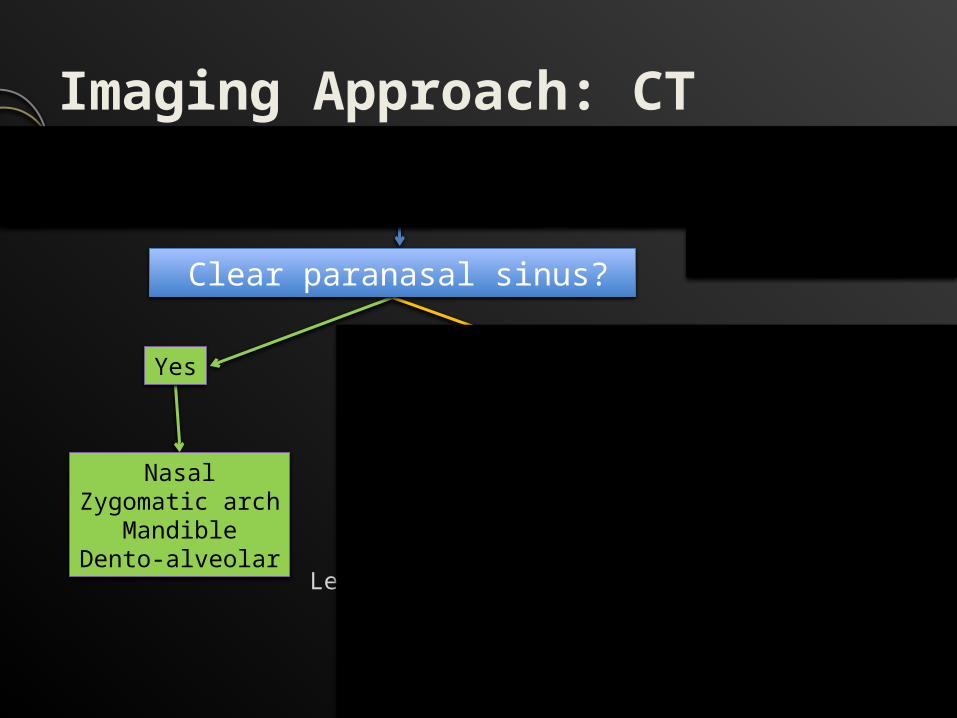
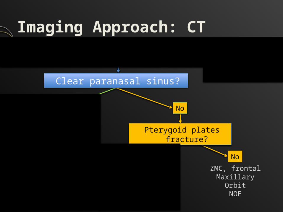
Imaging Approach: CTSpecifically search for critical findings
Yes No
NasalZygomatic arch
MandibleDento-alveolar
Le Fort I, II, III ZMC, frontalMaxillary
OrbitNOE
AirwayVision
NoYes
Clear paranasal sinus?
Pterygoid plates fracture?
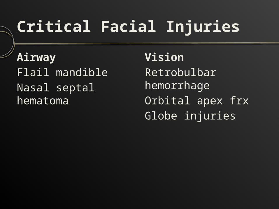
Critical Facial InjuriesAirwayFlail mandibleNasal septal hematoma
VisionRetrobulbar hemorrhageOrbital apex frxGlobe injuries
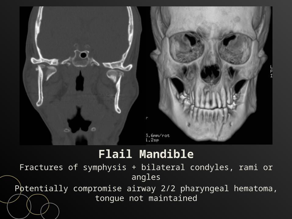
Flail MandibleFractures of symphysis + bilateral condyles, rami or anglesPotentially compromise airway 2/2 pharyngeal hematoma,
tongue not maintained
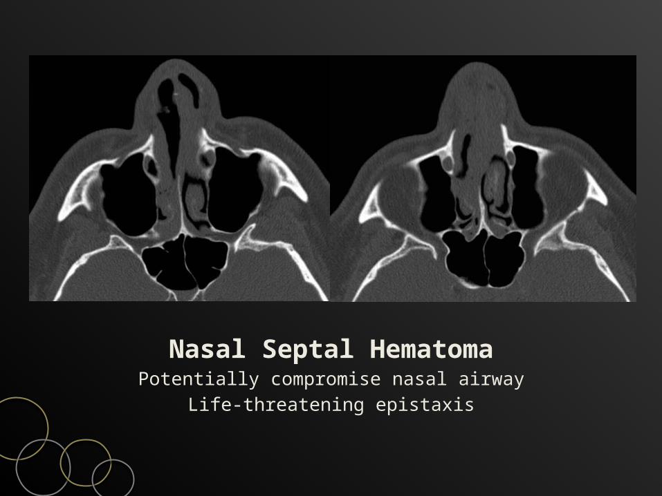
Nasal Septal HematomaPotentially compromise nasal airway
Life-threatening epistaxis
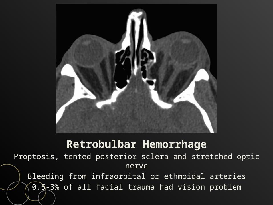
Retrobulbar HemorrhageProptosis, tented posterior sclera and stretched optic nerve
Bleeding from infraorbital or ethmoidal arteries0.5-3% of all facial trauma had vision problem
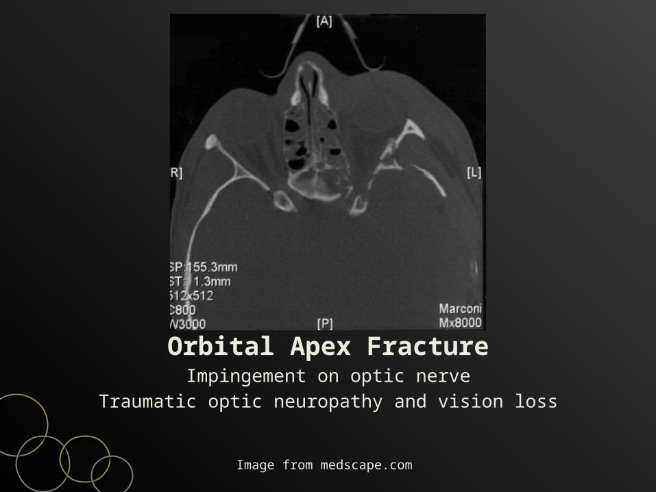
Orbital Apex FractureImpingement on optic nerve
Traumatic optic neuropathy and vision loss
Image from medscape.com

Globe RuptureFull thickness tear of sclera or cornea
Anterior surface common but posterior occult on clinical examCT helps diagnosis (SE60-75, SP76-100), FB, other injuries
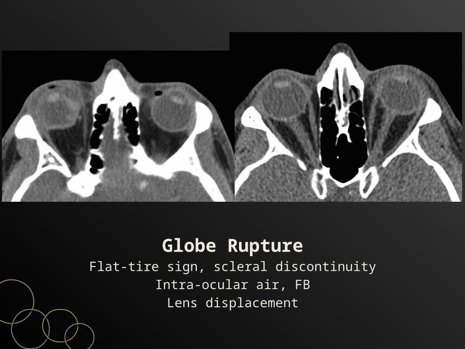
Globe RuptureFlat-tire sign, scleral discontinuity
Intra-ocular air, FBLens displacement
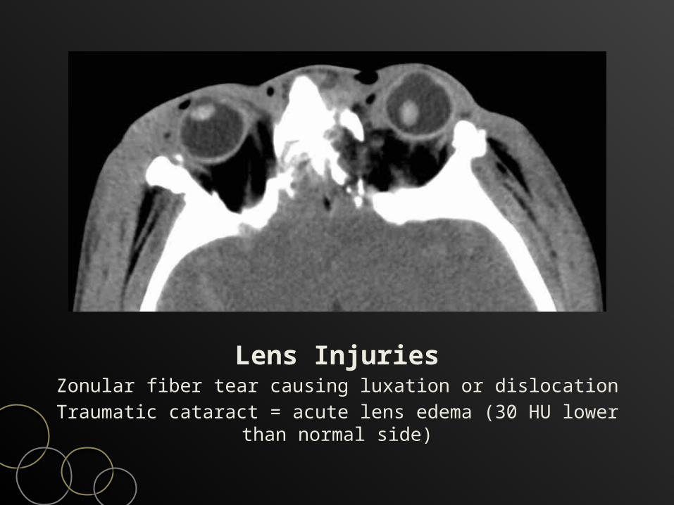
Lens InjuriesZonular fiber tear causing luxation or dislocation
Traumatic cataract = acute lens edema (30 HU lower than normal side)
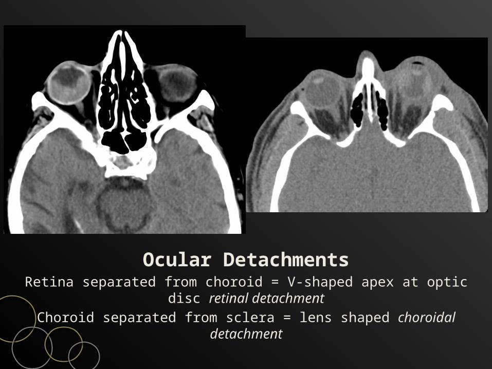
Ocular DetachmentsRetina separated from choroid = V-shaped apex at optic disc
retinal detachmentChoroid separated from sclera = lens shaped choroidal
detachment
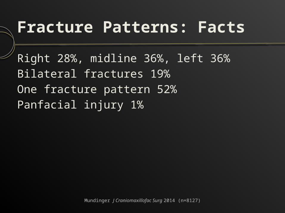
Fracture Patterns: FactsRight 28%, midline 36%, left 36%Bilateral fractures 19%One fracture pattern 52%Panfacial injury 1%
Mundinger J Craniomaxillofac Surg 2014 (n=8127)
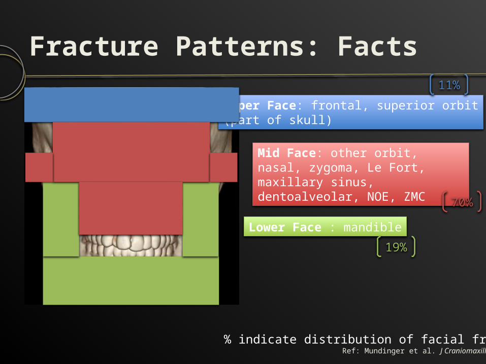
Fracture Patterns: Facts
Upper Face: frontal, superior orbit(part of skull)
Lower Face : mandible
Mid Face: other orbit, nasal, zygoma, Le Fort, maxillary sinus, dentoalveolar, NOE, ZMC
11%
70%
19%
% indicate distribution of facial fracturesRef: Mundinger et al. J Craniomaxillofac Surg 2014
Fracture Patterns: FactsNasal – Naso-orbital-ethmoidal – Nasal spineZygomatic arch – Zygomatic complex (ZMC)Maxillary sinus – Lefort – Dento-alveolar Frontal sinus MandibleOrbit
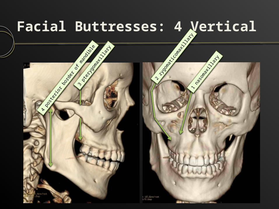
Facial Buttresses: 4 Vertical
1 nas
omax
illary
2 zyg
omati
comax
illary
3 pter
ygom
axilla
ry
4 pos
terior
borde
r of m
andib
le
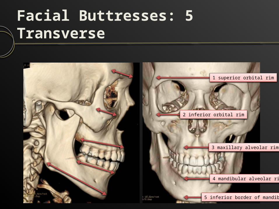
Facial Buttresses: 5 Transverse
1 superior orbital rim
2 inferior orbital rim
3 maxillary alveolar rim
4 mandibular alveolar rim
5 inferior border of mandible
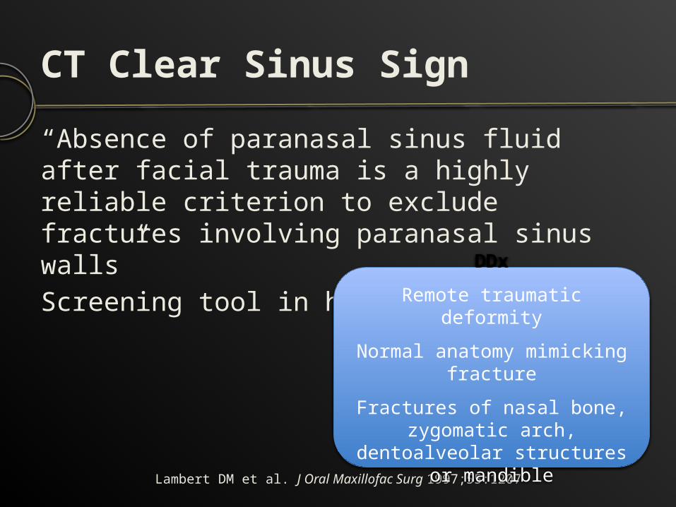
CT Clear Sinus Sign “Absence of paranasal sinus fluid after facial trauma is a highly reliable criterion to exclude fractures involving paranasal sinus walls”Screening tool in head CT
Lambert DM et al. J Oral Maxillofac Surg 1997;55:1207
DDxRemote traumatic deformityNormal anatomy mimicking
fractureFractures of nasal bone,
zygomatic arch, dentoalveolar structures or
mandible
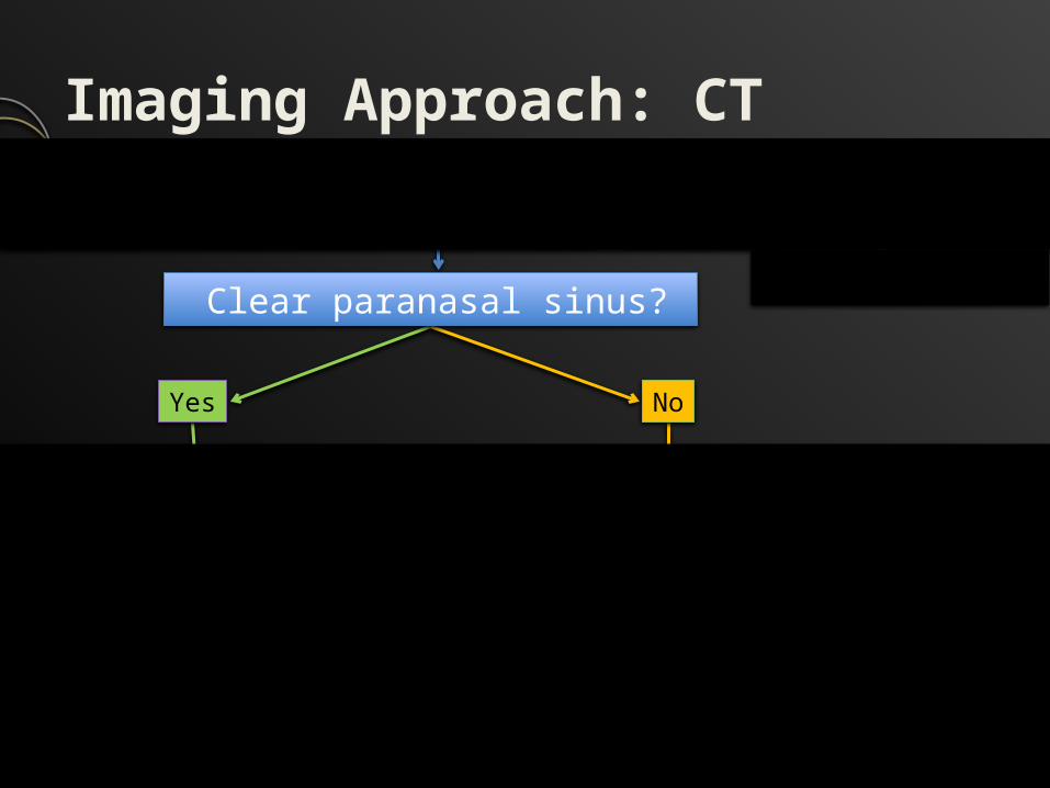
Imaging Approach: CTSpecifically search for critical findings
Yes No
NasalZygomatic arch
MandibleDento-alveolar
Le Fort I, II, III ZMC, frontalMaxillary
OrbitNOE
AirwayVision
NoYes
Clear paranasal sinus?
Pterygoid plates fracture?
Imaging Approach: CTSpecifically search for critical findings
Yes No
NasalZygomatic arch
MandibleDento-alveolar
Le Fort I, II, III ZMC, frontalMaxillary
OrbitNOE
AirwayVision
NoYes
Clear paranasal sinus?
Pterygoid plates fracture?
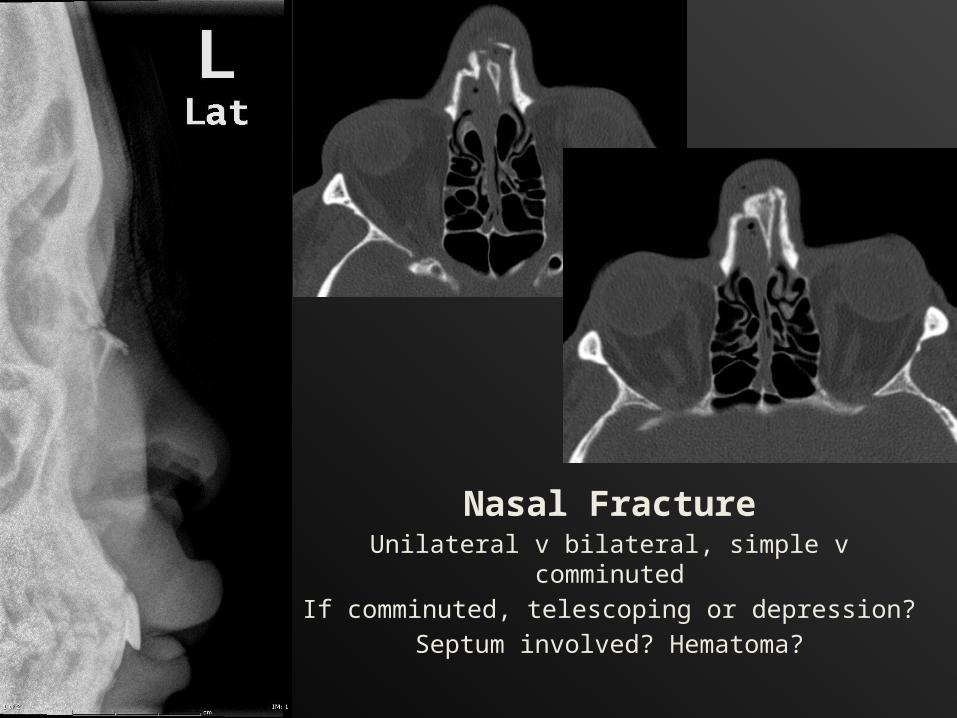
Nasal FractureUnilateral v bilateral, simple v comminutedIf comminuted, telescoping or depression?
Septum involved? Hematoma?
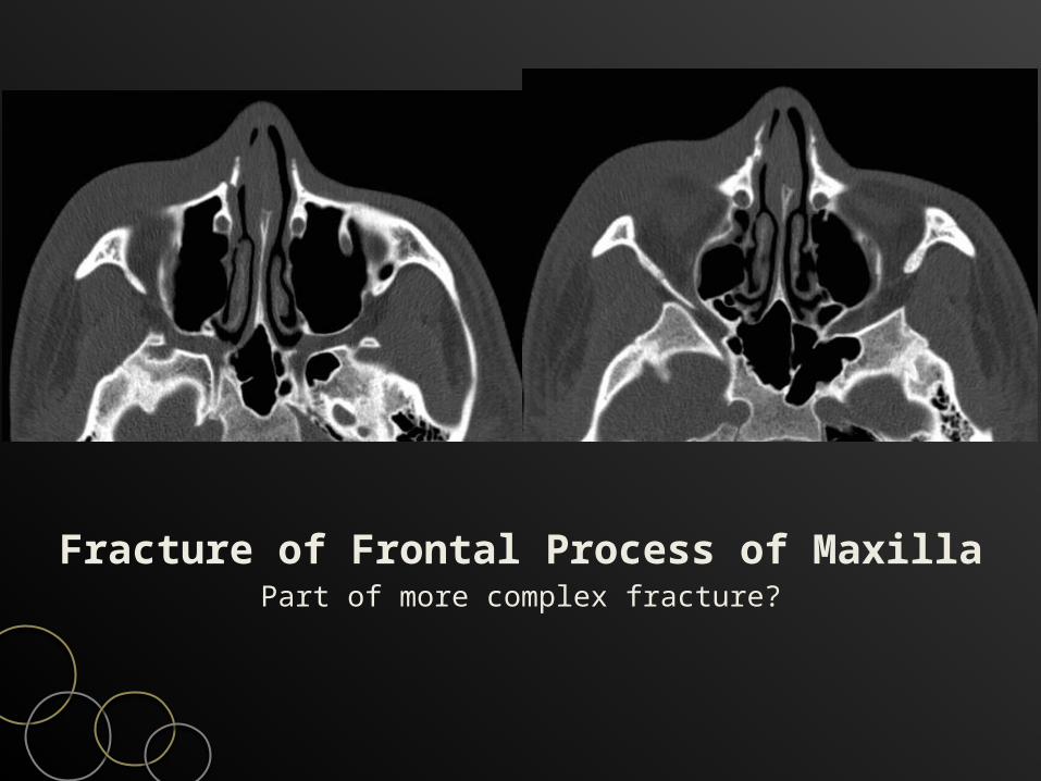
Fracture of Frontal Process of MaxillaPart of more complex fracture?
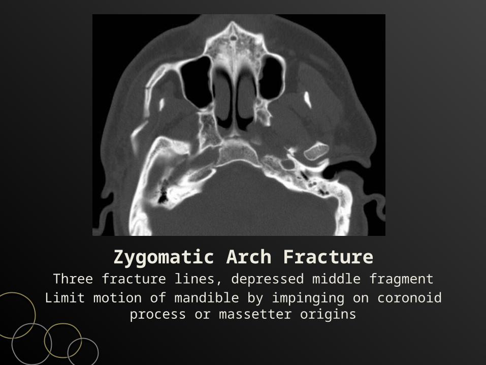
Zygomatic Arch FractureThree fracture lines, depressed middle fragment
Limit motion of mandible by impinging on coronoid process or massetter origins
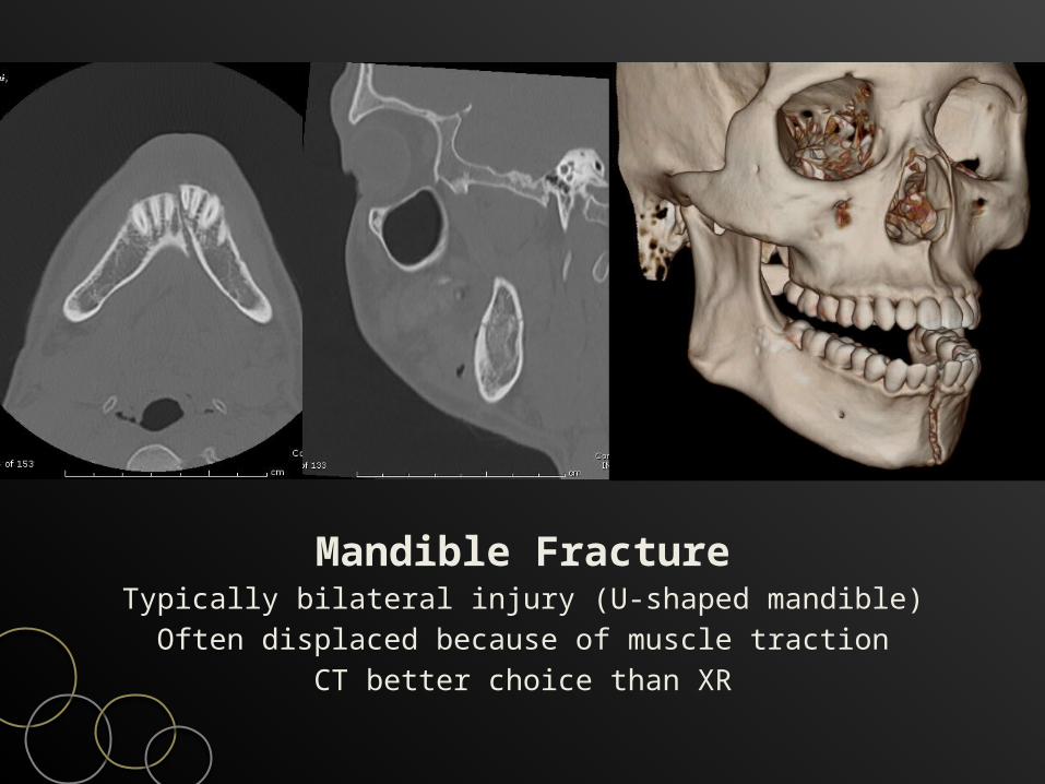
Mandible FractureTypically bilateral injury (U-shaped mandible)Often displaced because of muscle traction
CT better choice than XR
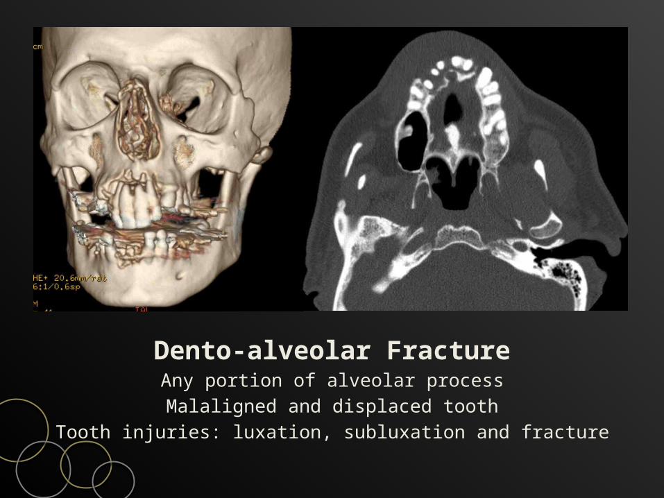
Dento-alveolar FractureAny portion of alveolar processMalaligned and displaced tooth
Tooth injuries: luxation, subluxation and fracture
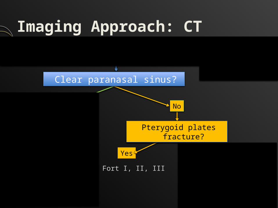
Imaging Approach: CTSpecifically search for critical findings
Yes No
NasalZygomatic arch
MandibleDento-alveolar
Le Fort I, II, III ZMC, frontalMaxillary
OrbitNOE
AirwayVision
NoYes
Clear paranasal sinus?
Pterygoid plates fracture?
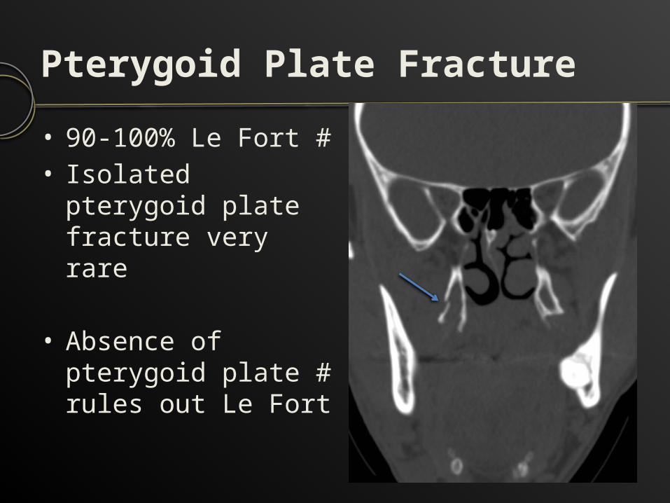
Pterygoid Plate Fracture• 90-100% Le Fort #• Isolated pterygoid
plate fracture very rare
• Absence of pterygoid plate # rules out Le Fort
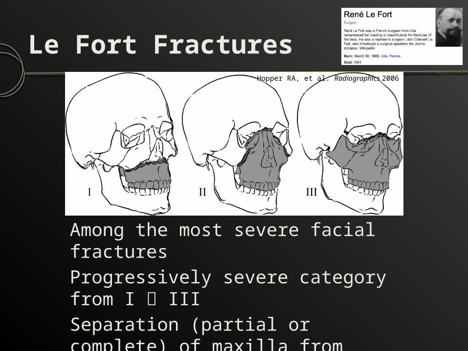
Le Fort Fractures
Among the most severe facial fracturesProgressively severe category from I IIISeparation (partial or complete) of maxilla from remainder face
Hopper RA, et al. Radiographics 2006
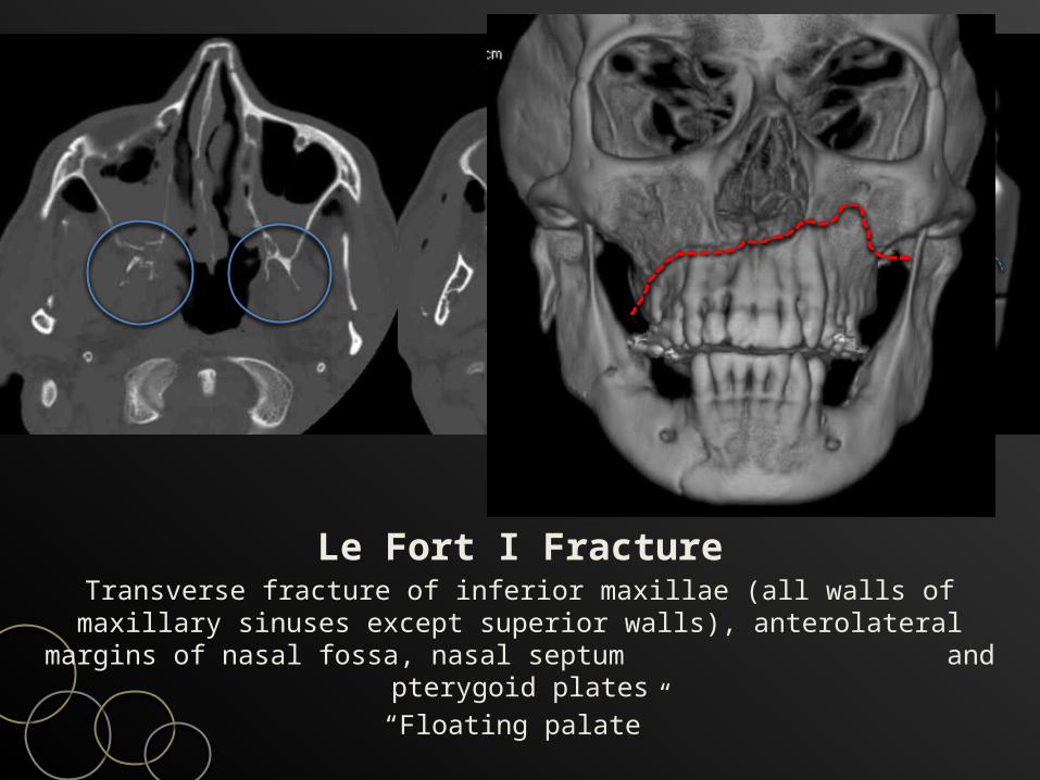
Le Fort I FractureTransverse fracture of inferior maxillae (all walls of maxillary sinuses except superior walls), anterolateral margins of nasal
fossa, nasal septum and pterygoid plates“Floating palate”
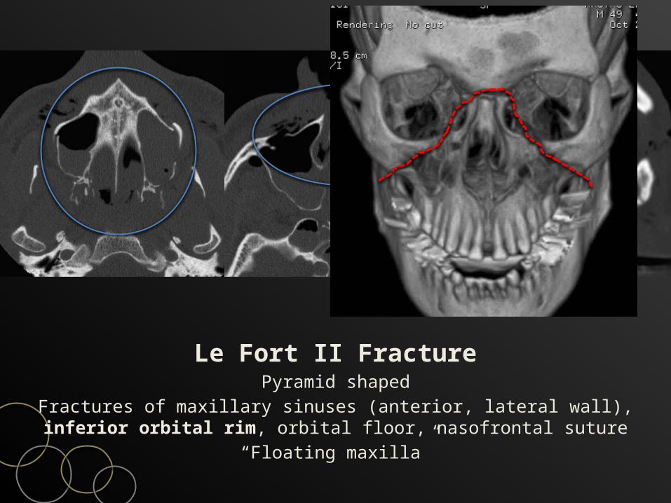
Le Fort II FracturePyramid shaped
Fractures of maxillary sinuses (anterior, lateral wall), inferior orbital rim, orbital floor, nasofrontal suture
“Floating maxilla”
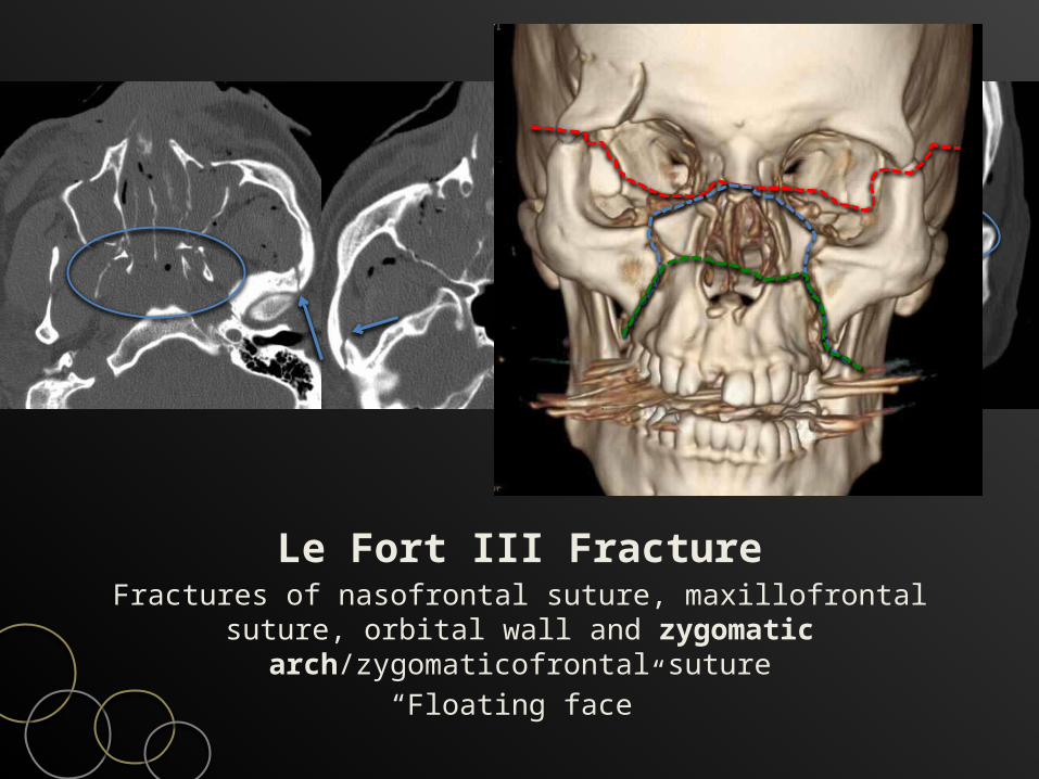
Le Fort III FractureFractures of nasofrontal suture, maxillofrontal suture, orbital wall
and zygomatic arch/zygomaticofrontal suture“Floating face”
Imaging Approach: CTSpecifically search for critical findings
Yes No
NasalZygomatic arch
MandibleDento-alveolar
Le Fort I, II, III ZMC, frontalMaxillary
OrbitNOE
AirwayVision
NoYes
Clear paranasal sinus?
Pterygoid plates fracture?
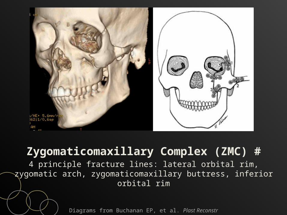
Zygomaticomaxillary Complex (ZMC) #4 principle fracture lines: lateral orbital rim, zygomatic arch,
zygomaticomaxillary buttress, inferior orbital rim
Diagrams from Buchanan EP, et al. Plast Reconstr Surg 2012
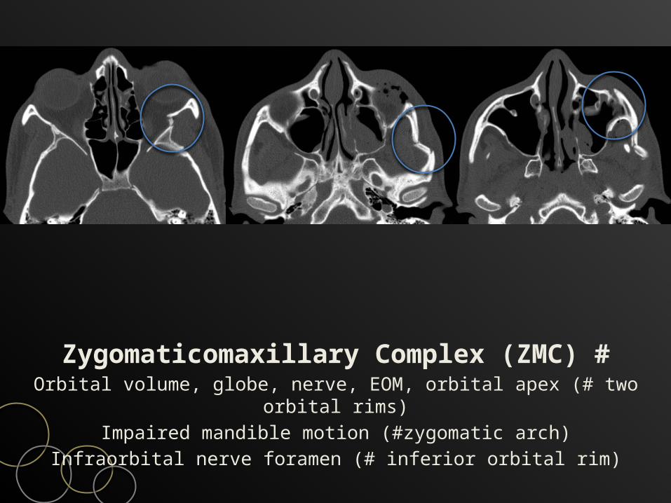
Zygomaticomaxillary Complex (ZMC) #Orbital volume, globe, nerve, EOM, orbital apex (# two orbital
rims)Impaired mandible motion (#zygomatic arch)
Infraorbital nerve foramen (# inferior orbital rim)
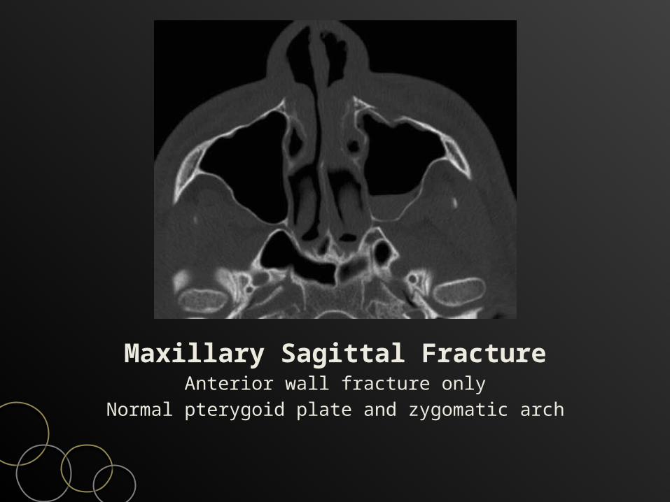
Maxillary Sagittal FractureAnterior wall fracture only
Normal pterygoid plate and zygomatic arch
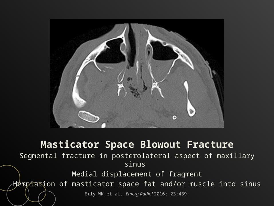
Masticator Space Blowout FractureSegmental fracture in posterolateral aspect of maxillary sinus
Medial displacement of fragmentHerniation of masticator space fat and/or muscle into sinus
Erly WK et al. Emerg Radiol 2016; 23:439.
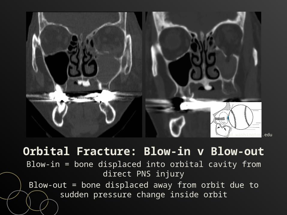
Orbital Fracture: Blow-in v Blow-outBlow-in = bone displaced into orbital cavity from direct PNS
injuryBlow-out = bone displaced away from orbit due to sudden
pressure change inside orbit
Rad.washington.edu
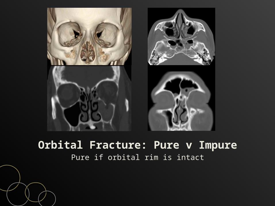
Orbital Fracture: Pure v ImpurePure if orbital rim is intact
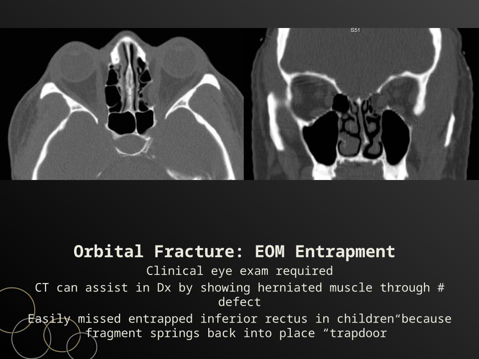
Orbital Fracture: EOM Entrapment Clinical eye exam required
CT can assist in Dx by showing herniated muscle through # defectEasily missed entrapped inferior rectus in children because
fragment springs back into place “trapdoor”
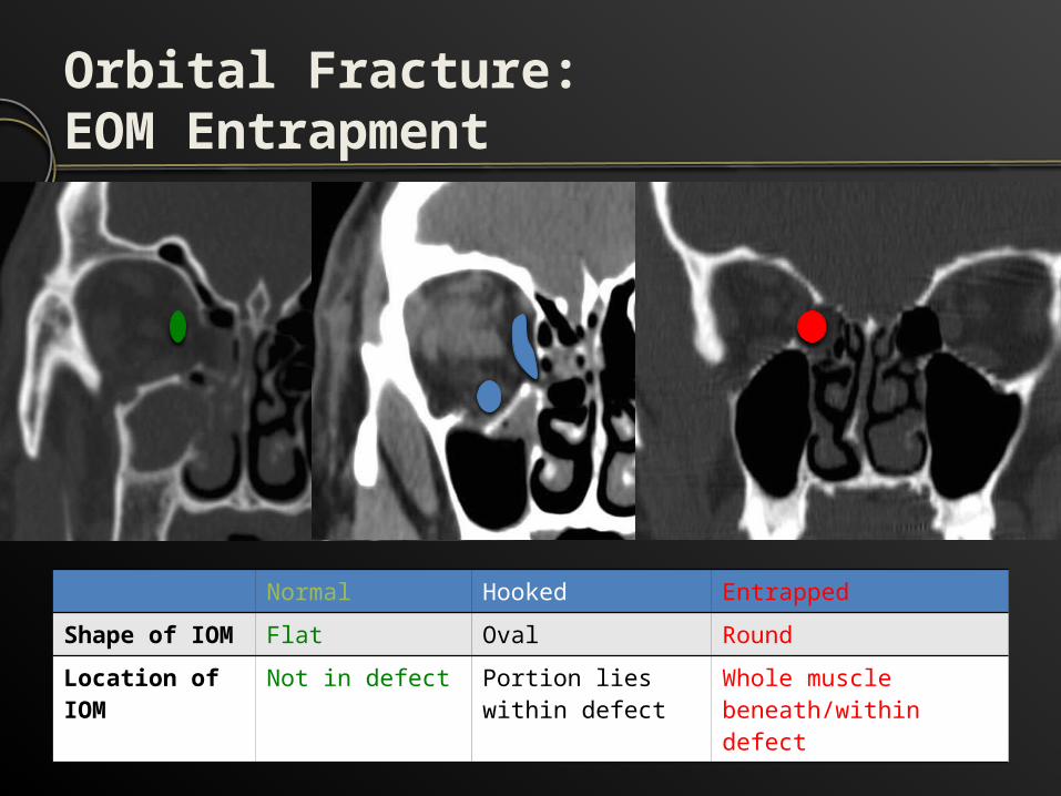
Orbital Fracture: EOM Entrapment
Normal Hooked EntrappedShape of IOM Flat Oval RoundLocation of IOM
Not in defect Portion lies within defect
Whole muscle beneath/within defect
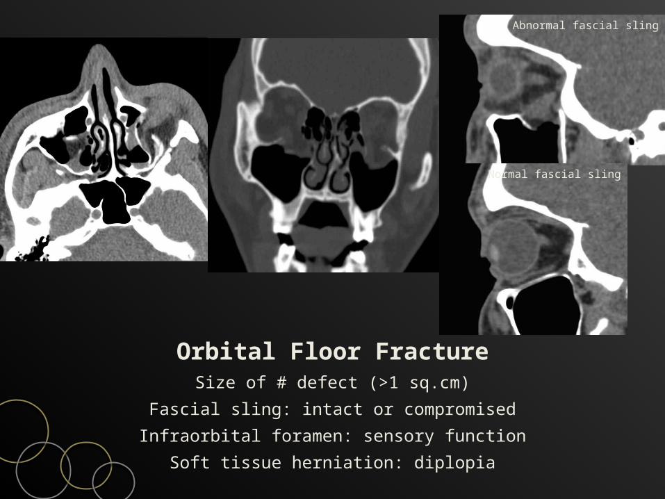
Orbital Floor FractureSize of # defect (>1 sq.cm)
Fascial sling: intact or compromisedInfraorbital foramen: sensory function
Soft tissue herniation: diplopia
Abnormal fascial sling
Normal fascial sling
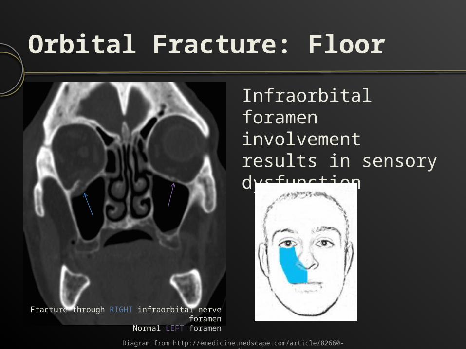
Orbital Fracture: FloorInfraorbital foramen involvement results in sensory dysfunction
Diagram from http://emedicine.medscape.com/article/82660-overview
Fracture through RIGHT infraorbital nerve foramen
Normal LEFT foramen
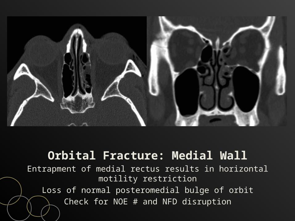
Orbital Fracture: Medial WallEntrapment of medial rectus results in horizontal motility
restrictionLoss of normal posteromedial bulge of orbit
Check for NOE # and NFD disruption
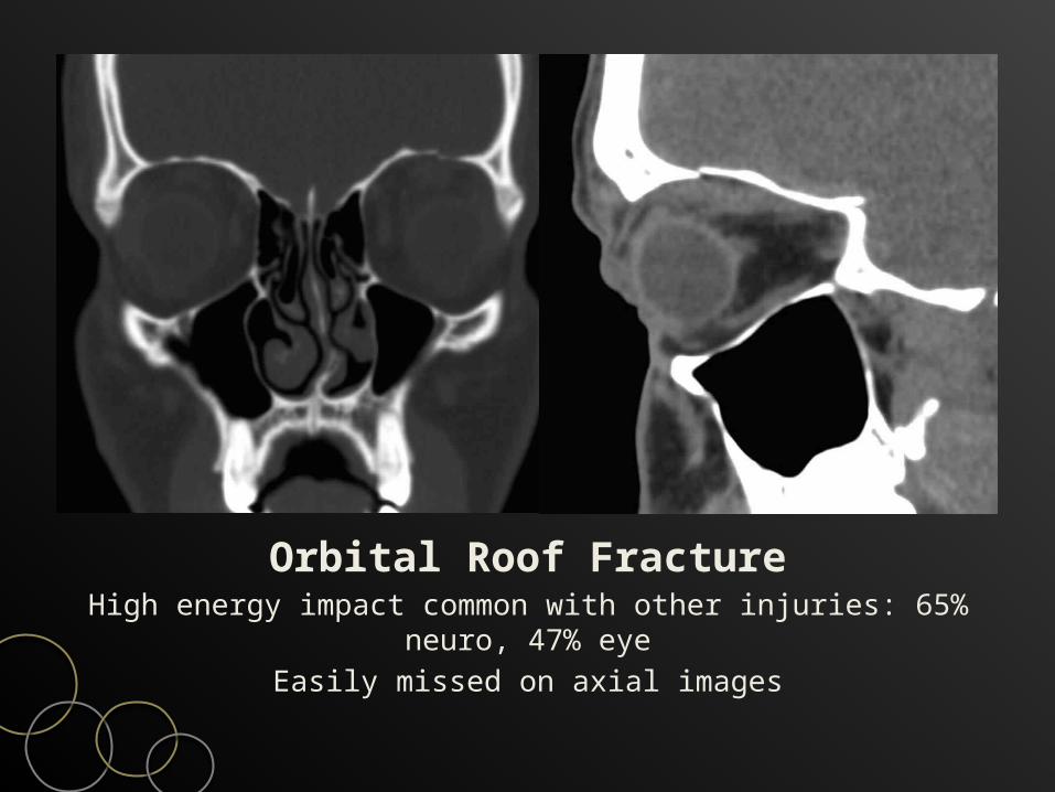
Orbital Roof FractureHigh energy impact common with other injuries: 65% neuro, 47%
eyeEasily missed on axial images
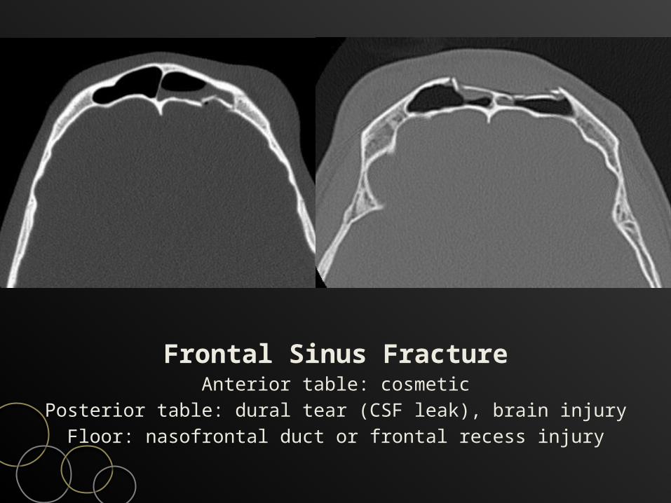
Frontal Sinus FractureAnterior table: cosmetic
Posterior table: dural tear (CSF leak), brain injuryFloor: nasofrontal duct or frontal recess injury
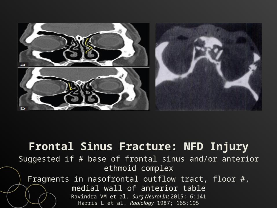
Frontal Sinus Fracture: NFD InjurySuggested if # base of frontal sinus and/or anterior ethmoid
complexFragments in nasofrontal outflow tract, floor #, medial wall of
anterior tableRavindra VM et al. Surg Neurol Int 2015; 6:141
Harris L et al. Radiology 1987; 165:195
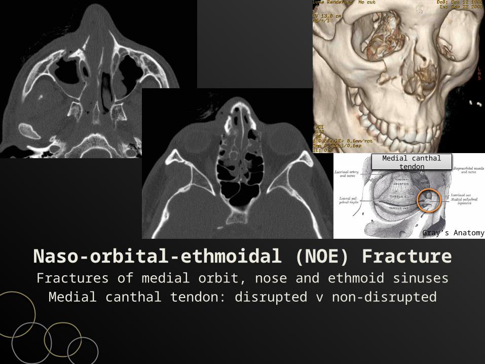
Naso-orbital-ethmoidal (NOE) FractureFractures of medial orbit, nose and ethmoid sinusesMedial canthal tendon: disrupted v non-disrupted
Medial canthal tendon
Gray’s Anatomy
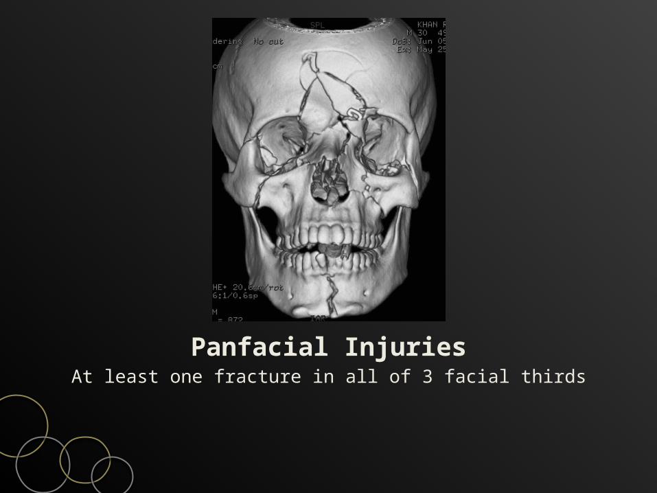
Panfacial InjuriesAt least one fracture in all of 3 facial thirds
ConclusionsAlways check intracranial & C-spine injuries firstTwo critical facial findings – airway and visionSystematic evaluation
Clear paranasal sinuses ?Pterygoid plate fracture ?
Try to fit all fractures into one or few patternsLook for potential complications