Embed Size (px)
Citation preview

ByMustafa Jamal

• Trachea
• Primary Bronchi
• Lobar bronchi
• Lobular bronchi
• Bronchioles
• Terminal bronchioles(5-7 per one bronchiole)

The trachea divides into two primary
bronchi that enter the lungs at the
hilum. At each hilum, arteries enter
and veins and lymphatic vessels
leave. These structures are
surrounded by dense connective
tissue and form a unit called the
pulmonary root.


After entering the lungs, the primary bronchi
course downward and outward, giving rise to
three bronchi in the right lung and two in the left
lung each of which supplies a pulmonary lobe.
These lobar bronchi divide repeatedly, giving
rise to smaller bronchi, whose terminal
branches are called bronchioles. Each
bronchiole enters a pulmonary lobule, where it
branches to form five to seven terminal
bronchioles.

The pulmonary lobules are pyramid shaped, with the apex directed toward the pulmonary
hilum. Each lobule is delineated by a thin connective tissue septum, best seen in the fetus. In adults, these septa are frequently incomplete,
resulting in a poor delineation of the lobules.

Structure of a bronchus. Smooth muscle is present in the entire bronchiolar tree, including the respiratory bronchiole. Contraction of this muscle induces folding of the mucosa. The elastic fibers in the bronchus continue into the bronchiole. The lower portion of the drawing represents a region with its connective tissue removed to show the presence of elastic fibers and smooth muscle.

Section of a bronchus wall showing the respiratory epithelium with goblet cells and columnar ciliated cells. The connective tissue of the lamina propria contains serous glands and smooth muscle (SM) In the lower half of the photomicrograph is a large piece of hyaline cartilage.


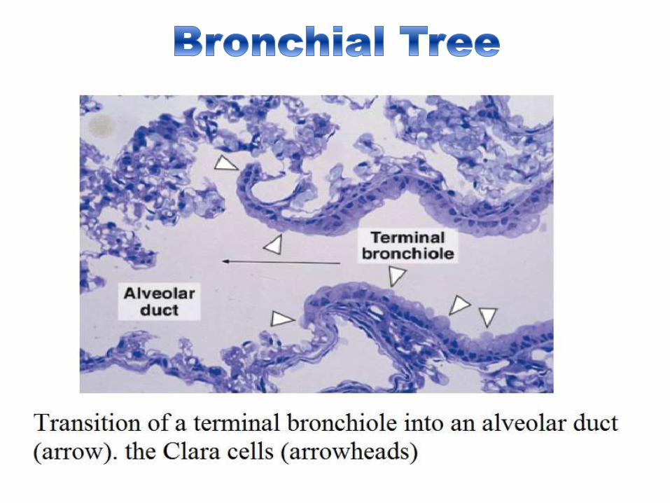
Bronchioles
Bronchioles, intralobular airways with diameters of 5 mm or less, have neither cartilage nor glands in their
mucosa; there are only scattered goblet cells within the epithelium of the initial segments. In the larger
bronchioles, the epithelium is ciliated pseudostratified columnar, which decreases in height and complexity to
become ciliated simple columnar in the smaller terminal bronchioles.
The epithelium of terminal bronchioles also contains Clara cells