Embed Size (px)
Citation preview
Emergencies and Complications in Gastroenterology
12 figures, 1 in color, and 14 tables, 2003
Editor
Petr DíteZ , Brno
Basel � Freiburg � Paris � London � New York �
Bangalore � Bangkok � Singapore � Tokyo � Sydney

S. KargerMedical and Scientific PublishersBasel � Freiburg � Paris � LondonNew York � Bangalore � BangkokSingapore � Tokyo � Sydney
Drug DosageThe authors and the publisher have exerted every effort to en-sure that drug selection and dosage set forth in this text are inaccord with current recommendations and practice at the timeof publication. However, in view of ongoing research, changesin government regulations, and the constant flow of informa-tion relating to drug therapy and drug reactions, the reader isurged to check the package insert for each drug for any changein indications and dosage and for added warnings and precau-tions. This is particularly important when the recommendedagent is a new and/or infrequently employed drug.
All rights reserved.No part of this publication may be translated into otherlanguages, reproduced or utilized in any form or by any means,electronic or mechanical, including photocopying, recording,microcopying, or by any information storage and retrievalsystem, without permission in writing from the publisher or, inthe case of photocopying, direct payment of a specified fee tothe Copyright Clearance Center (see ‘General Information’).
© Copyright 2003 by S. Karger AG,P.O. Box, CH–4009 Basel (Switzerland)Printed in Switzerland on acid-free paper byReinhardt Druck, BaselISBN 3–8055–7584–X
Fax + 41 61 306 12 34E-Mail [email protected]
Vol. 21, No. 1, 2003
Contents
© 2003 S. Karger AG, Basel
Fax + 41 61 306 12 34 Access to full text and tables of contents,E-Mail [email protected] including tentative ones for forthcoming issues:www.karger.com www.karger.com/ddi_issues
5 Editorial
DíteZ , P. (Brno)
Review Articles
6 Management of Acute Variceal Bleeding
Lata, J. (Brno); Hulek, P.; Vanasek, T. (Hradec Králové)
16 Upper Gastrointestinal Haemorrhage – Surgical Aspects
Lundell, L. (Stockholm)
19 Lower Gastrointestinal Bleeding – The Role of Endoscopy
Messmann, H. (Augsburg)
25 Management of Acute Cholangitis
Gouma, D.J. (Amsterdam)
30 Acute Pancreatitis: Treatment Strategies
Kahl, S.; Zimmermann, S.; Malfertheiner, P. (Magdeburg)
38 Modern Phase-Specific Management of Acute Pancreatitis
Werner, J.; Uhl, W.; Hartwig, W.; Hackert, T.; Müller, C.; Strobel, O.; Büchler, M.W.(Heidelberg)
46 Severe Inflammatory Bowel Disease: Medical Management
Farthing, M.J.G. (Glasgow)
54 Surgical Treatment of Severe Inflammatory Bowel Diseases
Leowardi, C.; Heuschen, G.; Kienle, P.; Heuschen, U.; Schmidt, J. (Heidelberg)
63 Intestinal Obstruction and Perforation – The Role of the Gastroenterologist
DíteZ , P.; Lata, J.; Novotný, I. (Brno)
68 Intestinal Obstruction and Perforation – The Role of the Surgeon
Dervenis, C.; Delis, S.; Filippou, D.; Avgerinos, C. (Athens)
77 Author Index and Subject Index

This page intentionally left blank
Dig Dis 2003;21:5DOI: 10.1159/000071332
Editorial
ABCFax + 41 61 306 12 34E-Mail [email protected]
© 2003 S. Karger AG, Basel0257–2753/03/0211–0005$19.50/0
Accessible online at:www.karger.com/ddi
Acute emergencies in gastroenterology are extraordi-narily severe conditions with high morbidity and mortali-ty. Particularly severe diseases include acute pancreatitis,a difficult course of non-specific intestinal inflammationsmanifested by toxic colon or acute intestinal obstruction,and even acutely developed intestinal pseudo-obstruction(Ogilvie’s syndrome) or variceal and non-variceal bleed-ing into the gastrointestinal tract. Undoubtedly seriousfactors influencing the accuracy of diagnostics and effec-tivity of therapy are the etiological multifactorial charac-teristics of changes that induce the acute state. Polymor-bidity is also frequent among these patients and requires acomplex diagnostic approach, often limiting the possibili-ty of using an optimal therapeutic approach.
Effective diagnostics and therapy for acute conditionsin gastroenterology requires a multidisciplinary team ap-proach. In diagnostics, endoscopic examination enablinga simultaneous therapeutical solution is of fundamentalimportance in managing most diseases, which is valid forexample in patients with acute bleeding into the alimenta-ry tract, in acute pancreatitis, acute cholangitis or acuteintestinal obstruction. However, endoscopy is an invasivemethod, and as many of these patients suffer from poly-morbidity, the usage of endoscopic approaches is limitedby the general clinical condition of patients, particularlywith respect to cardiopulmonary risks. In such cases, theapplication of non-invasive diagnostic methods is suit-able. These involve imaging methods such as ultrasoundabdominal examination, computer tomography or nu-clear magnetic resonance. Moreover, modifications ofthese methods, e.g. CT enteroclysis or CT colonography,
provide very precise and immediate results that allow theadoption of an optimal strategic course. Due to theirincreasing sensitivity and specificity, the above-men-tioned methods may be expected to substitute, in future,endoscopic examinations, whose present efficiency re-mains of the highest value.
Optimal therapy for acute states in gastroenterology isunthinkable without the close cooperation of a number ofdisciplines, particularly gastroenterology and surgery.Correct timing in determining whether conservative ther-apy is an effective and safe treatment for a patient in agiven situation or whether immediate surgery should beperformed is the basic requirement for the disease out-come of a patient. Severe states in particular should bemanaged at centers that have sufficient experience withsuch problems, possess a complete range of diagnosticmethods, carry out therapeutic endoscopy, and haveavailable acute surgical care, i.e. provide complex diag-nostic and therapeutical services.
Although acute conditions in gastroenterology and gas-troenterological complications are undoubtedly extraor-dinarily severe states, systematically processed data aboutrational and correct diagnostics and therapy from theviewpoint of gastroenterologists and surgeons have notbeen sufficient and therefore they could not be general-ized and utilized as recommendations for a rationalapproach in these states.
We believe that the topics published in this issue ofDigestive Diseases will help, at least in part, fill this gap.
Petr Dıte
Review Article
Dig Dis 2003;21:6–15DOI: 10.1159/000071333
Management of Acute Variceal Bleeding
Jan Lataa Petr Hulekb Tomas Vanasekb
aDepartment of Internal Medicine and Gastroentrology, Faculty of Medicine, Masaryk University, Brno andbDepartment of Internal Medicine, Faculty of Medicine, Charles University, Hradec Kralové, Czech Republic
Jan Lata, MD, PhD, Assoc. Prof. Med.Department of Internal Medicine and GastroentrologyFaculty of Medicine, Masaryk UniversityJihlavska 20, CS–625 00 Brno (Czech Republic)Tel. +420 547193465, Fax +420 47193701, E-Mail [email protected]
ABCFax + 41 61 306 12 34E-Mail [email protected]
© 2003 S. Karger AG, Basel0257–2753/03/0211–0006$19.50/0
Accessible online at:www.karger.com/ddi
Key WordsLiver cirrhosis W Variceal bleeding W Treatment W
Transjugular intrahepatic portosystemic shunt
AbstractPortal hypertension as a consequence of liver cirrhosis isresponsible for its most common complications: asci-tes, spontaneous bacterial peritonitis, hepatorenal syn-drome, hepatic encephalopathy and the most importantone – variceal hemorrhage. Variceal bleeding results inconsiderable morbidity and mortality. This review cov-ers all areas of importance in the therapy of acute va-riceal hemorrhage – endoscopic and pharmacologi-cal treatment, transjugular intrahepatic portosystemicshunt, surgery and balloon tamponade. Indications andlimitations of these therapeutic modalities are widelydiscussed.
Copyright © 2003 S. Karger AG, Basel
Introduction
One of the most important consequences of liver cir-rhosis and portal hypertension is increased pressure ingastric and esophageal venous systems, dilatation of relat-ed vessels and increased blood flow through developedportosystemic shunts. The most enlarged are deep innerveins under the lamina propria and muscularis mucosae;
first manifestation is usually seen in the so-called perfo-rating zone of the distal esophagus. Clinically, the mostimportant factor is the appearance of esophageal varicesobserved after increase of the hepatic venous pressure gra-dient (HVPG) 110 mm Hg. About 50% of patients withnewly diagnosed liver cirrhosis have varices at the time ofdiagnosis and this number increase annually by 6% [1].
When the HVPG increases 112 mm Hg, the probabili-ty of variceal rupture is high. The first variceal bleedingwas described in 1840 [2] and the relationship of esopha-geal varices, bleeding and liver disease in 1900 [3]. Vari-ceal bleeding affects 30–60% of cirrhotic patients. Inpatients with compensated liver disease, bleeding occursin only 30% of cases, and 60% in groups with decompen-sated liver disease. About one third of patients bleed with-in 2 years after the diagnosis of varices. Out of all gastroin-testinal hemorrhages, variceal bleeding represents about5–15% cases but 50% of severe bleeders – the presence ofboth decompensated liver disease and varices as source ofthe bleeding are independent predictors of high risk ofgastrointestinal bleeding [4].
The spontaneous cessation of bleeding episode hap-pens in up to 60% of cases, but untreated patients arejeopardized by rebleeding. This occurs in 30–40% withina 3-day interval and in 60% within 1 week. The mortalitywithin 6 weeks from the onset of bleeding is described ashigh as 30–50%. The cause of death is multifactorial,most of patients do not die due to exsanguinations butdue to complications of the hemorrhage, namely liver fail-
Management of Acute Variceal Bleeding Dig Dis 2003;21:6–15 7
ure. The most important factor predicting mortality is theliver disease. Thus, not only the incidence of bleeding butalso its mortality correlates with the Child-Pugh classifi-cation and the mortality of patients with class C is 70–80% [5]. Patients 165 years are threatened also by isch-emia and acute myocardial infarction due to anemia [6].
The Baveno III consensus conference [7] was held toupdate the consensus on the definitions of key eventsregarding the bleeding. Clinically significant portal hyper-tension (CSPH) was defined as an increase in the portalpressure gradient 110 mm Hg. The presence of varices,variceal hemorrhage, and/or ascites, is indicative of thepresence of CSPH. Measurement of the HVPG and endo-scopic assessment of esophageal varices are satisfactorytools for the diagnosis of CSPH.
General Measures
The first and most important measure is the hemody-namic stabilization of the patient and prevention of aspi-ration of vomited blood. The intravenous access shouldalways be ensured by large-bore and preferably multipleperipheral catheters, the central venous catheter is indi-cated in the presence of tachycardia 1100/min and sys-tolic pressure !100 mm Hg. These limits, together withthe need of application of more than 2 blood units within24 h, were recognized as attributes of severe bleeding bythe Baveno II conference [8]. First laboratory tests includeassessment of the blood group, blood count (hematocrit,hemoglobin, thrombocytes) and prothrombin time. Leu-kocytosis 18,500/mm3 is a prognostic factor predictingmore severe course of the disease [9]. The most commonapproach includes volume replacement with crystalloidsfirst and subsequently with blood derivates. Sodium over-load is unfavorable in ascitic patients. Intensive replace-ment of the blood volume is necessary for maintenance ofthe renal perfusion, but overload attributes to rebleedingdue to portal pressure increase. The optimal parametersare 2–5 mm Hg of the central venous pressure, hematocritbetween 25 and 30% and hemoglobin not 1100 g/l.Remarked hypovolemia with systolic pressure !90 mmHg and tachycardia 1120/min together with signs ofperipheral hypoperfusion are common indications for theapplication of oxygen (4 l/min). Vitamin K is indicated inmost patients. Though cirrhotic bleeders do often havevarious blood coagulation abnormalities, there is no evi-dence that general application of fresh-frozen plasma orthrombocytes is helpful.
The importance of infection in the etiopathogenesis ofvariceal bleeding and the need for prevention of the sys-temic infection is an indication for antibiotic treatment(amoxicillin-clavulanic acid, norfloxacin). A meta-analy-sis of studies of the use of prophylactic antibiotics in thissetting suggests that antibiotic prophylaxis substantiallyincreases the number of patients who remain free frominfection and improves short-term survival in patientswith cirrhosis and variceal hemorrhage [10].
The increase of the ammonium in the gastrointestinaltract due to bleeding can cause development or worseningof the encephalopathy. Thus, gastric large-bore tube andearly application of the lactulose are indicated, as well asvigorous correction of mineral unbalance, especially thepotassium and magnesium levels.
Endoscopic Therapy
Diagnostic endoscopy should be organized in acutelybleeding patients as soon as possible to determine the siteof bleeding. Even patients with portal hypertension anddocumented varices can bleed from other sources thanvarices. If varices are found to be the real source of hemor-rhage, endoscopic treatment is proved to decrease theshort-term mortality and to decrease further bleeding.Methods in question include sclerotherapy, application oftissue adhesives, banding of the varices, application ofdetachable loops for strangulation of varices and someothers [11].
Historically the first method introduced into the clini-cal practice was sclerotherapy. Which sclerosant is themost effective cannot be concluded. Comparative trialsare lacking a sufficient volume of patients and uniformmethodological standards regarding concentrations anddoses, intervals between sessions, and patient population,etc. Basically, all of these agents have been documented tobe effective in clinical trials. The intravariceal techniqueis perhaps more effective in controlling active bleedingthan paravariceal injection, but more studies are neededto confirm this. On the other hand, it was shown thatpunctures intended to be intravariceal are in fact paravar-iceal around 35–45% of the time [12]. Trials of sclerother-apy in acute bleeding are also influenced by the experi-ence of operators, schedule of follow-up and the numberof patients who were not actively bleeding at the time ofendoscopy. The experience of the operator is extremelyimportant in decision-making in common clinical prac-tice.
8 Dig Dis 2003;21:6–15 Lata/Hulek/Vanasek
Compared to balloon tamponade, sclerotherapy has asignificantly higher control of bleeding, specifically lowerrebleeding which occurs in up to 50% of cases after defla-tion of the balloon. Trials comparing somatostatin withsclerotherapy in general found no significant differencesin failure to control bleeding, rebleeding or mortality[13].
Variceal band ligation is superior to sclerotherapy inthe rate of complications and perhaps improvement insurvival. Control of active bleeding was in some trialsachieved more readily with ligation than with sclerothera-py, but some trials found no significant differences [14]. Itseems that severe bleeding responds better to banding andboth methods are equally effective in mild bleeding. How-ever, technically it is more difficult to employ banding insevere hemorrhage due to reduction of the visibility by thecylinder of the banding device and the further decrease offield of view by blood, which usually fills the cylinder tosome degree. New clear outer cylinders improved the easeof use of banding devices and multi-shot instrumentsshortened the time necessary for placement of a sufficientnumber of rings. The expert dependence plays a majorrole in this situation.
Combination of sclerotherapy and banding is also pos-sible. The so-called sandwich (ligation, sclerotherapy,ligation) approach was shown to be superior to ligationalone in prevention of recurrence of varices, but mortalityeradication rates, recurrent bleeding and complicationrates were similar for sandwich technique and bandingalone. Technically this approach means deployment ofthe rubber band at the most distal point of the varicealcolumn followed by the injection of 1–2 ml of the sclero-sant (5% ethanolamine oleate in this study) proximal tothe applied band, with another band subsequently beingapplied over the same column 3–4 cm proximal to theinjection site [15]. Another approach uses utilization ofthe argon plasma coagulation to induce mucosal fibrosisin the distal esophagus. It was shown that the recurrence-free rate at 24 months after treatment is significantly high-er with this treatment than with ligation alone [16]. Allthose attempts of technical improvement are intended toovercome the tendency of a higher recurrence rate of var-ices after banding as it does not obliterate deeper varices(peri- and para-esophageal varices) and perforating veins.At the moment, more studies are needed to evaluate theclinical benefit of application of newer methods in ques-tion. In individual patients it seems that it is not a mistaketo choose banding or sclerotherapy according to the size ofthe varices, the degree of fibrosis of the esophageal wall(affecting the feasibility of sucking of the vessel into the
cylinder), and the capability to obtain a good view in thedistal esophagus during active bleeding, etc.
In patients resistant to endoscopic treatment, it is clearthat more than two sessions of sclerotherapy are not help-ful, do not improve control of bleeding and bring in-creased risk of aspiration, perforation and sepsis [17].Development of deep post-sclerotherapy ulcers and mul-tiple sessions of sclerotherapy cause general deteriorationof the patient by itself. Vasoactive drugs can improve thetechnical feasibility of endoscopic therapy.
Tissue adhesives show a more than 90% rate of controlof bleeding but were not generally proved significantlybetter in application in esophageal varices in terms ofrebleeding and mortality [18]. This treatment is associat-ed with a significant risk of complications as cerebrovas-cular accidents or jeopardizes the scope. Furthermore, theagents that are used are more costly. Some benefit was,however, proved in patients with progressed liver disease(Child-Pugh C) in a randomized prospective trial compar-ing cyanoacrylate and sclerotherapy with ethanolamineoleate. The immediate hemostasis achieved by cyanoacry-late was significantly more often observed than with scle-rotherapy. This resulted in significantly lower rebleedingrates, need for surgery or transjugular intrahepatic porto-systemic shunt (TIPS) and mortality [19].
Complications of endoscopic therapy include local andsystemic events. The incidence of esophageal stricture for-mation and ulcer bleeding were significantly higher insclerotherapy (both appearing up to 25%) compared withband ligation (incidence less than 5%). In fact, most ulcerbleeding episodes require no therapeutic interventionsand strictures are usually treated with balloon dilatations.Major disasters as esophageal perforation and massiveesophageal hematoma are infrequent in both techniques.Pulmonary complications and mediastinitis are signifi-cantly more common after sclerotherapy [20].
Generally, for control of acute bleeding episode, vari-ceal band ligation is the method of first choice. If thisproves to be technically difficult, endoscopic variceal scle-rotherapy should be performed. Vasoactive drugs shouldbe used parallel to endoscopic therapy for 5 days. In fail-ure to control the bleeding, balloon tamponade can beused as a temporary measure en route to the radiologicalor surgical suite.
Management of Acute Variceal Bleeding Dig Dis 2003;21:6–15 9
Pharmacological Therapy
The biggest advantage of pharmacotherapy is its feasi-bility. It can be applied instantly without the need for spe-cialized instruments and is independent on the physi-cian’s skill and practice. Its efficacy was proved to be sim-ilar to endoscopic measures but optimal in their combina-tion.
Most drugs used for this indication cause splanchnicvasoconstriction. Vasoconstrictors decrease splanchnicperfusion and portal flow which results in decrease of theportal pressure. The decrease of blood flow and pressure isachieved in varices, too. The first drugs clinically used forthis indication were hormones, vasopressin and somato-statin. Currently their synthetic analogues, terlipressinand octreotide, are more widely used.
VasopressinThis is a hormone of the posterior lobe of the hypophy-
sis (also causes reabsorption of water in kidneys) whichwas the first vasoconstrictor used in the treatment ofbleeding due to portal hypertension [21] and was provedto be effective. It causes vasoconstriction in the splanch-nic area but also in the systemic circulation. Its major dis-advantage are side effects due to ischemia, especiallymyocardial [22]. It causes discontinuation of the treat-ment in up to 30% of cases. The combination withnitrates decreases the incidence of side effects but is notmore potent than other therapeutical options [23]. Vaso-pressin is no longer used for this indication in Europe incontrast to the USA where it is still an alternative in com-bination with nitrates.
TerlipressinTerlipressin is an N-triglycyl-8-lysine-vasopressin, a
synthetic analogue of the vasopressin, developed in 1964in Prague. It causes splanchnic vasoconstriction with aconsequent decrease of the portal pressure and blood flowin portosystemic collaterals. In comparison with vaso-pressin, it has minimum side effects and a prolonged bio-logical turnover (half-time 3.4 h) and this enables inter-mittent administration. In sufficient dose it decreases sig-nificantly not only the pressure in hepatic veins but alsothe intravariceal pressure [24]. The dose of 2 mg of terli-pressin significantly decreases portal flow and flow in theazygos veins in a 4-hour interval and the dose of 1 mg hasa similar effect [25]. Interesting is the combination withoctreotide. In rats, administration of both drugs alone sig-nificantly decreases portal pressure and cardiac index. Ifoctreotide is administered in animals pretreated with ter-
lipressin, the effect is not changed, if terlipressin is admin-istered in animals pretreated with octreotide, both sys-temic and splanchnic vasoconstriction are increased [26].The combination with ·1-adrenoreceptor antagonist in-creased the effect of terlipressin in animals [27]. Terlipres-sin in animals decreases portal flow significantly and thusthe hepatic inflow through the portal vein, but the arterialinflow increases which is important from the point ofhepatic function [28].
Clinically, terlipressin was proved to be significantlymore effective than placebo in the treatment of varicealbleeding [29]. Its efficacy is similar to balloon tamponade[30], somatostatin [31], octreotide [32] or endoscopic scle-rotherapy [33]. It is the only drug shown to decrease themortality related to acute bleeding episode. It is impor-tant to note the effect of its pre-hospital administrationduring the transport which significantly improves the suc-cess of consequent treatment [34]. A recent large multi-center trial of terlipressin versus sclerotherapy in thetreatment of acute variceal bleeding has shown similareffects of both treatment measures in terms of bleedingcontrol, rebleeding rate and 6-week mortality, number ofblood units transfused, stay in the intensive care unit, andhospital stay. Side effects were similar, but less frequent inthe terlipressin group [33].
SomatostatinSomatostatin is a hormone produced namely in the
hypothalamus and in the gastrointestinal tract. It was firstisolated in 1973 and subsequently synthesized. Its mainfunction is regulation of the somatotropin. It also has var-ious other effects as decreasing the flow in the splanchnicregion, inhibition of secretion of a variety of hormones(glucagons, insulin, gastrointestinal hormones) and de-creases also the gastric, biliary and intestinal motility andsecretion of the stomach and pancreas. The hemody-namic effect of the somatostatin and its analogue, octreo-tide, is not fully explained. In animal models it decreasesportal pressure by decreasing the inflow [35]; this, how-ever, was not confirmed in cirrhotic patients [36]. Somestudies have shown its vasoconstrictive effect on thesplanchnic region, but others did not confirm this. In cir-rhotics it probably has an effect on the decrease of gluca-gons which contributes to vasodilatation. Also, somato-statin contributes to the decrease of blood volume andprevention of postprandial hyperemia in the splanchnicregion. Its continuous administration in acute bleeding,however, decreases HVPG. Its disadvantage is namelyvery short biological half-time (approx. 2 min) requiringadministration as a continuous infusion. Somatostatin
10 Dig Dis 2003;21:6–15 Lata/Hulek/Vanasek
significantly decreases not only the portal pressure butalso the gastric mucosa blood flow (GMBF) [37], which ispotentially important in the bleeding from portal hyper-tensive gastropathy. However, trial data are conflicting.Meta-analyses have shown better control of bleeding com-pared with vasopressin [38]. A meta-analysis did not showsignificantly better efficacy in comparison to placebo [39].Smaller studies, however, found a similar efficacy com-pared to sclerotherapy [40], terlipressin [31] and found alesser need for blood transfusions and other urgent thera-pies [41].
OctreotideOctreotide is a synthetic octapeptide derivate of so-
matostatin, first described in 1982. Besides octreotide,more than 20 synthetic analogues of the somatostatin areknown. Lanreotide was tested mainly in animal models.Vapreotide was better in comparison with placebo andwas proved to increase the efficacy of endoscopic treat-ment in variceal bleeding in humans [42]. None of theseother analogues are currently used in common clinicalpractice.
Octreotide has a similar pharmacological effect as so-matostatin. The differences are dependent on its bindingto three out of five somatostatin receptors. In comparisonto somatostatin, its advantages are its longer half-time(90–120 min) and especially longer pharmacological ac-tion (8–12 h). Octreotide (as well as somatostatin) de-creases significantly the portal pressure in animals [43],but its influence on hemodynamics in cirrhotics, includingdecrease of the portal pressure, was not significantlyproved [44]. It probably also influences the mesenteric cir-culation [45]. Meta-analysis studies using octreotide orsomatostatin have shown a lower rate of complicationsand a similar effect as sclerotherapy or balloon tamponade[46]. A newer meta-analysis comparing octreotide to othermedical therapy and placebo has shown a better effect ofthe octreotide on the bleeding control compared to place-bo and other drugs and side effects comparable to placeboor no treatment [47]. The administration of the octreotideafter sclerotherapy decreases the portal pressure and re-bleeding rate compared to sclerotherapy alone [48, 49]; theeffect on mortality, however, was not proved.
NitratesIntravenous nitrates are mostly used to counteract the
vasoconstriction effect of vasopressin, of which isosor-bide-5-dinitrate is the most common. Its hypotensiveeffects limits its use in the acute phase of the bleeding epi-sode.
Mechanical-Balloon Tamponade of the Varices
The balloon tamponade may have a life-saving effectbut its inappropriate application has many complications.The ability to place properly balloon tamponade is sur-prisingly low outside specialized centers. Generally, now-adays it is seldom indicated. Currently it is accepted as atemporary measure after second unsuccessful endoscopictreatment en route to portosystemic decompression (sur-gical or TIPS). If indicated, the patient should be man-aged in the specialized intensive care unit. Most commonis the three-lumen double-balloon (Sengstaken-Blake-more). In case of bleeding from subcardial- fundal gastricvarices, the single-balloon (Linton-Nachlas) tamponade ismore appropriate. The Minnesota balloon is a modifica-tion of the double-balloon device with four lumens; thefourth is used for sucking from the space above the esoph-ageal balloon, thus it prevents aspiration better. Balloonsmust be inflated by the air, not liquid. Water, due to itsweight, changes the shape of the balloon, which results inmalfunction of the device, and is therefore not an appro-priate filling medium. The gastric balloon is inflated first,then traction is ensured and the esophageal balloon isinflated. Its pressure should be higher than portal pres-sure, 40 mm Hg is usually sufficient, overinflation is con-traproductive and causes complications. Suction shouldbe provided for gastric content and swallowed saliva. Thecorrect location of the balloon tube should be checked byX-ray.
The balloon should not be insufflated more than 24 h.Some authors recommend deflation of the balloon every4–6 h for 30 min [50]. Up to 50% of patients do haverebleeding after balloon decompression. Thus this tempo-rary measure should always be combined with othermethods [51]. The complications include aspiration, re-trosternal pain, esophageal or gastric rupture and mainlyesophageal and gastric ulcerations. Overinflated or water-filled balloons or dislocated balloons as well as multiplesclerotization sessions cause significant damage to theesophagus which replaces varices as bleeding source. Sel-dom the upright movement of the inflated esophageal bal-loon causes obstruction of the airways and suffocation,most such cases are due to the rupture of the gastric bal-loon. In this case the cross section of the lumen causingimmediate decompression of the balloon and subsequentextraction are indicated.
Management of Acute Variceal Bleeding Dig Dis 2003;21:6–15 11
Transjugular Intrahepatic Portosystemic Shunt(TIPS)
TIPS is a calibrated portosystemic shunt which re-duces quickly portosystemic gradient and opens access toendovasal treatment of varices (endovasal obliteration bysealants). Therefore, it is highly effective in stopping vari-ceal bleeding [52]. TIPS is indicated only when first-linemethods (medical and endoscopic) have failed. This hap-pens as ‘chronic’ or ‘acute’ failure. ‘Chronic’ means thatpatients do have repeated bleeding episodes despite ade-quate application of first-line treatment. An ‘elective’TIPS may be indicated. ‘Acute’ failure means bleedingrefractory to other measures and ‘urgent – salvage’ TIPS isoften a life-saving procedure.
It is difficult to organize a study comparing the TIPSprocedure as ‘salvage treatment’ as there is difficulty insetting up a comparable alternative. Even the first paperreporting TIPS dealt with uncontrolled bleeding in Child-Pugh class C patients and showed reasonably good results[53]. Most relevant papers investigating ‘salvage TIPS’showed immediate control of bleeding in 91–100% ofcases, 30-day rebleeding 7–30% and 1-month (or 42 days)mortality 28–55%. Child class C patients formed in mostof them more than 60% of cases [54–56] and in one 41%of cases [57]. Retrospective comparison with esophagealtransection [58] significantly favored TIPS (30-day mor-tality was 42 vs. 79%, rebleeding 16 vs. 26%). The role ofTIPS is especially important in patients bleeding fromgastric varices, which have a worse response to sclerother-apy and in bleeding portal hypertensive gastropathywhich cannot be treated endoscopically at all. Gastric var-ices in rescue TIPS series form up to 73% of cases [55].These impressive data show that rescue TIPS definitivelyhas its place in therapeutic algorithm for bleeding pa-tients. Most of TIPS procedures in question are per-formed with a combination of endovasal obliteration ofvarices as ‘urgent’ operations. It was proved that uncon-trolled bleeding can be effectively treated with TIPS, andTIPS has lower morbidity and mortality compared to sur-gery.
Indications of TIPS and TIPS-Related Procedures inBleeding PatientsIn general, accepted indications are patients with
bleeding that is uncontrolled by pharmacological andendoscopic therapy. This is true both for emergency situa-tions (urgent TIPS) and for patients with repeated epi-sodes of hemorrhage despite adequate preventive treat-ment who are not surgical candidates (elective TIPS).
These conclusions were confirmed by both the Restonand Baveno consensus meetings. Most patients appearwith gastroesophageal varices. Clinical situations aschronic anemia due to portal hypertensive gastropathy,prevention of rebleeding from large gastric or intestinalvarices, fresh portal vein thrombosis contributing tobleeding can be added to the list. Rare indications pub-lished include treatment of massive hemoptysis second-ary to bronchial collaterals [59], bleeding from stomal var-ices in patients after external enteric diversion [60], bleed-ing from colonic variceal veins and intestinal varices [61]and traumatic bleeding from cirrhotic liver [62].
Limitations of TIPS in Control of BleedingNot all cases with refractory or repeated bleeding are
indicated for TIPS. Contraindications are technical andclinical. Technical contraindications are mainly due toportal vein obstruction. However, successful placement ofTIPS is feasible also in selected cases of chronic occlusion[63], sometimes with the use of local thrombolysis [64].Favorable clinical outcome was reported in retrospectivestudies and fairly good technical success reaching 75%[65]. Even in patients with cavernomatous transforma-tion of the portal vein, successful TIPS placement is feasi-ble by combined percutaneous and intravasal approaches.Further relative contraindication for TIPS placement ispolycystic liver disease. Rare conditions include extremeobesity with body weight beyond the technical limits ofX-ray equipment.
Clinical contraindication means a situation where re-lief of portal hypertension is likely to deteriorate the liverfunction or the decrease of HVPG cannot improve thegeneral condition of the patient. Contraindication to elec-tive TIPS is also sepsis and heart failure. It is obvious thatTIPS can treat the complications of portal hypertensionand not the liver disease. In a recent consensus confer-ence, most investigators refused to perform TIPS with aChild-Pugh score of 12 points or above, so a jaundicedpatient in coma with renal insufficiency and need of arti-ficial ventilation is definitively not a candidate for TIPS[66]. Others have searched for individual variablesand pointed out emergent TIPS, ALT level 1100 IU/l(1.7 Ìkat/l), bilirubin 13 mg/dl (51 Ìmol/l) and pre-TIPSencephalopathy to predict overall mortality after TIPS[67]. Another important factor is renal insufficiency [68].
One should have in mind, however, that in cirrhoticsprotracted attack of esophageal bleeding has a deteriorat-ing effect on liver function and the general status of thepatient. Marked improvement is usually seen after cessa-tion of the bleeding period and therefore the exclusion of
12 Dig Dis 2003;21:6–15 Lata/Hulek/Vanasek
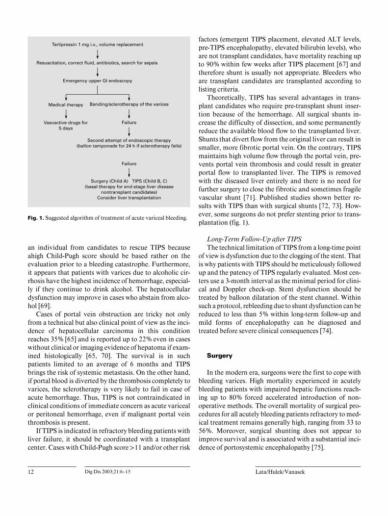
Fig. 1. Suggested algorithm of treatment of acute variceal bleeding.
an individual from candidates to rescue TIPS becauseahigh Child-Pugh score should be based rather on theevaluation prior to a bleeding catastrophe. Furthermore,it appears that patients with varices due to alcoholic cir-rhosis have the highest incidence of hemorrhage, especial-ly if they continue to drink alcohol. The hepatocellulardysfunction may improve in cases who abstain from alco-hol [69].
Cases of portal vein obstruction are tricky not onlyfrom a technical but also clinical point of view as the inci-dence of hepatocellular carcinoma in this conditionreaches 35% [65] and is reported up to 22% even in caseswithout clinical or imaging evidence of hepatoma if exam-ined histologically [65, 70]. The survival is in suchpatients limited to an average of 6 months and TIPSbrings the risk of systemic metastasis. On the other hand,if portal blood is diverted by the thrombosis completely tovarices, the sclerotherapy is very likely to fail in case ofacute hemorrhage. Thus, TIPS is not contraindicated inclinical conditions of immediate concern as acute varicealor peritoneal hemorrhage, even if malignant portal veinthrombosis is present.
If TIPS is indicated in refractory bleeding patients withliver failure, it should be coordinated with a transplantcenter. Cases with Child-Pugh score 111 and/or other risk
factors (emergent TIPS placement, elevated ALT levels,pre-TIPS encephalopathy, elevated bilirubin levels), whoare not transplant candidates, have mortality reaching upto 90% within few weeks after TIPS placement [67] andtherefore shunt is usually not appropriate. Bleeders whoare transplant candidates are transplanted according tolisting criteria.
Theoretically, TIPS has several advantages in trans-plant candidates who require pre-transplant shunt inser-tion because of the hemorrhage. All surgical shunts in-crease the difficulty of dissection, and some permanentlyreduce the available blood flow to the transplanted liver.Shunts that divert flow from the original liver can result insmaller, more fibrotic portal vein. On the contrary, TIPSmaintains high volume flow through the portal vein, pre-vents portal vein thrombosis and could result in greaterportal flow to transplanted liver. The TIPS is removedwith the diseased liver entirely and there is no need forfurther surgery to close the fibrotic and sometimes fragilevascular shunt [71]. Published studies shown better re-sults with TIPS than with surgical shunts [72, 73]. How-ever, some surgeons do not prefer stenting prior to trans-plantation (fig. 1).
Long-Term Follow-Up after TIPSThe technical limitation of TIPS from a long-time point
of view is dysfunction due to the clogging of the stent. Thatis why patients with TIPS should be meticulously followedup and the patency of TIPS regularly evaluated. Most cen-ters use a 3-month interval as the minimal period for clini-cal and Doppler check-up. Stent dysfunction should betreated by balloon dilatation of the stent channel. Withinsuch a protocol, rebleeding due to shunt dysfunction can bereduced to less than 5% within long-term follow-up andmild forms of encephalopathy can be diagnosed andtreated before severe clinical consequences [74].
Surgery
In the modern era, surgeons were the first to cope withbleeding varices. High mortality experienced in acutelybleeding patients with impaired hepatic functions reach-ing up to 80% forced accelerated introduction of non-operative methods. The overall mortality of surgical pro-cedures for all acutely bleeding patients refractory to med-ical treatment remains generally high, ranging from 33 to56%. Moreover, surgical shunting does not appear toimprove survival and is associated with a substantial inci-dence of portosystemic encephalopathy [75].
Management of Acute Variceal Bleeding Dig Dis 2003;21:6–15 13
Currently the first-line methods (vasoactive drugs andendoscopic therapy) reach up to 90% success in cessationof a bleeding episode. The remaining 10% of cases are oneof the most difficult groups to manage in hepatogastroen-terology. In the pre-TIPS era, the only ‘salvage therapy’accepted was surgery, but most patients with progressedliver diseases are excluded as surgical candidates. In surgi-cally treated patients, mortality reached 82% in patientswith Child class C [76]. Procedures as esophageal tran-section plus gastric devascularization and variety ofshunt operations are technically possible. Portal-systemicshunts can be separated into two basic types: nonselective(total) shunts and selective shunts. Total shunts are de-signed to divert portal blood away from the liver andinclude end-to-side portacaval shunts, side-to-side porto-caval shunts, interposition portocaval shunt, splenorenalshunts and mesocaval shunts. End-to-side shunts anatom-ically prevent any portal venous perfusion of the liver andtheoretically tends to more rapid liver failure, worsenedPSE and poor control of ascites, but this technique is tech-nically simpler and is recommended in the emergency sit-uation. Studies comparing different surgical shuntingtechniques are difficult to interpret and still remain anarea of considerably controversy [77]. Randomized stud-ies have shown that surgical shunts have a better hemo-static effect than local surgical treatment of bleeding ves-sels alone. In high-risk patients, sclerotherapy had a simi-lar effect with fewer complications than transection of theesophagus, thus transection does not seem to be a goodchoice [78]. It can be concluded that surgery possibly stillhas a place in the treatment of patients in otherwise goodcondition, but practically it is rare for cirrhotics in goodcondition to have refractory bleeding. The most impor-tant objective measure for comparing invasive methodstreating refractory bleeding is the 30-day mortality. Un-
fortunately, at the moment no studies are available fulfill-ing requirements for comparison of surgery and radioin-terventions (TIPS). The only randomized study [79] isquestioned from the point of imbalanced distribution ofgender, Child class, and urgent timing disfavoring theTIPS group. The results of this study showed comparable30-day mortality in 6 of 35 patients of the TIPS group and5 of the 35 patients treated by the H-graft. Another uncon-trolled large study comparing TIPS and surgical shunt[80] demonstrated 0% 30-day mortality in the surgicalgroup and 26% mortality in the TIPS group. Child-Pughclass C patients were not operated at all, but receivedexclusively TIPS and formed 57% of the TIPS group.Comparison of this large surgical experience with resultsof the Freiburg group [81] shows similar results in termsof mortality and rebleeding for patients with less pro-gressed disease (mortality 0% for Child A patients and11% for Child B patients). The rebleeding from variceswas demonstrated by two meta-analyses [82, 83] to besimilar after TIPS (19%) and after surgical shunts (3–45%) [1].
Orthotopic liver transplantation is not a treatmentmeasure of an acute bleeding episode but all bleedersshould be evaluated as transplant candidates and thosefulfilling standard criteria placed upon a waiting list.Transplantation of the liver is the treatment option thatoffers the best survival rates. The major mortality associ-ated with the procedure occurs in the first year. Thereported survival rate of patients with liver transplanta-tion because of variceal hemorrhage is 79% at 1 year and71% at 5 years [84]. The greatest survival advantage isconferred on the patient who falls in the Child’s C class.Unfortunately, access to this procedure will never be opento all patients due to limited sources of grafts, and ethicaland financial problems.
References
1 D’Amico G, Pagliaro L, Bosch J: The treatmentof portal hypertension: A meta-analytic review.Hepatology 1995;22:332–354.
2 Power W: Contributions to pathology. MDMed Surg J 1940;306–318.
3 Preble RB: Conclusions based on sixty cases offatal gastrointestinal hemorrhage due to cirrho-sis of the liver. Am J Med Sci 1900;119:263–268.
4 Terdiman JP: Update on upper gastrointestinalbleeding. Postgrad Med 1998;103:43–64.
5 Mann NS, Hillis A, Mann SK, Buerk CA, Pra-sad VM: In cirrhotic patients variceal bleedingis more frequent in the evening and correlateswith severity of liver disease. Hepatogastroen-terology 1999;46:391–394.
6 Emenike E, Srivastava S, Amoateng-AdjepongY, Al-Kharrat T, Zarich S, Manthous AC:Myocardial infarction complicating gastroin-testinal hemorrhage. Mayo Clin Proc 1999;74:235–241.
7 De Franchis R (ed): Portal Hypertension. III.Proceedings of the Third Baveno InternationalConsensus Workshop on Definitions, Method-ology and Therapeutic Strategies. Oxford,Blackwell Science, 2001.
8 De Franchis R (ed): Portal Hypertension. II.Proceedings of the Second Baveno Internation-al Consensus Workshop on Definitions, Meth-odology and Therapeutic Strategies. Oxford,Blackwell Science, 1996, pp 10–17.
9 Chalasani N, Patel K, Clark WS, Wilcox CM:The prevalence and significance of leukocytosisin upper gastrointestinal bleeding. Am J MedSci 1998;315:233–236.
10 Bernard B, Grange JD, Nyugen Khao E, et al:Antibiotic prophylaxis for the prevention ofbacterial infections in cirrhotic patients withgastrointestinal bleeding: A meta-analysis. He-patology 1999;29:1655–1661.
14 Dig Dis 2003;21:6–15 Lata/Hulek/Vanasek
11 Bhasin DK, Malhi NJS: Variceal bleeding andportal hypertension: Much to learn, much toexplore. Endoscopy 2002;43:119–128.
12 Waring JP, Sanowski RA, Pardy K, et al: Doesthe addition of methylene blue to the sclerosantimprove the accuracy of endoscopic varicealsclerotherapy? Am J Gastroenterol 1990;85:1227.
13 Burroughs AK, McCormick PA, Hughes MD,et al: Randomized, double-blind placebo-con-trolled trial of somatostatin for variceal bleed-ing. Gastroenterology 1990;99:1388–1395.
14 Lo GH, Lai KH, Cheng JS, et al: Emergencybanding ligation versus sclerotherapy for thecontrol of active bleeding from esophageal var-ices. Hepatology 1997;25:1101–1104.
15 Hou MC, Chen WC, Lin HC, et al: A new‘sandwich’ method of combined endoscopicvariceal ligation and sclerotherapy versus liga-tion alone in the treatment of esophageal vari-ceal bleeding: A randomized trial. GastrointestEndosc 2001;53:572–578.
16 Nakamura S, MItsunaga A, Murata Y, et al:Endoscopic induction of mucosal fibrosis byargon plasma coagulation (APC) for esophagealvarices: A prospective randomized trial of liga-tion plus APC vs. ligation alone. Endoscopy2001;33:210–215.
17 Bornman P, Terblanche J, Kahn D, et al: Limi-tations of multiple injection sclerotherapy ses-sions for acute variceal bleeding. S Afr Med J1986;70:34–36.
18 Sung JY, Yor W, Suen R, et al: Cyanoacrylatevs. sodium tetradecyl sulphate for the injectionof bleeding varices in patients with hepatocel-lular carcinoma: A prospective randomizedstudy. Gastrointest Endosc 1997;45:AB85.
19 Maluf-Filho F, Sakai P, Ishioka S, et al: Endo-scopic sclerosis versus cyanoacrylate endoscop-ic injection for the first episode of varicealbleeding: A prospective, control and random-ized study in Child-Pugh class C patients. En-doscopy 2001;33:421–427.
20 Lain L, Cook D: Endoscopic ligation comparedwith sclerotherapy for treatment of esophagealvariceal bleeding: A meta-analysis. Ann InternMed 1995;22:663–665.
21 Merigan TC, Poltkin GR, Davidson CS: Effectof intravenously administered posterior pitu-itary extract on haemorrhage from bleedingesophageal varices. N Engl J Med 1962;266:134–135.
22 Gimson AES, Westaby D, Hegarty J, et al: Arandomized trial of vasopressin and vasopres-sin plus nitroglycerin in the control of acutevariceal hemorrhage. Hepatology 1986;6:410–413.
23 Jenkins SA, Baxter JN, Corbett WA, et al: Aprospective randomised controlled clinical trialcomparing somatostatin and vasopressin incontrolling acute variceal hemorrhage. Br MedJ 1985;290:275–278.
24 Cestari R, Graga M, Missale G, et al: Haemo-dynamic effect of triglycyl-lysine-vasopressin(glypressin) on intravascular oesophageal vari-ceal pressure in patients with cirrhosis. A ran-domized placebo-controlled trial. J Hepatol1990;10:205–210.
25 Escorsell A, Bandi JC, Moitinho E, et al: Timeprofile of the haemodynamic effects of terli-pressin in portal hypertension. J Hepatol 1997;26:621–627.
26 Moreau R, Cailmail S, Valla D, et al: Haemo-dynamic responses to a combination of terli-pressin and octreotide in portal hypertensiverats. Aliment Pharmacol Ther 1997;11:993–997.
27 Huang YT, Lin LC, Chern JW, et al: Portalhypotensive effects of combined terlipressinand DL-028, a synthetic ·1-adrenoreceptor an-tagonist administration on anesthetized portalhypertensive rats. Liver 1999;19:129–134.
28 Hansen EF, Strandberg C, Hojgaard L, et al:Splanchnic haemodynamic after intravenousterlipressin in anaesthetised healthy pig. J He-patol 1999;30:503–510.
29 Soderlund C, Magnusson I, Torngren S, et al:Terlipressin (triglycyl-lysine vasopressin) con-trols acute bleeding oesophageal varices. Adouble-blind, randomized, placebo-controlledtrial. Scand J Gastroenterol 1990;25:622–630.
30 Fort E, Sautereau D, Silvain C, et al: A ran-domized trial of terlipressin plus nitroglycerinvs. balloon tamponade in the control of acutevariceal hemorrhage. Hepatology 1990;11:678–681.
31 Walker S, Kreichgauer HP, Bode JC: Terlipres-sin vs. somatostatin in bleeding esophageal var-ices: A controlled, double-blind study. Hepa-tology 1992;15:1023–1030.
32 Pedretti G: Octreotide vs. terlipressin in acutevariceal haemorrhage in liver cirrhosis. ClinInvest 1994;72:653–659.
33 Escorsell A, Ruiz del Arbol L, Planas R, et al:Multicenter, randomised controlled trial of ter-lipressin versus sclerotherapy in the treatmentof acute variceal bleeding: The TEST Study.Hepatology 2000;32:471–476.
34 Levacher S, Letoumelin P, Paterson D, et al:Early administration of terlipressin plus glyc-eryl trinitrate to control active upper gastroin-testinal bleeding in cirrhotic patients. Lancet1995;346:865–868.
35 Cerini R, Lee SS, Hadengue A, Koshy A, et al:Circulatory effects of somatostatin analogue intwo conscious rat models of portal hyperten-sion. Gastroenterology 1988;94:703–708.
36 Hanisch E, Doertenbach J, Usadel KH: So-matostatin in acute bleeding oesophageal var-ices. Pharmacology and rational for use. Drug1992;44(suppl):24–35.
37 Li MK, Sung JJ, Woo KS, et al: Somatostatinreduces gastric mucosal blood flow in patientswith portal hypertensive gastropathy: A ran-domized, double-blind crossover study. DigDis Sci 1996;41:2440–2446.
38 Imperiale T, Teran J, McCullough AJ: A meta-analysis of somatostatin versus vasopressin inthe management of acute esophageal varicealhemorrhage. Gastroenterology 1995;109:1289–1294.
39 Gotzsche P, Gjorup I, Bonnen H, et al: So-matostatin vs. placebo in bleeding esophagealvarices – Randomised trial and meta-analysis.BMJ 1995;310:1495–1498.
40 Shields R, Jenkins SA, Baxter JN, et al: A pro-spective randomised controlled trial compar-ing the efficacy of somatostatin with injectionsclerotherapy in the control of bleeding oesoph-ageal varices. J Hepatol 1992;16:128–137.
41 Burroughs AK, McCormick PA, Hughes MD,et al: Randomized, double-blind placebo-con-trolled trial of somatostatin for variceal bleed-ing. Gastroenterology 1990;99:1388–1395.
42 Cales P, Masliah C, Bernad B, et al: Earlyadministration of vapreotide for varicealbleeding in patients with cirrhosis. N Engl JMed 2001;344:23–28.
43 Jenkins SA, Baxter JN, Corbett WA, et al: Theeffects of a somatostatin analogue SMS 201-995 on hepatic haemodynamics in the cirrhoticrat. Br J Surg 1985;72:864–867.
44 Moller S, Brinch K, Henriksen JH, et al: Effectof octreotide on systemic, central and splanch-nic haemodynamics in cirrhosis. J Hepatol1997;26:1026–1033.
45 Escorsell A, Bandi JC, Andreu V, et al: Desen-sitization to the effect of intravenous octreotidein cirrhotic patients with portal hypertension.Gastroenterology 2001;120:161–169.
46 Avgerinos A, Armonis A, Raptis S: Somato-statin and octreotide in the management ofacute variceal hemorrhage. Hepatogastroenter-ology 1995;42:145–150.
47 Corley D, Cello J, Adkisson W, et al: Octreo-tide for acute esophageal variceal bleeding: Ameta-analysis. Gastroenterology 2001;120:161–169.
48 Besson I, Ingrand P, Person B, et al: Sclerother-apy with or without octreotide for acute vari-ceal bleeding. N Engl J Med 1995;9:555–560.
49 Jenkins SA, Baxter JN, Critchley M, et al: Ran-domised trial of octreotide for long-term man-agement of cirrhosis after variceal haemor-rhage. BMJ 1997;315:1338–1341.
50 Stanley AJ, Adrian J, Hayes PC: Portal hyper-tension and variceal haemorrhage. Lancet1997;350:1235–1239.
51 Vlavianos P, Gimson AES, Westaby D, Wil-liams R: Balloon tamponade in variceal bleed-ing: Use and misuse. BMJ 1989;298:1158–1165.
52 Garcıa-Villareal L, Martınez-Lagares F, SierraA, et al: Transjugular intrahepatic portosys-temic shunt versus endoscopic sclerotherapyfor the prevention of variceal rebleeding afterrecent variceal hemorrhage. Hepatology 1999;29:27–32.
53 Richter GM, Palmaz JC, Nöldge G, et al: Dertransjugulare intrahepatische portosystemischeStent-Shunt (TIPSS). Radiologie 1989;29:406–411.
54 Chau TN, Patch D, Chan YW, Nagral A, DickR, Burroughs AK: ‘Salvage’ transjugular intra-hepatic portosystemic shunts: Gastric fundalcompared with esophageal variceal bleeding.Gastroenterology 1998;114:981–987.
55 Gerbes AL, Gülberg V, Waggershauser T, HollJ, Reiser M: Transjugular intrahepatic porto-systemic shunt (TIPS) for variceal bleeding inportal hypertension: Comparison of emergencyand elective interventions. Dig Dis Sci 1998;43:2463–2469.
Management of Acute Variceal Bleeding Dig Dis 2003;21:6–15 15
56 McCormick PA, Dick R, Panagou EB, ChinJK, Greenslade L, McIntyre N, Burroughs AK:Emergency transjugular intrahepatic portosys-temic stent shunting as salvage treatment foruncontrolled variceal bleeding. Br J Surg 1994;81:1324–1327.
57 Banares R, Casado M, Rodrıguez-Laiz JM, etal: Urgent transjugular intrahepatic portosys-temic shunt for control of acute variceal bleed-ing. Am J Gastroenterol 1998;93:75–79.
58 Jalan R, John TG, Redhead DN, Garden OJ,Simpson KJ, Finlayson ND, Hayes PC: A com-parative study of emergency transjugular intra-hepatic portosystemic stent-shunt and esopha-geal transection in the management of uncon-trolled variceal hemorrhage. Am J Gastroen-terol 1995;90:1932–1937.
59 Mansilla AV, Putman SG, Cohen GS, et al:Massive hemoptysis secondary to bronchialcollaterals: Treatment with use of TIPS andembolization. J Vasc Interv Radiol 1999;10:372–374.
60 Ryu RK, Nemcek AA, Chrisman HB, et al:Treatment of stomal variceal hemorrhage withTIPS: Case report and review of the literature.Cardiovasc Intervent Radiol 2000;23:301–303.
61 Haskal ZJ, Scott M, Rubin RA, et al: Intestinalvarices: Treatment with the transjugular intra-hepatic portosystemic shunt. Radiology 1994;191:183–187.
62 Ludwig D, Borsa JJ, Maier RV: Transjugularintrahepatic portosystemic shunt for trauma? JTrauma 1999;48:954–956.
63 Radosevich PM, Ring EJ, LaBerge JM, etal: Transjugular intrahepatic portosystemicshunts in patients with portal vein occlusion.Radiology 1993;186:523–527.
64 Blum U, Haag K, Rossle M, et al: Noncaverno-matous portal vein thrombosis in hepatic cir-rhosis: Treatment with transjugular intrahepat-ic portosystemic shunt and local thrombolysis.Radiology 1995;195:153–157.
65 Walser EM, McNees SW, DeLa Pena O, et al:Portal venous thrombosis: percutaneous thera-py and outcome. J Vasc Interv Radiol 1998;9:119–127.
66 Bosch J: Transjugular intrahepatic portosys-temic shunt (TIPS); in De Franchis R (ed): Por-tal Hypertension. II. Oxford, BlackwellScience, 1996, pp 127–137.
67 Chalasani N, Clark WS, Martin LG, et al:Determinants of mortality in patients with ad-vanced cirrhosis after transjugular intrahepaticportosystemic shunting. Gastroenterology2000;118:138–144.
68 Ochs A, Rössle M, Haag K, et al: The transju-gular intrahepatic portosystemic stent-shuntprocedure for refractory ascites. N Engl J Med1995;332:1192–1197.
69 De Franchis R, Primignani M: Why do varicesbleed? Gastroenterol Clin North Am 1992;21:85–101.
70 Cedrona A, Rapaccini GL, Pompili M, et al:Portal vein thrombosis complicating hepato-cellular carcinoma: Value of ultrasound-guidedfine-needle biopsy of the thrombus in the thera-peutic management. Liver 1996;16:94–98.
71 Reed MH: TIPS: A liver transplant surgeon’sview. Semin Interv Radiol 1995;12:396–400.
72 Abouljoud MS, Levy MF, Rees CR, et al: Acomparison of treatment with transjugular in-trahepatic portosystemic shunt or distal sple-norenal shunt in the management of varicealbleeding prior to liver transplantation. Trans-plantation 1995;59:226–229.
73 Menegaux F, Kneefe EB, Baker E, et al: Com-parison of transjugular and surgical portosys-temic shunts on the outcome of liver transplan-tation. Ann Surg 1994;129:1018–1024.
74 Zizka J, Elias P, Krajina A, et al: Value ofDoppler sonography in revealing transjugularintrahepatic portosystemic shunt malfunction:A 5-year experience in 216 patients. AJR 2000;175:145–148.
75 Rikkers LF, Sorrell WT, Gongliang J: Whichportosystemic shunt is the best? GastoenterolClin North Am 1992;21:179–196.
76 Willson PD, Kunkler R, Blair SD, ReynoldsKW: Emergency oesophageal transection foruncontrolled variceal haemorrhage. Br J Surg1994;81:992–995.
77 Holt DR, Klein AS: The surgical treatment ofportal hypertension: Patient and procedure se-lection; in Perler B, Becker G (eds): A ClinicalApproach to Vascular Intervention. New York,Thieme, 1996, pp 603–608.
78 Terés J, Baroni R, Bordas JM, Visa J, Pera C,Rodés J: Randomized trial of portacaval shunt,stapling transection and endoscopic sclerother-apy in uncontrolled variceal bleeding. J Hepa-tol 1987;4:2, 159–167.
79 Rosemurgy AS, Bloomston M, Zervos EE, et al:Transjugular intrahepatic portosystemic shuntversus H-graft portacaval shunt in the manage-ment of bleeding varices: A cost-benefit analy-sis. Surgery 1997;122:794–800.
80 Henderson JM, Nagle A, Curtas S, et al: Surgi-cal shunts and TIPS for variceal decompres-sion in the 1990s. Surgery 2000;128:540–547.
81 Rössle M: Is there still a need for surgical inter-vention in portal hypertension? The internist’spoint of view; in Krajina A, Hulek P (eds): Cur-rent Practice of TIPS, 2001, pp 202–204.
82 Luca A, D’Amico G, La Galla R, Midiri M,Morabito A, Pagliaro L: TIPS for prevention ofrecurrent bleeding in patients with cirrhosis:Meta-analysis of randomized trials. Radiology1999;212:411–421.
83 Papatheodoridis GV, Goulis J, Leandro G,Patch D, Burroughs AK: Transjugular intrahe-patic portosystemic shunt compared with en-doscopic treatment for the prevention of vari-ceal rebleeding: A meta-analysis. Hepatology1999;30:612–622.
84 Millikan WJ Jr, Henderson JM, Galloway JR,Dodson TF, Shires GT 3rd, Stewart M: Surgi-cal rescue for failures of cirrhotic sclerotherapy.Am J Surg 1990;160:117–121.
Review Article
Dig Dis 2003;21:16–18DOI: 10.1159/000071334
Upper Gastrointestinal Hemorrhage –Surgical Aspects
Lars Lundell
Department of Surgery, Huddinge University Hospital, Stockholm, Sweden
Lars Lundell, MD, PhDDepartment of SurgeryHuddinge University HospitalS–14186 Stockholm (Sweden)Tel. +46 858 580 549, Fax +46 858 582 340, E-Mail [email protected]
ABCFax + 41 61 306 12 34E-Mail [email protected]
© 2003 S. Karger AG, Basel0257–2753/03/0211–0016$19.50/0
Accessible online at:www.karger.com/ddi
Key WordsGastrointestinal hemorrhage W Endoscopic therapy W
Peptic ulcer W Variceal bleeding W Acute surgery
AbstractDuring the last decades, significant advantages havebeen achieved with the use of emergency endoscopyand respective hemostatic interventions. Rebleeding,however, remains a significant clinical problem, and cur-rently re-endoscopy or surgical intervention offers ad-vantages and disadvantages. With the discovery of Heli-cobacter pylori as a main causative factor behind pepticulcer disease, a more conservative surgical approach ismandated even in situations with significant rebleeding.In case of large gastric ulcer, however, resection is a wisestrategy depending on the risk of malignancy. Livertransplantation has immensely improved the prognosesfor variceal bleeding in end-stage liver disease in careful-ly selected patients.
Copyright © 2003 S. Karger AG, Basel
Acute upper gastrointestinal bleeding is a frequentevent with an incidence of around 40–50 cases per100,000 persons per year. Since the early 1970s, emergen-cy endoscopy has been widely used in the diagnosis andmanagement of upper gastrointestinal hemorrhage. Acid-
suppressive drugs have become available and since theintroduction of endoscopic intervention modalities in the1980s, the mortality rate from this severe clinical mani-festation has decreased slightly but still remains around10%. One of the main reasons for the remaining high mor-tality is probably the fact that the patients are at anadvanced age and have concomitant complicated dis-eases. A quarter of the admitted patients are older than 80years. Another factor might be the extensive use ofNSAIDs and anticoagulants [1–22].
If endoscopy is performed within 24 h of admission,the cause of bleeding is identified in more than 90%.However, in large epidemiological studies, the percent-ages of undiagnosed patients vary widely between 0 and25% (table 1). Gastroduodenal peptic ulcers account forabout 40% of the cases, where duodenal ulcers are mostfrequently seen followed by hemorrhagic gastritis, vari-ceal bleeding, esophagitis, duodenitis, Mallory-Weisstears and malignancies (1–5%). A meta-analysis showedthat endoscopic therapy, including injection therapy, waseffective in reducing the risk of rebleeding and need foremergency surgery and mortality in patients with activebleeding or non-bleeding visible vessels. Furthermore, theroutine use of a second endoscopic treatment in the caseof rebleeding has been suggested, although a more wide-spread consensus and acceptance of this strategy has notbeen achieved. Rebleeding and requirement for emergen-cy and urgent surgical intervention remains and for
Upper Gastrointestinal Hemorrhage –Surgical Aspects
Dig Dis 2003;21:16–18 17
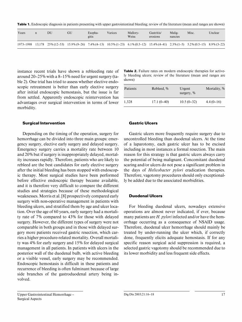
Table 1. Endoscopic diagnosis in patients presenting with upper gastrointestinal bleeding; review of the literature (mean and ranges are shown)
Years n DU GU Esopha-gitis
Varices Mallory-Weiss
Gastritis/erosions
Malig-nancies
Misc. Unclear
1973–1998 13,178 25% (12–53) 15.9% (9–26) 7.4% (4–13) 10.5% (1–23) 6.1% (0.5–12) 15.4% (4–41) 2.3% (1–5) 5.2% (0.5–15) 8.9% (3–22)
instance recent trials have shown a rebleeding rate ofaround 20–25% with a 8–15% need for urgent surgery (ta-ble 2). One trial has tried to assess whether elective endo-scopic retreatment is better than early elective surgeryafter initial endoscopic hemostasis, but the issue is farfrom settled. Apparently endoscopic reintervention hasadvantages over surgical intervention in terms of lowermorbidity.
Surgical Intervention
Depending on the timing of the operation, surgery forhemorrhage can be divided into three main groups: emer-gency surgery, elective early surgery and delayed surgery.Emergency surgery carries a mortality rate between 10and 20% but if surgery is inappropriately delayed, mortal-ity increases rapidly. Therefore, patients who are likely torebleed are the best candidates for early elective surgeryafter the initial bleeding has been stopped with endoscop-ic therapy. Most surgical studies have been performedbefore effective endoscopic therapy became available,and it is therefore very difficult to compare the differentstudies and strategies because of these methodologicalweaknesses. Morris et al. [8] prospectively compared earlysurgery with non-operative management in patients withbleeding ulcers, and stratified them by age and ulcer loca-tion. Over the age of 60 years, early surgery had a mortali-ty rate of 7% compared to 43% for those with delayedsurgery. However, the different types of surgery were notcomparable in both groups and in those with delayed sur-gery more patients received gastric resection, which car-ries a higher procedure-related mortality. Overall mortali-ty was 4% for early surgery and 15% for delayed surgicalmanagement in all patients. In patients with ulcers in theposterior wall of the duodenal bulb, with active bleedingor a visible vessel, early surgery may be recommended.Endoscopic hemostasis is difficult in these patients andrecurrence of bleeding is often fulminant because of largeside branches of the gastroduodenal artery being in-volved.
Table 2. Failure rates on modern endoscopic therapies for active-ly bleeding ulcers; review of the literature (mean and ranges areshown)
Patients Rebleed, % Urgentsurgery, %
Mortality, %
1,328 17.1 (0–40) 10.5 (0–32) 4.4 (0–16)
Gastric Ulcers
Gastric ulcers more frequently require surgery due touncontrolled bleeding than duodenal ulcers. At the timeof a laparotomy, each gastric ulcer has to be excisedincluding in most instances a formal resection. The mainreason for this strategy is that gastric ulcers always carrythe potential of being malignant. Concomitant duodenalscaring and/or ulcers do not pose a significant problem inthe days of Helicobacter pylori eradication therapies.Therefore, vagotomy procedures should only exceptional-ly be added due to the associated morbidities.
Duodenal Ulcers
For bleeding duodenal ulcers, nowadays extensiveoperations are almost never indicated, if ever, becausemany patients are H. pylori infected and/or have the hem-orrhage occurring as a consequence of NSAID usage.Therefore, duodenal ulcer hemorrhage should mainly betreated by under-running the ulcer which, if correctlydone, frequently elicits adequate hemostasis. If for anyspecific reason surgical acid suppression is required, aselected gastric vagotomy should be recommended due toits lower morbidity and less frequent side effects.
18 Dig Dis 2003;21:16–18 Lundell
Variceal Bleeding
In many institutions, operative portosystemic shuntsare no longer used as treatment for variceal bleeding.When the first-line options of non-selective ß-blockade orendoscopic treatment fail to control bleeding, a transjugu-lar intrahepatic portosystemic shunt (TIPS) is usuallyplaced. The advantages of TIPS are that it is non-opera-tive, it effectively decompresses the portal venous circula-tion during the short-term perspective and early compli-cations and procedure-related mortality are infrequent.However, late TIPS failure rates are high, with thrombo-sis or stenosis developing in approximately in 50% ofpatients within 1–2 years. Although TIPS revisions aresuccessful in many patients, in most series, rebleeding
rates after TIPS are considerably higher (10–30%) thanafter surgically constructed shunts (!10%). When patent,TIPS is usually a non-selective shunt with encephalopathyrates in most trials similar to those seen after a portocavalshunt. Despite these disadvantages, TIPS is an excellentoption for patients in whom endoscopic treatment isunsuccessful and who require relatively short-lasting por-tal decompression while on the waiting list for a livertransplant or whose anticipated survival is limited due tothe underlying liver disease.
Long-term survival has been particularly impressivefor patients undergoing surgery since the advent of livertransplantation, especially for those who are potential liv-er transplantation candidates and who can be salvaged bythis procedure when hepatic failure develops.
References and Suggested Reading
1 Vreeburg EM: Acute upper gastrointestinalbleeding. A prospective valuation of diagnosisant therapy in the Amsterdam area; thesis, Am-sterdam 1997.
2 Cook DJ, Guyatt GH, Salena BJ, Laine LA:Endoscopic therapy for acute nonvariceal up-per gastrointestinal hemorrhage: A meta-analy-sis. Gastroenterology 1992;102:139–148.
3 Labenz J, Borsch G. Role of Helicobacter pylorieradication in the prevention of peptic ulcerbleeding relapse. Digestion 1994;55:19–23.
4 Langman MJ: Epidemiologic evidence on theassociation between peptic ulceration and anti-inflammatory drug use. Gastroenterology1989;96(suppl):640–646.
5 Langman MJ, Morgan L, Worrall A: Use ofanti-inflammatory drugs by patients admittedwith small or large bowel perforations andhaemorrhage. Br Med J 1985;290:347–349.
6 Forrest JAH, Finlayson NDC, Sherman DJC:Endoscopy in gastro-intestinal bleeding. Lan-cet 1974;ii:391–397.
7 Hunt PS: Surgical management of bleedingchronic peptic ulcer. A 10-year study prospec-tive study. Ann Surg 1984;199:44–50.
8 Morris DI, Hawker PC, Brearly S, Simms M,Dykes PW, Keighley MR: Optimal timing ofoperation for bleeding peptic ulcer: Prospectiverandomised trial. Br Med J 1984;288:1277–1280.
9 Wheatley KE, Snyman JH, Brearley S, Keigh-ley MR, Dykes PW: Mortality in patients withbleeding peptic ulcer when those aged 60 orover are operated on early. BMJ 1990;330:272.
10 Pimpl W, Boeckl O, Heinerman M, Dapunt O:Emergency endoscopy: A basis for therapeuticdecisions in the treatment of severe gastroduo-denal bleeding. World J Surg 1989;13:592–597.
11 Heldwein W, Schreiner J, Pedrazzoli J, LehnertP: Is the Forrest classification a useful tool forplanning endoscopic therapy of bleeding pepticulcers? Endoscopy 1989;21:258–262.
12 Schein M, Gecelter G: Apache II score in mas-sive upper gastrointestinal hemorrhage frompeptic ulcer: Prognostic value and potentialclinical applications. Br J Surg 1989;76:733–736.
13 Saperas E, Pique JM, Perez Ayuso R, BordasJM, Teres J, Pera C: Conservative manage-ment of bleeding duodenal ulcer without a visi-ble vessel: Prognostic randomised trial. Br JSurg 1987;74:784–786.
14 Kubba AK, Choudari C, Rajgopal C, PalmerKR: The outcome of urgent surgery for majorpeptic ulcer hemorrhage following failed endo-scopic therapy. Eur J Gastroenterol Hepatol1996;8:1175–1178.
15 Qvist P, Arnesen KE, Jacobsen CD, RosselandAR: Endoscopic treatment and restrictive sur-gical policy in the management of peptic ulcerbleeding. Scand J Gastroenterol 1994;29:569–576.
16 Jordan PH: Surgery for peptic ulcer disease.Curr Probl Surg 1991;28:265–330.
17 Cochran TA: Bleeding peptic ulcer: Surgicaltherapy. Gastroent Clin North Am 1993;22:751–778.
18 Starlinger M, Becker HD: Upper gastrointesti-nal bleeding – indications and results in sur-gery. Hepatogastroenterology 1991;38:216–219.
19 Hasselgren G: Peptic ulcer bleeding 2000: Im-proved outcome; thesis, Gothenburg 1998.
20 Layton F, Rikkers MD: The changing spectrumof treatment for variceal bleeding. Ann Surgery1998;228:536–546.
21 Iwatsuki S, Starzl TE, Todo S, Gordon RD,Tzakis AG, Marsh JW, Makowka L, Koneru B,Stieber A, Klintmalm G, Husberg B, van ThielD: Liver transplantation in the treatment ofbleeding esophageal varices. Surgery 1988;104:697–705.
22 Mercado MA, Orozco H, Ramirez-Cisneros FJ,Hinojosa CA, Plata JJ, Alvarez-Tostado J: Di-minished morbidity and mortality in portal hy-pertension surgery: Relocation in the thera-peutic armamentarium. J Gastroenterol Surg2001;5:499–502.
Review Article
Dig Dis 2003;21:19–24DOI: 10.1159/000071335
Lower Gastrointestinal Bleeding –The Role of Endoscopy
Helmut Messmann
III. Medizinische Klinik, Klinikum Augsburg, Deutschland
Dr. H. Messmann, PDIII. Medizinische Klinik, Klinikum AugsburgPostfach 1019 20, DE–86009 Augsburg (Germany)Tel. +49 821 400 7351, Fax +49 821 400 3331E-Mail [email protected]
ABCFax + 41 61 306 12 34E-Mail [email protected]
© 2003 S. Karger AG, Basel0257–2753/03/0211–0019$19.50/0
Accessible online at:www.karger.com/ddi
Key WordsLower gastrointestinal bleeding W Endoscopy
AbstractEndoscopy is the method of choice in diagnosing thecause of lower gastrointestinal bleeding, and it offers theopportunity to treat patients suffering from lower gas-trointestinal bleeding. Endoscopic procedures must beintegrated with other approaches to reach a correct diag-nosis rapidly, safely, and economically. In all patients,evaluation begins with a history and physical examina-tion. The sequence of other tests depends on many fac-tors, especially the rate of bleeding. New technologiessuch as wireless capsule endoscopy will influence themanagement of patients with lower gastrointestinalbleeding.
Copyright © 2003 S. Karger AG, Basel
Definition
Lower intestinal bleeding is defined as acute or chronicabnormal blood loss distal to the ligament of Treitz. 10–20% of all gastrointestinal bleeding disorders occur distal
of this point, but bleeding of the small intestine is a rarecondition (3–5%).
Acute bleeding is arbitrarily defined as bleeding of !3days’ duration resulting in instability of vital signs, ane-mia, and/or need for blood transfusion [1, 2]. Hematoche-zia is the most common clinical symptom in patients withacute lower gastrointestinal bleeding (LGIB).
Chronic bleeding is defined as slow blood loss over aperiod of several days or longer presenting with symptomsof occult fecal blood, intermittent melena or scant he-matochezia. Occult bleeding means that the amounts ofblood in the feces are too small to be seen but detectableby chemical tests [3]. In 48–71% the source will be foundand an origin in the colorectum is to be expected in 20–30% [3].
Obscure gastrointestinal bleeding often presents asLGIB and means a bleeding from an unclear site, that per-sists or recurs after a negative initial or primary endosco-py. In 6% a repeat colonoscopy will identify the lesion inthe colon. Push enteroscopy will be helpful in 38–75% tofind the bleeding lesion, however, in two thirds the lesionsare detectable within the range of a conventional gastro-scope [3].
20 Dig Dis 2003;21:19–24 Messmann
General Aspects of Lower GastrointestinalBleeding
The incidence of lower gastrointestinal bleeding is onlyone fifth of that of the upper gastrointestinal tract and isestimated to be 21–27 cases per 100,000 adults/year [4,5]. LGIB usually is chronic and self-limiting and can betreated on an outpatient basis. Nevertheless, 21 of100,000 adults/year require hospitalization due to severebleeding. Among those, male gender and older patientssuffer from more severe LGIB [4]. There is a 200-foldincrease from the third to the ninth decade due to diver-ticulosis and angiodysplasia [6].
There is some evidence that upper gastrointestinalbleeding (UGIB) differs in acuity and severity fromLGIB: Patients with LGIB are significantly less in shock(19 vs. 35%, respectively), require fewer blood transfu-sions (36 vs. 64%) and have a significantly higher hemo-globin level (84 vs. 61%) [7, 8]. Similar to UGIB, themajority of bleeding disorders (80–85%) in the lower gas-trointestinal tract will stop spontaneously.
Mortality and morbidity increase with age. The overallmortality rate varies between 2.0 and 3.6%. Those pa-tients with bleeding episodes after hospital admissionhave significantly higher mortality rates (23.1%) com-pared to those who bleed before hospital admission [4].
Diagnosis
Endoscopy is the method of choice to diagnose and ifpossible to treat lower gastrointestinal bleeding. Whilecolonoscopy has been accepted for years in patients withchronic bleeding, urgent colonoscopy in acute bleedinghas been evaluated in the last few years and is meanwhilealso accepted as a safe method.
Before starting colonoscopy, history and clinical exam-ination should lead to a tentative diagnosis in order toplan the diagnostic procedures. In patients with chronicLGIB, colonoscopy is the first diagnostic step. The timepoint of colonoscopy is elective and optimal bowel prepa-ration is standard. If the origin of bleeding cannot bedetected, further steps are necessary.
In contrast, patients with acute LGIB are a challengefor optimal diagnostic procedures and there are still openquestions. It is generally accepted that in patients withhematochezia, especially in combination with circulationinstability, an UGIB must be excluded, since in 11%patients with suspected acute LGIB have their bleedingsource proximal to the ligament of Treitz. Although place-
ment of a nasogastric tube is safe and easy, it missesUGIB in 7%. The rate might even be higher in patientswith duodenal ulcer since pylorospasm can prevent refluxof blood into the stomach [9, 10].
While anoscopy and sigmoidoscopy were mandatoryprocedures in the pre-colonoscopy era, their role is lessobvious in the era of emergency and early colonoscopy. Inrecent years it could be demonstrated that in experiencedhands colonoscopy plays the same role in acute LGIB asupper gastrointestinal endoscopy in acute UGIB.
All patients with acute LGIB must be stabilized andcontraindications for colonoscopy are severe active in-flammation and also inadequate visual conditions. Fur-thermore, the endoscopy should be aborted if the patientbecomes unstable, the bleeding is so severe that identifica-tion of a bleeding source is impossible, or the risk of perfo-ration is too high. It is unclear whether urgent unpreparedcolonoscopy is more effective in detecting the bleedingsource as compared to prepared colonoscopy with a delayof several hours, since no randomized trial exists to thisquestion.
The amount, location or pattern of blood are impor-tant signs which make a detection of the bleeding sourcein a circumscribed segment of the colon easier. Most stud-ies, however, prefer bowel preparation before urgent co-lonoscopy. Their arguments are the frequent spontaneousbleeding stop and the improvement of visualization. Thebowel preparation can be performed by enemas and/orpolyethylene glycol solutions administered by mouth orvia a nasogastric tube. There exist no data that cleaningthe bowel might reactivate bleeding.
The detection rate of the bleeding source after bowelpreparation varies between 62 and 78%, and in patientswithout preparation the urgent unprepared colonoscopiescould identify the bleeding source in 76% [8, 11, 12].Therefore, urgent colonoscopy seems to be reasonable inmost patients.
In patients with intermittent or obscure gastrointesti-nal bleeding, wireless capsule endoscopy may become aninteresting diagnostic approach. In two trials, capsuleendoscopy was compared to X-ray of the small bowel orpush enteroscopy.
Costamagna et al. [13] could demonstrate that in 13patients with intermittent bleeding, the capsule was ableto detect the bleeding source in 11 cases while X-ray onlyin 1 case, respectively. Ell et al. [14] examined 32 pa-tients – the capsule detected a pathologic lesion in 66%and the X-ray in 28%, respectively.
Lower Gastrointestinal Bleeding –The Role of Endoscopy
Dig Dis 2003;21:19–24 21
Differential Diagnosis
Acute LGIB occurs most frequently in diverticular(35%), followed by vascular malformation (21%), colitis(16%), neoplasia/postpolypectomy (10%), anorectal dis-eases (5%), and small bowel (5%). In 11% the acute UGIBis falsely diagnosed as LGIB. Differential diagnosis ofsevere acute LGIB is mainly dependent on the patient’sage. While in children and young adults inflammatorybowel disease and Meckel’s diverticulum are the mainbleeding sources, diverticula are predominantly found inadults up to 60 years, and in the elderly, angiodysplasia isthe most common cause for severe LGIB.
Diverticular DiseaseThe true incidence of diverticular disease is difficult to
measure, mainly because most patients are asymptomat-ic. The incidence however clearly increases with age from10% under 40 years to an estimated 50–66% in patientsolder than 80 [15, 16]. The estimated risk of a severebleeding has been reported to be 3–5% [16, 17], butincluding milder forms of bleeding a risk up to 48% hasbeen described [18]. Among LGIB disorders, diverticulaare the cause in 15–27% [19]. The clinical presentation ofpatients with diverticular bleeding is mostly abrupt witha painless onset, associated with mild lower abdominalcramps and the urge to defecate. The stool consists of redvoluminous or maroon blood or clots. Melena is uncom-mon [16]. Approximately 80% of the bleeding episodesstop spontaneously. The risk of a first rebleeding is 25%but increases with definite bleeding stigmata (activebleeding, nonbleeding visible vessel, adherent clot: 67, 50and 43%, respectively) [19–21]. A third bleed after a sec-ond episode will occur in 50%, therefore surgical resec-tion is recommended after a second bleeding episode[16].
ColitisLGIB from IBD are rarely life-threatening (0.1% ulcer-
ative colitis, 1.3% Crohn’s disease), bleeding stops mostlyspontaneously and endoscopic treatment is not necessaryin most cases with diffuse bleeding. Bleeding from isch-emic colitis occurs mainly in elderly patients (165 years)and is associated with pain. Vascular diseases and atrialfibrillation are risk factors which are associated with isch-emic colitis. Patients with infectious colitis suffer mainlyfrom diffuse bleeding similar to ulcerative colitis. Amongbacteria, Salmonella, Shigella, Yersinia, Campylobacterand Escherichia coli, especially enterohemorrhagic E. coli(EHEC), most notably 0157:H7, are the most frequent
infectious agents. Acute radiation colitis occurs a few daysafter radiation but bloody diarrhea is uncommon at thistime point. Most patients complain of transient diarrheaand tenesmus. The endoscopic picture is similar to ulcer-ative colitis with edema, fragility, hemorrhage and someerosions or ulcers [16]. The clinical manifestation ofchronic radiation colitis occurs after 1–2 years. Pale mu-cosa with teleangiectasia and rarefaction of mucosal ves-sels is typical in mild forms. In severe radiation colitis,excessive hemorrhage, necrosis and ulcerations occurleading to extensive bleeding [16].
NeoplasiaAcute bleeding in colon cancer or polyps is not fre-
quent but has been described in 2–33 and 5–11%, respec-tively [16, 17]. The majority of these lesions present withchronic bleeding. Among patients with LGIB, postpoly-pectomy bleeding occurs in about 4% [4]. Bleeding occurseither immediately (within 24 h) or delayed (occurring aslong as 21 days after colonoscopy) [22]. The risk of bleed-ing depends on several factors: polyp size, type of polyp(pedunculated or sessile), hemostatic disorders, medica-tion and endoscopist’s experience influence the postpol-ypectomy hemorrhage risk. Although the use of NSAIDdid increase the incidence of minor self-limited bleeding,an increase in the rate of major bleeding was not observed[23]. The overall risk of bleeding after polypectomy rangesfrom 0.4 to 2% [24].
AngiodysplasiaIn patients with LGIB, angiodysplasia is the responsi-
ble bleeding disorder in 3–12% [4, 5]. Bleeding can bechronic, slow, intermittent or recurrent. Massive bleedinghas been described in 2% of the cases, but bleeding stopsspontaneously in up to 90%. Unfortunately the rebleedingrate is high and can reach values up to 85% [16].
The prevalence of angiodysplasia among healthyasymptomatic people was 0.83%. 87% of these usuallysmall lesions (4 mm) were located in the right colon, andthere was no risk for later bleeding [25]. Angiodysplasiaoften appears together with systemic diseases such as car-diovascular disorders (aortic stenosis) or chronic renalfailure [26, 27]. However, there exist also systematicexaminations which could not confirm an association ofangiodysplasia and aortic valve disease [28]. Capsuleendoscopy may improve the detection of these lesions inthe small bowel in the near future.
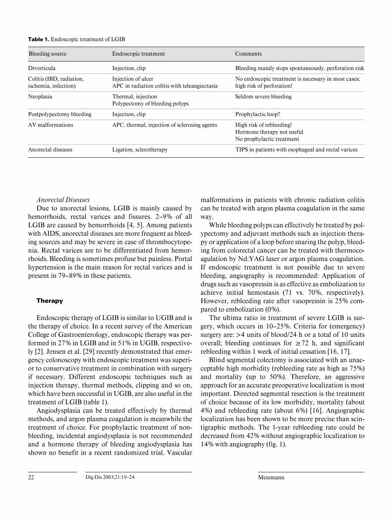
22 Dig Dis 2003;21:19–24 Messmann
Table 1. Endoscopic treatment of LGIB
Bleeding source Endoscopic treatment Comments
Diverticula Injection, clip Bleeding mainly stops spontaneously, perforation risk
Colitis (IBD, radiation,ischemia, infection)
Injection of ulcerAPC in radiation colitis with teleangiectasia
No endoscopic treatment is necessary in most cases;high risk of perforation!
Neoplasia Thermal, injectionPolypectomy of bleeding polyps
Seldom severe bleeding
Postpolypectomy bleeding Injection, clip Prophylactic loop?
AV malformations APC, thermal, injection of sclerosing agents High risk of rebleeding!Hormone therapy not usefulNo prophylactic treatment
Anorectal diseases Ligation, sclerotherapy TIPS in patients with esophageal and rectal varices
Anorectal DiseasesDue to anorectal lesions, LGIB is mainly caused by
hemorrhoids, rectal varices and fissures. 2–9% of allLGIB are caused by hemorrhoids [4, 5]. Among patientswith AIDS, anorectal diseases are more frequent as bleed-ing sources and may be severe in case of thrombocytope-nia. Rectal varices are to be differentiated from hemor-rhoids. Bleeding is sometimes profuse but painless. Portalhypertension is the main reason for rectal varices and ispresent in 79–89% in these patients.
Therapy
Endoscopic therapy of LGIB is similar to UGIB and isthe therapy of choice. In a recent survey of the AmericanCollege of Gastroenterology, endoscopic therapy was per-formed in 27% in LGIB and in 51% in UGIB, respective-ly [2]. Jensen et al. [29] recently demonstrated that emer-gency colonoscopy with endoscopic treatment was superi-or to conservative treatment in combination with surgeryif necessary. Different endoscopic techniques such asinjection therapy, thermal methods, clipping and so on,which have been successful in UGIB, are also useful in thetreatment of LGIB (table 1).
Angiodysplasia can be treated effectively by thermalmethods, and argon plasma coagulation is meanwhile thetreatment of choice. For prophylactic treatment of non-bleeding, incidental angiodysplasia is not recommendedand a hormone therapy of bleeding angiodysplasia hasshown no benefit in a recent randomized trial. Vascular
malformations in patients with chronic radiation colitiscan be treated with argon plasma coagulation in the sameway.
While bleeding polyps can effectively be treated by pol-ypectomy and adjuvant methods such as injection thera-py or application of a loop before snaring the polyp, bleed-ing from colorectal cancer can be treated with thermoco-agulation by Nd:YAG laser or argon plasma coagulation.If endoscopic treatment is not possible due to severebleeding, angiography is recommended: Application ofdrugs such as vasopressin is as effective as embolization toachieve initial hemostasis (71 vs. 70%, respectively).However, rebleeding rate after vasopressin is 25% com-pared to embolization (0%).
The ultima ratio in treatment of severe LGIB is sur-gery, which occurs in 10–25%. Criteria for (emergency)surgery are: 14 units of blood/24 h or a total of 10 unitsoverall; bleeding continues for 672 h, and significantrebleeding within 1 week of initial cessation [16, 17].
Blind segmental colectomy is associated with an unac-ceptable high morbidity (rebleeding rate as high as 75%)and mortality (up to 50%). Therefore, an aggressiveapproach for an accurate preoperative localization is mostimportant. Directed segmental resection is the treatmentof choice because of its low morbidity, mortality (about4%) and rebleeding rate (about 6%) [16]. Angiographiclocalization has been shown to be more precise than scin-tigraphic methods. The 1-year rebleeding rate could bedecreased from 42% without angiographic localization to14% with angiography (fig. 1).
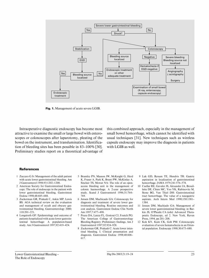
Lower Gastrointestinal Bleeding –The Role of Endoscopy
Dig Dis 2003;21:19–24 23
Fig. 1. Management of acute severe LGIB.
Intraoperative diagnostic endoscopy has become mostattractive to examine the small or large bowel with entero-scopes or colonoscopes after laparotomy, pleating of thebowel on the instrument, and translumination. Identifica-tion of bleeding sites has been possible in 83–100% [30].Preliminary studies report on a theoretical advantage of
this combined approach, especially in the management ofsmall bowel hemorrhage, which cannot be identified withusual techniques [31]. New techniques such as wirelesscapsule endoscopy may improve the diagnosis in patientswith LGIB as well.
References
1 Zuccaro G Jr: Management of the adult patientwith acute lower gastrointestinal bleeding. AmJ Gastroenterol 1998;93:1202–1208.
2 American Society for Gastrointestinal Endos-copy: The role of endoscopy in the patient withlower gastrointestinal bleeding. GastrointestEndosc 1998;48:685–688.
3 Zuckerman GR, Prakash C, Askin MP, LewisBS: AGA technical review on the evaluationand management of occult and obscure gas-trointestinal bleeding. Gastroenterology 2000;118:201–221.
4 Longstreth GF: Epidemiology and outcome ofpatients hospitalized with acute lower gastroin-testinal hemorrhage: A population-basedstudy. Am J Gastroenterol 1997;92:419–424.
5 Bramley PN, Masson JW, McKnight G, HerdK, Fraser A, Park K, Brunt PW, McKinlay A,Sinclair TS, Mowat NA: The role of an open-access bleeding unit in the management ofcolonic haemorrhage. A 2-year prospectivestudy. Scand J Gastroenterol 1996;31:764–769.
6 Jensen DM, Machicado GA: Colonoscopy fordiagnosis and treatment of severe lower gas-trointestinal bleeding. Routine outcomes andcost analysis. Gastrointest Endosc Clin NorthAm 1997;7:477–498.
7 Peura DA, Lanza FL, Gostout CJ, Foutch PG:The American College of GastroenterologyBleeding Registry: Preliminary findings. Am JGastroenterol 1997;92:924–928.
8 Zuckerman GR, Prakash C: Acute lower intes-tinal bleeding. I. Clinical presentation anddiagnosis. Gastrointest Endosc 1998;48:606–617.
9 Luk GD, Bynum TE, Hendrix TR: Gastricaspiration in localization of gastrointestinalhemorrhage. JAMA 1979;241:576–578.
10 Cuellar RE, Gavaler JS, Alexander JA, Brouil-lette DE, Chien MC, Yoo YK, Rabinovitz M,Stone BG, Van Thiel DH: Gastrointestinaltract hemorrhage. The value of a nasogastricaspirate. Arch Intern Med 1990;150:1381–1384.
11 Jensen DM, Machiado GA: Management ofsevere lower gastrointestinal bleeding; in Bar-kin JS, O’Phealn CA (eds): Advanced Thera-peutic Endoscopy, ed 2. New York, RavenPress, 1994, pp 201–208.
12 Kok KY, Kum CK, Goh PM: Colonoscopicevaluation of severe hematochezia in an Orien-tal population. Endoscopy 1998;30:675–680.
24 Dig Dis 2003;21:19–24 Messmann
13 Costamagna G, Shah SK, Riccioni ME, Fos-chia F, Mutignani M, Perri V, Vecchioli A, Bri-zi MG, Picciocchi A, Marano P: A prospectivetrial comparing small bowel radiographs andvideo capsule for suspected small bowel dis-ease. Gastroenterology 2002;123:999–1005.
14 Ell C, Remke S, May A, Helou L, Henrich R,Mayer G: The first prospective controlled trialcomparing wireless capsule endoscopy withpush enteroscopy in chronic gastrointestinalbleeding. Endoscopy 2002;34:685–689.
15 Laine L: Acute and chronic gastrointestinalbleeding; in Feldman M, Scharschmidt BF,Sleisinger MH (eds): Sleisinger’s & Fordtran’sGastrointestinal and Liver Disease. Philadel-phia, Saunders, 1999, pp 198–219.
16 Vernava AM 3rd, Moore BA, Longo WE, John-son FE: Lower gastrointestinal bleeding. DisColon Rectum 1997;40:846–858.
17 Zuckerman GR, Prakash C: Acute lower intes-tinal bleeding. II. Etiology, therapy and out-comes. Gastrointest Endosc 1999;49:228–238.
18 Winkler R: Ursachen und Klinik der peranalenBlutung; in Häring R (ed): GastrointestinaleBlutung. Berlin, Blackwell, 1990, pp 313–319.
19 Almy TP, Howell DA: Medical progress. Di-verticular disease of the colon. N Engl J Med1980;302:324–331.
20 So JB, Kok K, Ngoi SS: Right-sided colonicdiverticular disease as a source of lower gas-trointestinal bleeding. Am Surg 1999;65:299–302.
21 Stollman NH, Raskin JB: Diagnosis and man-agement of diverticular disease of the colon inadults. Ad Hoc Practice Parameters Commit-tee of the American College of Gastroenterol-ogy. Am J Gastroenterol 1999;94:3110–3121.
22 Gibbs DH, Opelka FG, Beck DE, Hicks TC,Timmcke AE, Gathright JB Jr: Postpolypecto-my colonic hemorrhage. Dis Colon Rectum1996;39:806–810.
23 Shiffman ML, Farrel MT, Yee YS: Risk ofbleeding after endoscopic biopsy or polypecto-my in patients taking aspirin or other NSAIDS.Gastrointest Endosc 1994;40:458–462.
24 Mergener K, Baillie J: Complications of endos-copy. Endoscopy 1998;30:230–243.
25 Foutch PG: Angiodysplasia of the gastrointesti-nal tract. Am J Gastroenterol 1993;88:807–818.
26 Weaver GA, Alpern HD, Davis JS, RamseyWH, Reichelderfer M: Gastrointestinal angio-dysplasia associated with aortic valve disease:Part of a spectrum of angiodysplasia of the gut.Gastroenterology 1979;77:1–11.
27 Foutch PG: Angiodysplasia of the gastrointesti-nal tract. Am J Gastroenterol 1993;88:807–818.
28 Bhutani MS, Gupta SC, Markert RJ, Barde CJ,Donese R, Gopalswamy N: A prospective con-trolled evaluation of endoscopic detection ofangiodysplasia and its association with aorticvalve disease. Gastrointest Endosc 1995;42:398–402.
29 Jensen DM, Machicado GA, Jutabha R, Ko-vacs TO: Urgent colonoscopy for the diagnosisand treatment of severe diverticular hemor-rhage. N Engl J Med 2000;342:78–82.
30 Lewis BS: Small intestinal bleeding. Gastroen-terol Clin North Am 2000;29:67–95.
31 Ingresso M, Pete F, Pisani A, et al: Laparosco-pically-assisted total enteroscopy: A new ap-proach to small intestinal disease. GastrointestEndosc 1999;49:651–653.
Review Article
Dig Dis 2003;21:25–29DOI: 10.1159/000071336
Management of Acute Cholangitis
Dirk J. Gouma
Department of Surgery, Academic Medical Center, Amsterdam, The Netherlands
Prof. Dirk J. Gouma, MDDepartment of Surgery, Academic Medical CenterMeibergdreef 9NL–1105 Amsterdam (The Netherlands)Fax +31 20 5669 243, E-Mail [email protected]
ABCFax + 41 61 306 12 34E-Mail [email protected]
© 2003 S. Karger AG, Basel0257–2753/03/0211–0025$19.50/0
Accessible online at:www.karger.com/ddi
Key WordsAcute cholangitis W Endoscopic sphincterotomy W
Laparoscopic CBD exploration W Common bile ductstones
AbstractEndoscopic sphincterotomy (ES) is the treatment ofchoice for patients with (severe) acute cholangitis. For fitpatients without co-morbidity with mild cholangitis andCBD stones with a gallbladder in situ, the one-stage lapa-roscopic approach could be considered as an alternativein centers with sufficient experience. The results of bothprocedures are comparable. Open surgery is relativelysafe. It has a high success rate, good/excellent long-termresults, but is not very attractive for the patient andshould not be used routinely nowadays. Therefore, theindication should be limited for management of severecomplications after ES as perforations of the duodenum,large CBD stones and patients with Mirizzi’s syndrome orintrahepatic stones with stenosis of the bile duct. ES asprimary treatment for CBD stones should be followed bylaparoscopic cholecystectomy in ‘fit’ patients. In patientswith malignant disease, particularly after repeated stentfailure and subsequent cholangitis, bypass surgeryshould be considered in patients with a life expectancy of13 months.
Copyright © 2003 S. Karger AG, Basel
After the introduction of endoscopic sphincterotomy(ES) and percutaneous drainage procedures, the indica-tion for different surgical and non-surgical approaches ofbiliary disorders changed radically and is still subject ofcontroversy. There is however general agreement thatpatients with severe cholangitis should preferably betreated non-surgically by ES instead of (open) CBD explo-ration after a randomized trial of Lai et al. [1] clearlyshowed a reduction in morbidity from 66 to 34% and areduction in hospital mortality from 32 to 10%. Recently,another trial has been published showing that even in theabsence of CBD stones during the attack of cholangitis,ES decreased the duration of fever in patients with acutecholangitis and reduced hospital stay from 4.3 to 2.2 daysand 9.1 to 8.1 days, respectively [2]. However, it did notdecrease the incidence of recurrent acute cholangitis dur-ing follow-up.
The development of high-quality ES in general hospi-tals has resulted in a decrease of surgical procedures foracute cholangitis as well as for the initial management ofCBD stones without cholangitis in many European coun-tries, particularly in The Netherlands and Germany. InThe Netherlands only 20% of patients with CBD stonesunderwent a surgical approach during the past decade. Aminority of these patients suffered from severe cholangi-tis, the others having symptomatic CBD stones.
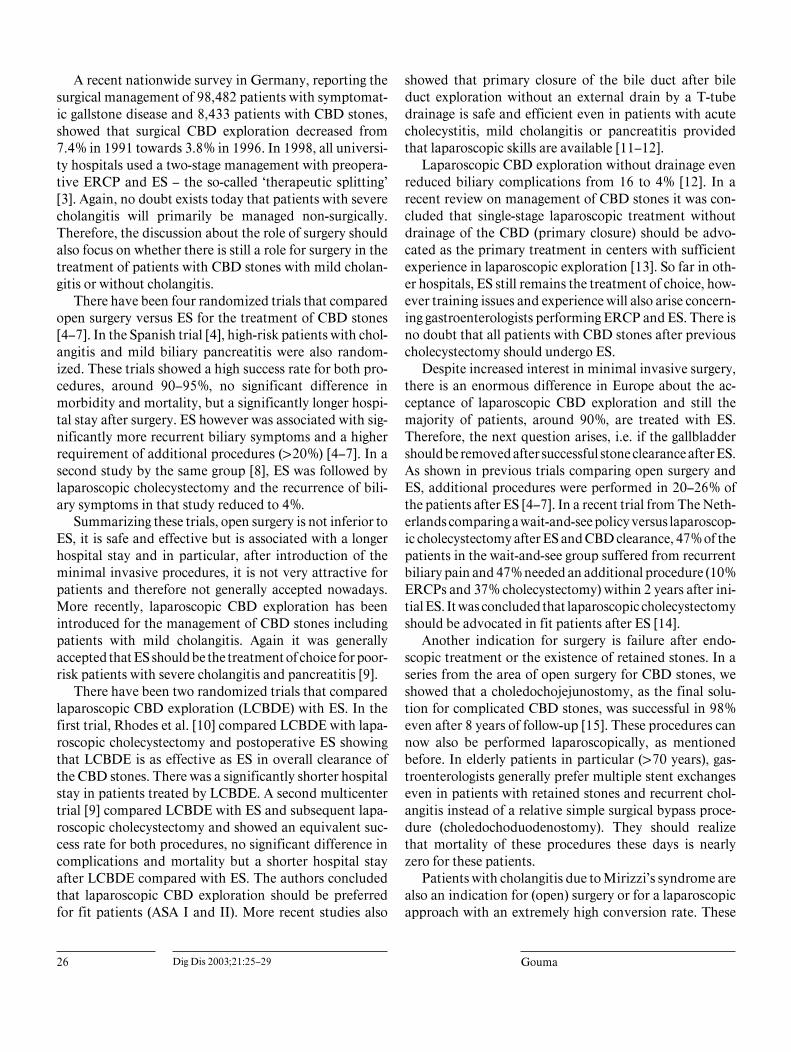
26 Dig Dis 2003;21:25–29 Gouma
A recent nationwide survey in Germany, reporting thesurgical management of 98,482 patients with symptomat-ic gallstone disease and 8,433 patients with CBD stones,showed that surgical CBD exploration decreased from7.4% in 1991 towards 3.8% in 1996. In 1998, all universi-ty hospitals used a two-stage management with preopera-tive ERCP and ES – the so-called ‘therapeutic splitting’[3]. Again, no doubt exists today that patients with severecholangitis will primarily be managed non-surgically.Therefore, the discussion about the role of surgery shouldalso focus on whether there is still a role for surgery in thetreatment of patients with CBD stones with mild cholan-gitis or without cholangitis.
There have been four randomized trials that comparedopen surgery versus ES for the treatment of CBD stones[4–7]. In the Spanish trial [4], high-risk patients with chol-angitis and mild biliary pancreatitis were also random-ized. These trials showed a high success rate for both pro-cedures, around 90–95%, no significant difference inmorbidity and mortality, but a significantly longer hospi-tal stay after surgery. ES however was associated with sig-nificantly more recurrent biliary symptoms and a higherrequirement of additional procedures (120%) [4–7]. In asecond study by the same group [8], ES was followed bylaparoscopic cholecystectomy and the recurrence of bili-ary symptoms in that study reduced to 4%.
Summarizing these trials, open surgery is not inferior toES, it is safe and effective but is associated with a longerhospital stay and in particular, after introduction of theminimal invasive procedures, it is not very attractive forpatients and therefore not generally accepted nowadays.More recently, laparoscopic CBD exploration has beenintroduced for the management of CBD stones includingpatients with mild cholangitis. Again it was generallyaccepted that ES should be the treatment of choice for poor-risk patients with severe cholangitis and pancreatitis [9].
There have been two randomized trials that comparedlaparoscopic CBD exploration (LCBDE) with ES. In thefirst trial, Rhodes et al. [10] compared LCBDE with lapa-roscopic cholecystectomy and postoperative ES showingthat LCBDE is as effective as ES in overall clearance ofthe CBD stones. There was a significantly shorter hospitalstay in patients treated by LCBDE. A second multicentertrial [9] compared LCBDE with ES and subsequent lapa-roscopic cholecystectomy and showed an equivalent suc-cess rate for both procedures, no significant difference incomplications and mortality but a shorter hospital stayafter LCBDE compared with ES. The authors concludedthat laparoscopic CBD exploration should be preferredfor fit patients (ASA I and II). More recent studies also
showed that primary closure of the bile duct after bileduct exploration without an external drain by a T-tubedrainage is safe and efficient even in patients with acutecholecystitis, mild cholangitis or pancreatitis providedthat laparoscopic skills are available [11–12].
Laparoscopic CBD exploration without drainage evenreduced biliary complications from 16 to 4% [12]. In arecent review on management of CBD stones it was con-cluded that single-stage laparoscopic treatment withoutdrainage of the CBD (primary closure) should be advo-cated as the primary treatment in centers with sufficientexperience in laparoscopic exploration [13]. So far in oth-er hospitals, ES still remains the treatment of choice, how-ever training issues and experience will also arise concern-ing gastroenterologists performing ERCP and ES. There isno doubt that all patients with CBD stones after previouscholecystectomy should undergo ES.
Despite increased interest in minimal invasive surgery,there is an enormous difference in Europe about the ac-ceptance of laparoscopic CBD exploration and still themajority of patients, around 90%, are treated with ES.Therefore, the next question arises, i.e. if the gallbladdershould be removed after successful stone clearance after ES.As shown in previous trials comparing open surgery andES, additional procedures were performed in 20–26% ofthe patients after ES [4–7]. In a recent trial from The Neth-erlands comparing a wait-and-see policy versus laparoscop-ic cholecystectomy after ES and CBD clearance, 47% of thepatients in the wait-and-see group suffered from recurrentbiliary pain and 47% needed an additional procedure (10%ERCPs and 37% cholecystectomy) within 2 years after ini-tial ES. It was concluded that laparoscopic cholecystectomyshould be advocated in fit patients after ES [14].
Another indication for surgery is failure after endo-scopic treatment or the existence of retained stones. In aseries from the area of open surgery for CBD stones, weshowed that a choledochojejunostomy, as the final solu-tion for complicated CBD stones, was successful in 98%even after 8 years of follow-up [15]. These procedures cannow also be performed laparoscopically, as mentionedbefore. In elderly patients in particular (170 years), gas-troenterologists generally prefer multiple stent exchangeseven in patients with retained stones and recurrent chol-angitis instead of a relative simple surgical bypass proce-dure (choledochoduodenostomy). They should realizethat mortality of these procedures these days is nearlyzero for these patients.
Patients with cholangitis due to Mirizzi’s syndrome arealso an indication for (open) surgery or for a laparoscopicapproach with an extremely high conversion rate. These
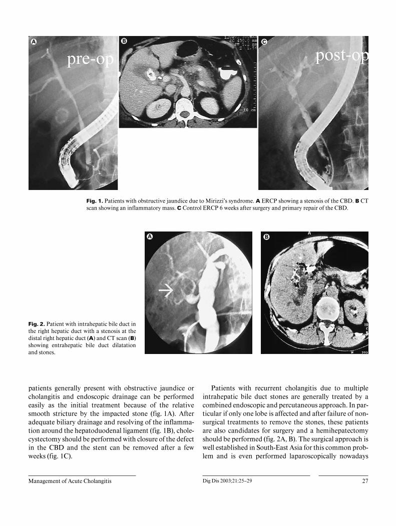
Management of Acute Cholangitis Dig Dis 2003;21:25–29 27
Fig. 1. Patients with obstructive jaundice due to Mirizzi’s syndrome. A ERCP showing a stenosis of the CBD. B CTscan showing an inflammatory mass. C Control ERCP 6 weeks after surgery and primary repair of the CBD.
Fig. 2. Patient with intrahepatic bile duct inthe right hepatic duct with a stenosis at thedistal right hepatic duct (A) and CT scan (B)showing entrahepatic bile duct dilatationand stones.
patients generally present with obstructive jaundice orcholangitis and endoscopic drainage can be performedeasily as the initial treatment because of the relativesmooth stricture by the impacted stone (fig. 1A). Afteradequate biliary drainage and resolving of the inflamma-tion around the hepatoduodenal ligament (fig. 1B), chole-cystectomy should be performed with closure of the defectin the CBD and the stent can be removed after a fewweeks (fig. 1C).
Patients with recurrent cholangitis due to multipleintrahepatic bile duct stones are generally treated by acombined endoscopic and percutaneous approach. In par-ticular if only one lobe is affected and after failure of non-surgical treatments to remove the stones, these patientsare also candidates for surgery and a hemihepatectomyshould be performed (fig. 2A, B). The surgical approach iswell established in South-East Asia for this common prob-lem and is even performed laparoscopically nowadays
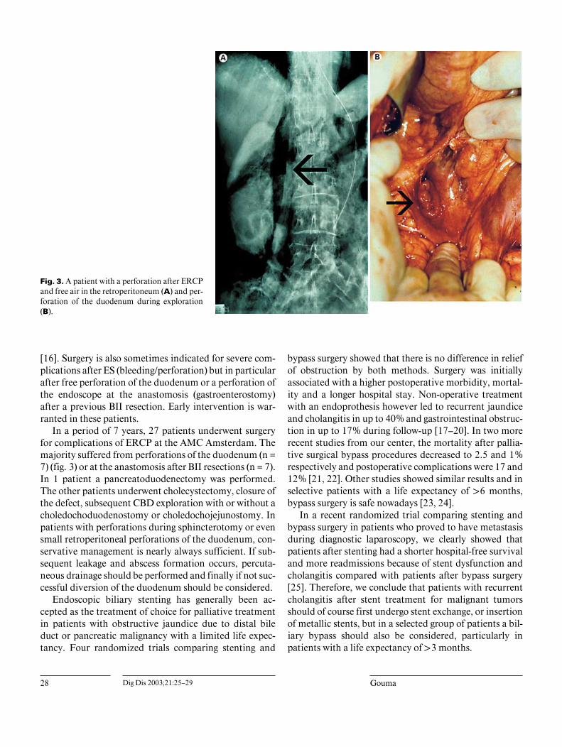
28 Dig Dis 2003;21:25–29 Gouma
Fig. 3. A patient with a perforation after ERCPand free air in the retroperitoneum (A) and per-foration of the duodenum during exploration(B).
[16]. Surgery is also sometimes indicated for severe com-plications after ES (bleeding/perforation) but in particularafter free perforation of the duodenum or a perforation ofthe endoscope at the anastomosis (gastroenterostomy)after a previous BII resection. Early intervention is war-ranted in these patients.
In a period of 7 years, 27 patients underwent surgeryfor complications of ERCP at the AMC Amsterdam. Themajority suffered from perforations of the duodenum (n =7) (fig. 3) or at the anastomosis after BII resections (n = 7).In 1 patient a pancreatoduodenectomy was performed.The other patients underwent cholecystectomy, closure ofthe defect, subsequent CBD exploration with or without acholedochoduodenostomy or choledochojejunostomy. Inpatients with perforations during sphincterotomy or evensmall retroperitoneal perforations of the duodenum, con-servative management is nearly always sufficient. If sub-sequent leakage and abscess formation occurs, percuta-neous drainage should be performed and finally if not suc-cessful diversion of the duodenum should be considered.
Endoscopic biliary stenting has generally been ac-cepted as the treatment of choice for palliative treatmentin patients with obstructive jaundice due to distal bileduct or pancreatic malignancy with a limited life expec-tancy. Four randomized trials comparing stenting and
bypass surgery showed that there is no difference in reliefof obstruction by both methods. Surgery was initiallyassociated with a higher postoperative morbidity, mortal-ity and a longer hospital stay. Non-operative treatmentwith an endoprothesis however led to recurrent jaundiceand cholangitis in up to 40% and gastrointestinal obstruc-tion in up to 17% during follow-up [17–20]. In two morerecent studies from our center, the mortality after pallia-tive surgical bypass procedures decreased to 2.5 and 1%respectively and postoperative complications were 17 and12% [21, 22]. Other studies showed similar results and inselective patients with a life expectancy of 16 months,bypass surgery is safe nowadays [23, 24].
In a recent randomized trial comparing stenting andbypass surgery in patients who proved to have metastasisduring diagnostic laparoscopy, we clearly showed thatpatients after stenting had a shorter hospital-free survivaland more readmissions because of stent dysfunction andcholangitis compared with patients after bypass surgery[25]. Therefore, we conclude that patients with recurrentcholangitis after stent treatment for malignant tumorsshould of course first undergo stent exchange, or insertionof metallic stents, but in a selected group of patients a bil-iary bypass should also be considered, particularly inpatients with a life expectancy of 13 months.
Management of Acute Cholangitis Dig Dis 2003;21:25–29 29
References
1 Lai EC, Mok FP, Tan ES, Lo CM, Fan ST, YouKT, Wong J: Endoscopic biliary drainage forsevere acute cholangitis. N Engl J Med 1992;326:1582–1586.
2 Hui CK, Lai KC, Wong WM, Yuen MF, LamSK, Lai CL: A randomised controlled trial ofendoscopic sphincterotomy in acute cholangi-tis without common bile duct stones. Gut 2002;51:245–247.
3 Huttl TP, Hrdina C, Geiger TK, et al: Manage-ment of common bile duct stones – Results of anationwide survey with analysis of 8,433 com-mon bile duct explorations in Germany. Zen-tralbl Chir 2002;127:282–289.
4 Targarona EM, Ayuso RM, Bordas JM, et al:Randomised trial of endoscopic sphincteroto-my with gallbladder left in situ versus open sur-gery for common bile duct calculi in high-riskpatients. Lancet 1996;347:926–929.
5 Hammerstrom LE, Holmin T, Stridberg H.Ihse I: Long-term follow-up of a prospectiverandomized study of endoscopic versus surgi-cal treatment of bile duct calculi in patientswith gallbladder in situ. Br J Surg 1995;82:1516–1521.
6 Stain SC, Cohen H, Tsuishoysha M, DonovanAJ: Choledocholithiasis. Endoscopic sphincter-otomy or common bile duct exploration. AnnSurg 1991;213:627–633; discussion: 633–634.
7 Suc B, Escat J, Cherqui D, Fourtanier G, HayJM, Fingerhut A, et al: Surgery vs. endoscopyas primary treatment in symptomatic patientswith suspected common bile duct stones: Amulticenter randomized trial. French Associa-tions for Surgical Research. Arch Surg 1998;133:702–708.
8 Trias M, Targarona EM, Ros E, Bordas JM,Perez Ayuso RM, Balague C, Pros I, Teres J:Prospective evaluation of a minimally invasiveapproach for treatment of bile-duct calculi inrisk patient. Surg Endosc 1997;11:632–635.
9 Cuschieri A, Lezoche E, Morino M, Croce E,Lacy A, Toouli J, et al: EAES multicenter pro-spective randomized trial comparing two-stagevs. single-stage management of patients withgallstone disease and ductal calculi. Surg En-dosc 1999;13:952–957.
10 Rhodes M. Sussman L, Cohen L, Lewis MP:Randomised trial of laparoscopic explorationof common bile duct versus postoperative en-doscopic retrograde cholangiography for com-mon bile duct stones. Lancet 1998;351:159–161.
11 Decker G, Borie F, Millat B, Berthou JC, De-leuze A, Drouard F, Guillon F, Rodier JG, Fin-gerhut A: One hundred laparoscopic choledo-chotomies with primary closure of the commonbile duct. Surg Endosc 2003;17:12–18.
12 Thompson MH, Tranter SE: Laparoscopic ex-ploration of the common bile duct. The resultsof an all-comers policy. Br J Surg 2002;89:1608–1612.
13 Tranter SE, Thompson MH: Comparison ofendoscopic sphincterotomy and laparoscopicexploration of the common bile duct. Br J Surg2002;89:1495–1504.
14 Boerma D, Rauws EA, Keulemans YC, JanssenIM, Bolwerk CJ, Timmer R, Boerma EJ, Ober-top H, Huibregtse K, Gouma DJ: Wait-and-seepolicy or laparoscopic cholecystectomy afterendoscopic sphincterotomy for bile ductstones: A randomised trial. Lancet 2002;360:761–765.
15 Gouma DJ, Konsten J, Soeters PB, et al: Long-term follow-up after choledochojejunostomyfor bile duct stones with complex clearance ofthe bile duct. Br J Surg 1989;76:451–453.
16 Tang CN, Li MK: Hand-assisted laparoscopicsegmentectomy in recurrent pyogenic cholangi-tis. Surg Endosc 2003;17:324–327.
17 Bornman PC, Harries-Jones EP, Tobias R, etal: Prospective controlled trial of transhepaticbiliary endoprosthesis versus bypass surgeryfor incurable carcinoma of head of pancreas.Lancet 1986;i:69–71.
18 Shepard HA, Royle G, Ross APR, et al: Endo-scopic biliary endoprosthesis in the palliationof malignant obstruction of the distal commonbile duct: A randomized trial. Br J Surg 1988;75:1166–1168.
19 Andersen JR, Sorensen SM, Kruse A, et al:Randomized trial of endoscopic endoprosthe-sis versus operative bypass in malignant ob-structive jaundice. Gut 1989;30:1132–1135.
20 Smith AC, Dowsett JF, Russell RCG, et al:Randomised trial of endoscopic stenting versussurgical bypass in malignant low bile duct ob-struction. Lancet 1994;344:1655–1660.
21 Van Wagensveld BA, Coene PP, Van GulikTM, et al: Outcome of palliative biliary andgastric bypass surgery for pancreatic head car-cinoma in 126 patients. Br J Surg 1997;84:1402–1406.
22 Van Geenen R, Keyzer-Dekker CM, Van Tien-hoven G, et al: Pain management of patientswith unresectable peripancreatic carcinoma.World J Surg 2002;26:715–720.
23 Lillemoe KD, Sauter PK, Pitt HA, et al: Cur-rent status of surgical palliation of periampulla-ry carcinoma. Surg Gynecol Obstet 1993;176:1–10.
24 Parks RW, Johnston GW, Rowlands BJ: Surgi-cal biliary bypass for benign and malignantextrahepatic biliary tract disease. Br J Surg1997;84:488–492.
25 Nieveen Van Dijkum EJ, Romijn MG, TerweeCB, De Wit LT, Van Der Meulen JH, LamerisHS, Rauws EA, Obertop H, Van Eyck CH, Bos-suyt PM, Gouma DJ: Laparoscopic staging andsubsequent palliation in patients with peripan-creatic carcinoma. Ann Surg 2003;237:66–73.
Review Article
Dig Dis 2003;21:30–37DOI: 10.1159/000071337
Acute Pancreatitis: Treatment Strategies
Stefan Kahl Sandra Zimmermann Peter Malfertheiner
Department of Gastroenterology, Hepatology and Infectious Diseases, Otto von Guericke University,Magdeburg, Germany
Stefan Kahl, MDDepartment of Gastroenterology, Otto von Guericke University MagdeburgLeipziger Strasse 44, DE–39120 Magdeburg (Germany)Tel. +49 391 6713100, Fax +49 391 6713105E-Mail [email protected]
ABCFax + 41 61 306 12 34E-Mail [email protected]
© 2003 S. Karger AG, Basel0257–2753/03/0211–0030$19.50/0
Accessible online at:www.karger.com/ddi
Key WordsPancreatitis W Endoscopy W Pain W Drug therapy W Enteralnutrition
AbstractAcute pancreatitis is an acute painful abdominal diseaseof sudden onset that ranges from a mild and self-limitedillness to a severe and severe life-threatening condition.In spite of decades of intensive research, there are nocausal therapeutic options. Treatment relies on suppor-tive treatment principles based on adequate volumereplacement to compensate for fluid loss in the intraperi-toneal space and analgesics for pain relief. In cases withacute pancreatitis predicted to have a severe course ofthe disease, antibiotic therapy is recommended to avoidinfection of pancreatic necrosis. Despite a substantial setof clinical trials in favor of antibiotic treatment to reducemorbidity, there is no general consensus on the prophy-lactic antibiotic treatment. Adequate nutritional supportis required for patients with severe acute pancreatitisand a protracted course of the disease. Enteral nutritionappears to be superior to enteral nutrition.
Copyright © 2003 S. Karger AG, Basel
Introduction
Acute pancreatitis is characterized by severe pain withsudden onset (fig. 1). The course of the disease rangesfrom a mild and self-limited illness to a severe and rapidlyor delayed progressive severe or life-threatening condi-tion. The ratio of mild to severe acute pancreatitis isapproximately 5:1. Patients with severe acute pancreatitismay develop systemic complications due to either the sys-temic inflammatory response syndrome (SIRS) or to sep-sis which may lead to multiorgan failure (MOF). Thedeath rate of severe acute pancreatitis, despite importantprogress in clinical management, is still within the rangeof 10–20% [1–7].
Etiology and Prognostic Assessment
The clinical assessment of acute pancreatitis requirescertainty in diagnosis, identification of etiology and prog-nostic evaluation.
Alcohol and gallstones represent 75–80% of all causesof acute pancreatitis in Western industrialized countries,but the prevalence of these two different factors varieswidely between countries in different parts of the world[8]. Around 20% of patients with acute pancreatitis willhave the severe from of the disease with a significantly
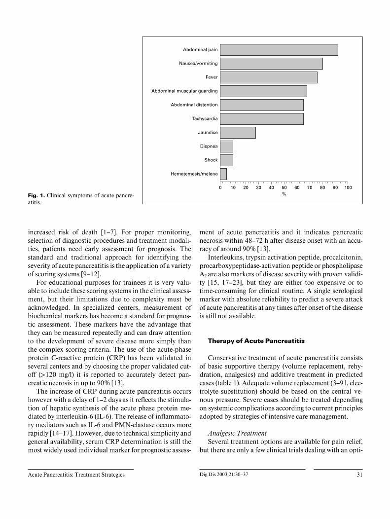
Acute Pancreatitis: Treatment Strategies Dig Dis 2003;21:30–37 31
Fig. 1. Clinical symptoms of acute pancre-atitis.
increased risk of death [1–7]. For proper monitoring,selection of diagnostic procedures and treatment modali-ties, patients need early assessment for prognosis. Thestandard and traditional approach for identifying theseverity of acute pancreatitis is the application of a varietyof scoring systems [9–12].
For educational purposes for trainees it is very valu-able to include these scoring systems in the clinical assess-ment, but their limitations due to complexity must beacknowledged. In specialized centers, measurement ofbiochemical markers has become a standard for prognos-tic assessment. These markers have the advantage thatthey can be measured repeatedly and can draw attentionto the development of severe disease more simply thanthe complex scoring criteria. The use of the acute-phaseprotein C-reactive protein (CRP) has been validated inseveral centers and by choosing the proper validated cut-off (1120 mg/l) it is reported to accurately detect pan-creatic necrosis in up to 90% [13].
The increase of CRP during acute pancreatitis occurshowever with a delay of 1–2 days as it reflects the stimula-tion of hepatic synthesis of the acute phase protein me-diated by interleukin-6 (IL-6). The release of inflammato-ry mediators such as IL-6 and PMN-elastase occurs morerapidly [14–17]. However, due to technical simplicity andgeneral availability, serum CRP determination is still themost widely used individual marker for prognostic assess-
ment of acute pancreatitis and it indicates pancreaticnecrosis within 48–72 h after disease onset with an accu-racy of around 90% [13].
Interleukins, trypsin activation peptide, procalcitonin,procarboxypeptidase-activation peptide or phospholipaseA2 are also markers of disease severity with proven validi-ty [15, 17–23], but they are either too expensive or totime-consuming for clinical routine. A single serologicalmarker with absolute reliability to predict a severe attackof acute pancreatitis at any times after onset of the diseaseis still not available.
Therapy of Acute Pancreatitis
Conservative treatment of acute pancreatitis consistsof basic supportive therapy (volume replacement, rehy-dration, analgesics) and additive treatment in predictedcases (table 1). Adequate volume replacement (3–9 l, elec-trolyte substitution) should be based on the central ve-nous pressure. Severe cases should be treated dependingon systemic complications according to current principlesadopted by strategies of intensive care management.
Analgesic TreatmentSeveral treatment options are available for pain relief,
but there are only a few clinical trials dealing with an opti-
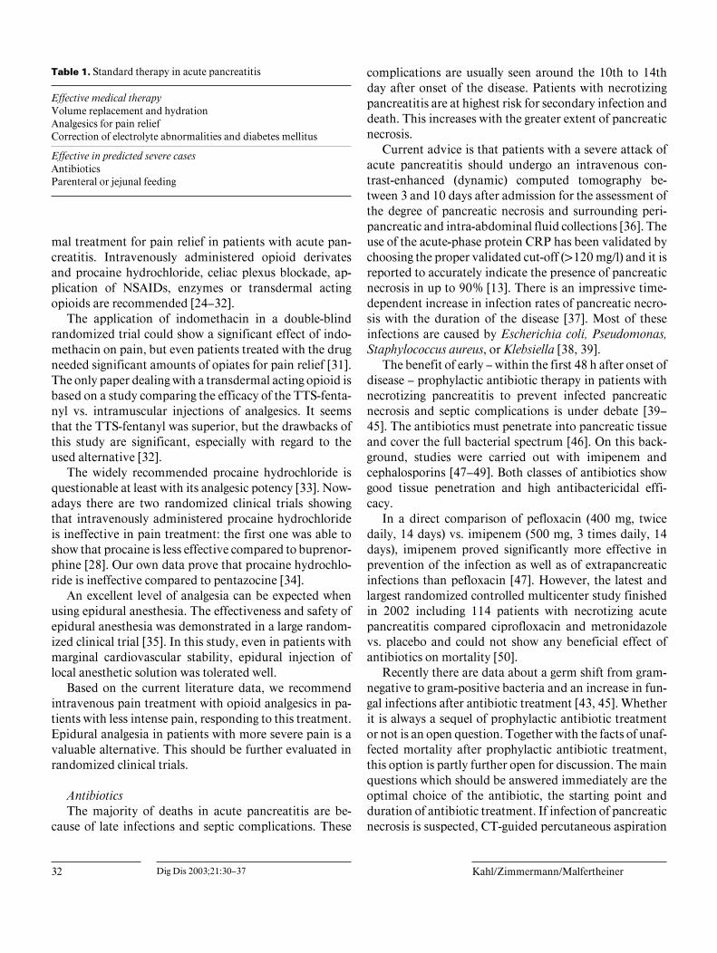
32 Dig Dis 2003;21:30–37 Kahl/Zimmermann/Malfertheiner
Table 1. Standard therapy in acute pancreatitis
Effective medical therapyVolume replacement and hydrationAnalgesics for pain reliefCorrection of electrolyte abnormalities and diabetes mellitus
Effective in predicted severe casesAntibioticsParenteral or jejunal feeding
mal treatment for pain relief in patients with acute pan-creatitis. Intravenously administered opioid derivatesand procaine hydrochloride, celiac plexus blockade, ap-plication of NSAIDs, enzymes or transdermal actingopioids are recommended [24–32].
The application of indomethacin in a double-blindrandomized trial could show a significant effect of indo-methacin on pain, but even patients treated with the drugneeded significant amounts of opiates for pain relief [31].The only paper dealing with a transdermal acting opioid isbased on a study comparing the efficacy of the TTS-fenta-nyl vs. intramuscular injections of analgesics. It seemsthat the TTS-fentanyl was superior, but the drawbacks ofthis study are significant, especially with regard to theused alternative [32].
The widely recommended procaine hydrochloride isquestionable at least with its analgesic potency [33]. Now-adays there are two randomized clinical trials showingthat intravenously administered procaine hydrochlorideis ineffective in pain treatment: the first one was able toshow that procaine is less effective compared to buprenor-phine [28]. Our own data prove that procaine hydrochlo-ride is ineffective compared to pentazocine [34].
An excellent level of analgesia can be expected whenusing epidural anesthesia. The effectiveness and safety ofepidural anesthesia was demonstrated in a large random-ized clinical trial [35]. In this study, even in patients withmarginal cardiovascular stability, epidural injection oflocal anesthetic solution was tolerated well.
Based on the current literature data, we recommendintravenous pain treatment with opioid analgesics in pa-tients with less intense pain, responding to this treatment.Epidural analgesia in patients with more severe pain is avaluable alternative. This should be further evaluated inrandomized clinical trials.
AntibioticsThe majority of deaths in acute pancreatitis are be-
cause of late infections and septic complications. These
complications are usually seen around the 10th to 14thday after onset of the disease. Patients with necrotizingpancreatitis are at highest risk for secondary infection anddeath. This increases with the greater extent of pancreaticnecrosis.
Current advice is that patients with a severe attack ofacute pancreatitis should undergo an intravenous con-trast-enhanced (dynamic) computed tomography be-tween 3 and 10 days after admission for the assessment ofthe degree of pancreatic necrosis and surrounding peri-pancreatic and intra-abdominal fluid collections [36]. Theuse of the acute-phase protein CRP has been validated bychoosing the proper validated cut-off (1120 mg/l) and it isreported to accurately indicate the presence of pancreaticnecrosis in up to 90% [13]. There is an impressive time-dependent increase in infection rates of pancreatic necro-sis with the duration of the disease [37]. Most of theseinfections are caused by Escherichia coli, Pseudomonas,Staphylococcus aureus, or Klebsiella [38, 39].
The benefit of early – within the first 48 h after onset ofdisease – prophylactic antibiotic therapy in patients withnecrotizing pancreatitis to prevent infected pancreaticnecrosis and septic complications is under debate [39–45]. The antibiotics must penetrate into pancreatic tissueand cover the full bacterial spectrum [46]. On this back-ground, studies were carried out with imipenem andcephalosporins [47–49]. Both classes of antibiotics showgood tissue penetration and high antibactericidal effi-cacy.
In a direct comparison of pefloxacin (400 mg, twicedaily, 14 days) vs. imipenem (500 mg, 3 times daily, 14days), imipenem proved significantly more effective inprevention of the infection as well as of extrapancreaticinfections than pefloxacin [47]. However, the latest andlargest randomized controlled multicenter study finishedin 2002 including 114 patients with necrotizing acutepancreatitis compared ciprofloxacin and metronidazolevs. placebo and could not show any beneficial effect ofantibiotics on mortality [50].
Recently there are data about a germ shift from gram-negative to gram-positive bacteria and an increase in fun-gal infections after antibiotic treatment [43, 45]. Whetherit is always a sequel of prophylactic antibiotic treatmentor not is an open question. Together with the facts of unaf-fected mortality after prophylactic antibiotic treatment,this option is partly further open for discussion. The mainquestions which should be answered immediately are theoptimal choice of the antibiotic, the starting point andduration of antibiotic treatment. If infection of pancreaticnecrosis is suspected, CT-guided percutaneous aspiration
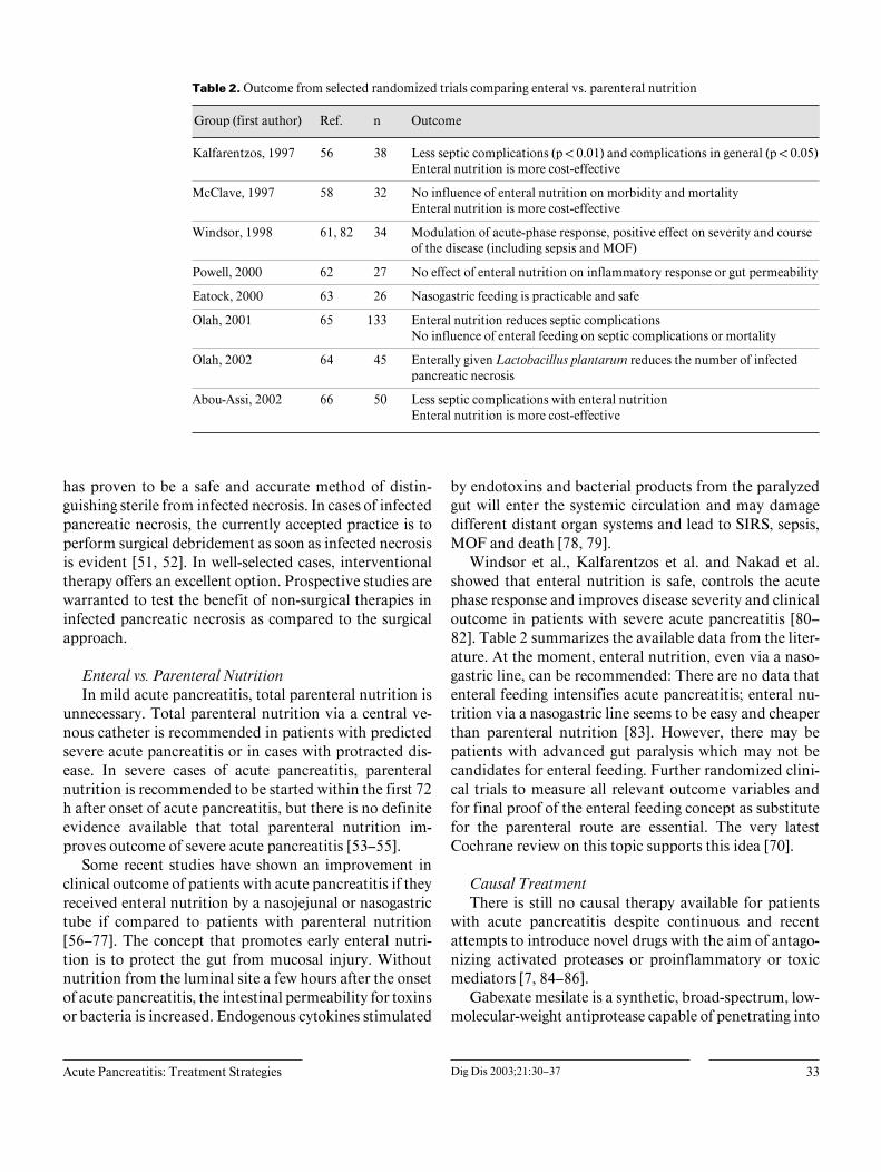
Acute Pancreatitis: Treatment Strategies Dig Dis 2003;21:30–37 33
Table 2. Outcome from selected randomized trials comparing enteral vs. parenteral nutrition
Group (first author) Ref. n Outcome
Kalfarentzos, 1997 56 38 Less septic complications (p ! 0.01) and complications in general (p ! 0.05)Enteral nutrition is more cost-effective
McClave, 1997 58 32 No influence of enteral nutrition on morbidity and mortalityEnteral nutrition is more cost-effective
Windsor, 1998 61, 82 34 Modulation of acute-phase response, positive effect on severity and courseof the disease (including sepsis and MOF)
Powell, 2000 62 27 No effect of enteral nutrition on inflammatory response or gut permeability
Eatock, 2000 63 26 Nasogastric feeding is practicable and safe
Olah, 2001 65 133 Enteral nutrition reduces septic complicationsNo influence of enteral feeding on septic complications or mortality
Olah, 2002 64 45 Enterally given Lactobacillus plantarum reduces the number of infectedpancreatic necrosis
Abou-Assi, 2002 66 50 Less septic complications with enteral nutritionEnteral nutrition is more cost-effective
has proven to be a safe and accurate method of distin-guishing sterile from infected necrosis. In cases of infectedpancreatic necrosis, the currently accepted practice is toperform surgical debridement as soon as infected necrosisis evident [51, 52]. In well-selected cases, interventionaltherapy offers an excellent option. Prospective studies arewarranted to test the benefit of non-surgical therapies ininfected pancreatic necrosis as compared to the surgicalapproach.
Enteral vs. Parenteral NutritionIn mild acute pancreatitis, total parenteral nutrition is
unnecessary. Total parenteral nutrition via a central ve-nous catheter is recommended in patients with predictedsevere acute pancreatitis or in cases with protracted dis-ease. In severe cases of acute pancreatitis, parenteralnutrition is recommended to be started within the first 72h after onset of acute pancreatitis, but there is no definiteevidence available that total parenteral nutrition im-proves outcome of severe acute pancreatitis [53–55].
Some recent studies have shown an improvement inclinical outcome of patients with acute pancreatitis if theyreceived enteral nutrition by a nasojejunal or nasogastrictube if compared to patients with parenteral nutrition[56–77]. The concept that promotes early enteral nutri-tion is to protect the gut from mucosal injury. Withoutnutrition from the luminal site a few hours after the onsetof acute pancreatitis, the intestinal permeability for toxinsor bacteria is increased. Endogenous cytokines stimulated
by endotoxins and bacterial products from the paralyzedgut will enter the systemic circulation and may damagedifferent distant organ systems and lead to SIRS, sepsis,MOF and death [78, 79].
Windsor et al., Kalfarentzos et al. and Nakad et al.showed that enteral nutrition is safe, controls the acutephase response and improves disease severity and clinicaloutcome in patients with severe acute pancreatitis [80–82]. Table 2 summarizes the available data from the liter-ature. At the moment, enteral nutrition, even via a naso-gastric line, can be recommended: There are no data thatenteral feeding intensifies acute pancreatitis; enteral nu-trition via a nasogastric line seems to be easy and cheaperthan parenteral nutrition [83]. However, there may bepatients with advanced gut paralysis which may not becandidates for enteral feeding. Further randomized clini-cal trials to measure all relevant outcome variables andfor final proof of the enteral feeding concept as substitutefor the parenteral route are essential. The very latestCochrane review on this topic supports this idea [70].
Causal TreatmentThere is still no causal therapy available for patients
with acute pancreatitis despite continuous and recentattempts to introduce novel drugs with the aim of antago-nizing activated proteases or proinflammatory or toxicmediators [7, 84–86].
Gabexate mesilate is a synthetic, broad-spectrum, low-molecular-weight antiprotease capable of penetrating into
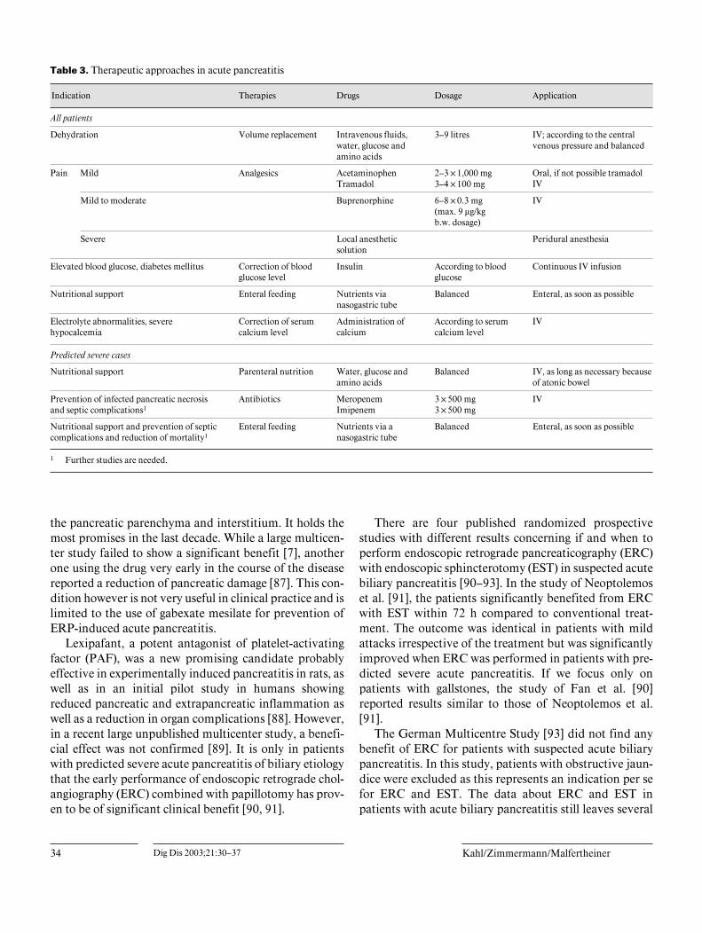
34 Dig Dis 2003;21:30–37 Kahl/Zimmermann/Malfertheiner
Table 3. Therapeutic approaches in acute pancreatitis
Indication Therapies Drugs Dosage Application
All patients
Dehydration Volume replacement Intravenous fluids,water, glucose andamino acids
3–9 litres IV; according to the centralvenous pressure and balanced
Pain Mild Analgesics AcetaminophenTramadol
2–3!1,000 mg3–4!100 mg
Oral, if not possible tramadolIV
Mild to moderate Buprenorphine 6–8!0.3 mg(max. 9 Ìg/kgb.w. dosage)
IV
Severe Local anestheticsolution
Peridural anesthesia
Elevated blood glucose, diabetes mellitus Correction of bloodglucose level
Insulin According to bloodglucose
Continuous IV infusion
Nutritional support Enteral feeding Nutrients vianasogastric tube
Balanced Enteral, as soon as possible
Electrolyte abnormalities, severehypocalcemia
Correction of serumcalcium level
Administration ofcalcium
According to serumcalcium level
IV
Predicted severe cases
Nutritional support Parenteral nutrition Water, glucose andamino acids
Balanced IV, as long as necessary becauseof atonic bowel
Prevention of infected pancreatic necrosisand septic complications1
Antibiotics MeropenemImipenem
3!500 mg3!500 mg
IV
Nutritional support and prevention of septiccomplications and reduction of mortality1
Enteral feeding Nutrients via anasogastric tube
Balanced Enteral, as soon as possible
1 Further studies are needed.
the pancreatic parenchyma and interstitium. It holds themost promises in the last decade. While a large multicen-ter study failed to show a significant benefit [7], anotherone using the drug very early in the course of the diseasereported a reduction of pancreatic damage [87]. This con-dition however is not very useful in clinical practice and islimited to the use of gabexate mesilate for prevention ofERP-induced acute pancreatitis.
Lexipafant, a potent antagonist of platelet-activatingfactor (PAF), was a new promising candidate probablyeffective in experimentally induced pancreatitis in rats, aswell as in an initial pilot study in humans showingreduced pancreatic and extrapancreatic inflammation aswell as a reduction in organ complications [88]. However,in a recent large unpublished multicenter study, a benefi-cial effect was not confirmed [89]. It is only in patientswith predicted severe acute pancreatitis of biliary etiologythat the early performance of endoscopic retrograde chol-angiography (ERC) combined with papillotomy has prov-en to be of significant clinical benefit [90, 91].
There are four published randomized prospectivestudies with different results concerning if and when toperform endoscopic retrograde pancreaticography (ERC)with endoscopic sphincterotomy (EST) in suspected acutebiliary pancreatitis [90–93]. In the study of Neoptolemoset al. [91], the patients significantly benefited from ERCwith EST within 72 h compared to conventional treat-ment. The outcome was identical in patients with mildattacks irrespective of the treatment but was significantlyimproved when ERC was performed in patients with pre-dicted severe acute pancreatitis. If we focus only onpatients with gallstones, the study of Fan et al. [90]reported results similar to those of Neoptolemos et al.[91].
The German Multicentre Study [93] did not find anybenefit of ERC for patients with suspected acute biliarypancreatitis. In this study, patients with obstructive jaun-dice were excluded as this represents an indication per sefor ERC and EST. The data about ERC and EST inpatients with acute biliary pancreatitis still leaves several
Acute Pancreatitis: Treatment Strategies Dig Dis 2003;21:30–37 35
questions open. The studies published up to now do notanswer the question as to when to perform an interven-tional endoscopy. From the available data we would rec-ommend that an interventional endoscopy (ERC plusEST) should be performed in cases of acute biliary pancre-atitis with severe prognosis in specialized centers that pro-vide optimal trained personnel, and technical and logisticsupport.
Conclusion
Basic therapy in patients with acute pancreatitis con-sists of volume replacement and analgesic therapy. Forpain relief, opioid analgesics (intravenously given) are the
first choice. Epidural analgesia is a valuable alternative inpatients with more intense pain, who do not respond tointravenously administered opioids. In severe cases withsuspected pancreatic necrosis, antibiotics should be ad-ministered to prevent infection and to avoid surgery. Thisstrategy is not proven to be more effective at all, but itseems to offer advantages.
Enteral nutrition should be started as soon as possible.There are no controlled data from larger studies aboutpositive effects on morbidity or mortality in enterally fedpatients, compared to patients with parenteral nutrition.But in most cases, enteral nutrition is harmless and doesnot cause any negative side effects. Table 3 summarizesthe current management options for patients with acutepancreatitis.
References
1 Karimgani I, Porter KA, Langevin RE, BanksPA: Prognostic factors in sterile pancreatic ne-crosis. Gastroenterology 1992;103:1636–1640.
2 Beger HG, Büchler MW, Bittner R, Block S,Nevalainen TJ, Roscher R: Necrosectomy andpostoperative local lavage in necrotizing pan-creatitis. Br J Surg 1988;75:207–212.
3 Sarr MG, Nagorney DM, Mucha P, FarnellMB, Johnson CD: Acute necrotizing pancreati-tis: Management by planned, staged pancreaticnecrosectomy/debridement and delayed pri-mary wound closure over drains. Br J Surg1991;78:576–581.
4 Rattner DW, Legermate DA, Lee MJ, MuellerPR, Warshaw AL: Early surgical debridementof symptomatic pancreatic necrosis is benefi-cial irrespective of infection. Am J Surg 1992;163:105–109.
5 Steinberg WM, Schlesselman SE: Treatment ofacute pancreatitis. Comparison of animal andhuman studies. Gastroenterology 1987;93:1420–1427.
6 Leese T, Holliday M, Watkins M, ThomasWM, Neoptolemos JP, Hall C, Attard A: Amulticentre controlled clinical trial of high-vol-ume fresh-frozen plasma therapy in prognosti-cally severe acute pancreatitis. Ann R Coll SurgEngl 1991;73:207–214.
7 Büchler MW, Malfertheiner P, Uhl W, Schol-merich J, Stockmann F, Adler G, Gaus W,Rolle K, Beger HG: Gabexate mesilate in hu-man acute pancreatitis. German PancreatitisStudy Group. Gastroenterology 1993;104:1165–1170.
8 Schmid SW, Uhl W, Büchler MW: Does theetiology influence the severity of acute pancre-atitis?; in Büchler MW, Uhl W, Friess H, Mal-fertheiner P (eds): Acute Pancreatitis: NovelConcepts in Biology and Therapy. Berlin,Blackwell, 1999, pp 193–198.
9 Ranson JH, Rifkind KM, Roses DF, Fink SD,Eng K, Spencer FC: Prognostic signs and therole of operative management in acute pancre-atitis. Surg Gynecol Obstet 1974;139:69–81.
10 Imrie CW, Benjamin IS, Ferguson JC, McKayAJ, Mackenzie I, O’Neill J, Blumgart LH: Asingle-centre, double-blind trial of Trasyloltherapy in primary acute pancreatitis. Br J Surg1978;65:337–341.
11 Knaus WA, Draper EA, Wagner DP, Zimmer-man JE: Apache II: A severity of disease classi-fication system. Crit Care Med 1985;13:818–829.
12 Marshall JC, Cook DJ, Christou NV, BernardGR, Sprung CL, Sibbald WJ: Multiple organdysfunction score: A reliable descriptor of acomplex clinical outcome. Crit Care Med 1995;23:1638–1652.
13 Büchler MW, Malfertheiner P, SchoetensackC, Uhl W, Beger HG: Sensitivity of antipro-teases, complement factors and C-reactive pro-tein in detecting pancreatic necrosis. Results ofa prospective clinical study. Int J Pancreatol1986;1:227–235.
14 Viedma JA, Perez-Mateo M, Dominguez-Mu-noz JE, Carballo F: Role of interleukin-6 inacute pancreatitis. Comparison with C-reactiveprotein and phospholipase A. Gut 1992;33:1264–1267.
15 Viedma JA, Perez-Mateo M, Agullo J, Domin-guez-Munoz JE, Carballo F: Inflammatory re-sponse in the early prediction of severity inhuman acute pancreatitis. Gut 1994;35:822–827.
16 Dominguez-Munoz JE, Viedma JA, Perez-Ma-teo M, Carballo F, Garcia FM: Inflammatoryresponse in the initial phase of acute pancreati-tis: Relationship to the onset and severity of thedisease. Rev Esp Enferm Dig 1995;87:225–246.
17 Uhl W, Büchler MW, Malfertheiner P, MartiniM, Beger HG: PMN elastase in comparisonwith CRP, antiproteases and LDH as indica-tors of necrosis in human acute pancreatitis.Pancreas 1991;6:253–259.
18 Inagaki T, Hoshino M, Ohara H, Yamada T:Evaluation of phospholipase A2 measurementfor early prediction of the severity of acute pan-creatitis. Pancreas 1998;16:558–559.
19 Rau B, Steinbach G, Gansauge F, Mayer JM,Grunert A, Beger HG: The potential role ofprocalcitonin and interleukin-8 in the predic-tion of infected necrosis in acute pancreatitis.Gut 1997;41:832–840.
20 Rau B, Cebulla M, Uhl W, Schoenberg MH,Beger HG: The clinical value of human pan-creas-specific protein procarboxypeptidase Bas an indicator of necrosis in acute pancreatitis:Comparison to CRP and LDH. Pancreas 1998;17:134–139.
21 Berney T, Gasche Y, Robert J, Jenny A, MensiN, Grau G, Vermeulen B, Morel P: Serum pro-files of interleukin-6, interleukin-8 and inter-leukin-10 in patients with severe and mildacute pancreatitis. Pancreas 1999;18:371–377.
22 Appelros S, Thim L, Borgstrom A: Activationpeptide of carboxypeptidase B in serum andurine in acute pancreatitis. Gut 1998;42:97–102.
23 Büchler MW, Uhl W, Andren-Sandberg A:CAPAP in acute pancreatitis: Just anothermarker or real progress? Gut 1998;42:8–9.
24 Lankisch PG: Acute and chronic pancreatitis.An update on management. Drugs 1984;28:554–564.
25 Lankisch PG, Banks PA: Acute Pancreatitis.Berlin, Springer, 1996.
26 Goebell H, Singer MV: Acute pancreatitis:Standards of conservative treatment; in BegerHG, Büchler MW (eds): Acute Pancreatitis.Berlin, Springer, 1987, pp 259–265.
36 Dig Dis 2003;21:30–37 Kahl/Zimmermann/Malfertheiner
27 Mossner J, Keim V, Niederau C, Büchler MW,Singer MV, Lankisch PG, Goke B: Guidelinesfor therapy of chronic pancreatitis. ConsensusConference of the German Society of Digestiveand Metabolic Diseases, Halle, Nov 21–23,1996. Z Gastroenterol 1998;36:359–367.
28 Jakobs R, Adamek MU, von Bubnoff AC, Rie-mann JF: Buprenorphine or procaine for painrelief in acute pancreatitis. A prospective ran-domized study. Scand J Gastroenterol 2000;35:1319–1323.
29 Patankar RV, Chand R, Johnson CD: Pan-creatic enzyme supplementation in acute pan-creatitis. HPB Surg 1995;8:159–162.
30 Rykowski JJ, Hilgier M: Continuous celiacplexus block in acute pancreatitis. Reg Anesth1995;20:528–532.
31 Ebbehoj N, Friis J, Svendsen LB, Bulow S,Madsen P: Indomethacin treatment of acutepancreatitis. A controlled double-blind trial.Scand J Gastroenterol 1985;20:798–800.
32 Stevens M, Esler R, Asher G: Transdermal fen-tanyl for the management of acute pancreatitispain. Appl Nurs Res 2002;15:102–110.
33 Zwernemann C, Steffen J, Thürmann PA: Pro-cain als Schmerztherapeutikum bei der akutenPankreatitis. Dtsch Med Wochenschr 2000;125:1307.
34 Kahl S, Zimmermann S, Leodolter A, SchulzHU, Malfertheiner P: Procaine hydrochloridefails to treat pain in patients with acute pancre-atitis (abstract). Pancreatology 2002;2:299.
35 Bernhardt A, Kortgen A, Niesel HC, Goertz A:Using epidural anesthesia in patients withacute pancreatitis: Prospective study of 121patients. Anaesthesiol Reanim 2002;27:16–22.
36 Balthazar EJ, Robinson DL, Megibow AJ, Ran-son JH: Acute pancreatitis: Value of CT inestablishing prognosis. Radiology 1990;174:331–336.
37 Beger HG, Bittner R, Block S, Büchler MW:Bacterial contamination of pancreatic necrosis.A prospective clinical study. Gastroenterology1986;91:433–438.
38 Banks PA, Gerzof SG, Langevin RE, Silver-man SG, Sica GT, Hughes MD: CT-guidedaspiration of suspected pancreatic infection:Bacteriology and clinical outcome. Int J Pan-creatol 1995;18:265–270.
39 Sharma VK, Howden CW: Prophylactic antibi-otic administration reduces sepsis and mortali-ty in acute necrotizing pancreatitis: A meta-analysis. Pancreas 2001;22:28–31.
40 Kramer KM, Levy H: Prophylactic antibioticsfor severe acute pancreatitis: The beginning ofan era. Pharmacotherapy 1999;19:592–602.
41 Powell JJ, Campbell E, Johnson CD, Siriwar-dena AK: Survey of antibiotic prophylaxis inacute pancreatitis in the UK and Ireland. Br JSurg 1999;86:320–322.
42 Gloor B, Schmidt O, Uhl W, Büchler MW: Pro-phylactic antibiotics and pancreatic necrosis.Curr Gastroenterol Rep 2001;3:109–114.
43 Gloor B, Muller CA, Worni M, Stahel PF,Redaelli C, Uhl W, Büchler MW: Pancreaticinfection in severe pancreatitis: The role of fun-gus and multiresistant organisms. Arch Surg2001;136:592–596.
44 Slavin J, Neoptolemos JP: Antibiotic prophy-laxis in severe acute pancreatitis –What are thefacts? Langenbecks Arch Surg 2001;386:155–159.
45 Howard TJ, Temple MB: Prophylactic antibi-otics alter the bacteriology of infected necrosisin severe acute pancreatitis. J Am Coll Surg2002;195:759–767.
46 Büchler MW, Malfertheiner P, Friess H, Isen-mann R, Vanek E, Grimm H, Schlegel P, FriessT, Beger HG: Human pancreatic tissue concen-tration of bactericidal antibiotics. Gastroenter-ology 1992;103:1902–1908.
47 Bassi C, Falconi M, Talamini G, Uomo G,Papaccio G, Dervenis C, Salvia R, Minelli EB,Pederzoli P: Controlled clinical trial of pefloxa-cin versus imipenem in severe acute pancreati-tis. Gastroenterology 1998;115:1513–1517.
48 Pederzoli P, Bassi C, Vesentini S, CampedelliA: A randomized multicenter clinical trial ofantibiotic prophylaxis of septic complicationsin acute necrotizing pancreatitis with imipen-em. Surg Gynecol Obstet 1993;176:480–483.
49 Sainio V, Kemppainen E, Puolakkainen P,Taavitsainen M, Kivisaari L, Valtonen V, Haa-piainen R, Schroder T, Kivilaakso E: Earlyantibiotic treatment in acute necrotising pan-creatitis. Lancet 1995;346:663–667.
50 Isenmann R, Rünzi M, Goebell H, Beger HG:German multicentre trial on antibiotic treat-ment in severe acute pancreatitis (ASAP study)(pers commun, 2002).
51 Rau B, Uhl W, Büchler MW, Beger HG: Surgi-cal treatment of infected necrosis. World J Surg1997;21:155–161.
52 Banks PA: Practice guidelines in acute pancre-atitis. Am J Gastroenterol 1997;92:377–386.
53 Pisters PW, Ranson JH: Nutritional supportfor acute pancreatitis. Surg Gynecol Obstet1992;175:275–284.
54 Sitzmann JV, Steinborn PA, Zinner MJ, Came-ron JL: Total parenteral nutrition and alternateenergy substrates in treatment of severe acutepancreatitis. Surg Gynecol Obstet 1989;168:311–317.
55 Kalfarentzos FE, Karavias DD, Karatzas TM,Alevizatos BA, Androulakis JA: Total paren-teral nutrition in severe acute pancreatitis. JAm Coll Nutr 1991;10:156–162.
56 Kalfarentzos F, Kehagias J, Mead N, KokkinisK, Gogos CA: Enteral nutrition is superior toparenteral nutrition in severe acute pancreati-tis: Results of a randomized prospective trial.Br J Surg 1997;84:1665–1669.
57 Kotani J, Usami M, Nomura H, Iso A, Kasaha-ra H, Kuroda Y, Oyanagi H, Saitoh Y: Enteralnutrition prevents bacterial translocation butdoes not improve survival during acute pancre-atitis. Arch Surg 1999;134:287–292.
58 McClave SA, Greene LM, Snider HL, MakkLJ, Cheadle WG, Owens NA, Dukes LG,Goldsmith LJ: Comparison of the safety of ear-ly enteral vs. parenteral nutrition in mild acutepancreatitis. JPEN J Parenter Enteral Nutr1997;21:14–20.
59 McClave SA, Snider H, Owens N, Sexton LK:Clinical nutrition in pancreatitis. Dig Dis Sci1997;42:2035–2044.
60 Nakad A, Piessevaux H, Marot JC, Hoang P,Geubel A, Van Steenbergen W, Reynaert M: Isearly enteral nutrition in acute pancreatitisdangerous? About 20 patients fed by an endo-scopically placed nasogastrojejunal tube. Pan-creas 1998;17:187–193.
61 Windsor AC, Kanwar S, Li AG, Barnes E,Guthrie JA, Spark JI, Welsh F, Guillou PJ,Reynolds JV: Compared with parenteral nutri-tion, enteral feeding attenuates the acute phaseresponse and improves disease severity inacute pancreatitis. Gut 1998;42:431–435.
62 Powell JJ, Murchison JT, Fearon KC, Ross JA,Siriwardena AK: Randomized controlled trialof the effect of early enteral nutrition on mark-ers of the inflammatory response in predictedsevere acute pancreatitis. Br J Surg 2000;87:1375–1381.
63 Eatock FC, Brombacher GD, Steven A, ImrieCW, McKay CJ, Carter R: Nasogastric feedingin severe acute pancreatitis may be practicaland safe. Int J Pancreatol 2000;28:23–29.
64 Olah A, Belagyi T, Issekutz A, Gamal ME,Bengmark S: Randomized clinical trial of spe-cific lactobacillus and fibre supplement to earlyenteral nutrition in patients with acute pancre-atitis. Br J Surg 2002;89:1103–1107.
65 Olah A, Pardavi G, Belagyi T, Nagy A, IssekutzA, Mohamed GE: Early nasojejunal feeding inacute pancreatitis is associated with a lowercomplication rate. Nutrition 2002;18:259–262.
66 Abou-Assi S, Craig K, O’Keefe SJ: Hypocaloricjejunal feeding is better than total parenteralnutrition in acute pancreatitis: Results of a ran-domized comparative study. Am J Gastroen-terol 2002;97:2255–2262.
67 Lehocky P, Sarr MG: Early enteral feeding insevere acute pancreatitis: Can it prevent sec-ondary pancreatic (super)infection? Dig Surg2000;17:571–577.
68 Eckerwall G, Andersson R: Early enteral nutri-tion in severe acute pancreatitis: A way of pro-viding nutrients, gut barrier protection, immu-nomodulation, or all of them? Scand J Gas-troenterol 2001;36:449–458.
69 Sanabria A: Randomized controlled trial of theeffect of early enteral nutrition on markers ofthe inflammatory response in predicted severeacute pancreatitis. Br J Surg 2001;88:728.
70 Al Omran M, Groof A, Wilke D: Enteral versusparenteral nutrition for acute pancreatitis.Cochrane Database Syst Rev 2001;CD002837.
71 Rao MP, Mulleague L: Nutritional support inacute pancreatitis: The enteral vs. parenteraldilemma. Hosp Med 2001;62:580.
72 Chen QP: Enteral nutrition and acute pancre-atitis. World J Gastroenterol 2001;7:185–192.
73 Hamvas J, Schwab R, Pap A: Jejunal feeding inchronic pancreatitis with severe necrosis. JOP2001;2:112–116.
74 Fang J, DiSario JA: Nutritional managementof acute pancreatitis. Curr Gastroenterol Rep2002;4:120–127.
Acute Pancreatitis: Treatment Strategies Dig Dis 2003;21:30–37 37
75 Meier R, Beglinger C, Layer P, Gullo L, KeimV, Laugier R, Friess H, Schweitzer M, MacfieJ: ESPEN guidelines on nutrition in acute pan-creatitis. European Society of Parenteral andEnteral Nutrition. Clin Nutr 2002;21:173–183.
76 Imrie CW, Carter CR, McKay CJ: Enteral andparenteral nutrition in acute pancreatitis. BestPract Res Clin Gastroenterol 2002;16:391–397.
77 Al Omran M, Groof A, Wilke D: Enteral versusparenteral nutrition for acute pancreatitis(Cochrane Review). Cochrane Database SystRev 2003;CD002837.
78 Gardiner KR, Maxwell RJ, Rowlands BJ, Bar-clay GR: Intestinal permeability. Gut 1995;37:589.
79 Hallay J, Kovacs G, Szatmari K, Bako A,Szentkereszty Z, Lakos G, Sipka S, Sapy P: Ear-ly jejunal nutrition and changes in the immu-nological parameters of patients with acutepancreatitis. Hepatogastroenterology 2001;48:1488–1492.
80 Ammann RW: A clinically based classificationsystem for alcoholic chronic pancreatitis: Sum-mary of an international workshop on chronicpancreatitis. Pancreas 1997;14:215–221.
81 Ammann RW: Zürich workshop on alcoholicchronic pancreatitis. Int J Pancreatol 1998;23:81–82.
82 Ammann RW: Pain profile in alcoholic andnonalcoholic chronic pancreatitis. Pancreas1996;12:315–318.
83 McGregor CS, Marshall JC: Enteral feeding inacute pancreatitis: Just do it. Curr Opin CritCare 2001;7:89–91.
84 Niederau C, Schulz HU: Current conservativetreatment of acute pancreatitis: Evidence fromanimal and human studies. Hepatogastroenter-ology 1993;40:538–549.
85 Binder M, Uhl W, Friess H, Malfertheiner P,Büchler MW: Octreotide in the treatment ofacute pancreatitis: Results of a unicenter pro-spective trial with three different octreotidedosages. Digestion 1994;55(suppl 1):20–23.
86 Uhl W, Büchler MW, Malfertheiner P, BegerHG, Adler G, Gaus W: A randomised, double-blind, multicentre trial of octreotide in moder-ate to severe acute pancreatitis. Gut 1999;45:97–104.
87 Cavallini G, Tittobello A, Frulloni L, Masci E,Mariana A, Di Francesco V: Gabexate for theprevention of pancreatic damage related to en-doscopic retrograde cholangiopancreatogra-phy. Gabexate in digestive endoscopy – ItalianGroup. N Engl J Med 1996;335:919–923.
88 McKay CJ, Curran F, Sharples C, Baxter JN,Imrie CW: Prospective placebo-controlled ran-domized trial of lexipafant in predicted severeacute pancreatitis. Br J Surg 1997;84:1239–1243.
89 Johnson CD, Kingsnorth AN, Imrie CW,McMahon MJ, Neoptolemos JP, McKay C,Toh SK, Skaife P, Leeder PC, Wilson P, LarvinM, Curtis LD: Double-blind, randomised, pla-cebo-controlled study of a platelet-activatingfactor antagonist, lexipafant, in the treatmentand prevention of organ failure in predictedsevere acute pancreatitis. Gut 2001;48:62–69.
90 Fan ST, Lai EC, Mok FP, Lo CM, Zheng SS,Wong J: Early treatment of acute biliary pan-creatitis by endoscopic papillotomy. N Engl JMed 1993;328:228–232.
91 Neoptolemos JP, Carr-Locke DL, London NJ,Bailey IA, James D, Fossard DP: Controlledtrial of urgent endoscopic retrograde cholangio-pancreatography and endoscopic sphincteroto-my versus conservative treatment for acutepancreatitis due to gallstones. Lancet 1988;ii:979–983.
92 Nowak A, Nowakowska-Duzawa E, Marek TA,Rybicka J: Final results of the prospective, ran-domized, controlled study on endoscopicsphincterotomy versus conventional manage-ment in acute biliary pancreatitis (abstract).Gastroenterology 1995;108:A380.
93 Folsch UR, Nitsche R, Ludtke R, Hilgers RA,Creutzfeldt W: Early ERCP and papillotomycompared with conservative treatment foracute biliary pancreatitis. The German StudyGroup on Acute Biliary Pancreatitis. N Engl JMed 1997;336:237–242.
Review Article
Dig Dis 2003;21:38–45DOI: 10.1159/000071338
Modern Phase-Specific Management ofAcute Pancreatitis
Jens Werner Waldemar Uhl Werner Hartwig Thilo HackertChristophe Müller Oliver Strobel Markus W. Büchler
Department of General and Visceral Surgery, University of Heidelberg, Germany
Waldemar Uhl, MDDepartment of General and Visceral Surgery, University of HeidelbergIm Neuenheimer Feld 110, DE–69120 Heidelberg (Germany)Tel. +49 6221 566920, Fax +49 6221 566922E-Mail [email protected]
ABCFax + 41 61 306 12 34E-Mail [email protected]
© 2003 S. Karger AG, Basel0257–2753/03/0211–0038$19.50/0
Accessible online at:www.karger.com/ddi
Key WordsAcute pancreatitis W Pancreatic necrosis W Pancreaticinfection W Organ failure W Non-surgical management W
Surgical treatment
AbstractThe management of acute necrotizing pancreatitis haschanged significantly over the past years. In contrast tothe early surgical intervention of the past, there is now astrong tendency towards a more conservative approach.Initially, severe acute pancreatitis is characterized by thesystemic inflammatory response syndrome. Early man-agement is non-surgically and solely supportive. A spe-cific treatment still does not exist. In cases of necrotizingdisease, prophylactic antibiotics should be applied toreduce late septic complications. Today, more patientssurvive the first phase of severe pancreatitis due toimprovements of intensive care medicine, thus increas-ing the risk of later sepsis. Pancreatic infection is themajor risk factor with regard to morbidity and mortalityin the second phase of severe acute pancreatitis. Where-as early surgery and surgery for sterile necrosis can onlybe recommended in selected cases, pancreatic infectionis a well-accepted indication for surgical treatment in thesecond phase of the disease. Surgery should ideally bepostponed until 4 weeks after the onset of symptoms, as
necrosis is well demarcated at that time. Three surgicaltechniques can be performed with comparable resultsregarding mortality: necrosectomy combined with the(1) open packing technique, (2) planned staged relaparot-omies with repeated lavage, or (3) closed continuouslavage of the retroperitoneum. However, the latter meth-od seems to be associated with the lowest morbiditycompared to the other approaches.
Copyright © 2003 S. Karger AG, Basel
Introduction
The management of acute pancreatitis has been con-troversial for more than 100 years, varying between a con-servative medical approach on the one hand and a surgi-cal approach on the other. There has been great improve-ment in knowledge of the natural course and pathophysi-ology of acute pancreatitis over the past 20 years [1–8].The clinical course of acute pancreatitis varies from amild transitory form to a severe necrotizing disease. Mostepisodes of acute pancreatitis (80%) are mild and self-lim-iting, subsiding spontaneously within 3–5 days. Patientswith mild pancreatitis respond well to medical treatmentand generally do not need intensive care treatment or pan-creatic surgery. Thus, morbidity and mortality rates arebelow 1% [9–13]. In contrast, severe pancreatitis is associ-
Management of Acute Pancreatitis Dig Dis 2003;21:38–45 39
ated with organ failure and/or local complications such asnecrosis, abscess formation, or pseudocysts [14]. Severepancreatitis can be observed in 15–20% of all cases.
In general, severe pancreatitis develops in two phases.The first 2 weeks after onset of symptoms are character-ized by the systemic inflammatory response syndrome(SIRS). The release of proinflammatory mediators isthought to contribute to the pathogenesis of SIRS-associ-ated pulmonary, cardiovascular, and renal insufficiency.Mediators include pancreatic proteases, cytokines, reac-tive oxygen species, and many more [5, 6, 15–17]. In par-allel, pancreatic necrosis develops within the first 4 daysafter the onset of symptoms to its full extent [18]. How-ever, it is important that SIRS in the early phase of severepancreatitis may be found in the absence of significantpancreatic necrosis and is frequently found in the absenceof pancreatic infection [19, 20]. In contrast, infection ofpancreatic necrosis is still the major risk factor of sepsis-related multiple organ failure and the main life-threaten-ing complication in the second phase of severe acute pan-creatitis [2, 9, 21]. Infection of pancreatic necrosis mostcommonly develops 2–3 weeks after the onset of symp-toms and can be observed in 40–70% of patients withnecrotizing disease [18, 22, 23]. The risk of infectionincreases with the extent of intra- and extrapancreaticnecrosis [18, 21]. The present article presents the differentnon-surgical and surgical strategies of acute pancreatitisin the two phases of the disease.
Management of Acute Pancreatitis in Phase I
Although the majority of patients will have mild dis-ease that resolves spontaneously, it is difficult to detectpatients at risk of complications early on admission to thehospital. The main problem has been the lack of accuratepredictors of disease severity indicating development ofnecrosis and organ failure in the early stages, and infectednecrosis, multi-organ failure, and sepsis in the later phase.On admission, clinical assessment of severity has beenshown to be inaccurate [24, 25]. Contrast-enhanced com-puted tomography (CE-CT) is the ‘gold standard’ for thediagnosis of pancreatic necrosis [7, 26]. However, it willnot reveal the complete extent of pancreatic necrosisbefore the fourth day after the onset of the disease [18]. Inmost cases, CE-CT is not capable of revealing the pres-ence of superinfected necrosis in the later course of thedisease [7, 26, 27], and the diagnosis of pancreatic necro-sis does not predict the development of remote organcomplications [19, 20]. Several scoring systems for the
assessment of severity of acute pancreatitis exist, includ-ing the Ranson, Glasgow, and APACHE II score [28, 29].These multiple factor scoring systems have been designedto assess the risk of complications in patients with acutepancreatitis, and to categorize patients into groups at highrisk of complications. However, they are only moderatelyaccurate in assessing the disease severity of an individualpatient. Moreover, due to their complexity, the scoringsystems are rarely used in the clinical practice [30].Although multiple single markers have been proposed aspredictors of disease severity, CRP is still the referenceparameter of all single indicators [31]. CRP predictssevere pancreatitis and pancreatic necrosis accuratelyfrom the third day after onset of symptoms onwards [31–33]. Moreover, measurement of CRP is readily availablealmost everywhere. In contrast, no single parameter hasbeen developed which is suitable for early prediction ofinfected pancreatic necrosis. Consequently, it is wise totreat every patient aggressively until disease severity hasbeen established [9–13].
There are two primary objectives in the treatment ofpatients with acute pancreatitis. The first is to providesupportive therapy and treat the specific complicationswhich may occur. The second is to limit both the severityof pancreatic inflammation and necrosis as well as the sys-temic inflammatory response by specifically interruptingtheir pathogenesis.
All patients with signs of moderate to severe acute pan-creatitis should be admitted to an intensive care unit(ICU) and referred to specialized centers for maximumsupportive care [10, 12, 13]. Since complications maydevelop at any time, frequent reassessment and contin-uous monitoring are necessary. The most important sup-portive therapy is an adequate and prompt fluid resuscita-tion with intravenous fluids and supplemental oxygenwith a liberal indication for assisted or controlled ventila-tion to guarantee optimal oxygen transport [34–36]. Car-dioinotropic drugs, hemofiltration or dialysis may also beneeded to allow optimal fluid therapy despite acute renalfailure or hypoperfusion. Due to the popular belief thatthe pancreas should be put to ‘rest’ during acute pancre-atitis, the parenteral route of administrating nutrition isstill predominantly used in acute pancreatitis [12, 13, 37].However, there has been increasing concern about the gutbeing the main source of microorganisms causing infec-tious pancreatic complications and multiple organ failure[38]. In patients with severe pancreatitis, oral intake isinhibited by nausea and subileus. Whereas some reportsdemonstrated that enteral feeding is possible in acute pan-creatitis and associated with fewer septic complications
40 Dig Dis 2003;21:38–45 Werner/Uhl/Hartwig/Hackert/Müller/Strobel/Büchler
[39, 40], others did not show any beneficial effects [41].Although the evidence is not conclusive to support enteralnutrition in all patients with severe acute pancreatitis, theenteral route may be used if that can be tolerated. Thesupportive therapy also includes an adequate analgesia[34, 35]. Several treatment regimens including opioids,procaine infusion, epidural blockade have been widelyadvocated. However, these strategies of pain managementare rather based on empirical experience than on resultsof controlled, prospective trials [42].
In addition to the sole supportive care, the principles ofintensive care therapy in severe pancreatitis include elim-ination of the cause of the primary insult whenever possi-ble. A causative therapy exists for severe gallstone pancre-atitis with an impacted stone, biliary sepsis, or obstructivejaundice [43–45]. Endoscopic retrograde cholangiopan-creatography (ERCP) and endoscopic sphincterotomyameliorate symptoms and progression of the disease whenapplied early [46]. Secondary causes of organ failure suchas hypovolemia, tissue hypoperfusion, and hypoxemiamust also be identified and treated promptly. There issome evidence that vigorous fluid resuscitation may beassociated with resolution of organ failure [47]. As plasmaexpanders are more effective and long-acting, colloidsshould be preferred compared to crystalloids [35, 36].Dextran 60 seems to be the most potent colloid availablefor treatment of acute pancreatitis, as it is characterizednot only by a long intravascular persistence, but also byantithrombotic properties and inhibitory effects on leuko-cyte adhesion [48, 49]. Moreover, a clinical trial indicatedthat dextran can be applied safely in acute pancreatitis[50].
Multiple mediators of the inflammatory cascade, in-cluding oxygen free radicals, vasoactive mediators, cyto-kines, as well as leukocyte and endothelial activation andpancreatic ischemia, have been identified as importantsteps in the pathogenesis of acute necrotizing pancreatitisand its systemic complications [5, 6, 15–17, 51–56]. Inexperimental studies, several drugs which inhibit thosepathogenetic steps specifically, e.g. protease inhibitor, ox-ygen free radical scavenger, cytokine antagonists, nitricoxide agonists, and inhibitors of adhesion molecules,attenuated biochemical and histological changes. How-ever, until today neither the inhibition of pancreaticautodigestion nor the inhibition of any other single patho-genetic step has effectively reduced mortality or increasedlong-term survival in severe acute pancreatitis [5, 57–59].Thus, treatment of acute pancreatitis is still symptomatic,with no specific medication being available today.
The most significant change in the clinical course ofacute pancreatitis over the last decade has undoubtedlybeen the decrease in mortality. Overall mortality is nowabout 5% and for severe cases in the range of 10–20% [9,19, 60–62]. The major improvements include intensivecare medicine, the accurate diagnosis of necrosis by CE-CT, the reliable diagnosis of infected necrosis by FNA, theERCP concept in gallstone pancreatitis, administration ofprophylactic antibiotics in severe necrotizing pancreatitis,and the improved surgical procedures [62]. Despite thereduction in overall mortality in severe pancreatitis, thepercentage of early mortality of the disease differs be-tween less than 10 and 85% among various centers andcountries [1, 5, 9, 19, 63]. This wide variation in earlymortality may partially be explained by differences of thehealth systems, socio-economic reasons, or patient selec-tion.
Management of Acute Pancreatitis in Phase II
Today, more patients survive the first phase of severeacute pancreatitis due to improvements of intensive caremedicine, thus increasing the risk of later sepsis [9, 64–66]. There is no doubt that pancreatic infection is themajor risk factor in necrotizing pancreatitis with regard tomorbidity and mortality in the second phase of the dis-ease [9, 18, 67]. Infection of pancreatic necrosis developsmost frequently 2–3 weeks after the onset of symptoms.Naturally pancreatic infection correlates with the dura-tion of the disease, and up to 70% of all patients withnecrotizing disease present with infected pancreatic ne-crosis 4 weeks after the onset of the disease [18, 22, 23].Moreover, the risk of infection increases with the extentof intra- and extrapancreatic necrosis [18, 21]. Thereforeit appears that the presence of more than 50% of pancreat-ic necrosis on CT scanning is predictive for severe disease,and helps to identify patients who might develop septiccomplications [68].
Unlike the use of antibiotics in the treatment of proveninfection, the rationale for the use of prophylactic antibi-otics in severe pancreatitis is to prevent infection fromaffecting areas of pancreatic necrosis and consequentlyreduce the need for surgery and mortality. Evidence forthe effectiveness of prophylactic antibiotics in the reduc-tion of septic complications and mortality of necrotizingpancreatitis has been demonstrated by several random-ized controlled trials [69–73]. A meta-analysis of eightpreviously published trials about prophylactic antibioticsin acute pancreatitis has shown a positive benefit for anti-
Management of Acute Pancreatitis Dig Dis 2003;21:38–45 41
biotics in reducing mortality [74]. However, the advan-tage was limited to patients with severe pancreatitis whoreceived broad-spectrum antibiotics that achieved thera-peutic pancreatic tissue levels. Büchler [75–77] and othershave identified imipenem as the antibiotic agent of firstchoice because it reached higher pancreatic tissue levelsand provided higher bactericidal activity against most ofthe bacteria present in pancreatic infection compared toother types of antibiotics. An alternative antibiotic regi-men is either ciprofloxacin or ofloxacin in combinationwith metronidazole, although a previous trial has notshown any benefit with this regimen [78].
When pancreatic necrosis has developed, the differen-tiation between sterile and infected necrosis is essentialfor the management of patients. Infection of necrotic pan-creatic tissue is usually suspected in patients who developclinical signs of sepsis [11]. These patients should undergoCT- or ultrasonography-guided fine-needle aspiration(FNA) of pancreatic or peripancreatic necrosis [9, 11].FNA is an accurate, safe and reliable approach to differ-entiate between sterile and infected necrosis [22, 79].Complication rates of this procedure are low with onlyvery few serious complications such as bleeding, aggrava-tion of acute pancreatitis or death reported in the litera-ture [80, 81]. With bacterial testing including Gram stain-ing and culture of the aspiration material, a diagnosticsensitivity and specificity of 88 and 90%, respectively, hasbeen reported for this procedure when guided by ultraso-nography [82].
Two distinctive forms of infection in acute pancreatitisneed to be differentiated: infected pancreatic necrosis andpancreatic abscess. At the 1992 Atlanta Consensus Con-ference [14] these terms were defined as follows: Pan-creatic necrosis is a diffuse or focal area of non-viable pan-creatic parenchyma which is typically associated withpancreatic fat necrosis. In contrast, a pancreatic abscess isa circumscribed intra-abdominal collection of pus, usual-ly in proximity to the pancreatic necrosis, which arises asa consequence of acute pancreatitis. Probably pancreaticabscesses are a consequence of limited necrosis with sub-sequent liquefaction and secondary infection. It is impor-tant to distinguish between infected pancreatic necrosisand pancreatic abscesses since significantly lower mortali-ty rates are described for patients with pancreatic ab-scesses [83]. Furthermore, pancreatic abscesses in generaldevelop later in the course of disease (usually after 5weeks), whereas infected pancreatic necrosis may alreadybe found within the first week after onset of symptoms[18]. Due to their less aggressive behavior, several groupshave introduced minimal invasive treatment strategies
for pancreatic abscesses [84–86]. However, their roleremains to be defined in randomized controlled clinicaltrials.
Indications for SurgeryProven infected necrosis as well as septic complica-
tions resulting from pancreatic infection are well-acceptedindications for surgical treatment [9, 61, 87]. The mortali-ty rate for these patients is higher than 30%, and morethan 80% of fatal outcomes in acute pancreatitis are dueto septic complications [9, 18, 63]. When treated non-sur-gically, mortality rates of up to 100% have been reportedfor infected necrosis associated with multiple organ fail-ure [67]. With surgical treatment, the mortality rate forpatients with infected pancreatic necrosis was decreasedto about 20–30% in various specialized centers [9, 61, 63,88–90].
While surgical debridement is mandatory in pancreat-ic infection, a conservative approach is accepted in sterilenecrosis as long as the patient responds to therapy [9, 67,89, 91, 92]. In a series of 38 patients with necrotizing pan-creatitis, Bradley and Allen [60] reported an overall sur-vival rate of 100% in patients with sterile necrosis treatedconservatively. However, when sterile necrosis is associat-ed with organ failure, the role of surgery remains contro-versial [92–95]. It is still unclear why some patients withsterile necrosis can be treated non-surgically while othersdie without timely intervention. The manifestation of sin-gle or multiple organ failure in acute pancreatitis is associ-ated with mortality rates of 23–75% [19, 94–96]. There-fore, some authors favored early surgical therapy inextended pancreatic necrosis, as in theory necrosectomyeliminates the risk of necrosis getting infected. Further-more, removal of necrosis is thought to prevent or reducethe risk of inflammatory mediators and toxic substancesbeing released into the systemic circulation, thereby ame-liorating the systemic inflammatory response. However,since proinflammatory mediators are released very earlyin the course of the disease [55], surgery is not the tool tointerfere with the stimulation of the various cascade sys-tems contributing to SIRS. Another drawback of earlysurgery is the risk of secondary infection of preoperativesterile necrosis, which has been shown in about 30% ofpatients [92, 97]. Thus, surgical intervention in sterilenecrosis even seems harmful with worsening the progno-sis of patients. Intensive care therapy including prophy-lactic antibiotic treatment has been shown to generatebetter survival [9, 97, 98].
Nevertheless, some patients with sterile necrosis donot improve despite maximal therapy in the ICU. In this
42 Dig Dis 2003;21:38–45 Werner/Uhl/Hartwig/Hackert/Müller/Strobel/Büchler
subset of patients, some authors advocate surgery. In alarge retrospective series of 172 patients with sterilenecrosis published by Beger’s group [92], 62% of patientswere managed surgically whereas the remainder weretreated conservatively. Mortality rates were not signifi-cantly different between the two groups, with 13.1% forsurgically treated patients and 6.2% for those treated non-surgically. Therefore, persistent or progressive organ com-plications despite maximal ICU treatment is an indica-tion for surgery in patients with sterile necrosis [11]. How-ever, there is no established uniform definition of when apatient should be considered a ‘non-responder’ to ICUtherapy. Also in the rare event of rapidly progressive mul-tiple organ failure in the first days of acute pancreatitisdespite ICU therapy, so-called ‘fulminant acute pancre-atitis’ surgery may be indicated [11]. Nevertheless, giventhe poor outcome with both surgical and conservativetherapy and the lack of published data, the optimal thera-py for this subset of patients remains unclear.
As defined at the 2002 IAP Consensus Conference[11], indications for surgical treatment of acute necrotiz-ing pancreatitis comprise (1) infected pancreatic necrosisand (2) sterile necrosis in case of (a) ‘fulminant acute pan-creatitis’ or (b) persistent severe pancreatitis (‘non-re-sponder’).
Timing of SurgeryPatients with severe necrotizing pancreatitis can pro-
gress to a critical condition within a few hours or daysafter onset of symptoms. Years ago, early surgical inter-vention was favored, especially if systemic organ compli-cations required a quick response [95, 99]. Furthermore,if diagnosis remained unclear despite various examina-tions, surgery was requested [28]. Today, there is generalagreement that surgery in severe pancreatitis should beperformed as late as possible [11]. The rationale for latesurgery is the ease of identifying well-demarcated necrotictissue from the viable parenchyma, with the effect of lim-iting the extent of surgery to pure debridement. Thisapproach decreases the risk of bleeding and minimizes thesurgery-related loss of vital tissue which leads to surgery-induced endocrine and exocrine pancreatic insufficiency[93, 100, 101].
Mortality rates of up to 65% have been described withearly surgery in severe pancreatitis [18, 102, 103], ques-tioning the benefit of surgical intervention within the firstdays after onset of symptoms. In the single prospectiveand randomized clinical trial comparing early (within 48–72 h of symptoms) versus late (at least 12 days after onset)debridement in patients with severe pancreatitis, the mor-
tality rates were 56 and 27%, respectively [103]. Althoughthe difference did not reach statistical significance, thetrial was terminated because of the evident risk of earlysurgery. In our experience, surgery should not be per-formed earlier than 4 weeks after the onset of symptoms.The optimal surgical conditions for necrosectomy arepresent at the later phase of the disease, when necrosis hasbeen demarcated. The initial hemodynamic instabilitycan be treated effectively in the ICU. As we avoided sur-gery in the early course of the disease, we hardly had anyearly deaths, even in patients with multiple organ failure[9, 63]. In conclusion, only in the case of proven infectednecrosis or in the rare case of a complication, such as mas-sive bleeding or bowel perforation, must early surgery beperformed [9, 11].
Surgical ProceduresIn most patients with necrotizing pancreatitis, surgery
is performed to remove infected pancreatic necrosis. Theaim is to control the focus, so that further complicationsare avoided by stopping the progress of infection and therelease of proinflammatory mediators. However, resec-tion procedures such as partial or total pancreatico-duo-denectomy, that also remove vital pancreatic tissue orhealthy organs, are associated with high rates of mortalityand postoperative exo- and endocrine insufficiency [99,104, 105]. In many cases of necrotizing pancreatitis, onlythe external parts of the gland are necrotic, whereas theparenchyma in the center is not affected. This so-called‘superficial necrotizing pancreatitis’ can mistakenly beconsidered as total pancreatic necrosis, leading to a wrongsurgical procedure. Therefore, the surgeon should beaware of the preoperative morphology of the pancreas,and should use modern imaging techniques, such as CE-CT, which provide reliable information about viable pan-creatic parenchyma [7, 26]. Thus, pancreatic resectionprocedures with subsequent exo- and endocrine insuffi-ciency can be avoided in most cases.
In the past, various surgical procedures have beenpropagated for the treatment of necrotizing pancreatitis[105–108], but mortality rates remained high. Conse-quently, surgical procedures that combined necrosectomywith a postoperative concept that maximizes furtherevacuation of debris and exudate have been advocated:necrosectomy combined with the open packing technique[101], planned, staged relaparotomies with repeated la-vage [61], and closed continuous lavage of the retroperito-neum [93]. In hands of experienced surgeons, mortalityrates below 15% have been described for all three tech-niques. However, a positive correlation between repeated
Management of Acute Pancreatitis Dig Dis 2003;21:38–45 43
surgical interventions and morbidity including gastroin-testinal fistula, stomach outlet stenosis, incisional hernia,and local bleeding have frequently been observed.
Both the open packing technique [87] and the planned,staged relaparotomy with repeated lavage [61] are charac-terized by a relatively high morbidity. Especially the num-ber of pancreatic and colonic fistula was significantlyhigher compared to necrosectomy with subsequent closedcontinuous lavage of the lesser sac [9]. At our institution asingle surgical approach was successful in 83%, and re-laparotomy or reintervention had to be performed in only17%.
Recently, non-surgical approaches such as interven-tional drainage of pancreatic necrosis using percutaneoustechniques have been introduced. Even in infected necro-sis, a few specialized centers reported that some patientsrecover with non-surgical or limited surgical managementin selected cases [84, 86, 109]. However, about 50% ofpatients managed by percutaneous drainage had to bereoperated on at a later time point. Therefore, the non-surgical management of infected necrosis has to be re-garded as an experimental approach, and should strictlybe limited to well-defined subsets of patients enrolled inrandomized controlled trials.
References
1 Buter A, Imrie C, Carter C, et al: Dynamicnature of early organ dysfunction determinesoutcome in acute pancreatitis. Br J Surg 2002;89:298–302.
2 Gloor B, Müller CA, Worni M, et al: Pancreaticinfection in severe pancreatitis: The role of fun-gus and multiresistant organisms. Arch Surg2001;36:592–596.
3 Büchler MW, Malfertheiner P, Schadlich H, etal: Role of phospholipase A2 in human acutepancreatitis. Gastroenterology 1989;97:1521–1526.
4 Isenmann R, Beger H: Natural history of acutepancreatitis and the role of infection. BaillieresPract Res Clin Gastroenterol 1999;13:291–301.
5 Johnson C, Kingsnorth A, Imrie C, et al:Double-blind, randomised, placebo-controlledstudy of a platelet-activating factor antagonist,lexipafant, in the treatment and prevention oforgan failure in predicted severe acute pancre-atitis. Gut 2001;48:62–69.
6 Norman J: The role of cytokines in the patho-genesis of acute pancreatitis. Am J Surg 1998;175:76–83.
7 Uhl W, Roggo A, Kirschstein T, et al: Influenceof contrast-enhanced computed tomographyon course and outcome in patients with acutepancreatitis. Pancreas 2002;24:191–197.
8 Klar E, Werner J: New pathophysiologicalfindings in acute pancreatitis. Chirurg 2000;71:253–264.
9 Büchler MW, Gloor B, Müller CA, et al: Acutenecrotizing pancreatitis: Treatment strategy ac-cording to the status of infection. Ann Surg2000;232:619–626.
10 Rünzi M, Layer P, Büchler MW: The therapyof acute pancreatitis. General guidelines.Working Group of the Society for Scientific-Medical Specialties. Z Gastroenterol 2000;38:571–581.
11 Uhl W, Warshaw AL, Imrie C, et al: IAPGuidelines for the surgical management ofacute pancreatitis. Pancreatology 2002;175.
12 United Kingdom guidelines for the manage-ment of acute pancreatitis. Gut 1998;42:S1–S13.
13 Dervenis C, Johnson CD, Bassi C, et al: Diag-nosis, objective assessment of severity, andmanagement of acute pancreatitis. Int J Pan-creatol 1999;25:195–200.
14 Bradley EL 3rd: A clinically based classifica-tion system for acute pancreatitis. Arch Surg1993;128:586–590.
15 Steer ML, Meldolesi J: The cell biology ofexperimental pancreatitis. N Engl J Med 1987;316:144–150.
16 Werner J, Z’graggen K, Fernandez-del CastilloC, et al: Specific therapy for local and systemiccomplications of acute pancreatitis with mono-clonal antibodies against ICAM-1. Ann Surg1999;229:834–840.
17 Büchler MW, Deller A, Malfertheiner P, et al:Serum phospholipase A2 in intensive care pa-tients with peritonitis, multiple injury, and ne-crotizing pancreatitis. Klin Wochenschr 1989;67:217–221.
18 Beger HG, Bittner R, Block S, Büchler MW:Bacterial contamination of pancreatic necrosis– A prospective clinical study. Gastroenterol-ogy 1986;91:433–441.
19 Tenner S, Sica G, Hughes M, et al: Relation-ship of necrosis to organ failure in severe acutepancreatitis. Gastroenterology 1997;113:899–903.
20 Lankisch P, Pflichthofer D, Lehnick D: Nostrict correlation between necrosis and organfailure in acute pancreatitis. Pancreas 2000;20:319–322.
21 Isenmann R, Rau B, Beger H: Bacterial infec-tion and extent of necrosis are determinants oforgan failure in patients with acute necrotizingpancreatitis. Br J Surg 1999;86:1020–1024.
22 Gerzof SG, Banks PA, Robbins AH, et al: Earlydiagnosis of pancreatic infection by computedtomography-guided aspiration. Gastroenterol-ogy 1987;93:1315–1320.
23 Bassi C, Falconi M, et al: Microbiological find-ings in pancreatitis. Surg Res Commun 1989;5:1–4.
24 Larvin M, McMahon M: APACHE II score forassessment and monitoring of cute pancreati-tis. Lancet 1989;ii:201–205.
25 Corfield AP, Cooper MJ, Williamson RCN:Prediction of severity in acute pancreatitis. Aprospective comparison of three prognostic in-dices. Lancet 1985;ii:403–406.
26 Balthazar E, Robinson D, Megibow A, RansonJ: Acute pancreatitis: Value of CT in estab-lishing prognosis. Radiology 1990;174:331–336.
27 Kemppainen E, Sainio V, Haapiainen R, et al:Early localization of necrosis by contrast-en-hanced computed tomography can predict out-come in severe acute pancreatitis. Br J Surg1996;83:924–929.
28 Ranson J, Rifkind K, Roses D, et al: Prognosticsigns and the role of operative management inacute pancreatitis. Surg Gynecol Obstet 1974;139:69–81.
29 Wilson C, Heath DI, Imrie CW: Prediction ofoutcome in acute pancreatitis: A comparativestudy of APACHE II, clinical assessment andmultiple factor scoring systems. Br J Surg 1990;77:1260–1264.
30 Toh S, Phillips S, Johnson C: A prospectiveaudit against management of acute pancreatitisin the south of England. Gut 2000;46:239–243.
31 Werner J, Hartwig W, Uhl W, et al: Acute pan-creatitis: Are there useful markers for monitor-ing disease progression? Pancreatology 2003;3:115–127.
32 Wilson C, Heads A, Shenkin A, Imrie CW: C-reactive protein, antiproteases and comple-ment factors as objective markers of severity inacute pancreatitis. Br J Surg 1989;76:177–181.
33 Büchler MW, Malfertheiner P, SchoetensackC, et al: Sensitivity of antiproteases, comple-ment factors and C-reactive protein in detect-ing pancreatic necrosis: Results of a prospec-tive clinical trial study. Int J Pancreatol 1986;1:227–235.
44 Dig Dis 2003;21:38–45 Werner/Uhl/Hartwig/Hackert/Müller/Strobel/Büchler
34 Niederau C, Schulz H: Current conservativetreatment of acute pancreatitis: Evidence fromanimal and human studies. Hepatogastroenter-ology 1993;40:538–549.
35 Sigurdson G: Acute pancreatitis: Therapeuticoptions in multiple organ failure; in BüchlerMW, Uhl W, Friess H, Malfertheiner P (eds):Acute Pancreatitis: Novel Concepts in Biologyand Therapy. Oxford, Blackwell Science, 1999,pp 395–410.
36 Werner J, Klar E: Effective treatment regimensin the management of acute pancreatitis. ChirGastroenterol 1999;15:328–333.
37 Grant J, James S, Grabowski V, Trexler K:Total parenteral nutrition in pancreatic dis-ease. Ann Surg 1984;200:627–631.
38 Runkel N, Rodriguez L, Moody F: Mecha-nisms of sepsis in acute pancreatitis in opos-sums. Am J Surg 1995;169:227–232.
39 Kalfarentzos F, Kehagias J, Mead N, et al:Enteral nutrition is superior to parenteral nu-trition in severe acute pancreatitis: Results of arandomized prospective trial. Br J Surg 1997;84:1665–1669.
40 Windsor A, Kanwar S, Li A, et al: Comparedwith parenteral nutrition, enteral feeding atten-uates the acute phase response and improvesseverity in acute pancreatitis. Gut 1998;42:431–435.
41 Powell J, Murchison J, Fearon K, et al: Ran-domized controlled trial of the effect of earlyenteral nutrition on markers of the inflamma-tory response in predicted severe acute pancre-atitis. Br J Surg 2000;87:1375–1381.
42 Mössner J: Current standards for the manage-ment of acute pancreatitis and pain; in BüchlerMW, Uhl W, Friess H, Malfertheiner P (eds):Acute Pancreatitis: Novel Concepts in Biologyand Therapy. Oxford, Blackwell Science, 1999,pp 293–297.
43 Fan ST, Lai EC, Mok FP, et al: Early treatmentof acute biliary pancreatitis by endoscopicpapillotomy. 1993;328:282–232.
44 Fölsch UR, Nitsche R, Ludtke R, et al: EarlyERCP and papillotomy compared with conser-vative treatment for acute biliary pancreatitis.N Engl J Med 1997;336:237–242.
45 Neoptolemos JP, Carr-Locke DL, London NJ,et al: Controlled trial of urgent endoscopic ret-rograde cholangiopancreatography and endo-scopic sphincterotomy versus conservativetreatment for acute pancreatitis due to gall-stones. Lancet 1988;ii:979–983.
46 Uhl W, Warshaw A, Imrie C, et al: IAP guide-lines for the surgical management of acute pan-creatitis. Pancreatology 2002;175.
47 Brown A, Baillargeon J, Hughes M, Banks P:Can fluid resuscitation prevent pancreatic ne-crosis in severe acute pancreatitis? Pancrea-tology 2002;2:104–107.
48 Schmidt J, Huch K, Mithofer K, et al: Benefitsof various dextrans after delayed therapy innecrotizing pancreatitis of the rat. IntensiveCare Med 1996;22:1207–1213.
49 Werner J, Schmidt J, Gebhard MM, et al:Superiority of dextran compared to other col-loids and crystalloids in inhibiting the leuko-cyte-endothelium interaction in experimentalnecrotizing pancreatitis. Langenbecks Arch1996(suppl I):467–470.
50 Klar E, Foitzik T, Buhr H, et al: Isovolemichemodilution with dextran 60 as treatment ofpancreatic ischemia in acute pancreatitis. Clin-ical practicability of an experimental concept.Ann Surg 1993;217:369–374.
51 Klar E, Messmer K, Warshaw AL, Herfarth C:Pancreatic ischaemia in experimental acutepancreatitis: Mechanism, significance andtherapy. Br J Surg 1990;77:1205–1210.
52 Fernandez-del Castillo C, Schmidt J, WarshawAL, Rattner DW: Interstitial protease activa-tion is the central event in progression to necro-tizing pancreatitis. Surgery 1994;116:497–504.
53 Hartwig W, Jimenez RE, Werner J, et al: Inter-stitial trypsinogen release and its relevance tothe transformation of mild into necrotizingpancreatitis in rats. Gastroenterology 1999;117:717–725.
54 Werner J, Fernandez-del Castillo C, Rivera JA,et al: On the protective mechanisms of nitricoxide in acute pancreatitis. Gut 1998;43:401–407.
55 Formela LJ, Galloway SW, Kingsnorth AN:Inflammatory mediators in acute pancreatitis.Br J Surg 1995;82:6–13.
56 Schönberg MH, Büchler MW, Younes M, et al:Effect of antioxidant treatment in rats withacute hemorrhagic pancreatitis. Dig Dis Sci1994;39:1034–1040.
57 Imrie C, Benjamin I, Ferguson J, et al: A single-center, double-blind trial of Trasylol therapy inprimary acute pancreatitis. Br J Surg 1978;65:337–341.
58 Uhl W, Büchler MW, Malfertheiner P, et al: Arandomised, double-blind, multicentre trial ofoctreotide in moderate to severe acute pancre-atitis. Gut 1999;45:97–104.
59 Büchler MW, Malfertheiner P, Uhl W, et al:Gabexate mesilate in human acute pancreati-tis. Gastroenterology 1993;104:1165–1170.
60 Bradley A, Allen K: A prospective longitudinalstudy of observation versus surgical interven-tion in the management of necrotizing pancre-atitis. Am J Surg 1991;161:19–24.
61 Sarr MG, Nagorney DM, Mucha P Jr, et al:Acute necrotizing pancreatitis: Managementby planned, staged pancreatic necrosectomy/debridement and delayed primary wound clo-sure over drains. Br J Surg 1991;78:576–581.
62 Bank S: Clinical course of acute pancreatitis:What has changed in recent years? In BüchlerMW, Uhl W, Friess H, Malfertheiner P (eds):Acute Pancreatitis: Novel Concepts in Biologyand Therapy. Berlin, Blackwell Science, 1999,pp 164–169.
63 Gloor B, Müller C, Worni M, et al: Late mortal-ity in patients with severe acute pancreatitis. BrJ Surg 2001;88:975–979.
64 Heath D, Alexander D, Wilson C, et al: Whichcomplications of acute pancreatitis are mostlethal? A prospective multicenter clinical studyof 719 episodes. Gut 1995;36:478.
65 Gullo A, Berlot G: Ingredients of organ dys-function or failure. World J Surg 1996;20:430–436.
66 Neoptolemos JP, Raraty M, Finch M, SuttonR: Acute pancreatitis: The substantial humanand financial cases. Gut 1998;42:886–891.
67 Widdison AL, Karanjia ND: Pancreatic infec-tion complicating acute pancreatitis. Br J Surg1993;80:148–54.
68 Isenmann R, Büchler MW, Uhl W, et al: Pan-creatic necrosis: An early finding in severeacute pancreatitis. Pancreas 1993;8:358–361.
69 Delcenserie R, Yzet T, Ducroix J: Prophylacticantibiotics in the treatment of severe acutenecrotising pancreatitis. Pancreas 1996;13:198–201.
70 Bassi C, Falconi M, Talamini G: Controlledclinical trial of pefloxacin versus imipenem insevere acute pancreatitis. Gastroenterology1998;115:1513–1517.
71 Pederzoli P, Bassi C, Vesentini S, CampedelliA: A randomized multicenter clinical trial ofantibiotic prophylaxis of septic complicationsin acute necrotizing pancreatitis with imipen-em. Surg Gynecol Obstet 1993;176:480–487.
72 Nordback I, Sand J, Saaristo R, Paajanen H:Early treatment with antibiotics reduces theneed for surgery in acute necrotizing pancreati-tis – A single-center randomized study. J Gas-trointestinal Surg 2001;5:113–120.
73 Sainio V, Kemppainen E, Puolakkainen P: Ear-ly antibiotic treatment of severe acute alcoholicpancreatitis. Lancet 1995;346:663–667.
74 Golub R, Siddiqi F, Pohl D: Role of antibioticsin acute pancreatitis: A meta-analysis. J. Gas-trointest Surg 1998;2:496–503.
75 Büchler MW, Malfertheiner P, Friess H, et al:The penetration of antibiotics into human pan-creas. Infection 1989;17:20–25.
76 Büchler MW, Malfertheiner P, Friess H, et al:Human pancreatic tissue concentration of bac-tericidal antibiotics. Gastroenterology 1992;103:1902–1908.
77 Bassi C, Pederzoli P, Vesentini S: Behaviour ofantibiotics during human necrotizing pancre-atitis. Antimicrob Agents Chemother 1994;38:830–836.
78 Isenmann R, Rünzi M, Kron M, et al: Cipro-floxacin/metronidazole in patients with severeacute pancreatitis – Results of a multi-centertrial (abstract). Pancreas 2002;25:433.
79 Banks P, Gerzof S, Langevin R, et al: CT-guided aspiration of suspected pancreatic in-fection: Bacteriology and clinical outcome. IntJ Pancreatol 1995;18:265–270.
80 Evans W, Ho C, McLoughlin M, Tao L: Fatalnecrotizing pancreatitis following fine-needleaspiration biopsy of the pancreas. Radiology1981;141:61–62.
81 Levin D, Bret P: Percutaneous fine-needle as-piration biopsy of the pancreas resulting indeath. Gastrointest Radiol 1991;16:67–69.
82 Rau B, Pralle U, Mayer J, Beger H: Role ofultrasonographically guided fine-needle aspira-tion cytology in the diagnosis of infected pan-creatic necrosis. Br J Surg 1998;85:179–184.
Management of Acute Pancreatitis Dig Dis 2003;21:38–45 45
83 Howard T, Wiebke E, Mogavero G: Classifica-tion and treatment of local septic complica-tions in acute pancreatitis. Am J Surg 1995;170:44–50.
84 Baril N, Ralls P, Wren S, et al: Does an infectedperipancreatic fluid collection or abscess man-date operation? Ann Surg 2000;231:361–367.
85 Carter C, McKay CJ, Imrie C: Percutaneousnecrosectomy and sinus tract endoscopy in themanagement of infected pancreatic necrosis:An initial experience. Ann Surg 2000;232:175–180.
86 Freeny P, Hauptmann E, Althaus S, et al: Per-cutaneous CT-guided catheter drainage of in-fected acute necrotizing pancreatitis: Tech-niques and results. Am J Roentgenol 1998;170:969–975.
87 Bradley EL 3rd: A 15-year experience withopen drainage for infected pancreatic necrosis.Surg Gynecol Obstet 1993;177:215–222.
88 Tsiotos GG, Luque-de Leon E, Sarr MG: Long-term outcome of necrotizing pancreatitistreated by necrosectomy. Br J Surg 1998;85:1650–1653.
89 Fernandez-del Castillo C, Rattner DW, Maka-ry MA, et al: Debridement and closed packingfor the treatment of necrotizing pancreatitis.Ann Surg 1998;228:676–684.
90 Bradley EL 3rd: Operative management ofacute pancreatitis: Ventral open packing. He-patogastroenterology 1991;38:134–138.
91 Bradley EL 3rd: Operative vs. non-operativemanagement in sterile necrotizing pancreatitis.HPB Surg 1997;10:188–191.
92 Rau B, Pralle U, Uhl W, et al: Management ofsterile necrosis in instances of severe acute pan-creatitis. J Am Coll Surg 1995;181:279.
93 Beger HG, Büchler MW, Bittner R, et al: Ne-crosectomy and postoperative local lavage innecrotizing pancreatitis. Br J Surg 1988;75:207–212.
94 Karimgani I, Porter K, Langevin R, Banks P:Prognostic factors in sterile pancreatic necro-sis. Gastroenterology 1992;103:1636–1640.
95 Rattner DW, Legermate DA, Lee MJ, et al:Early surgical debridement of symptomaticpancreatic necrosis is beneficial irrespective ofinfection. Am J Surg 1992;163:105–109.
96 Tran DD, et al: Prevalence and mortality fromacute pancreatitis in the Netherlands during1971–1990. Digestion 1994;55:342–343.
97 Smadja C, Bismuth H: Pancreatic debridementin acute necrotizing pancreatitis: An obsoleteprocedure? Br J Surg 1986;73:408–410.
98 Uomo G, Visconti M, Manes G, et al: Nonsur-gical treatment of acute necrotizing pancreati-tis. Pancreas 1996;73:408–410.
99 Kivilaakso E, Fraki O, Nikki P, Lempinen M:Resection of the pancreas for fulminant pan-creatitis. Surg Gynecol Obstet 1981;152:493–498.
100 Uhl W, Schrag H, Büchler MW: Acute pan-creatitis: Necrosectomy and closed contin-uous lavage of the retroperitoneum. Dig Surg1994;11:245–251.
101 Bradley EL, 3rd: Management of infectedpancreatic necrosis by open drainage. AnnSurg 1987;206:542–550.
102 Kelly T, Wagner D: Gallstone pancreatitis: Aprospective randomized trial of the timing ofsurgery. Surgery 1988;104:600–605.
103 Mier J, Leon E, Castillo A, et al: Early versuslate necrosectomy in severe necrotizing pan-creatitis. Am J Surg 1997;173:71–75.
104 Nordback IH, Auvinen OA: Long-term re-sults after pancreas resection for acute necro-tizing pancreatitis. Br J Surg 1985;72:687–689.
105 Alexandre J, Guerrieri M: Role of total pan-createctomy in the treatment of necrotizingpancreatitis. World J Surg 1981;5:369–377.
106 Mayer A, McMahon M, Corfield A: Con-trolled clinical trial of peritoneal lavage forthe treatment of severe acute pancreatitis. NEngl J Med 1985;312:399–404.
107 McCarthy M, Dickermann R: Surgical man-agement of severe acute pancreatitis. ArchSurg 1982;117:476–480.
108 Warshaw AL, Jin GL: Improved survival in45 patients with pancreatic abscess. Ann Surg1985;202:408–417.
109 Rünzi M, Layer P: Nonsurgical managementof acute pancreatitis. Use of antibiotics. SurgClin North Am 1999;79:759–765.
Review Article
Dig Dis 2003;21:46–53DOI: 10.1159/000071339
Severe Inflammatory Bowel Disease:Medical Management
Michael J.G. Farthing
Faculty of Medicine, University of Glasgow, Glasgow, UK
Prof. Michael J.G. Farthing, Dsc(Med), MD, FRCPFaculty of MedicineUniversity of GlasgowGlasgow, G12 6QQ (UK)Tel. +44 141 3303362, Fax +44 141 3303360, E-Mail [email protected]
ABCFax + 41 61 306 12 34E-Mail [email protected]
© 2003 S. Karger AG, Basel0257–2753/03/0211–0046$19.50/0
Accessible online at:www.karger.com/ddi
Key WordsUlcerative colitis W Crohn’s disease W Toxic megacolon W
Intestinal failure, treatment
AbstractThe majority of patients with inflammatory bowel dis-ease (IBD) have mild or moderate disease. However, aminority have a severe attack requiring hospital admis-sion. Acute severe colitis (ulcerative colitis and Crohn’scolitis) continues to be a medical emergency requiringcareful joint management by physicians and surgeons.Extensive Crohn’s jejuno-ileitis can also present majormanagement problems, particularly in children. The evi-dence base for the management of this potentially se-vere form of Crohn’s disease is limited and thus treat-ment has to be largely tailor-made for individual cases.Acute intestinal failure occurs in Crohn’s disease in avariety of clinical settings, but the most challengingproblem in the acute phase is the management of themajor losses of fluid and electrolytes.
Copyright © 2003 S. Karger AG, Basel
Introduction
The majority of patients with inflammatory bowel dis-ease (IBD) have mild or moderate disease which respondswell to medical therapy, remains uncomplicated and doesnot require hospital admission. However, about 15% ofpatients with ulcerative colitis (UC) will have a severeattack requiring hospital admission. Twenty-five percentof these patients will fail to respond adequately to cortico-steroid therapy and require an alternative medical inter-vention such as cyclosporin or if that fails, surgery. Moni-toring of these patients during the first 5–7 days of thera-py is absolutely vital to minimise the chances of develop-ing complications and to ensure timely, appropriate sur-gery. Crohn’s colitis may also present as acute severe totalcolitis that must be managed with similar care.
Another form of Crohn’s disease that can presentmajor management problems is diffuse, extensive jejuno-ileitis. This form of the disease is not common but canhave important metabolic effects such as hypoalbumin-aemia, weight loss and in children, growth failure. Theevidence base for the management of this potentiallysevere form of Crohn’s disease is limited and thus treat-ment has to be largely tailor-made for individual cases.
Intestinal failure has been defined as an impairment ofabsorptive capacity necessitating prolonged fluid and/or
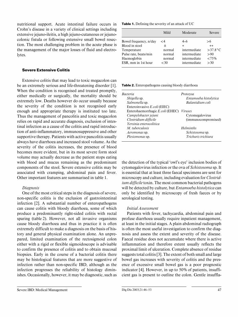
Severe IBD: Medical Management Dig Dis 2003;21:46–53 47
nutritional support. Acute intestinal failure occurs inCrohn’s disease in a variety of clinical settings includingextensive jejuno-ileitis, a high jejuno-cutaneous or jejuno-colonic fistula or following extensive small bowel resec-tion. The most challenging problem in the acute phase isthe management of the major losses of fluid and electro-lytes.
Severe Extensive Colitis
Extensive colitis that may lead to toxic megacolon canbe an extremely serious and life-threatening disorder [1].When the condition is recognised and treated promptly,either medically or surgically, the mortality should beextremely low. Deaths however do occur usually becausethe severity of the condition is not recognised earlyenough and appropriate therapy is instituted too late.Thus the management of pancolitis and toxic megacolonrelies on rapid and accurate diagnosis, exclusion of intes-tinal infection as a cause of the colitis and rapid introduc-tion of anti-inflammatory, immunosuppressive and othersupportive therapy. Patients with active pancolitis usuallyalways have diarrhoea and increased stool volume. As theseverity of the colitis increases, the presence of bloodbecomes more evident, but in its most severe form stoolvolume may actually decrease as the patient stops eatingwith blood and mucus remaining as the predominantcomponents of the stool. Severe extensive colitis may beassociated with cramping, abdominal pain and fever.Other important features are summarised in table 1.
DiagnosisOne of the most critical steps in the diagnosis of severe,
non-specific colitis is the exclusion of gastrointestinalinfection [2]. A substantial number of enteropathogenscan cause colitis with bloody diarrhoea, some of whichproduce a predominantly right-sided colitis with rectalsparing (table 2). However, not all invasive organismscause bloody diarrhoea and thus in practice it is oftenextremely difficult to make a diagnosis on the basis of his-tory and general physical examination alone. An unpre-pared, limited examination of the rectosigmoid coloneither with a rigid or flexible sigmoidoscope is advisableto confirm the presence of colitis and to obtain mucosalbiopsies. Early in the course of a bacterial colitis theremay be histological features that are more suggestive ofinfection rather than non-specific IBD, although as theinfection progresses the reliability of histology dimin-ishes. Occasionally, however, it may be diagnostic, such as
Table 1. Defining the severity of an attack of UC
Mild Moderate Severe
Bowel frequency, n/day !4 4–6 16Blood in stool B + ++Temperature normal intermediate 137.8°CPulse rate, beats/min normal intermediate 190Haemoglobin normal intermediate !75%ESR, mm in 1st hour !30 intermediate 130
Table 2. Enteropathogens causing bloody diarrhoea
BacteriaShigella sp.Salmonella sp.Enteroinvasive E.coli (EIEC)Enterohaemorrhagic E.coli (EHEC)Campylobacter jejuniClostridium difficileYersinia enterocoliticaM. tuberculosisAeromonas sp.Plesiomonas sp.
ProtozoaEntamoeba histolyticaBalantidium coli
VirusesCytomegalovirus(immunocompromised)
HelminthsSchistosoma sp.Trichuris trichiura
the detection of the typical ‘owl’s eye’ inclusion bodies ofcytomegalovirus infection or the ova of Schistosoma sp. Itis essential that at least three faecal specimens are sent formicroscopy and culture, including evaluation for Clostrid-ium difficile toxin. The most common bacterial pathogenswill be detected by culture, but Entamoeba histolytica canonly be identified by microscopy of fresh faeces or byserological testing.
Initial AssessmentPatients with fever, tachycardia, abdominal pain and
profuse diarrhoea usually require inpatient management,at least in the initial stages. A plain abdominal radiographis often the most useful investigation to confirm the diag-nosis and assess the extent and severity of the disease.Faecal residue does not accumulate where there is activeinflammation and therefore extent usually reflects theproximal limit of ulceration. Complete absence of residuesuggests total colitis [3]. The extent of both small and largebowel gas increases with severity of colitis and the pres-ence of excessive small bowel gas is a poor prognosticindicator [4]. However, in up to 50% of patients, insuffi-cient gas is present to outline the colon. Gentle insuffla-
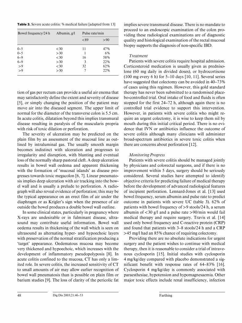
48 Dig Dis 2003;21:46–53 Farthing
Table 3. Severe acute colitis: % medical failure [adapted from 13]
Bowel frequency/24 h Albumin, g/l Pulse rate/min
!89 190
0–5 !30 11 47%0–5 130 1 6%6–9 !30 16 58%6–9 130 3 22%19 !30 32 62%19 130 7 22%
tion of gas per rectum can provide a useful air enema thatmay satisfactorily define the extent and severity of disease[5], or simply changing the position of the patient maymove air into the diseased segment. The upper limit ofnormal for the diameter of the transverse colon is 5.5 cm.In acute colitis, dilatation beyond this implies transmuraldisease resulting in paralysis of the muscularis propriawith risk of toxic dilation or perforation.
The severity of ulceration may be predicted on theplain film by an assessment of the mucosal line as out-lined by intraluminal gas. The usually smooth marginbecomes indistinct with ulceration and progresses toirregularity and disruption, with blunting and eventualloss of the normally sharp pastoral cleft. A deep ulcerationresults in bowel wall oedema and apparent thickeningwith the formation of ‘mucosal islands’ as disease pro-gresses towards toxic megacolon [6, 7]. Linear pneumato-sis implies deep ulceration with air tracking into the bow-el wall and is usually a prelude to perforation. A radio-graph will also reveal evidence of perforation; this may bethe typical appearance on an erect film of air under thediaphragm or as Krigler’s sign when the presence of airoutside the bowel produces a double bowel wall outline.
In some clinical states, particularly in pregnancy whereX-rays are undesirable or in fulminant disease, ultra-sound may contribute useful information. Bowel walloedema results in thickening of the wall which is seen onultrasound as alternating hyper- and hypoechoic layerswith preservation of the normal stratification producing a‘target’ appearance. Oedematous mucosa may becomevery thickened and hypoechoic, which increases with thedevelopment of inflammatory pseudopolyposis [8]. Inacute colitis confined to the mucosa, CT has only a lim-ited role. In severe colitis, the increased sensitivity of CTto small amounts of air may allow earlier recognition ofbowel wall pneumatosis than is possible on plain film orbarium studies [9]. The loss of clarity of the pericolic fat
implies severe transmural disease. There is no mandate toproceed to an endoscopic examination of the colon pro-viding these radiological examinations are of diagnosticquality and histological examination of the rectal mucosalbiopsy supports the diagnosis of non-specific IBD.
TreatmentPatients with severe colitis require hospital admission.
Corticosteroid medication is usually given as predniso-lone (60 mg daily in divided doses), or hydrocortisone(100 mg every 6 h) for 5–10 days [10, 11]. Several serieshave suggested that colectomy can be avoided in 40–73%of cases using this regimen. However, this gold standardtherapy has never been submitted to a randomised place-bo-controlled trial. Oral intake of food and fluids is oftenstopped for the first 24–72 h, although again there is nocontrolled trial evidence to support this intervention.However, in patients with severe colitis who might re-quire an urgent colectomy, it is wise to keep them nil bymouth during this initial critical period. There is no evi-dence that IVN or antibiotics influence the outcome ofsevere colitis although many clinicians will administerbroad-spectrum antibiotics in severe toxic colitis whenthere are concerns about perforation [12].
Monitoring ProgressPatients with severe colitis should be managed jointly
by physicians and colorectal surgeons, and if there is noimprovement within 5 days, surgery should be seriouslyconsidered. Several studies have attempted to identifyobjective criteria for predicting failure of medical therapybefore the development of advanced radiological featuresof incipient perforation. Lennard-Jones et al. [13] usedbowel frequency, serum albumin and pulse rate to predictoutcome in patients with severe UC (table 3). 62% ofpatients with bowel frequency of 19 stools/24 h, a serumalbumin of !30 g/l and a pulse rate 190/min would failmedical therapy and require surgery. Travis et al. [14]used only bowel frequency and C-reactive protein (CRP)and found that patients with 3–8 stools/24 h and a CRP145 mg/l had an 85% chance of requiring colectomy.
Providing there are no absolute indications for urgentsurgery and the patient wishes to continue with medicaltherapy, then it is reasonable to consider a trial of intrave-nous cyclosporin [15]. Initial studies with cyclosporin4 mg/kg/day compared with placebo demonstrated a sig-nificant benefit with response rates of 64–83% [16].Cyclosporin 4 mg/kg/day is commonly associated withparaesthesiae, hypotension and hypomagnesaemia. Othermajor toxic effects include renal insufficiency, infection
Severe IBD: Medical Management Dig Dis 2003;21:46–53 49
and seizures. Cyclosporin 2 mg/kg/day has a lower rate oftoxicity. Hypercholesterolaemia and hypomagnesaemiaincrease the risk of seizures. Maintenance of the responseto cyclosporin is significantly improved by the addition ofazathioprine as maintenance therapy.
Toxic Megacolon
Toxic megacolon is a severe complication of colitischaracterised by generalised toxic state (fever, prostrationand usually abdominal pain) associated with dilatation ofthe colon radiologically [17–19]. Toxic megacolon occursin approximately 2% of patients with chronic UC. Theprevalence rises to 10% in ill patients requiring hospitali-sation. These patients usually have a high fever 138°C,tachycardia, abdominal distension and abdominal painwhich may be diffuse or localised. There may be local ten-derness with rebound and if perforation has alreadyoccurred, this may be widespread in the abdomen. Bowelsounds are usually reduced or absent. Dilatation on anabdominal radiograph is the hallmark of this complica-tion with the colonic diameters reported between 8 and9 cm. Dilatation may be localised to a short segment ormay be generalised. In an acute attack, daily abdominalfilms are justified to monitor colonic diameter and thestate of the mucosa, to determine the need for surgicalintervention. Intraperitoneal perforation is the most seri-ous complication, the risk being highest in the initialattack [20]. The first sign of impending perforation islinear pneumatosis paralleling the bowel wall, commonlyfirst seen in the sigmoid colon. This may be more sensi-tively detected on CT than on plain films. Toxic megaco-lon is usually associated with anaemia, neutrophil leuko-cytosis and raised inflammatory markers such as ESR andCRP. The albumin is usually reduced. Once the colon isdilated on a plain abdominal radiograph, there is a stronglikelihood that colectomy will be required. The presenceof ‘mucosal islands’ is indicative of severe mucosal lossand disruption of smooth muscle function of the colonicwall. Although such patients do occasionally respond tomedical therapy, there is no doubt that the safest course ofaction is to recommend colectomy [21–23]. Patients withsevere fulminant colitis are at risk of thrombo-emboliccomplications before and particularly after surgery. Pro-viding rectal bleeding is not heavy it is wise to instituteprophylaxis with subcutaneous low-molecular-weightheparin.
Severe pancolitis and toxic megacolon also occur inCrohn’s disease. The clinical approach to the patient is
identical to that in severe UC. However, in patients whoare refractory to intravenous corticosteroids and immu-nosuppressive agents such as azathioprine and methotrex-ate, anti-TNF-· therapy (infliximab) should be consid-ered, providing there are no absolute indications forcolectomy.
Severe Crohn’s Jejuno-Ileitis
Diffuse involvement of a large proportion of the smallintestine is uncommon but can present a major therapeu-tic challenge. These patients often have major nutritionalproblems including profound weight loss and hypoalbu-minaemia. The condition can have particularly seriousclinical effects in children and adolescents such as retar-dation of growth and development. Diagnosis is based onconventional small bowel radiology (barium follow-through or enteroclysis), colonoscopy and small intestinalbiopsy. In the absence of colonic involvement the radio-logic appearances may be confused with ulcerative jejuni-tis associated with gluten-sensitive enteropathy and smallbowel lymphoma. It is advisable therefore to always con-firm the diagnosis histologically.
In adults, the therapeutic approach is similar to that forother forms of Crohn’s disease and should include theconventional treatment escalation, beginning with corti-costeroids, azathioprine or methotrexate and finally anti-TNF-· therapy (infliximab) if the disease is refractory tostandard immunosuppressive therapy [24]. However, inchildren and adolescents with remaining growth poten-tial, it is wise to avoid corticosteroid therapy. There isnow compelling evidence that enteral feeding with poly-meric diets can induce remission in children with Crohn’sdisease while at the same time optimising the opportunityfor growth promotion [25]. Evidence in animal models ofIBD have shown clearly that growth failure is due to acombination of anorexia and impaired food intake and toan effect which relates specifically to the inflammatoryprocess which is independent of the effect on appetite[26]. Limited anecdotal evidence in patients with exten-sive jejuno-ileitis suggests that long-term liquid enteralfeeding with a polymeric diet may contribute to achievingremission while supporting nutritional status. Althoughmeta-analysis has shown that both elemental diets andpolymeric liquid diets are inferior to corticosteroids in thetreatment of adults with active Crohn’s disease [27], effi-cacy appears to be more impressive in children and youngadults [25], particularly when there is associated growthretardation.
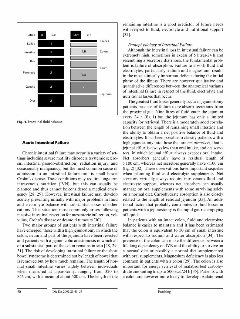
50 Dig Dis 2003;21:46–53 Farthing
Fig. 1. Intestinal fluid balance.
Acute Intestinal Failure
Chronic intestinal failure may occur in a variety of set-tings including severe motility disorders (systemic sclero-sis, intestinal pseudo-obstruction), radiation injury, andoccasionally malignancy, but the most common cause ofadmission to an intestinal failure unit is small bowelCrohn’s disease. These conditions may require long-termintravenous nutrition (IVN), but this can usually beplanned and thus cannot be considered a medical emer-gency [28, 29]. However, intestinal failure may developacutely presenting initially with major problems in fluidand electrolyte balance with substantial losses of othercations. This situation most commonly arises followingmassive intestinal resection for mesenteric infarction, vol-vulus, Crohn’s disease or desmoid tumours [30].
Two major groups of patients with intestinal failurehave emerged; those with a high jejunostomy in which thecolon, ileum and part of the jejunum have been resectedand patients with a jejuno-colic anastomosis in which allor a substantial part of the colon remains in situ [28, 29,31]. The risk of developing intestinal failure or the shortbowel syndrome is determined not by length of bowel thatis removed but by how much remains. The length of nor-mal small intestine varies widely between individualswhen measured at laparotomy, ranging from 320 to846 cm, with a mean of about 500 cm. The length of the
remaining intestine is a good predictor of future needswith respect to fluid, electrolyte and nutritional support[32].
Pathophysiology of Intestinal FailureAlthough the intestinal loss in intestinal failure can be
extremely high, sometimes in excess of 5 litres/24 h andresembling a secretory diarrhoea, the fundamental prob-lem is failure of absorption. Failure to absorb fluid andelectrolytes, particularly sodium and magnesium, resultsin the most clinically important deficits during the initialphase of the illness. There are however qualitative andquantitative differences between the anatomical variantsof intestinal failure in respect of the fluid, electrolyte andnutritional losses that occur.
The greatest fluid losses generally occur in jejunostomypatients because of failure to re-absorb secretions fromthe proximal gut. Nine litres of fluid enter the jejunumevery 24 h (fig. 1) but the jejunum has only a limitedcapacity for retrieval. There is a moderately good correla-tion between the length of remaining small intestine andthe ability to obtain a net positive balance of fluid andelectrolytes. It has been possible to classify patients with ahigh jejunostomy into those that are net absorbers, that isjejunal efflux is always less than oral intake, and net secre-tors, in which jejunal efflux always exceeds oral intake.Net absorbers generally have a residual length of1100 cm, whereas net secretors generally have !100 cm(fig. 2) [32]. These observations have important sequelaewhen planning fluid and electrolyte supplements. Netsecretors virtually always require intravenous fluid andelectrolyte support, whereas net absorbers can usuallymanage on oral supplements with some surviving solelyon a normal diet. Carbohydrate absorption is also closelyrelated to the length of residual jejunum [33]. An addi-tional factor that probably contributes to fluid losses inpatients with a jejunostomy is the rapid gastric emptyingof liquids.
In patients with an intact colon, fluid and electrolytebalance is easier to maintain and it has been estimatedthat the colon is equivalent to 50 cm of small intestinewith respect to sodium and water absorption [34]. Thepresence of the colon can make the difference between alife-long dependency on IVN and the ability to survive ona normal diet or possibly a normal diet supplementedwith oral supplements. Magnesium deficiency is also lesscommon in patients with a colon [29]. The colon is alsoimportant for energy retrieval of malabsorbed carbohy-drate amounting to up to 500 kcal/24 h [35]. Patients witha colon are however more likely to develop oxalate renal

Severe IBD: Medical Management Dig Dis 2003;21:46–53 51
Fig. 2. Daily oral intake and intestinal ef-fluent. IVN = Intravenous nutrition; IVF =intravenous fluid; OS = oral supplements[adapted from 30].
stones due to enhanced oxalate absorption from the colon[34].
Initial Management of Acute Intestinal FailureThe rational management of intestinal failure depends
on an assessment of fluid, electrolyte and nutrient losses.The aims of this assessment are two-fold, namely to rapid-ly correct any major deficiencies that have occurred dur-ing the early phase of the condition and secondly to planthe long-term management, particularly to predict wheth-er or not there will be a need for IVN [28, 29].
Although intestinal losses in excess of 2 litres/24 h areoften indicative that some form of intravenous supportwill be required, it is essential that the initial assessmentbe carried out when the patient is fluid and electrolytereplete. Patients may have been drinking vast quantitiesof low sodium liquids in an attempt to deal with thirstpromoted by dehydration and hyponatraemia. This willexacerbate sodium and magnesium deficiency and in-crease intestinal effluent.
It is advisable therefore to stabilise the situation by giv-ing appropriate volumes of intravenous saline to rehy-drate until body weight is stable and confirm that there isadequate sodium in the urine (120 mmol/l). When rehy-dration and sodium repletion is achieved, the patient can
then be progressively transferred to a normal diet andintestinal effluent volume (or weight) assessed. If intesti-nal losses continue to exceed 2 litres/24 h then it is highlylikely that intravenous replacement of saline will berequired, and as losses approach 3–4 litres/day then thiswill be essential. If losses are less than 2 litres/24 h, it islikely that fluid and electrolyte homeostasis can be main-tained orally, but such patients may require supplementa-tion with 1–2 litres of a high sodium (190 mmol/l) glu-cose-electrolyte solution [36, 37]. Many of the commer-cially available oral rehydration solutions have inade-quate sodium concentrations for patients with a high out-put jejunostomy. It may be necessary to make up anappropriate solution in the home or hospital pharmacy.
In patients with intestinal effluents exceeding 2 litres/24 h there is always the risk of magnesium deficiency [28].The risk is substantially reduced when the colon isretained. Deficiency should be screened for during the ini-tial assessment by measuring plasma magnesium concen-tration although deficiency may be apparent clinicallywith symptoms in the peripheral and central nervous sys-tem including paraesthesiae, tetany, lassitude, depressionand occasionally convulsions. There may also be muscleweakness. In symptomatic cases of magnesium deficien-cy, potassium and calcium concentrations are also re-
52 Dig Dis 2003;21:46–53 Farthing
duced. In severe acute deficiency, magnesium sulphateshould be given intravenously with careful monitoring ofplasma magnesium concentration. Many patients with achronically high intestinal effluent will require replace-ment on a regular basis, magnesium oxide (12–24 mmol/24 h) being the preferred preparation.
Potassium deficiency is uncommon in intestinal fail-ure and is usually only seen when there is !50 cm of resid-ual small intestine. Hypokalaemia in jejunostomy pa-tients may be indicative of sodium depletion as a result ofeither secondary hyperaldosteronism or a magnesium def-icit.
Drug Therapy to Reduce Intestinal EffluentPharmacological approaches to reducing intestinal ef-
fluent are only modestly effective and in general areunable to change a patient’s status from being dependenton IVN or IV fluids to an individual who can survive onoral intake alone. However, a reduction in effluent can beachieved by either improving intestinal absorption or byinhibiting intestinal secretion. Synthetic opioid drugssuch as loperamide or the opiate, codeine phosphate, arethe first-line medications to be evaluated. Although it hasbeen difficult to unequivocally demonstrate efficacy be-cause of the relatively small numbers of patients that are
available for inclusion in clinical trials, detailed balancestudies in an individual patient clearly show beneficialeffects with respect to reducing sodium and fluid loss [38].An alternative approach is to use the somatostatin ana-logue, octreotide, that slows intestinal transit and reducesgastric, pancreatic and biliary secretion. A variety of smallstudies have shown that octreotide reduces intestinal out-put and some have also shown a reduction in sodium andpotassium loss [39]. These effects have been sustainedlong term and no major adverse effects have been re-ported. Unfortunately these effects are insufficient to con-vert a patient from being a net secretor to a net absorberor render a patient no longer dependent on IVN. How-ever, reducing intravenous fluid requirements for a pa-tient will decrease the time that the individual needs to beconnected to the infusion system.
An alternative approach to reducing secretion into thegut is to use an H2-receptor antagonist or a proton pumpinhibitor [40]. The efficacy of these drugs is probablywithin the same range as octreotide although responses inindividual patients may be idiosyncratic and it thereforeworthwhile beginning in a hierarchical way with the acidinhibitors and then moving on to octreotide to determinewhether additional benefits can be achieved.
References
1 Farthing MJG, McLean AM: Pancolitis andtoxic megacolon; in Gassull MA, Obrador A,Chantar C (eds): Management of InflammatoryBowel Disease. Prous Science Publishers, 1994,pp 195–209.
2 Farthing MJG: Infectious diarrhea; in IrvineEJ, Hunt RH (eds): Evidence-Based Gastroen-terology. Hamilton, BC Decker, 2001, pp 323–341.
3 Prantera C, Lorenzetti T, Cerro P, et al: Theplain abdominal film accurately estimates ex-tent of active ulcerative colitis. J Clin Gas-troenterol 1991;13:231–234.
4 Bartram CI: Radiology in the current assess-ment of ulcerative colitis. Gastrointest Radiol1977;1:383–392.
5 Vernia P, Colaneri O, Tomei E, Caprilla R:Intestinal gas in ulcerative colitis. Dis ColonRectum 1979;22:346–349.
6 Bartram CI, Preston DM, Lennard-Jones JE:The ‘air enema’ in acute colitis. GastrointestRadiol 1983;8:61–65.
7 Halpert RD: Toxic dilation of the colon. RadiolClin North Am 1987;25:147–155.
8 Hata J, Haruma K, Suenga K, et al: Ultrasonicassessment of inflammatory bowel disease. AmJ Gastroentrol 1992;87:443–447.
9 Gore RM: CT of inflammatory bowel disease.Radiol Clin North Am 1989;27:717–730.
10 Meyers S, Janowitz HD: Systemic corticoste-roid therapy of ulcerative colitis. Gastroenter-ology 1985;89:1189–1191.
11 Truelove SC, Jewell DP: Intensive intravenousregimen for severe attacks of ulcerative colitis.Lancet 1974;i:1067–1070.
12 Jarnerot G, Rolny P, Sandberg-Gertzen H: In-tensive intravenous treatment of ulcerative co-litis. Gastroenterology 1985;89:1005–1013.
13 Lennard-Jones JE, Ritchie JK, Hilder W, Spic-er CC: Assessment of severity in colitis: A pre-liminary study. Gut 1975;16:579–584
14 Travis SPL, Farrant JM, Nolan DJ, MortensenNM, Kettlewell MGW, Jewell DP: Predictingoutcome in severe ulcerative colitis. Gut 1996;38:905–910.
15 Lichtiger S, Present DH: Preliminary report:Cyclosporin in treatment of severe active ulcer-ative colitis. Lancet 1990;336:16–19.
16 Loftus CG, Loftus EV, Sandborn WJ: Cyclo-sporin for refractory ulcerative colitis. Gut2002;52:172–173.
17 Binder SC, et al: Toxic megacolon in ulcerativecolitis. Gastroenterology 1974;66:909–915.
18 Odyniec NA, Judd ES, Sauer WG: Toxic mega-colon in ulcerative colitis. Gastroenterology1974;66:909.
19 Grant CS, Dozois RR: Toxic megacolon: Ulti-mate fate of patients after successful medicalmanagement. Am J Surg 1984;147:106–110.
20 DeDombal FT, Watts JMK, Watkinson G,Goligher JC: Intraperitoneal perforation of thecolon in ulcerative colitis. Proc R Soc Med1965;58:713–715.
21 Odyniec ND, Dudd ES, Sauer WG: Toxicmegacolon: Significant improvement in surgi-cal management. Arch Surg 1967;94:638–643.
22 Goligher JC, et al: Early surgery in the manage-ment of severe ulcerative colitis. Br Med J1967;iii:193.
23 Sirinek KR, et al: Total proctocolectomy andileostomy: Procedure of choice for acute toxicmegacolon. Arch Surg 1985;112:518.
24 Podolsky DK: Inflammatory bowel disease. NEngl J Med 2002;347:417–429.
25 Heuschkel RB, Menache CC, Megerian JT,Baird AE: Enteral nutrition and corticosteroidsin the treatment of acute Crohn’s disease inchildren. J Pediatr Gastroenterol Nutr 2000;31:8–15.
Severe IBD: Medical Management Dig Dis 2003;21:46–53 53
26 Ballinger AB, Azooz O, El-Haj T, Poole S,Farthing MJG: Growth failure occurs througha decrease in insulin-like growth factor-1 whichis independent of undernutrition in a rat modelof colitis. Gut 2000;46:694–700.
27 Griffiths AM, Ohlsson A, Sherman PM, Suth-erland LR: Meta-analysis of enteral nutrition asa primary treatment of active Crohn’s disease.Gastroenterology 1995,32:1056–1067.
28 Lennard-Jones JE: Practical management ofthe short bowel. Aliment Pharmacol Ther1994;8:563–577.
29 Farthing MJG: Intestinal failure; in FarthingMJG (ed): Clinical Challenges in Gastroenter-ology. London, Dunitz, 1996, pp 86–103.
30 Nightingale JMD, Walker ER, Burnham WR,et al: Short bowel syndrome. Digestion 1990;45(suppl 1):77–83.
31 Nightingale JMD: The short bowel. Eur J Gas-trohepatol 1995;7:514–520.
32 Nightingale JMD, Lennard-Jones JE, WalkerER, Farthing MJG: Jejunal efflux in short-bow-el syndrome. Lancet 1990;336:765–768.
33 Rodrigues CA, Lennard-Jones JE, ThompsonDG, Farthing MJG: Energy absorption as ameasure of intestinal failure in the short bowelsyndrome. Gut 1989;30:176–183.
34 Nightingale JMD, Lennard-Jones JE, GertnerDJ, Wood SR, Bartram CI: Colonic preserva-tion reduces the need for parenteral therapy,increases the incidence of renal stones but doesnot change the high prevalence of gallstones inpatients with a short bowel. Gut 1992;33:1493–1497.
35 Nordgaard I, Hansen BS, Mortensen PB: Colonas a digestive jejunostomy output in patientswith severe short-bowel syndrome. Lancet1994;343:373–376.
36 Lennard-Jones JE: Oral rehydration solutionsin short bowel syndrome. Clin Ther 1990;12(suppl A):129–137.
37 Nightingale JMD, Lennard-Jones JE, WalkerER, Farthing MJG: Oral salt supplements tocompensate for jejunstomy losses: Comparisonof sodium chloride capsules, glucose electrolytesolution and glucose polymer electrolyte solu-tion (Maxijul). Gut 1992;33:759–761.
38 Rodrigues CA, Lennard-Jones JE, Walker ER,Thompson DG, Farthing MJG: The effects ofoctreotide, soy polysaccharide, codeine and lo-peramide on nutrient, fluid and electrolyte ab-sorption in the short bowel syndrome. AlimentPharmacol Ther 1989;3:159–169.
39 Nightingale JMD, Walker ER, Burnham WR,Farthing MJG, Lennard-Jones JE: Octreotide(a somatostatin analogue) improves the qualityof life in some patients with a short intestine.Aliment Pharmacol Ther 1989;3:367–373.
40 Nightingale JMD, Walker ER, Farthing MJG,Lennard-Jones JE: Effect of omeprazole on in-testinal output in the short-bowel syndrome.Aliment Pharmacol Ther 1991;5:405–412.
Review Article
Dig Dis 2003;21:54–62DOI: 10.1159/000071340
Surgical Treatment of SevereInflammatory Bowel Diseases
Christine Leowardi Gundi Heuschen Peter Kienle Udo HeuschenJan Schmidt
Department of Surgery, University of Heidelberg, Germany
Jan Schmidt, MDDepartment of Surgery, University of HeidelbergKirschnerstrase 1, DE–69120 Heidelberg (Germany)Tel. +49 6221 566204, Fax +49 6221 565781E-Mail [email protected]
ABCFax + 41 61 306 12 34E-Mail [email protected]
© 2003 S. Karger AG, Basel0257–2753/03/0211–0054$19.50/0
Accessible online at:www.karger.com/ddi
Key WordsCrohn’s disease W Ulcerative colitis W Surgical treatment
AbstractSurgical treatment of severe inflammatory bowel dis-eases is required in failed medical treatment, in emer-gencies and for complications. Indications for surgeryand operative techniques have changed significantlyover the last few years. There is a clear tendency towardsearlier and less invasive surgical interventions per-formed in specialized and experienced centers. Im-proved quality of life of patients with Crohn’s disease orulcerative colitis after surgical therapy supports an ear-lier consideration of the surgical treatment option. Aclose cooperation with the involved gastroenterologist ismandatory in this context.
Copyright © 2003 S. Karger AG, Basel
Introduction
Ulcerative colitis (UC) and Crohn’s disease (CD) areinflammatory disorders of the gastrointestinal tract ofunknown etiology. Both diseases are primarily a domainof conservative medicine. However, about one third of
patients with CD or UC do not respond to conventionalmedical treatment. In this subgroup of patients withsevere inflammatory bowel diseases, surgery can lead to asignificant relief of symptoms and in UC patients evencure the disease.
Crohn’s Disease
CD is an idiopathic, chronic inflammatory disease ofthe gastrointestinal tract that primarily affects the smallintestine and colon, which may be caused by environmen-tal and genetic factors.
The incidence rate varies between different geographi-cal regions, with an average of 3–6 cases/100,000/year [1].There is a typical ‘bimodal’ age distribution at diagnosiswith a first peak between the age of 15 and 30 and a sec-ond peak later in life in the sixth or seventh decade.Regarding the gender distribution, several studies de-scribed a slight female predominance, with an increasedrisk for women of about 20–30%. CD appears to be asso-ciated with a significant genetic predisposition with anincreased relative risk for first-degree relatives of affectedpatients between the age of 18 and 36. Proven risk factorsare smoking [2], oral contraception [3] and a high socio-economic status.
Surgical Treatment of Inflammatory BowelDiseases
Dig Dis 2003;21:54–62 55
The etiology of CD is still unknown, but three funda-mental theories are presently being discussed [4–6]: (1) animpaired intestinal epithelial barrier function with a lossof tolerance towards intraluminal antigens; (2) a dis-turbed immunological response in the intestinal walltowards ubiquitous luminal antigens, and (3) a specificinfection.
CD is a transmural, predominantly submucosal in-flammatory disease that most commonly affects the distalileum and colon but may occur in any part of the gastroin-testinal tract. Macroscopically, segments of affected bow-el are characteristically sharply demarcated from adjacentnormal bowel (‘skip lesions’). Transmural inflammationleads to bowel wall thickening and lymph edema and canresult in extensive fibrosis with strictures. Patchy, muco-sal longitudinal and transverse ulcers with interveningmucosal edema can develop which then appear as the typ-ical cobblestone relief. Often the attached mesentery ismarkedly thickened and lymph edematous with adher-ence of the inflamed segment to neighboring organs,forming conglomerates with sometimes interenteric orblind fistulas and abscesses. Mesenteric fat typically ex-tends on over the serosal surface of the bowel. Microscopi-cally, there are submucosal edemas, lymphoid aggrega-tions, lymphoplasmacellular infiltrates, ulcers and fibro-sis with influx and proliferation of macrophages. Nonca-seating granulomas with multinucleated giant cells aredetectable in up to 60% of patients.
Clinical Symptoms and ComplicationsClinical symptoms vary with the location of the in-
flamed region. Chronic diarrhea with abdominal pain,fever, anorexia, weight loss, and a right lower quadrantmass or fullness are the most common presenting fea-tures. Many patients are first seen with an acute abdomendue to intestinal obstruction, sometimes simulating acuteappendicitis. In the selected surgical setting, there is anincreased percentage of patients with perianal fistulas.Extraintestinal manifestations include joints (arthritis),skin (pyoderma gangrenosum), kidneys and the urinarytract (stones, fistulas), gallbladder and bile ducts (stones,sclerosing cholangitis).
Due to the varying locations of the disease, the devel-opment of complications has a wide spectrum. Intestinalbleeding, perforation, obstructions, development of ente-roenteric, enterovesical, retroperitoneal, or enterocuta-neous fistulas, and abscess formations are common com-plications in CD, often requiring surgical intervention.The risk of developing a CD-associated carcinoma isincreased about 5- to 6-fold [7].
Surgical TherapyThe mainstay of CD treatment remains medical thera-
py, which is beyond the scope of this review. Interestedreaders are referred to the literature [8] or the CochraneLibrary (www.update-software.com).
Patients suffering from severe CD require surgery ei-ther to manage complications or in case of failure of medi-cal treatment. 2,070 cases with CD were treated at theSurgical Department of the University of Heidelbergbetween 1982 and January 2003.
Surgery, as well as conservative medical treatment,cannot cure the disease. However, more than 90% of allpatients treated surgically in our institution declared thatthey experienced a complete remission of symptoms(68%), or a significant relief of complaints. Nevertheless,the recurrence rate in the following 10–15 years in thesepatients was still high (50%) [9]. Most of these recurrencescan be effectively treated with a further operation. Theformer widespread fear of a ‘short-bowel syndrome’ isnow unfounded. The modern principles of Crohn’s sur-gery restrict resection to inflamed sections only withoutso-called ‘security margins’ as practised in cancer surgery[10]. Short fibrotic strictures can be treated with stricturo-plasty, also known as ‘conservative surgery’. Minimallyinvasive techniques can now be used in a large number ofcases. Therefore, surgical therapy should be consideredearly in the treatment of symptomatic stenoses, fistulas,septic complications and situations refractory to conser-vative treatment. Furthermore, complications of long-term therapy with glucocorticoids or immunosuppres-sants, as well as malignant transformation may beavoided by surgical treatment.
Specific Indications for SurgeryControversy still remains regarding the right time for
surgery. A major reason for early surgical intervention isthe high rate of symptomatic relief after surgery. Further-more, the resected bowel parts are mostly without func-tion. Opponents of this concept state that delayed surgeryis associated with fewer resections and therefore a lowerrisk of short-bowel syndrome. We believe that time of sur-gery should be based on the clinical symptoms. It isimportant to consider the preoperative medication withits side effects and the potential increase of perioperativecomplications due to the medications. The application ofthese principles should lead to a reasonable decisionregarding the time of surgery with a maximum relief ofcomplaints and a minimum incidence of surgery-relateddisadvantages. These principles, however, are not yet ade-quately considered. Scott and Hughes [11] found that
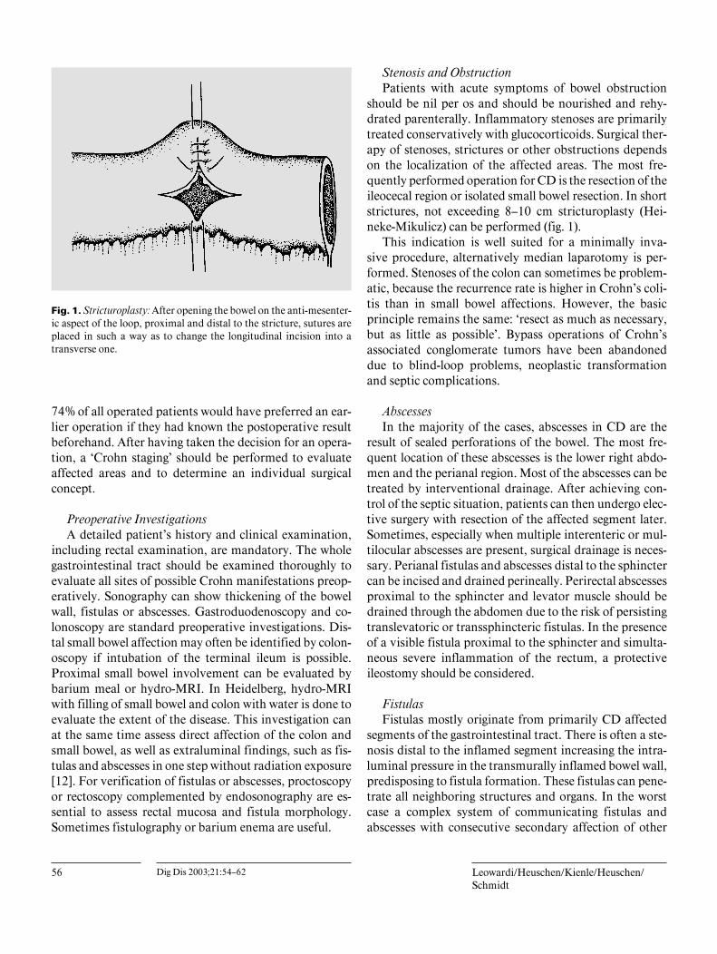
56 Dig Dis 2003;21:54–62 Leowardi/Heuschen/Kienle/Heuschen/Schmidt
Fig. 1. Stricturoplasty: After opening the bowel on the anti-mesenter-ic aspect of the loop, proximal and distal to the stricture, sutures areplaced in such a way as to change the longitudinal incision into atransverse one.
74% of all operated patients would have preferred an ear-lier operation if they had known the postoperative resultbeforehand. After having taken the decision for an opera-tion, a ‘Crohn staging’ should be performed to evaluateaffected areas and to determine an individual surgicalconcept.
Preoperative InvestigationsA detailed patient’s history and clinical examination,
including rectal examination, are mandatory. The wholegastrointestinal tract should be examined thoroughly toevaluate all sites of possible Crohn manifestations preop-eratively. Sonography can show thickening of the bowelwall, fistulas or abscesses. Gastroduodenoscopy and co-lonoscopy are standard preoperative investigations. Dis-tal small bowel affection may often be identified by colon-oscopy if intubation of the terminal ileum is possible.Proximal small bowel involvement can be evaluated bybarium meal or hydro-MRI. In Heidelberg, hydro-MRIwith filling of small bowel and colon with water is done toevaluate the extent of the disease. This investigation canat the same time assess direct affection of the colon andsmall bowel, as well as extraluminal findings, such as fis-tulas and abscesses in one step without radiation exposure[12]. For verification of fistulas or abscesses, proctoscopyor rectoscopy complemented by endosonography are es-sential to assess rectal mucosa and fistula morphology.Sometimes fistulography or barium enema are useful.
Stenosis and ObstructionPatients with acute symptoms of bowel obstruction
should be nil per os and should be nourished and rehy-drated parenterally. Inflammatory stenoses are primarilytreated conservatively with glucocorticoids. Surgical ther-apy of stenoses, strictures or other obstructions dependson the localization of the affected areas. The most fre-quently performed operation for CD is the resection of theileocecal region or isolated small bowel resection. In shortstrictures, not exceeding 8–10 cm stricturoplasty (Hei-neke-Mikulicz) can be performed (fig. 1).
This indication is well suited for a minimally inva-sive procedure, alternatively median laparotomy is per-formed. Stenoses of the colon can sometimes be problem-atic, because the recurrence rate is higher in Crohn’s coli-tis than in small bowel affections. However, the basicprinciple remains the same: ‘resect as much as necessary,but as little as possible’. Bypass operations of Crohn’sassociated conglomerate tumors have been abandoneddue to blind-loop problems, neoplastic transformationand septic complications.
AbscessesIn the majority of the cases, abscesses in CD are the
result of sealed perforations of the bowel. The most fre-quent location of these abscesses is the lower right abdo-men and the perianal region. Most of the abscesses can betreated by interventional drainage. After achieving con-trol of the septic situation, patients can then undergo elec-tive surgery with resection of the affected segment later.Sometimes, especially when multiple interenteric or mul-tilocular abscesses are present, surgical drainage is neces-sary. Perianal fistulas and abscesses distal to the sphinctercan be incised and drained perineally. Perirectal abscessesproximal to the sphincter and levator muscle should bedrained through the abdomen due to the risk of persistingtranslevatoric or transsphincteric fistulas. In the presenceof a visible fistula proximal to the sphincter and simulta-neous severe inflammation of the rectum, a protectiveileostomy should be considered.
FistulasFistulas mostly originate from primarily CD affected
segments of the gastrointestinal tract. There is often a ste-nosis distal to the inflamed segment increasing the intra-luminal pressure in the transmurally inflamed bowel wall,predisposing to fistula formation. These fistulas can pene-trate all neighboring structures and organs. In the worstcase a complex system of communicating fistulas andabscesses with consecutive secondary affection of other
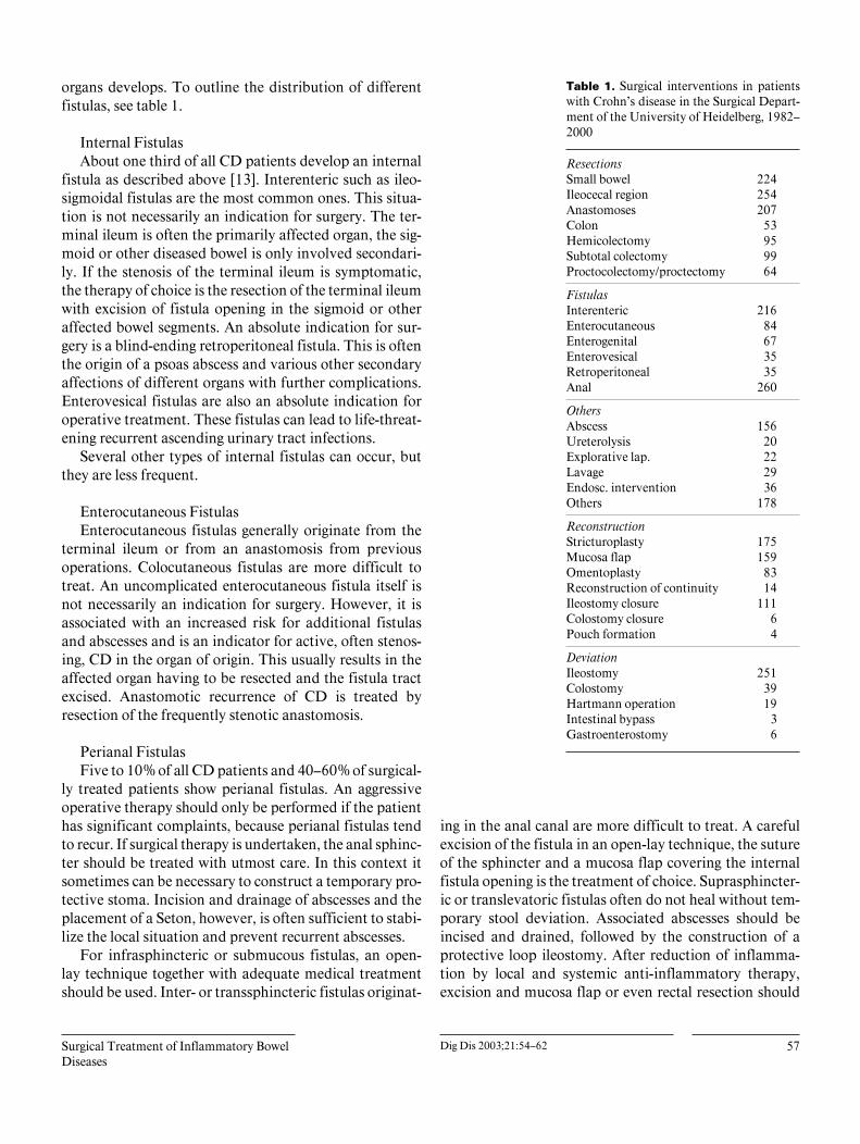
Surgical Treatment of Inflammatory BowelDiseases
Dig Dis 2003;21:54–62 57
organs develops. To outline the distribution of differentfistulas, see table 1.
Internal FistulasAbout one third of all CD patients develop an internal
fistula as described above [13]. Interenteric such as ileo-sigmoidal fistulas are the most common ones. This situa-tion is not necessarily an indication for surgery. The ter-minal ileum is often the primarily affected organ, the sig-moid or other diseased bowel is only involved secondari-ly. If the stenosis of the terminal ileum is symptomatic,the therapy of choice is the resection of the terminal ileumwith excision of fistula opening in the sigmoid or otheraffected bowel segments. An absolute indication for sur-gery is a blind-ending retroperitoneal fistula. This is oftenthe origin of a psoas abscess and various other secondaryaffections of different organs with further complications.Enterovesical fistulas are also an absolute indication foroperative treatment. These fistulas can lead to life-threat-ening recurrent ascending urinary tract infections.
Several other types of internal fistulas can occur, butthey are less frequent.
Enterocutaneous FistulasEnterocutaneous fistulas generally originate from the
terminal ileum or from an anastomosis from previousoperations. Colocutaneous fistulas are more difficult totreat. An uncomplicated enterocutaneous fistula itself isnot necessarily an indication for surgery. However, it isassociated with an increased risk for additional fistulasand abscesses and is an indicator for active, often stenos-ing, CD in the organ of origin. This usually results in theaffected organ having to be resected and the fistula tractexcised. Anastomotic recurrence of CD is treated byresection of the frequently stenotic anastomosis.
Perianal FistulasFive to 10% of all CD patients and 40–60% of surgical-
ly treated patients show perianal fistulas. An aggressiveoperative therapy should only be performed if the patienthas significant complaints, because perianal fistulas tendto recur. If surgical therapy is undertaken, the anal sphinc-ter should be treated with utmost care. In this context itsometimes can be necessary to construct a temporary pro-tective stoma. Incision and drainage of abscesses and theplacement of a Seton, however, is often sufficient to stabi-lize the local situation and prevent recurrent abscesses.
For infrasphincteric or submucous fistulas, an open-lay technique together with adequate medical treatmentshould be used. Inter- or transsphincteric fistulas originat-
Table 1. Surgical interventions in patientswith Crohn’s disease in the Surgical Depart-ment of the University of Heidelberg, 1982–2000
ResectionsSmall bowel 224Ileocecal region 254Anastomoses 207Colon 53Hemicolectomy 95Subtotal colectomy 99Proctocolectomy/proctectomy 64
FistulasInterenteric 216Enterocutaneous 84Enterogenital 67Enterovesical 35Retroperitoneal 35Anal 260
OthersAbscess 156Ureterolysis 20Explorative lap. 22Lavage 29Endosc. intervention 36Others 178
ReconstructionStricturoplasty 175Mucosa flap 159Omentoplasty 83Reconstruction of continuity 14Ileostomy closure 111Colostomy closure 6Pouch formation 4
DeviationIleostomy 251Colostomy 39Hartmann operation 19Intestinal bypass 3Gastroenterostomy 6
ing in the anal canal are more difficult to treat. A carefulexcision of the fistula in an open-lay technique, the sutureof the sphincter and a mucosa flap covering the internalfistula opening is the treatment of choice. Suprasphincter-ic or translevatoric fistulas often do not heal without tem-porary stool deviation. Associated abscesses should beincised and drained, followed by the construction of aprotective loop ileostomy. After reduction of inflamma-tion by local and systemic anti-inflammatory therapy,excision and mucosa flap or even rectal resection should
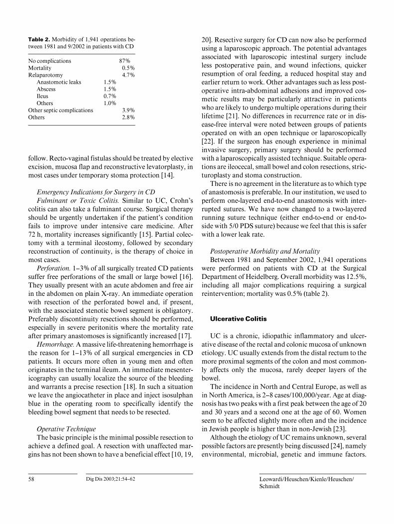
58 Dig Dis 2003;21:54–62 Leowardi/Heuschen/Kienle/Heuschen/Schmidt
Table 2. Morbidity of 1,941 operations be-tween 1981 and 9/2002 in patients with CD
No complications 87%Mortality 0.5%Relaparotomy 4.7%
Anastomotic leaks 1.5%Abscess 1.5%Ileus 0.7%Others 1.0%
Other septic complications 3.9%Others 2.8%
follow. Recto-vaginal fistulas should be treated by electiveexcision, mucosa flap and reconstructive levatorplasty, inmost cases under temporary stoma protection [14].
Emergency Indications for Surgery in CDFulminant or Toxic Colitis. Similar to UC, Crohn’s
colitis can also take a fulminant course. Surgical therapyshould be urgently undertaken if the patient’s conditionfails to improve under intensive care medicine. After72 h, mortality increases significantly [15]. Partial colec-tomy with a terminal ileostomy, followed by secondaryreconstruction of continuity, is the therapy of choice inmost cases.
Perforation. 1–3% of all surgically treated CD patientssuffer free perforations of the small or large bowel [16].They usually present with an acute abdomen and free airin the abdomen on plain X-ray. An immediate operationwith resection of the perforated bowel and, if present,with the associated stenotic bowel segment is obligatory.Preferably discontinuity resections should be performed,especially in severe peritonitis where the mortality rateafter primary anastomoses is significantly increased [17].
Hemorrhage. A massive life-threatening hemorrhage isthe reason for 1–13% of all surgical emergencies in CDpatients. It occurs more often in young men and oftenoriginates in the terminal ileum. An immediate mesenter-icography can usually localize the source of the bleedingand warrants a precise resection [18]. In such a situationwe leave the angiocatheter in place and inject isosulphanblue in the operating room to specifically identify thebleeding bowel segment that needs to be resected.
Operative TechniqueThe basic principle is the minimal possible resection to
achieve a defined goal. A resection with unaffected mar-gins has not been shown to have a beneficial effect [10, 19,
20]. Resective surgery for CD can now also be performedusing a laparoscopic approach. The potential advantagesassociated with laparoscopic intestinal surgery includeless postoperative pain, and wound infections, quickerresumption of oral feeding, a reduced hospital stay andearlier return to work. Other advantages such as less post-operative intra-abdominal adhesions and improved cos-metic results may be particularly attractive in patientswho are likely to undergo multiple operations during theirlifetime [21]. No differences in recurrence rate or in dis-ease-free interval were noted between groups of patientsoperated on with an open technique or laparoscopically[22]. If the surgeon has enough experience in minimalinvasive surgery, primary surgery should be performedwith a laparoscopically assisted technique. Suitable opera-tions are ileocecal, small bowel and colon resections, stric-turoplasty and stoma construction.
There is no agreement in the literature as to which typeof anastomosis is preferable. In our institution, we used toperform one-layered end-to-end anastomosis with inter-rupted sutures. We have now changed to a two-layeredrunning suture technique (either end-to-end or end-to-side with 5/0 PDS suture) because we feel that this is saferwith a lower leak rate.
Postoperative Morbidity and MortalityBetween 1981 and September 2002, 1,941 operations
were performed on patients with CD at the SurgicalDepartment of Heidelberg. Overall morbidity was 12.5%,including all major complications requiring a surgicalreintervention; mortality was 0.5% (table 2).
Ulcerative Colitis
UC is a chronic, idiopathic inflammatory and ulcer-ative disease of the rectal and colonic mucosa of unknownetiology. UC usually extends from the distal rectum to themore proximal segments of the colon and most common-ly affects only the mucosa, rarely deeper layers of thebowel.
The incidence in North and Central Europe, as well asin North America, is 2–8 cases/100,000/year. Age at diag-nosis has two peaks with a first peak between the age of 20and 30 years and a second one at the age of 60. Womenseem to be affected slightly more often and the incidencein Jewish people is higher than in non-Jewish [23].
Although the etiology of UC remains unknown, severalpossible factors are presently being discussed [24], namelyenvironmental, microbial, genetic and immune factors.
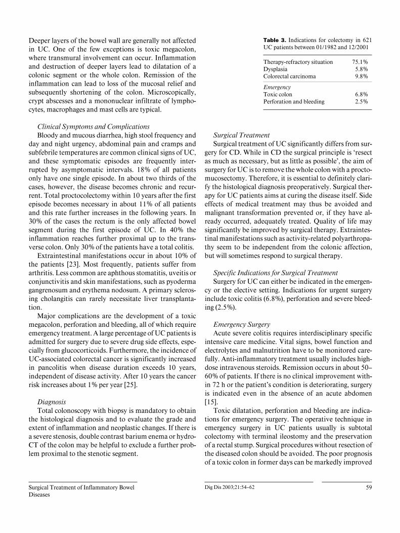
Surgical Treatment of Inflammatory BowelDiseases
Dig Dis 2003;21:54–62 59
Deeper layers of the bowel wall are generally not affectedin UC. One of the few exceptions is toxic megacolon,where transmural involvement can occur. Inflammationand destruction of deeper layers lead to dilatation of acolonic segment or the whole colon. Remission of theinflammation can lead to loss of the mucosal relief andsubsequently shortening of the colon. Microscopically,crypt abscesses and a mononuclear infiltrate of lympho-cytes, macrophages and mast cells are typical.
Clinical Symptoms and ComplicationsBloody and mucous diarrhea, high stool frequency and
day and night urgency, abdominal pain and cramps andsubfebrile temperatures are common clinical signs of UC,and these symptomatic episodes are frequently inter-rupted by asymptomatic intervals. 18% of all patientsonly have one single episode. In about two thirds of thecases, however, the disease becomes chronic and recur-rent. Total proctocolectomy within 10 years after the firstepisode becomes necessary in about 11% of all patientsand this rate further increases in the following years. In30% of the cases the rectum is the only affected bowelsegment during the first episode of UC. In 40% theinflammation reaches further proximal up to the trans-verse colon. Only 30% of the patients have a total colitis.
Extraintestinal manifestations occur in about 10% ofthe patients [23]. Most frequently, patients suffer fromarthritis. Less common are aphthous stomatitis, uveitis orconjunctivitis and skin manifestations, such as pyodermagangrenosum and erythema nodosum. A primary scleros-ing cholangitis can rarely necessitate liver transplanta-tion.
Major complications are the development of a toxicmegacolon, perforation and bleeding, all of which requireemergency treatment. A large percentage of UC patients isadmitted for surgery due to severe drug side effects, espe-cially from glucocorticoids. Furthermore, the incidence ofUC-associated colorectal cancer is significantly increasedin pancolitis when disease duration exceeds 10 years,independent of disease activity. After 10 years the cancerrisk increases about 1% per year [25].
DiagnosisTotal colonoscopy with biopsy is mandatory to obtain
the histological diagnosis and to evaluate the grade andextent of inflammation and neoplastic changes. If there isa severe stenosis, double contrast barium enema or hydro-CT of the colon may be helpful to exclude a further prob-lem proximal to the stenotic segment.
Table 3. Indications for colectomy in 621UC patients between 01/1982 and 12/2001
Therapy-refractory situation 75.1%Dysplasia 5.8%Colorectal carcinoma 9.8%
EmergencyToxic colon 6.8%Perforation and bleeding 2.5%
Surgical TreatmentSurgical treatment of UC significantly differs from sur-
gery for CD. While in CD the surgical principle is ‘resectas much as necessary, but as little as possible’, the aim ofsurgery for UC is to remove the whole colon with a procto-mucosectomy. Therefore, it is essential to definitely clari-fy the histological diagnosis preoperatively. Surgical ther-apy for UC patients aims at curing the disease itself. Sideeffects of medical treatment may thus be avoided andmalignant transformation prevented or, if they have al-ready occurred, adequately treated. Quality of life maysignificantly be improved by surgical therapy. Extraintes-tinal manifestations such as activity-related polyarthropa-thy seem to be independent from the colonic affection,but will sometimes respond to surgical therapy.
Specific Indications for Surgical TreatmentSurgery for UC can either be indicated in the emergen-
cy or the elective setting. Indications for urgent surgeryinclude toxic colitis (6.8%), perforation and severe bleed-ing (2.5%).
Emergency SurgeryAcute severe colitis requires interdisciplinary specific
intensive care medicine. Vital signs, bowel function andelectrolytes and malnutrition have to be monitored care-fully. Anti-inflammatory treatment usually includes high-dose intravenous steroids. Remission occurs in about 50–60% of patients. If there is no clinical improvement with-in 72 h or the patient’s condition is deteriorating, surgeryis indicated even in the absence of an acute abdomen[15].
Toxic dilatation, perforation and bleeding are indica-tions for emergency surgery. The operative technique inemergency surgery in UC patients usually is subtotalcolectomy with terminal ileostomy and the preservationof a rectal stump. Surgical procedures without resection ofthe diseased colon should be avoided. The poor prognosisof a toxic colon in former days can be markedly improved
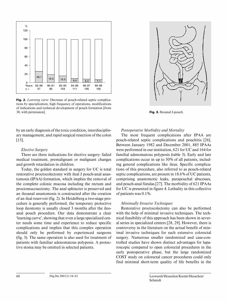
60 Dig Dis 2003;21:54–62 Leowardi/Heuschen/Kienle/Heuschen/Schmidt
Fig. 2. Learning curve: Decrease of pouch-related septic complica-tions by specialization, high frequency of operations, modificationsof indications and technical development of pouch formation [from38, with permission]. Fig. 3. Ileoanal J-pouch.
by an early diagnosis of the toxic condition, interdisciplin-ary management, and rapid surgical resection of the colon[15].
Elective SurgeryThere are three indications for elective surgery: failed
medical treatment, premalignant or malignant changesand growth retardation in children.
Today, the golden standard in surgery for UC is totalrestorative proctocolectomy with ileal J pouch-anal anas-tomosis (IPAA) formation, which implies the removal ofthe complete colonic mucosa including the rectum andproctomucosectomy. The anal sphincter is preserved andan ileoanal anastomosis is constructed after the creationof an ileal reservoir (fig. 2). In Heidelberg a two-stage pro-cedure is generally performed, the temporary protectiveloop ileostomy is usually closed 3 months after the ileo-anal pouch procedure. Our data demonstrate a clear‘learning curve’, showing that even a large specialized cen-ter needs some time and experience to reduce specificcomplications and implies that this complex operationshould only be performed by experienced surgeons(fig. 3). The same operation is also used for treatment ofpatients with familiar adenomatous polyposis. A protec-tive stoma may be omitted in selected patients.
Postoperative Morbidity and MortalityThe most frequent complications after IPAA are
pouch-related septic complications and pouchitis [26].Between January 1982 and December 2001, 885 IPAAswere performed in our institution, 621 for UC and 164 forfamilial adenomatous polyposis (table 3). Early and latecomplications occur in up to 50% of all patients, includ-ing general complications like ileus. Specific complica-tions of this procedure, also referred to as pouch-relatedseptic complications, are present in 18.6% of UC patients,comprising anastomotic leaks, parapouchal abscesses,and pouch-anal fistulas [27]. The morbidity of 621 IPAAsfor UC is presented in figure 4. Lethality in this collectiveof patients was 0.1%.
Minimally Invasive TechniquesRestorative proctocolectomy can also be performed
with the help of minimal invasive techniques. The tech-nical feasibility of this approach has been shown in sever-al series in specialized centers [28, 29]. However, there iscontroversy in the literature on the actual benefit of min-imal invasive techniques for such extensive colorectalsurgery. Numerous smaller randomized and case-con-trolled studies have shown distinct advantages for lapa-roscopic compared to open colorectal procedures in theearly postoperative phase, but the large randomizedCOST study on colorectal cancer procedures could onlyfind minimal short-term quality of life benefits in the
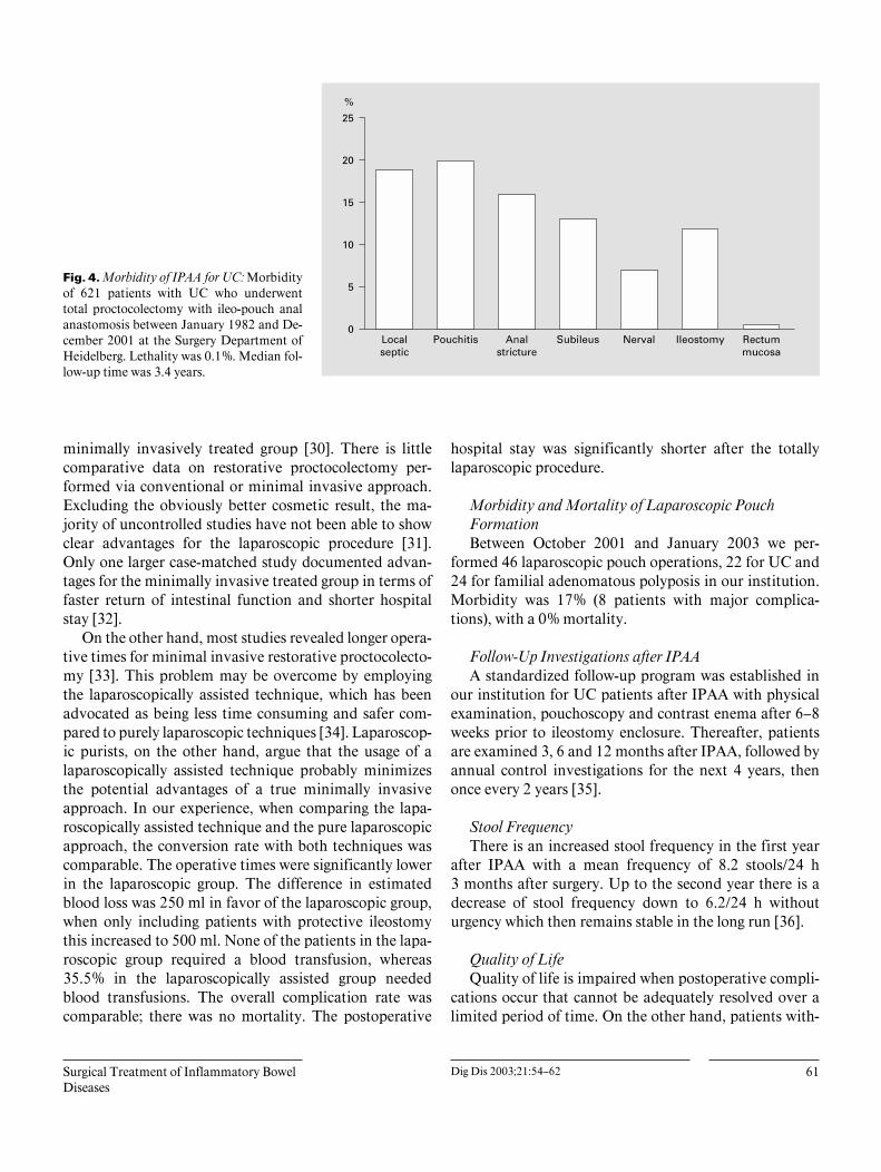
Surgical Treatment of Inflammatory BowelDiseases
Dig Dis 2003;21:54–62 61
Fig. 4. Morbidity of IPAA for UC: Morbidityof 621 patients with UC who underwenttotal proctocolectomy with ileo-pouch analanastomosis between January 1982 and De-cember 2001 at the Surgery Department ofHeidelberg. Lethality was 0.1%. Median fol-low-up time was 3.4 years.
minimally invasively treated group [30]. There is littlecomparative data on restorative proctocolectomy per-formed via conventional or minimal invasive approach.Excluding the obviously better cosmetic result, the ma-jority of uncontrolled studies have not been able to showclear advantages for the laparoscopic procedure [31].Only one larger case-matched study documented advan-tages for the minimally invasive treated group in terms offaster return of intestinal function and shorter hospitalstay [32].
On the other hand, most studies revealed longer opera-tive times for minimal invasive restorative proctocolecto-my [33]. This problem may be overcome by employingthe laparoscopically assisted technique, which has beenadvocated as being less time consuming and safer com-pared to purely laparoscopic techniques [34]. Laparoscop-ic purists, on the other hand, argue that the usage of alaparoscopically assisted technique probably minimizesthe potential advantages of a true minimally invasiveapproach. In our experience, when comparing the lapa-roscopically assisted technique and the pure laparoscopicapproach, the conversion rate with both techniques wascomparable. The operative times were significantly lowerin the laparoscopic group. The difference in estimatedblood loss was 250 ml in favor of the laparoscopic group,when only including patients with protective ileostomythis increased to 500 ml. None of the patients in the lapa-roscopic group required a blood transfusion, whereas35.5% in the laparoscopically assisted group neededblood transfusions. The overall complication rate wascomparable; there was no mortality. The postoperative
hospital stay was significantly shorter after the totallylaparoscopic procedure.
Morbidity and Mortality of Laparoscopic PouchFormationBetween October 2001 and January 2003 we per-
formed 46 laparoscopic pouch operations, 22 for UC and24 for familial adenomatous polyposis in our institution.Morbidity was 17% (8 patients with major complica-tions), with a 0% mortality.
Follow-Up Investigations after IPAAA standardized follow-up program was established in
our institution for UC patients after IPAA with physicalexamination, pouchoscopy and contrast enema after 6–8weeks prior to ileostomy enclosure. Thereafter, patientsare examined 3, 6 and 12 months after IPAA, followed byannual control investigations for the next 4 years, thenonce every 2 years [35].
Stool FrequencyThere is an increased stool frequency in the first year
after IPAA with a mean frequency of 8.2 stools/24 h3 months after surgery. Up to the second year there is adecrease of stool frequency down to 6.2/24 h withouturgency which then remains stable in the long run [36].
Quality of LifeQuality of life is impaired when postoperative compli-
cations occur that cannot be adequately resolved over alimited period of time. On the other hand, patients with-
62 Dig Dis 2003;21:54–62 Leowardi/Heuschen/Kienle/Heuschen/Schmidt
out complications and with good function after the ileo-anal pouch procedure may achieve a quality of life com-parable to that of healthy controls [36, 37]. Surgical expe-rience, technical modifications concerning pouch designand fashioning of the pouch-anal anastomosis are impor-tant for further improving this complex procedure and forreducing the complication rate.
Conclusion
Surgery for severe IBD has changed dramatically overthe last decade. There is a clear trend towards earlier, butless invasive operations. When the indications are wellreflected, most patients experience a substantial clinicalbenefit and improvement of quality of life after surgery.Most patients would have agreed to an earlier surgicalprocedure in retrospect if they had known the result ofthis procedure beforehand. This clearly indicates that gas-troenterologists should probably consider involving anexperienced surgeon earlier than practised to date.
References
1 Logan RF: IBD: Incidence: Up, down or un-changed? Gut 1998;42:309–311.
2 Mayberry JF: Recent epidemiology of ulcer-ative colitis and Crohn’s disease. Int J Colorec-tal Dis 1989;4:59–66.
3 Katschinski B, Fingerle D, Scherbaum B, et al:Oral contraceptives and cigarette smoking inCrohn’s disease. Dig Dis Sci 1993;38:1596–1600.
4 Meddings J: Barrier dysfunction and Crohn’sdisease. Ann NY Acad Sci 2000;915:333–338.
5 Church JM: Molecular genetics and Crohn’sdisease. Surg Clin North Am 2001;81:31–38.
6 Okabe N: The pathogenesis of Crohn’s disease.Digestion 2001;63:52–59.
7 Bernstein R, Rogers A: Malignancy in Crohn’sdisease. Am J Gastroenterol 1996;91:434–440.
8 Stein RB, Lichtenstein GR: Medical therapyfor Crohn’s disease: The state of the art. SurgClin North Am 2001;81:71–101.
9 Post S, Herfarth C, Bohm E, Timmermanns G,Schumacher H, Schurmann G, Golling M: Theimpact of disease pattern, surgical manage-ment and individual surgeons on the risk forrelaparotomy for recurrent Crohn’s disease.Ann Surg 1996;223:253–260.
10 Ewe K, Herfarth C, Malchow H, et al: Postop-erative recurrence of Crohn’s disease in rela-tion to radicality of operation and sulfasalazineprophylaxis: A multicenter trial. Digestion1989;42:224–232.
11 Scott NA, Hughes LE: Timing of ileocolonicresection for symptomatic Crohn’s disease –The patient’s view. Gut 1994;35:656–657.
12 Hansmann HJ, Kosa R, Düx M, Brado M,Goeser T, Roeren T, Stremmel W, KauffmannGW: Hydro-MRT chronisch entzündlicherDarmerkrankungen. Fortschr Roentgenstr1997;167:132–138.
13 McNamara MJ, Fazio VW, Lavery IC, et al:Surgical treatment of enterovesical fistulas inCrohn’s disease. Dis Colon Rectum 1990;33:271–276.
14 Herzog L, Herzog A, Glaser F, Herfarth C:Rektovaginale Fisteln bei Patienten mit Mor-bus Crohn: Therapie und Prognose. Langen-becks Arch Chir Suppl 1998;II:1002–1003.
15 Heuschen G, Heuschen UA, Klar E: Notfallin-dikationen und Operationsverfahren bei toxi-scher Colitis ulcerosa. Chir Gastroenterol2002;18:238–243.
16 Mowatt JI, Burnstein MJ: Free perforation ofsmall bowel Crohn’s disease: A case report andreview. Can J Gastroenterol 1993;7:300–302.
17 Greenstein AJ: The surgery of Crohn’s disease.Surg Clin North Am 1987;67:573–596.
18 Cirocco WC, Reilly JC, Rusin LC: Life-threat-ening hemorrhage and exsanguination fromCrohn’s disease. Report of four cases. Dis Co-lon Rectum 1995;38:85–95.
19 Chardavayne R, Flint GW, Pollack S, et al:Factors affecting recurrence following resectionfor Crohn’s disease. Dis Colon Rectum 1986;29:495.
20 Papaioannau N, Piris J, Lee ECG, et al: Therelationship between histological inflammationin the cut ends after resection of Crohn’s dis-ease and recurrence. Gut 1979;20:A916.
21 Bemelman WA, Slors JF, Dunker MS, van Ho-gezand RA, van Deventer SJ, Ringers J, Grif-fioen G, Gouma DJ: Laparoscopic-assisted vs.open ileocolic resection for Crohn’s disease. Acomparative study. Surg Endosc 2000;14:721–725.
22 Tabet J, Hong D, Kim CW, Wong J, GoodacreR, Anvari M: Laparoscopic versus open bowelresection for Crohn’s disease. Can J Gastroen-terol 2001;15:237–242.
23 Herfarth C, Stern J: Colitis ulcerosa, Adenoma-tosis coli – Funktionserhaltende Therapie. Ber-lin, Springer, 1990.
24 Farrell RJ, Peppercorn MA: Ulcerative colitis.Lancet 2002;359:331–340.
25 Soetikno RM, Lin OS, Heidenreich PA, YoungHS, Blackstone MO: Increased risk of colorec-tal neoplasia in patients with primary scleros-ing cholangitis and ulcerative colitis: A meta-analysis. Gastrointest Endosc 2002;56:48–54.
26 Heuschen UA, Hinz U, Allemeyer EH, Autsch-bach F, Stern J, Lucas M, Herfarth C, Heu-schen G: Risk factors for ileoanal J pouch-relat-ed septic complications in ulcerative colitis andfamilial adenomatous polyposis. Ann Surg2002;235:207–216.
27 Heuschen UA, Allemeyer EH, Hinz U, LucasM, Herfarth C, Heuschen G: Outcome afterseptic complications in J pouch procedures. BrJ Surg 2002;89:1–9.
28 Thibault C, Poulin EC: Total laparoscopicproctocoletomy and laparoscopy-assisted proc-tocolectomy for inflammatory bowel disease:Operative techniques and preliminary report.Surg Laparoscopy Endosc 1995;5:472–476.
29 Kienle P, Weitz J, Benner A, Herfarth C,Schmidt J: Laparoscopically assisted colecto-my and ileoanal pouch procedure with andwithout protective ileostomy. Surg Endosc2003;6 (epub).
30 Weeks JC, Nelson H, Gelber S et al, for theClinical Outcomes of Surgical Therapy (COST)Study Group. JAMA 2002;287:321–328.
31 Dunker MS, Bemelman WA, Slors JFM, et al:Functional outcome, quality of life, bodyimage, and cosmesis in patients after laparo-scopic-assisted and conventional restorativeproctocolectomy. Dis Colon Rectum 2001;44:1800–1807.
32 Marcello P, Milsom J, Wong S, et al: Laparo-scopic restorative proctocolectomy. Dis ColonRectum 2000;43:604–608.
33 Sardinha TC, Wexner SD: Laparoscopy forinflammatory bowel disease: Pros and cons.World J Surg 1998;22:370–374.
34 Darzi A: Hand-assisted laparoscopic colorectalsurgery. Semin Laparosc Surg 2001;8:153–160.
35 Heuschen UA, Autschbach F, Allemeyer EH,Zöllinger AM, Heuschen G, Uehlein T, Her-farth C, Stern J: Long-term follow-up afterileoanal pouch procedure. Dis Col Rec 2000;44.
36 Heuschen UA, Heuschen G, Herfarth C: Le-bensqualität nach Proktocolektomie wegen Co-litis ulcerosa. Chirurg 1998;69:1045–1051.
37 Heuschen UA, Heuschen G, Herfarth C: Derileoanale Pouch als Rectumersatz. Chirurg1999;70:530–542.
Review Article
Dig Dis 2003;21:63–67DOI: 10.1159/000071341
Intestinal Obstruction and Perforation –The Role of the Gastroenterologist
Petr Dıte Jan Lata Ivo Novotny
Department of Internal Medicine and Gastroenterology, Faculty of Medicine, Masaryk University,Brno, Czech Republic
Petr Dıte, MD, DSc, Prof. Med.Department of Internal Medicine and GastroenterologyFaculty of Medicine, Masaryk University BrnoJihlavska 20, CS–625 00 Brno (Czech Republic)Tel. +420 532 233500, Fax +420 532 233254, E-Mail [email protected]
ABCFax + 41 61 306 12 34E-Mail [email protected]
© 2003 S. Karger AG, Basel0257–2753/03/0211–0063$19.50/0
Accessible online at:www.karger.com/ddi
Key WordsSmall bowel W Large bowel W Obstruction W Perforation W
Endoscopic stenting
AbstractIntestinal obstruction belongs to highly severe condi-tions in gastroenterology, namely from the viewpoint ofquick and correct diagnosis as well as at determiningrational and effective therapy. Etiological multifactorialcharacteristics leading to processes resulting in mechan-ical or dynamic obstruction of the intestine, often re-ferred to as paralytic ileus, are undoubtedly serious fac-tors influencing the accuracy of diagnosis and therapeu-tic approach. Digestive endoscopy is a mandatory meth-od in the diagnosis of intestinal obstructions. Diagnosticendoscopy, colonoscopy in the involvement of the largeintestine or enteroscopy in the case of incomplete ob-struction of the small intestine are the methods indicatedin the majority of obstructive intestinal lesions. Besidestheir diagnostic importance, they also enable an effectivetherapeutic approach which may immediately follow thediagnostic intervention. Besides endoscopy that – due tothe nature of performance – belongs to invasive meth-ods, the diagnosis of obstructive intestinal processes isunthinkable without the use of non-invasive imagingmethods. Abdominal ultrasound examination, a widelyapplied method, provides – under optimal examinationconditions – information, e.g., about the width of theintestinal lumen or about the intestinal wall thickness;however, the specificity of investigation is not alwayssufficient. Both specificity and sensitivity of exploration
are increased by a plain X-ray of the abdomen supple-menting the ultrasound examination. Better results areachieved when the abdominal cavity is inspected bymeans of spiral CT examination that is nowadays notfashionably but highly effectively applied in the modifi-cation of the so-called CT enteroclysis or CT colonogra-phy. The usage of magnetic resonance (e.g. virtual co-lonography) is similar, but its efficacy is lower than thatof CT examination. From a gastroenterologist’s perspec-tive, endoscopic examination is the fundamental diag-nostic and therapeutic method. However, endoscopicexamination is initially limited by the cardiopulmonarystate of the patient – in a number of cases, first the car-diopulmonary condition must be stabilized, dysbalanceof water and mineral state must be restored, and onlythen can endoscopic investigation be carried out. Theapplication of enteroscopy in small intestine disorders isonly suitable in cases where air must be aspirated fromthe region of the stomach and mainly small intestine as ithappens, for example, in acute intestinal pseudo-ob-struction. The success of complex conservative therapyin these states is reached in 80% of the cases. In acuteand complete intestinal obstruction, a surgical treatmentperformed in time is the only method. In these cases, theimportance of identification of obstruction and timing ofthe intervention performance from the viewpoint of thepatient’s survival is explicitly the principal and life-savingconcern. In acute intestinal obstructions developing inpatients with malignant affection of the intestine, it isnecessary to choose – according to the obstruction loca-tion and general state of the patient – either urgently per-formed surgery or palliative endoscopic intervention
64 Dig Dis 2003;21:63–67 Dıte/Lata/Novotny
which is the reduction of the intestinal lumen of thegrowing tumor mass and following insertion of a drain.This method also concerns lesions localized in the lefthalf of the abdominal cavity, i.e. in the region of the rec-tosigmoid and descending part of the colon. Most pa-tients in whom acute intestinal obstruction developed onthe basis of malignant disease are risk and polymorbidsubjects, and acute surgical intervention may be eitherimpracticable or highly stressing. In such cases it istherefore helpful to insert a drain and to bridge theobstructed area after restoring the cardiopulmonarystate including adjustment of the aqueous and mineralenvironment. Later, the performance of an elective surgi-cal intervention is safer. Another alternative before in-serting a drain is the dilatation of the stenotic site bymeans of a balloon, followed by stenting. Up until today,various types of intestinal drains have been introduced –they have always been self-expanding metallic stents.Just the application of self-expanding stents in patientswith malignant intestinal obstruction and the endoscopicpossibility of dilatations of benign intestinal obstructionswith dilatation balloons are the most significant thera-peutic contributions of digestive endoscopy in thesestates.
Copyright © 2003 S. Karger AG, Basel
Definition
Intestinal obstruction is caused by mechanical blockageor insufficient peristalsis and may be complete or partial.The condition can also be classified by the level ofobstruction – small bowel or colon [25].
The synonym of this condition is ileus. The term func-tional obstruction is a possible alternative, but it is slightlyconfusing, because ‘functional’ could imply a psychologi-cal component to some, as in functional bowel disorder,‘obstruction’ implies an anatomic impediment to flow.
Motor paralysis and paresis describe the physiologicmalfunction of the bowel – paralytic ileus (adynamicileus).
Pseudo-obstruction is often used in describing achronic abnormality of function simulating mechanicalobstruction but without anatomic cause [26, 29]. Acutecolonic pseudo-obstruction (Ogilvie’s syndrome) is a sud-den massive idiopathic bowel dilatation [21].
The special sort of ileus in which severe transmuralinflammation produces atony of the colonic muscle istoxic megacolon.
In the toxic megacolon the mucosal barrier is dis-rupted, resulting in systemic toxemia [2]. The term ‘ob-struction’ is a synonym that implies that the process isintraluminal with the inability of intestinal contents topass through the digestive tract. The term closed-loopobstruction is used if the lumen is obliterated at two sites.In partial obstruction, the passage continues but is im-paired [20].
Causes of Mechanical Obstruction –Extrinsic and Intrinsic Lesions
Extrinsic LesionsExtrinsic masses can compress the bowel or mesentery
and cause obstruction.
AdhesionsAdhesions are the most common cause of small intes-
tine obstruction in adults. Adhesions may occur afterabdominal surgery, infection or radiation.
Congenital BandsCongenital bands behave clinically in much the same
way as adhesions, but they may occur in association withmalrotation, but very often in the absence of any knowncause.
Hernias– External– Internal– Pelvic hernias– Diaphragmatic
Hernias may cause either simple obstruction or closed-loop obstruction. Strangulation is common in incarcer-ated hernias, because blood supply is compromised by thehernial ring.
Volvulus– Gastric– Midgut– Cecal– Sigmoid
Volvulus of the small intestine is relatively frequent innewborns but rare in adults. Volvulus of the stomach isoften associated with large defects in the diaphragm orlarge paraesophageal hernias. Volvulus involves the sig-moid colon in 70–80% of the cases, and the cecum in 10–20% of the cases [28].
Intestinal Obstruction and Perforation –The Role of the Gastroenterologist
Dig Dis 2003;21:63–67 65
Intrinsic LesionsTumorsBenign and malignant tumors may narrow or obstruct
the lumen. Malignant obstruction may be primary or met-astatic. Primary malignancies of the small bowel are mostoften carcinoids, lymphomas or adenocarcinomas.
Inflammatory and Ischemic ProcessesMost frequent etiologic agents are blunt abdominal
trauma, hematomas as a result of severe thrombocytope-nia or vascular fragility (Henoch-Schönlein purpura).
Intussusception and Congenital DefectA leading segment of the bowel invaginates into an
accepting segment. Intrinsic bowel lesion – e.g. Meckeldiverticulum or tumor – usually initiates the process[23].– Malrotation/volvulus– Mesenteric cysts– Annular pancreas– Hirschsprung’s disease– Intestinal atresia
Intraluminal Objects– Meconium ileus– Barium impaction– Fecal impaction– Gallstone ileus– Foreign bodies
Causes of Adynamic Obstructions
Reflex Inhibition– Laparotomy– Renal transplantation– Abdominal trauma
Inflammatory Processes– Perforation or penetration– Peritonitis– Acute pancreatitis, acute cholecystitis– IBD– Celiac disease
Abdominal Injury and Abdominal IrradiationIschemic Processes
– Venous thrombosis– Arterial insufficiency– Mesenteric arteritis
Infection Processes– Bacterial peritonitis– Diverticulitis– Appendicitis
Retroperitoneal Processes– Pyelonephritis– Retroperitoneal hemorrhage– Pheochromocytoma– Ureteropelvic stones
Drugs– Opiates– Chemotherapeutics– Anticholinergic– Phenothiazines
Metabolic Abnormalities– Diabetes mellitus– Uremia– Septicemia– Electrolyte dysbalances– Pulmonary failure– Porphyria
Pathophysiology of Bowel ObstructionThe pathophysiology of bowel obstruction is character-
ized by proximal colon dilatation; it occurs above theobstruction, mucosal edema, and impairs venous andarterial blood flow. Ischemia of the bowel wall can lead tobowel perforation. An important factor is the increase ofbowel mucosal permeability with bacterial translocation,systematic toxicity, dehydration and electrolyte imbal-ances [29].
Diagnostic Procedures
Diagnostic procedures include the history and evalua-tion of symptoms, laboratory (biochemical) examina-tions, gastrointestinal tests and endoscopy [28]. Diagnos-tic procedures are similar in small and large intestineobstructions.
Clinical symptoms are relatively typical; in patientswith ‘high’ obstruction it is vomiting, very frequentlyabdominal pain connected with abdominal distension,absolute constipation, signs of peritonism and hypoten-sion, tachycardia and oliguria.
In patients with large bowel obstruction, malignantlesions are the most frequent etiological factor of the
66 Dig Dis 2003;21:63–67 Dıte/Lata/Novotny
obstruction. Carcinomas are the cause of obstruction in60–65%, diverticulitis in 20% and volvulus in 5%.
Clinical symptoms of the large colon obstruction aresimilar to those of patients with small bowel obstruction –abdominal pain, vomiting, dehydration and sepsis. Symp-toms of peritonism can be found very often.
X-ray examination – supine abdominal X-ray can giveinformation about the colon distention and air or liquidin the colonic lumen. ‘Free’ air in the abdominal cavity isa typical sign in patients with colon perforation [20].
Plain abdominal radiography can demonstrate the ab-sence of rectal gas and distended colon in cases withclosed-loop obstruction with large bowel obstruction. Sig-moid volvulus is presented radiographically as a ‘bentinner tube’ and cecal volvulus as a ‘coffee bean’.
Abdominal sonography is effective in some cases andcan describe the changes of lumen diameter and thicknessof the bowel wall [12]. Ultrasound can be a useful adjunctto the plain film when CT is not practicable or desirable.
CT scan sensibility for high-grade obstruction is about90%, for low-grade obstruction approximately 50% [6,16]. CT is superior in comparison with abdominal X-ray,ultrasonography and MRI for locating the site of obstruc-tion and diagnosis of bowel ischemia [15, 18].
A new effective diagnostic approach is CT enteroclysis[4], which, as a diagnostic procedure of the small bowelobstruction, is the gold standard for detecting low-gradeobstruction and predicting the site of obstruction. How-ever, enteroclysis is contraindicated if bowel ischemia issuspected. CT enteroclysis offers a novel technique fordiagnostically challenging cases.
An essential diagnostic method is endoscopy. Endo-scopical methods can locate obstructive lesions. The pro-cedure must be performed without air insufflation andwithout biopsy, especially in cases where bowel perfora-tion is suspected.
Endoscopy is a mandatory examination in obstructionsof the small bowel and colon, with high efficacy as diag-nostic procedures, but can be used as a therapeutic modal-ity as well.
Enteroscopes are available to examine the more distalsmall bowel as a diagnostic procedure [20] and desuffla-tion of the small bowel (e.g. early postoperative bowelobstruction) can be used as a therapeutic procedure [11].
Colonoscopy is indicated in examination of the rectum,colon and ileocecal valve and in desufflation of the colon,tumor mass ablation, stent insertion or colonic stricturedilatation [7, 24].
Therapy
Acute complete bowel obstruction is a surgical emer-gency. The effect of endoscopical therapy in uncompli-cated obstruction is dependent on the patient’s cardio-respiratory status stabilization which is the first step oftherapy in acute colonic disorders [10].
Together with nasogastric tube insertion, the correc-tion of the fluid and electrolyte dysbalances [10] and erad-ication of the sources of sepsis by using broad-spectrumantibiotics (third-generation cephalosporins, metronida-zole or amoxiclav) are mandatory therapeutic ap-proaches. Uncomplicated obstruction can be treated con-servatively in 80% of the cases, providing there are signsof resolution within 24 h.
Endoscopical bowel decompression together with fast-ing, nasogastric tube insertion and regular changes ofpatient position are indicated in bowel obstruction [5]. Inpatients with pseudo-obstruction, colonoscopic decom-pression is successful in more than 80% of the cases andfurther colonoscopy successfully treats the majority ofrecurrences [16]. After 24 h, the clinical situation has to bereviewed and a decision made if there is a need for furthersurgical intervention.
The rates of colonic perforation in patients with acutecolonic pseudo-obstruction vary from 3.0 to 15% [24].The cecum is the most common site of perforation. Perfo-ration leads to increased mortality which can be between43 and 46% [26]. Perforation leads to surgery, which isassociated with increased mortality as well. It is extremelyimportant to decide the correct timing between conserva-tive and surgical therapy as a prevention of perforation.Endoscopical therapy is indicated in patients with benignbowel stricture [22], but this situation sometimes leads toacute colonic obstruction. Recent balloons are flexibleand well suited to placement in the tortuous colon. Newerballoons with controlled radial expansion can be ex-panded in a controlled fashion.
The optimal time for inflation and number of dilata-tion procedures are still not known. Savary dilators can beused in patients with anastomotic strictures. These dila-tors predominantly exert their force in the axial directionand this may lead to a greater risk of complications andlower effect than balloons [27].
Endoscopical therapeutic procedures in patients withtumor colonic obstruction are tumor mass ablation [30]and stenting of the colon [1, 2, 19]. Metallic stents havebeen used since the beginning of 1990s (this method wasfirst described by Spinelli in 1992). Endoscopical place-ment of self-expanding metallic stents over placement by
Intestinal Obstruction and Perforation –The Role of the Gastroenterologist
Dig Dis 2003;21:63–67 67
interventional radiology has its advantages; the endoscop-ical technique is able to pass some stents directly by theworking channel of the endoscope. This advantage isespecially useful when the obstruction is proximal to therectosigmoid region or in patients with angulated rectosig-moid anatomy [8, 13]. However, both techniques, endo-scopical and radiological, can usually be used effectively[2].
Endoscopic stenting can be performed with the thera-peutic endoscope with a minimal working channel of4.2 mm in diameter. Three stents are recommended –colonic Z stents with a 25-mm diameter in the body,enteral Wallstent (22-mm diameter) and BARD Memo-therm stent (30-mm diameter). Technical success is, ofcourse, dependent on the experience of the endoscopist,the optimal is success rate being 90–95% insertions. Thelimitation is the inability to pass a guide-wire through thestricture or anatomic difficulties [3, 9]. Clinical success is
defined as successful bowel decompression and stool defe-cation [14].
Early complications after the procedure are stent mi-gration, bowel perforation and bleeding [25]. Late compli-cations are similar and stent migration is the most fre-quent. This complication can be asymptomatic or symp-tomatically patients can have tenesmus. Proximal stentmigration is very rare. Stenting is the first method ofchoice in patients with tumor localization in the leftcolon, especially in the rectosigmoid junction or in therectum [27]. Surgical resection or bypass operation isindicated in patients with proximal colon obstruction.Patients with total colonic obstruction are frequently illwith severe medical conditions. In these patients the self-expanding metallic stent insertion can help in the medicalstabilization and later performed colon resection, whenthe tumor and stent are resected en bloc at the time ofresection with greater safety [17].
References
1 Baron TH: Expandable metal stents for thetreatment of cancerous obstruction of the gas-trointestinal tract. N Engl J Med 2001;344:1681–1687.
2 Baron TH, Rey JF, Spinelly P: Expandablemetal stent placement for malignant colorectalobstruction. Endoscopy 2002;34:823–830.
3 Binkert CA, Ledermann H, Jost R, et al: Acutecolonic obstruction: Clinical aspects and cost-effectiveness of preoperative and palliativetreatment with self-expanding metallic stents –a preliminary report. Radiology 1998;206:199–204.
4 Boudiaf M, Soyer P, Jaff A, et al: How we per-form CT enteroclysis. Feuill Radiol 2002;42:253–258.
5 Brolin RE: The role of gastrointestinal tubedecompression in the treatment of mechanicalintestinal obstruction. Am Surg 1983;49:131.
6 Burkill G, Bell J, Healy J: Small bowel obstruc-tion: The role of computed tomography in thisdiagnosis and management with reference toother imaging modalities. Eur Radiol 2001;11:1405–1422.
7 Colquhoum PD, Vernava AM: Therapeutic co-lonoscopy. Clin Colon Rectal Surg 2001;14:347–357.
8 Dauphine CE, Tan P, Beart RW, et al: Place-ment of self-expanding metal stents for acutemalignant large-bowel obstruction: A collectivereview. Am Surg Oncol 2002;9:574–579.
9 Del Valle Hernandez E: Transanal self-expand-ing metal stents as an alternative to palliativecolostomy in selected patients with malignantobstruction of the left colon. Br J Surg 1998;85:232–236.
10 Gajic O, Urrutia LE, Sewani H, et al: Acuteabdomen in the medical intensive care unit.Crit Care Med 2002;30:1187–1190.
11 Gersin KS, Ponsky JL, Fanell RD: Enteroscop-ic treatment of early postoperative small bowelobstruction. Surg Endosc 2002;16:115–116.
12 Grunshaw ND, Rewick IGH, Scarisbrick G,Nasmyth DG: Prospective evaluation of theultrasound in distal ileal and colonic obstruc-tion. Clin Radiol 2000;55:546–562.
13 Harris GJ, Senagore AJ, Lavery IC, et al: Themanagement of neoplastic colorectal obstruc-tion with colonic endoluminal stenting devices.Am J Surg 2001;181:499–506.
14 Law WL, Chu KW, Ho JW, et al: Self-expand-ing metallic stent in the treatment of colonicobstruct ion caused by advanced malignancies.Dis Colon Rectum 2000;43:1522–1527.
15 Lee JM, Jung SE, Lee KY: Small-bowel ob-struction caused by phytobezoar: MR imagingfindings. Am J Roetgenol 2002;79:538–539.
16 Lopez-Kostner F, Hool GR, Lavery IC: Man-agement and causes of acute large bowel ob-struction. Surg Clin North Am 1997;77:1265–1270.
17 Martinez-Santos C, Lobato RF, Fradejas JM,et al: Self-expandable stent before elective sur-gery vs. emergency surgery for the treatment ofmalignant colorectal obstructions: Comparisonof primary anastomosis and morbidity rates.Dis Colon Rectum 2002;45:40–406.
18 Matsuoka H, Takanara T, Masaki T, et al: Pre-operative evaluation by magnetic resonanceimaging in patients with bowel obstruction.Am J Surg 2002;183:614–617.
19 Mauro MA, Roechler RE, Baron TH: Ad-vances in gastrointestinal intervention: Thetreatment of gastroduodenal and colorectal ob-struction with metallic stents. Radiology 2000;215:659–669.
20 Mucha P: Small intestinal obstruction. SurgClin North Am 1987;67:597–630.
21 Ogilvie H: Large intestine colic due sympathet-ic deprivation: A new clinical syndrome. BrMed J 1948;ii:671.
22 Paul L, Pinto I, Gomez H, et al: Metallic stentsin the treatment of benign diseases of the colon:Preliminary experiments in 10 cases. Radiolo-gy 2002;223:715–722.
23 Prall RT, Bannon MP, Bharucha AE: Meckel’sdiverticulum causing intestinal obstruction.Am J Gastroent 2001;96:3426–3427.
24 Rex VW: Colonoscopy and acute colonic ob-struction. Gastroent Endosc Clin North Am1997;7:499–508.
25 Seymour K, Johnson R, Marsh R, Corson J:Palliative stenting of malignant large bowel ob-struction. Colorect Dis 2002;4:240–245.
26 Soreide O, Bjerkeset T, Fossdal JE: Pseudo-obstruction of the colon (Ogilvie’s syndrome), agenuine clinical condition? Dis Colon Rectum1977;20:587–491.
27 Spinelli P, Mancini A: Use of self-expandingmetal stents for palliation of rectosigmoid can-cer. Gastrointest Endosc 2001;344:1681–1687.
28 Tham T, Collins J: Gastrointestinal Emergen-cies. London, BMJ Books, 2000, pp 187–196.
29 Vanek ,VW, Al-Salti M: Acute pseudo-obstruc-tion of the colon (Ogilvie’s syndrome): An anal-ysis of 400 cases. Dis Colon Rectum 1986;29:203–210.
30 Xinopoulos D, Dimitroupoulos D, TsamakidisK, et al: Treatment al malignant colonic ob-struction with metal stents and laser. Hepato-gastroenterology 2002;49:359–362.
Review Article
Dig Dis 2003;21:68–76DOI: 10.1159/000071342
Intestinal Obstruction and Perforation –The Role of the Surgeon
Christos Dervenis Spiros Delis Dimitrios Filippou Costas Avgerinos
Pancreatic Unit, 1st Department of Surgery, Agia Olga Hospital, Athens, Greece
Christos Dervenis, MD, PhDHead, 1st Department of SurgeryAgia Olga Hospital, 3–5, Agias Olgas str.GR–14233 Athens (Greece)Tel. +30 210 2775 467, Fax +30 210 2793 969, E-Mail [email protected]
ABCFax + 41 61 306 12 34E-Mail [email protected]
© 2003 S. Karger AG, Basel0257–2753/03/0211–0068$19.50/0
Accessible online at:www.karger.com/ddi
Key WordsIntestinal obstruction W Perforation W Small bowelobstruction W Large bowel obstruction
AbstractIntestinal obstruction and perforation are always a chal-lenge for the surgeon, not only in respect to the surgicaloption offered to the patient, but also to the ability toaccurately diagnose and stage the disease. The under-standing of the underlying pathophysiological mecha-nism is also very important in order to classify eachpatient in order to receive the more appropriate treat-ment. Mechanisms of obstruction and perforation, meth-ods of diagnosis as well as prevention and treatment ofthe disease were reviewed.
Copyright © 2003 S. Karger AG, Basel
Introduction
Obstruction of the small and large intestine seems to bea major health problem all over the world. Fifty years agothe most common cause of small bowel obstruction wasexternal hernia. Nowadays, postoperative adhesions com-prise more than half of small bowel obstructions, due to
the increased number of surgical procedures and earlyelective hernia repair [1]. On the other hand, althoughmany improvements have been achieved concerning largebowel obstruction and pseudo-obstruction, the maincause, i.e. malignancy, still remains unchanged. In thepast 20 years, the rate of morbidity and mortality for elec-tive colon operations has dropped significantly, but mor-tality for emergency colon operations is still twice as highcompared to elective ones [2].
The mechanism of obstruction (mechanical vs. func-tional), the presence of vascular compromise, the level ofobstruction (proximal or distal), the rate of progression ofobstruction and the location of the responsible patholo-gies are of great importance in intestine’s obstruction clas-sification. Correct and immediate diagnosis in small andlarge bowel is of great importance as far as morbidity andmortality are concerned. Many pathophysiologic conse-quences implicate in clinical manifestation of this situa-tion. Symptoms such as colicky pain, tenderness, peritoni-tis, signs of dehydration, abdominal distention and aus-cultation may indicate bowel obstruction. Laboratorytests are not helpful to identify obstruction. Radiologicalexams (X-rays, CT) and digital exams are essential notonly for diagnosis confirmation, but also for locating theobstruction area.
Intestinal Obstruction and Perforation Dig Dis 2003;21:68–76 69
Although many partial obstructions can be treated con-servatively or endoscopically, surgery still remains thecornerstone of treatment. The time of operation, indica-tions, and the specific surgical procedures are relateddirectly to the nature of the problem. Perforation can bedue to several causes, malignant or benign pathologies,and leads to local or generalized peritonitis. Radiological,laboratory and clinical findings are essential in estab-lishing the diagnosis. Surgery is the gold standard in thetreatment of perforations.
In the present article, small and large intestine obstruc-tion and perforation will be described separately with spe-cial references in the new advances in diagnosis and treat-ment of these pathologies [3].
Small Bowel Obstruction
The causes of small bowel obstruction (SBO) can bedivided into three groups, extraluminal causes (hernias,adhesions, carcinomas and abscesses), obstructions in-trinsic to the wall (tumors, tuberculosis, actinomycosis,malrotation, cysts, diverticula, hematomas, strictures,etc.), and in intraluminal causes as enterolith, gallstone,foreign bodies, etc. At the beginning of the 20th century,hernias were the leading cause of small intestinal obstruc-tion, but with routine elective hernia repair, adhesionssecondary to previous surgery became by far the mostcommon cause [4].
Postoperative adhesions are responsible for more than70% of all causes of SBO, particularly after pelvic proce-dures because small intestine is more mobile in the pelvis[5].
Tumors are the second leading cause accounting forabout 20% of SBO, especially metastatic lesions fromintra-abdominal primary tumor (e.g. ovarian, gastric, co-lonic, etc), and rarely from extra-abdominal primarytumors (e.g. breast, lung, melanoma, etc). Large intestinemalignant neoplasm may present with small intestineobstruction. Hernias are the third leading cause (ventral,inguinal, and internal) and inflammatory bowel diseases(Crohn’s disease) is the fourth cause, resulting from acuteinflammation and edema [6]. Intra-abdominal abscessesmay present as SBO (local ileus). Other miscellaneouscauses (enterolith, gallstone, foreign bodies, diverticula,and polyps), while very rare (!2%), should be consideredin the differential diagnosis (table 1) [7, 8].
Table 1. Small bowel obstruction causes inadults
Extrinsic lesionsAdhesions
PostoperativePrimary
NeoplasmsBenignMalignant
Intra-abdominal carcinomatosisExtraintestinal tumor
HerniasInternal (paraduodenal,diaphragmatic, etc.)External (inguinal, umbilical, etc.)
Intra-abdominal abscess
Intestinal wall, intrinsic lesionsNeoplasms
PrimaryMetastatic
InflammatoryCrohn’s diseaseInfectious diseases
ActinomycosisTuberculosis
DiverticulitisCongenital
MalrotationIntestinal wall cystsDuplication
Miscellaneous lesionsHematomaIschemiaStricturePost-radiation enteritisEndometriosisIntussusception
Intraluminal causesEnterolithGallstoneForeign bodyTrichopilimma
DiagnosisIn the majority of patients, a thorough history and
physical examination are very important to establish thediagnosis and treatment. The above should be comple-mented with abdomen X-rays, although more sophisti-cated exams (US, CT, MRI, endoscopy, or laparoscopy)may be necessary in cases with uncertain diagnosis. Themain symptoms of SBO are colicky abdominal pain, nau-sea and vomiting (more common in higher obstruction),
70 Dig Dis 2003;21:68–76 Dervenis/Delis/Filippou/Avgerinos
abdominal distention and failure to pass feces and flatus[2, 4, 7].
Physical examination may reveal important signs, in-cluding tachycardia, hypotension, severe dehydration,and fever. In the early stages, peristaltic waves can beobserved, and the auscultation may demonstrate hyperac-tive bowel sounds, but in the late stages minimal or nobowel sounds are present. Other findings include tender-ness, palpable mass, rebound, and localized pain (localperitonitis). Physical examination may include rectal andstool exam and careful examination for incarcerated her-nias, in the groin, femoral triangle and in obturator fora-men [1–3].
Most of the patients with SBO have a simple mechani-cal blockage in luminal contents flow without disturbanceof intestinal wall viability. In rarer cases intestinal stran-gulation may occur. Strangulation obstruction involves aclosed-loop obstruction with compromised vascular sup-ply of a particular intestinal segment leading to infarction,which is associated with higher morbidity and mortalityrates. Tachycardia, fever, leukocytosis, and constant ab-dominal pain present the most common symptoms ofstrangulation obstruction [1, 6].
Laboratory and Radiologic ExaminationsLaboratory examinations are not helpful in the actual
diagnosis. The most common findings in routine patients’laboratory measurements are elevated hematocrit due todehydration, serum electrolytes (sodium, potassium, bi-carbonate) and creatinine changes and leukocytosis (par-ticularly in patients with strangulation), although elevatedwhite blood cell count does not always denote strangula-tion [9].
Various serum determinations (amylase, electrolytes,ALP and ammonia) have been tried to find an associationwith the strangulated obstruction, with no result. Thepresent studies indicate that serum D-lactate, CK-BBisoenzyme and intestinal fatty acid binding protein maybe associated with strangulated obstruction.
Radiological studies confirm the clinical suspicion anddefine more accurately the site of obstruction. Plain abdo-men X-rays present 60% accuracy in defining the causeand site of SBO. Most common findings are dilated smallintestine loops, colonic distention, multiple air-fluid lev-els, and foreign bodies or gallstones. In more complicatedcases, further diagnostic evaluations may be necessary[10, 11].
In these cases, CT scanning proved to be beneficial,although this exam seems to be less sensitive in patientswith partial SBO. CT has dramatically improved the
management of patients with suspected obstruction, al-though its accuracy varies [12].
Multi-slice or helical CT has better outcomes. Intrave-nous contrast is needed in every case because it highlightsthe abdominal viscera and lesions of the bowel or sur-rounding area, and allows examination of the bowel’swall, particularly in cases where strangulation is sus-pected. Thin slices are useful when the site of obstructionis estimated [13].
In full obstructions it is of great importance to accu-rately determine the cause and the site of the obstruction,even if the cause is extrinsic (tumor, abscess, etc). Bariumstudies and particularly enteroclysis may be helpful in theassessment of obstruction, as it can precisely demonstratethe level and cause of the obstruction in patients with low-grade intermittent SBO and uncertain diagnosis. Thereare doubts about the diagnostic value of ultrasound, whileMRI appears not to be better diagnostically compared toCT scan [14–16].
Richards et al. [17] tried to detect mesenteric ischemiaassociated with altered small intestine’s basic electricrhythm with a non-invasive technique using a supercon-ducting quantum interference magnetometer. This proce-dure may be useful in strangulated obstruction diagnosisand is at present under clinical evaluation and assess-ment.
TreatmentConservative TreatmentPatients with SBO are usually dehydrated, and de-
pleted of electrolytes. These patients require intravenousreplacement of body fluids and electrolytes. The appro-priate replacement is intravenously with the patient mon-itored. Serial blood measurements (electrolytes, hemato-crit and white blood cells) must be performed in order toassess the adequacy of resuscitation and the patient’s gen-eral condition. In some cases, intravenous fluid replace-ment may require central venous catheter and Swan-Ganz catheter. In complicated patients with increasedwhite blood cells number, antibiotics prophylaxis or treat-ment may be useful [18].
Additionally with body fluid and electrolyte replace-ment, the patient’s SBO support requires nasogastric suc-tion with a nasogastric (Levin) tube, which empties thestomach and reduces the risk of gastric fluid pulmonaryaspiration. Several prospective studies demonstrate nosignificant difference concerning the non-operative treat-ment or the morbidity-mortality rates after surgical inter-ference, with or without nasogastric decompression [19].
Intestinal Obstruction and Perforation Dig Dis 2003;21:68–76 71
Conservative treatment may be successful in patientswith partial SBO. Resolution of the symptoms and pa-tient’s discharge have been reported in 65–80% of thepatients. The small intestine’s distention in abdominal X-rays during conservative treatment with a nasogastrictube is an indication for immediate surgical intervention[20]. Choi et al. [21] suggest that the use of Gastrographinin adhesive SBO is safe and reduces the need for surgerywhen conservative treatment fails. These authors treatedpatients with partial obstruction (contrast reaches thecolon within 24 h) conservatively, and suggest that Gas-trographin helped in the conservative treatment of thepatients.
Adhesion PreventionIntestinal obstruction due to abdominopelvic adhe-
sions has developed into a serious health problem world-wide. Research towards reduction of possible adhesionetiopathogenic factors as well as prevention of adhesionformation has shown that various individual factors suchas nutritional status, disease state like diabetes and thepresence of concurrent infectious processes, affect adhe-sion formation. In addition, various clinical techniquesand agents seem to decrease the incidence of postopera-tive adhesions [22, 23].
Surgical techniques that inhibit adhesions include lim-iting trauma to intra-abdominal structures [24], meticu-lous hemostasis [25] (free blood provides a source offibrin and thromboplastin that activate the clotting cas-cade), delicate handling of the bowel [24], tissues moisten-ing [26] and use of minimally invasive surgery when pos-sible. All these maneuvers have been shown to protectagainst adhesion formation and should always be fol-lowed.
On the other hand, despite the fact that many phar-macological agents have been tried against adhesions,several points need to be clarified before the enthusiasmfor the development of the ideal adhesion barrier be-comes scientific certainty [27]. This ‘adjuvant’ therapypoints either to the various causes of the inflammatoryprocess (NSAIDs [28], glucocorticoids and antihista-mines [29], progesterone/estrogen [27], anticoagulants[27], fibrinolytics [30], antibiotics) or directly to adhe-sion formation. The latter fall into two main categories:macromolecular solutions (crystalloids, 32% dextran 70,hyaluronic acid, HA-PBS/Sepracoat, carboxymethylcel-lulose) and solid barriers [27] (autologous peritonealtransplants, PTFE, oxidized-regenerated cellulose, HA-CMC [31]).
Surgical TreatmentPatients with complete SBO require surgical interven-
tion, although some authors suggest that many patientscan be managed conservatively with nasogastric suctionand fluid-electrolyte replacement. Several studies indicatethat a 12- to 24-hour delay of the surgical treatment seemsto be safe, although the incidence of strangulation of othercomplications may increase. During that period, conser-vative means can be used in severe patients’ (with fever,tachycardia, tenderness and leukocytosis) resuscitation.
The nature of the problem indicates the appropriateoperation in patients with SBO. In patients with a historyof cancer and intra-abdominal widespread metastasis,conservative treatment may be more useful in those caseswhere operation is contraindicated. In non-complicatedcases, tumor resection or bowel bypass may solve theproblem temporarily or permanently. In SBO, due toCrohn’s disease, conservative treatment may be useful,whereas bowel resection is indicated in strictures or fistu-las. Intra-abdominal abscess causing SBO needs drainage.The operation type depends on the nature of SBO. Incomplicated cases where bowel viability is questionable,the obstructed segment is released and placed in warmsaline gausses for 15–20 min, then re-estimated, and ifthere is still doubt, partial bowel resection should be per-formed [32].
Several authors suggest that SBO estimation shouldstart laparoscopically, because most of the cases are due tosecondary adhesions. Laparoscopic treatment of SBOseems to be effective and in selected patients leads to ashorter hospital stay. Several criteria have been proposedfor patient selections. Appropriate for laparoscopic inter-vention patients include those with mild abdominal dis-tention, proximal obstruction, and partial and anticipatedsingle band obstruction. Advanced, complete or distalSBO is a contraindication for laparoscopic treatment. Asalready reported, patients with adhesions, widespreadcarcinomatosis, or inducing distention after nasogastricsuction should be managed with open laparotomy [33,34].
Recent studies support the role of minimal invasiveprocedures in SBO treatment. Endoscopical stent place-ment in cancer obstructions seems to temporarily releaseobstruction, and become a good alternative in patientswith widespread carcinomatosis, where surgical treat-ment is contraindicated and conventional managementhas no effect.
72 Dig Dis 2003;21:68–76 Dervenis/Delis/Filippou/Avgerinos
Large Bowel Obstruction
Along the colon, many segments are completely intra-peritoneal, while others are partially retroperitoneal, whichmeans that the colon is more fixed and its parts have differ-ent mobility depending on the laxity of the mesocolon. Thecauses for large bowel obstruction are quite different tothose of the small intestine. The main causes are the follow-ing: tumor (benign or malignant: 8–29%), ulcerative coli-tis, large diverticula, adhesions, enterolith, intra-abdomi-nal abscess and volvulus. Because of its anatomic differ-ence (different mobility) from small intestine, volvulus is aspecific for colon obstruction cause. The main risk of vol-vulus is the infarction of the segment involved, via torsionof the supplying vessels that interrupts the in- and outflowin the colon wall, or via torsion of colon or/and ileum (incecal volvuli), creation of a closed loop and strangulation.In both cases, fatal arterial obstruction is the final result.The most common volvulus sites are cecum and sigmoidcolon, and rarely transverse colon [35].
DiagnosisAs already referred to, malignancy seems to be the
most common cause of colonic obstruction. This patholo-gy rarely requires immediate operative treatment, so inthe majority a preoperative evaluation should take place.Diagnosis can be achieved through contrast enema orcolonoscopy. The value of contrast enema is in the diag-nosis of pseudo-obstruction in which barium has not onlydiagnostic but also therapeutic use. Colonoscopy, on theother hand, is useful in identifying tumors and takingbiopsies [1, 4].
Radiological studies confirm the clinical suspicion anddefine more accurately the site of obstruction. Most com-mon findings are colonic distention and multiple air-fluidlevels. In more complicated cases, further diagnostic eval-uations may be necessary. In these cases, CT scanningproved to be beneficial, ultrasound does not offer much,while MRI appears not to be better diagnostically com-pared to CT scan [2, 7, 16].
Laboratory examinations are not helpful in the actualdiagnosis. The most common findings in routine patientlaboratory measurements are serum electrolyte distur-bances (sodium, potassium, bicarbonate), creatininechanges and leukocytosis (particularly in patients withstrangulation). Various serum determinations (amylase,electrolytes, ALP and ammonia) have been tried to findan association with the obstruction, with no result, whilefibrinogen and d-dimers seems to increase in cases ofcolonic necrosis [3].
Diagnosis of colonic obstruction caused by ulcerativecolitis is difficult and needs not only radiological, but alsopathologic and laboratory definition. No matter which isthe cause, diagnosis of colon obstruction can be achievedin most of the cases, with abdominal X-rays, enema con-trast studies, colonoscopy and CT. The final diagnosis canbe defined intra- or postoperatively [36].
TreatmentColon obstruction treatment is rarely conservative. In
most of the cases, the cause of obstruction should beremoved or bypassed, with surgical or endoscopic proce-dures. According to Ballantyne et al. [37], sigmoid volvu-lus is responsible for 6.1% of colon obstructions. Sigmoid-oscopic decompression by insertion of a rectal tube orendoscopicaly relieves the obstructions in uncomplicatedcases (absence of necrosis or perforation). The recurrencerate after endoscopic or conservative treatment variesfrom 40 to 85% or more in different series, which indi-cates surgical treatment (resection of the involved colon)to be a definitive therapy [38].
There are doubts about the appropriate surgical treat-ment of obstructive colon malignancies. Many surgeonssuggest resection of the lesion and primary anastomosis atthe same time, while others support other methods as forexample intraoperative colonic irrigation primary toanastomosis [39]. These techniques have significant com-plication rates when they are performed in patients withacute or chronic obstruction: wound infection rate is 12%,severe anastomotic leakage rate 6%, morbidity rate 14%and total mortality rate is 8%. The decision for the type ofoperation depends on the surgeon, on prognostic factorsand staging of malignancy, on the existence of intraperito-neal microcarcinomatosis and on parameters associatedwith the patient’s general situation and other health prob-lems. An effective alternative to primary anastomosis iscolostomy, which is indicated particularly in high mortali-ty risk patients and in cases where tumor resection isimpossible. Bypass may be useful in cases where it is noteasy to perform either colostomy or primary anastomosis[3, 7, 38].
Treatment of acute colorectal obstruction by usingmetallic stents is a recent advance that has been mainlyfor preoperative decompression. Despite limited reportsin the literature, endoscopic stent placement for colonicobstruction relief seems to be a highly feasible and safemethod [40]. Successful stent implantation is achieved in85–100% of the patients, depending on the center’s expe-rience. Colonic stent can be also placed under fluoroscop-ic guidance or with a combination of these two methods.
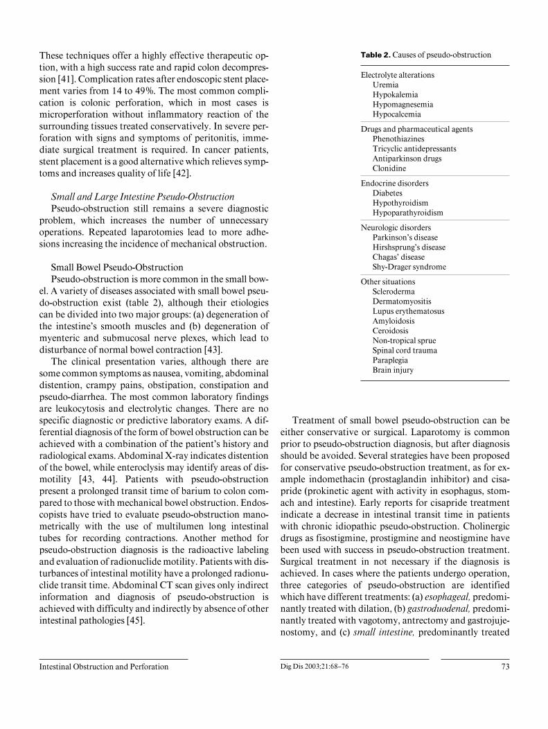
Intestinal Obstruction and Perforation Dig Dis 2003;21:68–76 73
These techniques offer a highly effective therapeutic op-tion, with a high success rate and rapid colon decompres-sion [41]. Complication rates after endoscopic stent place-ment varies from 14 to 49%. The most common compli-cation is colonic perforation, which in most cases ismicroperforation without inflammatory reaction of thesurrounding tissues treated conservatively. In severe per-foration with signs and symptoms of peritonitis, imme-diate surgical treatment is required. In cancer patients,stent placement is a good alternative which relieves symp-toms and increases quality of life [42].
Small and Large Intestine Pseudo-ObstructionPseudo-obstruction still remains a severe diagnostic
problem, which increases the number of unnecessaryoperations. Repeated laparotomies lead to more adhe-sions increasing the incidence of mechanical obstruction.
Small Bowel Pseudo-ObstructionPseudo-obstruction is more common in the small bow-
el. A variety of diseases associated with small bowel pseu-do-obstruction exist (table 2), although their etiologiescan be divided into two major groups: (a) degeneration ofthe intestine’s smooth muscles and (b) degeneration ofmyenteric and submucosal nerve plexes, which lead todisturbance of normal bowel contraction [43].
The clinical presentation varies, although there aresome common symptoms as nausea, vomiting, abdominaldistention, crampy pains, obstipation, constipation andpseudo-diarrhea. The most common laboratory findingsare leukocytosis and electrolytic changes. There are nospecific diagnostic or predictive laboratory exams. A dif-ferential diagnosis of the form of bowel obstruction can beachieved with a combination of the patient’s history andradiological exams. Abdominal X-ray indicates distentionof the bowel, while enteroclysis may identify areas of dis-motility [43, 44]. Patients with pseudo-obstructionpresent a prolonged transit time of barium to colon com-pared to those with mechanical bowel obstruction. Endos-copists have tried to evaluate pseudo-obstruction mano-metrically with the use of multilumen long intestinaltubes for recording contractions. Another method forpseudo-obstruction diagnosis is the radioactive labelingand evaluation of radionuclide motility. Patients with dis-turbances of intestinal motility have a prolonged radionu-clide transit time. Abdominal CT scan gives only indirectinformation and diagnosis of pseudo-obstruction isachieved with difficulty and indirectly by absence of otherintestinal pathologies [45].
Table 2. Causes of pseudo-obstruction
Electrolyte alterationsUremiaHypokalemiaHypomagnesemiaHypocalcemia
Drugs and pharmaceutical agentsPhenothiazinesTricyclic antidepressantsAntiparkinson drugsClonidine
Endocrine disordersDiabetesHypothyroidismHypoparathyroidism
Neurologic disordersParkinson’s diseaseHirshsprung’s diseaseChagas’ diseaseShy-Drager syndrome
Other situationsSclerodermaDermatomyositisLupus erythematosusAmyloidosisCeroidosisNon-tropical sprueSpinal cord traumaParaplegiaBrain injury
Treatment of small bowel pseudo-obstruction can beeither conservative or surgical. Laparotomy is commonprior to pseudo-obstruction diagnosis, but after diagnosisshould be avoided. Several strategies have been proposedfor conservative pseudo-obstruction treatment, as for ex-ample indomethacin (prostaglandin inhibitor) and cisa-pride (prokinetic agent with activity in esophagus, stom-ach and intestine). Early reports for cisapride treatmentindicate a decrease in intestinal transit time in patientswith chronic idiopathic pseudo-obstruction. Cholinergicdrugs as fisostigmine, prostigmine and neostigmine havebeen used with success in pseudo-obstruction treatment.Surgical treatment in not necessary if the diagnosis isachieved. In cases where the patients undergo operation,three categories of pseudo-obstruction are identifiedwhich have different treatments: (a) esophageal, predomi-nantly treated with dilation, (b) gastroduodenal, predomi-nantly treated with vagotomy, antrectomy and gastrojuje-nostomy, and (c) small intestine, predominantly treated
74 Dig Dis 2003;21:68–76 Dervenis/Delis/Filippou/Avgerinos
with gastrostomy or jujenostomy. Prior to surgical treat-ment, decompression with a nasogastric tube should beperformed, combined with intravenous alimentation forrestoration of fluids and calories in these patients [46,47].
Large Bowel Pseudo-Obstruction (LBPO)LBPO is also known as Ogilvie’s syndrome. Ogilvie
[48] first described 2 patients with colonic distentionwithout mechanical obstruction, suggesting that it may bedue to imbalance between parasympathetic and sympa-thetic colon innervation. The clinical features of LBPOare similar to those of LBO, with predominant symptomsof abdominal distention, nausea, vomiting, fever, painand obstipation, although flatus and liquid stools may notbe inhibited. Laboratory findings include leukocytosisand electrolytic disorders [49, 50].
Diagnosis can be achieved with several radiologicaland endoscopical exams. Abdomen X-ray presents enor-mous colonic distention and dilation, while CT scanpresents no obvious obstruction. Colonoscopy is the ap-propriate diagnostic and therapeutic treatment [51]. Ab-sence of obstruction can be well established while decom-pression of cecum reduces distention. Colonoscopic de-compression is indicated when the cecum diameter ex-ceeds 12 cm. Other authors suggest a long tube placementthrough the rectum for continuing large bowel decompres-sion. In rare cases where colon function and motility can-not be re-established, surgical decompression of the colon(colostomy or cecostomy) should be performed [52–54].
Small and Large Bowel PerforationIntestinal perforation has a variety of causes. The main
causes can be divided into three groups: (a) benign condi-tions, as appendicitis, Meckel diverticula, diverticulitis,volvulus, intussusception, ulcerations, etc.; (b) malignantdisease, as small bowel and colon tumors, and (c) othersituations, as foreign bodies, iatrogenic injuries (aftercolonoscopy, or endoscopical polypectomy), inflammato-ry bowel diseases, etc. [55].
Diagnosis of intestinal perforation is not difficult, be-cause it leads to local or generalized peritonitis andpresents with acute abdomen. A patient’s history may notindicate an obvious cause for perforation, but physicalexamination results in diagnosis of acute abdomen. Com-mon symptoms and signs of intestinal perforation arefever, nausea, severe pain (localized or all over the abdo-men), and contraction of abdomen, rebound, vomiting,dizziness, paleness, tachycardia, hypotension and absenceof enteral sounds. The patient’s condition worsens with
time. Common findings from laboratory examinationsare leukocytosis (116,000) with granulocyte domination,alterations in fluid and electrolyte balance, arterial bloodgases and pH alterations [56, 57].
Diagnosis of intestinal perforation is achieved radio-logically. Abdomen X-ray with the patient standing canpresent free air in the abdominal cavity and especiallysubdiaphragmatic. An X-ray can also demonstrate intesti-nal distention or air-fluid levels. CT scan is useful inestablishing the diagnosis, demonstrating free air or liquidin the abdominal cavity pointing in some cases to the per-forated part of the bowel. US has also significant diagnos-tic value by presenting free air and liquid in the abdomen.No further diagnostic procedures need to be developed, asperforation diagnosis is a more clinical challenge thanradiological evidence [58, 59].
Patients with intestinal perforation should be resusci-tated first. When admitted to hospital, most of them aresuffering from shock. Intravenous fluids and electrolytes,antibiotics and analgesics should be administrated beforeoperation. A nasogastric tube (Levin) helps in gastricdecompression. The treatment of perforation is operative,while conservative means have a supportive role [60].
The type of operation is based on perforation etiology.The primary goal is to remove all contaminated materialfrom the abdominal cavity, preventing further contami-nation. Definitive treatment is based on etiology, localconditions and the patient’s general status. A one-stageoperation is preferable if it is possible [56, 60]. If a tumoris present its removal is of great importance. In inflamma-tory diseases, partial enterectomy or colectomy may beperformed. In cases of foreign bodies, the material isremoved and the intestine is sutured, while in iatrogenicinjuries an attempt must be made to suture the bowel. Incases of minimal contamination, a primary anastomosisshould be performed, otherwise a two-stage restorationtakes place, with primary stoma creation and restorationof the intestinal continuity the second time [61, 62].
Intestinal Obstruction and Perforation Dig Dis 2003;21:68–76 75
References
1 Soybel DI: Ileus and bowel obstruction; inGreenfield LJ, Mulholland MW, Oldham KTet al (eds): Surgery: Scientific Principles andPractice, ed 2. Philadelphia, Lippincott-Raven,1997, pp 817–831.
2 Scott Jones R: Intestinal obstruction; in Sabis-ton DC Jr, Lyerly HK (eds): Textbook of Sur-gery: The Biological Basis of Modern SurgicalPractice, ed 15. Philadelphia, Saunders, 1997,pp 915–992.
3 Forrest APM, Carter DC, Macleod IB: Intesti-nal obstruction; in Forrest APM, Carter DC,Macleod IB (eds): Principles and Practice ofSurgery. Edinburgh, Churchill Livingstone,1995, pp 443–451.
4 Thompson J: Pathogenesis and prevention ofadhesion formation. Dig Surg 1998;15:153–157.
5 Pickleman J, Lee RM: The management ofpatients with suspected early postoperativesmall bowel obstruction. Ann Surg 1989;210:216–220.
6 Oishi SN, Page CP, Schwesinger WH: Compli-cated presentation of groin hernias. Am J Surg1991;162:568–5717.
7 Tito WA, Sarr MG: Intestinal obstruction; inZuidema GD (ed): Shackelford’s Surgery of theAlimentary tract, ed 4. Philadelphia, Saunders,1996, pp 375–416.
8 Matter I, Khalemsky L, Abrahamson J, et al:Does the index operation influence the courseand the outcome of adhesive intestinal obstruc-tion. Eur J Surg 1997;163:767–772.
9 Sarr MG, Bulkley GB, Zuidema GD: Preopera-tive recognition of intestinal strangulation ob-struction: Prospective evaluation of diagnosticcapability. Am J Surg 1983;145:176–182.
10 Czechowski J: Conventional radiography andultrasonography in the diagnosis of small bowelobstruction and strangulation. Acta Radiol1995;37:186–189.
11 Anderson CA, Humphrey WT: Contrast radi-ography in small bowel obstruction: A prospec-tive randomized trial. Milit Med 1996;162:749–775.
12 Donckier V, Closset J, van Gansbeke D: Con-tribution of computer tomography to decision-making in the management of adhesive smallbowel obstruction. Br J Surg 1998;85:1071–1074.
13 Taourel P, Kessler N, Lesnik A, et al: Non-trau-matic abdominal emergencies: Imaging ofacute intestinal obstruction. Eur Radiol 2002;12:2151–2160.
14 Maglinte DDT, Peterson LA, Vahey TN, et al:Enteroclysis in partial small bowel obstruction.Am J Surg 1984;147:325–329.
15 Chen SC, Lin FY, Lee PH, et al: Water-solublecontrast study predicts the need for early sur-gery in adhesive small bowel obstruction. Br JSurg 1998;85:1692–1694.
16 Ericksen AS, Krasma MJ, Mast BA, et al: Useof gastrointestinal contrast studies in obstruc-tion of the small and large bowel. Dis ColonRectum 1990;33:56–64.
17 Richards WO, Garrard CL, Allos SH, et al:Noninvasive diagnosis of mesenteric ischemiausing a SQUID magnetometer. Ann Surg 1995;221:696–705.
18 Helmkamp BF, Kimmel J: Conservative man-agement of small bowel obstruction. Am J Ob-stet Gynecol 1985;152:677–679.
19 Brolin RE: The role of gastrointestinal tubedecompression in the treatment of mechanicalintestinal obstruction. Am Surg 1983;49:13–137.
20 Brolin RE: Partial small bowel obstruction.Surgery 1984;95:145–149.
21 Choi HK, Chu AW, Law WL: Therapeutic val-ue of Gastrographin in adhesive small bowelobstruction after unsuccessful conservativetreatment. Ann Surg 2002;236:1–6.
22 Ray NF, Larsen JW, Stillmann RJ, et al: Eco-nomic impact of hospitalizations for lower ab-dominal adhesiolysis in the United States in1988. Surg Gynecol Obstet 1993;176:271–276.
23 Montz FJ, Shimanuki T, di Zerega GS: Post-surgical mesothelial remesothelialization; in deCherney AH, Polan ML (eds): ReproductiveSurgery. Chicago, Year Book Medical, 1986,pp 31–47.
24 Gomel V, Urban B, Gurgan T: Pathophysiolo-gy of adhesion formation and strategies for pre-vention. J Reprod Med 1996;41:35–41.
25 Diamond MP, de Chernay AH: Pathogenesisof adhesion formation/reformation: Applica-tion to reproductive pelvic surgery. Microsur-gery 1987;8:103–107.
26 De Cherney AH, di Zerega GS: Clinical prob-lem in intraperitoneal postsurgical adhesionformation following general surgery and the useof adhesion prevention barriers. Surg ClinNorth Am 1997;77:671–688.
27 Liakakos T, Thomakos N, Fine PM, DervenisC, Young RL: Peritoneal adhesions: Etiology,pathophysiology and clinical significance. DisSurg 2001;18 :260–273.
28 Risberg BO: Adhesions: Preventive strategies.Eur J Surg Suppl 1997;577:32–39.
29 Wiseman D: Polymers for the prevention ofsurgical adhesions; in Domb AJ (ed): Poly-meric Site-Specific Pharmacotherapy. NewYork, Wiley, 1994, pp 370–421.
30 Montz FJ, Fowler JM, Wolff AJ, et al: The abil-ity of recombinant plasminogen activator toinhibit post-radial pelvic surgery adhesion inthe dog model. Am J Obstet Gynecol 1991;43:141–145.
31 Diamond MP: Reduction of adhesions afteruterine myomectomy by Seprafilm membrane(HAL-F): A blinded prospective, randomized,multicenter clinical study. Fertil Steril 1996;66:904–910.
32 Sosa J, Gardner B: Management of patientsdiagnosed as acute intestinal obstruction sec-ondary to adhesions. Am Surg 1993;59:125–130.
33 Ibrahim IM, Wolodiger E, Sussmann B, et al:Laparoscopic management of acute small bow-el obstruction. Surg Endosc 1996;10:1014–1015.
34 Leon EL, Metzger A, Tsiotos GG, et al: Lapa-roscopic management of small bowel obstruc-tion: Indications and outcome. J GastrointestSurg 1998;2:132–140.
35 Back MP, Boley SJ: Sigmoid volvulus in elderlypatients. Am J Surg 1986;151:71–75.
36 Stewart J, Finan PJ, Courtney DF, et al: Does awater-soluble contrast enema assist in the man-agement of acute large bowel obstruction? Aprospective study of 117 cases. Br J Surg 1984;71:799–801.
37 Ballantyne GH, Brandner MD, Beart RW Jr, etal: Volvulus of the colon: Incidence and mortal-ity. Ann Surg 1985;202:83–92.
38 Ballantyne GH: Review of sigmoid volvulus:Clinical patterns and pathogenesis. Dis ColonRectum 1982;25:494–501.
39 Radcliffe AG, Dudley HAF: Intraoperative an-tegrade irrigation of the large intestine. SurgGynecol Obstet 1983;156:721–723.
40 Camunez F, Echenagusia A, Simo G, et al:Malignant colorectal obstruction treated bymeans of self-expanding metallic stents: Effec-tiveness before surgery and in palliation. Radi-ology 2000;216:492–497.
41 Harris GJ, Senagore AJ, Lavery AC, FazioVW: The management of neoplastic colorectalobstruction with colonic endolumenal stentingdevices. Am J Surg 2001;181:499–506.
42 Khot UP, Lang AW, Murali K, Parker MC:Systematic review of the efficacy and safety ofcolorectal stents. Br J Surg 2002;89:1096–1102.
43 Schuffer MD, Rohrmann CA, Chaffee RG:Chronic intestinal pseudo-obstruction. Medi-cine 1981;60:173–196.
44 Dudley HAF, Paterson-Brown S: Pseudo-ob-struction. Br Med J 1986;292:1157–1158.
45 Alwan MH, van Rij AM: Acute colonic pseudo-obstruction. Aust NZ J Surg 1988;68:129–132.
46 Pitt HN, Mann LL, Berquist WE, et al: Chronicintestinal pseudo-obstruction: Managementwith total parenteral nutrition and venting en-terostomy. Arch Surg 1985;120:614–618.
47 Wilson MS, Ellis H, Menzies D, et al: A reviewof the management of small bowel obstruction.Ann R Coll Surg Engl 1999;115:433–437.
48 Ogilvie H: Large intestine colic due to sympa-thetic deprivation: A new clinical syndrome. BrMed J 1948;ii:671–673.
49 Guivarc’h M, Hakim M, Roullet-Audy JC,Mosnier H: Ogilvie’s syndrome or colonicpseudo-obstruction: Apropos of 26 cases. JChir 1996;133:301–306.
50 Nanni G, Garcini A, Luchetti P: Ogilvie’s syn-drome (acute colonic pseudo-obstruction): Re-view of the literature (Oct 1948–March 1980)and report of four additional cases. Dis ColonRectum 1982;25:157–161.
76 Dig Dis 2003;21:68–76 Dervenis/Delis/Filippou/Avgerinos
51 Geller A, Petersen BT, Gostout CJ: Endoscopicdecompression for acute colonic pseudo-ob-struction. Gastrointest Endosc 1996;44:144–150.
52 Vanek VW, Al-Salti M: Acute pseudo-obstruc-tion of the colon (Ogilvie’s syndrome). Dis Co-lon Rectum 1986;29:203–210.
53 Strodel WE, Norstrant TT, Eckhauser JE, et al:Ann Surg 1983;197:416–421.
54 Vanek VW, Al-Salti M: Acute pseudo-obstruc-tion of the colon (Ogilvie’s syndrome): An anal-ysis of 400 cases. Dis Colon Rectum 1986;29:203–207.
55 Gatto NM, Frucht H, Sundararajan V, Jacob-son JS, Grann VR, Neugut AI: Risk of perfora-tion after colonoscopy and sigmoidoscopy: Apopulation-based study. J Natl Cancer Inst2003;95:230–236.
56 Chitkara N, Gupta R, Singla SL, Bansal V:Small bowel perforation. Trop Doct 2002;32:186.
57 Makela J, Kiviniemi H, Laitinen S: Prevalenceof perforated sigmoid diverticulitis is increas-ing. Dis Colon Rectum 2002;45:955–961.
58 Feezor RJ, Huber TS, Welborn MB 3rd, SchellSR: Duodenal perforation with an inferiorvena cava filter: An unusual cause of abdomi-nal pain. J Vasc Surg 2002;35:1010–1012.
59 Ray D, Sen T, Mukherjee AI, Gupta A: Smallbowel perforation. Trop Doct 2001;31:119–120.
60 Soffer D, Zait S, Klausner J, Kluger Y: Perito-neal cultures and antibiotic treatment in pa-tients with perforated appendicitis. Eur J Surg2001;167:214–216.
61 Biondo S, Pares D, Marti Rague J, De Oca J,Toral D, Borobia FG, Jaurrieta E: Emergencyoperations for nondiverticular perforation ofthe left colon. Am J Surg 2002;183:256–260.
62 Araghizadeh FY, Timmcke AE, Opelka FG,Hicks TC, Beck DE: Colonoscopic perfora-tions. Dis Colon Rectum 2001;44:713–716.
Author Index Vol. 21, No. 1, 2003
ABCFax + 41 61 306 12 34E-Mail [email protected]
© 2003 S. Karger AG, Basel
Accessible online at:www.karger.com/ddi
Avgerinos, C. 68Büchler, M.W. 38Delis, S. 68Dervenis, C. 68Dıte, P. 5, 63Farthing, M.J.G. 46Filippou, D. 68Gouma, D.J. 25Hackert, T. 38Hartwig, W. 38Heuschen, G. 54Heuschen, U. 54Hulek, P. 6Kahl, S. 30
Kienle, P. 54Lata, J. 6, 63Leowardi, C. 54Lundell, L. 16Malfertheiner, P. 30Messmann, H. 19Müller, C. 38Novotny, I. 63Schmidt, J. 54Strobel, O. 38Uhl, W. 38Vanasek, T. 6Werner, J. 38Zimmermann, S. 30
Subject Index Vol. 21, No. 1, 2003
Acute cholangitis 25– pancreatitis 38– surgery 16Common bile duct stones 25Crohn’s disease 46, 54Drug therapy 30Endoscopic sphincterotomy 25– stenting 63– therapy 16Endoscopy 19, 30Enteral nutrition 30Gastrointestinal hemorrhage 16Intestinal failure, treatment 46– obstruction 68Laparoscopic CBD exploration 25Large bowel 63– – obstruction 68Liver cirrhosis 6Lower gastrointestinal bleeding 19
Non-surgical management 38Obstruction 63Organ failure 38Pain 30Pancreatic infection 38– necrosis 38Pancreatitis 30Peptic ulcer 16Perforation 63, 68Small bowel 63– – obstruction 68Surgical treatment 38, 54Toxic megacolon 46Transjugular intrahepatic portosystemic
shunt 6Treatment 6Ulcerative colitis 46, 54Variceal bleeding 6, 16