Embed Size (px)
DESCRIPTION
ไข้เฉียบพลัน (Acute febrile illness). ภิรุญ มุตสิกพันธุ์ พบ . หน่วยโรคติดเชื้อ ภาควิชาอายุรศาสตร์ คณะแพทยศาสตร์ มหาวิทยาลัยขอนแก่น. เนื้อหา. ความหมายและสาเหตุของไข้เฉียบพลันโดยทั่วไป สถานการณ์และระบาดวิทยาของไข้เฉียบพลันในประเทศไทยและพิษณุโลก โรคติดเชื้อที่ทำให้เกิดโรคไข้เฉียบพลันพบบ่อย - PowerPoint PPT Presentation
Citation preview
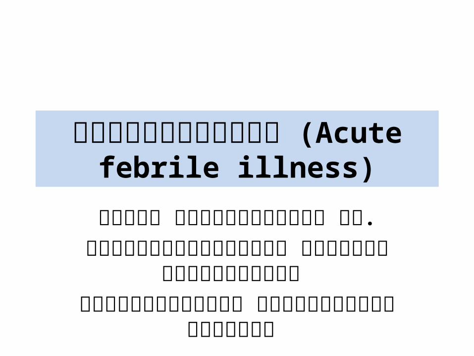
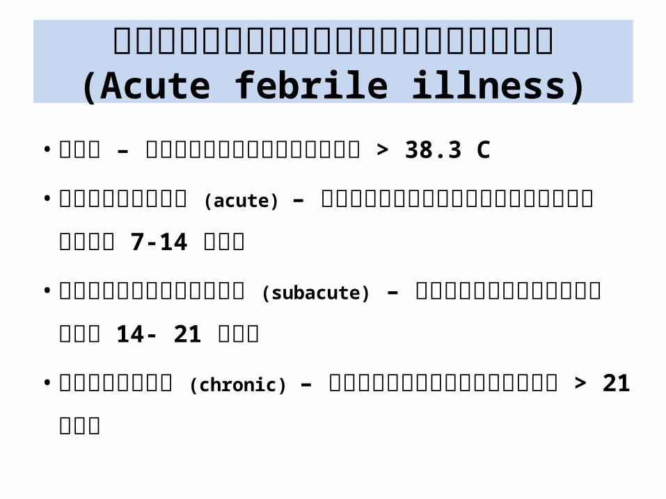
ไข้�เฉี�ยบพลั�น (Acute febrile illness)
ภิ�รุ�ญ มุ�ตสิ�กพ�นธุ์�� พบ.หน�วยโรุคต�ดเชื้ !อ ภิาคว�ชื้า
อาย�รุศาสิตรุ� คณะแพทยศาสิตรุ� มุหาว�ทยาลั�ย
ข้อนแก�น

เน !อหา• ความุหมุายแลัะสิาเหต�ข้องไข้�เฉี�ยบพลั�นโดย
ท�*วไป• สิถานการุณ�แลัะรุะบาดว�ทยาข้องไข้�
เฉี�ยบพลั�นในปรุะเทศไทยแลัะพ�ษณ�โลัก• โรุคต�ดเชื้ !อท�*ท/าให�เก�ดโรุคไข้�เฉี�ยบพลั�นพบ
บ�อย - อาการุทางคลั�น�ก - แนวทางการุว�น�จฉี�ยแลัะรุ�กษา
น�ยามุข้องไข้�เฉี�ยบพลั�น (Acute febrile illness)
• ไข้� – อ�ณหภิ1มุ�รุ�างกาย > 38.3 C
• เฉี�ยบพลั�น (acute) – รุะยะเวลัาท�*มุ�ไข้�ไมุ�เก�น 7-14 ว�น
• ก2*งเฉี�ยบพลั�น (subacute) – รุะยะเวลัาท�*มุ�ไข้� 14- 21
ว�น
• เรุ !อรุ�ง (chronic) – รุะยะเวลัาท�*มุ�ไข้� > 21 ว�น
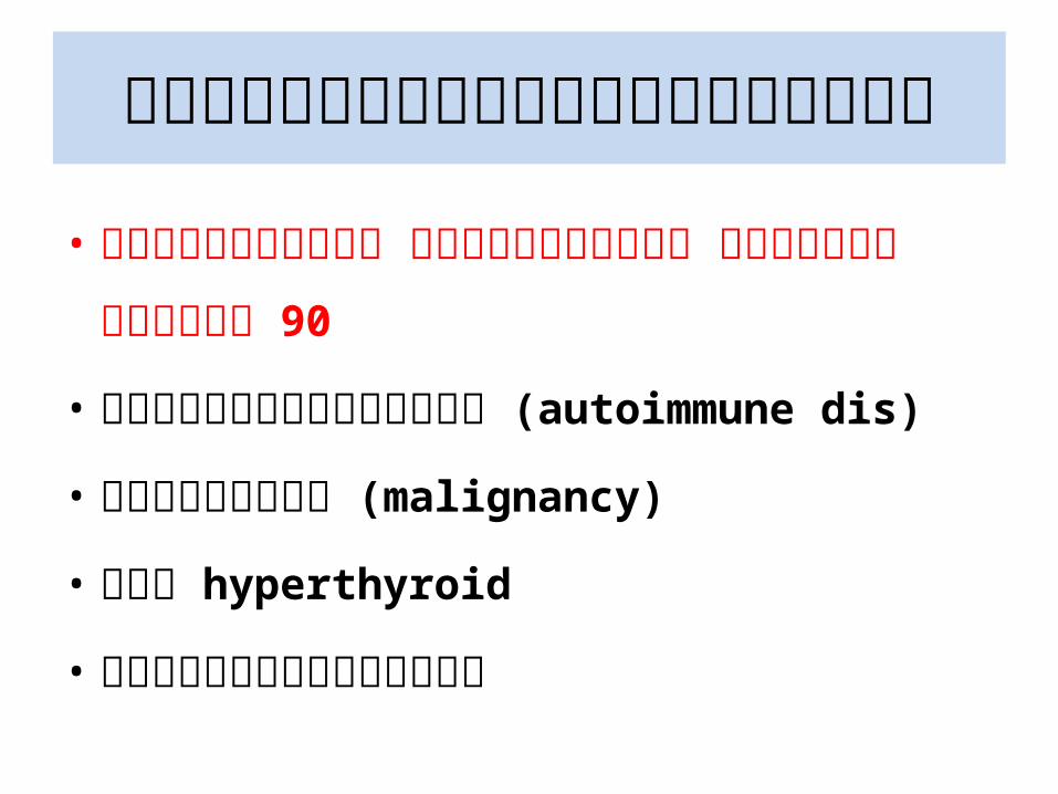
สิาเหต�ข้องไข้�เฉี�ยบพลั�น
• โรุคต�ดเชื้ !อ พบมุากท�*สิ�ด มุากกว�ารุ�อยลัะ 90
• โรุคแพ�ภิ1มุ�ตนเอง (autoimmune dis)
• โรุคมุะเรุ3ง (malignancy)
• โรุค hyperthyroid
• แพ�ยาหรุ อสิารุพ�ษ
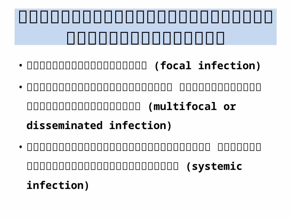
โรุคต�ดเชื้ !อท�*เป4นสิาเหต�ข้องไข้�เฉี�ยบพลั�น
• การุต�ดเชื้ !อเฉีพาะท�* (focal infection)
• การุต�ดเชื้ !อหลัายต/าแหน�ง ท�*ย�อมุพบหรุ อเพาะเชื้ !อก�อโรุคได� (multifocal or disseminated
infection)
• การุต�ดเชื้ !อท�*มุ�อาการุหลัายรุะบบ ท�*ย�อมุไมุ�พบหรุ อเพาะเชื้ !อไมุ�ได� (systemic infection)
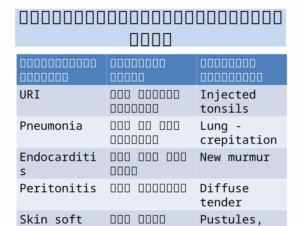
การุต�ดเชื้ !อเฉีพาะท�*ท�*พบบ�อยโรุคต�ดเชื้ !อตามุรุะบบ
ต�วอย�างอาการุ ต�วอย�างอาการุแสิดง
URI ไข้� เจ3บคอ น/!ามุ1ก Injected tonsilsPneumonia ไข้� ไอ หอบ มุ�
เสิมุหะLung - crepitation
Endocarditis ไข้� หอบ บวมุน/!า New murmurPeritonitis ไข้� ปวดท�อง Diffuse tenderSkin soft tissue infection
ไข้� ผื่ *น ผื่�วหน�ง Pustules, vesicles, bleb
Urinary tract infection
ไข้� ป6สิสิาวะ แสิบข้�ด ข้��น
CVA - tender
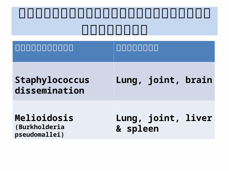
การุต�ดเชื้ !อเฉีพาะท�*หลัายต/าแหน�งโรุคต�ดเชื้ !อ ต�วอย�าง
Staphylococcus dissemination
Lung, joint, brain
Melioidosis(Burkholderia pseudomallei)
Lung, joint, liver & spleen
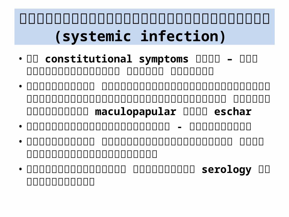
การุต�ดเชื้ !อท�*มุ�อาการุหลัายรุะบบ (systemic infection)
• มุ� constitutional symptoms เด�น – ไข้� ปวดเมุ *อยตามุต�ว ปวดข้�อ อ�อนลั�า
• ตรุวจรุ�างกาย ไมุ�พบความุผื่�ดปรุกต�เฉีพาะท�*ใดชื้�ดเจนหรุ อมุ�ผื่�ดปรุกต�หลัายต/าแหน�ง มุ�ผื่ *นผื่�วหน�งแบบ maculopapular หรุ อ eschar
• ผื่ลัตรุวจทางห�องปฏิ�บ�ต�การุ - ไมุ�จ/าเพาะ• ไมุ�พบฝี9หนอง ในต/าแหน�งท�*ต�ดเชื้ !อ ย�อมุสิารุค�ดหลั�*ง
ไมุ�พบเชื้ !อ• เพาะเชื้ !อไมุ�ข้2!น ใชื้�การุตรุวจ serology ในการุว�น�จฉี�ย
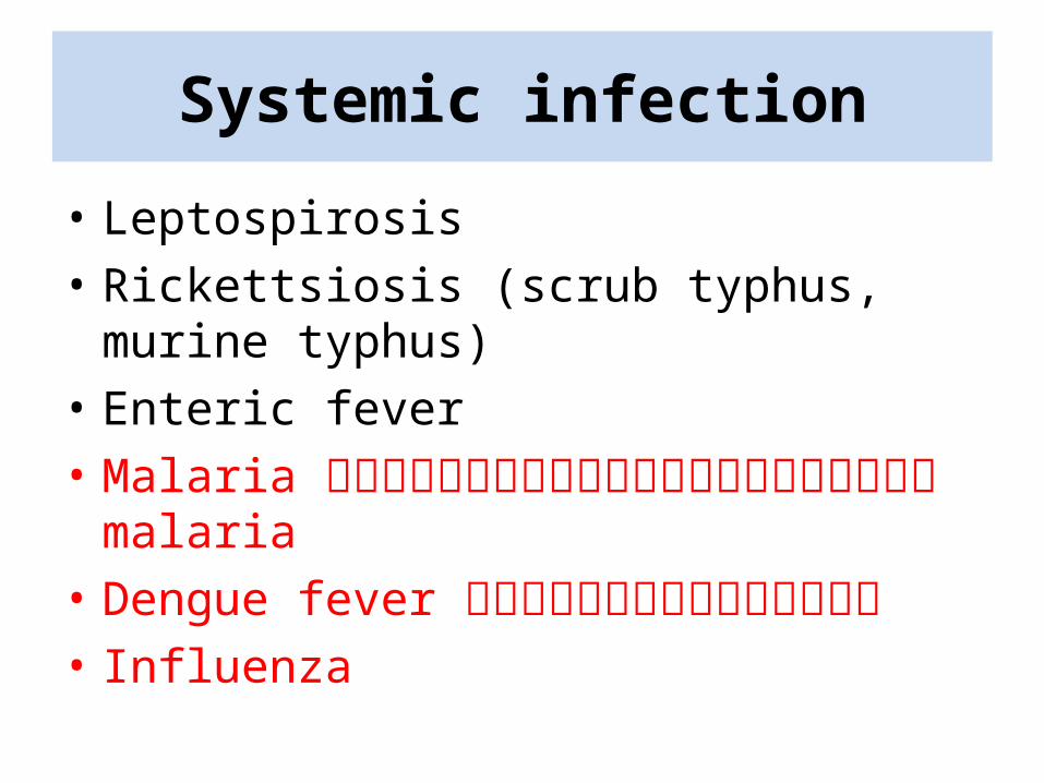
Systemic infection
• Leptospirosis• Rickettsiosis (scrub typhus, murine typhus)• Enteric fever• Malaria ก่�อนจะตรวจเลื�อดพบเชื้��อ malaria• Dengue fever ก่�อนจะตรวจเลื�อด• Influenza
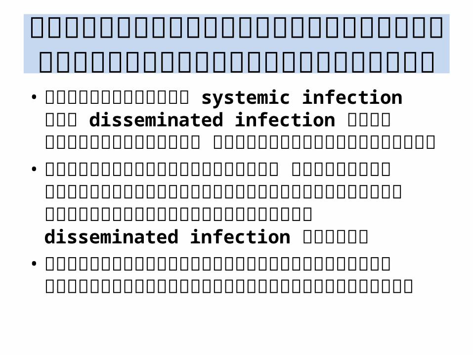
ข้�อควรุรุะว�งในการุว�น�จฉี�ยโรุคต�ดเชื้ !อไข้�เฉี�ยบพลั�น
• การุแยกรุะหว�าง systemic infection ก�บ disseminated infection ด�วยอาการุทางคลั�น�ก อาจท/าได�ยากในบางรุาย
• ในรุายท�*มุ�อาการุรุ�นแรุง หรุ อมุ�โรุคปรุะจ/าต�วท�*เสิ�*ยงต�อการุต�ดเชื้ !อรุ�นแรุงให�ค�ดถ2งหรุ อรุ�กษา disseminated infection ไปด�วย
• มุ�การุปรุะเมุ�นภิาวะแทรุกซ้�อนหรุ อการุเปลั�*ยนแปลังข้องอาการุอย�างต�อเน *อง
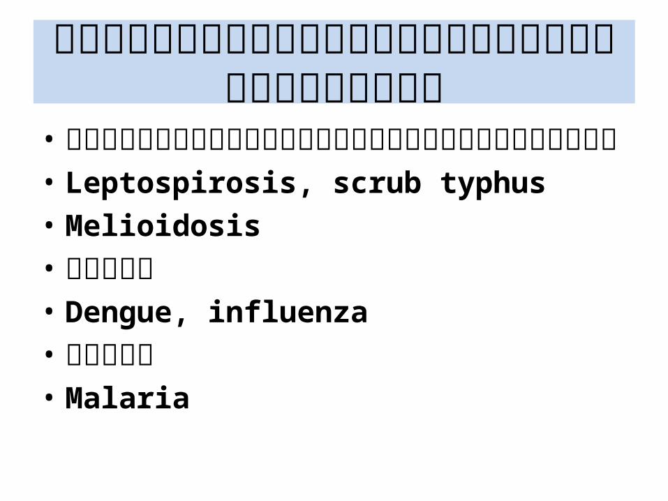
โรุคต�ดเชื้ !อไข้�เฉี�ยบพลั�นท�*พบบ�อย• การุต�ดเชื้ !อเฉีพาะท�*จากแบคท�เรุ�ย• Leptospirosis, scrub typhus• Melioidosis• ไวรุ�สิ• Dengue, influenza• ปรุสิ�ต• Malaria
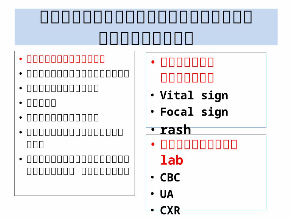
แนวทางการุว�น�จฉี�ยไข้�เฉี�ยบพลั�น• การุซ้�กปรุะว�ต�• อาการุเจ3บป;วยหลั�ก• โรุคปรุะจ/าต�ว• อาชื้�พ• ท�*อย1�อาศ�ย• ปรุะว�ต�การุสิ�มุผื่�สิโรุค• ปรุะว�ต�การุรุะบาดใน
ครุอบครุ�ว หมุ1�บ�าน
• การุตรุวจรุ�างกาย• Vital sign• Focal sign
• rash
• การุตรุวจทาง lab• CBC• UA• CXR
ต�วอย�างโรุคต�ดเชื้ !อไข้�เฉี�ยบพลั�นจากการุต�ดเชื้ !อเฉีพาะท�*
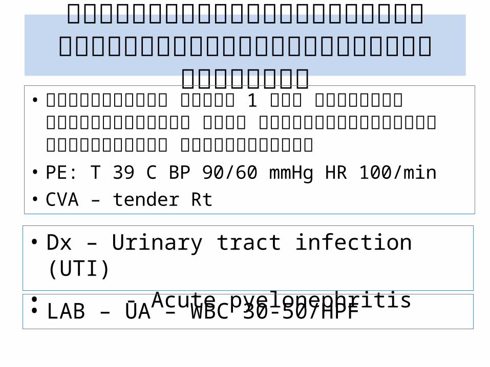
• ผู้��ป่�วยหญิ�ง มี�ไข้� 1 ว!น หนาวสั่!$น ป่%สั่สั่าวะแสั่บข้!ด ข้'�น ป่วดเหน�อห!วเหน�า ป่%สั่สั่าวะบ�อย ก่ลื!�นไมี�อย��
• PE: T 39 C BP 90/60 mmHg HR 100/min• CVA – tender Rt
• Dx – Urinary tract infection (UTI)• - Acute pyelonephritis
• LAB – UA – WBC 30-50/HPF
ต�วอย�างโรุคต�ดเชื้ !อไข้�เฉี�ยบพลั�นจากการุต�ดเชื้ !อพาะท�*
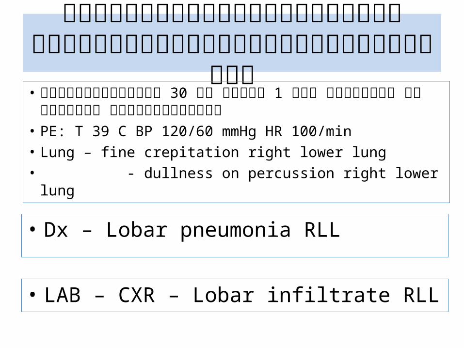
• ผู้��ป่�วยชื้ายอาย' 30 ป่( มี�ไข้� 1 ว!น หนาวสั่!$น ไอ มี�เสั่มีหะ เจ)บหน�าอก่ข้วา
• PE: T 39 C BP 120/60 mmHg HR 100/min• Lung – fine crepitation right lower lung• - dullness on percussion right lower lung
• Dx – Lobar pneumonia RLL
• LAB – CXR – Lobar infiltrate RLL
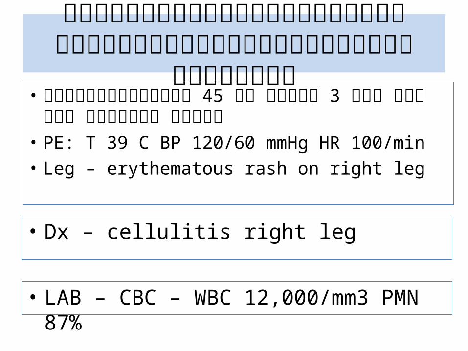
ต�วอย�างโรุคต�ดเชื้ !อไข้�เฉี�ยบพลั�นจากการุต�ดเชื้ !อเฉีพาะท�*
• ผู้��ป่�วยชื้ายอาย' 45 ป่( มี�ไข้� 3 ว!น ป่วดบวมี แดงร�อน ข้าชื้วา
• PE: T 39 C BP 120/60 mmHg HR 100/min• Leg – erythematous rash on right leg
• Dx – cellulitis right leg
• LAB – CBC – WBC 12,000/mm3 PMN 87%
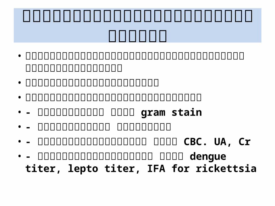
การุว�น�จฉี�ยเชื้ !อท�*เป4นสิาเหต�• จากรุะบาดว�ทยาข้องเชื้ !อท�*พบบ�อยข้องโรุคต�ดเชื้ !อ
รุะบบน�!น• ลั�กษณะจ/าเพาะทางคลั�น�ก• จากผื่ลัการุตรุวจทางห�องปฏิ�บ�ต�การุ• - การุตรุวจย�อมุ เชื้�น gram stain• - การุเพาะเชื้ !อ แบคท�เรุ�ย• - การุตรุวจเลั อดท�*วไป เชื้�น CBC. UA, Cr • - การุตรุวจเลั อดจ/าเพาะ เชื้�น dengue titer, lepto
titer, IFA for rickettsia
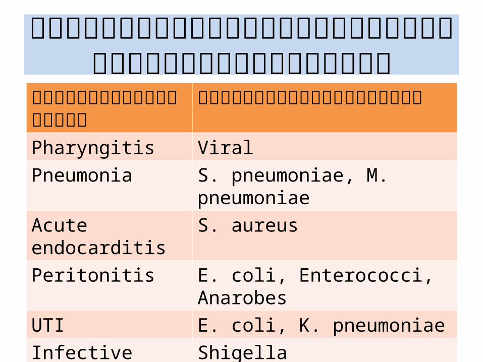
ต�วอย�างเชื้ !อก�อโรุคท�*พบบ�อยตามุรุะบาดว�ทยา
โรุคต�ดเชื้ !อในชื้�มุชื้น เชื้ !อก�อโรุคท�*พบบ�อยPharyngitis Viral Pneumonia S. pneumoniae, M. pneumoniaeAcute endocarditis S. aureusPeritonitis E. coli, Enterococci, AnarobesUTI E. coli, K. pneumoniae Infective diarrhea Shigella Cellulitis S. aureus, Strept gr A
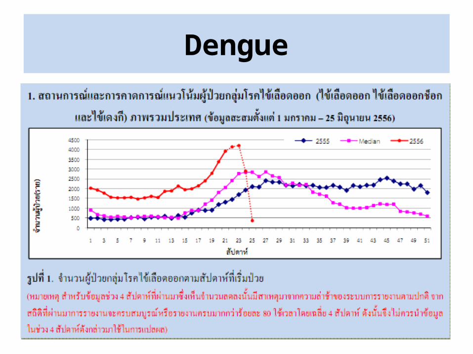
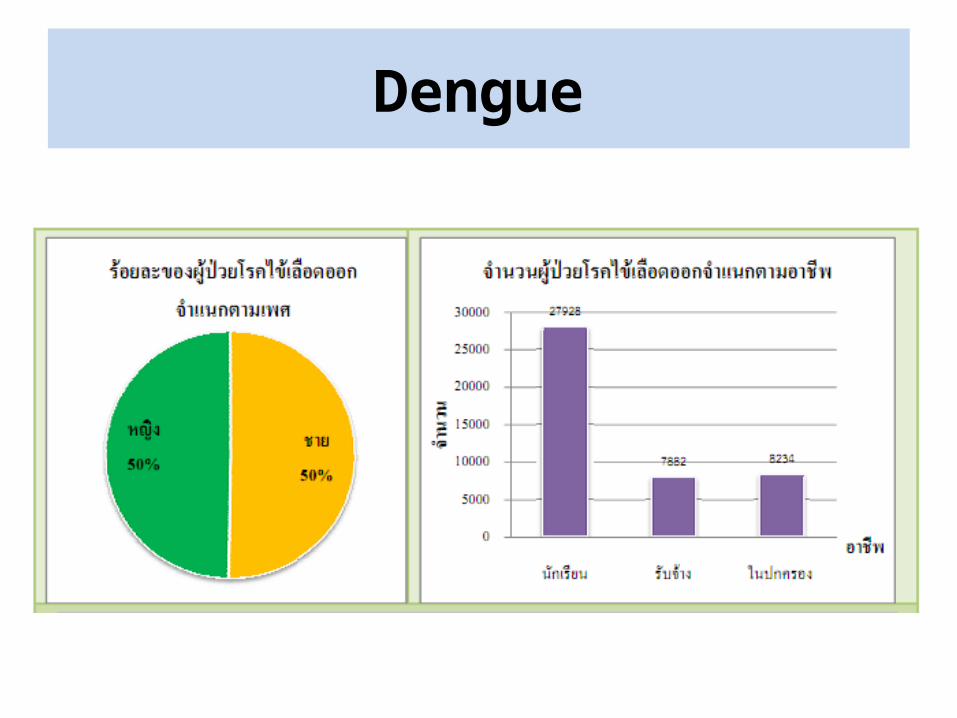
Dengue
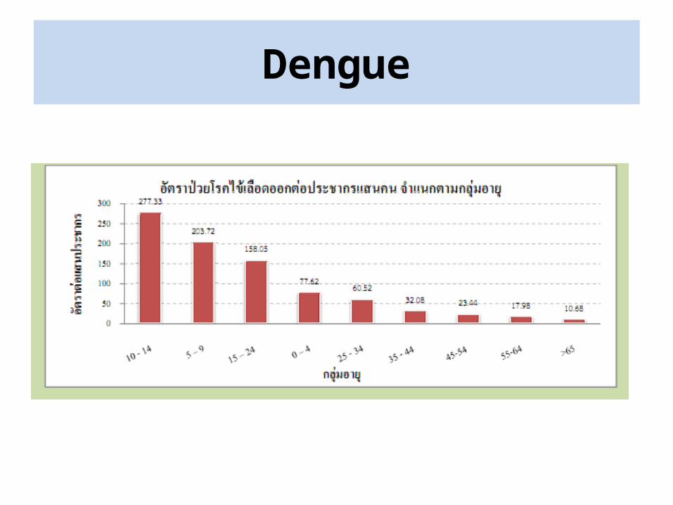
Dengue
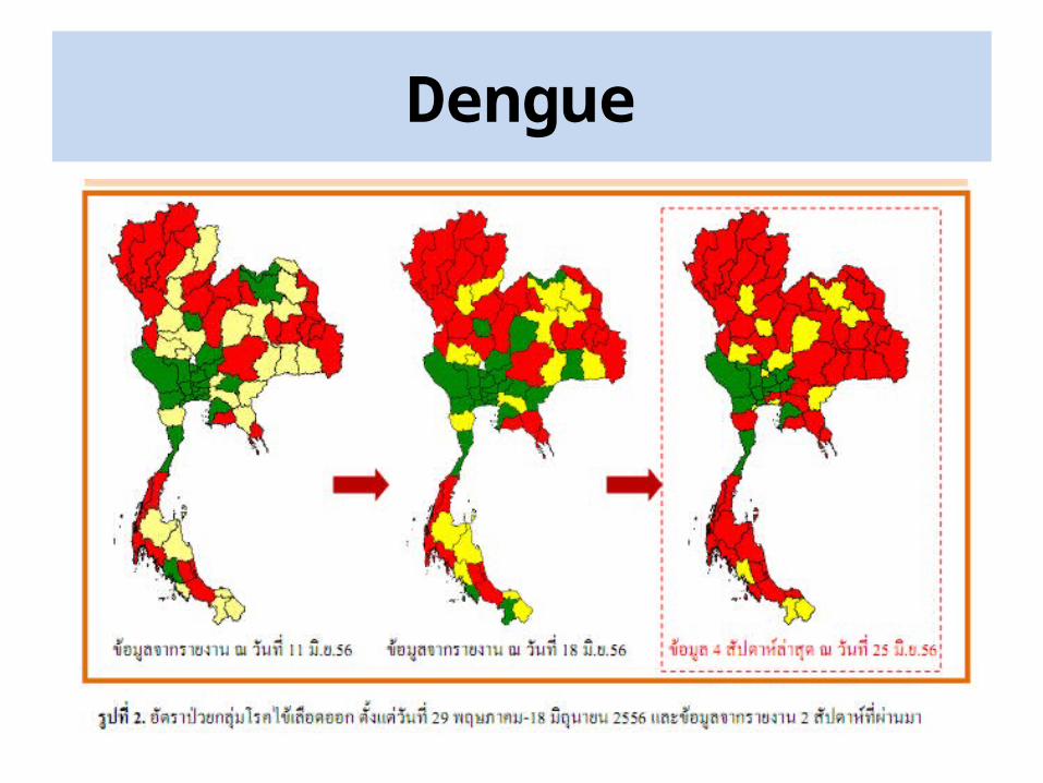
Dengue
Dengue
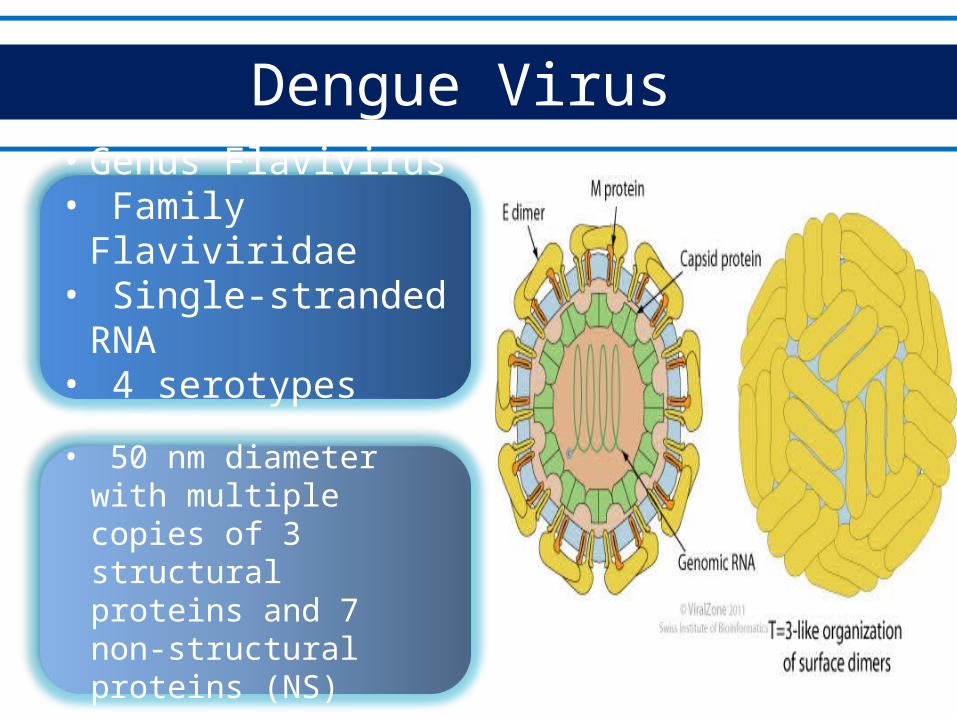
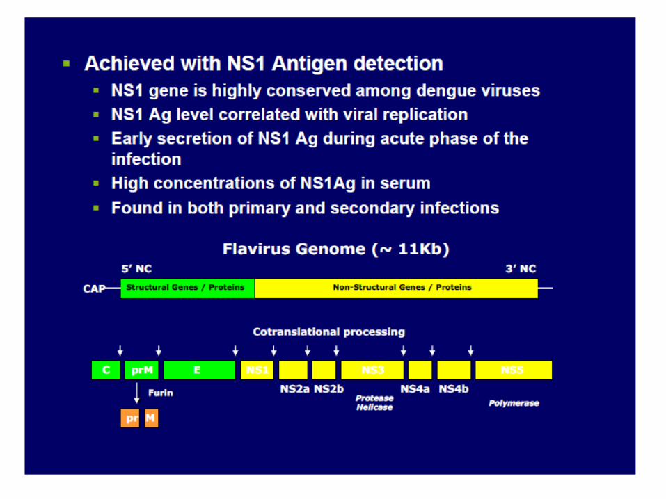
Dengue Virus
• Genus Flavivirus• Family Flaviviridae• Single-stranded RNA• 4 serotypes (DEN-1 to 4)
• 50 nm diameter with multiple copies of 3 structural proteins and 7 non-structural proteins (NS)
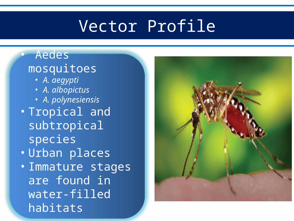
Vector Profile
• Aedes mosquitoes• A. aegypti• A. albopictus• A. polynesiensis
• Tropical and subtropical species
• Urban places• Immature stages are
found in water-filled habitats
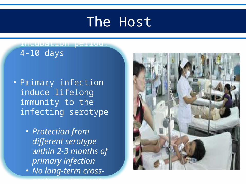
The Host
• Incubation period: 4-10 days
• Primary infection induce lifelong immunity to the infecting serotype
• Protection from different serotype within 2-3 months of primary infection
• No long-term cross-protective immunity
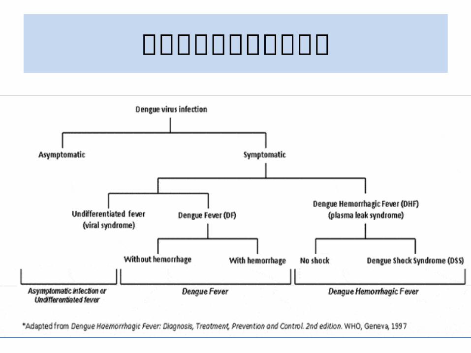
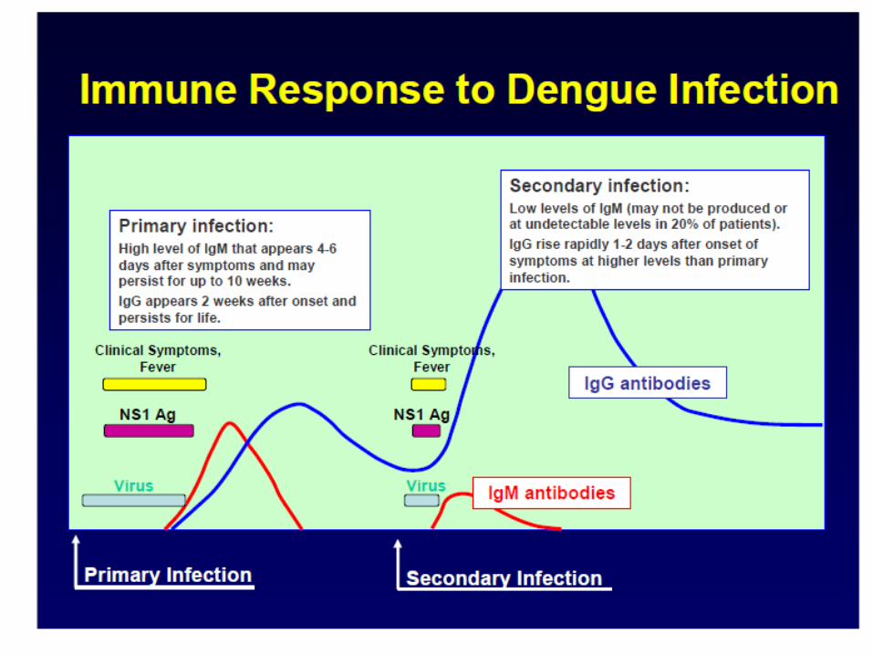
ไข้�เลั อดออก
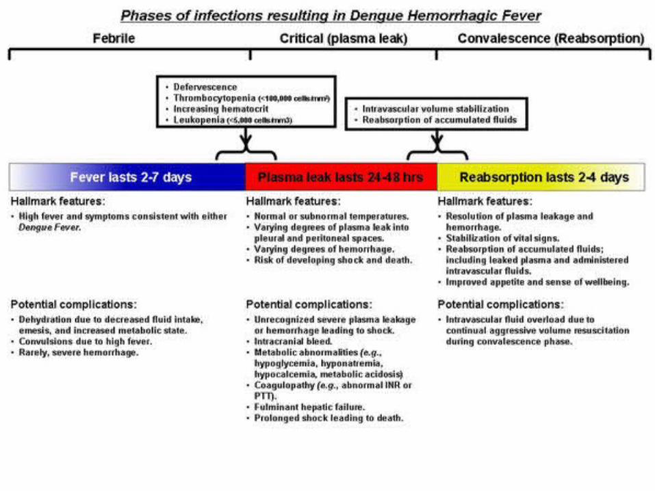
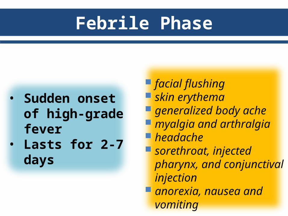
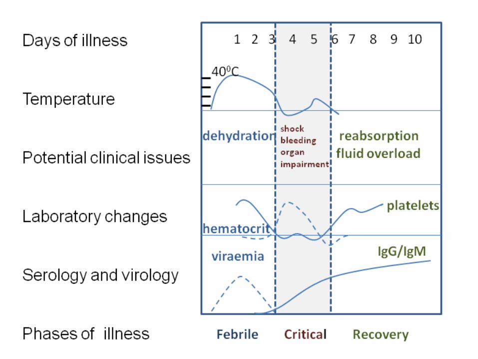
Febrile Phase
facial flushing skin erythema generalized body ache myalgia and arthralgia headache sorethroat, injected
pharynx, and conjunctival injection
anorexia, nausea and vomiting
• Sudden onset of high-grade fever
• Lasts for 2-7 days
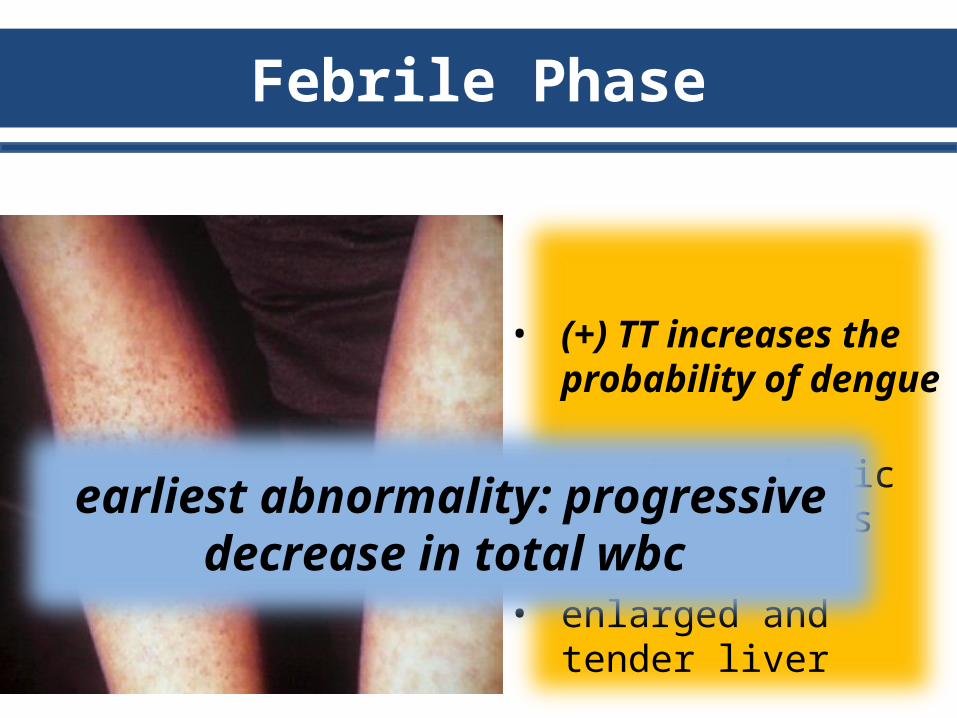
Febrile Phase
• (+) TT increases the probability of dengue
• (+) hemorrhagic manifestations
• enlarged and tender liver
earliest abnormality: progressive decrease in total wbc
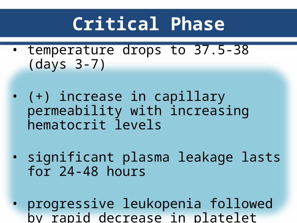
Critical Phase
• temperature drops to 37.5-38 (days 3-7)
• (+) increase in capillary permeability with increasing hematocrit levels
• significant plasma leakage lasts for 24-48 hours
• progressive leukopenia followed by rapid decrease in platelet precedes plasma leakage
Critical Phase
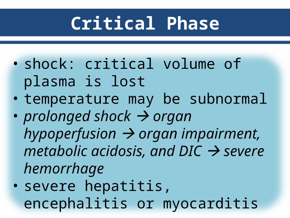
• shock: critical volume of plasma is lost• temperature may be subnormal• prolonged shock organ hypoperfusion
organ impairment, metabolic acidosis, and DIC severe hemorrhage
• severe hepatitis, encephalitis or myocarditis
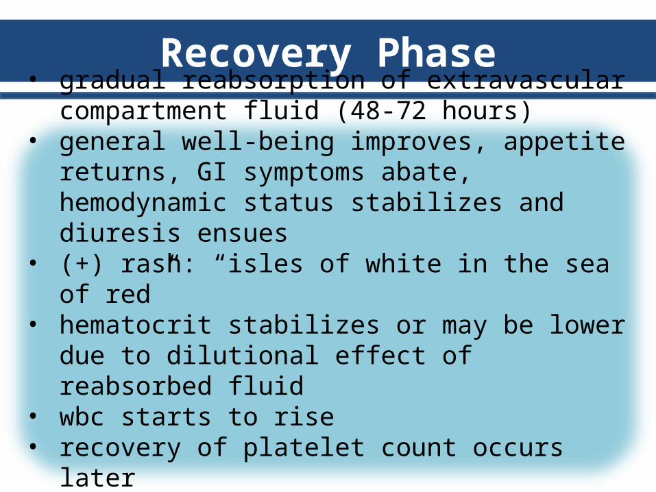
Recovery Phase
• gradual reabsorption of extravascular compartment fluid (48-72 hours)
• general well-being improves, appetite returns, GI symptoms abate, hemodynamic status stabilizes and diuresis ensues
• (+) rash: “isles of white in the sea of red”• hematocrit stabilizes or may be lower due to dilutional
effect of reabsorbed fluid• wbc starts to rise• recovery of platelet count occurs later
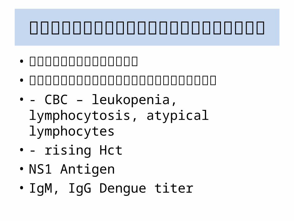
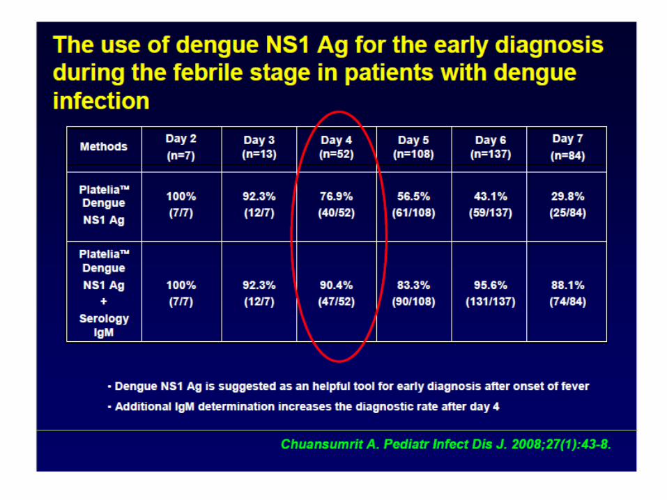
การุว�น�จฉี�ยไข้�เลั อดออก• อาการุทางคลั�น�ก• การุตรุวจทางห�องปฏิ�บ�ต�การุ• - CBC – leukopenia, lymphocytosis, atypical
lymphocytes• - rising Hct• NS1 Antigen• IgM, IgG Dengue titer
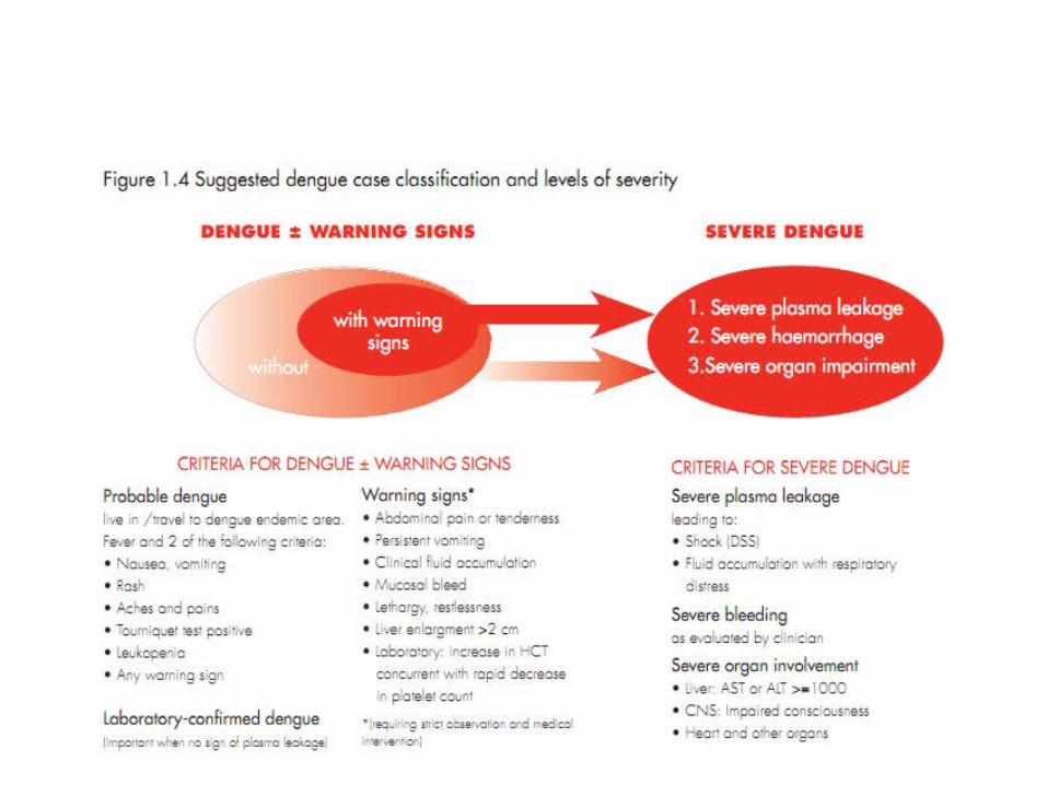
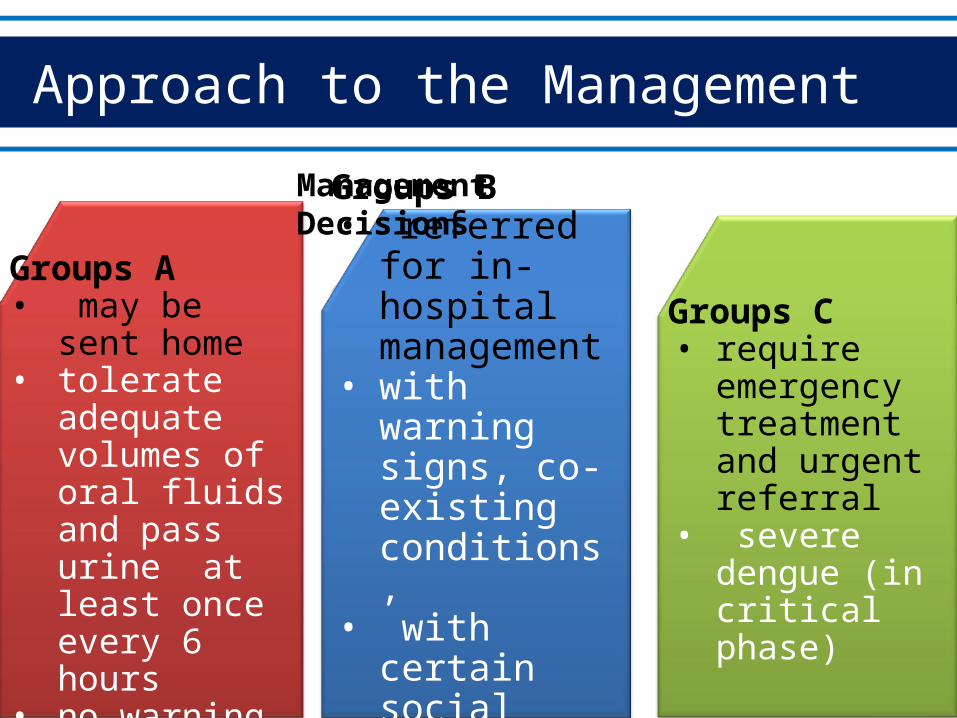
Approach to the Management
Groups A• may be sent
home• tolerate
adequate volumes of oral fluids and pass urine at least once every 6 hours
• no warning signs
Groups B• referred for in-
hospital management
• with warning signs, co-existing conditions,
• with certain social circumstances
Groups C• require
emergency treatment and urgent referral
• severe dengue (in critical phase)
Management Decisions
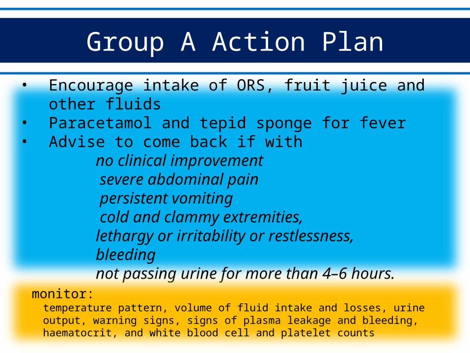
Group A Action Plan
• Encourage intake of ORS, fruit juice and other fluids• Paracetamol and tepid sponge for fever• Advise to come back if with
no clinical improvement severe abdominal pain persistent vomiting cold and clammy extremities,lethargy or irritability or restlessness, bleeding
not passing urine for more than 4–6 hours.
monitor: temperature pattern, volume of fluid intake and losses, urine output, warning signs, signs of plasma leakage and bleeding, haematocrit, and white blood cell and platelet counts
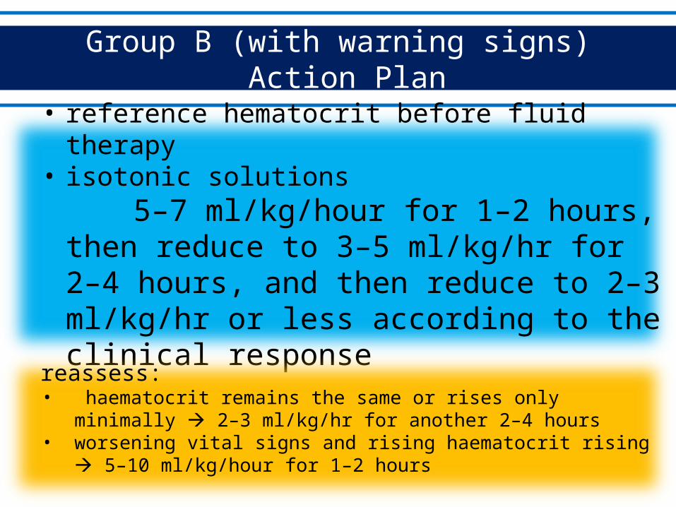
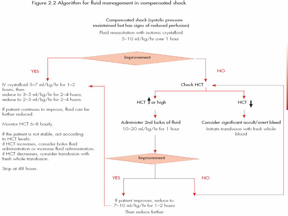
Group B (with warning signs) Action Plan
• reference hematocrit before fluid therapy• isotonic solutions
5–7 ml/kg/hour for 1–2 hours, then reduce to 3–5 ml/kg/hr for 2–4 hours, and then reduce to 2–3 ml/kg/hr or less according to the clinical response
reassess:• haematocrit remains the same or rises only minimally 2–3 ml/kg/hr for
another 2–4 hours • worsening vital signs and rising haematocrit rising 5–10 ml/kg/hour for 1–
2 hours
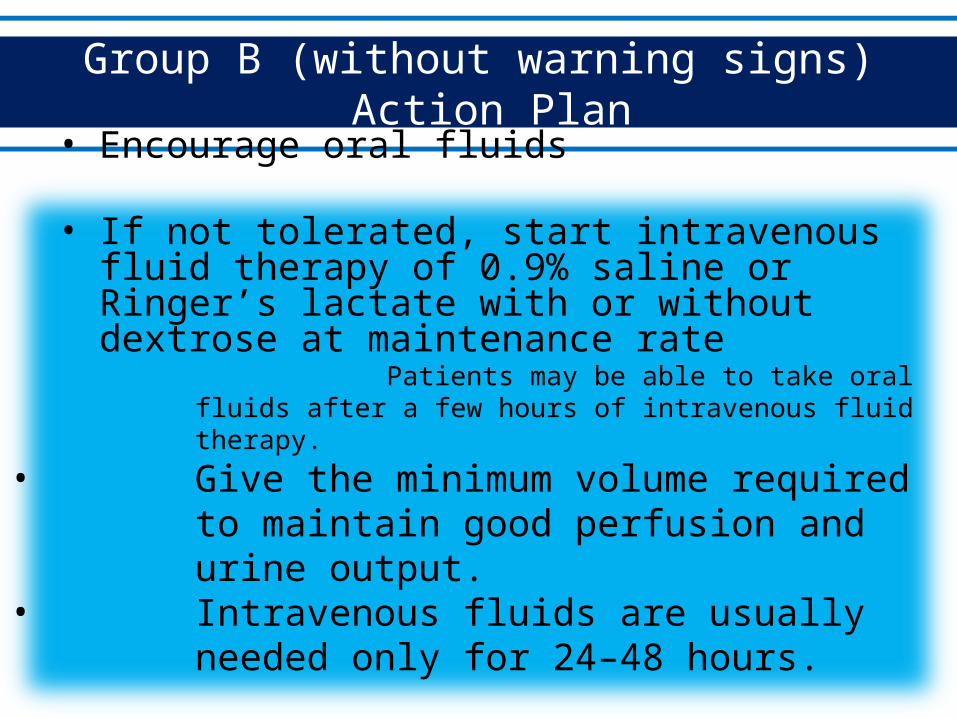
Group B (without warning signs) Action Plan
• Encourage oral fluids
• If not tolerated, start intravenous fluid therapy of 0.9% saline or Ringer’s lactate with or without dextrose at maintenance rate
Patients may be able to take oral fluids after a few hours of intravenous fluid therapy.
• Give the minimum volume required to maintain good perfusion and urine output.
• Intravenous fluids are usually needed only for 24–48 hours.
• Close monitoring
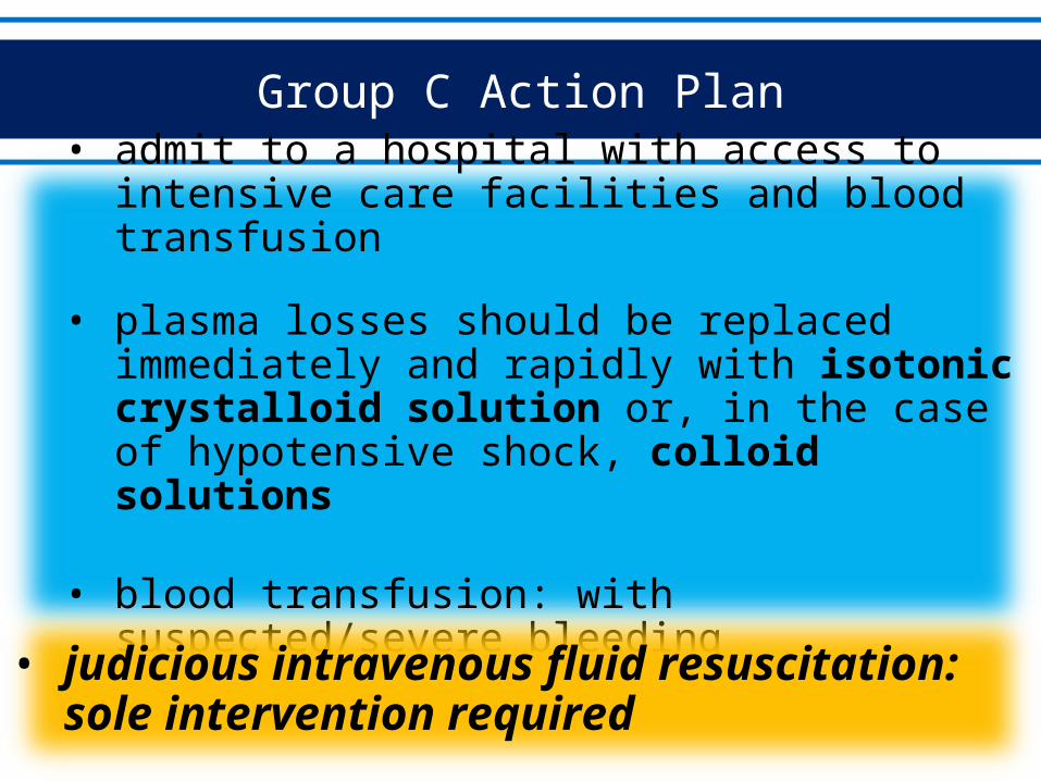
Group C Action Plan
• admit to a hospital with access to intensive care facilities and blood transfusion
• plasma losses should be replaced immediately and rapidly with isotonic crystalloid solution or, in the case of hypotensive shock, colloid solutions
• blood transfusion: with suspected/severe bleeding
• judicious intravenous fluid resuscitation: sole intervention required
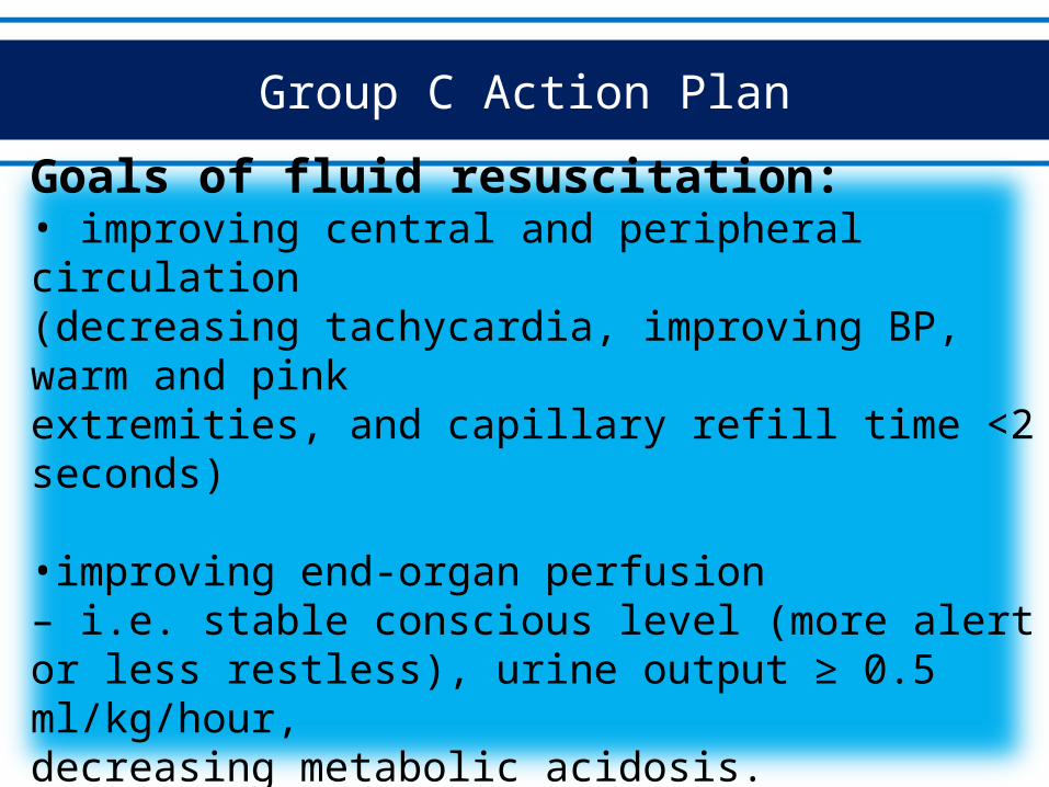
Group C Action Plan
Goals of fluid resuscitation:• improving central and peripheral circulation(decreasing tachycardia, improving BP, warm and pinkextremities, and capillary refill time <2 seconds)
•improving end-organ perfusion– i.e. stable conscious level (more alert or less restless), urine output ≥ 0.5 ml/kg/hour,decreasing metabolic acidosis.
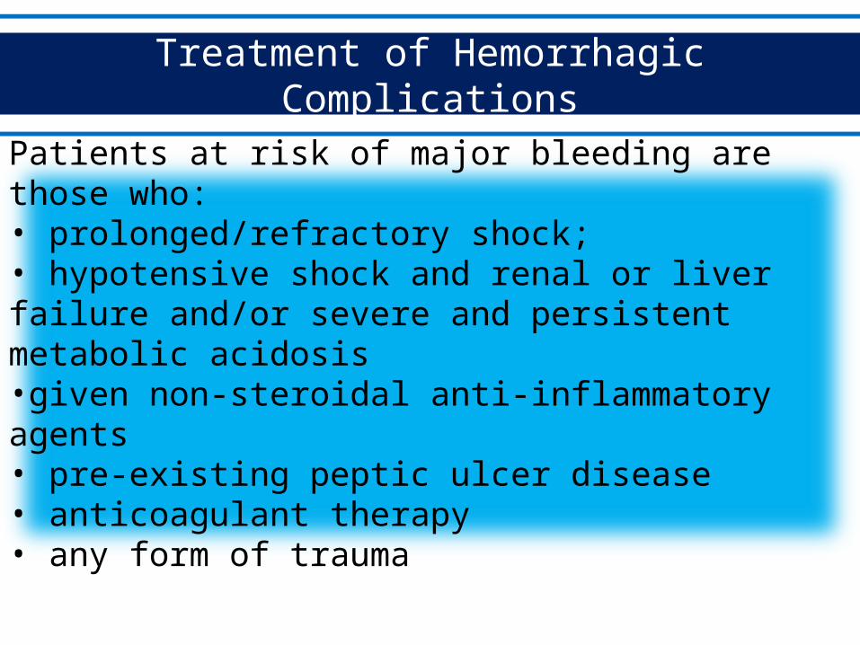
Treatment of Hemorrhagic Complications
Patients at risk of major bleeding are those who:• prolonged/refractory shock;• hypotensive shock and renal or liver failure and/or severe and persistent metabolic acidosis•given non-steroidal anti-inflammatory agents• pre-existing peptic ulcer disease• anticoagulant therapy• any form of trauma
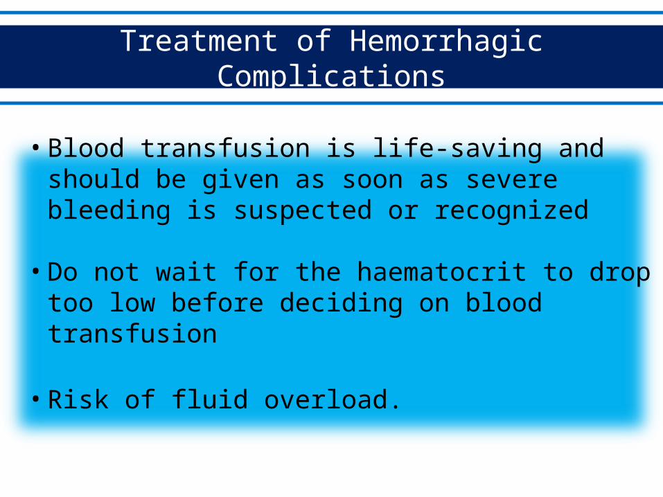
Treatment of Hemorrhagic Complications
• Blood transfusion is life-saving and should be given as soon as severe bleeding is suspected or recognized
• Do not wait for the haematocrit to drop too low before deciding on blood transfusion
• Risk of fluid overload.
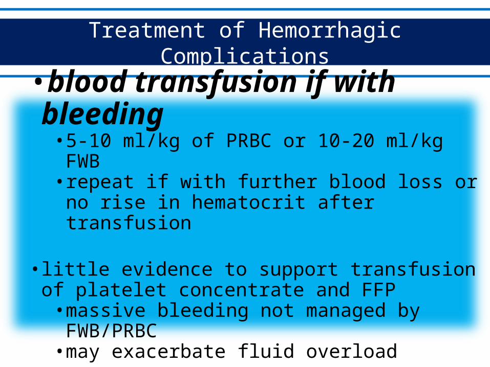
Treatment of Hemorrhagic Complications
•blood transfusion if with bleeding• 5-10 ml/kg of PRBC or 10-20 ml/kg FWB• repeat if with further blood loss or no rise in
hematocrit after transfusion
• little evidence to support transfusion of platelet concentrate and FFP
• massive bleeding not managed by FWB/PRBC• may exacerbate fluid overload
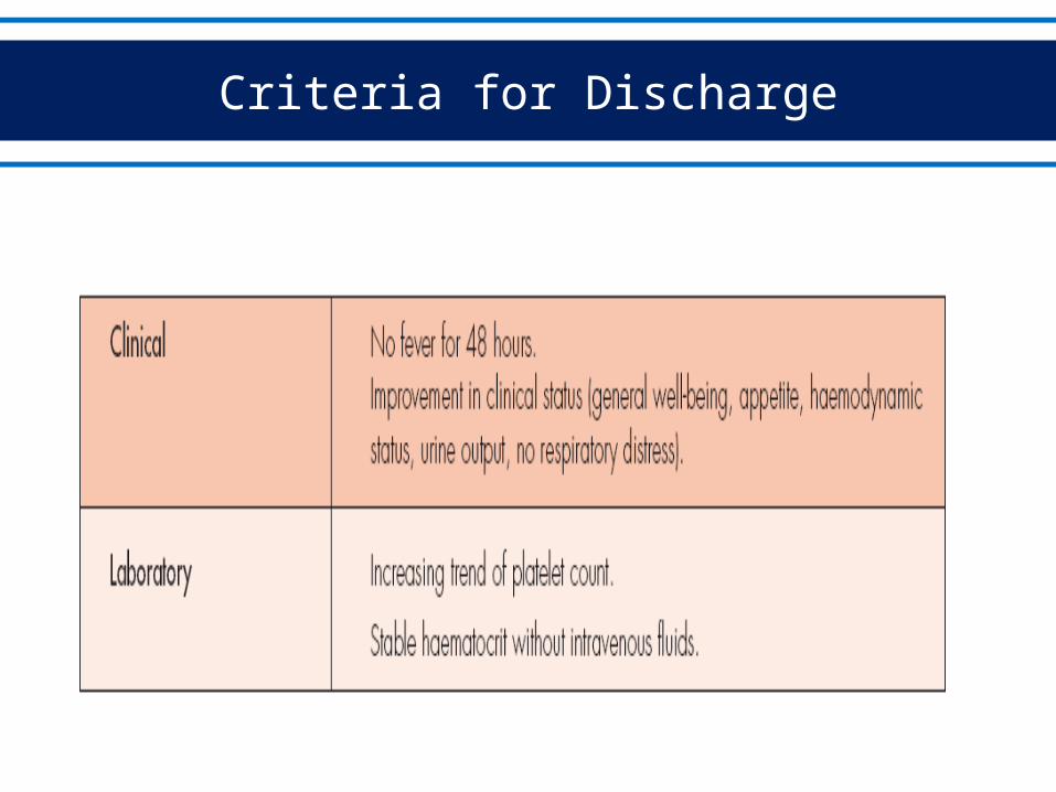
Criteria for Discharge
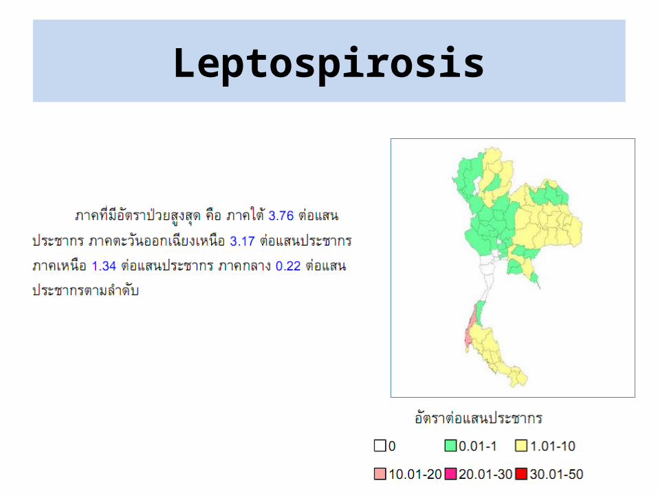
Leptospirosis
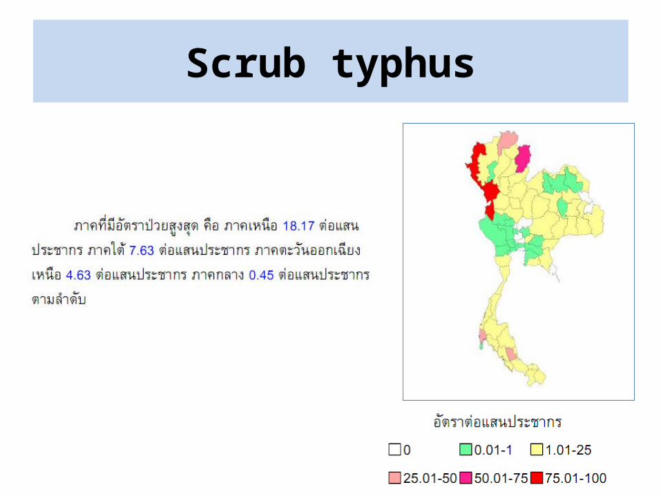
Scrub typhus
Leptospirosis
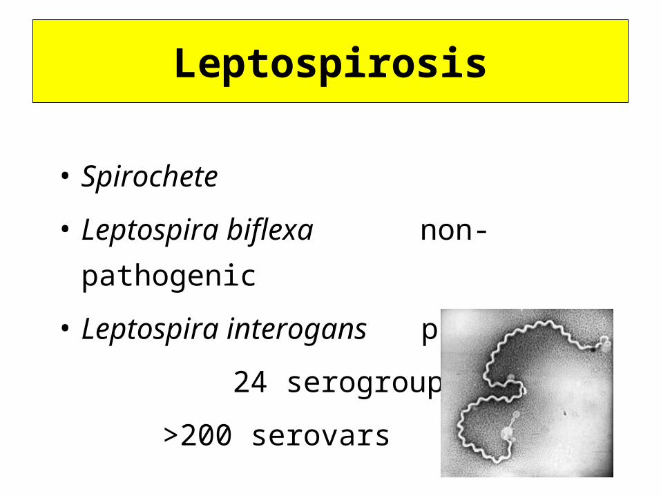
• Spirochete
• Leptospira biflexa non-pathogenic
• Leptospira interogans pathogenic
24 serogroups
>200 serovars
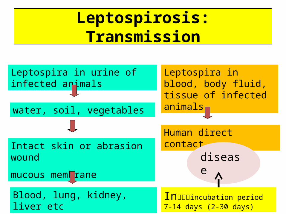
Leptospirosis: Transmission
Leptospira in urine of infected animals
water, soil, vegetables
Intact skin or abrasion wound
mucous membrane
Blood, lung, kidney, liver etc
Leptospira in blood, body fluid, tissue of infected animals
Human direct contact
Inรร�incubation period 7-14 days (2-30 days)
disease
อาก่ารทางคลื�น�ก่• ไมุ�มุ�อาการุ(symptomatic)• ไข้�เฉี�ยบพลั�น (acute uncomplicated febrile
illness)• ไข้�รุ�วมุก�บภิาวะแทรุกซ้�อน• ด�ซ้�าน(jaundice)• ไตวาย (acute renal failure)• ด�ซ้�านแลัะไตวาย (Weil’s syndrome)• เย *อห��มุสิมุองอ�กเสิบ (asepticmeningoencephalitis)
• เลั อดออกผื่�ดปรุกต� (hemorrhagic manifestation)
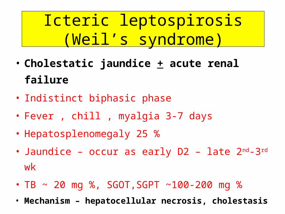
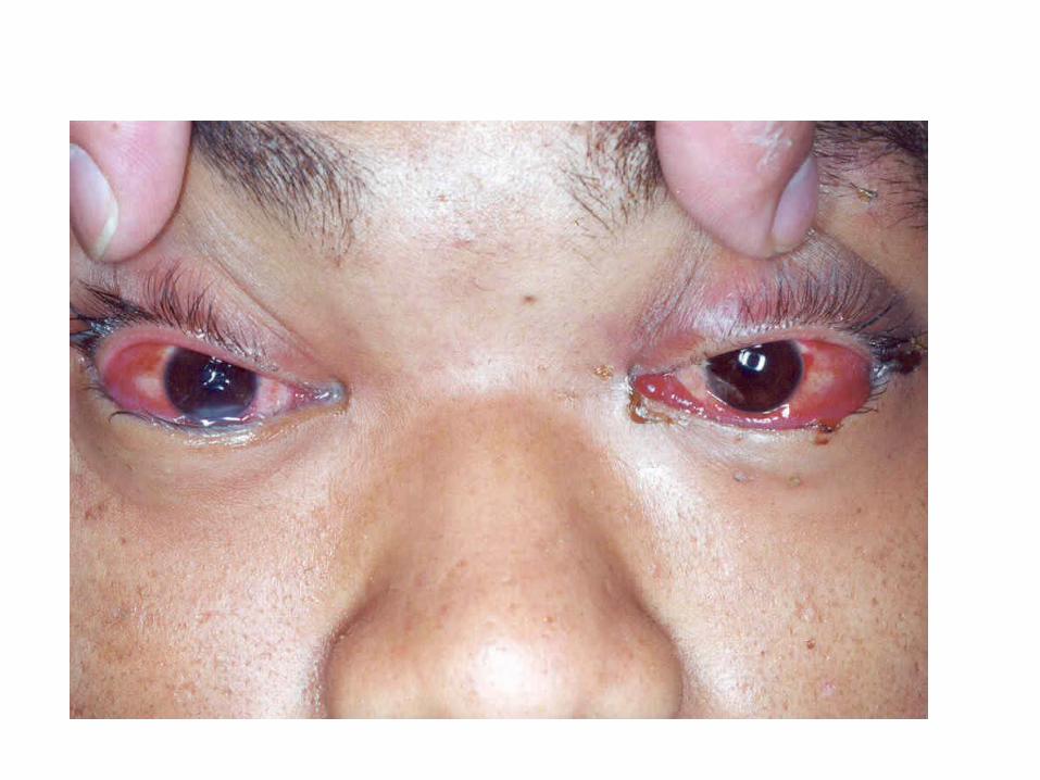
Icteric leptospirosis (Weil’s syndrome)
• Cholestatic jaundice + acute renal failure
• Indistinct biphasic phase
• Fever , chill , myalgia 3-7 days
• Hepatosplenomegaly 25 %
• Jaundice – occur as early D2 – late 2nd-3rd wk
• TB ~ 20 mg %, SGOT,SGPT ~100-200 mg %
• Mechanism – hepatocellular necrosis, cholestasis
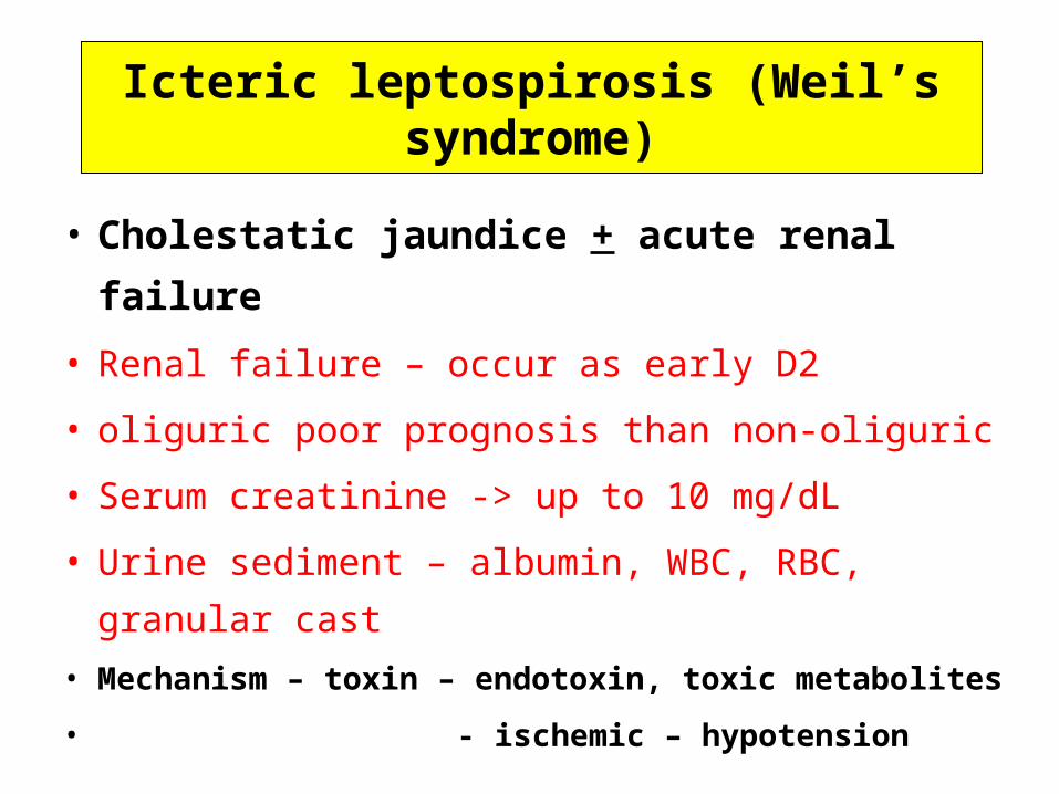
Icteric leptospirosis (Weil’s syndrome)
• Cholestatic jaundice + acute renal failure
• Renal failure – occur as early D2
• oliguric poor prognosis than non-oliguric
• Serum creatinine -> up to 10 mg/dL
• Urine sediment – albumin, WBC, RBC, granular
cast
• Mechanism – toxin – endotoxin, toxic metabolites
• - ischemic – hypotension
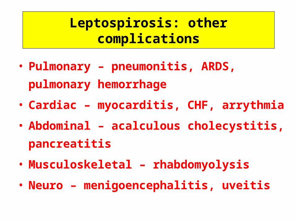
Leptospirosis: other complications
• Pulmonary – pneumonitis, ARDS, pulmonary
hemorrhage
• Cardiac – myocarditis, CHF, arrythmia
• Abdominal – acalculous cholecystitis,
pancreatitis
• Musculoskeletal – rhabdomyolysis
• Neuro – menigoencephalitis, uveitis
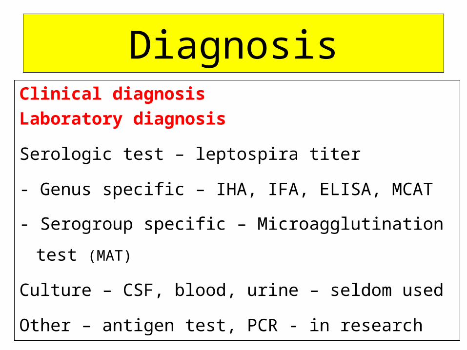
DiagnosisClinical diagnosis
Laboratory diagnosis
Serologic test – leptospira titer
- Genus specific – IHA, IFA, ELISA, MCAT
- Serogroup specific – Microagglutination test (MAT)
Culture – CSF, blood, urine – seldom used
Other – antigen test, PCR - in research
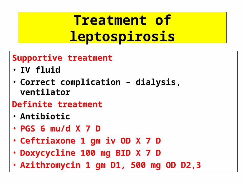
Treatment of leptospirosis
Supportive treatment• IV fluid• Correct complication – dialysis, ventilator
Definite treatment• Antibiotic• PGS 6 mu/d X 7 D• Ceftriaxone 1 gm iv OD X 7 D• Doxycycline 100 mg BID X 7 D• Azithromycin 1 gm D1, 500 mg OD D2,3
Scrub and murine typhus
Scrub typhus
• Zoonosis
• Pathogen - Orientia tsutsugamushi
• Gram negative coccobacilli
• Common in rural Southeast Asia
• No cross immunity
• Multiple infections can occur
Scrub typhus
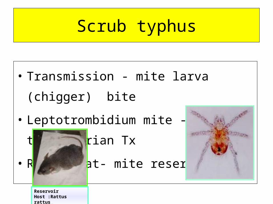
• Transmission - mite larva (chigger) bite
• Leptotrombidium mite - transovarian Tx
• Rattus rat- mite reservoir
Reservoir Host :Rattus rattus
Murine typhus
• Common zoonosis in Thailand
• Pathogen - Rickettsia typhi
Transmission • Rattus rattus, R. norvegicus rat- flea reservoir
• rat flea (Xenopsylla cheopis) -transovarian Tx
• flea bite -> defecation in wound -> Human
• No person to person transmission
Scrub typhus and murine typhus
Clinical symptoms
• Fever
• Headache
• Myalgia
• Conjunctival suffusion
• Regional lymphadenopathy
• Rash - maculopapular
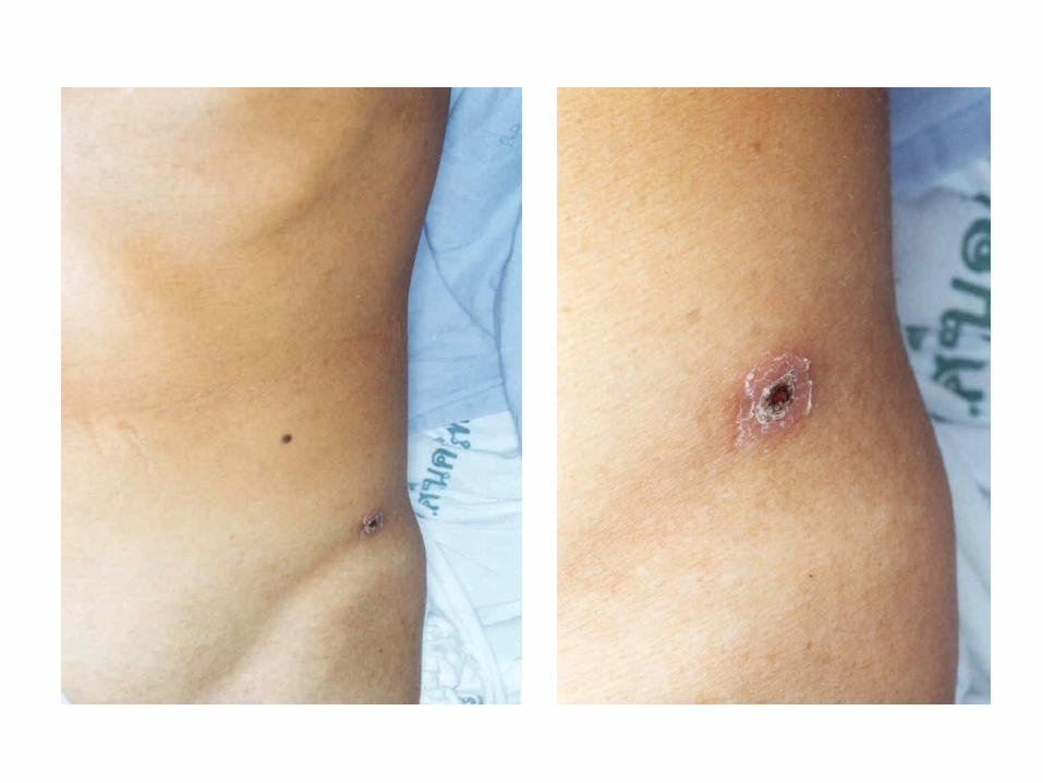
• Eschar only in scrub typhus
Scrub typhus and murine typhus
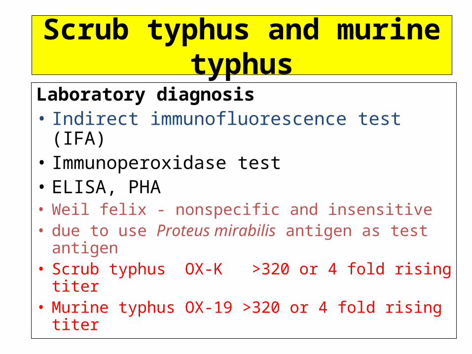
Laboratory diagnosis• Indirect immunofluorescence test (IFA)• Immunoperoxidase test• ELISA, PHA• Weil felix - nonspecific and insensitive• due to use Proteus mirabilis antigen as test
antigen • Scrub typhus OX-K >320 or 4 fold rising titer• Murine typhus OX-19 >320 or 4 fold rising titer
Scrub typhus and murine typhus
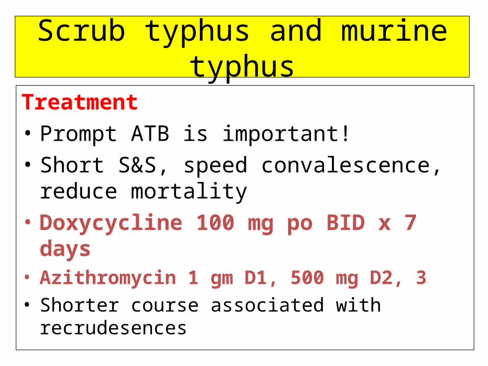
Treatment
• Prompt ATB is important!
• Short S&S, speed convalescence, reduce mortality
• Doxycycline 100 mg po BID x 7 days• Azithromycin 1 gm D1, 500 mg D2, 3• Shorter course associated with recrudesences