Embed Size (px)
Citation preview
Components ofاجزاء سيستم ايمني Immune System
لنفاوي سيستم اعضاء و LymphoidبافتهاOrgans
لنفاوي Lymphoid Cellsسلولهاي
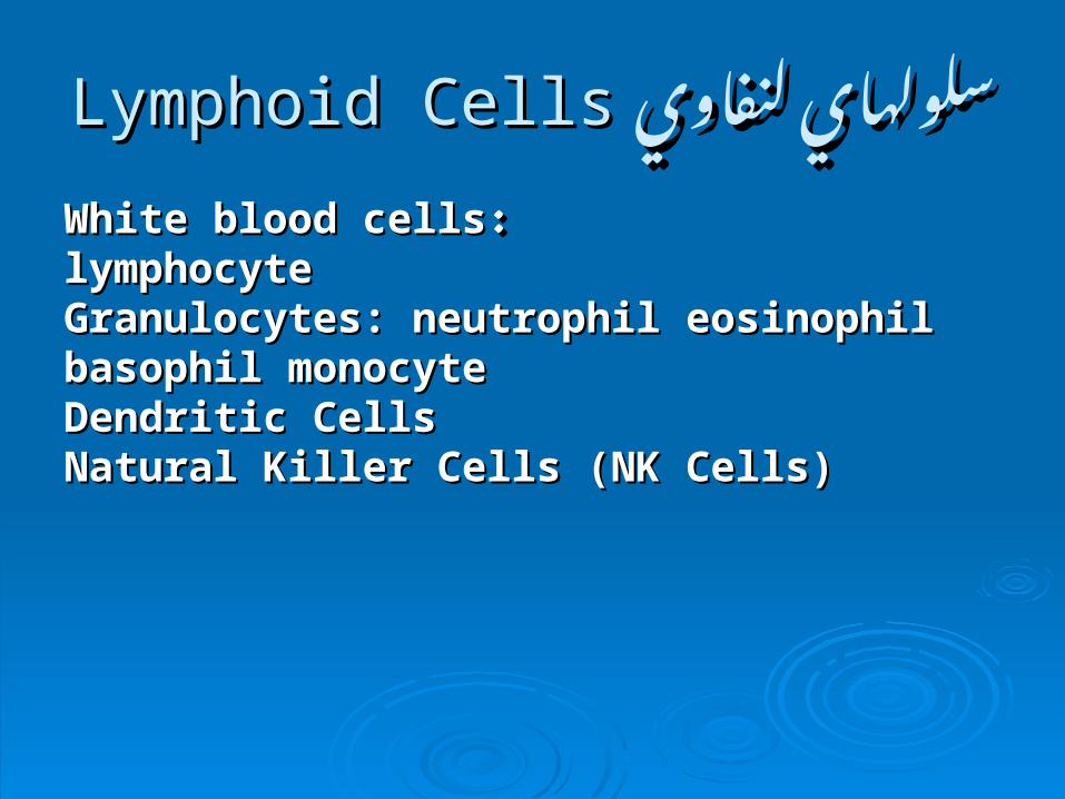
لنفاوي لنفاوي سلولهاي Lymphoid CellsLymphoid Cellsسلولهاي
White blood cellsWhite blood cells : : lymphocytelymphocyteGranulocytes: neutrophil eosinophil basophil Granulocytes: neutrophil eosinophil basophil monocyte monocyte Dendritic CellsDendritic Cells Natural Killer Cells (NK Cells)Natural Killer Cells (NK Cells)
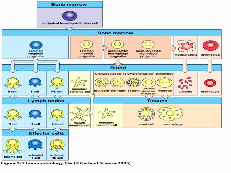
Figure 1-3
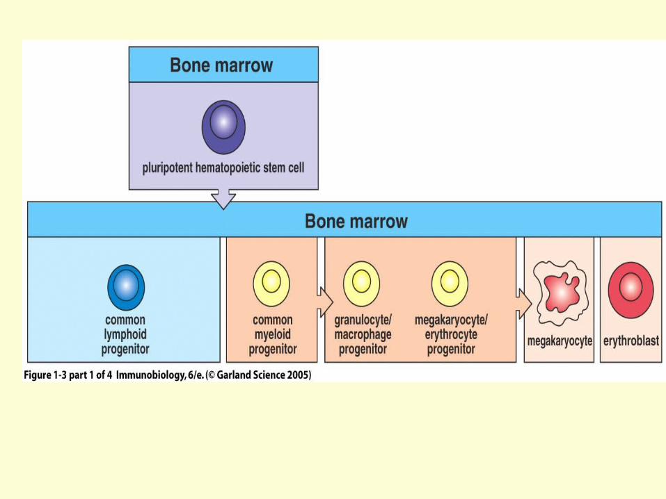
Figure 1-3 part 1 of 4
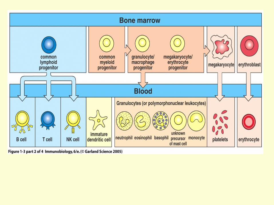
Figure 1-3 part 2 of 4
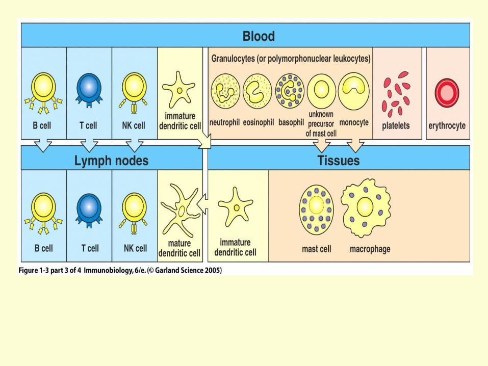
Figure 1-3 part 3 of 4
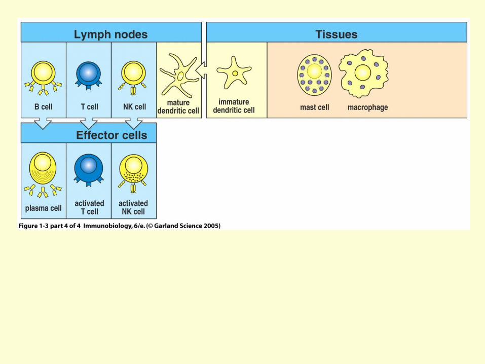
Figure 1-3 part 4 of 4
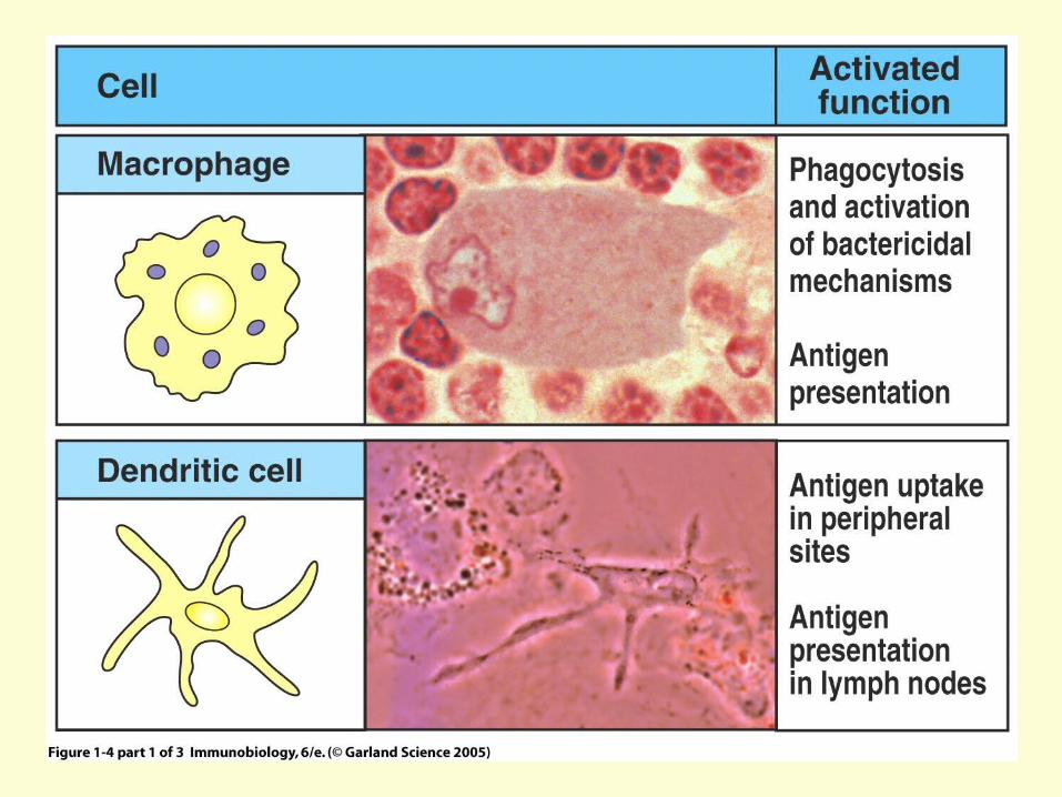
Figure 1-4 part 1 of 3
Figure 1-4 part 1 of 3
•(Greek: big eaters, from makros "large" + phagein "eat"; abbr. MΦ)•white blood cells within tissues, produced by the division of monocytes•Human macrophages are about 21 micrometres in diameter•Monocytes and macrophages are phagocytes, acting in both non-specific defense (or innate immunity) as well as to help initiate specific defense mechanisms (or cell-mediated immunity) of vertebrate animals.
Macrophages
Figure 1-4 part 1 of 3•Immune cells and form part of the mammalian immune system
•Their main function is to process antigen material and present it on the surface to other cells of the immune system, thus functioning as
antigen-presenting cells (APC)•Dendritic cells are present in small quantities in
tissues that are in contact with the external environment, mainly the skin (where there is a specialized dendritic cell type called Langerhans cells) and the inner lining of the nose, lungs, stomach and intestines. They can also be found in an immature state in the blood
Dendritic cells (DCs)
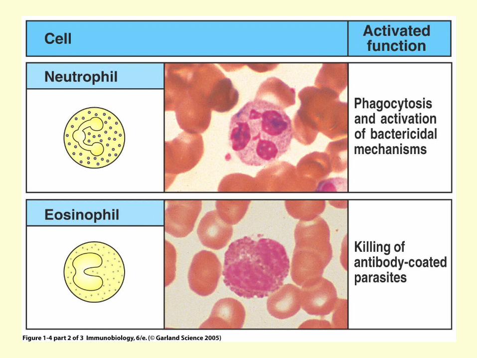
Figure 1-4 part 2 of 3
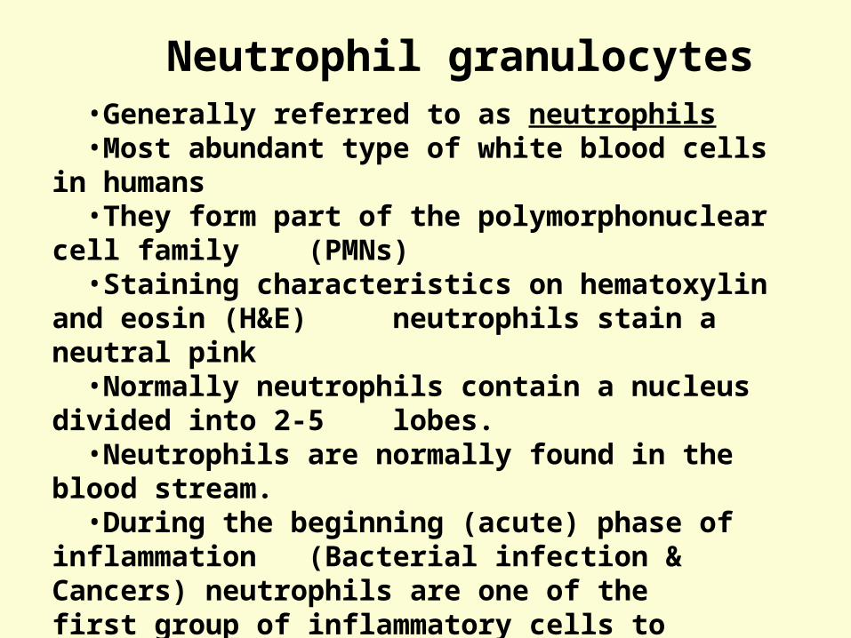
Figure 1-4 part 1 of 3•Generally referred to as neutrophils•Most abundant type of white blood cells in humans•They form part of the polymorphonuclear cell family (PMNs)•Staining characteristics on hematoxylin and eosin (H&E) neutrophils stain a neutral pink•Normally neutrophils contain a nucleus divided into 2-5 lobes.•Neutrophils are normally found in the blood stream.•During the beginning (acute) phase of inflammation
(Bacterial infection & Cancers) neutrophils are one of the first group of inflammatory cells to migrate toward the site of inflammation, firstly through the blood vessels, then through interstitial tissue
Neutrophil granulocytes
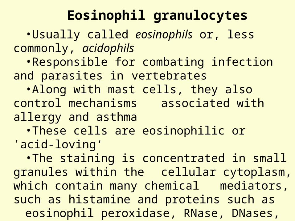
Figure 1-4 part 1 of 3•Usually called eosinophils or, less commonly, acidophils•Responsible for combating infection and parasites in
vertebrates•Along with mast cells, they also control mechanisms
associated with allergy and asthma•These cells are eosinophilic or 'acid-loving‘•The staining is concentrated in small granules within the cellular cytoplasm, which contain many chemical
mediators, such as histamine and proteins such as eosinophil peroxidase, RNase, DNases, lipase, plasminogen, and Major Basic Protein.
•These mediators are released by a process called degranulation following activation of the eosinophil, and are toxic to both parasite and host tissues.
Eosinophil granulocytes
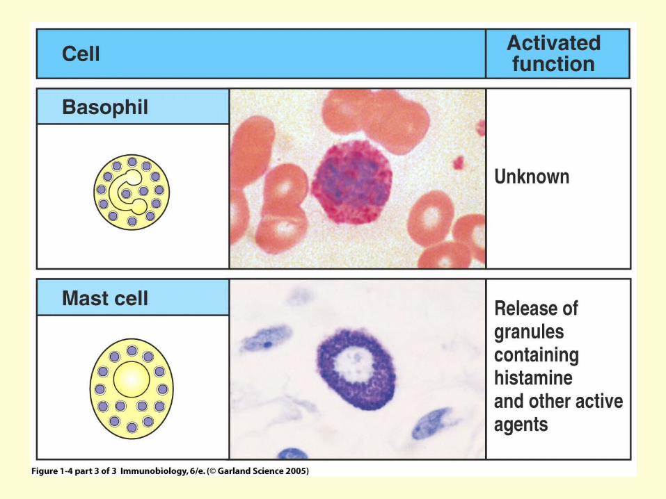
Figure 1-4 part 3 of 3
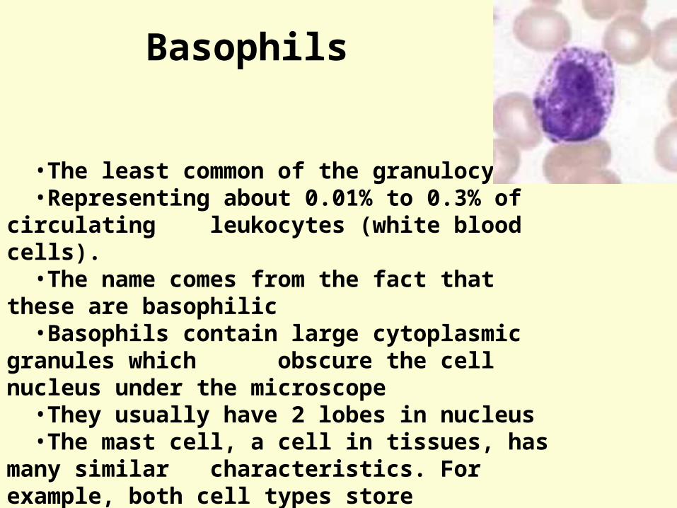
Figure 1-4 part 1 of 3
•The least common of the granulocytes•Representing about 0.01% to 0.3% of circulating
leukocytes (white blood cells).•The name comes from the fact that these are
basophilic•Basophils contain large cytoplasmic granules which
obscure the cell nucleus under the microscope•They usually have 2 lobes in nucleus•The mast cell, a cell in tissues, has many similar
characteristics. For example, both cell types store histamine
•Like all circulating granulocytes, basophils can be recruited out of the blood into a tissue when needed.
Basophils
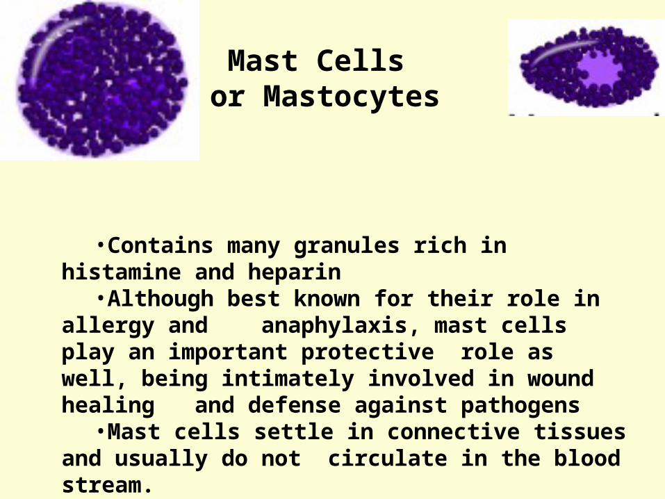
Figure 1-4 part 1 of 3
•Contains many granules rich in histamine and heparin•Although best known for their role in allergy and
anaphylaxis, mast cells play an important protective role as well, being intimately involved in wound healing and defense against pathogens
•Mast cells settle in connective tissues and usually do not circulate in the blood stream.
Mast Cells or Mastocytes
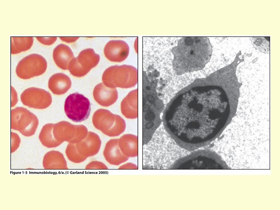
Figure 1-5
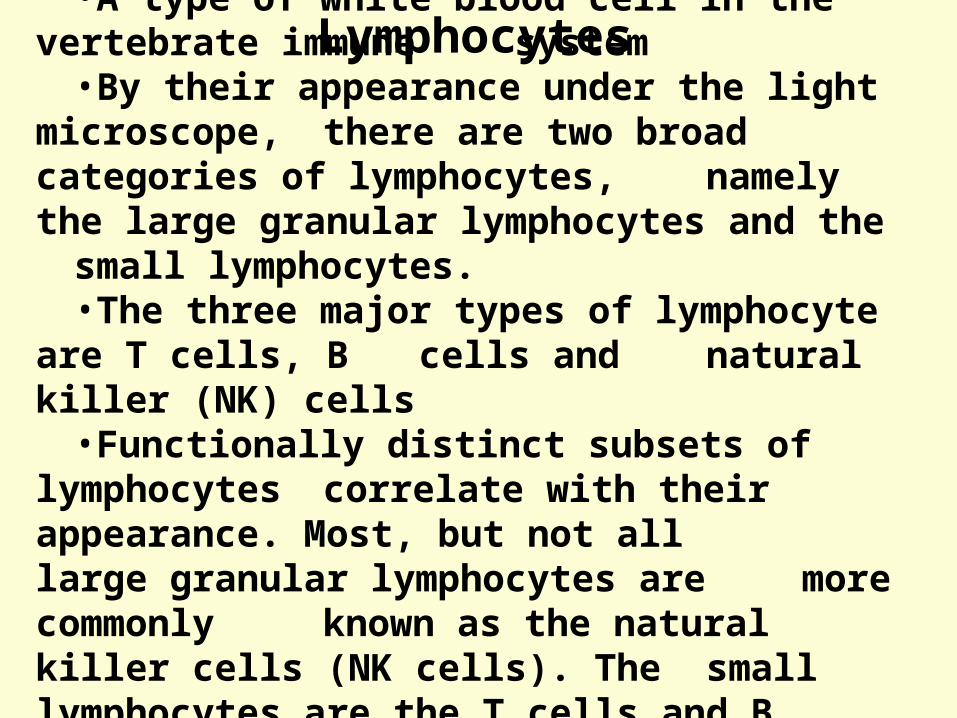
Figure 1-4 part 1 of 3•A type of white blood cell in the vertebrate immune system
•By their appearance under the light microscope, there are two broad categories of lymphocytes, namely the large granular lymphocytes and the small lymphocytes.
•The three major types of lymphocyte are T cells, B cells and natural killer (NK) cells•Functionally distinct subsets of lymphocytes
correlate with their appearance. Most, but not all large granular lymphocytes are more commonly known as the natural killer cells (NK cells). The small lymphocytes are the T cells and B cells
Lymphocytes
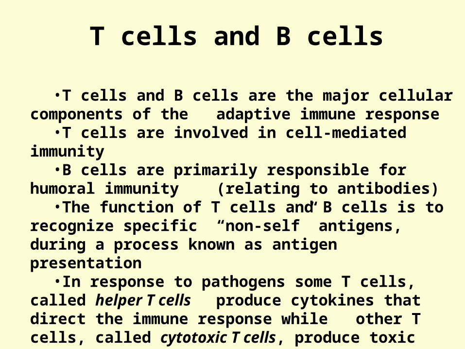
Figure 1-4 part 1 of 3•T cells and B cells are the major cellular components of the adaptive immune response •T cells are involved in cell-mediated immunity•B cells are primarily responsible for humoral immunity
(relating to antibodies)•The function of T cells and B cells is to recognize specific
“non-self” antigens, during a process known as antigen presentation
•In response to pathogens some T cells, called helper T cells produce cytokines that direct the immune response while other T cells, called cytotoxic T cells, produce toxic granules that induce the death of pathogen infected cells.
T cells and B cells
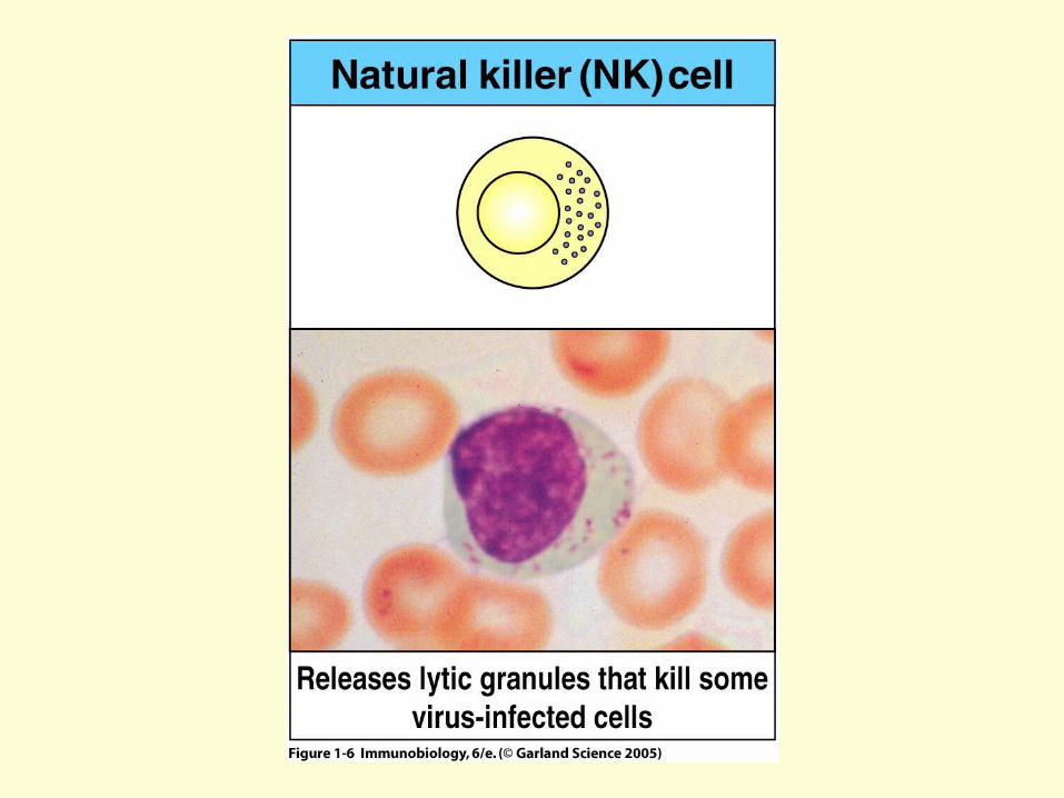
Figure 1-6
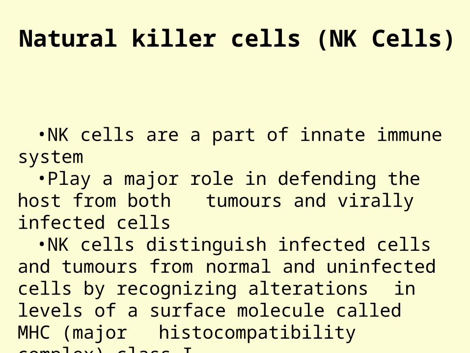
Figure 1-4 part 1 of 3
•NK cells are a part of innate immune system•Play a major role in defending the host from both
tumours and virally infected cells•NK cells distinguish infected cells and tumours from
normal and uninfected cells by recognizing alterations in levels of a surface molecule called MHC (major histocompatibility complex) class I
•NK cells kill infected cell similar to cytotoxic cells manner
Natural killer cells (NK Cells)