Embed Size (px)
Citation preview

Molecular Biology (Lecture Notebook)Cheju National University / Moon Eun Sung 1 / 12
2007년 3월 14일(수) / 3월 19일(월) 강의 내용
1. Ribose와 Deoxyribose
O
OHOH
HH
H
CH2
H
HOOH
O
HOH
HH
H
CH2
H
HOOH
1
23
4
5
1'
2'3'
4'
5'
RNA DNA
1) ribose와 deoxyribose는 모두 5탄당이다.
→ but, ribose의 2번 탄소에는 - OH group이 존재
deoxyribose 는 2번 탄소에 O(oxygen atom)가 한 개 빠진다. 즉, -H group만 존재
Lehninger PRINCIPLES OF BIOCHEMISTRY Fourth Edition(pp.277)
<Figure 8-7> Phosphodiester linkages in the
covalent backbone of DNA and RNA.
The phosphodiester bonds (one of which is
shaded in the DNA) link successive nucleotide
units. The backbone of alternating pentose and
phosphate groups in both types of nucleic acid
is highly polar. The 5' end of the macro
molecule lacks a nucleotide at the 5' position,
and the 3' end lacks a nucleotide at the 3'
position.
The successive nucleotides of both DNA and RNA are covalently linked through phosphate-group
"bridges," in which the 5'-phosphate group of one nucleotide unit is joined to the 3'-hydroxyl
group of the next nucleotide, creating a phosphodiester linkage. Thus the covalent backbones of
nucleic acids consist of alternating phosphate and pentose residues, and the nitrogenous bases
may be regarded as side groups joined to the backbone at regular intervals. The backbones of
both DNA and RNA are hydrophilic. The hydroxyl groups of the sugar residues form hydrogen
bonds with water. The phosphate groups, with a pKa near 0, are completely ionized and
negatively charged at pH 7, and the negative charges are generally neutralized by ionic
interactions with positive charges on proteins, metal ions, and polyamines.

Molecular Biology (Lecture Notebook)Cheju National University / Moon Eun Sung 2 / 12
All the phosphodiester linkages have the same orientation along the chain, giving each linear
nucleic acids strand a specific polarity and distinct 5' and 3' ends. By definition, the 5' end
lacks a nucleotide at the 5' position and the 3' end lacks a nucleotide at the 3' position. Other
groups (most often one or more phosphates) may be present on one or both ends.
The covalent backbone of DNA and RNA is subject to slow, nonenzymatic hydrolysis of the
phosphodiester bonds. In the test tube, RNA is hydrolyzed rapidly under alkaline conditions, but
DNA is not; the 2'-hydroxyl groups in RNA (absent in DNA) are directly involved in the process.
Cyclic 2',3'-monophosphate nucleotides are the first products of the action of alkali on RNA and
are rapidly hydrolyzed further to yield a mixture of 2'- and 3'-nucleoside monophosphates
2) ribose는 탄소에 1,2,3,4,5로 번호를 부여하나, deoxyribose는 1',2',3',4',5'로 번호를 부여한다.
3) base와 ribose의 결합
Lehninger PRINCIPLES OF BIOCHEMISTRY Fourth Edition(pp.274)
<Figure 8-1> Structure of nucleotides
(a) General structure showing the numbering
convention for the pentose ring. This is
a ribonucleotide. In deoxyribonucleotides
the - OH group on the 2' carbon (in red)
replaced with -H.
(b) The parent compounds of the pyrimidine
and purine bases of nucleotides and
nucleic acids, showing the numbering
conventions.
Basic concepts in Biochemistry(2/e) pp.36
The two complementary strands of the DNA double helix run in antiparallel directions. The
phosphodiester connection between individual deoxynucleotides is directional. It connects the 5'
hydroxyl group of one nucleotide with the 3' hydroxyl group of the next nucleotide. Think of it as
an arrow. If the top strand sequence is written with the 5' end on the left (this is the

Molecular Biology (Lecture Notebook)Cheju National University / Moon Eun Sung 3 / 12
conventional way), the bottom strand will have a complementary sequence, and the phosphate
backbone will run in the opposite direction; the 3'end will be on the left. The antiparallel
directionality of DNA is an important concept (i.e., it always appears on exams). Either of the
two strands could be written on top (just rotate the paper by 180°), but if the DNA codes for a
protein, the top strand is usually arranged so that it matches the sequence of the RNA that
would be made from the DNA.
* 예를 들어서 생각해 보자
adenine(base) + ribose = adenosine(nucleoside)
adenine(base) + deoxyribose = adenylate(nucleoside)
이것에 대한 명칭을 아래에 자세히 나타내어 보았다.
Lehninger PRINCIPLES OF BIOCHEMISTRY Fourth Edition(pp.275)
<Note>
"Nucleoside" and "nucleotide" are genetic terms that include both ribo- and deoxyribo-
forms. Also, ribonucleosides and ribonucleotides are here designated simply as nucleosides and
nucleotides (e.g., riboadenine as adenosine), and deoxyribonucleosides and deoxyribonucleotides
as deoxynucleosides and deoxynucleotides (e.g., deoxyriboadenine as deoxyadenosine). Both
forms of naming are aceptable, but the shortened names are more commonly used. Thymine is
an exception; "ribothymidine" is used to describe its unusual occurrence in RNA.
* 이제 본격적으로 nucleotide의 명칭에 대해서 생각을 해보도록 하자.
예를 들어서, adenosine에 결합하는 인산의 개수에 따른 명칭은 다음과 같다.
- 인산 1개 (Mono) : Adenosine 5-monophosphate (AMP)
- 인산 2개 (Di) : Adenosine 5-diphosphate (ADP)
- 인산 3개 (Tri) : Adenosine 5-triphosphate (ATP)
예를 들어서, deoxyadenosine에 결합하는 인산의 개수에 따른 명칭은 다음과 같다.
- 인산 1개 (Mono) : deoxyadenosine 5'-monophosphate (dAMP)
- 인산 2개 (Di) : deoxyadenosine 5'-diphosphate (dADP)
- 인산 3개 (Tri) : deoxyadenosine 5'-triphosphate (dATP)

Molecular Biology (Lecture Notebook)Cheju National University / Moon Eun Sung 4 / 12
실제 구조에 대해서는 아래에 나타내 보았다.
Lehninger PRINCIPLES OF BIOCHEMISTRY Fourth Edition(pp.275)
<Figure 8-4> Deoxyribonucleotides and ribonucleotides of nucleic acids.
All nucleotides are shown in their free form at pH 7.0. The nucleotide units of DNA (a) are usually
symbolized as A, G, T, and C, sometimes as dA, dG, dT, and dC; those of RNA (b) as A, G, U, and C.
In their free form the deoxyribonucleotides are commonly aabreviated dAMP, dGMP, dTMP, and
dCMP; the ribonucleotides, AMP, GMP, UMP, and CMP. For each nucleotide, the more common
name is followed by the complete mane in parentheses. All abbreviations assume that the phosphate
group is at the 5' position. The nucleoside portion of each molecule is shaded in light red. In this
and the following illustrations, the ring carbons are not shown.
* ATP는 α-β 사이와 β-γ 사이의 결합으로
에너지를 보관하고 있다가 필요시 인산기의
결합을 한 개 끊어 ADP로 전환하며 에너지를
방출한다.

Molecular Biology (Lecture Notebook)Cheju National University / Moon Eun Sung 5 / 12
Molecular Cell biology(Lodish, pp.109)
<Figure 4-9> Polymerization of ribonucleotides by RNA
polymerase during transcription
The ribonucleotide to be added at the 3' end of a
growing RNA strand is specified by base pairing between
the next base in the template DNA strand and the
complementary incoming ribonucleoside triphosphate(rNTP).
A phosphodiester bond is formed when RNA polymerase
catalyzed a reaction between the 3' O of the growing
strand and the a phosphate of a correctly base-paired
rNTP. RNA strands always are synthesized in the 5'→3'
direction and are opposite in polarity to their template
DNA strands.
A template DNA strand is transcribed into a complementary RNA chain by RNA polymerase.
During transcription of DNA, one DNA strand acts as template, determining the other in which
ribonucleoside triphosphate(rNTP) monomers are polymerized to form a complementary RNA
chain. Bases in the template DNA strand base-pair with complementary incoming rNTPs, which
then are joined in a polymerization reaction catalyzed by RNA polymerase. Polymerization involves
a nucleophilic attack by the 3' oxygen in the growing RNA chain on the α phosphate of the next
nucleotide precursor to be added, resulting in formation of a phosphodiester bond and release of
pyrophosphate(PPi). As a consequence of this mechanism, RNA molecules are always synthesized
the 5'→3' direction.
The energetics of the polymerization reaction strongly favors addition of ribonucleotides to the
growing RNA chain because the high-energy bond between the α and β phosphate of rNTP
monomers is replaced by the lower-energy phosphodiester bond between nucleotides. The
equilibrium for the reaction is driven further toward chain elongation by pyrophosphatase, an
enzyme that catalyzes cleavage of the released PPi into two molecules of inorganic phosphate.
Like the two strands in DNA, the template strand and the growing RNA strand that is base-paired
to it have opposite 5'→3' directionality.

Molecular Biology (Lecture Notebook)Cheju National University / Moon Eun Sung 6 / 12

Molecular Biology (Lecture Notebook)Cheju National University / Moon Eun Sung 7 / 12

Molecular Biology (Lecture Notebook)Cheju National University / Moon Eun Sung 8 / 12
2. DNA와 RNA의 구조
1) DNA 구조
- 5‘에 인산기가 있고, 3'에 OH기가 있음
- DNA의 염기는 4개로 구성된다.
* Adenine(A), Guanine(G), Cytosine(C), Thymine(T)
- DNA는 Chargaff‘s rule에 따라서 C는 G와, A는 T와 그 개수가 동일하다. 아울러, Watson과
Crick의 base-pairing rule에 따라 C는 G와 3개의 수소결합을 이루고, A와 T는 2개의 수소결합을
이룬다. 따라서, C≡G pair는 A=T pair 보다 결합수가 많기에 결합이 세며 C≡G pair가 많아질수록
DNA가 변성되는 온도가 높아진다.
Chargaff's rule
1. The base composition of DNA generally varies from one species to another.
2. DNA specimens isolated from different tissues of the same species have the same
base composition.
3. The base composition of DNA in a given species does not change with an organism's
age, nutritional state, or changing environment.
4. In all cellular DNAs, regardless of the species, the umber of adenosine residues is
equal to the number of thymidine residues (that is, A=T), and the number of
guanosine residues is equal to the number of cytidine residues (G=C). From these
relationships it follows that the sum of the purine residues equals the sum of the
pyrimidine residues; that is A+G = T+C
- 역평형 구조와 함께 상보적 염기가 쌍을 이루고 있다. 생물체의 염기 회수율 비교를 통해 A와 T,
G와 C는 같은 비율로 존재함을 알게 되었고, 이는 상보적 관계에 있는 염기는 서로 같은 수로
있다는 것을 알 수 있다.
- Watson과 Crick의 DNA 구조 규명시에는 DNA 복제에 있어 5'→3' 또는 3'→5'으로 복제가 가능
하다고 생각했으나, 이후의 연구 결과에 따르면 DNA polymerase는 5'→3'으로만 복제가 가능한
복제의 방향성을 가지고 있다. 이것으로 DNA 절편(okazaki fragment)이 만들어진다.
Okazaki fragment. 이중가닥 DNA가 반보존적으로 복제될 때 복제점 가까운 곳에서 부모 DNA
사슬과 상보적으로 새롭게 합성되는 짧은 DNA 단편. DNA 중합효소는 디옥시리보뉴클레오티드를
5‘→3’ 방향으로만 중합하는 것으로 알려져 있었는데, 반보존적 복제(semiconservative
replication)에서는 5'→3' 방향으로의 중합과 동시에 3‘→5’ 방향으로의 중합이 불가피하다는
모순이 오카자기 단편의 검출과 불연속적모델의 의해서 설명되었다.
- DNA는 유동적인 구조여서, A-form, B-form, Z-form을 가지고 있고, DNA 꼬이는 것이 틀리니
한번 꼬이는데 들어가는 염기수와 염기쌍의 기울기도 바뀐다. 일반적으로 B-form의 경우에는 한번
꼬이는데 10개의 염기가 들어간다.

Molecular Biology (Lecture Notebook)Cheju National University / Moon Eun Sung 9 / 12
2) RNA의 2차 구조
Basic concepts in Biochemistry(2/e) pp.4
<Figure 4-3> RNA Secondary Structure
A single molecule of RNA often contains segments of sequence that are complementary to
each other. These complementary sequence can base-pair and form helical regions of
secondary structure. Interactions between the secondary structures give RNA a significant
folded, three-dimensional structure.
RNA is often depicted as a single-stranded molecule. However, in many RNAꡑs, internal
complementarity may result in secondary (and tertiary) structure in which one part of the RNA
molecule forms a double-stranded region with another part of the same molecule. There are
usually a number of mismatches in these structures. Names have been given to some of these
structural features.

Molecular Biology (Lecture Notebook)Cheju National University / Moon Eun Sung 10 / 12
3) DNA 변성(denaturation) 온도 ; Tm(melting temperature)
- DNA는 온도가 높아짐에 따라 어느 순간 갑자기 변성된다.
- Tm에 영향을 미치는 요인
: A와 T 보다는 G와 C의 결합력이 강하므로, G와 C의 수가 많을수록 변성 온도는 높아진다.
- G와 C의 함량은 각 생물종마다 틀리며, G와 C의 함량이 높을수록 고온에서도 생존이 가능하다.
- DNA의 정량은 260nm(자외선 영역)에서 측정 가능하다.
Tm으로 나타내는 가열된 DNA 이중가닥 사슬 사이의 수소결합이 끊어져 단일가닥 상태로
변화할 때의 (DNA의 변성)온도.
4) DNA의 혼성화(hybridization)
: 열에 의해 분리된 DNA의 단일 가닥이 식히면 원래 자신과 상보적인 서열의 DNA를 만나서 이중
나선을 만듬.
→ 응용분야
- 신원확인 시 원래 DNA와 신원 불명의 DNA를 변성시킨 후, 가닥 한 개씩만 합치도록 하여
일치하는 정도를 보고 신원확인.
- 친자 확인시 자신의 DNA는 각각 부와 모로부터 반반씩 물려받았으므로, 자식과 부모의
DNA를 혼성화시키면 일치하는 정도를 보고 신원을 확인 할 수 있다.
→ 혼성화(hybridization)
: 분리된 DNA 가닥들이 서로 상보적인 자신의 짝을 찾아서 잡종화 되는 것. mRNA는 인트론이
없기 때문에 DNA의 엑손 부분만 가지고 RNA와 혼성시키는 것도 가능하다.
잡종화(hybridization of nucleic acid)
DNA나 RNA에 상보적인 염기배열을 이용하여 인공적으로 이중가닥의 잡종핵산분자를 형성시
키는 기법. 종류에는 서던법, 노던법, 플라크하이브리드법, 콜로니하이브리드법, 현장하이브리
디제이션 등이 있으며, 핵산분자의 검출, 분리 등에 널리 사용되고 있다.
DNA-DNA hybridization
열에 의해 단일가닥 상태가 된 이중가닥 DNA를 적당한 염농도와 온도를 조절하며, 염기간의
수소결합 의해서 원래의 이중가닥 구조로 되돌리는 방법
DNA-RNA hybridization
상보적 염기순서를 갖는 단일가닥 DNA와 단일가닥 RNA를 혼합하여 적당한 염농도와 온도조
건을 부여하면 염기간에 수소결합을 형성하여 DNA-RNA 사이에 마치 이중가닥 DNA처럼 잡종
이중가닥 구조가 형성되는 것.
→ 복원(renaturation)
: 변성된 DNA나 RNA가 원래 상태로 돌아오는 것.
변성된 단백질 또는 핵산이 원래의 형태를 되찾게 되는 것.
5) DNA의 복제
- 대장균의 DNA 복제
: 대장균은 circular DNA를 가지고 있고, 그 길이가 짧다. 그래서 복제 개시점(ori, origin of
replication)이 한 개이다. 복제 개시점과 종결점이 만나기 때문에 복제 전․후 길이의 변함이
없다.

Molecular Biology (Lecture Notebook)Cheju National University / Moon Eun Sung 11 / 12
- 사람의 DNA 복제
: 사람은 linear DNA를 가지고 있고, 그 길이가 길다. 그래서 복제 개시점이 여러 군데가 있다.
복제 개시점과 종결점이 만나지 않으며, 복제할 때마다 DNA의 끝과 끝이 조금씩 짧아진다.
고리모양(circular DNA)
일반적으로 폴리뉴클레오티드사슬의 5‘ 말단과 3’말단이 공유결합에 의해서 연결된 DNA 분자.
이중가닥 DNA의 경우 양사슬 모두 공유결합에 의해 연결된 것을 폐쇄고리모양 DNA(closed
circular DNA)라고 한다. 폐쇄고리모양 DNA에 틈이 생기면 개방환상 DNA(open circular DNA)가
된다.
3. 유전자(Gene)의 정의와 열린 해독틀(Open Reading Frame, ORF)
1) 유전자(Gene)
: Gene이라는 것은 단백질을 만드는 DNA 부분 중에서 mRNA의 시작점(ATG)과 종결점(TAA,
TAG, TGA)을 포함하는 부분이다. 그리고 그 유전자가 발현되어서(단백질이 되어) 가지는 특징
및 특성에 따라서 이름이 지어진다. RNA의 경우는 예외가 있는데, mRNA는 단백질을 합성하는데
template로 작용하고, tRNA는 아미노산을 운반하는 역할을 담당하고, rRNA는 단백질을 합성하는
ribosome에 들어 있는데 이들은 자신들 스스로는 translation이 되지는 못하나 translation과정에
참여하여 유전자 발현에 관여하기 때문에 이들 또한 유전자라고 부른다.
2) 열린 해독틀(Opne Reading Frame, ORF)
: ORF는 개시코돈과 종결코돈이 있는 DNA상의 특정 영역으로써, mRNA의 시작점과 끝점이 만들어
지는 부위이다. mRNA의 시작점은 언제나 DNA의 ATG 부분에서 만들어지며, 끝점은 DNA의
TAG, TAA, TGA 부분에서 만들어진다.
- mRNA의 AUG 부분은 단백질에서 Methionine(Met)이 됨
- 만약 ORF에 돌연변이가 있을 경우 단백질에 많은 영향을 미치게 됨.
(e.g., TCA에서 C가 G로 바뀌면, 단백질이 합성되는 중간에 종결이 되게 된다)
- 참고적으로 mRNA는 개시점과 종결점에 연연하지 않고, 앞과 뒷부분에 조금 더 염기를 붙여서
만들어짐.

Molecular Biology (Lecture Notebook)Cheju National University / Moon Eun Sung 12 / 12
3. Additional reference (from Macmillan Science Library Biology)
1) nucleotide, Vol.3(143-145)
2) DNA, Vol.1(222-224)
3) RNA, RNA processing, Vol.4(75-78)
4) gene, Vol.2(117-124)

Viewed in the electron microscope, a nucleolus has two distinct parts:the fibrillar component and the granular component. The fibrillar compo-nent can be subdivided into two compartments: the dense fibrillar compo-nent and the fibrillar center. Fibrillar centers contain large amounts of RNApolymerase I, which transcribes rRNA. Transcription of rRNA genes isthought to occur at the interface between the dense fibrillar component andthe fibrillar center. Later stages of ribosome assembly take place in the gran-ular component.
Human chromosomes contain five nucleolar organizer regions(called NORs), located on the short arms of the chromosomes 13, 14, 15,21, and 22. In humans, each NOR contains approximately one hundredtandemly repeated rRNA gene copies. The NORs of different chromo-somes typically come together in interphase. Thus, a single nucleolus isoften made up of rRNA genes from two or more different NORs. Somespecies have only a single NOR-bearing chromosome and thus a singlenucleolus.
In addition to the well-established function of nucleoli in ribosome as-sembly, recent evidence suggests that nucleoli are also involved in severalother cellular processes, including assembly and modification of varioussmall ribonucleoproteins (RNPs), sequestration of important cell-cycleregulatory proteins, export of other nonribosomal RNAs, and control of cel-lular senescence or aging. SEE ALSO Chromosome, Eukaryotic; NuclearTransport; Nucleus; Ribosome; RNA; Transcription
A. Gregory Matera
Bibliography
Olson, M.O., M. Dundr, and A. Szebeni. “The Nucleolus: An Old Factory with Un-expected Capabilities.” Trends in Cell Biology (2000) 10: 189-196.
Visintin, R., and A. Amon. “The Nucleolus: The Magician’s Hat for Cell CycleTricks.” Current Opinions in Cell Biology (2000) 12: 372-377.
NucleotidesNucleotides are the subunits that are linked to form the nucleic acids ri-bonucleic acid (RNA) and deoxyribonucleic acid (DNA), which serve as thecell’s storehouse of genetic information. Free nucleotides play importantroles in cell signaling and metabolism, serving as convenient and universalcarriers of metabolic energy and high-energy electrons.
All nucleotides are composed of three parts: a five-carbon sugar, a phos-phate, and a nitrogen-rich structure called a nitrogenous base. The sugarcan be ribose, which is found in ribonucleotides and RNA, or deoxyribose,which is found in deoxyribonucleotides and DNA. The only difference be-tween these two sugars is that deoxyribose has one fewer oxygen atom thanribose. The five carbon atoms in the sugar are numbered sequentially. Todistinguish these carbon atoms from those of the nitrogenous base, whichare also numbered, they are designated as 1� (prime), 2�, and so on.
There are five nitrogenous bases. The so-called pyrimidines (cytosine,thymine, and uracil) are smaller, having only one ring structure. The largerpurines (adenine and guanine) have two rings. Adenine, guanine, and cyto-sine are found in both ribonucleotides and deoxyribonucleotides, while
Nucleotides
143
chromosome “coloredbody” in the cellnucleus; made of DNAand protein, and dividedfunctionally into genesand nongene regions
metabolism chemicalreactions within a cell
ribonucleoprotein com-bination of RNA and pro-tein

thymine occurs only in deoxyribonucleotides and uracil only in ribonu-cleotides.
The phosphate group is bonded to the 5� carbon of the sugar (seeFigure 2), and when nucleotides are joined to form RNA or DNA, thephosphate of one nucleotide is joined to the sugar of the next nucleotideat its 3� carbon, to form the sugar-phosphate backbone of the nucleicacid. In a free nucleotide, there may be one, two, or three phosphategroups attached to the sugar, as a chain of phosphates attached to the 5�
carbon.
Three nucleotides merit special consideration because of their special-ized roles in cellular function. These are adenosine triphosphate (ATP),flavin adenine dinucleotide (FAD), and nicotinamide adenine dinucleotide(NAD+). Most biosynthetic reactions require energy, which is usually sup-plied by ATP. When ATP is hydrolyzed to ADP (adenosine diphosphate)or AMP (adenosine monophosphate), energy is released. By coupling thisenergy release to a reaction requiring energy, that reaction can be made tooccur. Since ATP is so frequently used this way, it is commonly called the“energy currency of the cell.”
Adenine-containing molecules are also important coenzymes, serving tocarry chemical functional groups that are needed for enzyme activity. Threeimportant adenosine-containing coenzymes are coenzyme A (CoA), FAD,and NAD+. CoA carries acetyl groups into the Krebs cycle (the central meta-bolic pathway in mitochondria), and FAD and NAD+ carry high-energyelectrons from the Krebs cycle to the electron transport system, wheretheir energy is used to synthesize ATP from ADP and inorganic phosphate.
Another adenine-based molecule is important in cellular signaling. Whena hormone binds at a cell-surface receptor, it often promotes the produc-tion of cyclic AMP (cAMP) inside the cell. In cAMP, the phosphate groupis joined to the 3� and 5� carbons of the ribose, forming a small ring struc-
Nucleotides
144
O
C
HC NH
HC
NH
C
O
U
uracil
BASES
NH2
C
HC N
HC
NH
CC
cytosine
O
O
C
C NH
HC
NH
CT
thymine
O
H3C
NH2
C
C N
C
N
CH
adenine
N
N H
HC A
O
C
C NH
C
N
C
guanine
N
N H
HC G
N
N
4
3
21
6
5 N
N
6
1
23
4
5
N
N7
8
9
PYRIMIDINE PURINE
The bases are nitrogen-containing ring compounds, either purines or pyrimidines.
NH
The molecular structuresof the five nitrogenousbases.
biosynthetic forming acomplex molecule fromsimpler ones
hydrolyze to split apartusing water
AMP adenosinemonophosphate, form ofATP after removal of twophosphate groups
enzyme protein thatcontrols a reaction in acell
mitochondria subcellu-lar organelle that cre-ates ATP used forenergy-requiringprocesses in a cell
electron transportsystem membrane-bound system of pro-teins that extractsenergy from high-energyelectrons, found in mito-chondria and chloro-plasts
inorganic not bondedto carbon
hormone moleculereleased by one cell toinfluence another

ture. cAMP can activate or suppress various cell processes, thereby servingas an intracellular signal and messenger that responds to hormone binding.SEE ALSO DNA; Metabolism, Cellular; RNA; Vitamins and Coenzymes
David W. Tapley
Bibliography
Nelson, David L., and Michael M. Cox. “Nucleotides and Nucleic Acids.” In LehningerPrinciples of Biochemistry, 3rd ed. New York: Worth Publishers, 2000.
NucleusIn eukaryotic cells, chromosomes are found in a special compartment calledthe nucleus. The nucleus is a defining feature of eukaryotic cells, whichrange from single-celled yeasts to plants and humans. In contrast, bacteriaand other prokaryotes are more ancient in evolution and lack a nucleus. Thedevelopment of the nucleus contributed to the evolution of complex lifeforms by separating transcription (reading of genes, occurring inside thenucleus) from translation (protein synthesis, occurring in the cytoplasm)and by providing a structural framework for organizing and regulating largergenomes. In multicellular organisms, individual cells can express differentsubsets of genes and thereby form specialized tissues such as muscle or skin.
The Nuclear EnvelopeThe nuclear envelope surrounds the nucleus and creates and maintains aspecial environment inside it. The envelope consists of two nuclear mem-
Nucleus
145
chromosome “coloredbody” in the cellnucleus; made of DNAand protein, and dividedfunctionally into genesand nongene regions
transcription messen-ger RNA formation froma DNA sequence
gene portion of DNAthat codes for a proteinor RNA molecule
translation synthesis ofprotein using mRNAcode
cytoplasm material in acell, excluding thenucleus
genome total geneticmaterial in a cell ororganism
N
N O
NH2
O
O
BASE
SUGAR
PHOSPHATE
OH OH
NUCLEOTIDES
–O P O
O–
CH2
A nucleotide consists ofa nitrogen-containingbase, a 5-carbon sugar,and one or morephosphate groups. Thesugar dipicted is ribose.Deoxyribose has an Hinstead of an OH in theboxed position.
intracellular within acell
NUCLEOTIDES

Homeostasis; Neurologic Diseases; Parasitic Diseases; Psychiatric Dis-orders, Biology of; Sexually Transmitted Diseases; Viral Diseases
Roberta M. Meehan
Bibliography
Madigan, Michael T., John M. Martinko, and Jack Parker. Brock Biology of Micro-organisms, 9th ed. Upper Saddle River, NJ: Prentice Hall, 2000.
Thomas, Clayton L., ed. Taber’s Cyclopedic Medical Dictionary, 18th ed., Philadelphia,PA: F. A. Davis Company, 1997.
DNADNA (deoxyribonucleic acid) is the molecule that stores genetic informa-tion in living systems. Like other organic molecules, DNA mostly consistsof carbon, along with hydrogen, oxygen, nitrogen, and phosphorus. Thefundamental structural unit of DNA is the nucleotide, which has two parts:an unvarying portion composed of sugar and phosphate, attached to one offour nitrogen-containing bases named adenine, cytosine, guanine, orthymine (abbreviated A, C, G, T).
The Double HelixThe structure of DNA, deduced in 1953 by James Watson, Francis Crick,and Rosalind Franklin, resembles that of a twisted ladder or spinal staircasecomposed of two long chains of nucleotides that are coiled around each otherto form a double helix. The DNA ladder’s two sidepieces (its double-strandedbackbone) are made of alternating units of sugar and phosphate. The sugaris deoxyribose, which contains a ring of four carbons and one oxygen. A phos-phate is an atom of phosphorus bonded to four oxygens. Bases attached toopposing sugars project inward toward each other to form rungs or steps,called base pairs. In contrast to the strong covalent (electron-sharing) bondsbetween nucleotides in a strand, the two bases in a base pair are held to-gether only by much weaker hydrogen bonds. However, the cumulative at-tractive force of the hydrogen bonds in a chain of base pairs maintains DNAas a double-stranded molecule under physiological conditions. In the cell nu-cleus, DNA is bound to proteins to form chromosomes, and is coated witha layer of water molecules.
To make a sturdy rung, the two bases in a base pair have to interlocklike pieces of a jigsaw puzzle, which only happens if their shapes and hy-drogen-bonding characteristics are compatible. Only two combinations ful-fill these requirements in DNA: G–C and A–T. This rule makes the twostrands of a DNA molecule complementary, so that if the bases of onestrand are ordered GGTACAT, the bases of the opposite strand must beordered CCATGTA. The order of the bases on a strand (mirrored in thecomplementary strand) is called the sequence of the DNA, and embodiescoded instructions for making new biomolecules: proteins, ribonucleic acid(RNA), and DNA itself.
Complementarity and ReplicationEach strand of DNA has a direction in which it can be read by the cellularmachinery, arising from the arrangement of phosphates and sugars in the
DNA
222
organic composed ofcarbon, or derived fromliving organisms
nucleotide the buildingblock of RNA or DNA
base pair twonucleotides (either DNAor RNA) linked by weakbonds
hydrogen bond weakbond between the H ofone molecule or groupand a nitrogen oroxygen of another
nucleus membrane-bound portion of cellcontaining the chromo-somes
protein complex mol-ecule made from aminoacids; used in cells forstructure, signaling, andcontrolling reactions
chromosome “coloredbody” in the cellnucleus; made of DNAand protein, and dividedfunctionally into genesand non-gene regions
complementary match-ing opposite

backbone. The two strands of DNA are oriented antiparallel to each other,that is, they lie parallel to each other but are decoded in opposite directions.Because of the numbering convention for the combinations in sugar, the di-rections along the backbone are called 5 * * * 3 (“five-prime to three-prime”) or 3 * 5. The complementary nature of the two strands meansthat instructions for making new DNA can be read from both strands.
When DNA replicates, the weak hydrogen bonds of base pairs are bro-ken and the two strands separate. Each strand acts as a template for thesynthesis of a new complementary strand. Since the resulting new double-stranded molecule always contains one “old” (template) strand and one newlymade strand, DNA replication is said to be semiconservative; it would betermed conservative if the two original template strands rejoined. By a sim-ilar mechanism (transcription), a DNA strand can be a template for the syn-thesis of RNA, which is a single-stranded nucleic acid that carries codedinformation from the DNA to the protein synthesizing machinery of thecell. During protein synthesis, the genetic code is used to translate the or-der of bases originally found in the DNA sequence into the order of aminoacid building blocks in a protein.
Genes, Noncoding Sequences, and MethylationDNA exists in nature as a macromolecule millions of base pairs long. Inmulticelled organisms, the complete set of genetic information—thegenome—is divided among several DNA macromolecules (called chro-mosomes) in the cell nucleus. In contrast, the genomes of many one-celledorganisms consist of a single, often circular, chromosome. The humangenome contains 3.2 billion base pairs distributed among twenty-three chro-mosomes. Laid end to end, these would make a macromolecule 1.7 meters(5.5 feet) long; printed out, they would fill one thousand one-thousand-pagetelephone books. Furthermore, two copies of the genome are in almost everycell of humans and other diploid organisms. This vast amount of DNApacks into a cell nucleus, whose volume is only a few millionths of a cubicmeter, by first spooling around globular proteins called histones. TheDNA/histone complex then coils and curls up into even denser configura-
DNA
223
A helix-loop-helix dimerbound to DNA.
template master copy
genetic code relation-ship between triples ofRNA nucleotides andthe amino acids theycode for during proteinsynthesis
amino acid a buildingblock of protein
genome total geneticmaterial in a cell ororganism
macromolecules largemolecules such as pro-teins, carbohydrates,and nucleic acids
diploid having pairs ofchromosomes in thenucleus
histone protein aroundwhich DNA wraps toform chromosomes
A defect in the gene for amethylating enzyme causes Rettsyndrome, a disorder responsi-ble for mental retardation andmovement disorders in younggirls.

tions, like a rubber band does when one holds one end and rolls the otherend between one’s fingers. Yet the human genome isn’t nearly nature’sbiggest: the genome of a lily is just over ten times larger than a human’s,although its nuclei are not significantly larger.
The information storage capacity of DNA is vast; a microgram (one-millionth of a gram) of DNA theoretically could store as much informationas 1 million compact discs. The “useful” information contained in genomesconsists of the coded instructions for making proteins and RNA. These information-containing regions of a genome are called genes. However,genes comprise less than 5 percent of the human genome. Most genomesconsist largely of repetitive, noncoding DNA (sometimes called junk DNA)that is interspersed with genes and whose only apparent function is to repli-cate itself. Perhaps it helps to hold the chromosome together. The tenfoldgreater size of the lily genome compared to humans’ is due to the presenceof enormous amounts of repetitive DNA of unknown function.
While most cells of higher organisms contain all the genes in thegenome, specialized cells such as neurons or muscle require expression fromonly some of the genes. One strategy for silencing unneeded genes is methy-lation. A methyl group (–CH3) is added to cytosine nucleotides, but only ifthey are followed by a guanine in the sequence, that is, CG. Adding methylgroups to a region of DNA attracts repressive DNA-binding proteins to itand may also cause the region to compact even further, making it inacces-sible to proteins that make RNA from DNA (the first step of protein syn-thesis). During DNA replication the pattern of methylation is preserved byspecific proteins that add methyl groups to the new strand based on the lo-cation of CG methyl groups in the template strand. The most extreme caseof repression by methylation is X-inactivation, in which one of the two Xchromosomes in cells of a female mammal is entirely shut down, presum-ably because expression from one X provides enough protein in females, asit does in males (who have only one X chromosome). SEE ALSO Chromo-some, Eukaryotic; Control of Gene Expression; Crick, Francis; Gene;Mutation; Nucleotides; Replication; RNA; Watson, James
Steven A. Sullivan
Bibliography
Alberts, Bruce, et al. Molecular Biology of the Cell. New York: Garland Publishing,2000.
Felsenfeld, Gary. “DNA.” Scientific American 253 (1985): 58–67.
Levin, Benjamin. Genes VII. New York: Oxford University Press, 1999.
Watson, James D., and Francis H. Crick. “A Structure for Deoxyribose Nucleic Acid.”Nature 171 (1953): 737.
DNA SequencingThe genome of an organism is the sum total of its genetic information. Thegenome is not only a blueprint for the organism it also contains historicalnotes on the evolution of the organism. The ability to determine the se-quence of deoxyribonucleic acid (DNA) and thus read the messages in thegenome is of immense biological importance because it not only describesthe organism in detail but also indicates its evolutionary history.
DNA Sequencing
224
A human DNA fingerprinttaken from blood tomatch donor and patientin an organtransplantation.
WILKINS, MAURICE(1916– )
New Zealand–born British biolo-gist who helped James Watsonand Francis Crick deduce thestructure of deoxyribonucleicacid (DNA), for which the threemen received a 1962 NobelPrize. Wilkins secretly showedWatson an x-ray diffractionphoto of DNA taken byresearcher Rosalind Franklin.Watson and Crick later usedFranklin’s extensive unpublisheddata to build a model of DNA.
neuron nerve cell
methylation addition ofthe methyl group CH3
genome total geneticmaterial in a cell ororganism

ulation of these invertebrates is relatively small, however, so there are fewpredators in headwater streams; there is not enough for them to eat.
Rivers, being wider, have more surface exposed to sunlight, so their pri-mary productivity (photosynthesis) is greater. This is aided by inorganicnutrients such as nitrogen and phosphorus flowing down from the smaller-order streams. Fourth- to sixth-order rivers provide ideal conditions for al-gae and rooted aquatic plants because of their softer substrates and amplelight. Shredders become less abundant, grazers increase, and the relativepopulations of collectors and predators remain about the same. Species di-versity increases in these mid-order rivers, with fish and burrowing animalssuch as clams and worms becoming more common. High-altitude, cold, oxy-gen-rich midsized rivers are an ideal haven for trout, which feed on the in-sect community. The organisms in midsized rivers, where there is morephotosynthesis, produce more organic matter than they consume, and theexcess nourishes the larger rivers downstream.
Large rivers (seventh to twelfth order) are relatively deep and wide. Theyare rich in organic matter but also contain a lot of inorganic sediment pro-duced by erosion and runoff into the upland waters. Thus, the water is moreturbid (muddy), and there is insufficient light to support as much photosyn-thesis as in smaller rivers. Collectors and predators dominate the consumercommunity, and consumption exceeds primary production. Fish species suchas sturgeon and catfish, which feed on sediments, are more common herethan predatory fish.
All lotic organisms must adapt to drift, the incessant flow of water to-ward the sea, carrying nutrients and the organisms themselves downstream.Drift is particularly significant when spring snowmelts and heavy summerrains increase the current. River valleys offer especially rich farmland be-cause of the great quantities of nutrients deposited by periodic flooding. Nu-trient loss by drift is compensated for by the continual addition of riparianorganic matter to the lower-order upland streams, while animals compen-sate for drift by their rheotaxis and other means. Many aquatic insects flyupstream to lay their eggs, and fish such as trout and salmon are well knownfor their upstream spawning runs. The immature animals drift downstreamas they grow and typically reach maturity at lower altitudes, only to repeatthe process and deposit their offspring back in the headwaters. SEE ALSO
Ecosystem; Lakes and Ponds; LimnologistKenneth S. Saladin
Bibliography
Cole, Gerald A. Textbook of Limnology, 4th ed. Prospect Heights, IL: Waveland Press,1994.
Giller, Paul S., and Bjorn Malmqvist. The Biology of Streams and Rivers. New York:Oxford University Press, 1999.
Wetzel, Robert G. Limnology: Lake and River Ecosystems, 3rd ed. Burlington, MA: Aca-demic Press, 2001.
RNARibonucleic acid (RNA), like deoxyribonucleic acid (DNA), is a polymermade up of nucleotides. A nucleotide is composed of a pentose (5-carbon)
RNA
75
inorganic not bondedto carbon
lotic of, relating to, orliving in actively movingwater
nucleotide the buildingblock of RNA or DNA

RNA
76
O
O
OH OH
–O P O
O–
CH2
NH2
C
C N
C
N
C
N
N
CHH
HH
Adenine
Structure of an adenine RNA nucleotide
Nitrogenousbase
Phosphate
Sugar
Schematic structure of an RNA nucleotide
H H
RNA chains arecomposed of simplerunits called nucleotides.Four different bases areused in RNA; adenine isshown.
phosphodiester the linkbetween two nucleotidesin DNA or RNA
transcription messen-ger RNA formation froma DNA sequence
gene portion of DNAthat codes for a proteinor RNA molecule
protein complex mole-cule made from aminoacids; used in cells forstructure, signaling, andcontrolling reactions
amino acid a buildingblock of protein
heterogeneous com-posed of or containingdifferent parts or types
ribosome protein-RNAcomplex in cells thatsynthesizes protein
polypeptide chain ofamino acids
catalyze aid in thereaction of
sugar, a nitrogen-containing base, and phosphate. The pentose sugar foundin RNA nucleotides is ribose, whereas that in DNA is 2� (2-prime) de-oxyribose. The bases commonly found in RNA nucleotides are adenine (A),guanine (G), cytosine (C), and uracil (U). Bases found in DNA are A, G, C,and thymine (T instead of U). As in DNA, the individual nucleotides in thepolymer are joined together by phosphodiester bonds. Unlike DNA, RNAis single-stranded; however, many RNA molecules fold into complex three-dimensional structures.
During transcription the DNA code is read and copied into RNA. Thesequence of nucleotides in an RNA is therefore determined by the sequenceof nucleotides in the gene from which it was transcribed. Following tran-scription, RNA may be processed before it becomes functional.
There are three main classes of RNA: messenger RNA (mRNA), trans-fer RNA (tRNA), and ribosomal RNA (rRNA). Each of the classes is im-portant in some aspect of protein synthesis. The nucleotide sequence of amessenger RNA specifies the order of amino acids in the protein which itencodes. A cell contains many different mRNA molecules, each being theblueprint for a different protein. Although mRNAs are the least abundantclass of RNA, they are the most heterogeneous. Ribosomes play an im-portant role in protein synthesis, and ribosomal RNA (rRNA), is an im-portant structural component of ribosomes. rRNA is the most abundant typeof RNA. tRNAs act as adaptors in protein synthesis, in that they read thesequence of nucleotides in the mRNA and deliver the correct amino acid tothe growing polypeptide chain.
Most scientists believe that life has evolved from what was essentiallyan “RNA world.” In today’s world, most organisms store their genetic in-formation in DNA and use proteins (encoded by DNA) to catalyze bio-logically important chemical reactions. RNA molecules, however, arebelieved to have been the first biological catalysts. Through evolution, someof these RNA molecules gained the ability to replicate themselves, andthrough many rounds of replication, the RNA molecules gained new capa-

bilities, such as the ability to code for and synthesize proteins. Eventually,the RNA genome was replaced with DNA.
Scientists have uncovered a number of enzymatic RNA molecules,called ribozymes, believed to be typical of those in the RNA world. RNAenzymes can make phosphodiester bonds, suggesting that early RNA mol-ecules could reproduce their genetic material. In fact, it is now known thatRNA in the ribosome catalyzes the formation of peptide bonds during pro-tein synthesis, supporting the idea that RNA molecules were able to syn-thesize proteins. Even in the twenty-first century, not all genomes arecomposed of DNA: some very important viruses, such as the one that causesAIDS (acquired immunodeficiency syndrome), has RNA as its genetic ma-terial. However, the so-called RNA viruses express their genome only afterthey have turned it into DNA. SEE ALSO Ribosome; RNA Processing;Transfer RNA
James E. Blankenship
Bibliography
Alberts, Bruce, et al. Molecular Biology of the Cell, 4th ed. New York: Garland Pub-lishing, 2000.
Stryer, Lubert. Biochemistry, 4th ed. New York: W. H. Freeman and Company, 1995.
RNA ProcessingIn the appropriate cell type and at the correct developmental stage, ri-bonucleic acid (RNA) polymerase transcribes an RNA copy of a gene, theprimary transcript. However, the primary transcript may contain many morenucleotides than are needed to create the intended protein. In addition,the primary transcript is vulnerable to breakdown by RNA-degrading en-zymes.
Before the primary transcript can be used to guide protein synthesis, itmust be processed into a mature transcript, called messenger RNA (mRNA).This is especially true in eukaryotic cells. Processing events include pro-tection of both ends of the transcript and removal of intervening nonpro-tein-coding regions.
On an RNA molecule, the end formed earliest is known as the 5� (5-prime) end, whereas the trailing end is the 3� end. The ends of the primarytranscript are particularly susceptible to a class of degradative enzymes calledexonucleases. During processing, the 5� end of the primary transcript is pro-tected against the effects of these enzymes by the addition of a CAP. TheCAP uses an unusual linkage between nucleotides. Exonucleases do not rec-ognize this unusual structure and therefore cannot remove the CAP. Sinceexonucleases work only from an end, if the CAP nucleotide cannot be re-moved, the entire 5� end of the mRNA is protected. The 5� CAP also aidsin transport out of the nucleus and helps bind the mRNA to the ribosome.
To protect the 3� end against degradative exonucleases, a poly-A tail isadded by poly-A polymerase. Poly-A is a chain of adenine nucleotides, onehundred to two hundred units long. The poly-A tail has typical bonds thatare susceptible to degradation by exonucleases, but it does not have any pro-tein coding function so it does not particularly matter if some of the A
RNA Processing
77
genome total geneticmaterial in a cell ororganism
enzymatic related tofunction of an enzyme
enzyme protein thatcontrols a reaction in acell
peptide bond bondbetween two aminoacids
�-thalassemia, a hemoglobindisease, can be caused by anintron mutation that preventsrecognition of a splice site.
transcribe create anRNA copy of a DNAgene
nucleotide the buildingblock of RNA or DNA
eukaryotic cell a cellwith a nucleus
nucleus membrane-bound portion of cellcontaining the chromo-somes
ribosome protein-RNAcomplex in cells thatsynthesizes protein

residues are degraded. It actually takes quite some time for the poly-A tailto be completely lost, and during this time the protein coding portion ofthe mRNA remains intact. Without the poly-A tail, however, the exonu-cleases would rapidly degrade into the protein coding portion of the mRNA.An exception to the poly-A strategy is seen in the mRNA for histones, pro-teins that wrap deoxyribonucleic acid (DNA) into chromosomes. Instead ofpoly-A, histone mRNA uses a much smaller structure that is regulated byfactors present during DNA synthesis.
The most striking event in RNA processing occurs because the proteincoding region in eukaryotic genes is not continuous. A typical eukaryoticgene is composed of a number of protein coding regions, called exons, thatare separated by noncoding regions called introns. In fact, the number ofnucleotides in the introns can be much larger than the number of nucleotidesin the combined exons. The DNA gene contains the code for both the ex-ons and the introns, as does the primary RNA transcript, but the noncod-ing intron sequences must be removed to form the mRNA before proteinsynthesis.
The process by which introns are removed and exons are joined to oneanother is called RNA splicing, and it is catalyzed by complexes of proteinsand RNA called SNuRPs (small nuclear ribonucleoprotein particles).These complexes locate special RNA sequences that flank the exon/intronjunctions, bind to them, and catalyze the splicing reactions. Some primarytranscripts can be spliced in a few different ways. Such “alternate splicing”yields a range of related proteins.
After addition of the CAP to the 5� end, the poly-A tail to the 3� end,and splicing of the introns, the processing is complete and the mRNA istransported through nuclear pores to the cytoplasm of the eukaryotic cellwhere translation (protein synthesis) will occur. SEE ALSO Gene; NuclearTransport; Protein Synthesis; RNA; Transfer RNA; Transcription
James E. Blankenship
Bibliography
Alberts, Bruce, et al. Molecular Biology of the Cell, 4th ed. New York: Garland Pub-lishing, 2000.
Stryer, Lubert. Biochemistry, 4th ed. New York: W. H. Freeman and Company, 1995.
RNA Virus See Retrovirus
RootsPlants are autotrophic and make their own food via photosynthesis. How-ever, they must acquire the molecular building blocks for the production offood from the environment. Carbon dioxide (CO2), water, and a variety ofminerals are needed for photosynthesis to occur. While CO2 comes fromthe air, all plants get the majority of their water and minerals from the soilvia their roots. In addition, roots provide structural support for the plant.Moreover, roots can serve as storage houses for the food produced by theplant. Roots also act as the gatekeepers for the plant by actively regulatingthe entry of substances into the plant body.
RNA Virus
78
catalyze aid in thereaction of
ribonucleoprotein com-bination of RNA and pro-tein
cytoplasm material in acell, excluding thenucleus
minerals iron, calcium,sodium, and other ele-ments needed by livingorganisms
DNA3'
(b)
(c)
5'
5' cap Poly-A tail
Transcription
ModifiedmRNAtranscript
5' 3'
Introns removed;exons splicedtogether
5' 3'
To cytoplasmfor translation
MaturemRNA
Stages in the processingof an mRNA transcribedfrom a gene of aeukaryote. (a) Geneticdata are transcribed intoan RNA copy. (b) Thecopy is modified with acap at the 5� end and apoly-A tail at the 3� end.(c) The exons are splicedtogether. The maturemRNA then passes to thecytoplasm, where it istranslated into protein.

amphibian lungs have less surface area than reptilian lungs. SEE ALSO Blood;Amphibian; Arthropod; Bird; Circulatory Systems; Insect; Krebs Cy-cle; Mammal; Oxidative Phosphorylation; Reptile; Respiration
Margaret G. Ott
Bibliography
Guyton, Arthur C., and John E. Hall. Textbook of Medical Physiology. Philadelphia, PA:W. B Saunders, Co., 2000.
Hickman, Cleveland P. Biology of the Invertebrates. St. Louis, MO: C. V. Mosby Co.,1973.
Schmidt-Nielsen, Knut. Animal Physiology Adaptation and Environment. New York:Cambridge University Press, 1997.
GeneConsidering the central role that genes play in the understanding of biol-ogy, it is surprising that no single, simple definition of a gene exists. Thisis partly because genes are under multiple evolutionary constraints, andpartly because the concept of a gene has both structural and functional as-pects that do not always align perfectly. A modern description of a genemust consider not only its structure, as a length of DNA, but also its func-tion, as a unit of heredity in transmission from one generation to the nextand in development as a carrier of coded information of the sequence of aprotein or RNA molecule. In addition, the description should recognize themultiple roles a single gene can play in different tissues during various stagesof development and over the course of evolution.
In the table on page 118, some different sorts of geneticists are listedalong with the aspects of genes on which they focus and what kinds of phe-nomena they investigate. In order to understand someone who is discussinggenes, it is critical for the listener or reader to know sufficient context suchthat s/he can ferret out which of the possible interpretations of “gene” inthis list is most likely implied.
Units of HeredityThe modern conception of genes begins with the work of Gregor Mendel(1822–1884), who showed that inheritance involved discrete factors passedfrom parent to offspring. (While Mendel is given credit as the originator ofmodern genetics, the word “gene” was not coined until well after his death.)In this view, genes are those elements responsible for the “phenotype,” theset of observable traits that make up the organism. In the original Mendelianconception, genes came in pairs, as did possible phenotypes. Classic exam-ples include round versus wrinkled seeds in peas, or presence or absence ofhairs on the middle section of the fingers in humans.
The competing school of thought for the first thirty years of the twen-tieth century was Darwinism, which considered characters with a contin-uous distribution such as speed, strength, skin color, height, weight,number of progeny, etc., for which no simple paired set of elements couldaccount. By 1930, these seemingly incompatible views had been combinedin the “neo-Darwinian synthesis,” which incorporated features of both sidesof the debate. This involved a transformation of the “one gene, one trait”
Gene
117
WEISMANN, AUGUST(1834–1914)
German biologist who kept aliveEnglish naturalist CharlesDarwin’s theory of natural selec-tion as the mechanism for evo-lution, when most biologistswere looking for other mecha-nisms. Weismann also predictedthe existence of deoxyribonu-cleic acid (DNA), arguing thatparents pass traits, such as eyecolor, to their children by meansof molecules of some kind.
protein complex mole-cule made from aminoacids; used in cells forstructure, signaling, andcontrolling reactions
phenotype observablecharacteristics of anorganism
progeny offspring

relationship to a recognition that single inheritable genes could influencemany different observable traits (called pleiotropy), and a single definabletrait could be influenced by many different genes called polygenes.
Pleiotropy is a one-to-many genetic phenomenon. If a human has twocopies of the gene for hemoglobin S, then with high probability the indi-vidual is likely to develop a broad constellation of symptoms that constitutesickle cell disease. Complications of swelled heart, ulcerated skin, spleen fail-ure, and shortness of breath are all associated with this single gene.
On the other hand, polygenic inheritance, epistasis, gene interaction,operons, and regulatory circuits all involve a many-to-one relationship be-tween genotype and phenotype. Wheat color provides a good example ofpolygenic inheritance, the contribution of more than one gene to a singletrait. When a very dark red, completely homozygous individual is crossedwith a white, completely homozygous individual, all of their progeny are
Gene
118
Aspects of Genes of Kind of Biologist Major Concern Phenomena Investigated
Molecular Biologist A piece of DNA Physical isolation; knock-out experiments
Classical Geneticist A mapped position on a % recombination; chromosome; a new mutant; mappable and unique; a functional unit satisfy complementation or cis-trans test
Cytogeneticist A band or knob on a stained Presence or absence of chromosome (insertions, genetic function and deletions, translations) occurrence of physical chromosome feature
Quantitative Geneticist Contributing alleles in an Polygenic ratios; additive or multiplicative inbreeding effects; path fashion analysis
Population Geneticist Selection, mutation, Multigenerational change migration, genetic drift in allele or genotype frequency, polymorphism, heterozygosity
Molecular Evolutionist or Evolutionary tree of A traceable molecularPhylogenetic Systematicist changes in DNA sequence character inherited by all progeny
Bioinformatician One of six reading frames "Gene finding" by of DNA with a particular computer algorithms and pattern heuristics
Developmental Geneticist Homeotic mutant Embryonic changes
Genetic Epidemiologist Marker Can be studied for spatial distribution and diffusion
Sociobiologist Selfish genes; "junk DNA" Replication without function
X-ray Crystallographer Geometry Relationship of three- dimensional structure to function
Mathematical Biologist Topology Knots, Catenanes
Biotechnology Entrepreneur Commodity Commercial value
Genetic Therapist Surgically insertable piece Alleviation of cause of or "fixable" DNA symptoms
Ways of Investigating Genes
hemoglobin oxygen-carrying protein complexin red blood cells
epistasis suppressionof a characteristic ofone gene by the actionof another gene
homozygous containingtwo identical copies of aparticular gene

phenotypically red. When these red progeny are self-crossed, their offspringinclude individuals that are very dark red, dark red, red, light red, and white,in a ratio of 1:4:6:4:1. The inference drawn by geneticists is that two inde-pendently assorting genes are interacting to determine color, and that eachgene has two alleles, one that contributes red color and the other that doesnot. Hence, the genotypes range from four contributing alleles (making verydark red) to zero (making white). Involvement of more genes can give evenmore complex and more continuous distributions.
It is important to realize that in none of these cases is any informationprovided about the physical nature of the gene. In classical genetics, a geneis a unit of heredity, and understanding inheritance patterns does not re-quire knowledge of gene structure.
However, without an understanding of structure, it is tempting to thinkof genes as being “for” the trait they influence, in the sense that a hammeris “for” pounding nails or a CD player is “for” listening to music. However,the whole notion of “for” is an unacceptable concept to most research bi-ologists. “For” connotes a determinism that is inconsistent with our under-standing of the complexities of cellular processes. There is no gene forintelligence, although many genes influence intelligence through their ac-tions within individual cells. Intelligence, like any other complex trait, arisesas the result of many genes interacting.
Genes Are Carried on Chromosomes
Long before the discovery that genes were made of DNA, geneticists real-ized that hereditary factors—genes—were carried on chromosomes. Un-like genes themselves, chromosomes can be easily seen under themicroscope, and their movements can be followed during the processes ofmitosis and meiosis. Beginning around 1910, Thomas Morgan and col-leagues showed that the patterns of Mendelian inheritance could be corre-lated with the patterns of movement and recombination of thechromosomes. Morgan’s group showed that one of the central events ofmeiosis is crossing over, in which genes trade places between maternal andpaternal chromosomes. In this way, Morgan and colleagues developed thechromosomal theory of inheritance and gave a physical reality to the ab-stract concept of the gene.
From this point, much work was devoted to discovering the physical na-ture of the gene. Throughout the next several decades, a series of experi-ments showed that genes were made of DNA (deoxyribonucleic acid), andfinally that the double-helical structure of DNA accounted for the faithfulreplication and inheritance of genes.
Genes Encode Enzymes and Other Proteins
Parallel to the growing understanding of the structure of the gene came dis-coveries about how genes affect the phenotype. From patients who sufferedfrom Mendelian diseases and from experiments on bread mold, early re-searchers inferred that mutant genes were frequently associated with dis-functional enzymes that could not catalyze particular metabolic steps.Thus, they concluded that enzymes perform the actual functions in a cellthat lead to phenotype. These observations led to the first definition of a
Gene
119
CHASE, MARTHA (1927–)
American biologist who, withAlfred Hershey, used a friend’sblender to show that genes aremade of deoxyribonucleic acid(DNA). In their ingenious experi-ment, Chase and Hersheylabeled virus proteins with oneradioactive label and virus DNAwith another label. When theviruses then infected bacteria,Hershey and Chase found DNA,not protein, inside the bacteria.
allele a particular formof a gene
chromosome “coloredbody” in the cellnucleus; made of DNAand protein, and dividedfunctionally into genesand non-gene regions
mitosis separation ofreplicated chromosomes
meiosis cell divisionthat forms eggs orsperm
enzyme protein thatcontrols a reaction in acell
catalyze aid in thereaction of

gene that combined structure and function, stated as “one gene, one en-zyme.” In this formulation, a gene was thought to be enough DNA to bringabout the production of one enzyme. This view had to be modified slightlywith the realization that many enzymes are composed of several subunits,called polypeptides, whose corresponding DNA sequences (genes) may beon entirely different chromosomes. In addition, not all proteins are enzymes;there are structural proteins, transcription factors, and other types. Thisled to the reformulation “one gene, one polypeptide.”
Information Sequences that Code for Production of RNAThe discovery of the structure of DNA led quickly to an unraveling of themeans by which it controls protein production. RNA was discovered to bean intermediate between DNA and protein, and this led Francis Crick toformulate the “central dogma of molecular genetics”:
DNA * RNA * Protein
The sequence of DNA subunits, called nucleotides, was found to cor-respond to the sequence of amino acids in the resulting protein. This ledto the explicit formulation of a gene as a coded instruction.
Three major aspects of DNA as a code—a sequence of symbols thatcarry information—are widely employed. First, molecular biologists describegenes as messages that can be decoded or translated. The letters in the DNAalphabet (A, C, G, T) are transcribed into an RNA alphabet (A, C, G, U),which in turn is translated at the ribosome into a protein alphabet (twentyamino acids). A word in DNA or RNA is a sequence of three nucleotidesthat corresponds to a particular amino acid. Thus, translating the messen-ger RNA word AUG via the standard genetic code yield the amino acidmethionine.
In this conception, the gene is a DNA molecule with instructions writ-ten within it. The analogy to words, books, and libraries has been drawn re-peatedly, because it offers a way to understand the hierarchy of informationcontained in the genome.
Further work showed that not all DNA sequences are ultimately trans-lated into protein. Some are used only for production of RNA molecules,including transfer RNA (tRNA) and ribosomal RNA (rRNA). This led toyet another formulation of the gene definition, as the code for an RNA mol-ecule. This encompasses tRNA, rRNA, and the mRNA that ultimately isused to make proteins.
Genes Have Complex StructuresA surprising fact about gene structure was revealed in 1977 with the dis-covery of intron. Introns are segments of DNA within the gene that are notultimately translated into protein. The introns alternate with exons, seg-
Gene
120
5' 3'
PromoterTranscription
initiation Translated regionTranscription termination
Figure 1. This simplifiedgene is composed of fourregions. The promoterbinds to an RNApolymerase in an on-offfashion and controlswhether mRNA can bemade. The beginningstretch of RNA is notultimately translated intoprotein at the ribosome,and neither is theterminal region.
TONEGAWA, SUSUMU(1939– )
Japanese molecular biologistand immunologist who won the1987 Nobel Prize in physiologyfor discovering how the immunesystem makes billions of uniqueantibodies to fight disease andother unwanted intruders of thehuman body. Tonegawa showedthat white blood cells mix andmatch a few genes to make bil-lions of combinations that arethen translated into billions ofunique antibodies.
polypeptide chain ofamino acids
transcription factorprotein that increasesthe rate of transcriptionof a gene
nucleotide the buildingblock of RNA or DNA
amino acid a buildingblock of protein
transcribe creation ofan RNA copy of a DNAgene
ribosome protein-RNAcomplex in cells thatsynthesizes protein
genetic code relation-ship between triples ofRNA nucleotides andthe amino acids theycode for during proteinsynthesis
genome total geneticmaterial in a cell ororganism

ments that are translated. The entire gene is first transcribed to make RNA,but then the intronic sections are removed, and the RNA exons are splicedtogether to form mature mRNA. The transcribed DNA of a gene is alsoflanked by nontranslated and nontranscribed regions that are essential to itsfunction. These include the promoter region, a section of “upstream” DNAthat binds RNA polymerase, the enzyme that forms the RNA copy. In Fig-ure 1, an overly simplified version of a genetic message is presented. OtherDNA segments called enhancers also regulate gene transcription, and thesemay be located upstream, downstream, within the gene, or far from it.
Genes Have Complex FunctionsFurther complexity arose with the discovery of alternative splicing and
multiple promoters. In many eukaryotic genes, the exons can be combinedin different ways to make closely related but slightly different proteins, calledisoforms. There can be multiple promoters, some within the gene, that be-gin transcription at different sites within the gene. Such an example is il-lustrated in Figure 2. The dystrophin gene codes for a muscle protein that,when absent, causes Duchenne muscular dystrophy. Other isoforms of dy-strophin are expressed in white blood cells, neurons, and the Schwann cellsthat wrap neurons with insulation.
Thus, it is difficult to speak of “the” dystrophin gene because the al-ternative splicing of noncontiguous pieces of RNA produces a variety of
Gene
121
EUKARYOTIC DNA
P1 ex' P2 ex'' P3 ex''' P4 ex'''' P5 ex''''' Intron 1 Exon 2 Intron 2 Exon 3 Intron 4 Exon 4
poly A cap P4 ex''' Exon 2 Exon 3 Exon 4
transcription
primary RNA transcript
alternative RNA splicing and capping
messenger RNA
translation
protein
Figure 2. The dystrophingene codes for slightlydifferent proteins—isoforms—in a variety ofdifferentiated cell types.A simplified version isillustrated above. Thedystrophin gene isthought to have eightpromoters, each with itsown initial exon and asmany as seventy-eightdownstream exons.
promoter DNAsequence to which RNApolymerase binds tobegin transcription
neuron nerve cell

different proteins. Isoforms help generate the differences between tissues,and are thus partly responsible for the complexity of the fully differentiatedorganism. Similarly, the vast variety of antibodies we produce are coded fora much smaller number of exons, shuffled and expressed in a combinator-ial fashion.
With these complications, defining a gene becomes yet more compli-cated. While it would be possible to describe the set of dystrophin iso-forms as arising from an equal-numbered set of genes, most biologists findthat unnecessarily complex. Instead, the gene is defined as a DNA se-quence that is transcribed as a single unit, and one that encodes one setof closely related polypeptides or RNA molecules. Thus there is one dy-strophin gene, which at varies times in various tissues codes for each ofthe known dystrophin isoforms. This has been summarized as “one gene,many polypeptides.”
Genes Act in Evolution, Heredity, and DevelopmentFinally, some fruitful connections can be made by looking at genes in threedifferent contexts and from three different points of view. First, develop-
Gene
122
great great grandchild 4
Individual
great great grandchild 3
great great grandchild 2
great great grandchild 1
great grandchild 1
great grandchild 2
great grandchild 3
great grandchild 4
great grandchild 5
great grandchild 6
Figure 3. Gene treeillustrating the transfer ofgenes from one biologicalancestor to descendents.

mental biologists focus on the action of genes at different times and placesover the life history of an individual from conception to death. Over time,a particular gene will be expressed or silenced depending on stage of de-velopment and the tissue it is in. Second, geneticists focus on transmissionof information, assortment and recombination of markers, and reproduc-tion within families and populations within one species. Over time, a par-ticular gene will be copied and transmitted to offspring and may accumulatemutations in the process. Third, evolutionary biologists focus on history,mutation, variability, and gene duplication. Over time in different species,as mutation and natural selection have their effects, there is divergence ofeach duplicate’s structure and function.
These perspectives can be understood by displaying multiple views asgraphs called trees. In Figures 3 and 4, the general form of the tree, repre-senting the transfer of genes from one biological ancestor to descendents,can be identical, yet the diagrams illustrate a passage of genes with a vari-ety of spatial, temporal, and biological changes in different contexts.
A gene is a unit of both structure and function, whose exact meaningand boundaries are defined by the scientist in relation to the experiment he
Gene
123
skin
Fertilizedegg
(zygote)
nerves
eyes
brain
liver
intestine
lung
blood
heart
muscle
Figure 4. Gene treeillustrating the differentcell types that arise bydivision of one originalcell (a zygote; fertilizedegg) and differentiation ofsubsequent daughtercells.
or she is doing. Despite an inability to define a gene precisely, the conceptof gene has been a fruitful one for a century. In fact, these ambiguities havehelped scientists to develop a concept of “gene” that has attained a robust-ness. This dynamic richness of meaning has contributed to the enduranceof “the gene” in biologists’ vocabulary. All of these meanings will have valueas we face genetic problems in the future and try to establish wise policy inusing our knowledge of genes. SEE ALSO Gene Therapy; Genetic Analysis;Genetic Code; Genetic Control of Development; Genetic Diseases;History of Biology: Inheritance; Mendel, Gregor; Protein Synthesis
John R. Jungck
Bibliography
Condit, Celeste Michelle. The Meanings of the Gene: Public Debates About Human Hered-ity. Madison, WI: The University of Wisconsin Press, 2000.
Dawkins, Richard. The Selfish Gene. Oxford: Oxford University Press, 1989.Fowler, C., and P. Mooney. The Threatened Gene: Food, Politics, and the Loss of Genetic
Diversity. Cambridge: Lutterworth Press, 1990.Jones, Steve. The Language of Genes: Solving the Mysteries of Our Genetic Past, Present
and Future. New York: Anchor Books, 1993.Jungck, John R., and John N. Calley. “Genotype as Phenotype: How Genetic Engi-
neering Has Changed Our Fundamental Concepts of Biology.” American BiologyTeacher 46 (1984): 357, 405.
Mulligan, R.C. “The Basic Science of Gene Therapy.” Science 60 (1993): 926–932.Olby, Robert. Origins of Mendelism, 2nd edition. Chicago: University of Chicago Press,
1985.Singer, Maxine, and Paul Berg. Genes and Genomes. Mill Valley, CA: University Sci-
ence Books, 1991.Wallace, Bruce. The Search for the Gene. Ithaca, NY: Cornell University Press, 1992.
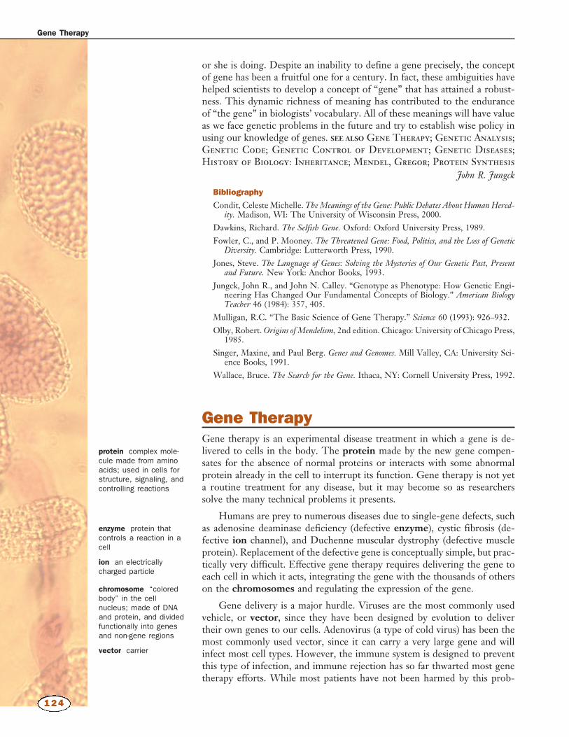
Gene TherapyGene therapy is an experimental disease treatment in which a gene is de-livered to cells in the body. The protein made by the new gene compen-sates for the absence of normal proteins or interacts with some abnormalprotein already in the cell to interrupt its function. Gene therapy is not yeta routine treatment for any disease, but it may become so as researcherssolve the many technical problems it presents.
Humans are prey to numerous diseases due to single-gene defects, suchas adenosine deaminase deficiency (defective enzyme), cystic fibrosis (de-fective ion channel), and Duchenne muscular dystrophy (defective muscleprotein). Replacement of the defective gene is conceptually simple, but prac-tically very difficult. Effective gene therapy requires delivering the gene toeach cell in which it acts, integrating the gene with the thousands of otherson the chromosomes and regulating the expression of the gene.
Gene delivery is a major hurdle. Viruses are the most commonly usedvehicle, or vector, since they have been designed by evolution to delivertheir own genes to our cells. Adenovirus (a type of cold virus) has been themost commonly used vector, since it can carry a very large gene and willinfect most cell types. However, the immune system is designed to preventthis type of infection, and immune rejection has so far thwarted most genetherapy efforts. While most patients have not been harmed by this prob-
Gene Therapy
124
protein complex mole-cule made from aminoacids; used in cells forstructure, signaling, andcontrolling reactions
enzyme protein thatcontrols a reaction in acell
ion an electricallycharged particle
chromosome “coloredbody” in the cellnucleus; made of DNAand protein, and dividedfunctionally into genesand non-gene regions
vector carrier


![지역브랜드그리고마케팅 - cfs6.blog.daum.netcfs6.blog.daum.net/upload_control/download.blog?fhandle... · [지역기획프로세스] 환경분석환경분석 지역자산조사지역자산조사](https://img.pdfslide.tips/doc/110x75/5e04b6f83675eb0f0a34b727/eoeeoeeoeeeeeoe-cfs6blogdaum-eeoe-eeee.jpg)