Embed Size (px)
Citation preview
366 M. GUARDIOLA, L. JOFRE, S. CAPDEVILA, S. BLANCH, J.ROMEU, 3D UWB MAGNITUDE-COMBINED TOMOGRAPHY...
3D UWB Magnitude-Combined Tomographic Imaging for Biomedical Applications. Algorithm Validation
Marta GUARDIOLA, Lluís JOFRE, Santiago CAPDEVILA, Sebastián BLANCH, Jordi ROMEU
AntennaLab, Universitat Politècnica de Catalunya, C/ Jordi Girona 3-4, 08034 Barcelona, Spain
[email protected], [email protected], [email protected], [email protected], [email protected]
Abstract. Biomedical microwave imaging is a topic of continuous research for its potential in different areas especially in breast cancer detection. In this paper, 3D UWB Magnitude-Combined tomographic algorithm is assessed for this recurrent application, but also for a more challenging one such as brain stroke detection. With the UWB Magnitude-Combined concept, the algorithm can take advantage of both the efficiency of Fourier Diffraction Theorem-based tomographic formulation and the robustness and image quality improvement provided by a multi-frequency combination.
Keywords Tomography, biomedical imaging, microwave imaging, breast cancer, brain stroke, experimental verification.
1. Introduction Microwave imaging is a topic of intense research for
its potential in biomedical applications and especially in breast cancer detection. X-ray mammography is the generally well-established clinical breast imaging technique for preventive screening and cancer treatment. Other imaging techniques including MRI (magnetic resonance imaging), ultrasounds or PET (positron emission tomography), are recommended for cases where X-ray mammography does not succeed, such as in women with dense breasts or with high cancer risk to avoid exposition to ionizing radiation, as reported in [1]. This, jointly with other concerns, such as the ionizing character of X-ray radiation, its uncomfortable (and even painful) application, motivate the research in complementary or alternative imaging methods exploiting other physical properties of tissues. In this framework, the potential of microwave imaging relies on the capability of microwaves to differentiate among tissues based on the contrast in dielectric properties, which is more important than those exploited by X-ray mammography (the attenuation of waves when passing through the breast structures) [2]. The advantages for its practical clinical usage are significant, including relatively low cost, the use of low-power non-ionizing radiation and patient comfort.
Active microwave imaging relies on obtaining information about a target from the scattered fields measured at a number of probes, when the target is illuminated with an incident field. This inverse scattering problem can be addressed either by radar-based techniques (refer to [3] for a review of UWB radar methods) or tomographic methods. Tomographic approaches try to solve the non-linear and ill-posed inverse scattering problem, by either linearizing it or iteratively approaching the solution. Many research groups are focused on iterative algorithms to obtain quantitative reconstructions of the dielectric properties of the target. Those are compu-tationally intensive, above all for 3D reconstructions, and usually contain some regularization scheme that requires a priori information about the target, having a direct influence into the algorithm convergence [4]. A number of different methods and optimization schemes have been proposed, [5]-[7], reporting useful 3D reconstructions of numerical models, phantoms and first 2D measurements on real patients [8]. However more research towards increasing computational efficiently of the algorithms is needed for a real time imaging. This opens the door to less computationally heavy algorithms as the ones based on linearizing approximations.
Linearizing approximations, on which the method proposed here is partially based, allow to obtain robust reconstructions, in a very efficient way, being however limited to small relatively low-contrast targets to produce quantitative reconstructions [9]. In general, biological organs do not accomplish these requirements, thus, line-arizing methods are restricted to qualitative recon-structions. In [10], useful qualitative images of a trans-versal cut of a human forearm were presented, retrieving clearly the two bones.
The use of multi-frequency information in a con-venient manner has been recognized as an opportunity for linearizing methods to improve the image quality in non-Born scenarios [11]. To this extent, it has not been used in linearized tomography methods due to the well-known frequency-dependent residual phase errors that appear when electrically large and highly contrasted targets are imaged. In the algorithm validated herein, namely 3D UWB Magnitude-Combined (UWB-MC) tomography, an amplitude (phase-less) multi-frequency combination is proposed to overcome this undesired effect [12].
RADIOENGINEERING, VOL. 20, NO. 2, JUNE 2011 367
Brain stroke detection is also addressed in this paper to investigate the potentiality of the proposed algorithm in such a challenging application, as proposed previously by [5], [13]. The motivation to explore this case is the difficulty to differentiate the cause of the stroke between a hemorrhage or a blood clot. Both present similar symp-toms, but opposite treatment, which must be given with the maximum promptness. Up to now, the diagnosis relies on bulky imaging methods, such as CT (computed tomography), PET and MRI, which are not available in all medical emergency units. This deficiency may definitely delay or complicate the decision and eventually cause important after-effects.
2. 3D UWB Magnitude-Combined Tomographic Imaging Algorithm 3D UWB Magnitude-Combined tomographic imag-
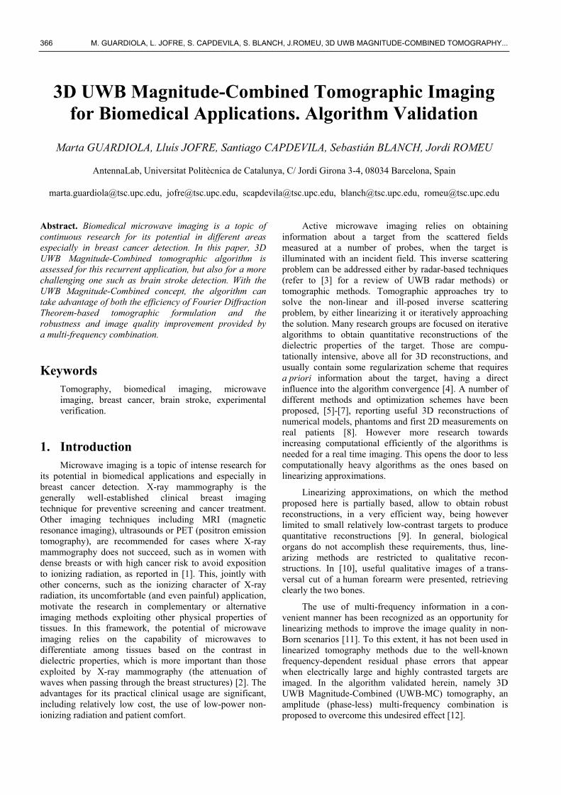
ing, as the name suggests, proposes a compound coherent multi-view image addition, which is typical of the linearized-tomography-based algorithms, followed with a magnitude multi-frequency image combination, in the last step of the algorithm. In this paper a 3D cylindrical geometry, as shown in Fig. 1 is studied. The cylindrical array of both the transmitting and the receiving antennas is composed by , 2 -diameter rings of angularly equispaced antennas. For a given transmitter, situated at , the scattered field is measured at the receiver positions, . This procedure is successively repeated for each transmitter to complete a maximum of acquisitions.
Fig. 1. The target of permittivity , is immersed in a me-
dium of permittivity . The measurement cylin-
drical array of antennas is composed by rings of antennas of radius , separated a distance ∆ . , refers to the position of the transmitting and receiving antennas respectively, and , is the direction of the synthesized plane wave.
The theoretical basis for 3D UWB-MC to obtain the dielectric contrast of the target is as follows. Let , be the dielectric contrast expressed as
, 1,
(1)
, and being the complex permittivities of the target and the external medium respectively, measured at a particular frequency .
The dielectric contrast can be related to the induced current on the target, , , , by
, , 2 , , ,
2 , , , (2)
where is the total electric field including the scattered and the incident field.
Using the reciprocity theorem (3), one can obtain the induced current on the target, , from the scattered field measured along the antenna
∭ ∭ . (3)
is the electric current on the cylindrical antenna acting as a transmitter which radiates a plane wave electric field, , propagating to a direction . is the electric current on the target induced by a plane wave incident field pro-pagating along the vector ( ). , is the scattered field produced by .
When the cylindrical array is composed by linear z-polarized antennas, is also z-directed, therefore, only the z component of the field is needed, thus permitting a scalar formulation.
Under Born approximation (the scattered field is negligible in front of the incident field), the induced current
can be expressed as
, , ≅ 2 , , , =
, , . (4)
Then, replacing (4) in (3), a Fourier transform ap-pears, and the spectrum of the contrast profile can be expressed as
, 2
, , ,
, , ,
, ;
, ; , ; (5)
where , ; is the scattered field measured at
a probe positioned at when an antenna placed at is
transmitting. , , ; , represents the amplitude to be applied to a probe situated at , to synthesize a plane wave towards , and vertical polarization [15] as a com-bination of cylindrical waves emanating from a number of probes.
From (5) it can be derived that for a given frequency, when the object is illuminated with an incident plane wave directed to , the Fourier transform of the scattered field obtained at a direction may be translated into the angular spectrum of the dielectric contrast of the target sampled on
y
z
xa
', ;iTE r f r0
' ', ;sR TE r f r0
,
˛ r f 0
', ; TJ r f r0
'Tr
'Rr
( )ext˛ f 0
, ˆ, ;a pwRE r f r0
, ˆ, ;i pw
TE r f r0
¢ z
368
thecensinangdepfrechadom
com
in tranforwaandlim
3.
UWto banddet
imagooof freantsysa cobt
8 M.
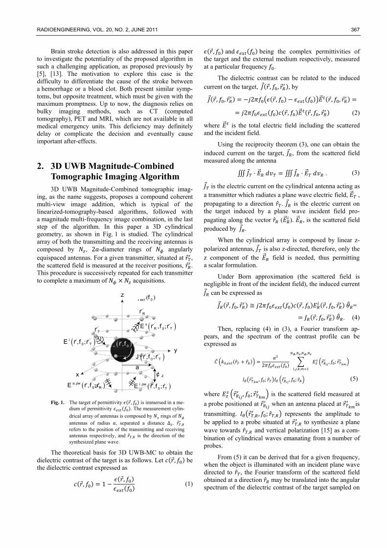
e surface of antered at n , sin ,gular scans wpicted in Fig. quency of thanges providmain.
Fig. 2. 2D ca singspect
a sphanoth
The multi-fmbining only
C
Due to the the z directinslated as a dr , close to aves may prodd should be av
mited between
SimulatThis sectio
WB-MC tomobiomedical imd a more chatection have b
The main gaging methodod spatial resoa good spatiaquencies; howtennas may bstem. Accordiertain value. tained using
GUARDIOLA, L
a sphere of ra
, , whercos ,
will fill the sp2 [14]. In a
e incident wading more i
cut of the dielectgle transmitter trum of the contr
here of radius her direction of in
frequency comthe magnitude
max
mi
f
UWB MCi f
C
limited extenion, there is deterioration o
the nadir or tduce the degrvoided. In ord45º and 135º.
tion Resuon gathers thographic algormaging. To doallenging applbeen considere
goal in biomedd with the beolution. Usingal resolution cwever the attecome excessingly, the freqIn this case a full wave
, ˆext Rk r0
, ˆext Tk r0
L. JOFRE, S. CA
adius ,
e , cos. Thus,
pectral domaisimilar way, ave, the radiuinformation i
tric spectrum of and incidence
rast is sampled o
0, centered atncidence the sphe
mbination is te of the multi-
x
,
in
1i extkFT C
nt of the measa truncation
of the synthesthe zenith. Unradation of thder to prevent.
ults he preliminaryrithm to studyo this, breast clication, suched.
dical imagingest sensitivityg microwaves,can be achievetenuation andsive, leading quency has tothe synthetic simulation so
,extk02
yk
, ˆext TC k r0
, ˆ ˆext T Rk r r0
APDEVILA, S. B
2
, sin ,
the successin knowledgeby changing
us of the sphin the spect
the contrast. Forfrequency, the
on the surface of
t , . Forere rotates.
then obtained -view images.
surement surferror, which
sized plane wanacceptable plahe reconstructt it , has be
y assessment y its applicabicancer detecti
h as brain stro
g is to develop, specificity a, the requiremed by using hd the number to an unfeasio be bounded
data have beoftware (FEK
xk
ˆT Rr
R
BLANCH, J.RO
sive e as the
here tral
r e f
r
d by .
(6)
face h is ave ane tion een
of lity ion, oke
p an and
ment high r of ible d to een
KO)
based betweelemethe Naliasindiamesepara
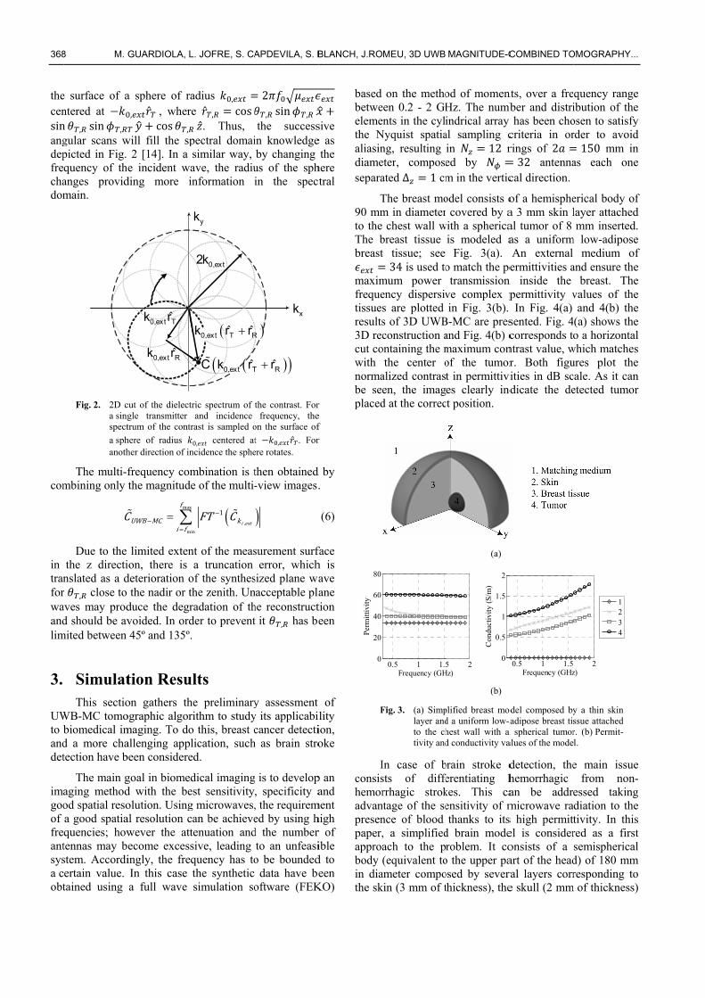
T90 mmto theThe bbreast
maximfrequetissueresults3D reccut cowithnormabe seplaced
F
Iconsishemoradvanpresenpaper,approabody in diathe sk
0
20
40
60
80
Perm
ittiv
ity
MEU, 3D UWB
on the methoeen 0.2 - 2 GHents in the cyliNyquist spatiang, resulting eter, composeated Δ 1 cm
The breast mom in diameter
chest wall wbreast tissue t tissue; see 34 is used to
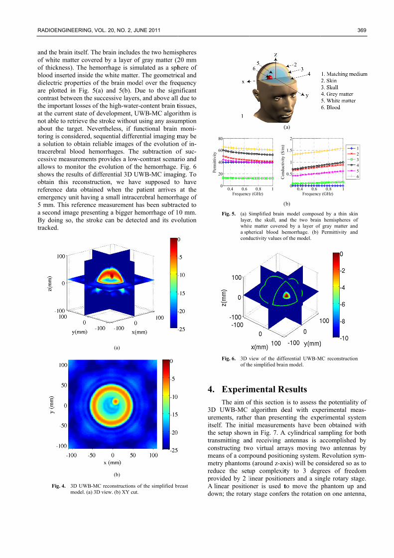
mum power ency dispersivs are plotteds of 3D UWBconstruction a
ontaining the mthe center o
alized contrasen, the imagd at the correc
Fig. 3. (a) Simplayer andto the chtivity and
In case of bsts of differrhagic strok
ntage of the sence of blood , a simplifiedach to the p(equivalent to
ameter compokin (3 mm of t
0.5 1 1.5Frequency (GH
MAGNITUDE-C
od of momenHz. The numbindrical array al sampling cin 12 red by m in the vertic
odel consists or covered by a
with a sphericais modeled a
Fig. 3(a). o match the petransmission
ve complex pin Fig. 3(b).
B-MC are presand Fig. 4(b) cmaximum conof the tumort in permittivies clearly ind
ct position.
(a)
(b)
plified breast mod a uniform low-ahest wall with a d conductivity va
brain stroke derentiating hkes. This caensitivity of mthanks to its
d brain moderoblem. It co
o the upper paosed by severathickness), the
5 2Hz)
0
0.5
1
1.5
2
Con
duct
ivity
(S/
m)
COMBINED TO
nts, over a frember and distriy has been chocriteria in orrings of 232 antenna
cal direction.
of a hemisphea 3 mm skin lal tumor of 8 as a uniformAn external ermittivities a
n inside the permittivity vIn Fig. 4(a)
sented. Fig. 4corresponds to
ntrast value, wr. Both figuities in dB sc
ndicate the de
odel composed byadipose breast tis
a spherical tumoralues of the mode
detection, thehemorrhagic an be addrmicrowave ras high permittel is consideronsists of a art of the headral layers corre skull (2 mm
0.5 1 1.5Frequency (GHz
OMOGRAPHY...
quency rangeibution of theosen to satisfyrder to avoid150 mm in
as each one
erical body oflayer attachedmm inserted.
m low-adiposemedium of
and ensure thebreast. The
values of theand 4(b) the
(a) shows theo a horizontal
which matchesures plot thecale. As it canetected tumor
y a thin skin ssue attached r. (b) Permit-l.
e main issuefrom non-
essed takingdiation to thetivity. In thisred as a firstsemisphericald) of 180 mmresponding to
m of thickness)
2z)
1234
.
e e y d n e
f d . e f e e e e e l s e n r
e -g e s t l
m o )
RADIOENGIN
and the brainof white matof thickness)blood insertedielectric proare plotted icontrast betwthe importanat the currennot able to reabout the tatoring is cona solution totracerebral bcessive measallows to moshows the reobtain this reference daemergency u5 mm. This a second imaBy doing sotracked.
Fig. 4. 3m
EERING, VOL.
n itself. The brtter covered b). The hemorred inside the woperties of thein Fig. 5(a) a
ween the succent losses of thent state of deveetrieve the stroarget. Neverthsidered, seque
o obtain reliabblood hemorrsurements proonitor the evosults of differreconstructio
ata obtained unit having a sreference mea
age presentingo, the stroke c
3D UWB-MC remodel. (a) 3D vie
20, NO. 2, JUN
rain includes tby a layer of grhage is simuwhite matter. Te brain modeland 5(b). Duessive layers, e high-water-celopment, UWoke without uheless, if funential differenble images of rhages. The
ovides a low-colution of the rential 3D UWon, we have when the pasmall intracereasurement hasg a bigger hemcan be detecte
(a)
(b)
econstructions of ew. (b) XY cut.
NE 2011
the two hemisgray matter (2
ulated as a sphThe geometricl over the freq
ue to the signand above all
content brain tWB-MC algorising any assumctional brain
ntial imaging mthe evolutionsubtraction o
contrast scenarhemorrhage.
WB-MC imagisupposed to
atient arrives ebral hemorrhs been subtracmorrhage of 1ed and its evo
the simplified br
spheres 20 mm here of cal and quency nificant l due to tissues, ithm is mption moni-
may be n of in-of suc-rio and Fig. 6
ing. To o have
at the hage of cted to
10 mm. olution
reast
4.
3DureitsthetracomemeredproAdo
Pitt
iit
Fig. 5. (a) Slayerwhita sphcond
Fig. 6. 3D vof th
. ExperimThe aim o
D UWB-MCements, ratheelf. The initie setup shownansmitting annstructing tweans of a cometry phantomsduce the setuovided by 2 llinear positio
own; the rotary
0.4 0.60
20
40
60
80
Frequency
Perm
ittiv
ity
(
(
Simplified brain r, the skull, andte matter coveredherical blood heductivity values o
view of the diffehe simplified brain
mental Ref this section algorithm de
r than presenal measuremen in Fig. 7. Ad receiving a
wo virtual arrampound positio
s (around z-axup complexitlinear positiononer is used ty stage confer
0.8 1y (GHz)
Con
duct
ivity
(S/
m)
(a)
(b)
model composed the two braind by a layer of emorrhage. (b)
of the model.
ferential UWB-Min model.
Results is to assess th
eal with expnting the expeents have bee
A cylindrical santennas is aays moving toning system. xis) will be coty to 3 degrners and a sinto move the rs the rotation
0.4 0.60
0.5
1
1.5
2
Frequency
y(
)
ed by a thin skinn hemispheres o
gray matter andPermittivity and
MC reconstruction
he potentialityperimental meerimental sysen obtained wampling for baccomplished two antennasRevolution sy
onsidered so arees of freedngle rotary staphantom up
n on one anten
0.8 1y (GHz)
369
n f d d
n
y of eas-tem
with both
by s by ym-
as to dom age. and
nna,
123456
370
whothbe is rfina csph2.1dep3Danybe
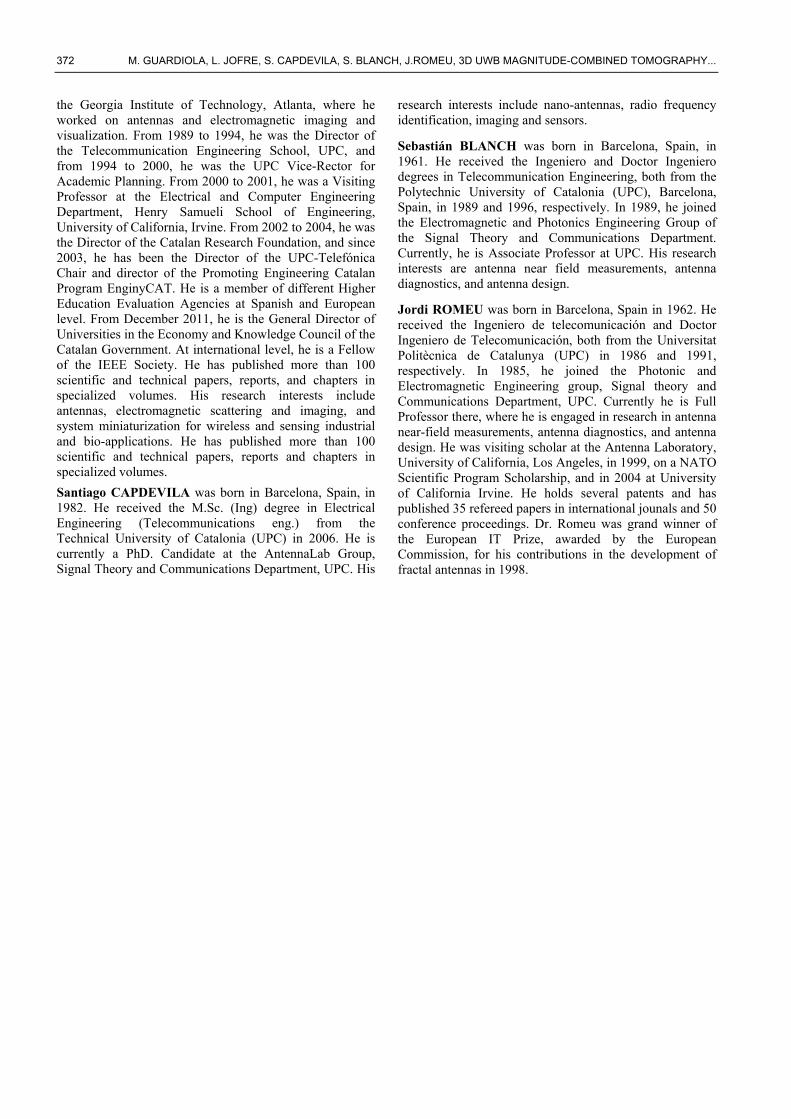
wabeetheperbiocomrecmeof bet
Figmeextimpconwestrusho
5.
Cotwoandcon
0 M.
hile the other her linear posobtained by tresponsible foally the wholomputer. Thehere (19) fixed withpicted in Fig.
D cylindrical my liquid as masurrounded by
Fig. 7. Expepermtargesupp
Preliminaryater using the en presented ie phantom conrmittivity to omedical simumpared to thconstructions easurements anhigh permittivtween 3 – 10 G
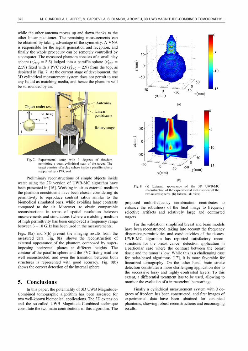
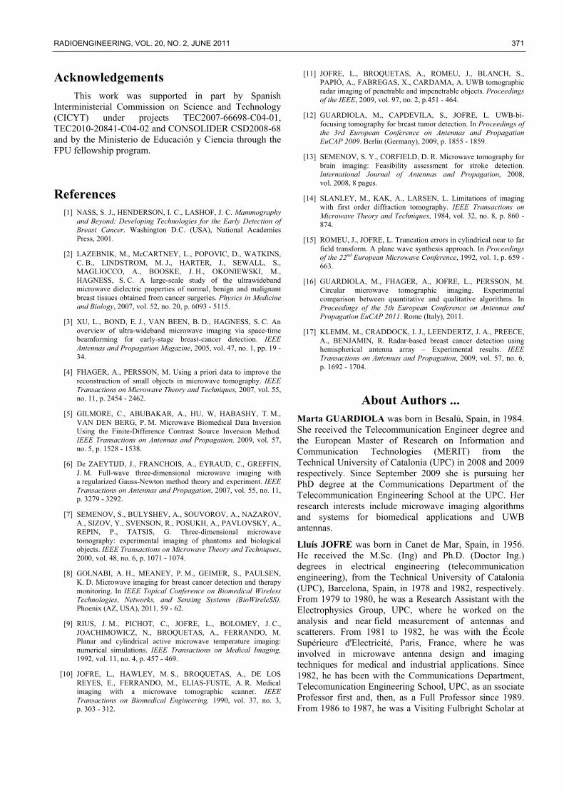
gs. 8(a) and 8easured data. ternal appearaposing horizntour of the pell reconstructuctures is repows the correc
ConclusIn this pape
ombined tomoo well-knownd the so-callenstitute the tw
GUARDIOLA, L
antenna movesitioner. The taking advantaor the signal gle procedure ce measured ph5.5) lodged i
h a PVC rod 7. At the curr
measurement satching mediay air.
erimental setup mitting a quasi-cyet consists of a claorted by a PVC r
y reconstructi2D version
in [16]. Worknstituents havreproduce co
ulated ones, whe air. Morein terms of
nd simulationvity has been GHz has been
8(b) present tFig. 8(a) s
ance of the pzontal planesparaffin sphereted, and evenpresented wict detection of
sions er, the potentiographic algon biomedical aed UWB Ma
wo main contri
L. JOFRE, S. CA
es up and dowremaining meage of the symgeneration ancan be remote
hantom consistinto a paraffin( 2.9) rent stage of dsystem does na, and hence t
with 3 degreylindrical scan ofay sphere inside rod.
ions of simplof UWB-MC
king in air as eve been chosenontrast ratioswhile avoidingeover, to obtf spatial resons (where a m
employed) a n used in the m
the imaging shows the rephantom comps at differene and the PVCn the transitioth good accuf the internal s
iality of 3D Uorithm has beapplications. Tagnitude-Comibutions of thi
APDEVILA, S. B
wn thanks to easurements cmmetry. A VN
nd reception, aely controlled ts of a small cn sphere (from the top,
development, not permit to uthe phantom w
ees of freedomf the target. Thea paraffin sphere
le objects insC algorithm haexternal medin considerings similar to g large contratain comparaolution betwe
matching medifrequency ran
measurements.
results from econstruction posed by sup
nt heights. TC fixing road on between buracy. Fig. 8sphere.
UWB Magnitueen assessed The 3D extens
mbined techniqis algorithm. T
BLANCH, J.RO
the can NA and
d by clay
, as the use will
m e e
side ave ium g its
the asts able een ium nge .
the of
per-The are
both 8(b)
ude-for
sion que The
F
propoenhanselectitarget
Fhave bdisperUWBstructia partitissue for ralinearidetectthe suextentmonit
Fgrees experiphantoresults
MEU, 3D UWB
Fig. 8. (a) Extreconstrutwo neste
sed multi-frnce the robusive artifacts s.
For the validabeen reconstrursive permittiv-MC algorithions for the icular case wand the tumo
adar-based algized tomogration constituteuccessive losst, a differentiaor the evolutio
Finally a cylinof freedom himental data oms, showings.
MAGNITUDE-C
(a
(b
ernal appearancuction of the expeed spheres. (b) In
equency comtness of the and relative
ation, simplifieucted, taking vities and conhm has repbreast cancer
where the conr is low. Whil
gorithms [17]aphy. On the es a more chalsy and highlyal treatment hon of a intrace
ndrical measuas been const
have been g robust recon
COMBINED TO
a)
b)
ce of the 3Derimental measur
nternal 3D view.
mbination cofinal image
ely large an
ed breast and into account tnductivities o
ported satisfaer detection aontrast betweele this is a cha], it is more other hand,
allenging appliy-contrasted lahas to be usederebral hemor
urement systetructed, and fi
n obtained fnstructions and
OMOGRAPHY...
D UWB-MC rement of the
ontributes toto frequency
nd contrasted
brain modelsthe frequency
of the tissues.actory recon-application inen the breastallenging casefavorable forbrain stroke
ication due toayers. To thisd, allowing torrhage.
em with 3 de-first images offor canonicald encouraging
.
o y d
s y . -n t e r e o s o
-f l g
RADIOENGINEERING, VOL. 20, NO. 2, JUNE 2011 371
Acknowledgements This work was supported in part by Spanish
Interministerial Commission on Science and Technology (CICYT) under projects TEC2007-66698-C04-01, TEC2010-20841-C04-02 and CONSOLIDER CSD2008-68 and by the Ministerio de Educación y Ciencia through the FPU fellowship program.
References [1] NASS, S. J., HENDERSON, I. C., LASHOF, J. C. Mammography
and Beyond: Developing Technologies for the Early Detection of Breast Cancer. Washington D.C. (USA), National Academies Press, 2001.
[2] LAZEBNIK, M., McCARTNEY, L., POPOVIC, D., WATKINS, C. B., LINDSTROM, M. J., HARTER, J., SEWALL, S., MAGLIOCCO, A., BOOSKE, J. H., OKONIEWSKI, M., HAGNESS, S. C. A large-scale study of the ultrawideband microwave dielectric properties of normal, benign and malignant breast tissues obtained from cancer surgeries. Physics in Medicine and Biology, 2007, vol. 52, no. 20, p. 6093 - 5115.
[3] XU, L., BOND, E. J., VAN BEEN, B. D., HAGNESS, S. C. An overview of ultra-wideband microwave imaging via space-time beamforming for early-stage breast-cancer detection. IEEE Antennas and Propagation Magazine, 2005, vol. 47, no. 1, pp. 19 - 34.
[4] FHAGER, A., PERSSON, M. Using a priori data to improve the reconstruction of small objects in microwave tomography. IEEE Transactions on Microwave Theory and Techniques, 2007, vol. 55, no. 11, p. 2454 - 2462.
[5] GILMORE, C., ABUBAKAR, A., HU, W, HABASHY, T. M., VAN DEN BERG, P. M. Microwave Biomedical Data Inversion Using the Finite-Difference Contrast Source Inversion Method. IEEE Transactions on Antennas and Propagation, 2009, vol. 57, no. 5, p. 1528 - 1538.
[6] De ZAEYTIJD, J., FRANCHOIS, A., EYRAUD, C., GREFFIN, J. M. Full-wave three-dimensional microwave imaging with a regularized Gauss-Newton method theory and experiment. IEEE Transactions on Antennas and Propagation, 2007, vol. 55, no. 11, p. 3279 - 3292.
[7] SEMENOV, S., BULYSHEV, A., SOUVOROV, A., NAZAROV, A., SIZOV, Y., SVENSON, R., POSUKH, A., PAVLOVSKY, A., REPIN, P., TATSIS, G. Three-dimensional microwave tomography: experimental imaging of phantoms and biological objects. IEEE Transactions on Microwave Theory and Techniques, 2000, vol. 48, no. 6, p. 1071 - 1074.
[8] GOLNABI, A. H., MEANEY, P. M., GEIMER, S., PAULSEN, K. D. Microwave imaging for breast cancer detection and therapy monitoring. In IEEE Topical Conference on Biomedical Wireless Technologies, Networks, and Sensing Systems (BioWireleSS). Phoenix (AZ, USA), 2011, 59 - 62.
[9] RIUS, J. M., PICHOT, C., JOFRE, L., BOLOMEY, J. C., JOACHIMOWICZ, N., BROQUETAS, A., FERRANDO, M. Planar and cylindrical active microwave temperature imaging: numerical simulations. IEEE Transactions on Medical Imaging, 1992, vol. 11, no. 4, p. 457 - 469.
[10] JOFRE, L., HAWLEY, M. S., BROQUETAS, A., DE LOS REYES, E., FERRANDO, M., ELIAS-FUSTE, A. R. Medical imaging with a microwave tomographic scanner. IEEE Transactions on Biomedical Engineering, 1990, vol. 37, no. 3, p. 303 - 312.
[11] JOFRE, L., BROQUETAS, A., ROMEU, J., BLANCH, S., PAPIÓ, A., FABREGAS, X., CARDAMA, A. UWB tomographic radar imaging of penetrable and impenetrable objects. Proceedings of the IEEE, 2009, vol. 97, no. 2, p.451 - 464.
[12] GUARDIOLA, M., CAPDEVILA, S., JOFRE, L. UWB-bi-focusing tomography for breast tumor detection. In Proceedings of the 3rd European Conference on Antennas and Propagation EuCAP 2009. Berlin (Germany), 2009, p. 1855 - 1859.
[13] SEMENOV, S. Y., CORFIELD, D. R. Microwave tomography for brain imaging: Feasibility assessment for stroke detection. International Journal of Antennas and Propagation, 2008, vol. 2008, 8 pages.
[14] SLANLEY, M., KAK, A., LARSEN, L. Limitations of imaging with first order diffraction tomography. IEEE Transactions on Microwave Theory and Techniques, 1984, vol. 32, no. 8, p. 860 - 874.
[15] ROMEU, J., JOFRE, L. Truncation errors in cylindrical near to far field transform. A plane wave synthesis approach. In Proceedings of the 22nd European Microwave Conference, 1992, vol. 1, p. 659 - 663.
[16] GUARDIOLA, M., FHAGER, A., JOFRE, L., PERSSON, M. Circular microwave tomographic imaging. Experimental comparison between quantitative and qualitative algorithms. In Proceedings of the 5th European Conference on Antennas and Propagation EuCAP 2011. Rome (Italy), 2011.
[17] KLEMM, M., CRADDOCK, I. J., LEENDERTZ, J. A., PREECE, A., BENJAMIN, R. Radar-based breast cancer detection using hemispherical antenna array – Experimental results. IEEE Transactions on Antennas and Propagation, 2009, vol. 57, no. 6, p. 1692 - 1704.
About Authors ... Marta GUARDIOLA was born in Besalú, Spain, in 1984. She received the Telecommunication Engineer degree and the European Master of Research on Information and Communication Technologies (MERIT) from the Technical University of Catalonia (UPC) in 2008 and 2009 respectively. Since September 2009 she is pursuing her PhD degree at the Communications Department of the Telecommunication Engineering School at the UPC. Her research interests include microwave imaging algorithms and systems for biomedical applications and UWB antennas.
Lluís JOFRE was born in Canet de Mar, Spain, in 1956. He received the M.Sc. (Ing) and Ph.D. (Doctor Ing.) degrees in electrical engineering (telecommunication engineering), from the Technical University of Catalonia (UPC), Barcelona, Spain, in 1978 and 1982, respectively. From 1979 to 1980, he was a Research Assistant with the Electrophysics Group, UPC, where he worked on the analysis and near field measurement of antennas and scatterers. From 1981 to 1982, he was with the École Supérieure d'Electricité, Paris, France, where he was involved in microwave antenna design and imaging techniques for medical and industrial applications. Since 1982, he has been with the Communications Department, Telecomunication Engineering School, UPC, as an ssociate Professor first and, then, as a Full Professor since 1989. From 1986 to 1987, he was a Visiting Fulbright Scholar at
372 M. GUARDIOLA, L. JOFRE, S. CAPDEVILA, S. BLANCH, J.ROMEU, 3D UWB MAGNITUDE-COMBINED TOMOGRAPHY...
the Georgia Institute of Technology, Atlanta, where he worked on antennas and electromagnetic imaging and visualization. From 1989 to 1994, he was the Director of the Telecommunication Engineering School, UPC, and from 1994 to 2000, he was the UPC Vice-Rector for Academic Planning. From 2000 to 2001, he was a Visiting Professor at the Electrical and Computer Engineering Department, Henry Samueli School of Engineering, University of California, Irvine. From 2002 to 2004, he was the Director of the Catalan Research Foundation, and since 2003, he has been the Director of the UPC-Telefónica Chair and director of the Promoting Engineering Catalan Program EnginyCAT. He is a member of different Higher Education Evaluation Agencies at Spanish and European level. From December 2011, he is the General Director of Universities in the Economy and Knowledge Council of the Catalan Government. At international level, he is a Fellow of the IEEE Society. He has published more than 100 scientific and technical papers, reports, and chapters in specialized volumes. His research interests include antennas, electromagnetic scattering and imaging, and system miniaturization for wireless and sensing industrial and bio-applications. He has published more than 100 scientific and technical papers, reports and chapters in specialized volumes.
Santiago CAPDEVILA was born in Barcelona, Spain, in 1982. He received the M.Sc. (Ing) degree in Electrical Engineering (Telecommunications eng.) from the Technical University of Catalonia (UPC) in 2006. He is currently a PhD. Candidate at the AntennaLab Group, Signal Theory and Communications Department, UPC. His
research interests include nano-antennas, radio frequency identification, imaging and sensors.
Sebastián BLANCH was born in Barcelona, Spain, in 1961. He received the Ingeniero and Doctor Ingeniero degrees in Telecommunication Engineering, both from the Polytechnic University of Catalonia (UPC), Barcelona, Spain, in 1989 and 1996, respectively. In 1989, he joined the Electromagnetic and Photonics Engineering Group of the Signal Theory and Communications Department. Currently, he is Associate Professor at UPC. His research interests are antenna near field measurements, antenna diagnostics, and antenna design.
Jordi ROMEU was born in Barcelona, Spain in 1962. He received the Ingeniero de telecomunicación and Doctor Ingeniero de Telecomunicación, both from the Universitat Politècnica de Catalunya (UPC) in 1986 and 1991, respectively. In 1985, he joined the Photonic and Electromagnetic Engineering group, Signal theory and Communications Department, UPC. Currently he is Full Professor there, where he is engaged in research in antenna near-field measurements, antenna diagnostics, and antenna design. He was visiting scholar at the Antenna Laboratory, University of California, Los Angeles, in 1999, on a NATO Scientific Program Scholarship, and in 2004 at University of California Irvine. He holds several patents and has published 35 refereed papers in international jounals and 50 conference proceedings. Dr. Romeu was grand winner of the European IT Prize, awarded by the European Commission, for his contributions in the development of fractal antennas in 1998.