Embed Size (px)
Citation preview
65
CASE REPORT
Nagoya J. Med. Sci. 79. 65 ~ 73, 2017doi:10.18999/nagjms.79.1.65
A case report of idiopathic iliopsoas hematoma which occurred soon after transfer to the wheelchair
after total hip arthroplasty
Tomoo Okumura, Hiroshi Fujita, Hideto Harada, Ryuuichi Nishimura and Tomohiro Tominaga
Institute for Joint Replacement, Department of Orthopedic Surgery, Kyoto Katsura Hospital, Kyoto, Japan
ABSTRACT
A 79-year-old woman was diagnosed with osteoarthritis of the left hip and scheduled for total hip arthroplasty. As two lesions were detected in branches of the coronary arteries, she was treated with catheter treatment. The patient was receiving anticoagulant treatment, which was suspended eight days before the operation; however, heparin was started at 6 days before the operation and was stopped 9 hours before the operation. On the 2nd postoperative day soon after transfer to the wheel chair, she was feeling unwell and was suffering from severe pain in her left buttock and left thigh. Her blood pressure had decreased to 70 mmHg. During abdominal contrast-enhanced CT, a hematoma was detected in the left iliopsoas muscle. Catheter therapy was started by the cardiovascular department and bleeding had stopped spontaneously. The patient was able to walk with a cane and was discharged on the 40th postoperative day.
Key Words: iliopsoas hematoma, total hip arthroplasty, anticoagulant treatmentAbbreviations: THA: total hip arthroplasty, CT: computed tomography, JOA score: Japanese Orthopaedic
Association hip score, HA: hydroxyapatite, PMMA: polymethyl-methacrylate, APTT: activated partial thromboplastin time
This is an Open Access article distributed under the Creative Commons Attribution-NonCommercial-NoDerivatives 4.0 International License. To view the details of this license, please visit (http://creativecommons.org/licenses/by-nc-nd/4.0/).
INTRODUCTION
Iliopsoas muscle hematomas are relatively rare. They are often complicated with hemophilia in cases involving hemorrhagic risk factors.1) Iliopsoas muscle hematomas can also occur as a rare complication of total hip arthroplasty (THA).2-7) We report a case in which an iliopsoas hematoma occurred after THA for osteoarthritis of the hip joint and perioperative antithrombotic treatment.
CASE REPORT
79-year-old Japanese female had suffered from left hip joint pain during walking for two years. Her height was 148 cm, and her body weight was 50.6 kg. She had a history of lung cancer
Received: August 8, 2016; accepted: November 2, 2016
Corresponding author: Hiroshi Fujita MD, PhD
Institute for Joint Replacement, Department of Orthopedic Surgery, Kyoto Katsura Hospital,
Yamada-hirao-cho, Nishikyo-ku, Kyoto 615-8256, Japan
Tel: +81-75-391-5811, Fax: +81-75-381-0090, E-mail: [email protected]
66
Tomoo Okumura et al.
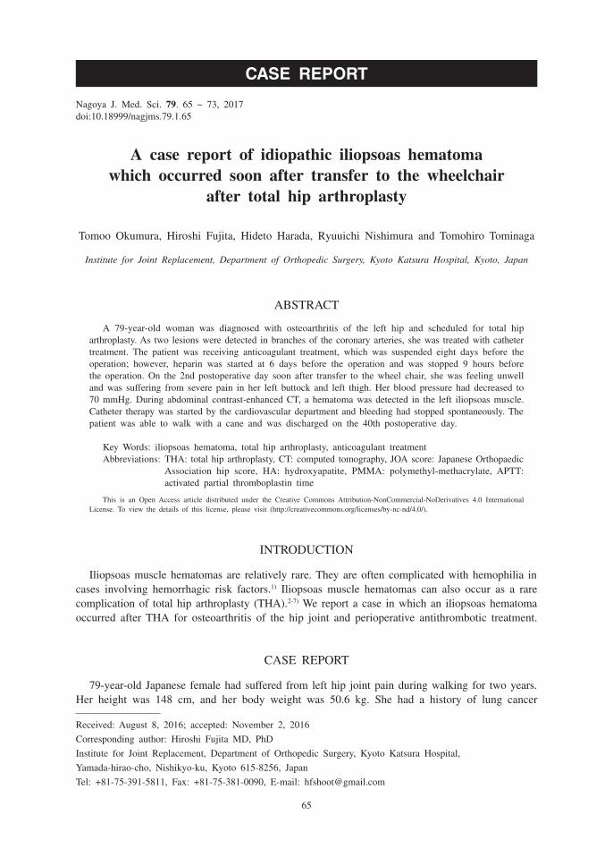
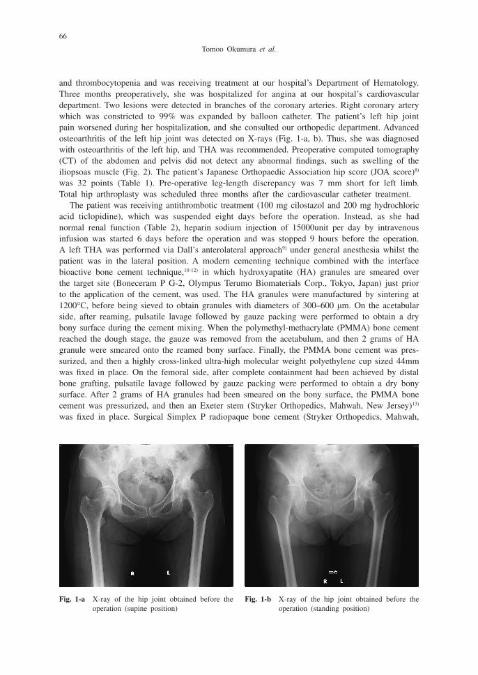
and thrombocytopenia and was receiving treatment at our hospital’s Department of Hematology. Three months preoperatively, she was hospitalized for angina at our hospital’s cardiovascular department. Two lesions were detected in branches of the coronary arteries. Right coronary artery which was constricted to 99% was expanded by balloon catheter. The patient’s left hip joint pain worsened during her hospitalization, and she consulted our orthopedic department. Advanced osteoarthritis of the left hip joint was detected on X-rays (Fig. 1-a, b). Thus, she was diagnosed with osteoarthritis of the left hip, and THA was recommended. Preoperative computed tomography (CT) of the abdomen and pelvis did not detect any abnormal findings, such as swelling of the iliopsoas muscle (Fig. 2). The patient’s Japanese Orthopaedic Association hip score (JOA score)8) was 32 points (Table 1). Pre-operative leg-length discrepancy was 7 mm short for left limb. Total hip arthroplasty was scheduled three months after the cardiovascular catheter treatment.
The patient was receiving antithrombotic treatment (100 mg cilostazol and 200 mg hydrochloric acid ticlopidine), which was suspended eight days before the operation. Instead, as she had normal renal function (Table 2), heparin sodium injection of 15000unit per day by intravenous infusion was started 6 days before the operation and was stopped 9 hours before the operation. A left THA was performed via Dall’s anterolateral approach9) under general anesthesia whilst the patient was in the lateral position. A modern cementing technique combined with the interface bioactive bone cement technique,10-12) in which hydroxyapatite (HA) granules are smeared over the target site (Boneceram P G-2, Olympus Terumo Biomaterials Corp., Tokyo, Japan) just prior to the application of the cement, was used. The HA granules were manufactured by sintering at 1200°C, before being sieved to obtain granules with diameters of 300–600 μm. On the acetabular side, after reaming, pulsatile lavage followed by gauze packing were performed to obtain a dry bony surface during the cement mixing. When the polymethyl-methacrylate (PMMA) bone cement reached the dough stage, the gauze was removed from the acetabulum, and then 2 grams of HA granule were smeared onto the reamed bony surface. Finally, the PMMA bone cement was pres-surized, and then a highly cross-linked ultra-high molecular weight polyethylene cup sized 44mm was fixed in place. On the femoral side, after complete containment had been achieved by distal bone grafting, pulsatile lavage followed by gauze packing were performed to obtain a dry bony surface. After 2 grams of HA granules had been smeared on the bony surface, the PMMA bone cement was pressurized, and then an Exeter stem (Stryker Orthopedics, Mahwah, New Jersey)13) was fixed in place. Surgical Simplex P radiopaque bone cement (Stryker Orthopedics, Mahwah,
Fig. 1-a X-ray of the hip joint obtained before the operation (supine position)
Fig. 1-b X-ray of the hip joint obtained before the operation (standing position)
67
Idiopathic iliopsoas hematoma after THA
New Jersey) was used as the PMMA bone cement. The operative duration was two hours and seven minutes, and 130 ml of intraoperative bleeding occurred. The fixation of the implant was performed without any problems, and the length of the patient’s left leg was extended by 7 mm. As a result, postoperative X-rays demonstrated that the discrepancy between the lengths of the patient’s legs had improved (Fig. 3). The extension of the hip joint was improved from –5 degrees to 2 degrees. Extension angle of the hip joint was measured in prone position using a goniometer. No problems with the patient’s vital signs were detected after the operation, and her blood pressure did not decrease during or after the operation.
Fig. 2 Plain CT image of the abdomen obtained before the operation (horizontal view)No abnormal findings, including swelling of the iliopsoas muscle, were observed.
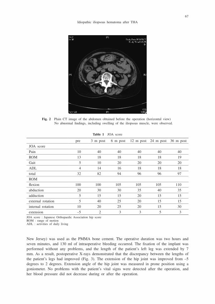
Table 1 JOA score
pre 3 m post 6 m post 12 m post 24 m post 36 m post
JOA score
Pain 10 40 40 40 40 40
ROM 13 18 18 18 18 19
Gait 5 10 20 20 20 20
ADL 4 14 16 18 18 18
total 32 82 94 96 96 97
ROM
flexion 100 100 105 105 105 110
abduction 20 30 30 35 40 35
adduction 5 15 15 20 15 15
external rotation 5 40 25 20 15 15
internal rotation 10 20 25 20 15 30
extension –5 2 3 3 5 3JOA score : Japanese Orthopaedic Association hip scoreROM : range of motionADL : activities of daily living
68
Tomoo Okumura et al.
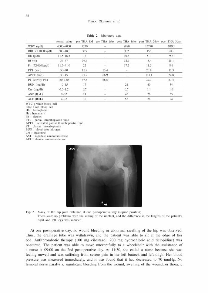
At one postoperative day, no wound bleeding or abnormal swelling of the hip was observed. Thus, the drainage tube was withdrawn, and the patient was able to sit at the edge of her bed. Antithrombotic therapy (100 mg cilostazol, 200 mg hydrochloric acid ticlopidine) was re-started. The patient was able to move uneventfully to a wheelchair with the assistance of a nurse at 09:00 on the 2nd postoperative day. At 11:30, she called a nurse because she was feeling unwell and was suffering from severe pain in her left buttock and left thigh. Her blood pressure was measured immediately, and it was found that it had decreased to 70 mmHg. No femoral nerve paralysis, significant bleeding from the wound, swelling of the wound, or thoracic
Table 2 laboratory data
normal value pre THA 1M pre THA 1day post THA 1day post THA 2day post THA 3day
WBC (/μdl) 4000–9000 5270 – 8880 13770 9290
RBC (X10000/μdl) 380–480 385 – 332 156 283
Hb (g/dl) 11.5–16.5 13 – 10.8 5.1 9.2
Ht (%) 37–47 39.7 – 32.7 15.4 25.1
Plt (X10000/μdl) 11.5–41.0 22 – 17.2 11.5 8.6
PTT (sec.) 50–70 11.9 13.4 – 20.8 12.3
APTT (sec.) 30–45 25.9 66.9 – 111.1 24.8
PT activity (%) 80–130 97.8 68.5 – 32.1 81.4
BUN (mg/dl) 10–15 17 – 21 40 34
Cre (mg/dl) 0.6–1.2 0.7 – 0.7 1.1 1.0
AST (IU/L) 9–32 21 – 45 26 35
ALT (IU/L) 4–37 16 – 53 28 24
WBC : white blood cellRBC : red blood cellHb : hemoglobinHt : hematocritPlt : plateletPTT : partial thromboplastin timeAPTT : activated partial thromboplastin timePT : plasma thromboplastinBUN : blood urea nitrogenCre : creatinineAST : aspartate aminotransferaseALT : alanine aminotransferase
Fig. 3 X-ray of the hip joint obtained at one postoperative day (supine position)There were no problems with the setting of the implant, and the difference in the lengths of the patient’s right and left legs was reduced.
69
Idiopathic iliopsoas hematoma after THA
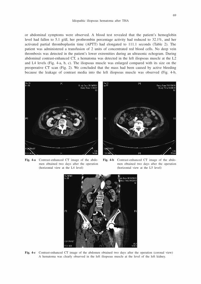
or abdominal symptoms were observed. A blood test revealed that the patient’s hemoglobin level had fallen to 5.1 g/dl, her prothrombin percentage activity had reduced to 32.1%, and her activated partial thromboplastin time (APTT) had elongated to 111.1 seconds (Table 2). The patient was administered a transfusion of 2 units of concentrated red blood cells. No deep vein thrombosis was detected in the patient’s lower extremities during an ultrasonic echogram. During abdominal contrast-enhanced CT, a hematoma was detected in the left iliopsoas muscle at the L2 and L4 levels (Fig. 4-a, b, c). The iliopsoas muscle was enlarged compared with its size on the preoperative CT scan (Fig. 2). We concluded that the mass had been caused by active bleeding because the leakage of contrast media into the left iliopsoas muscle was observed (Fig. 4-b,
Fig. 4-a Contrast-enhanced CT image of the abdo-men obtained two days after the operation (horizontal view at the L4 level)
Fig. 4-b Contrast-enhanced CT image of the abdo-men obtained two days after the operation (horizontal view at the L5 level)
Fig. 4-c Contrast-enhanced CT image of the abdomen obtained two days after the operation (coronal view)A hematoma was clearly observed in the left iliopsoas muscle at the level of the left kidney.
70
Tomoo Okumura et al.
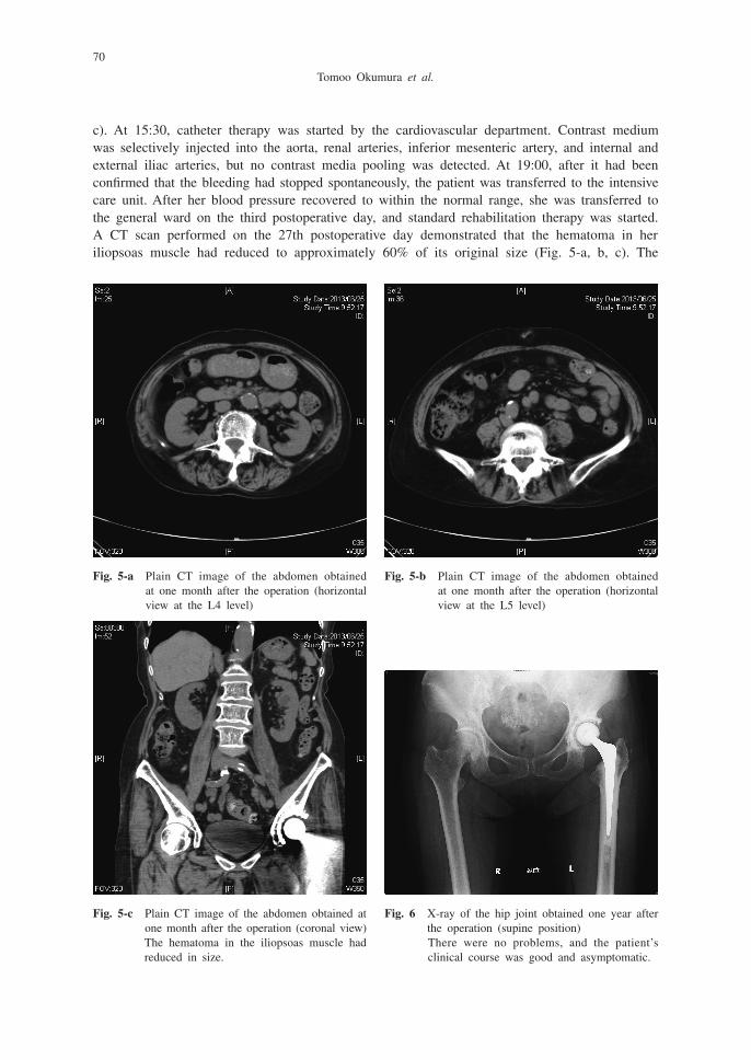
c). At 15:30, catheter therapy was started by the cardiovascular department. Contrast medium was selectively injected into the aorta, renal arteries, inferior mesenteric artery, and internal and external iliac arteries, but no contrast media pooling was detected. At 19:00, after it had been confirmed that the bleeding had stopped spontaneously, the patient was transferred to the intensive care unit. After her blood pressure recovered to within the normal range, she was transferred to the general ward on the third postoperative day, and standard rehabilitation therapy was started. A CT scan performed on the 27th postoperative day demonstrated that the hematoma in her iliopsoas muscle had reduced to approximately 60% of its original size (Fig. 5-a, b, c). The
Fig. 5-c Plain CT image of the abdomen obtained at one month after the operation (coronal view)The hematoma in the iliopsoas muscle had reduced in size.
Fig. 5-a Plain CT image of the abdomen obtained at one month after the operation (horizontal view at the L4 level)
Fig. 6 X-ray of the hip joint obtained one year after the operation (supine position)There were no problems, and the patient’s clinical course was good and asymptomatic.
Fig. 5-b Plain CT image of the abdomen obtained at one month after the operation (horizontal view at the L5 level)
71
Idiopathic iliopsoas hematoma after THA
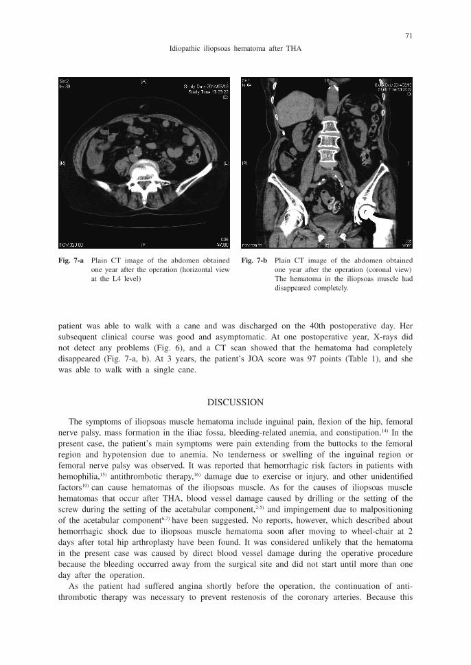
patient was able to walk with a cane and was discharged on the 40th postoperative day. Her subsequent clinical course was good and asymptomatic. At one postoperative year, X-rays did not detect any problems (Fig. 6), and a CT scan showed that the hematoma had completely disappeared (Fig. 7-a, b). At 3 years, the patient’s JOA score was 97 points (Table 1), and she was able to walk with a single cane.
DISCUSSION
The symptoms of iliopsoas muscle hematoma include inguinal pain, flexion of the hip, femoral nerve palsy, mass formation in the iliac fossa, bleeding-related anemia, and constipation.14) In the present case, the patient’s main symptoms were pain extending from the buttocks to the femoral region and hypotension due to anemia. No tenderness or swelling of the inguinal region or femoral nerve palsy was observed. It was reported that hemorrhagic risk factors in patients with hemophilia,15) antithrombotic therapy,16) damage due to exercise or injury, and other unidentified factors10) can cause hematomas of the iliopsoas muscle. As for the causes of iliopsoas muscle hematomas that occur after THA, blood vessel damage caused by drilling or the setting of the screw during the setting of the acetabular component,2-5) and impingement due to malpositioning of the acetabular component6,7) have been suggested. No reports, however, which described about hemorrhagic shock due to iliopsoas muscle hematoma soon after moving to wheel-chair at 2 days after total hip arthroplasty have been found. It was considered unlikely that the hematoma in the present case was caused by direct blood vessel damage during the operative procedure because the bleeding occurred away from the surgical site and did not start until more than one day after the operation.
As the patient had suffered angina shortly before the operation, the continuation of anti-thrombotic therapy was necessary to prevent restenosis of the coronary arteries. Because this
Fig. 7-a Plain CT image of the abdomen obtained one year after the operation (horizontal view at the L4 level)
Fig. 7-b Plain CT image of the abdomen obtained one year after the operation (coronal view)The hematoma in the iliopsoas muscle had disappeared completely.
72
Tomoo Okumura et al.
patient had a history of thrombopenia after aspirin was used orally, cilostazol and ticlopidine were reluctantly used instead of clopidrogel. Blood tests detected a reduction in the patient’s prothrombin percentage activity and an elongation of her APTT. It was thought that heparin was stopped nine hours before operation, an influence continued after the operation. Thus, the continuation of antithrombotic therapy clearly carried a risk of hemorrhaging.
It has been reported that rupturing of the iliopsoas muscle occurs during excessively strong contractions from an extended position.17,18) Preoperatively, extension of the left hip joint was restricted to –5 degrees. This had improved to 2 degrees at 3 months after the operation. The length of the patient’s left leg was extended by 7 mm, and X-rays obtained after the operation demonstrated that the discrepancy between the lengths of the patient’s legs had reduced (Fig. 3). Therefore, it was considered that the patient’s psoas muscle had been elongated by the THA, and the iliopsoas muscle had been subjected to excessive tension when the patient moved to a wheelchair, which had resulted in bleeding.
Generally treatment of iliopsoas hematomas depends on the speed onset, volume and neuro-logical impairment.16) In the present case there was no need to perform further therapy as the bleeding stopped when a catheter therapy was performed. It had been necessary, however, to perform an embolization for a bleeding point when bleeding had not stopped spontaneously.
In conclusion, the reason of the iliopsoas hematoma of the present case was a combination of mechanical effects on the muscle and insufficient coagulation management. When hemorrhagic shock occurs after THA in cases involving hemorrhagic risk factors, iliopsoas muscle hematoma should be included in the differential diagnoses, and contrast-enhanced CT should be performed as soon as possible.
CONCLUSIONS
1. We performed THA for osteoarthritis of the hip joint and continued antithrombotic therapy before the operation. A hematoma developed in the iliopsoas muscle soon after moving to wheel-chair on the second postoperative day.
2. When hemorrhagic shock occurs after THA in cases involving hemorrhagic risk factors, contrast-enhanced CT should be performed promptly in case an iliopsoas muscle hematoma has developed.
CONFLICTS OF INTEREST STATEMENT
The authors did not receive and will not receive any benefits or funding from any commercial party related directly or indirectly to this article.
REFERENCES
1) Bulloch W, Fildes P. Haemophilia: In Treasury of Human Inheritance. 1911, Eugemnetics Laboratory Memories, London.
2) Uchida K, Negoro K, Kokubo Y, Yayama T, Miyazaki T, Nakajima H, et al. Retroperitoneal hematoma with bone resorption around the acetabular component after total hip arthroplasty: a case report and review of the literature. J Med Case Rep, 2012 Sep 13; 6: 294.
3) Gorgus A, Ozturk C, Sirvanci M, Aydogan M, Hamzaoglu A. Femoral nerve palsy due to iliacus hematoma occurred after total hip arthroplasty. Arch Orthop Trauma Surg, 2008; 128: 657–660.
4) Ha YC, Ahn IO, Jeong ST, Park HB, Koo KH. Iliacs haematoma and femoral nerve palsy after revision
73
Idiopathic iliopsoas hematoma after THA
hip arthroplasty: a case report. Clin Orthop Rel Res, 2001; 385: 100–103. 5) Wooten SL, McLaughlin RE. Iliacs hematoma and subsequent femoral nerve palsy after penetration of the
medial acetabular wall during total hip arthroplasty. Report of a case. Clin Orthop Rel Res, 1984; 191: 221–223.
6) Bartelt RB, Sierra RJ. Recurrent hematomas within the iliopsoas muscle caused by impingement after total hip arthroplasty. J Arthroplasty, 2011 Jun; 26: 665.e1–665.e5.
7) Pouliot MA, Lee KB, Goodman SB. Retroperitoneal hematoma: an unusual cause of pain after total hip arthroplasty. J Arthroplasty, 2009 Oct; 24: 1144.e9–1144.e12.
8) Kuribayashi M, Takahashi KA, Fujioka M, Ueshima K, Inoue S, Kubo T. Reliability and validity of the Japanese Orthopaedic Association hip score. J Orthop Sci, 2010 Jul; 15: 452–458.
9) Dall D. Exposure of the hip by anterior osteotomy of the greater trochanter. A modified anterolateral approach. J Bone Joint Surg, 1986; 68B: 382–386.
10) Oonishi H, Kadoya Y, Iwaki H, Kin N. Total hip arthroplasty with a modified cementing technique using hydroxyapatite granules. J Arthroplasty, 2001; 784–789.
11) Oonishi H, Ohashi H, Oonishi H Jr, Kim SC. THA with hydroxyapatite granules at cement-bone interface: 15- to 20-year results. Clin Orthop Relat Res, 2008 Feb; 466: 373–379.
12) Fujita H, Oonishi H, Ito, Kim SC, Doukawa H. Radiological evaluation of the femoral component fixed with interface bioactive bone cement (IBBC) in revision THA. J Arthroplasty, 2008 Aug; 23: 689–693.
13) Fujita H, Katayama N, Iwase T, Otsuka H. Multi-center study of use of the Exeter stem in Japan: evaluation of 1000 primary THA. J Orthop Sci, 2012; 17: 370–376.
14) Goodfellow J, Fearn CB, Matthews JM. Iliacus haematoma; a common complication of haemophilia. J Bone Joint Surg, 1967; 49-B: 748–756.
15) Ashrani AA, Osip J, Christie B, Key NS. Iliopsoas haemorrhage in patients with bleeding disorders–experi-ence from one centre. Haemophilia, 2003 Nov; 9: 721–726.
16) Marquardt G, Barduzal Angles S, Leherta F, Seifert V. Spontaneous haematoma of the iliac psoas muscle: a case report and the literature. Arch Orthop Trauma Surg, 2002; 122: 109–111.
17) Kumar S, Anantham J, Wan Z. Posttraumatic hematoma of iliacus muscle with paralysis of the femoral nerve. J Orthop Trauma, 1992; 6: 110–112.
18) Takami H, Takahashi S, Ando M. Traumatic rupture of the iliacus muscle with femoral nerve paralysis. J Trauma,1983; 23: 253–254.