Embed Size (px)
Citation preview
A Primary Aorto-duodenal Fistula Associated with
an Inflammatory Abdominal Aortic Aneurysm:A Case Report
Osami Honjo , Yukio Yamada , Takashi Arata , Tsuyoshi Matsuno ,
Tatsuo Kurokawa, and Yoshio Kushida
Departments of Cardiovascular Surgery, Surgery and Pathology,Saiseikai Imabari Hospital, Imabari, Ehime 799-1592, Japan
Primary aorto-enteric fistula(PAEF)is a serious complication of abdominal aortic aneurysm(AAA).We report a patient with PAEF associated with inflammatory AAA who underwent emergent
surgery. A 52-year-old male presented with recurrent hematemesis. A computer tomography scan
showed a sealed rupture of the AAA adjacent to the duodenum. At surgery, a coin-sized PAEF was
noted. The aorta was replaced with a Dacron graft . Histological examination revealed the
characteristics of an inflammatory AAA. The postoperative course was uneventful, and there has
been no evidence of infection during a follow-up period of 3 years. We discuss the etiologic and
surgical considerations regarding this unusual entity.
Key words:primary aorto-enteric fistula, inflammatory abdominal aortic aneurysm, ruptured abdominal aortic
aneurysm
P rimary aorto-enteric fistula(PAEF)is a rare but
serious complication of abdominal aortic aneurysm(AAA), and the outcome of surgical treatment is poor[1,2]. The surgical results depend on preoperative condition
of the patient and degree of contamination of the operative
field[1, 2]. Inflammatory AAA is one of the causes of
PAEF, and a few cases of PAEF caused by
inflammatory AAA have been reported[3, 4]. We
report here the successful treatment of a case of PAEF
associated with inflammatory AAA and discuss the
etiologic and surgical considerations regarding this un-usual entity.
Case Report
A 52-year-old male was referred to our institution for
treatment of recurrent hematemesis and melena. He had
had hematemesis and melena 2 weeks before admission
and had visited a local hospital. Emergency exploratory
laparotomy was performed due to uncontrollable
hematemesis, but no obvious cause in the abdomen was
found. A repeat endoscopic examination showed an
extrinsic pulsating mass protruding into the internal lumen
of the second portion of the duodenum. The recurrent
gastrointestinal bleeding was uncontrollable, and the
patient was transferred to our institution. An abdominal
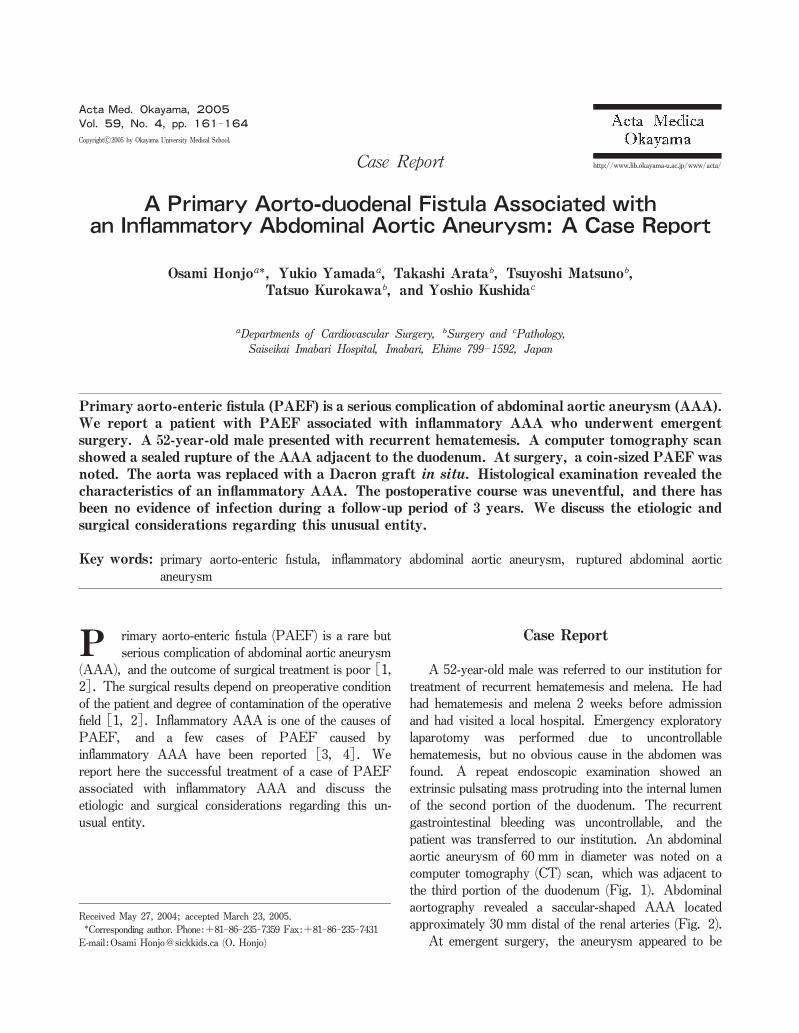
aortic aneurysm of 60 mm in diameter was noted on a
computer tomography(CT)scan, which was adjacent to
the third portion of the duodenum(Fig. 1). Abdominal
aortography revealed a saccular-shaped AAA located
approximately 30 mm distal of the renal arteries(Fig. 2).At emergent surgery, the aneurysm appeared to be
Received May 27,2004;accepted March 23,2005.Corresponding author.Phone:+81-86-235-7359 Fax:+81-86-235-7431
E-mail:Osami Honjo@sickkids.ca(O.Honjo)
http://www.lib.okayama-u.ac.jp/www/acta/
Acta Med. Okayama, 2005
Vol. 59 , No. 4, pp. 161-164
Case Report
Copyrightc2005by Okayama University Medical School.

inflammatory, and the right lateral wall of the aneurysm
had a communication with the third portion of the duode-num. Inspection of the internal lumen of the aorta showed
a coin-sized aorto-duodenal fistula(Fig. 3). The markedly
thickened arterial wall was resected to the extent that was
possible. The aorta was replaced with a Dacron Y graft
in situ. The perforated region of the duodenum was
resected and was anastomosed in an end-to-end fashion.The cavity was irrigated with 10 liters of saline. The
omentum was placed between the aorta and duodenum.A
culture of the aneurysmal wall showed the presence of
Candida species, and an antifungal agent was administer-ed for 2 months. The results of histological examination
showed that chronic inflammatory cells had focally
infiltrated into the aneurysmal wall (Fig. 4), and that
neutrophilic infiltration with abscess formation was seen
around the fistula. The patient’s postoperative course was
uneventful, and there has been no evidence of infection
during a follow-up period of 3 years.
Discussion
PAEF, direct communication between the aorta and
the intestinal tract, is mainly due to an atherosclerotic
aneurysm, and other causes include mycotic aneurysm,radiation trauma, and inflammatory AAA[1, 2].Although 5 of infrarenal AAAs have the characteristics
of inflammatory AAA, PAEFs resulting from
inflammatory AAA are extremely rare[3, 4]. Ikeda et
al. reported a 50-year-old woman who had PAEF
associated with multiple inflammatory AAAs[3], and
Mii et al. reported a 28-year-old man who had primary
aorto-jejunal fistula associated with inflammatory AAA[4]. Together with the present case, the clinical charac-teristics of those patients include relatively young age and
the presence of a rapidly developed saccular-shaped aneur-ysm. Interestingly, there were no symptoms associated
with systemic inflammation such as high-grade fever,
Honjo et al. Acta Med. Okayama Vol. 59 , No. 4 162
Fig.1 A CT scan showing the sealed rupture of the AAA adjacent
to the third portion of the duodenum.Fig. 2 Aortography revealing a saccular-shaped AAA with no
extravasation.
Fig.3 Intraoperative finding of a coin-sized aorto-duodenal fistula.The arrow shows the fistularization.

despite the fact that all 3 patients had elevated white blood
cells and C-reactive protein. The early to medium term
results of those patients were encouraging, even though
the overall surgical results in patients with PAEF have
been poor. Such cases must receive careful follow-up,because the native aortic tissue in the anastomotic sites
still has the potential of recurrent inflammation and subse-quent fistulization.It is controversial whether the chronic inflammation of
the aneurysmal wall results purely from the inflammatory
process, i.e., a so-called “non-specific inflammatory
aneurysm,”or from infection, i.e., a mycotic aneurysm.In the present case, a culture of the aneurysmal wall
showed the presence of Candida species;however, the
patient had no symptoms of active infection, and there
was also no evidence of endocarditis or other focuses.Furthermore, bacteriological study of mycotic aneurysm[5]showed that the common organisms cultured from
aneurismal walls included Staphylococcus aureus and
Salmonella, and mycotic aneurysm due to Candida
species was extremely rare[5]. In addition, the charac-teristic CT findings in mycotic aneurysms, which include
the sudden appearance of an aneurysm on a previously
normal aorta in a febrile patient, a soft tissue mass
surrounding the aneurysm, and an adjacent vertebral
osteomyelitis[6], were not fully correlated with findings
in present case. The clinical and pathological findings in
our case indicated that Candida species might not be a
primary cause of inflammatory aneurysm but might be a
secondary contamination through the fistula, and we
concluded that it was a non-specific inflammatory aneur-ysm rather than a mycotic aneurysm.Despite the presence of contamination,most surgeons
have reconstructed diseased aortae with in situ grafts[1-4]. Extra-anatomic bypass grafting is an alternative
approach for treatment of PAEF to avoid the use of a
prosthetic graft in the contaminated region[7]. How-ever, a high mortality rate has been reported in patients
treated with extra-anatomic bypass, since not only does
the procedure involve high risks of limb loss and aortic
stump blowout, but also it is often performed in severely
ill patients with gross infection in the operative field. The
selection of surgical approach depends on the presence of
sepsis and the degree of contamination. If a severe
purulent infection is present in periaortic tissue, extra-anatomic bypass should be selected. Our patient was
treated with an in situ graft because the contamination of
the operative field was localized. Careful irrigation of the
operative field is mandatory regardless of the presence or
absence of gross contamination, and the proximal end of
the prosthetic graft should be wrapped with the omentum
to prevent recurrent fistulization, especially in cases of
inflammatory aneurysm.
PAEF with Inflammatory AAA August 2005
Fig.4 Histological findings of the aneurysmal wall:chronic inflammatory cells infiltrated into the aneurysmal wall(A), and severe chronic
inflammatory cell infiltration and localized neutrophilic infiltration with abscess formation(B).
A B
163
References
1. Sweeney MS and Gadacz TR:Primary aortoduodenal fistula:Manifes-tation, diagnosis, and treatment. Surgery(1984)96:492-497.
2. Tareen AH and Schroeder TV:Primary aortoenteric fistula:Two new
case reports and review of 44 previously reported cases. Eur J Vasc
Endovasc Surg (1996)12:5-10.3. Ikeda K, Abe T, Itou M, Tamiya Y, Tanaka T and Kazui T:Successful
surgical treatment of primary aorto-duodenal fistula associated with
inflammatory abdominal aortic aneurysm:A case report. Ann Thorac
Cardiovasc Surg (1999)5:194-197.
4. Mii S, Onohara T, Okadome K, Fukuda A and Sugimachi K:Surgical
repair of primary aorto-jejunal fistula associated with non-specific
inflammatory abdominal aortic aneurysm. Eur J Vasc Surg (1991)5:355-357.
5. Brown SL, Busuttil RW, Baker JD, Machleder HL, Moore WS and
Barker WF:Bacterioligic and surgical determinants of survival in
patients with mycotic aneurysms. J Vasc Surg (1984)1:541-547.6. Parellada JA, Palmer J, Monill JM, Zidan A, Gimenez AM and
Moreno A:Mycotic aneurysm of the abdominal aorta:CT findings in
three patients. Abdom Imaging (1997)22:321-324.
7. Pagni S, Denatale RW, Sweeney T, McLauglin C and Ferneini AM:Primary aorto-duodenal fistula secondary to infected abdominal aortic
aneurysm:the role of local debridement and extra-anatomic bypass. J
Cardiovasc Surg (1999)40:31-35.
Honjo et al. Acta Med. Okayama Vol. 59 , No. 4 164