Embed Size (px)
Citation preview
153
□ CASE REPORT □
Acute Multiple Cerebral Infarction in a Patientwith an Accessory Mitral Valve
Ikuo Misumi 1, Asako Nagao 2, Katsuya Iwamoto 3, Tsuyoshi Honda 1, Masanobu Ishii 1,
Hidetsugu Ueyama 2, Yasushi Maeda 2, Masatoshi Ishizaki 2, Ryoichi Kurisaki 2,
Toshio Okazaki 2, Tetsuji Yamashita 2, Akiko Fujimoto 2 and Yumi Honda 2
Abstract
A 96-year-old woman developed hemiparesis 2 weeks after orthopedic surgery. Magnetic resonance imag-
ing revealed multiple cerebral infarctions in the bilateral hemisphere. Transthoracic echocardiography revealed
a mobile structure attached to the anterior mitral leaflet that protruded toward the left ventricular outflow
tract. The structure was identified as an accessory mitral valve. Doppler echocardiography showed that there
was no significant left ventricular outflow obstruction. This is a rare case of a silent accessory mitral valve
that was detected after multiple cerebral infarctions.
Key words: accessory mitral valve, left ventricular outflow obstruction, cerebral infarction
(Intern Med 56: 153-155, 2017)(DOI: 10.2169/internalmedicine.56.7649)
Introduction
An accessory mitral valve (AMV) is a rare congenital car-
diac malformation that may be accompanied by other con-
genital heart diseases, including left ventricular outflow tract
obstruction (1). We herein report the case of a patient with
an AMV that was diagnosed after multiple cerebral infarc-
tions.
Case Report
A 96-year-old woman fractured her thigh and was admit-
ted to our hospital. Two weeks after open surgery, she no-
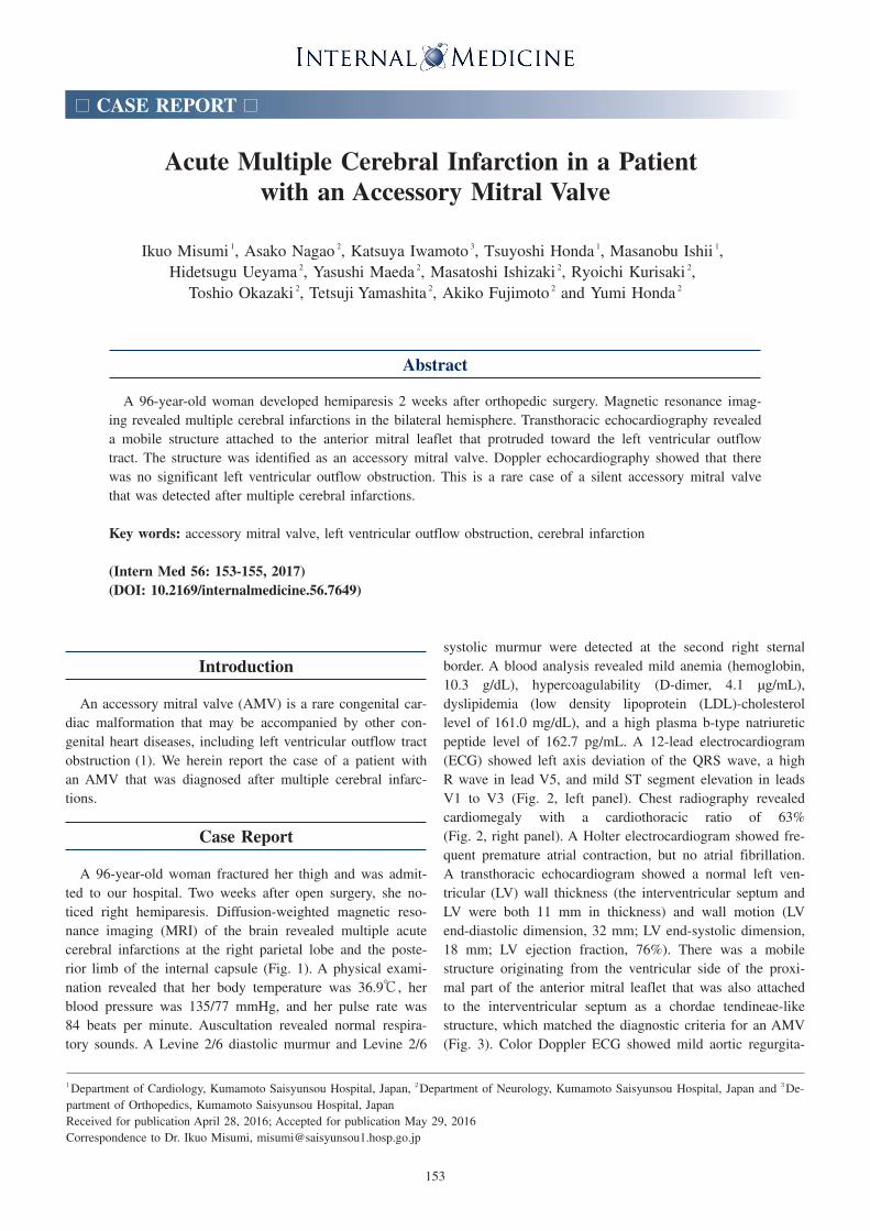
ticed right hemiparesis. Diffusion-weighted magnetic reso-
nance imaging (MRI) of the brain revealed multiple acute
cerebral infarctions at the right parietal lobe and the poste-
rior limb of the internal capsule (Fig. 1). A physical exami-
nation revealed that her body temperature was 36.9℃, her
blood pressure was 135/77 mmHg, and her pulse rate was
84 beats per minute. Auscultation revealed normal respira-
tory sounds. A Levine 2/6 diastolic murmur and Levine 2/6
systolic murmur were detected at the second right sternal
border. A blood analysis revealed mild anemia (hemoglobin,
10.3 g/dL), hypercoagulability (D-dimer, 4.1 μg/mL),
dyslipidemia (low density lipoprotein (LDL)-cholesterol
level of 161.0 mg/dL), and a high plasma b-type natriuretic
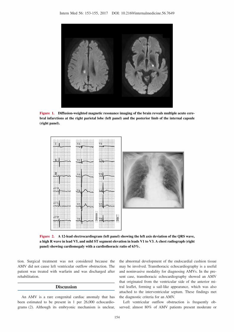
peptide level of 162.7 pg/mL. A 12-lead electrocardiogram
(ECG) showed left axis deviation of the QRS wave, a high
R wave in lead V5, and mild ST segment elevation in leads
V1 to V3 (Fig. 2, left panel). Chest radiography revealed
cardiomegaly with a cardiothoracic ratio of 63%
(Fig. 2, right panel). A Holter electrocardiogram showed fre-
quent premature atrial contraction, but no atrial fibrillation.
A transthoracic echocardiogram showed a normal left ven-
tricular (LV) wall thickness (the interventricular septum and
LV were both 11 mm in thickness) and wall motion (LV
end-diastolic dimension, 32 mm; LV end-systolic dimension,
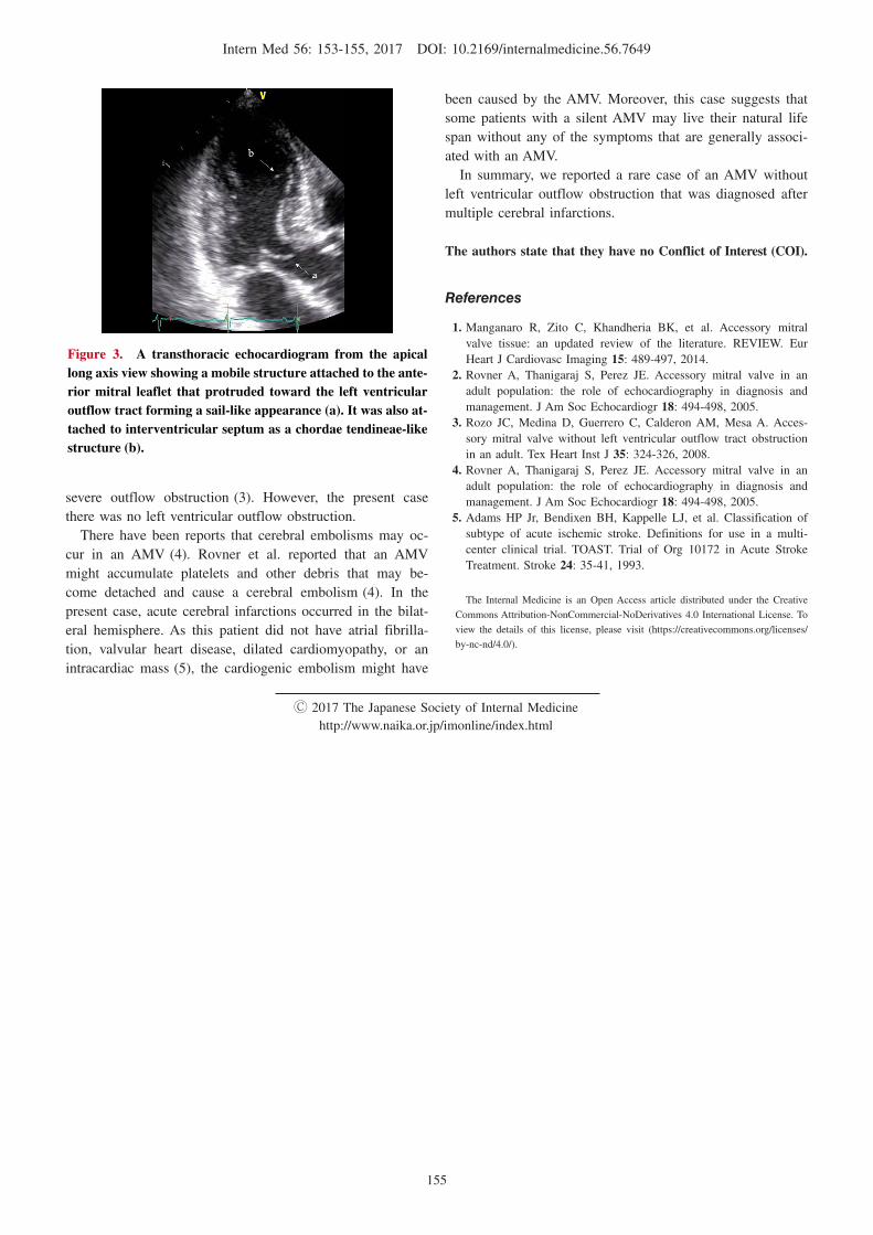
18 mm; LV ejection fraction, 76%). There was a mobile
structure originating from the ventricular side of the proxi-
mal part of the anterior mitral leaflet that was also attached
to the interventricular septum as a chordae tendineae-like
structure, which matched the diagnostic criteria for an AMV
(Fig. 3). Color Doppler ECG showed mild aortic regurgita-
1Department of Cardiology, Kumamoto Saisyunsou Hospital, Japan, 2Department of Neurology, Kumamoto Saisyunsou Hospital, Japan and 3De-
partment of Orthopedics, Kumamoto Saisyunsou Hospital, Japan
Received for publication April 28, 2016; Accepted for publication May 29, 2016
Correspondence to Dr. Ikuo Misumi, [email protected]
Intern Med 56: 153-155, 2017 DOI: 10.2169/internalmedicine.56.7649
154
Figure 1. Diffusion-weighted magnetic resonance imaging of the brain reveals multiple acute cere-bral infarctions at the right parietal lobe (left panel) and the posterior limb of the internal capsule (right panel).
Figure 2. A 12-lead electrocardiogram (left panel) showing the left axis deviation of the QRS wave, a high R wave in lead V5, and mild ST segment elevation in leads V1 to V3. A chest radiograph (right panel) showing cardiomegaly with a cardiothoracic ratio of 63%.
tion. Surgical treatment was not considered because the
AMV did not cause left ventricular outflow obstruction. The
patient was treated with warfarin and was discharged after
rehabilitation.
Discussion
An AMV is a rare congenital cardiac anomaly that has
been estimated to be present in 1 per 26,000 echocardio-
grams (2). Although its embryonic mechanism is unclear,
the abnormal development of the endocardial cushion tissue
may be involved. Transthoracic echocardiography is a useful
and noninvasive modality for diagnosing AMVs. In the pre-
sent case, transthoracic echocardiography showed an AMV
that originated from the ventricular side of the anterior mi-
tral leaflet, forming a sail-like appearance, which was also
attached to the interventricular septum. These findings met
the diagnostic criteria for an AMV.
Left ventricular outflow obstruction is frequently ob-
served; almost 80% of AMV patients present moderate or
Intern Med 56: 153-155, 2017 DOI: 10.2169/internalmedicine.56.7649
155
Figure 3. A transthoracic echocardiogram from the apical long axis view showing a mobile structure attached to the ante-rior mitral leaflet that protruded toward the left ventricular outflow tract forming a sail-like appearance (a). It was also at-tached to interventricular septum as a chordae tendineae-like structure (b).
severe outflow obstruction (3). However, the present case
there was no left ventricular outflow obstruction.
There have been reports that cerebral embolisms may oc-
cur in an AMV (4). Rovner et al. reported that an AMV
might accumulate platelets and other debris that may be-
come detached and cause a cerebral embolism (4). In the
present case, acute cerebral infarctions occurred in the bilat-
eral hemisphere. As this patient did not have atrial fibrilla-
tion, valvular heart disease, dilated cardiomyopathy, or an
intracardiac mass (5), the cardiogenic embolism might have
been caused by the AMV. Moreover, this case suggests that
some patients with a silent AMV may live their natural life
span without any of the symptoms that are generally associ-
ated with an AMV.
In summary, we reported a rare case of an AMV without
left ventricular outflow obstruction that was diagnosed after
multiple cerebral infarctions.
The authors state that they have no Conflict of Interest (COI).
References
1. Manganaro R, Zito C, Khandheria BK, et al. Accessory mitral
valve tissue: an updated review of the literature. REVIEW. Eur
Heart J Cardiovasc Imaging 15: 489-497, 2014.
2. Rovner A, Thanigaraj S, Perez JE. Accessory mitral valve in an
adult population: the role of echocardiography in diagnosis and
management. J Am Soc Echocardiogr 18: 494-498, 2005.
3. Rozo JC, Medina D, Guerrero C, Calderon AM, Mesa A. Acces-
sory mitral valve without left ventricular outflow tract obstruction
in an adult. Tex Heart Inst J 35: 324-326, 2008.
4. Rovner A, Thanigaraj S, Perez JE. Accessory mitral valve in an
adult population: the role of echocardiography in diagnosis and
management. J Am Soc Echocardiogr 18: 494-498, 2005.
5. Adams HP Jr, Bendixen BH, Kappelle LJ, et al. Classification of
subtype of acute ischemic stroke. Definitions for use in a multi-
center clinical trial. TOAST. Trial of Org 10172 in Acute Stroke
Treatment. Stroke 24: 35-41, 1993.
The Internal Medicine is an Open Access article distributed under the Creative
Commons Attribution-NonCommercial-NoDerivatives 4.0 International License. To
view the details of this license, please visit (https://creativecommons.org/licenses/
by-nc-nd/4.0/).
Ⓒ 2017 The Japanese Society of Internal Medicine
http://www.naika.or.jp/imonline/index.html


![SplenicInfarctioninAcuteCytomegalovirusandHuman … · 2019. 7. 30. · [9]S. Naviglio, M. V. Abate, M. Chinello, and A. Ventura, “Splenic infarction in acute infectious mononucleosis,”](https://img.pdfslide.tips/doc/110x75/613ec40eb946476b8b530f56/splenicinfarctioninacutecytomegalovirusandhuman-2019-7-30-9s-naviglio-m.jpg)