Embed Size (px)
Citation preview
8/2/2019 appi.ajp.2010.09091348
http://slidepdf.com/reader/full/appiajp201009091348 1/9
Article
Am J Psychiatry 167:8, August 2010 ajp.psychiatryonline.org 925
This article is eatured in this month’s AJP Audio, is the subject o a CME course (p. 1009),
and is discussed in an editorial by Drs. New and Stanley (p. 882).
(Am J Psychiatry 2010; 167:925–933)
depression display a negative emotional bias and di-
culties in shiting emotional states, those with borderline
personality disorder lack eective regulation o responses
to emotionally salient stimuli (5, 6). Whether development
o borderline personality disorder depends on genetic ac-
tors alone or is a variable combination o genetic vulner-
ability and environmental actors (4), exploration o the
underlying neurobiology o the disorder would provide
signicant insight into its pathophysiology.
Resting metabolic rate reductions in the prerontal cor-tical regions o patients with borderline personality dis-
order have been observed in some but not all studies (9,
10), as well as reductions in the cuneus and hippocampus
and increases in the cingulate and rontal gyri relative to
matched comparison subjects (11). Reductions in base-
line metabolic unction in the posterior cingulate cortex,
temporal lobe, and precuneus have been noted in patients
with both borderline personality disorder and posttrau-
matic stress disorder (12). Patients with borderline per-
E pidemiological data suggest that borderline person-
ality disorder has a lietime prevalence o 1% –5% (1), with
about 75% o clinical subjects being emale (2). Patients
with borderline personality disorder oten have axis I and
axis II comorbidity and high disability levels, and their uti-
lization o medical resources is disproportionate to their
numbers (3).
Borderline personality disorder is characterized by dys-
regulation o emotion processing, maniested as aective
lability and impulsive behaviors, including aggression andsel-harm (4). This dysregulation is exemplied by short-
duration, oten severe, rapidly changing mood states that
are highly reactive to environmental stimuli (5–7). Patients
appear to react more quickly, with greater intensity and a
slower return to baseline state (7).
Borderline personality disorder exhibits some clinical
and biological similarities to major depressive disorder,
but available evidence suggests that the two have distinct
pathophysiological proles (8). While patients with major
Alan R. Prossin, M.D.
Tiffany M. Love
Robert A. Koeppe, Ph.D.
Jon-Kar Zubieta, M.D., Ph.D.
Kenneth R. Silk, M.D.
Objective: Borderline personality dis-
order is characterized by a lack o eec-
tive regulation o emotional responses.
The authors investigated the role o the
endogenous opioid system and μ-opioid
receptors in emotion regulation in bor-
derline personality disorder.
Method: Mu-opioid receptor availability
in vivo (nondisplaceable binding poten-
tial, or BPND) was measured with positron
emission tomography and the selective
radiotracer [11C]carentanil during neutral
and sustained sadness states in 18 un-
medicated emale patients with border-
line personality disorder and 14 healthyemale comparison subjects.
Results: Patients showed greater re-
gional μ-opioid BPND than did comparison
subjects at baseline (neutral state) bilater-
ally in the orbitorontal cortex, caudate,
and nucleus accumbens and in the let
amygdala, but lower BPND in the poste-
rior thalamus. Sadness induction was as-
sociated with greater reductions in BPND
(endogenous opioid system activation)
in the patient group than in the com-
parison group in the pregenual anterior
cingulate, let orbitorontal cortex, let
ventral pallidum, let amygdala, and let
inerior temporal cortex. Patients showed
evidence o endogenous opioid system
deactivation in the let nucleus accum-
bens, the hypothalamus, and the right
hippocampus/parahippocampus relative
to comparison subjects. Correlations o
baseline measures with the Dissociative
Experiences Scale and endogenous opioid
system activation with the Barratt Impul-
siveness Scale did not remain signicant
ater correction or multiple comparisons.
Conclusions: Dierences exist betweenpatients with borderline personality dis-
order and comparison subjects in base-
line in vivo μ-opioid receptor concen-
trations and in the endogenous opioid
system response to a negative emotional
challenge that can be related to some
o the clinical characteristics o patients
with borderline personality disorder. The
regional network involved is implicated
in the representation and regulation o
emotion and stress responses.
Dysregulation o Regional Endogenous Opioid Functionin Borderline Personality Disorder
8/2/2019 appi.ajp.2010.09091348
http://slidepdf.com/reader/full/appiajp201009091348 2/9
ENDOGENOUS OPIOID FUNCTION IN BORDERLINE PERSONALITY DISORDER
926 ajp.psychiatryonline.org Am J Psychiatry 167:8, August 2010
line personality disorder and associated with dissocia-
tive symptoms and negative aect (23). In an MRI study,intensity-matched pain stimuli induced greater activity
in the prerontal cortex but lower activity in the amygdala
and anterior cingulate o patients with borderline person-
ality disorder relative to comparison subjects. These are
regions interacing emotion, pain, and stress regulation
and where μ-opioid receptors have important modulatory
roles (22, 23). These regions and systems also have been
ound to be related to trait impulsivity in healthy volun-
teers in response to a pain stressor (24).
We used positron emission tomography (PET) and a
selective μ-opioid receptor radiotracer to examine avail-
ability (nondisplaceable binding potential, or BPND) o these receptors under neutral (baseline) conditions and
during induction o a sustained sadness state in emale
borderline personality disorder patients and matched
comparison subjects (19, 20). Under challenge conditions,
acute reductions in BPND are thought to refect activation
o μ-opioid-receptor-mediated neurotransmission (e.g.,
competition o the endogenous ligand with the radiotrac-
er, receptor activation and recycling). Increases in BPND
would then refect deactivation o neurotransmission
(19, 20). We hypothesized that dierences between the
borderline personality disorder and comparison groups
would include greater baseline μ-opioid BPND in emotion
sonality disorder and a history o childhood abuse have
been ound to display reductions in amygdala and hippo-campal volumes (13). The relationship between metabolic
activity o the prerontal cortex and the amygdala was di-
minished in a sample o impulsive-aggressive borderline
personality disorder patients relative to comparison sub-
jects, supporting a hypothesis o poor prerontal control o
emotion-regulatory limbic structures (e.g., the amygdala)
in these patients (14, 15). Challenge studies employing
MRI and emotional stimuli support this hypothesis. Re-
ductions in cingulate and prerontal cortical unction and
increases in amygdala activity were ound in the presenta-
tion o ear aces and in attempted inhibition o negative
emotional stimuli (16, 17).In this study, we explored hypothesized neurochemical
mechanisms in borderline personality disorder patho-
physiology. The endogenous opioid system and μ-opioid
receptors have long been implicated in emotional and
stress response regulation. In animal models, reductions
in endogenous opioid system unction are associated with
attachment behavior decits and anxiety-like responses
(18), and in humans, with normal and pathological (e.g.,
major depressive disorder) emotion regulation (19, 20),
in addition to the system’s traditional role in modulat-
ing responses to physical and emotional stressors (21,
22). Pain thresholds are ound to be increased in border-
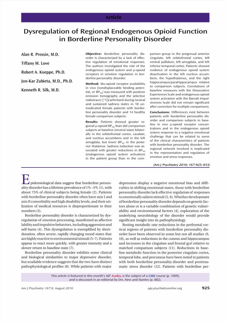
FIGURE 1. Time Course o PANAS Sadness Subscale Scores in Women With Borderline Personality Disorder (N=18) andHealthy Comparison Subjects (N=14)a
0
5
10
15
20
0 10 20 30 40 50 60 70 80 90 P A N A S S a d n e
s s S u b s c a l e
S c o r e
Time (minutes) After Tracer Administration
Neutral (10 to 45 minutes)
Sad (50 to 90 minutes)
Borderline Personality Disorder Patients
Comparison Subjects
0
5
10
15
20
0 10 20 30 40 50 60 70 80 90
Time (minutes) After Tracer Administration
Sad (10 to 45 minutes)
Neutral (50 to 90 minutes)
Borderline Personality Disorder Patients
Comparison Subjects
P A N A S S a d n e s s S u b s c a l e
S c o r e
a The order o conditions (sad, neutral) was randomized and counterbalanced between subjects. The top panel shows data rom studies inwhich the sadness condition was perormed second in order, and the bottom panel shows data rom studies in which the sadness conditionwas perormed frst. Bars indicate standard deviation.
8/2/2019 appi.ajp.2010.09091348
http://slidepdf.com/reader/full/appiajp201009091348 3/9
PROSSIN, LOVE, KOEPPE, ET AL.
Am J Psychiatry 167:8, August 2010 ajp.psychiatryonline.org 927
were instructed to try to create passive relaxation while avoiding any active cognitive process. During sadness induction, partici-pants recalled a previously rehearsed past autobiographical vi-gnette associated with sadness. Both states were practiced prior
to the actual scan and the specic events recorded. Participantsrated their experience every 10 minutes on the sadness subscaleo the Positive and Negative Aect Schedule (PANAS; 31) to ascer-tain maintenance o their emotional state; the subscale includes
the adjectives “sad,” “blue,” “downhearted,” “alone,” and “lonely.”The ull PANAS (60 adjectives, 20 o which are grouped into twomain aective subscales, positive and negative) was completedby participants at baseline and ater completion o neutral andsadness states (45 minutes and 90 minutes ater administrationo the radiotracer).
Neuroimaging Methods
One PET scan was acquired over 90 minutes with a Siemens(Knoxville, TN) HR+ scanner in three-dimensional mode (recon-
structed ull width at hal maximum resolution, ~5.5 mm). [11
C]carentanil, a selective μ-opioid receptor radiotracer, was admin-istered at a dose o 10–15 mCi and mass <0.03 μg/kg (tracer quan-tities). Acquisition, reconstruction, and emission image coregis-tration protocols were identical to those used in previous studies(19, 20).
Image data were transormed on a voxel-by-voxel basis into
two sets o parametric maps: a tracer transport measure (K 1 ratio)and receptor-related measures during neutral and sadness states.To avoid arterial blood sampling, these measures were calculatedemploying a modied Logan graphical analysis (32) using the oc-cipital cortex (an area devoid o μ-opioid receptors) as the reer-ence region. The slope o the Logan plot is proportional to [(Bmax /K d) +1] or this receptor site (Bmax = receptor concentration; K d=receptor anity or the radiotracer); Bmax /K d is the “in vivo recep-
tor availability” measure, or nondisplaceable binding potential,BPND. With the bolus-continuous inusion protocol, the Logan
plot becomes linear approximately 4–6 minutes ater adminis-tration o the radiotracer, allowing or quantication o receptorsites. BPND values or neutral and sadness states were calculatedrom data rom 10–45 minutes and 50–90 minutes ater radiotrac-er administration. Anatomical MRI scans were acquired on a GE3-T scanner (General Electric, Milwaukee). Acquisition sequenc-es were axial spoiled gradient-recall acquisition (echo time=3.4msec, repetition time=10.5 msec, inversion time=200 msec, fipangle =25°, number o excitations=1, using 124 contiguous im-ages 1.5 mm thick). K 1 and BPND images or each experimentalperiod and MR images were coregistered to each other and to
the Montreal Neurological Institute (MNI) stereotactic atlas ori-entation (33). Statistical parametric maps o dierences between
processing regions in the borderline personality disorder
group, refecting chronic lower levels o opioid regula-
tory control with compensatory receptor up-regulation.
During sustained sadness we expected an exaggeratedresponse in these same areas, refecting high sensitivity
o borderline personality disorder patients to emotional
stimuli, with greater activation o the stress regulatory en-
dogenous opioid system and μ-opioid receptors.
Method
Participants
Participants were 18 right-handed emale patients with bor-derline personality disorder (mean age=28 years [SD=9], meaneducational level=14 years [SD=2]) and 14 age-matched healthy
emale comparison subjects (mean age=35 years [SD=10], meaneducational level=17 years [SD=2]). Nine comparison subjects
were in a previous study (19), and nine in a second study (20), with an overlap o 10 comparison subjects between the presentstudy and previously published work; our comparison subjects
were unique to this study. Axis I and axis II diagnoses were made
via the Structured Clinical Interview or DSM-IV Axis I Disorders(25) and the Structured Clinical Interview or DSM-IV Axis II Dis-orders (26) and conrmed by an experienced clinician (K.R.S.).Patients with borderline personality disorder also met the DSM-IV borderline personality disorder criterion o aective instability.Exclusion criteria were concurrent axis I and III diagnoses (exceptor mood disorder); history o psychosis or head trauma; and cur-rent or recent (within 3 months) illicit substance use, abuse, ordependence. All participants were medication ree or at least 3
months. Comprehensive urine drug screens were negative priorto the PET scan.
Participants completed the NEO Personality Inventory–Re-vised (NEO-PI-R; 27), the Barratt Impulsiveness Scale (28), andthe Dissociative Experiences Scale (29) prior to scanning. To re-duce phenotypic and neurobiological variability, only women
were studied (30). All participants provided written inormed consent. Protocols
were approved by the Investigational Review Board and the Ra-dioactive Drug Research Committee at the University o Michigan.
Induction of Sustained Affective States
Neutral and sadness states were randomized, counterbalanced,and initiated either 5 minutes or 45 minutes ater administration
o the radiotracer. During the neutral condition, participants
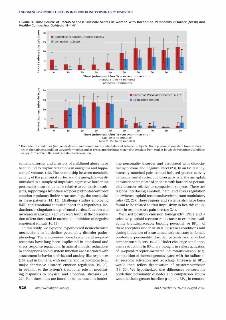
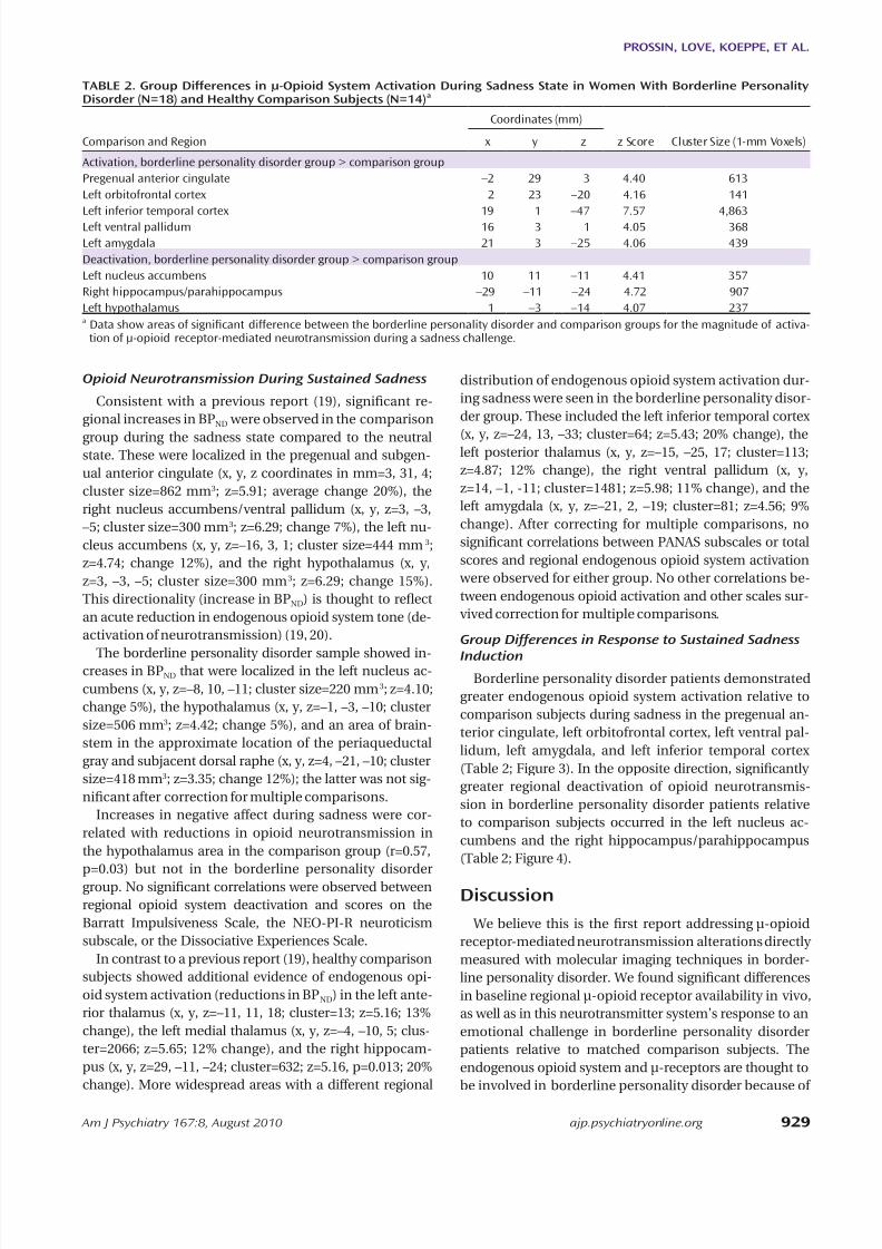
TABLE 1. Group Dierences in Baseline (Neutral State) μ-Opioid Receptor Binding Potential in Women With BorderlinePersonality Disorder (N=18) and Healthy Comparison Subjects (N=14)a
Comparison and Region
Coordinates (mm)
z Score Cluster Size (1-mm Voxels)x y z
Baseline, borderline personality disorder group > comparison group
Let orbitorontal cortex 14 38 –22 6.89 441
Right orbitorontal cortex –17 38 –20 6.03 1,294
Right caudate/nucleus accumbens –17 26 –2 6.10 972
Let caudate 19 26 5 4.41 517
Let nucleus accumbens 7 7 –12 7.98 1,780
Let amygdala 21 0 –23 5.42 6,719
Baseline, comparison group > borderline personality disorder group
Right posterior thalamus –10 –29 8 8.57 2,503
Let posterior thalamus 8 –29 10 7.66 3,891a Data show areas o signifcant dierence between the borderline personality disorder and comparison groups or baseline µ-opioid receptor
availability in vivo.
8/2/2019 appi.ajp.2010.09091348
http://slidepdf.com/reader/full/appiajp201009091348 4/9
ENDOGENOUS OPIOID FUNCTION IN BORDERLINE PERSONALITY DISORDER
928 ajp.psychiatryonline.org Am J Psychiatry 167:8, August 2010
insula, posterior thalamus, nucleus accumbens/ventral pallidum,and amygdala [19, 20]). Statistical thresholds or other regions werecorrected or type I error rate at p=0.05 or multiple comparisons(34). The BPND values were extracted rom image data by averaging the voxel values contained in an area where signicant dierences
were obtained down to a threshold o p<0.01. These values werethen used to plot the data and perorm correlation analyses, rul-ing out the presence o outliers. Planned analyses included Spear-man rank correlations between regional BP
NDvalues or sadness-
induced changes in BPND and the NEO-PI-R neuroticism subscore,the Barratt Impulsiveness Scale score, and the Dissociative Experi-ences Scale score and change in PANAS scores at p<0.05 with sub-sequent Bonerroni corrections or multiple comparisons.
Results
Sadness was maintained through the induced sadness
experimental period in both borderline personality disor-
der patients and comparison subjects (Figure 1). Repeated-
measures analysis o variance (ANOVA) showed signicant
eects o experimental condition (sadness > neutral [d=7,
1, 63; F=112.2, p<0.0001]) and an interaction between ex-
perimental condition and order (F=9.0, p=0.006), with
greater negative aect scores when sadness induction was
perormed rst. An eect o diagnosis was observed, with
the borderline personality disorder group showing greater
PANAS sadness subscale scores (F=4.2, p=0.05).
PANAS negative aect ratings acquired ater scan com-
pletion showed a mean score o 13 [SD=4] in the neutral
state and 19 [SD=7] in the sadness state in the comparison
group and 14 [SD=5] in the neutral state and 25 [SD=7] in
the sadness state in the borderline personality disorder
group. Repeated-measures ANOVA showed signicant e-
ects o condition (sadness > neutral [d=7, 1, 63; F=40.8,
p<0.000]) and diagnosis (borderline personality disorder
group > comparison group [F=5.5, p=0.03]). Order eects
did not achieve statistical signicance (F=3.8, p=0.06).
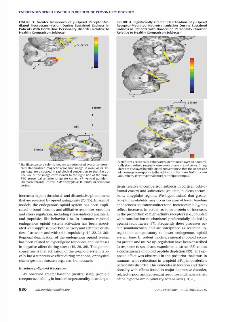
Baseline Measures
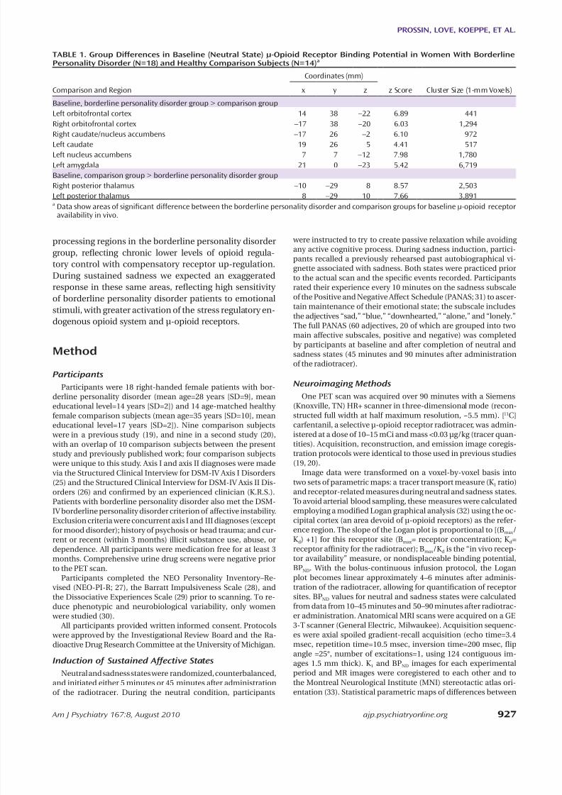
Baseline data (neutral state) comparisons between
groups showed signicantly greater μ-opioid BPND in bor-
derline personality disorder patients in the orbitorontal
cortex bilaterally; the caudate nucleus bilaterally, extending
into the nucleus accumbens on the right; the let nucleus
accumbens; and the let amygdala (Table 1, Figure 2). These
corresponded to average between-group dierences o 44%
and 47% or right and let orbitorontal cortex, respectively;44% and 32% or right and let caudate, respectively; 25%
or let nucleus accumbens; and 35% or let amygdala. The
comparison group demonstrated greater BPND in the poste-
rior thalamus bilaterally (Table 1), corresponding to group
dierences o 39% on the right and 37% on the let.
No signicant correlations were obtained between Bar-
ratt Impulsiveness Scale scores or NEO-PI-R neuroticism
subscale scores and baseline regional μ-opioid BPND. Dis-
sociative Experiences Scale scores were negatively cor-
related with BPND in the right caudate region (r=–0.57,
p=0.02), but this correlation did not persist ater correc-
tion or multiple comparisons.
conditions were generated by anatomically standardizing the T1 spoiled gradient-recall acquisition MRI o each participant to theMNI stereotactic atlas coordinates, with subsequent application
o this transormation to the receptor binding maps. The accu-racy o coregistration and nonlinear warping algorithms was con-rmed or each participant by comparing the transormed MRIand PET images to each other and to the atlas template.
Data Analysis
Dierences between groups and conditions were calculated with subtraction analyses and mapped into stereotactic space
with t maps o statistical signicance using the SPM2 ( WellcomeDepartment o Cognitive Neurology, University College, London)and MATLAB (MathWorks, Natick, Mass.) sotware packages with
a general linear model and correction or multiple comparisons.No global normalization was applied to the data; calculations pre-sented are based on absolute BPND estimates. Only regions withspecic μ-opioid receptor binding were included in the analyses(voxels with BPND values >0.1). Subtraction analyses were per-ormed on μ-opioid BPND images separately to assess main eects.For each subtraction analysis, one-sample paired t values werecalculated or each voxel using pooled variance across voxels. Sig-nicant dierences were detected using a statistical threshold o p<0.0001 or regions known to be involved in opioid modulation o aective and stress responses (the rostral anterior cingulate, me-
dial prerontal and orbitorontal cortex, inerior temporal cortex,
FIGURE 2. Greater Regional μ-Opioid BPND in Patients WithBorderline Personality Disorder Relative to Healthy Com-parison Subjectsa
Superior
Inferior
CAU/NAC
NAC
OFC
AMY
z Score
4
1
2
3
0
a Signifcant z score color values are superimposed over an anatomi-cally standardized magnetic resonance image in axial views. Im-age data are displayed in radiological convention so that the up-per side o the image corresponds to the right side o the brain.
CAU=nucleus caudate; NAC=nucleus accumbens; OFC=orbitorontalcortex, AMY=amygdala.
8/2/2019 appi.ajp.2010.09091348
http://slidepdf.com/reader/full/appiajp201009091348 5/9
PROSSIN, LOVE, KOEPPE, ET AL.
Am J Psychiatry 167:8, August 2010 ajp.psychiatryonline.org 929
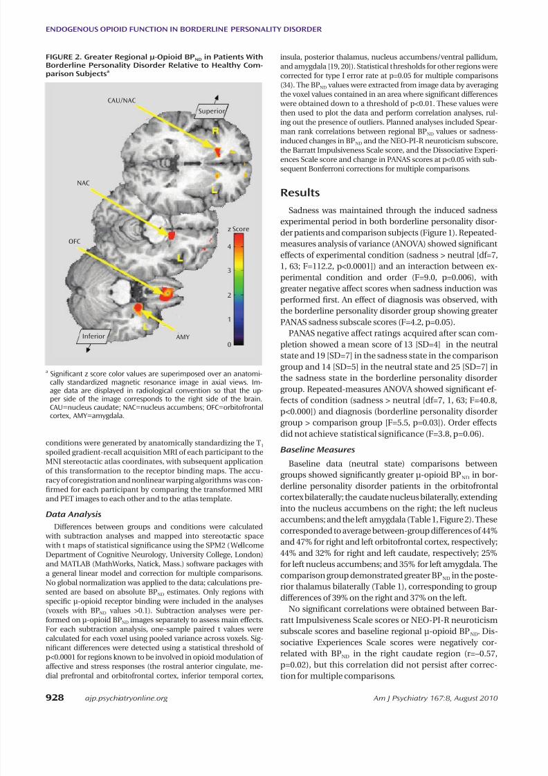
distribution o endogenous opioid system activation dur-
ing sadness were seen in the borderline personality disor-
der group. These included the let inerior temporal cortex
(x, y, z=–24, 13, –33; cluster=64; z=5.43; 20% change), thelet posterior thalamus (x, y, z=–15, –25, 17; cluster=113;
z=4.87; 12% change), the right ventral pallidum (x, y,
z=14, –1, -11; cluster=1481; z=5.98; 11% change), and the
let amygdala (x, y, z=–21, 2, –19; cluster=81; z=4.56; 9%
change). Ater correcting or multiple comparisons, no
signicant correlations between PANAS subscales or total
scores and regional endogenous opioid system activation
were observed or either group. No other correlations be-
tween endogenous opioid activation and other scales sur-
vived correction or multiple comparisons.
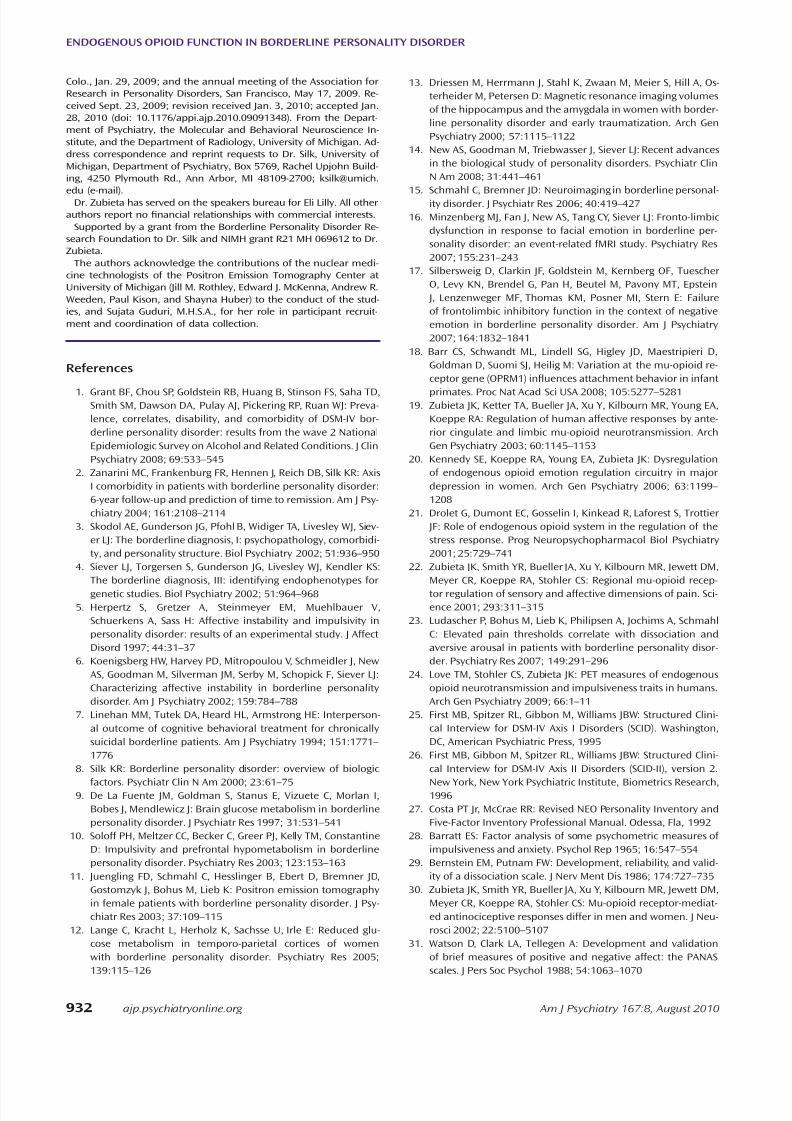
Group Differences in Response to Sustained Sadness
Induction
Borderline personality disorder patients demonstrated
greater endogenous opioid system activation relative to
comparison subjects during sadness in the pregenual an-
terior cingulate, let orbitorontal cortex, let ventral pal-
lidum, let amygdala, and let inerior temporal cortex
(Table 2; Figure 3). In the opposite direction, signicantly
greater regional deactivation o opioid neurotransmis-
sion in borderline personality disorder patients relative
to comparison subjects occurred in the let nucleus ac-
cumbens and the right hippocampus/parahippocampus
(Table 2; Figure 4).
Discussion
We believe this is the rst report addressing μ-opioid
receptor-mediated neurotransmission alterations directly
measured with molecular imaging techniques in border-
line personality disorder. We ound signicant dierences
in baseline regional μ-opioid receptor availability in vivo,
as well as in this neurotransmitter system’s response to an
emotional challenge in borderline personality disorder
patients relative to matched comparison subjects. The
endogenous opioid system and μ-receptors are thought to
be involved in borderline personality disorder because o
Opioid Neurotransmission During Sustained Sadness
Consistent with a previous report (19), signicant re-
gional increases in BPND were observed in the comparison
group during the sadness state compared to the neutralstate. These were localized in the pregenual and subgen-
ual anterior cingulate (x, y, z coordinates in mm=3, 31, 4;
cluster size=862 mm3; z=5.91; average change 20%), the
right nucleus accumbens/ventral pallidum (x, y, z=3, –3,
–5; cluster size=300 mm3; z=6.29; change 7%), the let nu-
cleus accumbens (x, y, z=–16, 3, 1; cluster size=444 mm3;
z=4.74; change 12%), and the right hypothalamus (x, y,
z=3, –3, –5; cluster size=300 mm3; z=6.29; change 15%).
This directionality (increase in BPND) is thought to refect
an acute reduction in endogenous opioid system tone (de-
activation o neurotransmission) (19, 20).
The borderline personality disorder sample showed in-creases in BPND that were localized in the let nucleus ac-
cumbens (x, y, z=–8, 10, –11; cluster size=220 mm3; z=4.10;
change 5%), the hypothalamus (x, y, z=–1, –3, –10; cluster
size=506 mm3; z=4.42; change 5%), and an area o brain-
stem in the approximate location o the periaqueductal
gray and subjacent dorsal raphe (x, y, z=4, –21, –10; cluster
size=418 mm3; z=3.35; change 12%); the latter was not sig-
nicant ater correction or multiple comparisons.
Increases in negative aect during sadness were cor-
related with reductions in opioid neurotransmission in
the hypothalamus area in the comparison group (r=0.57,
p=0.03) but not in the borderline personality disordergroup. No signicant correlations were observed between
regional opioid system deactivation and scores on the
Barratt Impulsiveness Scale, the NEO-PI-R neuroticism
subscale, or the Dissociative Experiences Scale.
In contrast to a previous report (19), healthy comparison
subjects showed additional evidence o endogenous opi-
oid system activation (reductions in BPND) in the let ante-
rior thalamus (x, y, z=–11, 11, 18; cluster=13; z=5.16; 13%
change), the let medial thalamus (x, y, z=–4, –10, 5; clus-
ter=2066; z=5.65; 12% change), and the right hippocam-
pus (x, y, z=29, –11, –24; cluster=632; z=5.16, p=0.013; 20%
change). More widespread areas with a dierent regional
TABLE 2. Group Dierences in μ-Opioid System Activation During Sadness State in Women With Borderline PersonalityDisorder (N=18) and Healthy Comparison Subjects (N=14)a
Comparison and Region
Coordinates (mm)
z Score Cluster Size (1-mm Voxels)x y z
Activation, borderline personality disorder group > comparison group
Pregenual anterior cingulate –2 29 3 4.40 613
Let orbitorontal cortex 2 23 –20 4.16 141
Let inerior temporal cortex 19 1 –47 7.57 4,863
Let ventral pallidum 16 3 1 4.05 368
Let amygdala 21 3 –25 4.06 439
Deactivation, borderline personality disorder group > comparison group
Let nucleus accumbens 10 11 –11 4.41 357
Right hippocampus/parahippocampus –29 –11 –24 4.72 907
Let hypothalamus 1 –3 –14 4.07 237a Data show areas o signifcant dierence between the borderline personality disorder and comparison groups or the magnitude o activa-
tion o µ-opioid receptor-mediated neurotransmission during a sadness challenge.
8/2/2019 appi.ajp.2010.09091348
http://slidepdf.com/reader/full/appiajp201009091348 6/9
ENDOGENOUS OPIOID FUNCTION IN BORDERLINE PERSONALITY DISORDER
930 ajp.psychiatryonline.org Am J Psychiatry 167:8, August 2010
tients relative to comparison subjects in cortical (orbito-
rontal cortex) and subcortical (caudate, nucleus accum-
bens, amygdala) regions. We hypothesized that greater
receptor availability may occur because o lower baseline
endogenous neurotransmitter tone. Increases in BPND may
refect increases in actual receptor protein or increases
in the proportion o high-anity receptors (i.e., coupled
with transduction mechanisms) preerentially labeled by
agonist radiotracers (37). Frequently these processes oc-
cur simultaneously and are interpreted as receptor up-
regulation compensatory to lower endogenous opioid
system tone. In rodent models, regional μ-opioid recep-
tor protein and mRNA up-regulation have been described
in response to social and experimental stress (38) and as
a consequence o opioid peptide depletion (39). The op-
posite eect was observed in the posterior thalamus in
humans, with reductions in μ-opioid BPND in borderline
personality disorder. This coincides in location and direc-
tionality with eects ound in major depressive disorder,
related to poor antidepressant response and hyperactivity
o the hypothalamic-pituitary-adrenal axis (19, 20).
increases in pain thresholds and dissociative phenomena
that are reversed by opioid antagonists (23, 35). In animal
models, the endogenous opioid system has been impli-
cated in bond-orming and aliative responses; emotion
and stress regulation, including stress-induced analgesia;
and impulsive-like behavior (18). In humans, regional
endogenous opioid system activation has been associ-
ated with suppression o both sensory and aective quali-
ties o stressors and with trait impulsivity (19, 22, 24, 36).
Regional deactivation o the endogenous opioid system
has been related to hyperalgesic responses and increases
in negative aect during stress (19, 29, 36). The general
consensus is that activation o the μ-opioid system typi-
cally has a suppressive eect during emotional or physical
challenges that threaten organism homeostasis.
Baseline μ-Opioid Receptors
We observed greater baseline (neutral state) μ-opioid
receptor availability in borderline personality disorder pa-
FIGURE 3. Greater Responses o μ-Opioid Receptor-Me-diated Neurotransmission During Sustained Sadness inPatients With Borderline Personality Disorder Relative toHealthy Comparison Subjectsa
Superior
Inferior
PGC
VP
OFC
AMY
z Score
4
1
2
3
0
ITC
Superior
Inferior
PGC
VP
FC
AMY
z Score
4
1
2
3
ITC
a Signifcant z score color values are superimposed over an anatomi-
cally standardized magnetic resonance image in axial views. Im-age data are displayed in radiological convention so that the up-per side o the image corresponds to the right side o the brain.PGC=pregenual anterior cingulate cortex; VP=ventral pallidum;OFC=orbitorontal cortex; AMY=amygdala; ITC=inerior temporalcortex.
FIGURE 4. Signifcantly Greater Deactivation o μ-OpioidReceptor-Mediated Neurotransmission During SustainedSadness in Patients With Borderline Personality DisorderRelative to Healthy Comparison Subjectsa
Superior
Inferior
NAC
HIP
z Score
4
1
2
3
0
HYP
a Signifcant z score color values are superimposed over an anatomi-cally standardized magnetic resonance image in axial views. Image
data are displayed in radiological convention so that the upper sideo the image corresponds to the right side o the brain. NAC=nucleusaccumbens; HYP=hypothalamus; HIP=hippocampus.
8/2/2019 appi.ajp.2010.09091348
http://slidepdf.com/reader/full/appiajp201009091348 7/9
PROSSIN, LOVE, KOEPPE, ET AL.
Am J Psychiatry 167:8, August 2010 ajp.psychiatryonline.org 931
was ound during sadness induction. I increases in baseline
receptor availability refect lower basal levels o dynamic
regulatory control o emotional states and stress by the
typically suppressive μ-opioid system, the response o these
circuits during recall o negative experiences would be con-
sistent with exaggerated stress-like responses to emotional
stimuli that were not observed in comparison subjects.
Greater μ-opioid availability and endogenous opioidrelease in response to painul stimuli appear to be asso-
ciated to impulsivity traits in healthy volunteers in loca-
tions overlapping with our ndings here (24). Serotonergic
mechanisms, long implicated in borderline personality
disorder pathophysiology (50, 51), along with dopaminer-
gic systems, have been associated with various orms o
impulsive behavior as well. Our ndings are consonant
with the concept that both borderline personality disor-
der and impulsivity are multiactorial, aecting relevant
circuits and involving various neurotransmitter systems,
including the endogenous opioid system (24). For exam-
ple, well-described neurotransmitter-neurotransmitterinteractions reveal regulation o serotonergic and dopa-
minergic cell ring by μ-opioid receptors in raphe (52) and
ventral tegmental nuclei (53), respectively.
This study was limited by using small numbers o study
subjects and including only women. While we think the
ndings are intriguing, we must be cautious in assuming
that they are readily generalizable.
Clinical Overview
Linehan et al. (7) suggested that borderline personality
disorder is characterized by a low threshold to becoming
emotionally dysregulated, ollowed by a rise to high lev-
els o emotional arousal, with a slow intensity decrement
over time. Greater emotional lability and dysregulation
may occur in patients with borderline personality disor-
der because they do not attribute the correct saliency to
an emotional event or stimulus (greater nucleus accum-
bens deactivation). Once they overattribute an emotion
to an event, they cannot shit away rom that emotion
or shit their emotional or interpersonal strategy (orbital
rontal cortex overactivation), even when the strategy is
not interpersonally productive. The stimulus may become
encoded with much greater emotional intensity (amyg-
dala-hippocampal eects) than it should have, leading to
greater reactivity to uture similar emotional events.Overall, we have described initial evidence o region-
al alterations in the unction o the endogenous opioid
system and μ-opioid receptors in brain regions involved
in emotion and stress processing, decision making, and
pain and neuroendocrine regulation. Further investiga-
tion into interindividual variations o the μ-opioid system
is warranted.
Presented in part at the 47th annual meeting o the American
College o Neuropsychopharmacology, Hollywood, Fla., Dec. 6–10,
2008; the Winter Conerence on Brain Research, Copper Mountain,
Response of μ-Opioid Neurotransmission During
Sustained Sadness
Relative to comparison subjects, patients with border-
line personality disorder demonstrated greater activation
o the endogenous opioid system in response to sustained
sadness in the pregenual anterior cingulate, let orbito-
rontal cortex, let ventral pallidum, and let amygdala.
These highly interconnected brain regions are implicatedin evaluation and behavioral responses to salient stimuli
and in decision making in healthy volunteers. Orbitoron-
tal cortex lesions are associated with poor decision making
and diculties in switching cognitive strategies (40). This
region has extensive connections with the cingulate gyrus,
nucleus accumbens, amygdala, hippocampus, and hypo-
thalamus (41), which together orm networks implicated
in assessing saliency, intensity, and valence o rewards and
stressors and in regulating behavioral responses (42, 43).
The let lateralization o many o the results is interest-
ing. Let lateralization has been described during success-
ul retrieval o emotional context in the amygdala and inrontotemporal networks (44).
Mu-opioid receptors in the ventral pallidum have been
implicated in encoding hedonic value o rewards (45), reg-
ulation o midbrain dopaminergic inputs, and integration
o amygdala and prerontal cortex connections, aecting
motivated behavior (46). Activation o the endogenous
opioid system in the orbitorontal cortex, anterior cin-
gulate, thalamus, nucleus accumbens, ventral pallidum,
and amygdala has been shown to suppress both physical
and emotional aspects o stressul challenges (19, 22, 47).
Consistent with those ndings, we observed negative but
nonsignicant correlations between endogenous opioidsystem activation in the let amygdala and Barratt Impul-
siveness Scale scores in the borderline personality disor-
der group. There were also negative but nonsignicant
correlations between let inerior temporal cortex activa-
tion and increases in negative aect during sadness, again
suggesting that in borderline personality disorder the en-
dogenous opioid system is involved in the suppression o
emotional responses. This interpretation corresponds to
reports o relie rom aversive arousal during cutting and
sel-injury (48) and increased pain thresholds in patients
with borderline personality disorder (23).
Patients with borderline personality disorder were alsodierentiated rom comparison subjects by a relative de-
activation o μ-opioid neurotransmission in the nucleus
accumbens, the hippocampus/parahippocampus, and
the hypothalamus. The nucleus accumbens is implicated
in the assignment o salience to both positive and nega-
tive events (42). The hippocampus and parahippocampus
have roles in explicit memory encoding and retrieval and
in autonomic nervous system and neuroendocrine regu-
lation through hypothalamic connections (49).
Brain regions where increases in neutral state μ-opioid
receptor availability were observed largely overlapped with
those in which greater endogenous opioid system activation
8/2/2019 appi.ajp.2010.09091348
http://slidepdf.com/reader/full/appiajp201009091348 8/9
ENDOGENOUS OPIOID FUNCTION IN BORDERLINE PERSONALITY DISORDER
932 ajp.psychiatryonline.org Am J Psychiatry 167:8, August 2010
13. Driessen M, Herrmann J, Stahl K, Zwaan M, Meier S, Hill A, Os-
terheider M, Petersen D: Magnetic resonance imaging volumes
o the hippocampus and the amygdala in women with border-
line personality disorder and early traumatization. Arch Gen
Psychiatry 2000; 57:1115–1122
14. New AS, Goodman M, Triebwasser J, Siever LJ: Recent advances
in the biological study o personality disorders. Psychiatr Clin
N Am 2008; 31:441–461
15. Schmahl C, Bremner JD: Neuroimaging in borderline personal-
ity disorder. J Psychiatr Res 2006; 40:419–427
16. Minzenberg MJ, Fan J, New AS, Tang CY, Siever LJ: Fronto-limbic
dysunction in response to acial emotion in borderline per-
sonality disorder: an event-related MRI study. Psychiatry Res
2007; 155:231–243
17. Silbersweig D, Clarkin JF, Goldstein M, Kernberg OF, Tuescher
O, Levy KN, Brendel G, Pan H, Beutel M, Pavony MT, Epstein
J, Lenzenweger MF, Thomas KM, Posner MI, Stern E: Failure
o rontolimbic inhibitory unction in the context o negative
emotion in borderline personality disorder. Am J Psychiatry
2007; 164:1832–1841
18. Barr CS, Schwandt ML, Lindell SG, Higley JD, Maestripieri D,
Goldman D, Suomi SJ, Heilig M: Variation at the mu-opioid re-
ceptor gene (OPRM1) infuences attachment behavior in inant
primates. Proc Nat Acad Sci USA 2008; 105:5277–528119. Zubieta JK, Ketter TA, Bueller JA, Xu Y, Kilbourn MR, Young EA,
Koeppe RA: Regulation o human aective responses by ante-
rior cingulate and limbic mu-opioid neurotransmission. Arch
Gen Psychiatry 2003; 60:1145–1153
20. Kennedy SE, Koeppe RA, Young EA, Zubieta JK: Dysregulation
o endogenous opioid emotion regulation circuitry in major
depression in women. Arch Gen Psychiatry 2006; 63:1199–
1208
21. Drolet G, Dumont EC, Gosselin I, Kinkead R, Laorest S, Trottier
JF: Role o endogenous opioid system in the regulation o the
stress response. Prog Neuropsychopharmacol Biol Psychiatry
2001; 25:729–741
22. Zubieta JK, Smith YR, Bueller JA, Xu Y, Kilbourn MR, Jewett DM,
Meyer CR, Koeppe RA, Stohler CS: Regional mu-opioid recep-
tor regulation o sensory and aective dimensions o pain. Sci-
ence 2001; 293:311–315
23. Ludascher P, Bohus M, Lieb K, Philipsen A, Jochims A, Schmahl
C: Elevated pain thresholds correlate with dissociation and
aversive arousal in patients with borderline personality disor-
der. Psychiatry Res 2007; 149:291–296
24. Love TM, Stohler CS, Zubieta JK: PET measures o endogenous
opioid neurotransmission and impulsiveness traits in humans.
Arch Gen Psychiatry 2009; 66:1–11
25. First MB, Spitzer RL, Gibbon M, Williams JBW: Structured Clini-
cal Interview or DSM-IV Axis I Disorders (SCID). Washington,
DC, American Psychiatric Press, 1995
26. First MB, Gibbon M, Spitzer RL, Williams JBW: Structured Clini-
cal Interview or DSM-IV Axis II Disorders (SCID-II), version 2.
New York, New York Psychiatric Institute, Biometrics Research,1996
27. Costa PT Jr, McCrae RR: Revised NEO Personality Inventory and
Five-Factor Inventory Proessional Manual. Odessa, Fla, 1992
28. Barratt ES: Factor analysis o some psychometric measures o
impulsiveness and anxiety. Psychol Rep 1965; 16:547–554
29. Bernstein EM, Putnam FW: Development, reliability, and valid-
ity o a dissociation scale. J Nerv Ment Dis 1986; 174:727–735
30. Zubieta JK, Smith YR, Bueller JA, Xu Y, Kilbourn MR, Jewett DM,
Meyer CR, Koeppe RA, Stohler CS: Mu-opioid receptor-mediat-
ed antinociceptive responses dier in men and women. J Neu-
rosci 2002; 22:5100–5107
31. Watson D, Clark LA, Tellegen A: Development and validation
o brie measures o positive and negative aect: the PANAS
scales. J Pers Soc Psychol 1988; 54:1063–1070
Colo., Jan. 29, 2009; and the annual meeting o the Association or
Research in Personality Disorders, San Francisco, May 17, 2009. Re-
ceived Sept. 23, 2009; revision received Jan. 3, 2010; accepted Jan.
28, 2010 (doi: 10.1176/appi.ajp.2010.09091348). From the Depart-
ment o Psychiatry, the Molecular and Behavioral Neuroscience In-
stitute, and the Department o Radiology, University o Michigan. Ad-
dress correspondence and reprint requests to Dr. Silk, University o
Michigan, Department o Psychiatry, Box 5769, Rachel Upjohn Build-
ing, 4250 Plymouth Rd., Ann Arbor, MI 48109-2700; ksilk@umich.
edu (e-mail).
Dr. Zubieta has served on the speakers bureau or Eli Lilly. All other
authors report no fnancial relationships with commercial interests.
Supported by a grant rom the Borderline Personality Disorder Re-
search Foundation to Dr. Silk and NIMH grant R21 MH 069612 to Dr.
Zubieta.
The authors acknowledge the contributions o the nuclear medi-
cine technologists o the Positron Emission Tomography Center at
University o Michigan (Jill M. Rothley, Edward J. McKenna, Andrew R.
Weeden, Paul Kison, and Shayna Huber) to the conduct o the stud-
ies, and Sujata Guduri, M.H.S.A., or her role in participant recruit-
ment and coordination o data collection.
Reerences
1. Grant BF, Chou SP, Goldstein RB, Huang B, Stinson FS, Saha TD,Smith SM, Dawson DA, Pulay AJ, Pickering RP, Ruan WJ: Preva-
lence, correlates, disability, and comorbidity o DSM-IV bor-
derline personality disorder: results rom the wave 2 National
Epidemiologic Survey on Alcohol and Related Conditions. J Clin
Psychiatry 2008; 69:533–545
2. Zanarini MC, Frankenburg FR, Hennen J, Reich DB, Silk KR: Axis
I comorbidity in patients with borderline personality disorder:
6-year ollow-up and prediction o time to remission. Am J Psy-
chiatry 2004; 161:2108–2114
3. Skodol AE, Gunderson JG, Pohl B, Widiger TA, Livesley WJ, Siev-
er LJ: The borderline diagnosis, I: psychopathology, comorbidi-
ty, and personality structure. Biol Psychiatry 2002; 51:936–950
4. Siever LJ, Torgersen S, Gunderson JG, Livesley WJ, Kendler KS:
The borderline diagnosis, III: identiying endophenotypes or
genetic studies. Biol Psychiatry 2002; 51:964–968
5. Herpertz S, Gretzer A, Steinmeyer EM, Muehlbauer V,
Schuerkens A, Sass H: Aective instability and impulsivity in
personality disorder: results o an experimental study. J Aect
Disord 1997; 44:31–37
6. Koenigsberg HW, Harvey PD, Mitropoulou V, Schmeidler J, New
AS, Goodman M, Silverman JM, Serby M, Schopick F, Siever LJ:
Characterizing aective instability in borderline personality
disorder. Am J Psychiatry 2002; 159:784–788
7. Linehan MM, Tutek DA, Heard HL, Armstrong HE: Interperson-
al outcome o cognitive behavioral treatment or chronically
suicidal borderline patients. Am J Psychiatry 1994; 151:1771–
1776
8. Silk KR: Borderline personality disorder: overview o biologic
actors. Psychiatr Clin N Am 2000; 23:61–759. De La Fuente JM, Goldman S, Stanus E, Vizuete C, Morlan I,
Bobes J, Mendlewicz J: Brain glucose metabolism in borderline
personality disorder. J Psychiatr Res 1997; 31:531–541
10. Solo PH, Meltzer CC, Becker C, Greer PJ, Kelly TM, Constantine
D: Impulsivity and prerontal hypometabolism in borderline
personality disorder. Psychiatry Res 2003; 123:153–163
11. Juengling FD, Schmahl C, Hesslinger B, Ebert D, Bremner JD,
Gostomzyk J, Bohus M, Lieb K: Positron emission tomography
in emale patients with borderline personality disorder. J Psy-
chiatr Res 2003; 37:109–115
12. Lange C, Kracht L, Herholz K, Sachsse U, Irle E: Reduced glu-
cose metabolism in temporo-parietal cortices o women
with borderline personality disorder. Psychiatry Res 2005;
139:115–126
8/2/2019 appi.ajp.2010.09091348
http://slidepdf.com/reader/full/appiajp201009091348 9/9
PROSSIN, LOVE, KOEPPE, ET AL.
Am J Psychiatry 167:8, August 2010 ajp.psychiatryonline.org 933
42. Tobler PN, Fiorillo CD, Schultz W: Adaptive coding o reward
value by dopamine neurons. Science 2005; 307:1642–1645
43. Tom SM, Fox CR, Trepel C, Poldrack RA: The neural basis o
loss aversion in decision-making under risk. Science 2007;
315:515–518
44. Smith AP, Henson RN, Rugg MD, Dolan RJ: Modulation o re-
trieval processing refects accuracy o emotional source mem-
ory. Learn Mem 2005; 12:472–479
45. Smith KS, Berridge KC: Opioid limbic circuit or reward: inter-
action between hedonic hotspots o nucleus accumbens and
ventral pallidum. J Neurosci 2007; 27:1594–1605
46. Horvitz JC: Mesolimbocortical and nigrostriatal dopamine
responses to salient non-reward events. Neuroscience 2000;
96:651–656
47. Ribeiro SC, Kennedy SE, Smith YR, Stohler CS, Zubieta JK: In-
terace o physical and emotional stress regulation through
the endogenous opioid system and mu-opioid receptors.
Prog Neuropsychopharmacol Biol Psychiatry 2005; 29:1264–
1280
48. Leibenlut EGD, Cowdry RW: The inner experience o the bor-
derline sel-mutilator. J Pers Disord 1987; 1:317–324
49. McEwen BS: Central eects o stress hormones in health and
disease: understanding the protective and damaging eects o
stress and stress mediators. Eur J Pharmacol 2008; 583:174– 185
50. Solo PH, Price JC, Meltzer CC, Fabio A, Frank GK, Kaye WH:
5HT2A receptor binding is increased in borderline personality
disorder. Biol Psychiatry 2007; 62:580–587
51. Wagner S, Baskaya O, Lieb K, Dahmen N, Tadic A: The 5-HT-
TLPR polymorphism modulates the association o serious lie
events (SLE) and impulsivity in patients with borderline person-
ality disorder. J Psychiatr Res 2009; 43:1067–1072
52. Jolas T, Nestler EJ, Aghajanian GK: Chronic morphine increases
GABA tone on serotonergic neurons o the dorsal raphe nucle-
us: association with an up-regulation o the cyclic AMP path-
way. Neuroscience 2000; 95:433–443
53. Svingos AL, Garzón M, Colago EE, Pickel VM: Mu-opioid recep-
tors in the ventral tegmental area are targeted to presynapti-
cally and directly modulate mesocortical projection neurons.
Synapse 2001; 41:221–229
32. Logan J, Fowler JS, Volkow ND, Wang GJ, Ding YS, Alexo DL:
Distribution volume ratios without blood sampling rom
graphical analysis o PET data. J Cereb Blood Flow Metab 1996;
16:834–840
33. Meyer CR, Boes JL, Kim B, Bland PH, Zasadny KR, Kison PV, Ko-
ral K, Frey KA, Wahl RL: Demonstration o accuracy and clinical
versatility o mutual inormation or automatic multimodality
image usion using ane and thin-plate spline warped geo-
metric deormations. Med Image Anal 1997; 1:195–206
34. Friston KJ, Frith CD, Liddle PF, Frackowiak RS: Comparing unc-
tional (PET) images: the assessment o signicant change. J
Cereb Blood Flow Metab 1991; 11:690–699
35. Bohus MJ, Landwehrmeyer GB, Stiglmayr CE, Limberger MF,
Bohme R, Schmahl CG: Naltrexone in the treatment o disso-
ciative symptoms in patients with borderline personality dis-
order: an open-label trial. J Clin Psychiatry 1999; 60:598–603
36. Scott DJ, Stohler CS, Egnatuk CM, Wang H, Koeppe RA, Zubieta
JK: Placebo and nocebo eects are dened by opposite opi-
oid and dopaminergic responses. Arch Gen Psychiatry 2008;
65:220–231
37. Narendran R, Hwang DR, Slistein M, Talbot PS, Erritzoe D,
Huang Y, Cooper TB, Martinez D, Kegeles LS, Abi-Dargham A,
Laruelle M: In vivo vulnerability to competition by endogenous
dopamine: comparison o the D2 receptor agonist radiotracer(-)-N-[11C]propyl-norapomorphine ([11C]NPA) with the D2 re-
ceptor antagonist radiotracer [11C]-raclopride. Synapse 2004;
52:188–208
38. Nikulina EM, Miczek KA, Hammer RP: Prolonged eects o re-
peated social deeat stress on mRNA expression and unction
o mu-opioid receptors in the ventral tegmental area o rats.
Neuropsychopharmacol 2005; 30:1096–1103
39. Brady LS, Herkenham M, Rothman RB, Partilla JS, Konig M,
Zimmer AM, Zimmer A: Region-specic up-regulation o opioid
receptor binding in enkephalin knockout mice. Molec Brain
Res 1999; 68:193–197
40. Wallis JD: Orbitorontal cortex and its contribution to decision-
making. Annu Rev Neurosci 2007; 30:31–56
41. Carmichael ST, Price JL: Limbic connections o the orbital and
medial prerontal cortex in macaque monkeys. J Comp Neurol
1995; 363:615–641
´


