Embed Size (px)
Citation preview
28
Anestezjologia i Ratownictwo 2016; 10: 28-33
Nauka praktyce / Science for medical practice
A R T Y K U Ł O R Y G I N A L N Y / O R I G I N A L PA P E R Submitted: 23.02.2016 • Accepted: 08.03.2016 © Akademia Medycyny
Application of ultrasound protocols in immediate postoperative care
Anna Róziewska, Ewa Puchalska, Elżbieta Nowacka, Lidia Jureczko, Tomasz Łazowski1stDepartmentofAnaesthesiologyandIntensiveCare,MedicalUniversityofWarsaw,Poland
Abstract
Background. Clinical application of point-of-care ultrasound in immediate postoperative period is still to be evaluated. The aim of this report is to present the possible application of the critical care ultrasound protocols for early detection of the most common postoperative complications. Material and methods. 73 women between 27 and 74 years of age, ASA (American Society of Anesthesiologists) grade 1-3, up to 6 hours after gynecologi-cal procedures or cesarean sections were evaluated. Examination consisted of lung ultrasound and fluid status assessment. Results. In 5 laparotomy cases interstitial pulmonary oedema was noted, which was associated with mildly decreased oxygen saturation readings of 92-95%. In only one of these cases fluid status indices were sug-gestive of hypervolemia, while the remaining four have shown the sonographic signs of hypovolemia. Subpleural consolidations were detected in one patient. In 2 cases pleural effusion and atelectasis were found. One case of postoperative hemorrhage was shown. In 23% of patients fluid status indices were suggestive of hypovolemia, while in 6% hypovolemia was shown to be severe. Conclusion. It appears that ultrasound imaging may help to improve the quality of postoperative care in terms of early detection of possible complications and fluid status assessment. Anestezjologia i Ratownictwo 2016; 10: 28-33.
Keywords: ultrasonography, fluid status assessment, recovery room, lung ultrasound
28
Introduction
Within the last few years the ultrasound imaging has invaded anaesthesia and intensive care. It caused major shifts in regional anaesthesia techniques, changed the way we approach the issues of central venous access and is widely used in the intensive care and pain man-agement. It is well established that the introduction of sonographic guidance has improved both quality and safety of the invasive procedures for which up to quite recently no other options than blind technique was at hand [1,2]. Improved availability of the smaller, less
complex and more efficient devices has made many doctors to change their initially reluctant approach and use the ultrasound for diagnostic and therapeutic purposes. Intensive care has improved significantly since it is possible to perform a point-of-care ultrasound examination. Non-invasive, repetitive exams allow to monitor the course of the disease and adjust the imple-mented therapy if found not to be sufficiently effective. There is also promising data suggesting that the number of X-Rays and CT scans that are ordered by intensive care units is much less in facilities where ultrasound was introduced as a routine diagnostic feature [3].
29
Anestezjologia i Ratownictwo 2016; 10: 28-33
Nauka praktyce / Science for medical practice
surgery varied: gynaecological laparoscopies (LPRS), laparotomies (LPRT) and cesarean sections (CS). Demographic preoperative data were noted, as well as the type of anaesthesia, blood loss, intraopera-tive and postoperative fluid management. Before the exam the vital signs were recorded (BP- blood pres-sure, HR-heart rate, oxygen saturation), as well as the presence of symptoms like nausea or shortness of breath. Ultrasound examination was performed up to 6 hours after the end of surgery. For the benefit of comparative analysis each type of surgical intervention was regarded separately. Patient evaluation consisted of lung ultrasound and fluid status assessment. Lung ultrasound was performed with either convex or sec-tor transducer, the results from three different points on both sides of chest were recorded (BLUE protocol) [6]. Any abnormality noted in BLUE protocol was an indication for further intercostal spaces to be assessed.
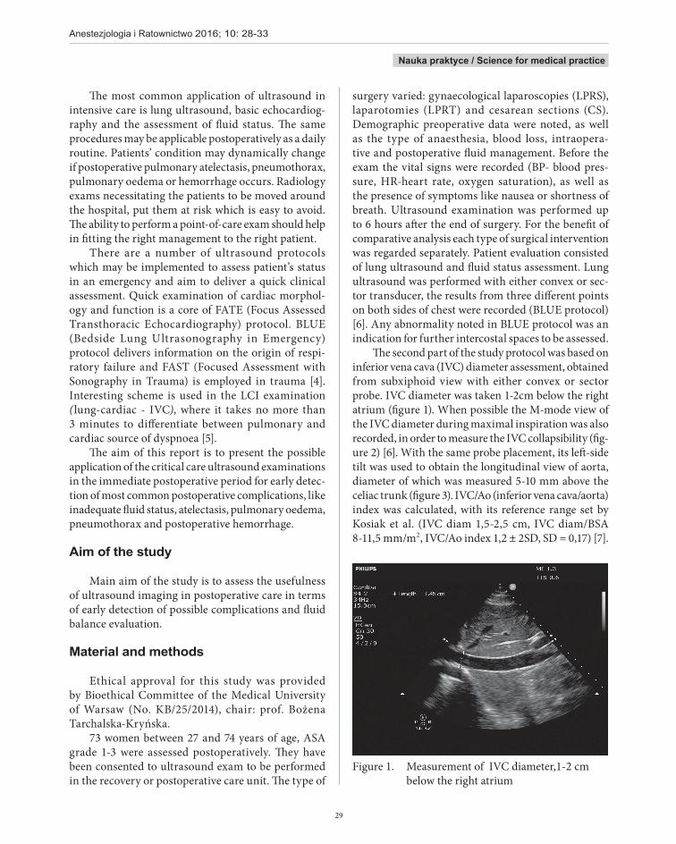
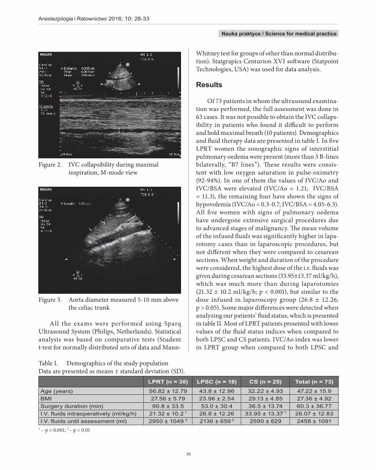
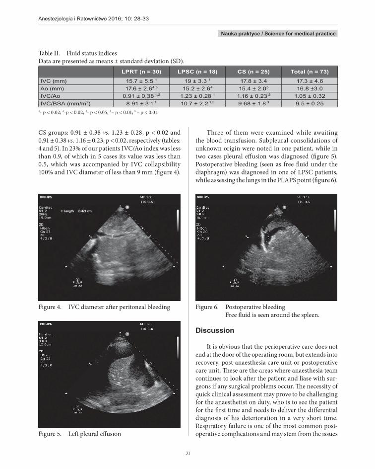
The second part of the study protocol was based on inferior vena cava (IVC) diameter assessment, obtained from subxiphoid view with either convex or sector probe. IVC diameter was taken 1-2cm below the right atrium (figure 1). When possible the M-mode view of the IVC diameter during maximal inspiration was also recorded, in order to measure the IVC collapsibility (fig-ure 2) [6]. With the same probe placement, its left-side tilt was used to obtain the longitudinal view of aorta, diameter of which was measured 5-10 mm above the celiac trunk (figure 3). IVC/Ao (inferior vena cava/aorta) index was calculated, with its reference range set by Kosiak et al. (IVC diam 1,5-2,5 cm, IVC diam/BSA 8-11,5 mm/m2, IVC/Ao index 1,2 ± 2SD, SD = 0,17) [7].
Figure 1. Measurement of IVC diameter,1-2 cm below the right atrium
The most common application of ultrasound in intensive care is lung ultrasound, basic echocardiog-raphy and the assessment of fluid status. The same procedures may be applicable postoperatively as a daily routine. Patients’ condition may dynamically change if postoperative pulmonary atelectasis, pneumothorax, pulmonary oedema or hemorrhage occurs. Radiology exams necessitating the patients to be moved around the hospital, put them at risk which is easy to avoid. The ability to perform a point-of-care exam should help in fitting the right management to the right patient.
There are a number of ultrasound protocols which may be implemented to assess patient’s status in an emergency and aim to deliver a quick clinical assessment. Quick examination of cardiac morphol-ogy and function is a core of FATE (Focus Assessed Transthoracic Echocardiography) protocol. BLUE (Bedside Lung Ultrasonography in Emergency) protocol delivers information on the origin of respi-ratory failure and FAST (Focused Assessment with Sonography in Trauma) is employed in trauma [4]. Interesting scheme is used in the LCI examination (lung-cardiac - IVC), where it takes no more than 3 minutes to differentiate between pulmonary and cardiac source of dyspnoea [5].
The aim of this report is to present the possible application of the critical care ultrasound examinations in the immediate postoperative period for early detec-tion of most common postoperative complications, like inadequate fluid status, atelectasis, pulmonary oedema, pneumothorax and postoperative hemorrhage.
Aim of the study
Main aim of the study is to assess the usefulness of ultrasound imaging in postoperative care in terms of early detection of possible complications and fluid balance evaluation.
Material and methods
Ethical approval for this study was provided by Bioethical Committee of the Medical University of Warsaw (No. KB/25/2014), chair: prof. Bożena Tarchalska-Kryńska.
73 women between 27 and 74 years of age, ASA grade 1-3 were assessed postoperatively. They have been consented to ultrasound exam to be performed in the recovery or postoperative care unit. The type of
30
Anestezjologia i Ratownictwo 2016; 10: 28-33
Nauka praktyce / Science for medical practice
Figure 2. IVC collapsibility during maximal inspiration, M-mode view
Figure 3. Aorta diameter measured 5-10 mm above the celiac trunk
All the exams were performed using Sparq Ultrasound System (Philips, Netherlands). Statistical analysis was based on comparative tests (Student t-test for normally distributed sets of data and Mann-
Whitney test for groups of other than normal distribu-tion). Statgrapics Centurion XVI software (Statpoint Technologies, USA) was used for data analysis.
Results
Of 73 patients in whom the ultrasound examina-tion was performed, the full assessment was done in 63 cases. It was not possible to obtain the IVC collaps-ibility in patients who found it difficult to perform and hold maximal breath (10 patients). Demographics and fluid therapy data are presented in table I. In five LPRT women the sonographic signs of interstitial pulmonary oedema were present (more than 3 B-lines bilaterally, “B7 lines”). These results were consis-tent with low oxygen saturation in pulse-oximetry (92-94%). In one of them the values of IVC/Ao and IVC/BSA were elevated (IVC/Ao = 1.21; IVC/BSA = 11.3), the remaining four have shown the signs of hypovolemia (IVC/Ao = 0.3-0.7; IVC/BSA = 4.05-6.3). All five women with signs of pulmonary oedema have undergone extensive surgical procedures due to advanced stages of malignancy. The mean volume of the infused fluids was significantly higher in lapa-rotomy cases than in laparoscopic procedures, but not different when they were compared to cesarean sections. When weight and duration of the procedure were considered, the highest dose of the i.v. fluids was given during cesarean sections (33.95±13.37 ml/kg/h), which was much more than during laparotomies (21.32 ± 10.2 ml/kg/h; p < 0.001), but similar to the dose infused in laparoscopy group (26.8 ± 12.26; p > 0.05). Some major differences were detected when analyzing our patients’ fluid status, which is presented in table II. Most of LPRT patients presented with lower values of the fluid status indices when compared to both LPSC and CS patients. IVC/Ao index was lower in LPRT group when compared to both LPSC and
Table I. Demographics of the study populationData are presented as means ± standard deviation (SD).
LPRT (n = 30) LPSC (n = 18) CS (n = 25) Total (n = 73)
Age(years) 56.82 ± 12.79 43.8 ± 12.96 32.22 ± 4.93 47.22 ± 15.9BMI 27.56 ± 5.79 23.96 ± 2.54 29.13 ± 4.85 27.36 ± 4.92Surgeryduration(min) 90.8 ± 33.5 53.0 ± 30.4 36.5 ± 13.74 60.3 ± 36.77I.V.fluidsintraoperatively(ml/kg/h) 21.32 ± 10.2 1 26.8 ± 12.26 33.95 ± 13.37 1 26.07 ± 12.83I.V.fluidsuntilassessment(ml) 2950 ± 1049 2 2136 ± 659 2 2590 ± 829 2458 ± 1091
1 – p < 0.001; 2 – p < 0.01
31
Anestezjologia i Ratownictwo 2016; 10: 28-33
Nauka praktyce / Science for medical practice
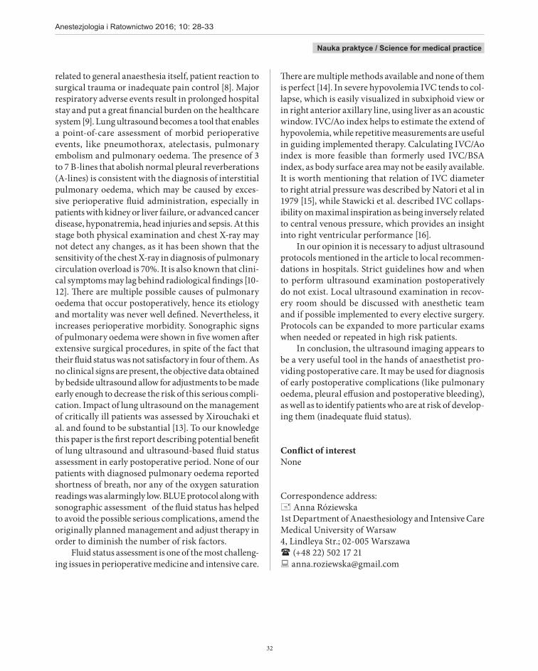
CS groups: 0.91 ± 0.38 vs. 1.23 ± 0.28, p < 0.02 and 0.91 ± 0.38 vs. 1.16 ± 0.23, p < 0.02, respectively (tables: 4 and 5). In 23% of our patients IVC/Ao index was less than 0.9, of which in 5 cases its value was less than 0.5, which was accompanied by IVC collapsibility 100% and IVC diameter of less than 9 mm (figure 4).
Figure 4. IVC diameter after peritoneal bleeding
Figure 5. Left pleural effusion
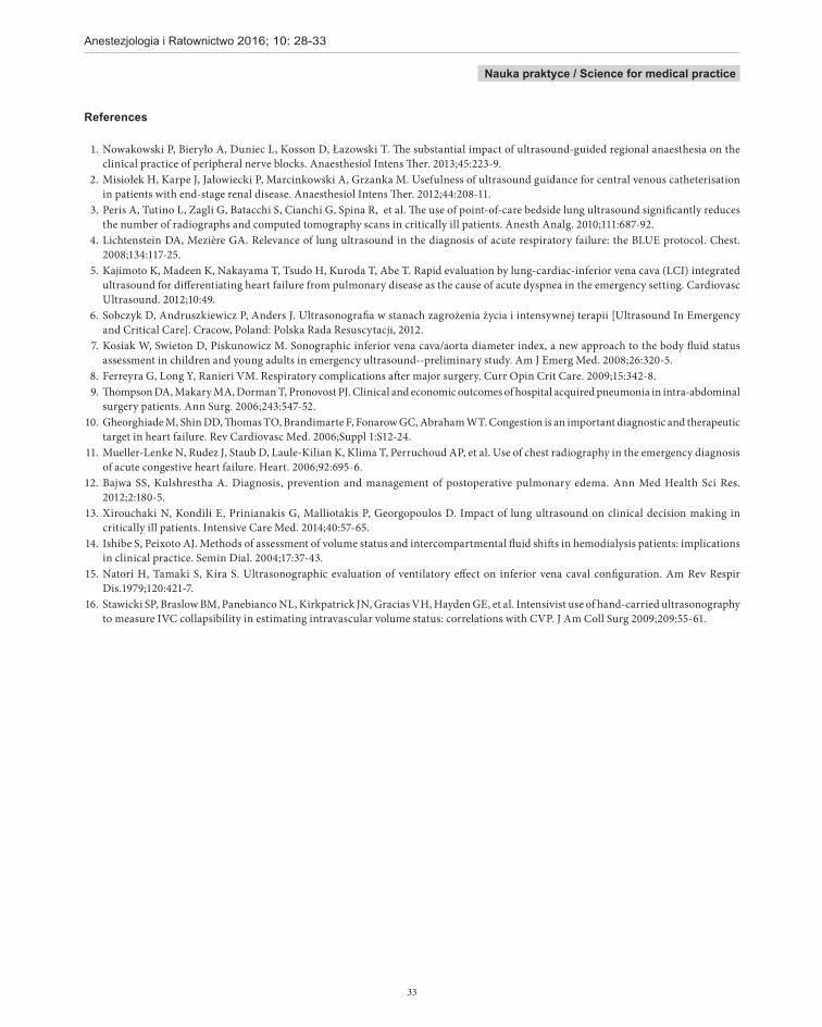
Three of them were examined while awaiting the blood transfusion. Subpleural consolidations of unknown origin were noted in one patient, while in two cases pleural effusion was diagnosed (figure 5). Postoperative bleeding (seen as free fluid under the diaphragm) was diagnosed in one of LPSC patients, while assessing the lungs in the PLAPS point (figure 6).
Figure 6. Postoperative bleeding Free fluid is seen around the spleen.
Discussion
It is obvious that the perioperative care does not end at the door of the operating room, but extends into recovery, post-anaesthesia care unit or postoperative care unit. These are the areas where anaesthesia team continues to look after the patient and liase with sur-geons if any surgical problems occur. The necessity of quick clinical assessment may prove to be challenging for the anaesthetist on duty, who is to see the patient for the first time and needs to deliver the differential diagnosis of his deterioration in a very short time. Respiratory failure is one of the most common post-operative complications and may stem from the issues
Table II. Fluid status indicesData are presented as means ± standard deviation (SD).
LPRT (n = 30) LPSC (n = 18) CS (n = 25) Total (n = 73)
IVC(mm) 15.7 ± 5.5 1 19 ± 3.3 1 17.8 ± 3.4 17.3 ± 4.6Ao(mm) 17.6 ± 2.64,5 15.2 ± 2.64 15.4 ± 2.05 16.8 ±3.0IVC/Ao 0.91 ± 0.381,2 1.23 ± 0.28 1 1.16 ± 0.23 2 1.05 ± 0.32IVC/BSA(mm/m2) 8.91 ± 3.1 1 10.7 ± 2.21,3 9.68 ± 1.8 3 9.5 ± 0.25
1- p < 0.02; 2-p < 0.02; 3- p < 0.05; 4– p < 0.01; 5 – p < 0.01.
32
Anestezjologia i Ratownictwo 2016; 10: 28-33
Nauka praktyce / Science for medical practice
related to general anaesthesia itself, patient reaction to surgical trauma or inadequate pain control [8]. Major respiratory adverse events result in prolonged hospital stay and put a great financial burden on the healthcare system [9]. Lung ultrasound becomes a tool that enables a point-of-care assessment of morbid perioperative events, like pneumothorax, atelectasis, pulmonary embolism and pulmonary oedema. The presence of 3 to 7 B-lines that abolish normal pleural reverberations (A-lines) is consistent with the diagnosis of interstitial pulmonary oedema, which may be caused by exces-sive perioperative fluid administration, especially in patients with kidney or liver failure, or advanced cancer disease, hyponatremia, head injuries and sepsis. At this stage both physical examination and chest X-ray may not detect any changes, as it has been shown that the sensitivity of the chest X-ray in diagnosis of pulmonary circulation overload is 70%. It is also known that clini-cal symptoms may lag behind radiological findings [10-12]. There are multiple possible causes of pulmonary oedema that occur postoperatively, hence its etiology and mortality was never well defined. Nevertheless, it increases perioperative morbidity. Sonographic signs of pulmonary oedema were shown in five women after extensive surgical procedures, in spite of the fact that their fluid status was not satisfactory in four of them. As no clinical signs are present, the objective data obtained by bedside ultrasound allow for adjustments to be made early enough to decrease the risk of this serious compli-cation. Impact of lung ultrasound on the management of critically ill patients was assessed by Xirouchaki et al. and found to be substantial [13]. To our knowledge this paper is the first report describing potential benefit of lung ultrasound and ultrasound-based fluid status assessment in early postoperative period. None of our patients with diagnosed pulmonary oedema reported shortness of breath, nor any of the oxygen saturation readings was alarmingly low. BLUE protocol along with sonographic assessment of the fluid status has helped to avoid the possible serious complications, amend the originally planned management and adjust therapy in order to diminish the number of risk factors.
Fluid status assessment is one of the most challeng-ing issues in perioperative medicine and intensive care.
There are multiple methods available and none of them is perfect [14]. In severe hypovolemia IVC tends to col-lapse, which is easily visualized in subxiphoid view or in right anterior axillary line, using liver as an acoustic window. IVC/Ao index helps to estimate the extend of hypovolemia, while repetitive measurements are useful in guiding implemented therapy. Calculating IVC/Ao index is more feasible than formerly used IVC/BSA index, as body surface area may not be easily available. It is worth mentioning that relation of IVC diameter to right atrial pressure was described by Natori et al in 1979 [15], while Stawicki et al. described IVC collaps-ibility on maximal inspiration as being inversely related to central venous pressure, which provides an insight into right ventricular performance [16].
In our opinion it is necessary to adjust ultrasound protocols mentioned in the article to local recommen-dations in hospitals. Strict guidelines how and when to perform ultrasound examination postoperatively do not exist. Local ultrasound examination in recov-ery room should be discussed with anesthetic team and if possible implemented to every elective surgery. Protocols can be expanded to more particular exams when needed or repeated in high risk patients.
In conclusion, the ultrasound imaging appears to be a very useful tool in the hands of anaesthetist pro-viding postoperative care. It may be used for diagnosis of early postoperative complications (like pulmonary oedema, pleural effusion and postoperative bleeding), as well as to identify patients who are at risk of develop-ing them (inadequate fluid status).
Conflict of interest None
Correspondence address: Anna Róziewska1st Department of Anaesthesiology and Intensive CareMedical University of Warsaw4, Lindleya Str.; 02-005 Warszawa (+48 22) 502 17 21 [email protected]
33
Anestezjologia i Ratownictwo 2016; 10: 28-33
Nauka praktyce / Science for medical practice
References
1. Nowakowski P, Bieryło A, Duniec L, Kosson D, Łazowski T. The substantial impact of ultrasound-guided regional anaesthesia on the clinical practice of peripheral nerve blocks. Anaesthesiol Intens Ther. 2013;45:223-9.
2. Misiołek H, Karpe J, Jałowiecki P, Marcinkowski A, Grzanka M. Usefulness of ultrasound guidance for central venous catheterisation in patients with end-stage renal disease. Anaesthesiol Intens Ther. 2012;44:208-11.
3. Peris A, Tutino L, Zagli G, Batacchi S, Cianchi G, Spina R, et al. The use of point-of-care bedside lung ultrasound significantly reduces the number of radiographs and computed tomography scans in critically ill patients. Anesth Analg. 2010;111:687-92.
4. Lichtenstein DA, Mezière GA. Relevance of lung ultrasound in the diagnosis of acute respiratory failure: the BLUE protocol. Chest. 2008;134:117-25.
5. Kajimoto K, Madeen K, Nakayama T, Tsudo H, Kuroda T, Abe T. Rapid evaluation by lung-cardiac-inferior vena cava (LCI) integrated ultrasound for differentiating heart failure from pulmonary disease as the cause of acute dyspnea in the emergency setting. Cardiovasc Ultrasound. 2012;10:49.
6. Sobczyk D, Andruszkiewicz P, Anders J. Ultrasonografia w stanach zagrożenia życia i intensywnej terapii [Ultrasound In Emergency and Critical Care]. Cracow, Poland: Polska Rada Resuscytacji, 2012.
7. Kosiak W, Swieton D, Piskunowicz M. Sonographic inferior vena cava/aorta diameter index, a new approach to the body fluid status assessment in children and young adults in emergency ultrasound--preliminary study. Am J Emerg Med. 2008;26:320-5.
8. Ferreyra G, Long Y, Ranieri VM. Respiratory complications after major surgery. Curr Opin Crit Care. 2009;15:342-8. 9. Thompson DA, Makary MA, Dorman T, Pronovost PJ. Clinical and economic outcomes of hospital acquired pneumonia in intra-abdominal
surgery patients. Ann Surg. 2006;243:547-52. 10. Gheorghiade M, Shin DD, Thomas TO, Brandimarte F, Fonarow GC, Abraham WT. Congestion is an important diagnostic and therapeutic
target in heart failure. Rev Cardiovasc Med. 2006;Suppl 1:S12-24. 11. Mueller-Lenke N, Rudez J, Staub D, Laule-Kilian K, Klima T, Perruchoud AP, et al. Use of chest radiography in the emergency diagnosis
of acute congestive heart failure. Heart. 2006;92:695-6. 12. Bajwa SS, Kulshrestha A. Diagnosis, prevention and management of postoperative pulmonary edema. Ann Med Health Sci Res.
2012;2:180-5. 13. Xirouchaki N, Kondili E, Prinianakis G, Malliotakis P, Georgopoulos D. Impact of lung ultrasound on clinical decision making in
critically ill patients. Intensive Care Med. 2014;40:57-65. 14. Ishibe S, Peixoto AJ. Methods of assessment of volume status and intercompartmental fluid shifts in hemodialysis patients: implications
in clinical practice. Semin Dial. 2004;17:37-43. 15. Natori H, Tamaki S, Kira S. Ultrasonographic evaluation of ventilatory effect on inferior vena caval configuration. Am Rev Respir
Dis.1979;120:421-7. 16. Stawicki SP, Braslow BM, Panebianco NL, Kirkpatrick JN, Gracias VH, Hayden GE, et al. Intensivist use of hand-carried ultrasonography
to measure IVC collapsibility in estimating intravascular volume status: correlations with CVP. J Am Coll Surg 2009;209:55-61.