Embed Size (px)
Citation preview
Chronic stress and intestinal barrier function Implications for infection and inflammation
in intensive salmon aquaculture
Akademisk Avhandling
för filosofie doktorsexamen i zoofysiologi som enligt naturvetenskapliga fakultetens beslut kommer att försvaras offentligt fredagen den 30 oktober
2009, kl. 10.00 i föreläsningssalen, Zoologiska institutionen, Medicinaregatan 18, Göteborg
av
Henrik Sundh
Department of Zoology
University of Gothenburg 2009

Published by the Department of Zoology University of Gothenburg, Sweden Cover picture of the intestinal epithelium of Atlantic salmon by Henrik Sundh Printed by Chalmers Reproservice, Göteborg 2009 © Henrik Sundh 2009 ISBN 978-91-628-7909-9 http://hdl.handle.net/2077/20826 Paper I is reprinted with permission from Wiley/Blackwell
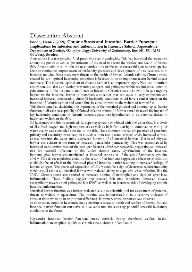
Dissertation Abstract Sundh, Henrik (2009). Chronic Stress and Intestinal Barrier Function: Implications for Infection and Inflammation in Intensive Salmon Aquaculture. Department of Zoology/Zoophysiology, University of Gothenburg, Box 463, SE-405 30 Göteborg, Sweden Aquaculture is a fast growing food-producing sector worldwide. This has increased the awareness among the public as well as governments of the need to secure the welfare and health of farmed fish. Atlantic salmon is, in non-Asian countries, one of the most successful aquacultured species. Despite continuous improvement of husbandry practices and development of new vaccines, both bacterial and viral diseases are main threats to the health of farmed Atlantic salmon. Chronic stress, created by sub- optimal husbandry conditions is believed to be an important factor behind disease outbreaks. The intestinal epithelium of Atlantic salmon is an important organ. Not just in nutrient absorption, but also as a barrier, preventing antigens and pathogens within the intestinal lumen to gain entrance to the host and thereby start an infection. Chronic stress is known to have a negative impact on the intestinal barrier in mammals, a situation that can cause a leaky epithelium and increased bacterial translocation. Stressful husbandry conditions could have a similar effect on the intestine of Atlantic salmon and would thus be a major threat to the welfare of farmed fish. This thesis aimed at elucidating the importance of the intestinal physical and immunological barrier function in disease susceptibility of farmed Atlantic salmon. It further aimed to reveal the impact of key husbandry conditions in Atlantic salmon aquaculture hypothesized to be potential threats to health and welfare of the fish. All husbandry conditions examined, i.e. hyperoxygenation combined with low water flow, low levels of dissolved oxygen and high temperature as well as high fish density in combination with poor water quality was concluded stressful to the fish. These common husbandry practises all generated primary and secondary stress responses such as increased plasma cortisol levels, increased cortisol release rate into the water and a decreased function of all intestinal barriers. Decreased physical barrier was evident in the form of increased paracellular permeability. This was accompanied by increased translocation rates of the pathogen bacteria Aeromonas salmonicida, suggesting an increased risk for bacterial infections in fish under chronic stress. Dysfunction of the intestinal immunological barrier was manifested as impaired expression of the pro-inflammatory cytokine IFN-γ. This down regulation could be the result of an immuno suppressive effect of cortisol but could also be an effect of the decreased physical intestinal barrier resulting in increased leakage of luminal antigens. The decreased expression of IFN-γ could be a sign of decreased cellular immunity which would render an intestinal barrier with reduced ability to cope with virus infections like the IPNV. Chronic stress also resulted in increased homing of neutrophils and signs of sever local inflammation. These findings suggest that stressed fish may experience increased disease susceptibility towards viral pathogens like IPNV as well as an increased risk of developing chronic intestinal inflammation. Intestinal barrier function was further evaluated as a new scientific tool for assessment of potential threats to welfare in aquaculture. The intestine was demonstrated to be a sensitive indicator of stress at times when no or only minor differences in primary stress responses was observed In conclusion, common husbandry may constitute a threat to health and welfare of farmed fish and intestinal barrier function can serve as a valuable tool for assessing potential stressful husbandry conditions in the future. Keywords: Intestinal barrier function, stress, cortisol, Ussing chambers, welfare, health, inflammation, neutrophils, cytokines, chronic stress, chronic inflammation
List of papers This thesis is based on following papers and manuscripts, which are referred to in the text by their Roman numerals: Paper I The effect of hyperoxygenation and reduced flow in fresh water and subsequent infectious pancreatic necrosis virus challenge in sea water, on the intestinal barrier integrity in Atlantic salmon, (Salmo salar L.) (2009) H.Sundh, R. E. Olsen, F. Fridell, K. Gadan, Ø. Evensen, J. Glette, G. L. Taranger, R. Myklebust and K. Sundell. Journal of Fish Diseases 32:687-698. Paper II Disturbed intestinal barrier function in Atlantic salmon (Salmo salar L.) post smolts caused by common sea cage environmental conditions: A possible physiological welfare indicator. H. Sundh, R. E. Olsen, T. Ellis, B. O. Kvamme, F. Fridell, G. L. Taranger and K. Sundell (Under revision for publication in BMC Physiology). Paper III High stocking density and poor water quality disturb physical and immunological barriers in Atlantic salmon (Salmo salar, L.) intestine. H Sundh, T. Ellis, F Fridell, G L Taranger and K Sundell (Submitted for publication in Fish and Shellfish Immunology). Paper IV Local innate immune responses in the intestinal mucosa of Atlantic Salmon (Salmo salar) after long term exposure to low oxygen levels and high temperature in the environment. L. Niklasson, H. Sundh, B. O. Kvamme, F. Fridell, G. L. Taranger and K. Sundell (Manuscript). Paper V Translocation of infectious pancreatic necrosis virus across the intestinal epithelium of Atlantic salmon (Salmo salar L.). H. Sundh, S. Calabrese, F. Jutfelt, L. Niklasson, R. E. Olsen and K. Sundell (Submitted for publication in Aquaculture).
Table of contents
Introduction Atlantic salmon 7 Life cycle 7
Aquaculture 8 Atlantic salmon farming 8 Aquaculture conditions 9 The stress response in fish 10 Welfare and health in aquaculture 12 Welfare 12 Health 13 Stress, welfare and health 14 Diseases in aquaculture 15 IPNV 15 Aeromonas salmonicida 16 The gastrointestinal tract of salmonids 16 Gross morphology 16 Functional morphology of the intestine 17 Barriers separating the internal environment from the external 18 Atlantic salmon intestinal barrier 18 The physical barrier 18 The extrinsic barrier 19 The immunological barrier 20 Host-pathogen interaction 22 IPNV 22 Stress and the intestinal barrier 22
Scientific aims Main objectives 24 Methodological considerations The Ussing chamber technique 26 Sampling, tissue preparation and methodology 26 Ussing chambers and intestinal barrier function 27 Complementary measures of permeability 29 Bacterial translocation as a measure of disease resistance 30
IPNV translocation 31 Immunohistochemistry 31 Results and Discussion Is everyday life stressful for farmed Atlantic salmon? 32 Stressors, stress and welfare 32 Hyperxygenation combined with poor water quality 32 Low DO levels at different temperatures 33 High fish density and poor water quality 34 Intestinal barrier function as a welfare indicator 35 Stress and the physical barrier 35 Corticosteroids 35 CRH and mast cells 37 Stress and the immune barrier 39 Neutrophils 39 Effects on the physical barrier 40 Neutrophils and intestinal barrier integrity: an extended view 41 Cytokines 43 Effects on disease resistance 43 Effects on the intestinal barrier function 44 Stress and intestinal inflammation 45 Diseases in aquaculture 46 Aeromonas salmonicida 46 IPNV 47 A hypothetical model for the effect of chronic stress in Atlantic salmon 48
Conclusions 50 Acknowledgements 52 Additional bibliography 54 References 55
7
Introduction Atlantic salmon Life cycle
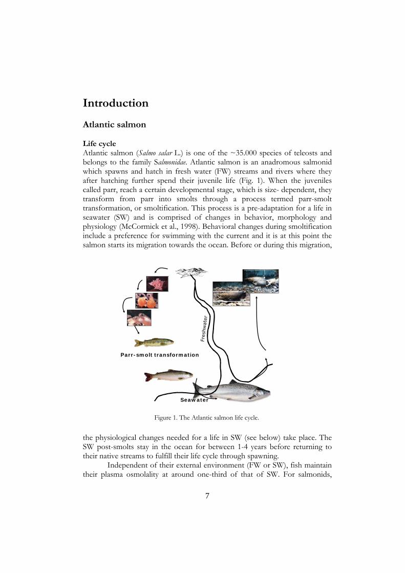
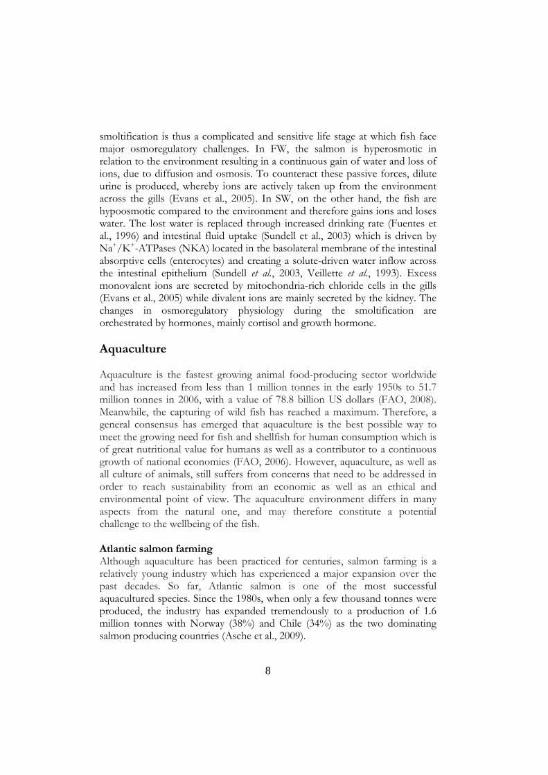
Atlantic salmon (Salmo salar L.) is one of the ~35.000 species of teleosts and belongs to the family Salmonidae. Atlantic salmon is an anadromous salmonid which spawns and hatch in fresh water (FW) streams and rivers where they after hatching further spend their juvenile life (Fig. 1). When the juveniles called parr, reach a certain developmental stage, which is size- dependent, they transform from parr into smolts through a process termed parr-smolt transformation, or smoltification. This process is a pre-adaptation for a life in seawater (SW) and is comprised of changes in behavior, morphology and physiology (McCormick et al., 1998). Behavioral changes during smoltification include a preference for swimming with the current and it is at this point the salmon starts its migration towards the ocean. Before or during this migration,
the physiological changes needed for a life in SW (see below) take place. The SW post-smolts stay in the ocean for between 1-4 years before returning to their native streams to fulfill their life cycle through spawning.
Independent of their external environment (FW or SW), fish maintain their plasma osmolality at around one-third of that of SW. For salmonids,
Parr-smolt transformation
Seawater
Figure 1. The Atlantic salmon life cycle.
8
smoltification is thus a complicated and sensitive life stage at which fish face major osmoregulatory challenges. In FW, the salmon is hyperosmotic in relation to the environment resulting in a continuous gain of water and loss of ions, due to diffusion and osmosis. To counteract these passive forces, dilute urine is produced, whereby ions are actively taken up from the environment across the gills (Evans et al., 2005). In SW, on the other hand, the fish are hypoosmotic compared to the environment and therefore gains ions and loses water. The lost water is replaced through increased drinking rate (Fuentes et al., 1996) and intestinal fluid uptake (Sundell et al., 2003) which is driven by Na+/K+-ATPases (NKA) located in the basolateral membrane of the intestinal absorptive cells (enterocytes) and creating a solute-driven water inflow across the intestinal epithelium (Sundell et al., 2003, Veillette et al., 1993). Excess monovalent ions are secreted by mitochondria-rich chloride cells in the gills (Evans et al., 2005) while divalent ions are mainly secreted by the kidney. The changes in osmoregulatory physiology during the smoltification are orchestrated by hormones, mainly cortisol and growth hormone. Aquaculture
Aquaculture is the fastest growing animal food-producing sector worldwide and has increased from less than 1 million tonnes in the early 1950s to 51.7 million tonnes in 2006, with a value of 78.8 billion US dollars (FAO, 2008). Meanwhile, the capturing of wild fish has reached a maximum. Therefore, a general consensus has emerged that aquaculture is the best possible way to meet the growing need for fish and shellfish for human consumption which is of great nutritional value for humans as well as a contributor to a continuous growth of national economies (FAO, 2006). However, aquaculture, as well as all culture of animals, still suffers from concerns that need to be addressed in order to reach sustainability from an economic as well as an ethical and environmental point of view. The aquaculture environment differs in many aspects from the natural one, and may therefore constitute a potential challenge to the wellbeing of the fish.
Atlantic salmon farming Although aquaculture has been practiced for centuries, salmon farming is a relatively young industry which has experienced a major expansion over the past decades. So far, Atlantic salmon is one of the most successful aquacultured species. Since the 1980s, when only a few thousand tonnes were produced, the industry has expanded tremendously to a production of 1.6 million tonnes with Norway (38%) and Chile (34%) as the two dominating salmon producing countries (Asche et al., 2009).
9
The fast growing aquaculture industry has increased the awareness among both the public as well as governments of the need to secure the welfare and health of farmed fish (Ashley, 2007, Conte, 2004, Ellis et al., 2002, Huntingford et al., 2006, Chandroo et al., 2004). Welfare of farmed fish is also a major issue for the industry, not only due to the perception of the public and thereby acceptance of the product, but also because of its major impact on production efficiency, quality and quantity (Ashley, 2007, FSBI, 2002). Aquaculture conditions Compared with the atmosphere, water is a harsh and restrictive medium which aquatic animals such as fish have to face. Partial pressure as well as concentration of atmospheric gas varies considerably compared with the situation in air. Oxygen depletions are naturally occurring events making respiration in this environment a challenge. As the surrounding water is the respiratory medium, the dissolved oxygen (DO) content is a critical and limiting water quality factor for salmon aquaculture. Moreover, the much higher density of water compared with air, adds physical restrictions. Higher water density increases the energetic cost of respiration and movement. However, the surrounding water is not only the source of oxygen for the fish, it also serves as a dilution medium for toxic metabolites and waste products that needs to be excreted. In ancient China, where aquaculture was initiated around 4000 years ago (Rabanal, 1988), fish was extensively cultured in static, or low flow through ponds. In this environment, water was thus required to provide physical living space for the fish, supply of oxygen, dilute toxic metabolites excreted by the fish and serve as a source of nutrition in the form of food organisms. The consequences of such a culturing system were a limitation in the biomass of fish which could be stocked in a pond at any given time. Providing food to the fish increased the production which, in turn stretched the limitations as the oxygen consumption of the fish as well as the accumulation of toxic metabolites rapidly became limiting factors. To overcome these bottle necks, increased production could be achieved by increasing the water flow through the pond. At this stage, when the production converted to depend on flow of water, the rearing of fish became intensive (Wedemeyer, 1997).
The intensification of Atlantic salmon smolt production includes high fish densities. In order to limit water usage oxygen is added in order to maintain adequate oxygen levels in the holding tanks (Wedemeyer, 1996). Many hatcheries can only obtain 20% of the water needed for a natural supply of sufficient oxygen levels in the tanks and hyperoxygenation of the inlet water or addition of oxygen to the holding tanks are the easiest ways around this problem. DO levels of 200 - 300% saturation have commonly been used in
10
aquaculture in order to reduce the specific water flow (L kg-1 min-1) (Brun, 2003). This procedure will have a negative impact on water quality in terms of increased levels of ammonia and carbon dioxide together with decreased pH (Fivelstad et al., 2004, Fivelstad et al., 1999, Ellis et al., 2002). In addition, the hyperoxic environment per se may be damaging to the fish due to increased levels of free oxygen radicals. Such environment has been hypothesized to be a contributing factor to the increased disease susceptibility to fish pathogens observed after SW transfer. However, scientific studies on the effect of the intense FW rearing on the physiology of the fish are currently scarce (Brun, 2003). During the growth phase in SW, Atlantic salmon are held in large cages at sea. According to Norwegian legislation, the density of fish in the cages should not to exceed 25 kg m-3. Moreover, it is stated that “the fish shall be kept in an environment that is adapted to the needs of each specific species and that protects them from unnecessary stress, pain and suffering” (FAO, 2009). In order to achieve this, it is important to define the composition of the environment which the fish are
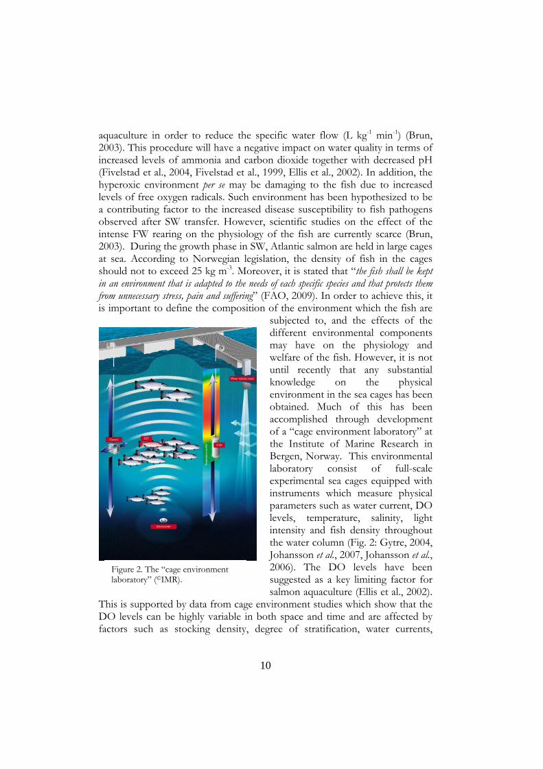
subjected to, and the effects of the different environmental components may have on the physiology and welfare of the fish. However, it is not until recently that any substantial knowledge on the physical environment in the sea cages has been obtained. Much of this has been accomplished through development of a “cage environment laboratory” at the Institute of Marine Research in Bergen, Norway. This environmental laboratory consist of full-scale experimental sea cages equipped with instruments which measure physical parameters such as water current, DO levels, temperature, salinity, light intensity and fish density throughout the water column (Fig. 2: Gytre, 2004, Johansson et al., 2007, Johansson et al., 2006). The DO levels have been suggested as a key limiting factor for salmon aquaculture (Ellis et al., 2002).
This is supported by data from cage environment studies which show that the DO levels can be highly variable in both space and time and are affected by factors such as stocking density, degree of stratification, water currents,
Figure 2. The “cage environment laboratory” (©IMR).

11
position of the sea cages and seasonal variations. Moreover, tidal cycles exhibit a major influence on the DO levels in farms situated in fjords, as these farms are sheltered from other causes of water movement like wind, waves and strong currents. Cyclic drops in DO levels, reaching as low as 50% (<5 mg ml-1) for several hours have been observed when the tidal current changes direction and the water current is close to zero (slack water). Further, during a study in late summer, the combination of slack water and high water temperature has been shown to cause the lowest and most critical DO levels within the cages (Johansson et al., 2007). Although the sea cage environment is now being described, the impact of this environment on the physiology of the fish remains largely to be explored. The stress response in fish All living organisms are adapted to the environment they inhabit through the natural selection of evolution. A prerequisite for life is being able to maintain stability of essential physiological systems (homeostasis) (McEwen and
Wingfield, 2003) independent of the external environment. In a changing external environment, the homeostasis of the internal milieu is constantly challenged and needs to be controlled by physiological mechanisms acting to
Stress
Brain - Hypothalamus
Pituitary
Interrenal tissue Chromaffin tissue
Primaryresponses
Secondaryresponses
Tertiaryresponses
ImmunologicalPrimary barriers
GrowthDisease resistanceSwimming capacityBehaviourReproduction
CellularIonic
MetabolicNutritional
CirculatoryRespiratory
CATCortisol
Figure 3. The stress response in fish

12
restore the internal milieu within the narrow ranges necessary for the survival of the animal (Stott, 1981).
Perhaps the most fundamental reaction preserved among all vertebrates is the stress response (Fig. 3) elicited by an environmental change, or a threat to homeostasis (a stressor). The, primary stress response, i.e. the release of stress hormones, is mediated by the hypothalamic-pituitary-interrenal (HPI) axis and initiated by the central nervous system (CNS) perceiving cues from the environment, a stressor. This is followed by a neuroendocrine release of corticotropin releasing hormone (CRH) from the hypothalamus. CRH, in turn, stimulates corticotropic cells of the anterior pituitary to secrete adrenocorticotropic hormone (ACTH) which stimulates the adrenal tissue in fish, as in other vertebrates, to synthesize and release corticosteroids (Barton, 2002, Schreck et al., 2007). Of these, cortisol is the main stress hormone in fish. In the circulation, it elicits a secondary stress response restricted to target tissues expressing glucocorticoid receptors (Mommsen et al., 1999). In fish, well documented secondary stress responses are metabolic, cellular, osmoregulatory, hematological and immunological changes, manifested among other things as increases in blood glucose and lactate, heat shock protein production, changes in ion composition, changes in haematocrit, lysozyme activity and antibody production (Barton, 2002, Barton and Iwama, 1991). Finally, this creates a tertiary stress response, affecting the long-term performance of the fish manifested as decreased growth, swimming capacity, disease resistance, feeding activity and altered behaviour (Barton, 2002). Welfare and health in aquaculture Welfare Welfare, or well-being, of an animal is a complex issue and it has been hard to agree on a unified definition. Most definitions take into account the animal feelings, functions and nature and are influenced by human concerns. Definitions based on feelings mainly refer to the mental state of the animal, whereas the function-based definitions focus on the ability of the animal to cope and adapt to its environment without being forced beyond its physical capacity. Nature-based definitions adopt the view that animals should be able to express its natural behaviour. In the current thesis, welfare is mostly discussed around the function-based definitions, even though changes in the biological function of the fish also may affect the mental state of the fish.
Feeling-based definition of welfare requires the conviction that animals are sentient and able to suffer, i.e. experience pain and fear (Chandroo et al., 2004). Whether fish are able to do so is a matter of an ongoing debate

13
(Rose, 2002, Chandroo et al., 2004, Sneddon et al., 2003). Some argues that fish lack the brain centres believed to be a prerequisite for a sentient animal capable of subjectively experiencing pain like humans, i.e. neocortex (Rose, 2002). However, the sensory structures for detection of harmful stimuli through nociceptors do exist in rainbow trout (Oncorhychus mykiss) (Sneddon et al., 2003). Further, fish possess brain centres giving them the ability of memory and learning (Huntingford et al., 2006) and it cannot be excluded that other brain regions than the neocortex can mediate different states of pain and fear (Huntingford et al., 2006, FSBS, 2002, Chandroo et al., 2004). Therefore, environments and husbandry conditions that may be perceived as causing suffering should be regarded as a potential threat to welfare from a feeling-based point of view (Huntingford et al., 2006, FSBS, 2002). From a function-based point of view, bad welfare arises when the biological functions of the animals are compromised (Moberg, 2000). Broom, (1986) defined the welfare of an individual as “its state as regards its attempts to cope with its environment” in a range from poor to good. This implies that that the animal is trying to maintain homeostasis and should be able to react and change its physiology in response to any changes in the surrounding environment. Thus, welfare decreases as the ability to cope with the physical environment decreases.
Health A well accepted definition of health is ”an animal's state as regards its attempts to cope with pathology” and pathology defined as “a detrimental derangement of molecules, cells, tissues and functions that occur in living organisms in response to injurious agents or deprivations” (Broom, 2007). Health and welfare are intimately connected in a complex manner. Although it may seem obvious that the physical health of an animal is fundamental for good welfare, poor health can, on the other hand, be both the cause and effect of poor welfare. Animals that are subjected to a change in their environment, but are unable to habituate, are subjected to chronic stress which is a pre-pathological state placing the animals at increased disease susceptibility due to the suppressive effects of prolonged stress on the immune system (Yada and Nakanishi, 2002, Weyts et al., 1999). In this state the health of the fish is not directly compromised, but the immune suppression increases future risk of developing pathological conditions. This increased risk of developing disease does place the fish in a state of bad welfare (Broom, 2007) and any sign of disease indicates that the animals are experiencing bad welfare (FSBS, 2002).
In 2007, Norwegian aquaculture reported an 8.7% loss of farmed Atlantic salmon due to mortality, declassification at harvest and unknown reasons (Norwegian Directorate of Fisheries (Fisheries, 2008)). Although no exact reasons for mortality were reported, this indicates that the husbandry

14
conditions may compromise the welfare and health of the fish. Moreover, disease outbreaks in aquaculture are still of major concern for the aquaculture industry (Skjelstad et al., 2007). The links between welfare, health and diseases suggest that any occurrence of disease can be a sign of an underlying problem with the environments the fish are subjected to (FSBI, 2002). However, the knowledge on how husbandry conditions affect the physiology of the fish or if these are stressful and constitute a threat to the welfare of the fish is currently obscure. Stress, welfare and health One of the most important cues affecting biological functions is stress. In this thesis, stress is defined as an adaptive biological response elicited in response to a threat to the homeostasis of the individual. The threat then constitutes the stressor. As previously discussed, in its struggle to maintain homeostasis, the body has to adapt physiologically in response to internal and/or external changes. This never-ending effort can be described in terms of allostasis, i.e. a strive to maintain homeostasis through changes in physiological systems responsible for maintaining the homeostasis (McEwen and Wingfield, 2003, McEwen, 2002, Landys et al., 2006, Korte et al., 2006, Korte et al., 2005). Allostasis allows an animal to actively adjust the physiology to predictable and unpredictable life events (Landys et al., 2006) through complex interplay between amongst others, the HPI-axis and the immune system (McEwen, 1998b, McEwen, 2002, McEwen, 1998a). Further discussion in this chapter will focus on the role of the HPI-axis and cortisol in allostasis. Every change, internally or externally, that elicits a primary stress response with elevated plasma cortisol levels as a result, contributes to the allostatic load which can be described as the energy needed to actively change the prior operation of physiological systems to fit the new environment in order to maintain homeostasis (McEwen and Wingfield, 2003). As the allostatic load increases, more energy is needed to maintain homeostasis. When available energy resources are approaching the energy needed for allostasis, more and more energy must be re-allocated from “less important”, energy demanding biological functions such as somatic growth, reproduction and immune function, and the organism is thereby subjected to a pre-pathological state. Less energy will be available for dealing with additional stressors, i.e. “its state as regards its attempts to cope with its environment” has decreased, meaning its welfare is becoming poorer and poorer. When the energy needed to maintain homeostasis exceeds the available energy recourses, the organism suffers allostatic overload or distress. At this stage, the risk of developing pathology is evident, and a major threat to the health of the fish. Importantly, not all stress is inherently bad and the intriguing task is to establish acceptable allostatic

15
load and to determine when allostatic load becomes allostatic overload, i.e. when stress becomes distress (Moberg, 2000). Diseases in aquaculture Viral diseases are currently the main threat to the health of farmed Atlantic salmon. Infectious pancreatic necrosis virus (IPNV) and pancreas disease (PD) are the two major viral diseases. Historically, furunculosis, caused by the gram negative bacteria Aeromonas salmonicida, was one of the most common diseases in salmon aquaculture. However, currently, winter ulcer, Moritella viscosa, is regarded as the major bacterial threat. These pathogens are of major concern for salmon farming industry (Skjelstad et al., 2007). IPNV IPNV is a member of the animal virus genus Aquabirnavirus belonging to the family Birnavirdae. The virions of IPNV are approximately 60 nm in diameter and are single-shelled, non-enveloped icosahedrons containing bi-segmented double-stranded RNA (dsRNA) (Wolf, 1988, Rodriguez Saint-Jean et al., 2003). The virus were initially a major problem in brook trout (Salvelinus fontinalis M.) and rainbow trout hatcheries in North America (M`Gonigle, 1941), but have subsequently also caused major concerns in Europe and Chile and has been described as the biggest health problem in the history of Norwegian aquaculture with annual losses estimated up to 100 million NOK (Brun, 2003). IPNV outbreaks can occur from first feeding, but are generally conceived as being inversely linked to the age of the fish; mortality being highest in young fish and relatively rare in older fish (Wolf, 1988).
In Atlantic salmon aquaculture, the transfer of fish from FW to SW is a particularly stressful event in the production cycle (Roberts and Pearson, 2005). During the first period after SW transfer, sudden and often extremely high mortalities caused by IPNV can occur (Roberts and Pearson, 2005, Smail et al., 1992, Melby et al., 1994, Jarp et al., 1995).
In salmonids suffering from an acute IPNV infection, common histopathological signs are necrosis of pancreatic acinar cells, kidney and liver tissue (Roberts and Pearson, 2005). Further, severe gut enteritis and complete sloughing of the intestinal mucosa are commonly observed in moribund post smolts (M`Gonigle, 1941, McKnight and Roberts, 1976, Johansen and Sommer, 2001, Roberts and Pearson, 2005, Wolf, 1988, Smail et al., 2006). It has further been suggested that these damages created to the intestine may be more acute than the necrosis of the pancreatic acinar cells (Smail et al., 1995, Wolf, 1988, M`Gonigle, 1941).
16
Aeromonas salmonicida Aeromonas salmonicida ssp salmonicida (A. salmonicida), a Gram-negative bacteria, is the causative agent of furunculosis. The disease is named after the characteristic furuncles, i.e. blisters occurring on the skin of infected fish. These are connected to chronic infection, but acute furunculosis can occur without any signs of furuncles (Cipriano and Bullock, 2001). A. salmonicida can cause acute mortality in susceptible salmonids, but have also been reported to infect other species of fish (Bricknell et al., 2006). Acute infections in salmonids are often manifested as internal haemorrhages to visceral organs. The fish usually stop feeding and thus, the gut is devoid of food, with a lumen containing sloughed-off epithelial cells, blood and mucus (Cipriano and Bullock, 2001). A. salmonicida can be found in the intestine of salmonids and the intestine has been proven as an infection route in both rainbow trout and Atlantic salmon (Ringø et al., 2009, Jutfelt et al., 2006, Jutfelt et al., 2007). Translocation rates of A. salmonicida across the intestinal epithelium are affected by the developmental stage of the fish, and by the composition of the diet (Jutfelt et al., 2007). However, it is currently un-known if the translocation rate also is affected by stressors in the aquaculture environment of Atlantic salmon.
The gastrointestinal tract of salmonids Gross morphology The gastrointestinal (GI) tract is basically a tube that extends from the mouth to the anus. The mucosa lining the GI tract constitutes the interface between the external and internal environment, and together with the associated organs, the pancreas, liver and gall bladder, the GI tract provides essential functions such as food digestion, nutrient uptake, osmoregulation and immunity. The GI tract of salmonids consists of several distinct anatomical regions, starting with a short muscular oesophagus leading in to the large cardiac, U-shaped stomach, which is followed by pyloric stomach and a pyloric sphincter controlling the release of food into the intestine (Burnstock, 1959). The teleost intestine in general is divided into a proximal and distal region. The anterior part of the proximal region of salmonids has pyloric caecas. These are finger-like extensions which increase the nutrient absorptive surface area in the proximal intestine without increasing its length or thickness (Clements and Raubenheimer, 2006). The distal region is radially larger and darker in appearance and with a thicker muscle coat. Further, the distal region is characterised by the annulo-spiral septa. These consist of circular musculature and sub-mucosa extending into intestinal lumen, forming ridges
17
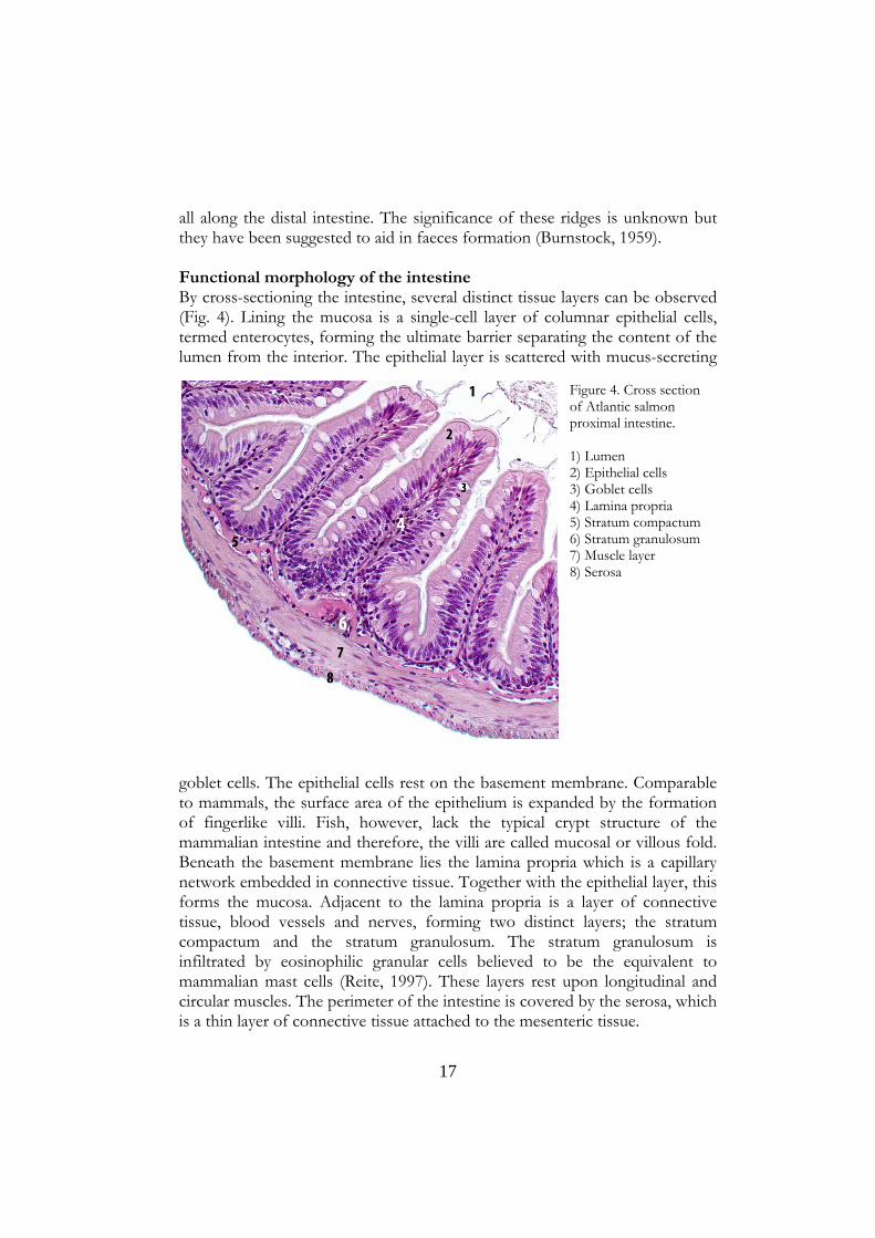
all along the distal intestine. The significance of these ridges is unknown but they have been suggested to aid in faeces formation (Burnstock, 1959). Functional morphology of the intestine By cross-sectioning the intestine, several distinct tissue layers can be observed (Fig. 4). Lining the mucosa is a single-cell layer of columnar epithelial cells, termed enterocytes, forming the ultimate barrier separating the content of the lumen from the interior. The epithelial layer is scattered with mucus-secreting
goblet cells. The epithelial cells rest on the basement membrane. Comparable to mammals, the surface area of the epithelium is expanded by the formation of fingerlike villi. Fish, however, lack the typical crypt structure of the mammalian intestine and therefore, the villi are called mucosal or villous fold. Beneath the basement membrane lies the lamina propria which is a capillary network embedded in connective tissue. Together with the epithelial layer, this forms the mucosa. Adjacent to the lamina propria is a layer of connective tissue, blood vessels and nerves, forming two distinct layers; the stratum compactum and the stratum granulosum. The stratum granulosum is infiltrated by eosinophilic granular cells believed to be the equivalent to mammalian mast cells (Reite, 1997). These layers rest upon longitudinal and circular muscles. The perimeter of the intestine is covered by the serosa, which is a thin layer of connective tissue attached to the mesenteric tissue.
Figure 4. Cross section of Atlantic salmon proximal intestine. 1) Lumen 2) Epithelial cells 3) Goblet cells 4) Lamina propria 5) Stratum compactum 6) Stratum granulosum 7) Muscle layer 8) Serosa
18
Barriers separating the internal environment from the external Fish are suggested to rely more upon innate immunity than adaptive immunity, as this part of the immune system displays a slow response (4-6 weeks for specific antibodies to be produced in salmonids), the memory is weak and the affinity of the antibodies is regarded as low compared to the mammalian system (Magnadottir, 2006, Ellis, 2001, Tort, 2003). Fish are in intimate contact with their aqueous environment, which is also a thriving habitat for microorganisms, both virus and bacteria (Wilhelm and Suttle, 1999). Many of these microorganisms are pathogenic and able to infect fish. The first line of defence a pathogen must breach in order to infect a host are the innate physical barriers separating the host from the physical environment. In fish, these are the skin, gills and gut (Press and Evensen, 1999). The gut is different from the other barriers as it is inhabited by a vast number of microbial cells. In mammals, microbial density has been estimated to be 1012 cells per millilitre of luminal content in the large intestine (Lukas et al., 2006). In fish, the bacterial density is lower, with reported numbers of 105 – 108 cells per millilitre of luminal content (Ringø et al., 1995). Normally, this indigenous microflora remains harmless in the lumen. Rather, the commensal flora is regarded as beneficial for the host by facilitating the digestion and providing the host with nutrients otherwise inaccessible, functioning as a protection against pathogenic bacteria by occupying ecological niches and other physical resources, and inhibiting growth of bacterial pathogens like A. salmonicida (Jöborn et al., 1997). In health, harmful luminal content is prevented host entry by several layers of barriers which in concert cooperate and communicate in order to prevent infection. If health is impaired, a breakdown in this balance may reduce the protective barriers and thus allow pathogens like IPNV and A. salmonicida to enter the host and induce infection. Atlantic salmon intestinal barrier The physical barrier Enterocytes are connected on the apical side of the membrane via the tight junctions (TJ) (Fig. 5). TJ do not represent a rigid barrier but rather a fence, regulating the passage of ions, water and other molecules as well as immune cells, through the paracellular pathway. Further, the TJs are situated at the apical membrane of the epithelial cells, generating a polarity to the epithelium, by restricting the diffusion of lipids and proteins between the apical and basolateral membrane (Fanning et al., 1999).

19
TJs observed on ultrathin section electron micrographs appear as a set of fusion points between the outermost parts of the cell membrane of adjacent cells (Tsukita and Furuse, 1999). TJ consists of several physiologically regulated proteins forming the circumferential seals around adjacent epithelial cells. Three of the main protein families found in TJs are occludins, claudins and junction-associated membrane proteins (JAM). The claudins and occludins forms the backbone of the TJ, while JAM appear to be important for controlling the traffic of immune cells like T-lymphocytes, neutrophils and dendritic cells through the paracellular pathway (González-Mariscal et al., 2003). For the claudins, 24 isoforms have been reported to date and two isoforms of occludins have been found. The claudin isoforms do, among other things, display different number of charged residues and claudin has therefore been suggested to be the major protein participating in creating the cation selectivity of the TJ. Most TJ show some cation selectivity and this physiological feature can be regulated by altering the claudin isoform present in the TJ (Fanning et al., 1999). To provide a functional seal of the paracellular pathway and an epithelial barrier, the TJ proteins are assembled and positioned by cytosolic plaque proteins consisting of numerous proteins like the zonula occludens (ZO-1, 2 and 3). In turn, the plaque proteins are connected to the actin-ring of the cytoskeleton, creating a continuous structure facing the apical side of the intestinal epithelium (Schneeberger and Lynch, 2004).
The extrinsic barrier The intestinal epithelial layer is protected by a mucus layer. This is created by the apical release of mucin glycoproteins from goblet cells (Fig. 6A)scattered in the intestinal epithelium (Shephard, 1994). An important role of the mucus layer is to prevent uncontrolled attachment of bacteria to the intestinal epithelial cells. This is achieved by the glycoproteins creating a matrix network
Lipidbilayercell 2
Lipidbilayercell 1
Occludin
ZO‐1
ZO‐2Actin
p130
N
C
Claudin
Figure 5. The tight junctions of Atlantic salmon proximal intestine, possible molecular arrangement.
20
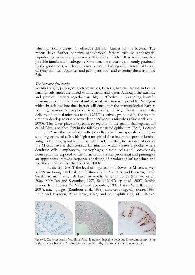
which physically creates an effective diffusion barrier for the bacteria. The mucus layer further contains antimicrobial factors such as antibacterial peptides, lysosyme and proteases (Ellis, 2001) which will actively neutralize possible intraluminal pathogens. Moreover, the mucus is constantly produced by the goblet cells, which results in a constant flushing of the intestinal lumen, carrying harmful substances and pathogens away and excreting them from the fish. The immunological barrier Within the gut, pathogens such as viruses, bacteria, bacterial toxins and other harmful substances are mixed with nutrients and water. Although the extrinsic and physical barriers together are highly effective in preventing harmful substances to enter the internal milieu, total exclusion is impossible. Pathogens which breach the intestinal barrier will encounter the immunological barrier, i.e. the gut-associated lymphoid tissue (GALT). In fact, at least in mammals, delivery of luminal microbes to the GALT is actively promoted by the host, in order to develop tolerance towards the indigenous microbes (Kucharzik et al., 2000). This takes place in specialized regions of the mammalian epithelium called Peyer’s patches (PP) in the follicle associated epithelium (FAE). Located to the PP are the microfold cells (M-cells) which are specialized antigen-sampling epithelial cells with high transepithelial vesicular transport of luminal antigens from the apical to the basolateral side. Further, the basolateral side of the M-cells have a characteristic invagination which creates a pocket where dendritic cells, lymphocytes, macrophages, plasma cells and occasionally neutrophils are exposed to the antigens for further processing and priming of an appropriate immune response consisting of production of cytokines and specific antibodies (Kucharzik et al., 2000).
In the fish GALT the level of organization is lower, as M-cells as well as PPs are thought to be absent (Dalmo et al., 1997, Press and Evensen, 1999). Similar to mammals, fish have intraepithelial lymphocytes (Bernard et al., 2006, McMillan and Secombes, 1997, Bakke-McKellep et al., 2007), lamina propria lymphocytes (McMillan and Secombes, 1997, Bakke-McKellep et al., 2007), macrophages (Rombout et al., 1989), mast cells (Fig. 6B) (Reite, 1998, Reite and Evensen, 2006, Reite, 1997) and neutrophils (Fig. 6C) (Bakke-
A B CFigure 6. Cross sections of proximal Atlantic salmon intestine depicting important components of the mucosal barriers. A.. intraepithelial goblet cells, B. mast cells and C. neutrophils

21
McKellep et al., 2000, Rombout et al., 1989). In fish, especially the second intestinal segment, the distal region, has been shown to transport macromolecules from the intestinal lumen through pinocytosis (Dalmo et al., 1997), and further, to present them to macrophages in lamina propria and the circulation (Rombout et al., 1985, Rombout and Van, 1989). Also, in absence of true M-cells, macromolecular transport by enterocytes in the distal intestinal region is considered to be of equal immunological importance as that of mammalian M-cells, rendering an induction of an immune response (Rombout and Van, 1989, Hart et al., 1988, Rombout et al., 1985, Nakamura et al., 2000). However, macromolecular transport can also occur in the proximal region of the salmonid intestine (Dalmo and Bogwald, 1996, Petrie and Ellis, 2006, Jutfelt et al., 2007, Jutfelt et al., 2006, Jutfelt et al., 2008, Rombout et al., 1985). Interestingly, immune cells expressing major histocompatibility complex (MHC) class II have been observed within the epithelium of the proximal intestine of Atlantic salmon (Koppang et al., 2003). However, the immunological importance of the proximal intestinal region in Atlantic salmon is currently obscure.
Immune surveillance and recognition of non-self, i.e. harmful pathogens, in the intestine is mediated by germ cell coded pattern recognition receptors such as toll-like receptors (TLRs). These receptors recognize molecular patterns which are common for a broad range of microbes including lipopolysaccarides (LPS), peptidoglucans, bacterial DNA, dsRNA and a range of other molecules expressed on the surface of pathogens. The TLRs are an evolutionary ancient set of receptors as indicated by their presence in plants, invertebrates as well as vertebrates. In mammals, 12 TLRs have been reported so far, with TLR3 and TLR4 recognizing dsRNA and LPS respectively (Blikslager et al., 2007). In mammalian enterocytes, TLRs are expressed on the cell surface for surveillance of the intestinal lumen, within the cytosol and endosomes for recognition of intracellular pathogens and on the basolateral side for recognition of pathogens successful in passing the intestinal barriers. Further, a wide range of other cells of the immune system express TLRs including mast cells, macrophages and neutrophils stressing their importance in the innate immune system. Upon activation, TLRs mediate intracellular signaling cascades that results in the production of pro-inflammatory cytokines and chemokines for recruitment of professional phagocytes such as neutrophils for clearance of the recognized pathogen. Fish possess more than 20 TLRs including orthologs to mammalian TLRs. However, fish also have unique TLRs (Seya et al., 2009). For virus dsRNA recognition fish have 2 separate TLRs, i.e. TLR3 which is expressed in endosomes and TLR22 expressed on the cell surface, sensing the environment for the presence of dsRNA (Rodriguez et al., 2005, Rebl et al., 2007).

22
Constitutive expression of both TLR3 and TLR22 has been reported in the pyloric caeca and intestine of rainbow trout (Rodriguez et al., 2005). Thus fish appear to have an innate immune system in the intestine capable of sensing the environment for virus. Host-pathogen interaction Animals have a well-developed innate defense system against pathogens. However, during the co-evolution of host and pathogen, the pathogens have evolved mechanisms which can utilize the host’s own defense mechanisms in order to facilitate the penetration of the barriers and infect the host. Further, epithelia such as the intestinal epithelial cells can serve as a site where viruses are able to infect the host cells and initiate replication, while some viruses may only use the epithelia such as the intestine for translocation in order to reach the circulation for distribution and infection of other tissues. IPNV Information regarding routes for virus infection in fish is scarce and the actual route of IPNV translocation and infection is unknown. However, viruses are likely to enter through the skin, gills and/or the digestive tract as clinical outbreaks can be induced by immersion challenge and coincide with first feeding of fry (Wolf, 1988). Data on interaction between IPNV and the intestinal epithelium of salmonid fish show that the virus can be found within the intestine early during a natural infection (Shankar and Yamamoto, 1994) and after an intraperitoneal (i.p.) injection (Swanson et al., 1982). Although these data indicate that the intestinal tract may serve as an entry route for IPNV it has not been excluded that the virus entry can occur elsewhere and reach the intestine through the systemic circulation. Stress and the intestinal barrier Stress in mammals has a profound effect on the GI tract. Both physiological stress, like trauma, burns and surgeries, and psychological stress can induce intestinal barrier dysfunction in mammals (Söderholm and Perdue, 2001). Acute and chronic psychological stress in mammals has been shown to lead to decreased transepithelial resistance (TER) concomitant with increased intestinal permeability to small, inert marker molecules (mannitol or Cr-
EDTA) (Velin et al., 2004, Söderholm et al., 2002, Santos et al., 2000, Santos et al., 2001, Saunders et al., 1994, Santos et al., 1999) as well as increased transport of macromolecules and bacteria from lumen through the intestinal epithelium to the circulation (Velin et al., 2004, Kiliaan et al., 1998, Santos et al., 1999, Santos et al., 2000).
23
Similar effects of stress on the GI tract have been observed in fish. Environmental stress in the form of netting, followed by a 4 h transportation, results in goblet cell depletion and detachment of the intestinal epithelial cells from the basement membrane in the common carp (Cyprinus carpio), the Japanese colored carp (Cyprinus carpio haematopterus), the European eel (Anguilla anguilla) and the African catfish (Clarias gariepinus) (Szakolczai, 1997). Social stress resulted in flattening of the mucosal folds and loosening of the cell-to-cell contact between enterocytes in the European eel (Peters, 1982). Increased paracellular permeability has been observed in Atlantic cod (Gadhus morhua L.) subjected to acute stress (Olsen et al., 2008). In Atlantic salmon subjected to 15 min of acute stress, damages to the intercellular junctional complexes has been observed up to 12 h after stress (Olsen et al., 2002). Similar intercellular damages have been observed in rainbow trout subjected to 15 min acute stress. Moreover, such stress results in intestinal barrier dysfunction measured as increased paracellular permeability to mannitol (Olsen et al., 2005). However, the effect of chronic environmental stress on the intestinal barrier function of Atlantic salmon is currently unexplored.

24
Scientific aims Main objectives The overall aim of this thesis has been to increase the knowledge on intestinal barrier function and host-pathogen interaction in Atlantic salmon and the understanding of how common husbandry conditions may affect this as well as the overall welfare and health of Atlantic salmon. Focus has been on three key husbandry conditions considered to be potential threats to the welfare of the fish.
1. In intensive production of Atlantic salmon smolts, FW supply is insufficient to maintain satisfactory oxygen levels. This is commonly compensated for by water hyperoxygenation. This environmental condition is hypothesised to be chronically stressful to the fish and an underlying cause of increased susceptibility to IPNV infections during the first weeks after transfer of the fish to SW.
2. During the growth phase in SW, the fish are kept in large sea cages. Within the cages, large spatial and temporal variations in DO levels occur due to changes in temperature and tidal currents. The most critical decrease in DO levels are observed during tidal reversals in late summer when the water temperature is elevated. It is hypothesised that low DO levels are chronically stressful to the fish.
3. The schooling behaviour of salmon within the sea cages results in local
densities much higher than the recommended limits. This leads to poor water quality. It is hypothesised that high density in combination with poor water quality is chronically stressful to the fish.
It is generally accepted that chronic stress is a threat to animal welfare. However, the degree of stress experienced by fish to different common husbandry practises has not been elucidated. Therefore, in each experiment, the physiological responses to stress have been monitored at several levels. The levels of plasma cortisol in combination with the release rate of cortisol into the water have been measured as indicators of the primary stress response in response to different husbandry conditions. The negative effects of stress on the intestinal barrier function in mammals are well described and similar observations are reported from several species of fish including Atlantic salmon. Therefore, intestinal barrier function has been
25
evaluated as a possible research tool for assessment of welfare and stressful husbandry conditions. Information on physiological function of the intestinal barriers in fish is scarce. This thesis aims at increasing the knowledge on the intestinal barrier of Atlantic salmon in relation to physiological and immunological factors. Intestinal inflammation is the result of barrier dysfunction in mammals and is influenced by chronic stress. It is hypothesised that chronic stress may have similar effects on the intestinal barrier of fish. To elucidate this hypothesis, intestinal inflammation, assessed as neutrophil infiltration and cytokine expression, have been measured in response to chronic stress. Chronic stress is known to lead to increased susceptibility to bacterial diseases but the underlying mechanisms have not been fully clarified. The intestine of fish has been demonstrated as a route for infection by bacterial pathogens. Therefore, the translocation rate of pathogenic bacteria has been measured to elucidate if this could be a cause of increased disease susceptibility observed during chronic stress. IPNV is currently a major problem in Atlantic salmon aquaculture. Mammalian viruses utilize the intestinal epithelium for host infection. Therefore, this thesis aims at developing a method to elucidate if the intestinal epithelium is a translocation route for IPNV. Chronic stress has been suggested to increase the disease susceptibility to IPNV. Therefore, it is hypothesized that decreased barrier function caused by chronic stress may be one important factor behind IPNV outbreaks.
26
Methodological considerations The Ussing chamber technique To study the effect of husbandry conditions on the intestinal barrier function of Atlantic salmon, the Ussing chamber methodology has been used (Ussing and Zerahn, 1951) Papers I, II, III and V). This is an established in vitro technique for measurements of active and passive transports and transfers across a live epithelial tissue.
Ussing chambers were invented and first described by the Danish physiologist Hans Ussing, and used to study the capacity of an epithelium to actively move ions and nutrients against an electrochemical and/or concentration gradient in the frog skin (Ussing and Zerahn, 1951). Since, the technique has been modified and developed further, although the basic principle remains the same. Today, modified Ussing chambers, diffusion chambers (Grass and Sweetana, 1988), are widely used to study physiological mechanisms in multitude of species and tissues for applications as ion transport mechanisms, nutrient uptake, protein absorption, drug absorption, host pathogen interactions and pathophysiological mechanisms (Papers I, II, III, V, Clarke, 2009, Jutfelt et al., 2007, Jutfelt et al., 2006, Jutfelt et al., 2008, Sundell et al., 2003, Velin et al., 2004, Santos et al., 2001, Santos et al., 2000, Söderholm and Perdue, 2001, Moeser et al., 2007, Saunders et al., 2002). Sampling, tissue preparation and methodology In order to preserve function, structure and viability, the intestine is dissected using blunt forceps; adipose tissue is removed and the intestine cut open before the tissue is washed and placed in ice cold buffered Ringer solution. Thereafter, the serosal layer is removed by the use of fine forceps under magnification. This is done for different reasons. Firstly, as with all in vitro methods, isolation of the intestine from the fish renders a preparation without blood circulation and the tissue has to rely solely on delivery of oxygen and nutrients from the Ringer solution. The serosal layer is therefore removed to facilitate delivery of these essential substances through passive diffusion to the tissue. Secondly, the epithelial diffusion distances in vivo are mostly shorter than can be obtained in whole tissues in vitro, as substances in vivo have access to the circulation directly after crossing the epithelial layer. Thus, in order to reduce diffusion distances, the serosal layer is removed. Thereafter, the intestine is mounted between two half-chambers filled with physiological, buffered Ringer solution. Each half-chamber is continuously gassed with air containing 0.3% CO2 through a gas-lift arrangement which further generates circulation, nutrient delivery and maintenance of O2 and pH closest to the tissue.
27
Fish are ectothermic, and thus, the body temperature varies depending on water temperature. In each experiment, the temperature in the chambers has been maintained at the acclimation temperature by the use of cooling mantles. The electrical properties: transepithelial resistance (TER), transepithelial potential difference (TEP) and short-circuit current (SCC) of the epithelium is continuously monitored as a viability control. Originally, Ussing (1951) short-circuited the epithelium in order to determine the SCC. This is a rather invasive procedure which may add net charges to the epithelium and thereby affect the active transporting mechanisms of the epithelium. The method used in the present thesis instead applies short alternating positive and negative pulses of current. For each current pulse, the corresponding response in TEP is recorded and the current-voltage pairs plotted. A linear, least-square analysis is fitted to the current/voltage pairs in which the slope describes the TER across the tissue and the intercept with the voltage axis determines the TEP. SCC is further calculated using the law of Ohm: SCC = –TEP/TER. After mounting the peeled intestinal segments into the Ussing chambers, the segments have been allowed a 60 min acclimation period in order for all electrical parameters to reach stable levels before the start of the 90 min experiment. During the experiment, electrical parameters is measured every 5 min and the results presented as the mean TER, TEP and SCC over the last 5 sampling points. Ussing chambers and intestinal barrier function In order to establish a gradient of charges between two compartments, an active transport is needed from one compartment to the other, concomitant with a physical barrier that can withhold the charges from leaking back. The intestinal epithelium creates this scenario with three different current sources and their corresponding resistances. Thus, the intestinal epithelium can be modelled as an electrical circuit consisting of two current sources in series (the apical and the basolateral membranes) which are coupled in parallel with a third current source, having its own resistance, the paracellular pathway and the TJ (Fig. 7). The active transports are attributed to ion transporters in the apical and basolateral membrane with the major driving force being the basolaterally located NKA. However, the fence property of the intrinsic barrier is not absolute as it allows some ions to passively leak back. There are, in theory, two pathways through which ions can pass across the epithelium: the transcellular and the paracellular route. In a leaky epithelium, as the intestine (Loretz, 1995), the apical and basolateral membranes of the enterocytes have at least three orders of magnitude higher resistance to ion fluxes compared to the paracellular route through the TJ (Blikslager et al.,

28
2007). Subsequently, the measurement of TER mainly reflects the tightness of TJ. When TER decreases, it means that the leakiness or permeability through the TJ has increased resulting in reduced barrier function.
The interactions between TER, TEP and SCC are such that if the permeability through the TJ increased, the TER through the TJ has decreased.
In order to maintain TEP constant, SCC must increase. If SCC is maintained and TER is reduced, so is TEP. These interactions between the electrical measures by use of the Ussing methodology can be described in terms of an analogy of “heating a house”. Consider the temperature outside to be -10 ºC and inside of the house you want to maintain 20 ºC. Thus, you want to maintain a temperature gradient (TEP) between the inside of the house (blood) and the outside (lumen). Heat (SCC) is provided by the radiators scattered along the walls (epithelial cells) of the house. The walls and the windows (TJ) constitute the physical barriers that prevent the cold air from the
2K+
3Na+HCO3-
Na+
Cl-
Mucosa/Lumen
Serosa/Blood
Transcellularpathway
Paracellularpathway
Na+
Ra
Rtj
Rb
Rics
HNNa+
2Cl-K+ Na+
Apicalmembrane
Basolateralmembrane
Nucleus
K+ HCO3-
K+ Cl-
PhagocytosisPinocytosis
Exocytosis
A. Transepithelial elictrical characteristics
Occludin
Claudin
Tight junctions
B. Transepithelial pathways
NKA
Co-transporters
Endosome
F-actin
Figure. 7. Schematic picture of the intestinal epithelium modeled as an electric circuit (A) consisting of two current sources in series with their own resistance (the apical (Ra) and the basolateral (Rb) membranes) which are coupled in parallel with a third current source, having its own resistance, the intercellular space (Rics) and the TJ(Rtj). The active transports are attributed to ion transporters in the apical and basolateral membrane with the major driving force being the basolaterally located NKA. (B) Schematic picture of theoretical transepitihelial pathways.

29
outside from mixing with the warm air on the inside. The walls are thick, insulated and cannot be opened and allow very little entry of cold air. On the other hand, the windows are very thin, leaky and can be opened. Thus, in general, most of the cold air that is allowed access to the house is through the windows. Therefore, the windows determine the barrier function of the house (TER). If a window is opened, more cold air can enter the house. If temperature (TEP) is to be maintained, the heat (SCC) must be turned up on the radiators. Otherwise, the temperature (TEP) will decrease. Complementary measures of permeability Intestinal permeability can also be assessed by adding molecules of different sizes and/or different chemical characters which will allow them to take different routes through the epithelium. For measurement of vesicular, transcellular transport bacteria, microspheres and horseradish peroxidase (HRP; approx. 44 kDa) are commonly used. Steroid hormones and other easily labelled small, lipophilic molecules, which can diffuse through the lipid membrane of the cell, are used for measurement transcellular permeability. For assessment of paracellular permeability through large pores, 51Cr-EDTA, disaccharides (lactulose, cellobiose) and polyethylene glycols (PEG) around 1 kDa are commonly used, whereas assessment of permeability through small pores is carried out using smaller monosaccarides like mannitol (~180 Da) as well as small PEGs. In the current thesis, the probe used for assessment of paracellular permeability was radio labelled mannitol (14C-mannitol) as it has been extensively used for this purpose both in vitro and in vivo studies and proven to work well also in fish (Papers I, II, III, V, Sundell et al., 2003, Jutfelt et al., 2007, Jutfelt et al., 2006, Jutfelt et al., 2008). The advantage of using two complementary measurements of paracellular permeability is that TER measurements do not easily distinguish between changes in TJ and physical damage to the epithelium. Physical damages are, however, are instantly revealed by mannitol flux.
Thus, discrepancies between the two paracellular measures are not infrequent (Papers II, V, Sundell et al., 2003, Jutfelt et al., 2008). The TJs consists of complex protein structures where the number of TJ strands are suggested to be proportional to TER (Schneeberger and Lynch, 2004). However, the protein composition of the TJ strands greatly affects the selective permeability for different molecules not solely dependent on molecular size, but also dependent on electrical charge. Especially, the claudins appear to be determinants of the selective size and charge-based permeability through TJ. Inert hydrophilic marker molecules with intact tertiary structure and shape, such as mannitol, mainly reflect the number and size of pores within the TJ as well as the tightness of the whole junctional complexes

30
including the paracellular spaces. These features are also reflected in the TER. However, TER also reflects the overall net-charges of the pores, as well as the influence of other barrier properties affecting the electrical resistance, such as the extrinsic mucus layer (Schep et al., 1997, Schep et al., 1998, Shephard, 1994) and the surface area of the exposed tissue. Thus, the observed discrepancy can be attributed to a differential expression and composition of claudin isoforms showing differentiated slectivity for charged molecules. In fish, 56 different claudin isoforms have been described in the pufferfish (Fugu rubripes) (Loh et al., 2004) and 26 isoforms in Atlantic salmon (Tipsmark et al., 2008). However, little information exists on the protein composition of TJs in fish and the role of these claudins in the regulation of intestinal, epithelial TJs of Atlantic salmon remains to be explored. However, discrepancies between the two paracellular markers could also be due altered mucus secretion rate and/or composition, a parameter that would affect the TER, but not the permeability for mannitol. Moreover, as TER is calculated on the basis of the exposed area in the serosal compartment rather than the actual surface area exposed on the mucosal side. Subsequently, an increase in TER does not inherently suggest that the paracellular permeability have decreased, but it may instead reflect an attenuation of the mucosal surface area exposed in the Ussing chambers caused by damages to the epithelium (Blikslager et al., 2007).
Bacterial translocation as a measure of disease resistance Previous studies on both fish and mammals show that the intestine is used by bacteria as well as viruses as a route of infection and stress has been shown in mammals to greatly reduce the intestinal barrier against bacteria. The effect of stress on the intestinal barrier against live A. salmonicida in fish was therefore addressed in order to elucidate if changes in the intestinal barrier could be a possible explanation to increased disease susceptibility during stress (Paper I). In paper II, it was not possible to use this method due to practical reasons as pathogens could not be used in the laboratory where the experiments in paper paper II were conducted. In paper III, this bottleneck was overcome by the use of heat-inactivated A. salmonicida (Jutfelt et al., 2008). However, Jutfelt (2008) and co-workers showed that the translocation rate of heat-inactivated A. salmonicida was significantly lower compared to live bacteria, in the distal intestinal region of rainbow trout. The authors suggested that the heat inactivation process may damage essential translocation facilitating mechanisms at the surface of the bacteria. This could thus generate an underestimation of the “true” translocation rate. The method nevertheless serves as a good estimation of the transcellular vesicle transport (Paper III).

31
IPNV translocation One of the aims of this thesis was to develop a technique to elucidate if the intestine is a route of infection for IPNV (Paper V). For this purpose, the Ussing chamber method was chosen as it is widely used to study bacterial translocation in both mammals and fish (Papers I, II, III, Jutfelt et al., 2007, Jutfelt et al., 2006, Jutfelt et al., 2008, Velin et al., 2004, Kurkchubasche et al., 1998). However, one challenge with the use of Ussing chambers in transport and translocation studies is how to detect the transported or translocated molecule/organism of target. In the present thesis, two complementary methods were used to detect the presence of IPNV in the serosal compartment of the Ussing chambers after 90 min of exposure. The classical method of demonstrating the presence of live and virulent IPNV was used: inoculation of serosal concentrates on to CHSE-214 cells and detection of cytopathic effect (CPE). Although this classical detection method is relatively sensitive, working with cells is time- and labour-intensive. Therefore, further quantification of possible translocated virions was carried out by real time quantitative (RQ)-PCR. The RQ-PCR technique is widely used in human clinical medicine for studies on virus replication (Watzinger et al., 2006, Watzinger et al., 2004) and is also a widely used technique for viral studies in fish (Fridell et al., 2007, Milne et al., 2006, Taksdal et al., 2001, Blake et al., 1995). In the current thesis, specific and sensitive primers were designed allowing sensitive and accurate determination of translocated IPNV. Immunohistochemistry Immunohistochemistry (IHC) was used to investigate if chronic stress in fish can lead to intestinal inflammation. To address this, the number of neutrophils in the intestine was chosen as a marker for inflammation, as neutrophil infiltration is a common sign of acute inflammation in mammals (Söderholm et al., 2002, Kucharzik et al., 2001). For this purpose, a monoclonal antibody (mAb) against Atlantic salmon neutrophils was used (Pettersen et al., 1995). The advantage of having a mAb is that it is possible to specifically visualize the cells expressing the antigen against which the antibody is raised. This is a major advantage compared to classical tissue staining where cells needs to be separated by morphology compared to colour by using IHC (Paper IV). Classical staining with toluidine blue visualises the epithelium nicely and was used for investigating possible structural alterations and/or damages to the epithelium created by the husbandry conditions (Papers I and II). Moreover, toluidine blue also stains mast cells which are easily distinguished from other cells in the intestinal epithelium due to the staining of its characteristic granules.

32
Results and Discussion Is everyday life stressful for farmed Atlantic salmon? In the current thesis, several different experiments have been conducted, all with the aim to investigate common husbandry conditions in salmon aquaculture. All husbandry conditions examined, i.e. hyperoxygenation combined with low water flow (Paper I), low DO levels at low and high temperature (Papers II and IV) and high fish density in combination with poor water quality (Paper III) resulted in primary stress responses. Stressors, stress and welfare The degrees of stress elicited by the different husbandry conditions tested can vary with number and type of stressors involved as well as with the severity of each stressor. Depending on the degree of stress the impact of a particular husbandry condition on the welfare of the fish may differ. In this section, the degree of stress between and within each husbandry condition is discussed in relation to the impact on the welfare and health of the fish. Hyperxygenation combined with poor water quality This husbandry condition resulted in chronically elevated plasma cortisol levels after 26 days (Paper I). The allostatic load was revealed as increased intestinal paracellular permeability and translocation of A. salmonicida in the distal intestine. The observed decrease in intestinal barrier function is suggested to increase the risk of infection. Indeed, fish subjected to a similar
Fig. 8. Cortisol release rate from Atlantic salmon subjected to hyperoxygenation and low water flow in FW and a subsequent co-habitant challenge with IPNV after SW transfer.
Normoxic-IPNV
Normoxic+IPNV
Hyperoxic-IPNV
Hyperoxic+IPNV
Cor
tisol
rel
ease
rat
e ng
g-1
biom
ass
h-1
0.0
0.2
0.4
0.6
0.8
1.0
*

33
experimental protocol revealed increased disease susceptibility to an IPNV challenge after subsequent transfer to SW (Fridell et al., 2007). Moreover, the IPNV challenge per se served as a stressor manifested by elevated levels of plasma cortisol. This was also reflected in the cortisol release rate (Fig. 8) into the water. Interestingly, fish subjected to stress in FW responded with a stronger primary stress response to the IPNV challenge compared to unstressed fish (Fig. 8). This reveals that stress in one life stage may affect the outcome of additional stressors in a subsequent life stages. The IPNV infection also decreased the intestinal barrier function. This was evident as both an increased paracellular permeability and increased translocation of A. salmonicida and may be one explanation behind the increased risk of disease from secondary bacterial infections observed during acute IPNV infections (Johansen and Sommer, 2001). All together, this husbandry condition is likely to reduce the ability of Atlantic salmon post-smolts to cope with additional stressors and represents therefore a major risk to the welfare of the fish. Low DO levels at different temperatures DO levels commonly observed in sea cages were stressful to the fish. At low temperature, fish subjected to DO levels of 50% and 60% had elevated plasma cortisol levels after 9 and 29 days, but not at later time points (Paper II). Thus, low DO levels and the accompanying deteriorations of water quality act as stressors for the fish and impose an allostatic load. According to plasma cortisol levels solely, the allostatic load decrease with time. However, the impact of the stressors as assessed as decreased intestinal barrier function was observed throughout the experimental time. Thus, this environment is stressful to the fish and although not reflected in plasma cortisol, 50% DO may be detrimental due to the impact on intestinal barrier function and hence a threat to the welfare of the fish. Low DO levels and poor water quality experienced at elevated temperature resulted in plasma cortisol levels with a similar pattern as observed at low temperatures, i.e. elevated plasma cortisol levels at 11 and 29 days but not at day 43. However, also in this experimental set up the intestinal barrier function was disturbed throughout the experimental time. Moreover, the detrimental effects on the intestinal barrier were more severe at the higher temperature. This indicates that elevated temperature per se acts as an additional stressor creating a higher allostatic load. The cyclic variations in DO created to mimic slack water during tidal reversal, on the other hand, seemed to be equally stressful to the fish as steady low DO levels of 50 and 60%. Low DO levels further affected the immunological barrier of the intestinal mucosa (Paper IV). This was observed as increased infiltration of neutrophils in the proximal intestine in fish subjected to 50% DO levels as

34
compared to 80% DO. Further, cytokine expression in the intestinal mucosa was clearly affected by both decreased DO levels and increased temperature. An immunosuppressive effect of the stressors was suggested as IFN-γ was down regulated at 50% DO and even further when fish were exposed to high temperature. Taken together, environmental situations commonly observed in sea cages during Atlantic salmon on growth, with low and cyclic oxygen levels, are apparently stressful to the fish. Increased water temperatures implies an additional and even more severe stressor indicating that certain situations in the sea cages, as during extreme summer conditions, can lead to increased risk for impaired health and welfare of the fish. High fish density and poor water quality This environment was stressful to the fish and the degree of stress increased with increased stocking density. Elevated plasma cortisol levels and increased cortisol release rate of cortisol could be observed in the highest density (70 kg m-3; Fig. 9), but were not as evident in the intermediate densities (30 and 50 kg m-3) (Paper III). The elevated plasma cortisol levels decreased with time, suggesting habituation. However, the intestinal barrier function decreased in a dose-dependent manner in response to the severity of the stress, i.e. the fish density. Again, this was observed at time points when no differences in plasma cortisol could be measured. Increased fish density resulted in decreased TER and increased paracellular permeability to mannitol. The ability of the epithelium to maintain TEP was also reduced while SCC increased. In general, it appears that the threshold density for mediating a decreased barrier function is between 30 and 50 kg m-3. Local signs of inflammation,was more evident in fish from the 70 kg m-3 as compared to 10 kg m-3. Also the expression of cytokines in the intestinal
Fig. 9. Experimental tanks from the stocking density experiment, showing Atlantic salmon kept at the two extreme densities: 10 kg*m-3 and 70 kg*m-3
10 kg m-3 70 kg m-3

35
mucosa was affected by density. IFN-γ was down regulated in the 70 kg m-3 compared to 10 kg m-3 suggesting a suppression of the mucosal immune barrier. Interestingly, there was difference between intestinal regions as the expression of all cytokines measured was higher in the distal region compared to the proximal region. In conclusion, high stocking density leading to poor water quality is chronically stressful to the fish as it elicits a primary stress response followed by disturbed physical as well as immunological intestinal barriers. Thus, this environment should be regarded as a threat to the welfare and health of the fish. Intestinal barrier function as a welfare indicator The effect of a chronic stress response is an allostatic load, a “wear and tear” on the animal which can be regarded as the cost for habituating to a sub-optimal environment. As measured by intestinal barrier function, all husbandry environments assessed in the present thesis revealed a “wear and tear” on the intestinal barrier observed as increased paracellular permeability (Papers I, II and III) and increased translocation of live A. salmonicida (Paper I), decreased ability to maintain an electrochemical gradient (Papers II and V), as well as decreased transport of ions (Papers II, III and V). Importantly, the intestinal barrier disturbances were apparent at time points where no or only minor changes in plasma cortisol levels could be observed, suggesting that monitoring the intestinal barrier function is a more sensitive method to reveal chronic stress compared to plasma cortisol and/or cortisol release rate (Papers II and III, Ellis et al., 2002, Schreck, 2000, Moberg, 2000). Moreover, the intestinal immunological barrier was disturbed by several of the stressors used. This was observed as increased infiltration of neutrophils (Paper IV), more frequent incidences of local inflammation (Paper III) and reduced expression of IFN-γ (Papers III and IV). Thus, assessment of intestinal barriers is suggested as important scientific welfare indicators to be used in further evaluation of husbandry conditions. This can be of importance for determining the legislation framework needed to secure the welfare of farmed Atlantic salmon as well as other farmed species. Stress and the physical barrier Corticosteroids Corticosteroids exhibit a major negative impact on the intestinal barrier function in both fish and mammals. In rainbow trout, this is demonstrated using slow-release cortisol implants. This treatment results in increased paracellular permeability to mannitol concomitant with decreased TER

36
(Sundell, unpublished results). In mammals, similar results have been obtained after subjecting rats to environmental stress. Intestinal permeability increases in response to degree of stress directly reflected in circulating glucocorticoid levels (Meddings and Swain, 2000). Adrenalectomy and treatment with the glucocorticoid receptor antagonist RU486 attenuates the stress effect (Meddings and Swain, 2000). The suggested mechanism behind the effect of corticosteroids on the intestinal barrier function is through reduction of bilary production of immunoglobulin A (IgA). The role of IgA in the in the intestinal lumen of mammals is to prevent adherence of bacteria to the intestinal epithelium. However, as IgA levels decrease, luminal pathogens are able to attach to the intestinal epithelium for interaction and initiation of translocation. Moreover, the interaction between bacteria and epithelium activates intracellular signalling pathways resulting in contraction of the f-actin cytoskeleton (Fig. 7A), loosening of tight junctions and increasing intestinal permeability (Spitz et al., 1994, Spitz et al., 1996). Interestingly, corticoid stress is most pronounced in areas of the intestine having the highest concentration of bacteria such as the mammalian colon (Spitz et al., 1994). This could be one plausible mechanism behind decreased barrier function in paper I. The role of IgM, the major immunoglobulin in teleosts, in the intestinal lumen is obscure (Bakke-McKellep et al., 2007, Hatten et al., 2001). Nevertheless, stress in salmonids has been demonstrated to cause a reduction in the protective mucous layer leading to increased epithelial-bacteria interaction (Olsen et al., 2005, Olsen et al., 2002, Szakolczai, 1997). Similar to mammals, the bacterial density is higher in the distal intestine of fish (Ringø et al., 2003). Thus it is plausible that the reduced barrier as observed in paper I, is a result of increased attachment of bacteria to the intestinal epithelium resulting in increased permeability and increased bacterial translocation as a result of stress induced mucus depletion (Spitz et al., 1996, Olsen et al., 2005).
Increased paracellular permeability could be observed after long term stress at time points when no, or only moderately elevated plasma cortisol levels were observed (Papers II and III). This does not inherently suggest a decreased activity of the corticosteroid system. Plasma cortisol concentration is a reflection of the balance between the production and plasma clearance rates of the hormone from the circulation and decreased plasma levels of a hormone can indicate increased receptor binding and subsequent clearing of the hormone (Mommsen et al., 1999). Thus, decreased circulating levels of cortisol may suggest increased physiological importance.

37
CRH and mast cells Another hormone released during stress, CRH, mediate stress-induced abnormalities in the intestinal mucosa in mammals (Wallon et al., 2008, Santos et al., 1999, Saunders et al., 2002, Moeser et al., 2007). CRH is released from the hypothalamus during the primary stress response and plasma CRH levels are elevated after stress (Moeser et al., 2007). CRH can also be released locally as “immune CRH” by enteric nerves (Tsigos and Chrousos, 2002). In the rat, decreased TER and increased transmucosal flux of HRP have been observed after acute stress and could be blocked by pretreatment with the CRH antagonist α helical CRH9–41 (Saunders et al., 2002, Santos et al., 1999). Moreover, also in vitro stimulation of intestinal segments with CRH in Ussing chambers mimicked the in vivo results (Saunders et al., 2002). In vitro stimulation of human colon biopsies with CRH results in increased transepithelial transport of HRP, but not paracellular permeability. Pretreatment with the CRH antagonist α helical CRH9–41 abolishes the increase in HRP transport (Wallon et al., 2008).
Interestingly, the mechanism behind increased paracellular permeability by CRH in mammals has been linked to mucosal mast cells which increase in number after stress. The stress effect in rats described CRH, was abolished in mast cell deficient rats (Söderholm et al., 2002). Moreover, mast cells express receptors for CRH, and by blocking these with a CRH-antagonist (Wallon et al., 2008, Saunders et al., 2002) or by administration of the mast cell stabilizer cromolyn (Moeser et al., 2007) the stress effect on the intestinal barrier was attenuated. Further, convincing data on the role of mast cells in barrier dysfunction have been obtained in experiments on early weaning piglets. Weaning is regarded as a stressful event for the piglets and high levels of corticosteroids and CRH can be measured. However, barrier dysfunction is dependent on timing of weaning where early weaning induces decreased TER and increased permeability to mannitol whereas if weaning is done later, the intestinal barrier is unaffected despite high levels of CRH in the plasma. This phenomenon is correlated to the expression of CRH- receptors on mast cells. The number of receptors is up-regulated in early weaning pigs compared to late weaning. Pre-administration of a mast cell stabilizer to early weaning pigs attenuates the effect of weaning on the intestinal permeability (Moeser et al., 2007). Moreover, other neuropeptides than CRH, such as substance P and acetylcholine, are released from enteric nerves and cause mast cell degranulation leading to increased paracellular intestinal permeability (Santos et al., 1999).
In the fish intestine, mast cells are resident in the stratum granulosum and highly involved in the inflammatory processes (Reite, 1998, Reite and Evensen, 2006, Ellis, 1985, Ellis, 1977, Reite, 1997). During IPNV infection,

38
mast cells accumulate in the proximal intestine, which is known to develop acute inflammation during infection (Paper I, Smail et al., 1992, Smail et al., 1995). Mast cells also increase in number during intestinal inflammation caused by inclusion of soybean meal in the fish diet (Bakke-McKellep et al., 2007). These intestinal inflammatory states are linked to increased paracellular permeability (Paper I, Knudsen et al., 2008). The knowledge on stress-mediated mast cell recruitment and regulation is scarce in fish. One study has shown both acute and chronic stress generates a 2-fold increase of mast cells in the gill epithelium of rainbow trout. However, in this study, the intestine was not analysed (Holland and Rowley, 1998). In vitro stimulation with LPS and TNF-α also significantly increased the number of mast cells in the gill epithelium (Holland and Rowley, 1998) suggesting that mast cell recruitment is an important mechanism in response to bacterial stress. Degranulation of mast cells has been observed in several species of salmonids after stimulation with noxious agents such as formalin-killed A. salmonicida and compound 48/80, a substance promoting histamine release in mammals (Reite, 1997). Interestingly, cortisol administration also results in mast cell degranulation which thus provides a direct link between the primary stress response and mast cell activation in fish (Reite, 1997). Moreover, similar to mammals, high levels of CRH can be found in the plasma of fish after acute stress (Pepels et al., 2004). Taken together, this suggests that CRH may contribute to the decreased barrier effects in response to stress also in fish. If plasma CRH levels are elevated during chronic stress, or if CRH is released locally in the intestinal mucosa remains to be determined. There is however evidence for mast cell regulation by enteric nerves. This has been shown as degranulation of intestinal mast cells in rainbow trout 1-4 hours after i.p. injection of substance P (Powell et al., 1993b). Mast cells are commonly found in close association to nerves in the intestine of rainbow trout (Powell et al., 1991). Upon stimulation extensive degranulation has been observed in the lamina propria and more moderate in the stratum granulosum (Powell et al., 1991). Thus, mast cell contents are released in close proximity to the epithelium where these substances may act and affect the barrier function of the intestine in fish (Papers I, II, III and V).
Upon stimulation, mammalian mast cells release tryptase, instantly modulates the integrity of the intestinal barrier (Jacob et al., 2005). In vitro studies on T84 colonic epithelial cells, revealed tryptase receptors located to the basolateral membrane (Jacob et al., 2005). When the tryptase receptors are stimulated with a tryptase agonist or mast cell supernatant, increased paracellular permeability is observed as decreased TER concomitant with increased transcellular permeability to HRP (Jacob et al., 2005), thus mimicking the effect of CRH. The mechanism behind increased paracellular

39
permeability has been attributed to redistribution of ZO-1, occludin and perijunctional f-actin to the cytosol (Jacob et al., 2005). Although not described in Atlantic salmon, mast cells containing tryptase have been observed in the intestine of the zebrafish (Danio rerio) (Dobson et al., 2008). Similar to mammalian mucosal mast cells, tryptase was released upon stimulation in response to the mast cell stimulator, compound 48/80. Thus, a similar role of the brain-gut axis may indeed exist in fish similar to mammals. It is therefore suggested that this axis is a potential mechanism of intestinal barrier dysfunction in Atlantic salmon (Papers I, II, III, V, Olsen et al., 2005, Knudsen et al., 2008, Szakolczai, 1997). Stress and the immune barrier Neutrophils In fish as well as in mammals, neutrophils are described as an innate phagocytozing leukocyte abundant in the circulation and quickly recruited from the blood to sites of inflammation where they are activated to phagocytose pathogens and then undergo apoptosis (Parkin and Cohen, 2001, Ellis, 1977, Do Vale et al., 2002, Afonso et al., 1998, Pettersen et al., 2003, Hamdani et al., 1998).
Neutrophils could be observed in both stressed and unstressed fish (Papers III and IV). Further, an increased homing of neutrophils was observed in the proximal intestine of Atlantic salmon after exposure to 50% as compared to 80% DO levels (Paper IV) and a similar pattern was seen in fish reared at high densities (Paper III). Further, neutrophils could be observed in the intercellular space between adjacent enterocytes (Papers III and IV) indicating that that neutrophils may migrate through the intestinal epithelium towards and into the lumen, a well studied phenomenon in mammals (Fig. 10;
Figure 10. Transmigrating, or antigen sampling neutrophil (arrow) between two adjacent enterocytes in the intestinal epithelium of Atlantic salmon.

40
0
0,000000
0,000001
0,000001
0,000002
0,000002
0,000003
0 50 100 150 200 250
P app
cm
s-1
Neutrophilscm-2
Gayle et al., 2000, Gayle et al., 2002, Jaye and Parkos, 2000, Chin and Parkos, 2006, Chin and Parkos, 2007). Microorganisms that breach the intestinal barrier attract neutrophils to the area. This is achieved by bacterial products as well as factors leaking out from damaged cells, which serve as chemo attractants for the neutrophils. The ability of the epithelium to function as a sentinel of the luminal environment by apically located TLRs plays an important role in mediating neutrophil transmigration. In mammals, adhesion of enteropathogenic bacteria such as E. coli to the apical surface results in a basolateral release of IL-8, a potent neutrophil attractant, which is followed by neutrophil transmigration into the lumen (Chin and Parkos, 2007). IL-8 is also up regulated in rainbow trout intestinal epithelial cells stimulated with A. salmonicida (Komatsu et al., 2009). In order to reach the lumen, the neutrophils must traverse the paracellular junctional proteins. Therefore, neutrophils express receptors on the cell surface allowing interaction with junctional proteins without causing any major alterations in permeability of the epithelium if migration is moderate. In fact, low level of neutrophil migration is an important mechanism in the surveillance of the luminal milieu (Jaye and Parkos, 2000, Parkos et al., 1992, Parkos et al., 1994). Effects on the physical barrier The number of neutrophils within the intestinal epithelium can be correlated to intestinal permeability (Fig. 11; Paper II and IV, Parkos et al., 1994). This increase can be both the effect and the cause of a increased permeability in the intestinal epithelium (Papers II and IV). In mammals, abundant migration of neutrophils across the intestinal epithelium can cause severe damage to the epithelium with loss of barrier function and leakage of luminal content and microorganisms as major deleterious consequences (Gayle et al., 2002, Gayle
Figure 11. Correlation between paracellular permeability to mannitol (Papp) and neutrophils in the intestinal epithelium of Atlantic salmon.

41
et al., 2000, Chin and Parkos, 2007). Thus, increased neutrophil transmigration could be one possible mechanism behind the increased paracellular permeability (Papers I, II and III). Neutrophil transmigration could also be a contributing factor to the increased translocation of bacteria observed in paper I. This has been observed for some mammalian bacterial pathogens that utilize the neutrophil mediated tissue destruction to cause host infection (Perdomo et al. 1994b, Zychlinsky et al. 1994, Perdomo et al. 1994a).
Thus, although the neutrophils are important in the clearance of antigens from the epithelium during an acute infection of the intestine (Kuhl et al., 2007), studies in mammals describe a cost-benefit relationship between migrating neutrophils and the integrity of the intestinal barrier. As components of the innate immune system, neutrophils rely on non-specific defence mechanisms such as the respiratory burst and the production of reactive oxygen species, as well as granules containing proteolytic enzymes (Weiss, 1989, Neumann et al., 2001). These factors are highly beneficial for the host in pathogen clearance, but if not controlled, abundant neutrophil activation and infiltration can generate tissue damage that can result in increased intestinal permeability (Weiss 1989, Gayle et al. 2000).
All together, tissue damage caused by non-specific defence mechanisms and/or abundant neutrophil transmigration could be plausible factors mediating increased permeability as observed in papers I, II and III. Neutrophils and intestinal barrier integrity: an extended view By using the monoclonal antibody E3D9 specific for Atlantic salmon neutrophils (Pettersen et al., 1995) it can be concluded that neutrophils appear to be resident within both the proximal and distal intestine of apparently unstressed, healthy control fish (Papers I, IV, Bakke-McKellep et al. 2000). The short lifespan of neutrophils in combination with apoptosis after bacterial phagocytosis is an important mechanism in the clearance of pathogens from the infection site. According to the classic classical description of neutrophils, the presence of neutrophils in apparently healthy, unstressed fish may appear contradictory. It could however be argued that neutrophils serve an important role in the maintenance of intestinal barrier integrity in fish by functioning as antigen presenting cells. Among other locations, neutrophils could be observed at the base of the epithelial cells (Papers III and IV) and they seemed to be more abundant in the annulospiral septum compared to the mucosal folds in the distal intestine (Paper IV). Interestingly, this location was strikingly similar to the location of T-cells in the distal intestine of Atlantic salmon (Bakke-McKellep et al. 2007). This suggests an interplay between neutrophils and T-cells. In line with this, a mechanism of neutrophil action presented in the mammalian literature describes a communication between

42
neutrophils and T-cells, acting as a bridge between the innate and adaptive immune system. When properly stimulated, mature neutrophils can escape apoptosis, change surface receptor pattern and acquire a dendritic cell (DC) like phenotype and function. DCs are immune cells capable of processing antigens and loading it onto MHC molecules for presentation to other immune cells (Basset et al., 2003). This has been demonstrated by cultivation of human neutrophils with granulocyte/macrophage-colony (GM-CSF) in combination with IFN-γ which results in induced expression of MHC class II and the co-stimulatory molecules CD80, CD86 and CD83 on the neutrophils (Iking-Konert et al., 2002, Hansch and Wagner, 2003). The latter is normally considered to be specific for DCs (Lechmann et al., 2002). With expression of MHC class II together with the co-stimulatory molecules, the neutrophils possess the prerequisite for being antigen-presenting cells and evidence of MHC class II presentation of antigen to T-cells in mammals is accumulating (Hansch and Wagner, 2003, Culshaw et al., 2008, Radsak et al., 2000). Thus far, perhaps the best data on neutrophils as antigen presenting cells have been proposed also in fish (Chaves-Pozo et al., 2005). In this study, the gilthead seabream (Sparus aurata) was i.p. injected with intact Vibrio anguillarum and an instant infiltration of neutrophils to the site of injection was observed. The neutrophils are suggested to participate in the transportation of antigens to the spleen where co-localization with lymphocytes was observed. Moreover, the neutrophils in the sea bream constitutively expressed MHC class II mRNA (Chaves-Pozo et al., 2005). If this scenario takes place in the intestine of Atlantic salmon, then other signs on antigen presentation, besides the histological co-localization of neutrophils and T-cells, must be found in support for the hypothesis of neutrophils as antigen presenting cells. Interestingly, expression of the co-stimulatory molecule CD83 has been observed in the intestine of rainbow trout (Ohta et al., 2004) and sea bream (Doñate et al., 2007). However, the expression of CD83 can not be regarded as a direct evidence for neutrophils, as CD 83 can also be expressed on macrophages or other immune-related cells within the intestine (Doñate et al., 2007, Erlend et al., 2006). Koppang et al. (2003) observed MHC class II positive cells within the intestinal epithelium of Atlantic salmon. In the distal region these cells were described as “DC- like” (Koppang et al., 2003). Moreover, intraepithelial leucocytes expressing MHC class II located close to the basal membrane the similar position as both the T-cells (Bakke-McKellep et al., 2007) and neutrophils (Papers IV and III, Bakke-McKellep et al. 2000) in Atlantic salmon indeed is in favour for such hypothesis. Although speculative, it could be proposed that the neutrophils observed in the epithelium in papers III and IV may be antigen presenting cells interacting with the T-cells and neutrophils observed between the intestinal epithelial cells

43
may be antigen sampling neutrophils (Fig. 10). Future studies are, however, needed to test this hypothesis. Cytokines Cytokines are well-known modulators of intestinal barrier function in mammals and most focus has been placed on TNF-α, IFN-γ and IL-1β (Capaldo and Nusrat, 2009, Al-Sadi and Ma, 2007, Tedelind et al., 2003, Bruewer et al., 2005, Utech et al., 2005). In the current thesis the expression of these cytokines was examined in relation to disease resistance, intestinal barrier function and inflammation (Papers III and IV).
T-cells are a major source of IFN-γ. Broadly, T-cells can be divided into cytotoxic cells (Tc) and helper cells (Th). The Tc cells express CD8+ and are involved in intracellular and antiviral immunity whereas CD4+ helper cells are mediators of the cell-mediated immune response by recognition of foreign antigens and subsequent activation of other parts of the cell-mediated immune response (Parkin and Cohen, 2001). Depending on the cytokines expressed, CD4+ Th-cells are divided into Th1 or Th2 cells. The Th1-cells mainly produce IL-2, IFN-γ, and TNF-α which mediate host defense against intracellular pathogens by activating macrophages and enhancing antigen presentation. On the other hand, Th2-cells mainly secrete IL-4, IL-5, IL-10 and IL-13 which promotes immune defense against extracellular pathogens by stimulating production of antibodies (Romagnani, 1999, Elenkov and Chrousos, 1999, Elenkov and Chrousos, 2002). Effects on disease resistance A marked effect in cytokine expression could be observed by different husbandry conditions (Papers III and IV). IFN-γ expression was reduced by both elevated temperature from 8°C to 16°C (paper IV) and high stocking density (paper III). This down regulation could be the result of the immune modulatory effects of cortisol acting on CD4+ lymphocytes. In mammals, glucocorticoids induces a shift in systemic cytokine production profile from an induction of Th1-cells to Th2-cells as observed by inhibition of Th1 cytokine production while stimulating the production of Th2 related cytokines (Calcagni and Elenkov, 2006, Elenkov, 2004). If such shift exist in fish, it is likely that elevated plasma cortisol levels for several weeks as observed in papers I, II and III could mediate such a down-regulation. In agreement, glucocorticoids down-regulates the expression of IFN-γ in intraepithelial lymphocytes (Zhang et al., 2005) suggesting that this shift may exist also at a local tissue level.
The down regulation of IFN-γ could also be the result of the decreased physical intestinal barrier, leading to increased leakage of luminal

44
antigens, generating an immune response (Paper III and IV, Komatsu et al., 2009, Mulder et al., 2007). As if Th1 cells are unable to eradicate the antigens from the intestinal mucosa, which could be the case if the physical intestinal barrier is reduced for a prolonged period, an acute inflammation can switch from a Th1 polarized inflammation to a less polar or even a Th2 dominated inflammation with up-regulated expression of cytokines such as IL-4, IL5, IL-10 and IL-13 (Romagnani, 1999). Thus, the down-regulation of IFN-γ could be a sign of chronic intestinal inflammation.
IFN-γ recruits phagocytic cells such as macrophages and stimulate the functional activity of Tc cells. A decreased expression of IFN-γ could thus be a sign of decreased cellular immunity in the intestinal mucosa. This suggests that stressed fish may be more susceptible to virus infections such as the IPNV (Romagnani, 1999). Thus, stress mediated down-regulation of cellular immunity in the intestinal mucosa could be one explanation behind the increased susceptibility to IPNV, commonly observed during stress in salmonids (Fridell et al., 2007, Roberts and Pearson, 2005, Smail et al., 1992, Melby et al., 1994, Jarp et al., 1995). Moreover, this supports the view that these husbandry conditions may render an intestinal barrier with reduced ability to cope with virus infections like the IPNV (Papers I, II and III). Effects on the intestinal barrier function In mammals, cytokines are well known modualtors of the physical intestinal barrier. In in vitro studies using mammalian epithelial cell lines, several mechanisms have been postulated for cytokine-mediated permeability increases. IL-1β, TNF-α and IFN-γ all down regulate the expression of occludin (Mankertz et al., 2000). Stimulation of epithelial cells with TNF-α and IFN-γ leads to internalization of TJ-associated proteins like JAM, occludin, claudin and ZO-1 (Bruewer et al., 2003, Bruewer et al., 2005, Turner, 2006). Moreover, these cytokines can also cause contraction of the f-actin filaments attached to the plaque proteins of the TJs to physically separate the enterocytes (Wang et al., 2005, Capaldo and Nusrat, 2009, Turner, 2006). In the current thesis, none of these cytokines can be suggested to participate in the maintenance of a leaky intestinal barrier in Atlantic salmon as no up-regulation, but rather decreased expression, of these cytokines was observed concomitant with increased barrier permeability (Paper I, II and III). They could however, still be suggested to initiate the increased permeability. Mammalian studies investigating the role of cytokines on intestinal barrier function are limited in time to around ten days. This is a shorter time span than used when the cytokine expression was investigated in the current thesis (Papers III and IV) and therefore, cytokines could be up-regulated shortly after initiation of the stressor and then affect the permeability. When the

45
cytokine expression decreases with time the increased permeability is maintained by other factors. Such a scenario has been described in mice, where the expression of IFN-γ in the colon is highest on day one where after the expression declines with time after a daily 2 h acute stress for 4 days (Ferrier et al., 2003). The protein concentration of IFN-γ follows the same pattern, although the increase is evident 1 day after the observed increase in mRNA expression. Interestingly, the change in mouse intestinal permeability was is not significant until after day 4, when both the mRNA expression and protein concentrations of IFN-γ is declining (Ferrier et al., 2003). Further, mice subjected to daily repeated stress for 5 days, decrease production of IFN-γ, without changing the number of intraepithelial lymphocytes. This down regulation is attributed to glucocorticoids (Zhang et al., 2005). Thus, it may be suggested that the down regulation of IFN-γ observed in fish subjected to poor water quality due to high density, low DO levels and/or high temperatures (Paper III and IV) can be attributed a glucocorticoid-mediated shift in cytokine production from a Th1-dominated pattern to a more Th2-dominated pattern. If so, there should be an up-regulation of Th2 cytokines which, similar to the Th1 pro-inflammatory cytokines IFN-γ and TNF-α, are able to modulate the tight TJ proteins and increase the paracellular pathway. Indeed, a series of Th2 cytokines, IL-4, IL-6 and IL-13 have been shown to cause increased paracellular permeability in epithelial cells of humans (Yang et al., 2003, Colgan et al., 1994, Capaldo and Nusrat, 2009, Di Leo et al., 2002). Interestingly, the two main groups of inflammatory bowel diseases (IBDs) in humans are broadly characterized either a Th1- or a Th2-dominating inflammation, depending on cytokine expression. Crohn’s disease is mainly characterized as a Th1 pro-inflammatory disorder whereas Ulcerative colitis is a Th2 dominated inflammatory disorder (Podolsky, 2002b, Podolsky, 2002a, Lukas et al., 2006, Fuss et al., 1996). Thus, the increase in intestinal permeability concomitant with down regulation of Th1-typical cytokines such as IFN-γ observed in the thesis can be speculated to be an Ulcerative colitis-like Th2 intestinal inflammation. Stress and intestinal inflammation Atlantic salmon subjected to stressful husbandry conditions experience increased homing of neutrophils (Papers III and IV) and mast cells (Paper I) into the intestine. Similar observations have been reported from mammals suffering from acute IBDs (Caso et al., 2008, Podolsky, 2002b, Gareau et al., 2008, Mawdsley and Rampton, 2005). The etiology of IBD is obscure, but is believed to result from an inappropriate and persistent activation of the mucosal immune system driven by the endogenous microflora (Podolsky,

46
2002b, Fasano and Shea-Donohue, 2005). One common sign amongst all IBD is a loss of barrier function (Turner, 2006, Arrieta et al., 2006). Studies report an association between stressful life events and relapse of disease (Levenstein et al., 2000). It is therefore hypothesized that the stress-induced reduction of the intestinal barrier resulting in leakage of luminal pathogens into the intestine, is the triggering factor behind IBDs (Kelsall, 2008, Söderholm et al., 2002, Söderholm and Perdue, 2001, Arrieta et al., 2006). However, there are also other important factors influencing the generation of IBDs such as genetic and environmental factors (Podolsky, 2002a, Podolsky, 2002b).
In comparison, the most striking resemblances between intestinal inflammation in fish and mammals are the reduction of the physical barrier measured as increased paracellular permeability (Papers I, II, IV, III, Knudsen et al., 2008, Velin et al., 2004, Söderholm et al., 2002, Santos et al., 2000, Santos et al., 2001, Saunders et al., 1994, Santos et al., 1999) and increased transport of pathogenic bacteria and/or luminal antigens (Paper I, Velin et al., 2004, Kiliaan et al., 1998, Santos et al., 1999, Santos et al., 2000). These similarities, in combination with increased homing of neutrophils (Papers III and IV) suggest that fish may suffer from a chronic intestinal inflammation comparable with mammalian IBD. This should be regarded s a major risk to the welfare of the fish.
Diseases in aquaculture Aeromonas salmonicida In combination with the increased permeability, the translocation rate of A. salmonicida was higher in the stressed fish (Paper I). This could be a result of increased antigen sampling by the enterocytes in the distal intestine. In agreement, rats subjected to chronic stress display increased intestinal transcellular uptake of HRP (Santos et al., 2000) and M-cell mediated transcellular transport of E. coli (Velin et al., 2004). For the bacteria to be translocated through the intestine, the first step is adherence to the apical membrane of the epithelial cells. Normally, the extrinsic mucus barrier protects the epithelium and blocks this step. However, in stressed Atlantic salmon, the mucus layer is sloughed off from the intestine and may allow bacteria to colonize the surface of the enterocytes thus allowing for initiation of translocation (Olsen et al., 2002). This agrees with studies on rats subjected to chronic stress, which led to mucus depletion followed by increased bacterial adherence and penetration into the enterocytes concomitant with increased macromolecular permeability (Söderholm et al., 2002). Thus, the increase in bacterial translocation could be an effect of weakening of the extrinsic barrier, allowing bacteria to adhere to the enterocytes which will induce translocation

47
in combination with increased transcellular transport. Moreover, increased bacterial adherence per se could explain the increased paracellular permeability observed. This, as bacterial adherence could initiate intracellular signal transduction rendering increased permeability (Spitz et al., 1996). IPNV If a cell is susceptible to a specific virus or not is mainly dependent on whether or not the cell expresses the right type of surface receptors that mediate viral attachment and entry into cell (Bergelson, 2003). Normally, cell surface proteins or surface lipids may function as receptors and therefore, the sensitivity of a cell to a specific virus is dependent on whether the cell expresses these molecules or not (Compans and Herrler, 2005). In the intestinal epithelium, the polarity of the receptor expression, i.e. on the apical or basolateral membrane, determines if the virus can infect via the intestinal lumen, or is dependent on transport to the basolateral membrane via the circulation (Compans and Herrler, 2005).
The intestine as a route for systemic infection can be utilized by viruses via several suggested pathways. Damages to the intestinal epithelium would permit viruses to gain access to the circulation and further to susceptible cells within the host (Bergelson, 2009, Bergelson, 2003). However, a damaged barrier is not essential for virus access, as certain viruses such as poliovirus, HIV-virus and reovirus utilize the M-cells for endocytosis from the lumen and transcytosis through the cells (Kucharzik et al., 2000). This would render a subsequent release at the basolateral side and a possibility for further infection in the submucosa or of other susceptible cells (Smith and Helenius, 2004, Bomsel and Alfsen, 2003). For direct infection of intestinal epithelial cells, virus may fuse with the epithelial membrane and be internalized into an endosome for further transport to the replication machinery within the cytosol (Bomsel and Alfsen, 2003). Moreover, some viruses can use TJ proteins like claudins and JAM attachment to initiate subsequent endocytosis and gain access to the cell interior (Bergelson, 2009, Guttman and Finlay, 2009). As mentioned in previous section, the intestine is a major target for IPNV during infection. However, the intestine as a route of IPNV infection has not been previously determined. The development of an IPNV translocation method (Paper V) made it possible to provide in vitro evidence showing that the intestine of Atlantic salmon indeed serves as an infection route for IPNV (Paper V). The translocation could not be correlated to the paracellular permeability (Fig. 12) and it is therefore suggested to be mainly transcellular (Paper V). More virions were translocated through the distal region of the intestine compared to the proximal region (Paper V). A well characterised feature of the distal intestine of fish is a high vesicular activity (Dalmo et al.,

48
1997) and a high rate of transport through pinocytosis in the epithelium. This would render a higher translocation rate also of virions from the lumen in this region. The high translocation rate in this intestinal region would also imply a direct translocation of the virions across the intestinal epithelium with possibility to enter the circulation and create a systemic infection. In the proximal intestine, however, the rate of translocation was lower and previous studies have shown a higher titre of virus in the epithelium of the proximal than the distal intestine after IPNV infection. This could indicate a stronger degree of replication occurring in the proximal intestine. The virions are endocytosed at the apical membrane of the proximal intestine but instead of a direct translocation across the epithelium for release into the circulation the endosomes are released within the cytosol and virus replication is initiated. The latter explanation is supported by the characteristic severe tissue damages observed in the proximal but not distal, intestinal regions during an acute IPNV infection (Paper I, M`Gonigle, 1941, McKnight and Roberts, 1976, Johansen and Sommer, 2001, Smail et al., 1992, Smail et al., 1995). A hypothetical model for the effect of chronic stress in Atlantic salmon After the recognition of a stressor by the fish, the CNS elicits a series of neuro-endocrine signals which results in elevated levels of plasma cortisol and thus initiates allostasis to change the physiological operations to meet the new demand. Depending on the nutritional status of the fish, i.e. to a certain extent feed appears to protect the individual from stress-induced increases in intestinal permeability (Olsen et al., 2005, Spitz et al., 1996), the stress
R² = 0,018
R² = 0,019
0
500
1000
1500
2000
2500
0.0 1.0 2.0 3.0 4.0 5.0
Tras
loca
ted
IPN
V T
CID
50m
l-1
Papp cm s-1
Proximal intestineDistal intestine
Figure 12. Correlation between intestinal permeability to mannitol (Papp) and translocated IPNV in the proximal and distal region of Atlantic salmon.

49
generates detachment of enterocytes from the basal membrane. A breakdown in the physical barrier, increases paracellular permeability (Paper I (Olsen et al., 2005, Meddings and Swain, 2000) and reduces levels of IgM in the intestine together with sloughing of the mucous layer is the result (Olsen et al., 2002, Olsen et al., 2005, Szakolczai, 1997). This allows luminal pathogenic bacteria to attach and interact with the epithelial cells even further decreasing the physical barrier (Spitz et al., 1996, Olsen et al., 2002, Olsen et al., 2005). In parallel, central (CRH) and local release of neuropeptides (CRH and substance P) causes degranulation of lamina propria mast cells which further exacerbate the breakdown of the physical barrier (Powell et al., 1993a, Powell et al., 1991). This allows luminal pathogens, such as A. salmonicida and IPNV, and other luminal antigens to enter the lamina propria where these are recognized by the mucosal immune system and thus, an inflammatory response is elicited (Komatsu et al., 2009, Mulder et al., 2007). The production of pro-inflammatory cytokines recruits leucocytes such as neutrophils (Papers III and IV) and mast cells (Söderholm et al., 2002, Holland and Rowley, 1998) into the intestinal epithelium for clearance of possible pathogens (Papers III and IV). Enterocytes recognizing translocated bacteria and/or bacterial products by TLRs, produce IL-8 (Komatsu et al., 2009) which guides the neutrophils through the intercellular space (Fig. 10) Paper III and IV). The neutrophils then pass through the TJ and further out into the lumen. Other neutrophils do not enter the lumen but instead sample the lumen for possible pathogens and present them to intraepithelial T-cells. Neutrophils that have reached the lumen start to release ROS and proteolytic enzymes, indeed killing the microbes, but in parallel also results in further damage to the epithelium if too abundant. Meanwhile, immune cells release cytokines such as IFN-γ, TNF-α and IL-β (Komatsu et al., 2009, Mulder et al., 2007) for recruitment of more immune cells but simultaneously causes a down regulation of TJ proteins and contraction of the f-actin cytoskeleton which further increases the intestinal permeability. This stage could be regarded as of crucial importance. Either, the intestinal barrier function is restored (Blikslager et al., 2007, Olsen et al., 2005) or maintained at a hyperpermeable state depending on the duration of the stress response. If the stressor is acute, the stress response may instantly be turned off and allow the physical barrier to reseal while Th1-related immune cells such as Tc-cells, macrophages and neutrophils (Paper III and IV) clear the mucosa from pathogens. In case of a chronic stressor, however, (Papers I, II and III), the breakdown of the physical barrier generates an increased, continuous flow of luminal pathogens and antigens to the mucosal immune system, concomitant with elevated plasma cortisol levels (Papers I, II and III). The immune system suffers from overload and the Th1 cells are unable to clear the mucosa from luminal

50
antigens concomitant with a cortisol mediated suppression of production by Th1 cells. These scenarios in concert polarize the inflammation progressively from an acute towards a chronic state with increased expression of Th2 related cytokines such as IL-4, IL-6, IL-10 and IL-13 of which IL-4 maintains the increase in paracellular permeability (Di Leo et al., 2002). Conclusions Based on the results from the current thesis, it is concluded that common husbandry conditions is chronically stressful to the fish. This can be observed as primary as well as secondary stress responses. It is also evident that the degree of stress differs between different stressors, as well as within each stressor.
Hyperoxygenation in combination with reduced water flow is chronically stressful to the fish and elevates the plasma cortisol levels as well as reduces intestinal barrier function and increases the disease susceptibility. These observations may explain the increased disease susceptibility to IPNV observed shortly after transferring the fish to SW. Stressful environments in one life stage will determine the response to additional stress in subsequent life stages. Thus, from these results it is suggested that this common husbandry procedure should be regarded as a risk to the welfare of the fish.
Variations in water DO levels commonly observed in sea cages are hazardous. As the DO levels approach 60% saturation, a primary stress response is elicited and plasma cortisol levels increases. Depending on duration of the stressor, the fish may habituate as observed by plasma cortisol levels. However, an allostatic load is still evident as demonstrated by decreased function of intestinal barriers. Moreover, the allostatic load increases even more as the temperature is elevated.
Intestinal barrier function is proposed as a new scientific tool for assessment of potential threats to welfare in aquaculture. This technique is a sensitive indicator of stress at times when no or only minor differences in primary stress responses can be observed and will be helpful in future studies on the impact of husbandry conditions on the physiology of the fish. These methods should be included in future studies related to determining the legislation framework needed to secure the welfare of farmed Atlantic salmon as well as other farmed species.
Chronic stress affects all the different intestinal barriers in Atlantic salmon. A decreased physical barrier is evident in the form of increased paracellular permeability. The decreased physical barrier is accompanied by increased translocation rates of the pathogen bacteria Aeromonas salmonicida, suggesting an increased risk for bacterial infections in fish under chronic

51
stress. A dysfunction of the intestinal immunological barrier is observed as impaired expression of several essential cytokines as well as increased infiltration of neutrophils. These findings suggest that the fish may experience increased disease susceptibility towards viral pathogens like IPNV as well as an increased risk of developing intestinal inflammation similar to IBD as observed in mammals.
The increased susceptibility to viral diseases is reinforced by the fact that the intestine is an infection route for IPNV. IPNV interact with the intestinal epithelium and elicits rapid changes in the active transports which could be mediated by innate recognition of the virus by factors within the intestinal epithelium. Moreover, prolonged exposure to IPNV further decreases the intestinal barrier function as well as the active processes in the intestinal mucosa leading to disturbed barrier integrity and nutrient uptake as well as increased risk for secondary infections. The translocation method presented is suitable for further research on how environmental and developmental factors affect the interaction between virus and the intestinal epithelium.

52
Acknowledgements I would like to acknowledge the following people for helping me accomplishing my, so far, biggest life achievement: Kristina “Snuttiluttan” Sundell…my supervisor…no matter where and no matter when you always take your time to help or chat or whatever. You are always supportive, encouraging and understanding. Every time I speak to you, either on the phone or face to face, I always leave with a smile on my face. Thank you for guiding me through this. Susanne Holmgren, my examiner, for providing the best working conditions. You are always supportive and encouraging and take really good care of us PhD students.! My assistant supervisor Thrandur for valuable support and collaboration and help with English grammar. The IMR crew in Bergen, Frode, Geir-Lasse, Rolf-Erik and Björn Olav for the fruitful collaboration and all the fun at the WEALTH meetings. Audun Nereland for teaching me about microbiology. Frode Fridell, thank you for always finding something to do at the “spare times” in Bergen. You made the time in Bergen, despite the weather, a jolly good time! Koestan Gadan and Oystein Evensen in Oslo for valuable collaboration and for teaching me about cells, virus and PCR. Heidrun Wergeland and Eirin Fausa Pettersen for allowing me to use the neutrophil Ab. Fredrik Jutfelt for always telling me to “tag down”, stop “markera på, markera av” or to turn of the data screen. I have enjoyed the argumentations during the luchbrakes! And I just what to say that “jag vet att tekniken har gått framåt och jag var där. Det räcker för att du skall kunna måla upp en djävulsk bild av mig. Det…det tror jag...”. Erik Sandblom for being a true friend. All the PhD-students at Zoologen for a good time. Daniel for always answering questions about cell culturing. Henrik, Albin, Lars, Daniel, Erik, Jon, and Andreas for a good time over a few glass of whiskey.

53
The rest of the FEL-group, Ingibjörg, Lisa, Peter, Susana, Barbro for providing a cheerful atmosphere and good working conditions and for grilling me. My roommate Lena, one of the boys, for conversations about everything. Sorry about the spex… All former PhD students that I have had the honor to characterize at the dissertation parties. The technical and administrative staff for all help and for running this place. And everyone else who I might have forgotten… Min familj, mamma, pappa och min storasyster. Tack för att ni finns. Tack er oändliga uppmuntran och stöd, för att ni alltid ställer upp, för att ni alltid lyssnar, för att ni alltid finns där när man behöver er. Jag älskar er. Och pappa, nu när det inte blev ett fotbollsproffs av mig så hoppas att det duger gott med en doktor till pôjk. Min älskling Karolina. Denna avhandling är lika mycket din. Tack för att du finns. Du har varit ett otroligt stöd för mig under denna tid. Jag hade aldrig klarat detta utan dig. Du är fantastisk, du lyssnar alltid på mig, stöttar mig alltid, jag känner mig trygg med dig och med vår lilla dotter. Tack för att du lade ditt liv, dina intressen och ambitioner åt sidan den sista tiden när jag gjorde klart avhandlingen. Du har hjälpt mig, stöttat mig och pushat mig genom min, hittills, största personliga prestation i livet. Nu lovar jag att jag skall hjälpa dig och stötta dig med att genomföra och förverkliga dina ambitioner och din dröm. Du är grym. Jag är stolt över dig. Jag älskar dig. Min älskade Hilma. Tack för att du finns. Tack för att du alltid ropar på pappa det första du gör när du vaknar på morgonen. Och gråter på kvällen när du ska lägga dig för att jag inte är där hos dig då jag sitter här på Zoologen och skriver klart avhandlingen. Du är så rolig, tokig och alldeles underbar. Du gör mig glad. Jag är den stoltaste pappan i hela världen. Nu kommer vi få mycket tid tillsammans du och jag. Min alldeles egna lilla Hilma. Jag älskar dig mer än någonting annat.

54
Additional bibliography Fredrik Jutfelt, Henrik Sundh, Johan Glette, Lisa Mellander,Björn Thrandur Björnsson, Kristina Sundell. The involvement of Aeromonas salmonicida virulence factors in bacterial translocation across the rainbow trout (Oncorhynchus mykiss) intestine. Journal of fish Diseases 31, 141-151(2008) David Knudsen, Fredrik Jutfelt, Henrik Sundh, Kristina Sundell, Wolfgang Koppe, Hanne Frøkiær. Dietary soyasaponins increase gut permeability and play a key role in the onset of soybean-induced enteritis in Atlantic salmon (Salmo salar L.). British Journal of Nutrition 2, 1-10 (2008) T. Ellis, J. James, H. Sundh, F. Fridell, K. Sundell, AP, and AP. Scott. Non-invasive measurement of cortisol and melatonin in tanks stocked with seawater Atlantic salmon. Aquaculture 272, 698-706 (2007)
Fridell F, Gadan K, Sundh H, Taranger GL, Glette J, Sundell K and Evensen Ø. Effect of hyperoxygenation on the susceptibility of Atlantic salmon (Salmo salar L.) to experimental challenge of IPN virus. Aquaculture 270, 23-35 (2007)
Sundh H, Larsson D and Sundell K. Environmental salinity regulates production rates of the antagonizing calciumreguatory hormones 1,25-dihydroxyvitamin D3 and 24,25- dihydroxyvitamin D3 in rainbow trout, Oncorhynchus mykiss. (General and Comparative Endocrinology 152, 252–258 (2007) Helena Elofsson, Katrien J. W. Van Look, Henrik Sundh, Kristina Sundell and Bertil Borg. Stickelback sperm saved by ovarian fluid. Journal of Experimental Biology 209, 4230-4237 (2006) Financial support was given by the WEALTH project (Welfare and health in sustainable aquaculture, EU FP6 project no: No 501984). Financial support was also, Helge Ax:son Johnsons Foundation, Wilhelm & Martina Lundgrens Foundation and Adlerbertska Scientific Foundation.

55
References AFONSO, A., LOUSADA, S., SILVA, J., ELLIS, A. E. & SILVA, M. T.
(1998) Neutrophil and macrophage responses to inflammation in the peritoneal cavity of rainbow trout Oncorhynchus mykiss. A light and electron microscopic cytochemical study. Dis Aquat Organ, 34, 27-37.
AL-SADI, R. M. & MA, T. Y. (2007) IL-1beta Causes an Increase in Intestinal Epithelial Tight Junction Permeability. J Immunol, 178, 4641-4649.
ARRIETA, M. C., BISTRITZ, L. & MEDDINGS, J. B. (2006) Alterations in intestinal permeability. Gut, 55, 1512-1520.
ASCHE, F., ROLL, K. H. & TVETERAS, R. (2009) Economic inefficiency and environmental impact: An application to aquaculture production. Journal of Environmental Economics and Management, 58, 93-105.
BAKKE-MCKELLEP, A. M., FROYSTAD, M. K., LILLEENG, E., DAPRA, F., REFSTIE, S., KROGDAHL & LANDSVERK, T. (2007) Response to soy: T-cell-like reactivity in the intestine of Atlantic salmon, Salmo salar L. Journal of Fish Diseases, 30, 13-25.
BAKKE-MCKELLEP, A. M., PRESS, C. M., BAEVERFJORD, G., Å, K. & LANDSVERK, T. (2000) Changes in immune and enzyme histochemical phenotypes of cells in the intestinal mucosa of Atlantic salmon, Salmo salar L., with soybean meal-induced enteritis. Journal of Fish Diseases, 23, 115-127.
BARTON, B. A. (2002) Stress in fishes: A diversity of responses with particular reference to changes in circulating corticosteroids. Integrative and Comparative Biology, 42, 517-525.
BARTON, B. A. & IWAMA, G. K. (1991) Physiological changes in fish from stress in aquaculture with emphasis on the response and effects of corticosteroids. Annual Review of Fish Diseases, 1, 3-26.
BASSET, C., HOLTON, J., O'MAHONY, R. & ROITT, I. (2003) Innate immunity and pathogen-host interaction. Vaccine, 21 Suppl 2, S12-23.
BERGELSON, J. M. (2003) Virus interactions with mucosal surfaces: alternative receptors, alternative pathways. Curr Opin Microbiol, 6, 386-91.
BERGELSON, J. M. (2009) Intercellular junctional proteins as receptors and barriers to virus infection and spread. Cell Host Microbe, 5, 517-21.
BERNARD, D., SIX, A., RIGOTTIER, G. L., MESSIAEN, S., CHILMONCZYK, S., QUILLET, E., BOUDINOT, P. & BENMANSOUR, A. (2006) Phenotypic and functional similarity of gut intraepithelial and systemic T cells in a teleost fish. Journal of Immunology, 176, 3942-3949.

56
BLAKE, S. L., SCHILL, W. B., MCALLISTER, P. E., LEE, M. K., SINGER, J. T. & NICHOLSON, B. L. (1995) Detection and identification of aquatic birnaviruses by PCR assay. J Clin Microbiol, 33, 835-9.
BLIKSLAGER, A. T., MOESER, A. J., GOOKIN, J. L., JONES, S. L. & ODLE, J. (2007) Restoration of Barrier Function in Injured Intestinal Mucosa. Physiol. Rev., 87, 545-564.
BOMSEL, M. & ALFSEN, A. (2003) Entry of viruses through the epithelial barrier: pathogenic trickery. Nat Rev Mol Cell Biol, 4, 57-68.
BRICKNELL, I. R., BRON, J. E. & BOWDEN, T. J. (2006) Diseases of gadoid fish in cultivation: a review. ICES J. Mar. Sci., 63, 253-266.
BROOM, D. M. (2007) Welfare in relation to feelings, stress and health. Revista electrónica de Veterinaria 8.
BRUEWER, M., LUEGERING, A., KUCHARZIK, T., PARKOS, C. A., MADARA, J. L., HOPKINS, A. M. & NUSRAT, A. (2003) Proinflammatory Cytokines Disrupt Epithelial Barrier Function by Apoptosis-Independent Mechanisms. J Immunol, 171, 6164-6172.
BRUEWER, M., UTECH, M., IVANOV, A. I., HOPKINS, A. M., PARKOS, C. A. & NUSRAT, A. (2005) Interferon-gamma induces internalization of epithelial tight junction proteins via a macropinocytosis-like process. FASEB J, 19, 923-33.
BRUN, E. (2003) In: IPN in salmonids. Epidemiology. In: B. Skjelstad, Editor, IPN in salmonids: A review (2003) ISBN 82-91743-16-9.
BURNSTOCK, G. (1959) The Morphology of the Gut of the Brown Trout (Salmo trutta). Quarterly Journal of Microscopical Science, s3-100, 183-198.
CALCAGNI, E. & ELENKOV, I. (2006) Stress System Activity, Innate and T Helper Cytokines, and Susceptibility to Immune-Related Diseases. Annals of the New York Academy of Sciences, 1069, 62-76.
CAPALDO, C. T. & NUSRAT, A. (2009) Cytokine regulation of tight junctions. Biochimica et Biophysica Acta (BBA) - Biomembranes, 1788, 864-871.
CASO, J. R., LEZA, J. C. & MENCHEN, L. (2008) The Effects of Physical and Psychological Stress on the Gastrointestinal Tract: Lessons from Animal Models. Current Molecular Medicine, 8, 299-312.
CHANDROO, K. P., YUE, S. & MOCCIA, R. D. (2004) An evaluation of current perspectives on consciousness and pain in fishes. Fish and Fisheries, 5, 281-295.
CHAVES-POZO, E., MUNOZ, P., LOPEZ-MUNOZ, A., PELEGRIN, P., GARCIA AYALA, A., MULERO, V. & MESEGUER, J. (2005) Early innate immune response and redistribution of inflammatory cells in the bony fish gilthead seabream experimentally infected with Vibrio anguillarum. Cell Tissue Res, 320, 61-8.

57
CHIN, A. C. & PARKOS, C. A. (2006) Neutrophil Transepithelial Migration and Epithelial Barrier Function in IBD. Annals of the New York Academy of Sciences, 1072, 276-287.
CHIN, A. C. & PARKOS, C. A. (2007) Pathobiology of Neutrophil Transepithelial Migration: Implications in Mediating Epithelial Injury. Annual Review of Pathology: Mechanisms of Disease, 2, 111-143.
CIPRIANO, C. R. & BULLOCK, G. (2001) Furunculosis and other diseases caused by Aeromonas salmonicida. In Fish Disease Leaflet, Vol. 66. USGS, National Fish Health Laboratory, Kearneysville, W Virginia.
CLARKE, L. L. (2009) A guide to Ussing chamber studies of mouse intestine. Am J Physiol Gastrointest Liver Physiol, 296, G1151-66.
CLEMENTS, K. D. & RAUBENHEIMER, D. (2006) Feeding and Nutrition. IN EVANS, D. H. & CLAIBORNE, J. B. (Eds.) The Physiology of Fishes. 3rd ed. Boca Raton, FL, USA, Taylor and Francis Group.
COLGAN, S., RESNICK, M., PARKOS, C., DELP-ARCHER, C., MCGUIRK, D., BACARRA, A., WELLER, P. & MADARA, J. (1994) IL-4 directly modulates function of a model human intestinal epithelium. J Immunol, 153, 2122-2129.
COMPANS, R. W. & HERRLER, G. (2005) Virus infection of epithelial cells. In: Mucosal immunology 3rd Edition, Edited by Mestecky J et. al., Elsevier, Academic press, .
CULSHAW, S., MILLINGTON, O. R., BREWER, J. M. & MCINNES, I. B. (2008) Murine neutrophils present Class II restricted antigen. Immunol Lett, 118, 49-54.
DALMO, R. A. & BOGWALD, J. (1996) Distribution of intravenously and perorally administered Aeromonas salmonicida lipopolysaccharide in Atlantic salmon, Salmo salar L. Fish and Shellfish Immunology, 6, 427-441.
DALMO, R. A., INGEBRIGTSEN, K. & BOGWALD, J. (1997) Non-specific defence mechanisms in fish, with particular reference to the reticuloendothelial system (RES). Journal of Fish Diseases, 20, 241-273.
DI LEO, V., YANG, P. C., BERIN, M. C. & PERDUE, M. H. (2002) Factors regulating the effect of IL-4 on intestinal epithelial barrier function. Int Arch Allergy Immunol, 129, 219-27.
DO VALE, A., AFONSO, A. & SILVA, M. T. (2002) The professional phagocytes of sea bass (Dicentrarchus labrax L.): cytochemical characterisation of neutrophils and macrophages in the normal and inflamed peritoneal cavity. Fish & Shellfish Immunology, 13, 183-198.
DOBSON, J. T., SEIBERT, J., TEH, E. M., DA'AS, S., FRASER, R. B., PAW, B. H., LIN, T.-J. & BERMAN, J. N. (2008) Carboxypeptidase A5 identifies a novel mast cell lineage in the zebrafish providing new insight into mast cell fate determination. Blood, 112, 2969-2972.

58
DOÑATE, C., ROHER, N., BALASCH, J. C., RIBAS, L., GOETZ, F. W., PLANAS, J. V., TORT, L. & MACKENZIE, S. (2007) CD83 expression in sea bream macrophages is a marker for the LPS-induced inflammatory response. Fish & Shellfish Immunology, 23, 877-885.
ELENKOV, I. J. (2004) Glucocorticoids and the Th1/Th2 balance. Ann N Y Acad Sci, 1024, 138-46.
ELENKOV, I. J. & CHROUSOS, G. P. (1999) Stress Hormones, Th1/Th2 patterns, Pro/Anti-inflammatory Cytokines and Susceptibility to Disease. Trends Endocrinol Metab, 10, 359-368.
ELENKOV, I. J. & CHROUSOS, G. P. (2002) Stress hormones, proinflammatory and antiinflammatory cytokines, and autoimmunity. Ann N Y Acad Sci, 966, 290-303.
ELLIS, A. E. (1977) The leucocytes of fish: A review. Journal of Fish Biology, 11, 453-491.
ELLIS, A. E. (1985) Eosinophilic granular cells (EGC) and histamine responses to toxins in rainbow trout. Developmental & Comparative Immunology, 9, 251-260.
ELLIS, A. E. (2001) Innate host defense mechanisms of fish against viruses and bacteria. Developmental & Comparative Immunology, 25, 827-839.
ELLIS, T., NORTH, B., SCOTT, A. P., BROMAGE, N. R., PORTER, M. & GADD, D. (2002) The relationships between stocking density and welfare in farmed rainbow trout. Journal of Fish Biology, 61, 493-531.
ERLEND, H., JIM, T., MORTEN, L., QIRONG, H. & ERLING OLAF, K. (2006) Melanogenesis and evidence for melanosome transport to the plasma membrane in a CD83+ teleost leukocyte cell line. Pigment Cell Research, 19, 214-225.
EVANS, D. H., PIERMARINI, P. M. & CHOE, K. P. (2005) The multifunctional fish gill: dominant site of gas exchange, osmoregulation, acid-base regulation, and excretion of nitrogenous waste. Physiol Rev, 85, 97-177.
FANNING, A. S., MITIC, L. L. & ANDERSON, J. M. (1999) Transmembrane proteins in the tight junction barrier. J Am Soc Nephrol, 10, 1337-45.
FAO (2006) FAO Fisheries Technical Paper. No. 500; State of world aquaculture 2006.
FAO (2008) The state of world fisheries and aquaculture. Food and Agriculture Organization of the United Nations
FAO, F. A. A. D. (2009) National Aquaculture Legislation Overview, Norway. FASANO, A. & SHEA-DONOHUE, T. (2005) Mechanisms of disease: the
role of intestinal barrier function in the pathogenesis of

59
gastrointestinal autoimmune diseases. Nat Clin Pract Gastroenterol Hepatol, 2, 416-22.
FERRIER, L., MAZELIN, L., CENAC, N., DESREUMAUX, P., JANIN, A., EMILIE, D., COLOMBEL, J. F., GARCIA-VILLAR, R., FIORAMONTI, J. & BUENO, L. (2003) Stress-induced disruption of colonic epithelial barrier: role of interferon-gamma and myosin light chain kinase in mice. Gastroenterology, 125, 795-804.
FISHERIES, N. D. O. (2008) Tap av laks, regnbueörret og örret i produksjonen fordelt på fylke og årsak. http://www.fiskeridir.no/english/content/download/11039/90363/version/5/file/sta-laks-mat-8-tap.xls.
FIVELSTAD, S., BERGHEIM, A., HØLLAND, P. M. & FJERMEDAL, A. B. (2004) Water flow requirements in the intensive production of Atlantic salmon (Salmo salar L.) parr-smolt at two salinity levels. Aquaculture, 231, 263-277.
FIVELSTAD, S., OLSEN, A. B., KLOFTEN, H., SKI, H. & STEFANSSON, S. (1999) Effects of carbon dioxide on Atlantic salmon (Salmo salar L.) smolts at constant pH in bicarbonate rich freshwater. Aquaculture, 178, 171-187.
FRIDELL, F., GADAN, K., SUNDH, H., TARANGER, G. L., GLETTE, J., OLSEN, R. E., SUNDELL, K. & EVENSEN, Ø. (2007) Effect of hyperoxygenation and low water flow on the primary stress response and susceptibility of Atlantic salmon Salmo salar L. to experimental challenge with IPN virus. Aquaculture, 270, 23-35.
FSBI (2002) FISH WELFARE. FISHERIES SOCIETY OF THE BRITISH ISLES.
FSBS (2002) Fisheries society of the brittish isles Fish welfare. FUENTES, J., BURY, N. R., CARROLL, S. & EDDY, F. B. (1996) Drinking
in Atlantic salmon presmolts (Salmo salar L.) and juvenile rainbow trout (Oncorhynchus mykiss Walbaum) in response to cortisol and sea water challenge. Aquaculture, 141, 129-137.
FUSS, I., NEURATH, M., BOIRIVANT, M., KLEIN, J., DE LA MOTTE, C., STRONG, S., FIOCCHI, C. & STROBER, W. (1996) Disparate CD4+ lamina propria (LP) lymphokine secretion profiles in inflammatory bowel disease. Crohn's disease LP cells manifest increased secretion of IFN-gamma, whereas ulcerative colitis LP cells manifest increased secretion of IL-5. J Immunol, 157, 1261-1270.
GAREAU, M. G., SILVA, M. A. & PERDUE, M. H. (2008) Pathophysiological Mechanisms of Stress-Induced Intestina Damage. Current Molecular Medicine, 8, 274-281.

60
GAYLE, J., JONES, S. L., ARGENZIO, R. A. & BLIKSLAGER, A. T. (2002) Neutrophils increase paracellular permeability of restituted ischemic-injured porcine ileum. Surgery, 132, 461-70.
GAYLE, J. M., BLIKSLAGER, A. T. & JONES, S. L. (2000) Role of neutrophils in intestinal mucosal injury. J Am Vet Med Assoc, 217, 498-500.
GONZÁLEZ-MARISCAL, L., BETANZOS, A., NAVA, P. & JARAMILLO, B. E. (2003) Tight junction proteins. Progress in Biophysics and Molecular Biology, 81, 1-44.
GRASS, G. M. & SWEETANA, S. A. (1988) In vitro measurement of gastrointestinal tissue permeability using a new diffusion cell. Pharm Res, 5, 372-6.
GUTTMAN, J. A. & FINLAY, B. B. (2009) Tight junctions as targets of infectious agents. Biochimica et Biophysica Acta (BBA) - Biomembranes, 1788, 832-841.
HAMDANI, S. H., MCMILLAN, D. N., PETTERSEN, E. F., WERGELAND, H., ENDRESEN, C., ELLIS, A. E. & SECOMBES, C. J. (1998) Isolation of rainbow trout neutrophils with an anti-granulocyte monoclonal antibody. Veterinary Immunology and Immunopathology, 63, 369-380.
HANSCH, G. M. & WAGNER, C. (2003) Expression of MHC class II antigen and coreceptor molecules in polymorphonuclear neutrophils. Chem Immunol Allergy, 83, 45-63.
HART, S., WRATHMELL, A. B., HARRIS, J. E. & GRAYSON, T. H. (1988) Gut immunology in fish: a review. Dev Comp Immunol, 12, 453-80.
HATTEN, F., FREDRIKSEN, Å., HORDVIK, I. & ENDRESEN, C. (2001) Presence of IgM in cutaneous mucus, but not in gut mucus of Atlantic salmon, Salmo salar. Serum IgM is rapidly degraded when added to gut mucus. Fish & Shellfish Immunology, 11, 257-268.
HOLLAND, J. W. & ROWLEY, A. F. (1998) Studies on the eosinophilic granule cells in the gills of the rainbow trout, Oncorhynchus mykiss. Comparative Biochemistry and Physiology Part C: Pharmacology, Toxicology and Endocrinology, 120, 321-328.
HUNTINGFORD, F. A., ADAMS, C., BRAITHWAITE, V. A., KADRI, S., POTTINGER, T. G., SANDOE, P. & TURNBULL, J. F. (2006) Current issues in fish welfare. Journal of Fish Biology, 68, 332-372.
IKING-KONERT, C., WAGNER, C., DENEFLEH, B., HUG, F., SCHNEIDER, M., ANDRASSY, K. & HANSCH, G. M. (2002) Up-regulation of the dendritic cell marker CD83 on polymorphonuclear neutrophils (PMN): divergent expression in acute bacterial infections and chronic inflammatory disease. Clin Exp Immunol, 130, 501-8.

61
JACOB, C., YANG, P.-C., DARMOUL, D., AMADESI, S., SAITO, T., COTTRELL, G. S., COELHO, A.-M., SINGH, P., GRADY, E. F., PERDUE, M. & BUNNETT, N. W. (2005) Mast Cell Tryptase Controls Paracellular Permeability of the Intestine: ROLE OF PROTEASE-ACTIVATED RECEPTOR 2 AND {beta, #812}-ARRESTINS. J. Biol. Chem., 280, 31936-31948.
JARP, J., GJEVRE, A. G., OLSEN, A. B. & BRUHEIM, T. (1995) Risk factors for furunculosis, infectious pancreatic necrosis and mortality in post-smolt of Atlantic salmon, Salmo salar L. Journal of Fish Diseases, 18, 67-78.
JAYE, D. L. & PARKOS, C. A. (2000) Neutrophil migration across intestinal epithelium. Ann N Y Acad Sci, 915, 151-61.
JOHANSEN, L. H. & SOMMER, A. I. (2001) Infectious pancreatic necrosis virus infection in Atlantic salmon Salmo salar post-smolts affects the outcome of secondary infections with infectious salmon anaemia virus or Vibrio salmonicida. Dis Aquat Organ, 47, 109-17.
JOHANSSON, D., JUELL, J.-E., OPPEDAL, F., STIANSEN, J.-E. & RUOHONEN, K. (2007) The influence of the pycnocline and cage resistance on current flow, oxygen flux and swimming behaviour of Atlantic salmon (Salmo salar L.) in production cages. Aquaculture, 265, 271-287.
JUTFELT, F., OLSEN, R. E., BJÖRNSSON, B. T. & SUNDELL, K. (2007) Parr-smolt transformation and dietary vegetable lipids affect intestinal nutrient uptake, barrier function and plasma cortisol levels in Atlantic salmon. Aquaculture, 273, 298-311.
JUTFELT, F., OLSEN, R. E., GLETTE, J., RINGO, E. & SUNDELL, K. (2006) Translocation of viable Aeromonas salmonicida across the intestine of rainbow trout, Oncorhynchus mykiss (Walbaum). Journal of Fish Diseases, 29, 255-262.
JUTFELT, F., SUNDH, H., GLETTE, J., MELLANDER, L., THRANDUR BJORNSSON, B. & SUNDELL, K. (2008) The involvement of Aeromonas salmonicida virulence factors in bacterial translocation across the rainbow trout, Oncorhynchus mykiss (Walbaum), intestine. J Fish Dis, 31, 141-51.
JÖBORN, A., OLSSON, J. C., WESTERDAHL, A., CONWAY, P. L. & KJELLEBERG, S. (1997) Colonization in the fish intestinal tract and production of inhibitory substances in intestinal mucus and faecal extracts by Carnobacterium sp. strain K1. The Journal of Fish Diseases, 20, 383-392.

62
KELSALL, B. (2008) Innate and adaptive mechanisms to control of pathological intestinal inflammation. The Journal of Pathology, 214, 242-259.
KILIAAN, A. J., SAUNDERS, P. R., BIJLSMA, P. B., BERIN, M. C., TAMINIAU, J. A., GROOT, J. A. & PERDUE, M. H. (1998) Stress stimulates transepithelial macromolecular uptake in rat jejunum. American Journal of Physiology, 275, G1037-G1044.
KNUDSEN, D., JUTFELT, F., SUNDH, H., SUNDELL, K., KOPPE, W. & FRØKIÆR, H. (2008) Dietary soya saponins increase gut permeability and play a key role in the onset of soyabean-induced enteritis in Atlantic salmon (Salmo salar L.). British Journal of Nutrition, 100, 120-129.
KOMATSU, K., TSUTSUI, S., HINO, K., ARAKI, K., YOSHIURA, Y., YAMAMOTO, A., NAKAMURA, O. & WATANABE, T. (2009) Expression profiles of cytokines released in intestinal epithelial cells of the rainbow trout, Oncorhynchus mykiss, in response to bacterial infection. Developmental & Comparative Immunology, 33, 499-506.
KOPPANG, E. O., HORDVIK, I., BJERKÅS, I., TORVUND, J., AUNE, L., THEVARAJAN, J. & ENDRESEN, C. (2003) Production of rabbit antisera against recombinant MHC class II [beta] chain and identification of immunoreactive cells in Atlantic salmon (Salmo salar). Fish & Shellfish Immunology, 14, 115-132.
KORTE, S. M., KOOLHAAS, J. M., WINGFIELD, J. C. & MCEWEN, B. S. (2005) The Darwinian concept of stress: benefits of allostasis and costs of allostatic load and the trade-offs in health and disease. Neurosci Biobehav Rev, 29, 3-38.
KORTE, S. M., OLIVIER, B. & KOOLHAAS, J. M. (2006) A new animal welfare concept based on allostasis. Physiol Behav.
KUCHARZIK, T., LUGERING, N., RAUTENBERG, K., LUGERING, A., SCHMIDT, M. A., STOLL, R. & DOMSCHKE, W. (2000) Role of M cells in intestinal barrier function. Ann N Y Acad Sci, 915, 171-83.
KUCHARZIK, T., WALSH, S. V., CHEN, J., PARKOS, C. A. & NUSRAT, A. (2001) Neutrophil transmigration in inflammatory bowel disease is associated with differential expression of epithelial intercellular junction proteins. Am J Pathol, 159, 2001-9.
KUHL, A. A., KAKIRMAN, H., JANOTTA, M., DREHER, S., CREMER, P., PAWLOWSKI, N. N., LODDENKEMPER, C., HEIMESAAT, M. M., GROLLICH, K., ZEITZ, M., FARKAS, S. & HOFFMANN, J. C. (2007) Aggravation of different types of experimental colitis by depletion or adhesion blockade of neutrophils. Gastroenterology, 133, 1882-92.

63
KURKCHUBASCHE, A. G., CARDONA, M., WATKINS, S. C., SMITH, S. D., ALBANESE, C. T., SIMMONS, R. L., ROWE, M. I. & FORD, H. R. (1998) Transmucosal passage of bacteria across rat intestinal epithelium in the Ussing chamber: effect of nutritional factors and bacterial virulence. Shock, 9, 121-7.
LANDYS, M. M., RAMENOFSKY, M. & WINGFIELD, J. C. (2006) Actions of glucocorticoids at a seasonal baseline as compared to stress-related levels in the regulation of periodic life processes. General and Comparative Endocrinology, 148, 132-149.
LECHMANN, M., ZINSER, E., GOLKA, A. & STEINKASSERER, A. (2002) Role of CD83 in the immunomodulation of dendritic cells. Int Arch Allergy Immunol, 129, 113-8.
LEVENSTEIN, S., PRANTERA, C., VARVO, V., SCRIBANO, M. L., ANDREOLI, A., LUZI, C., ARCÀ, M., BERTO, E., MILITE, G. & MARCHEGGIANO, A. (2000) Stress and exacerbation in ulcerative colitis: a prospective study of patients enrolled in remission. American Journal of Gastroenterology, 95, 1213-1220.
LOH, Y. H., CHRISTOFFELS, A., BRENNER, S., HUNZIKER, W. & VENKATESH, B. (2004) Extensive expansion of the claudin gene family in the teleost fish, Fugu rubripes. Genome Research, 14, 1248-1257.
LORETZ (1995) Electrophysiology of ion transport in teleost intestinal cells. In Fish Physiology (Shuttleworth, C.M.W.a.T.J. ed.), Vol. 14, pp. 25-57. Academic Press, Orlando.
LUKAS, M., BORTLIK, M. & MARATKA, Z. (2006) What is the origin of ulcerative colitis? Still more questions than answers. Postgrad Med J, 82, 620-625.
M`GONIGLE, R. H. (1941) Acute catharral enteritis of salmonid fingerlings. Trans. Am. Fish. Soc. , 70, 297-303.
MAGNADOTTIR, B. (2006) Innate immunity of fish (overview). Fish Shellfish Immunol, 20, 137-51.
MANKERTZ, J., TAVALALI, S., SCHMITZ, H., MANKERTZ, A., RIECKEN, E. O., FROMM, M. & SCHULZKE, J. D. (2000) Expression from the human occludin promoter is affected by tumor necrosis factor alpha and interferon gamma. J Cell Sci, 113, 2085-2090.
MAWDSLEY, J. E. & RAMPTON, D. S. (2005) Psychological stress in IBD: new insights into pathogenic and therapeutic implications. Gut, 54, 1481-91.
MCCORMICK, S. D., HANSEN, L. P., QUINN, T. P. & SAUNDERS, R. L. (1998) Movement, migration, and smolting of Atlantic salmon (Salmo salar). Canadian Journal of Fisheries and Aquatic Sciences, 55, 77-92.

64
MCEWEN, B. S. (1998a) Protective and damaging effects of stress mediators. New England Journal of Medicine, 338, 171-179.
MCEWEN, B. S. (1998b) Stress, adaptation, and disease. Allostasis and allostatic load. Ann N Y Acad Sci, 840, 33-44.
MCEWEN, B. S. (2002) Introduction: Protective and damaging effects of stress mediators: The good and bad sides of the response to stress. Metabolism, 51, 2-4.
MCEWEN, B. S. & WINGFIELD, J. C. (2003) The concept of allostasis in biology and biomedicine. Hormones and Behavior, 43, 2-15.
MCKNIGHT, I. J. & ROBERTS, R. J. (1976) The pathology of infectious pancreatic necrosis. I. The sequential histopathology of the naturally ocurring condition. Br Vet J, 132, 76-85.
MCMILLAN, D. N. & SECOMBES, C. J. (1997) Isolation of rainbow trout (Oncorhynchus mykiss) intestinal intraepithelial lymphocytes (IEL) and measurement of their cytotoxic activity. Fish & Shellfish Immunology, 7, 527-541.
MEDDINGS, J. B. & SWAIN, M. G. (2000) Environmental stress-induced gastrointestinal permeability is mediated by endogenous glucocorticoids in the rat. Gastroenterology, 119, 1019-28.
MELBY, H. P., CASWELL-RENO, P. & FALK, K. (1994) Antigenic analysis of Norwegian aquatic birnavirus isolates using monoclonal antibodies - with special reference to fish species, age and health status Journal of Fish Diseases, 17, 85-91.
MILNE, S. A., GALLACHER, S., CASH, P. & PORTER, A. J. (2006) A reliable RT-PCR-ELISA method for the detection of infectious pancreatic necrosis virus (IPNV) in farmed rainbow trout. J Virol Methods, 132, 92-6.
MOBERG, G. P. (2000) Biological response to stress: implications for animal welfare. BOK.
MOESER, A. J., RYAN, K. A., NIGHOT, P. K. & BLIKSLAGER, A. T. (2007) Gastrointestinal dysfunction induced by early weaning is attenuated by delayed weaning and mast cell blockade in pigs. Am J Physiol Gastrointest Liver Physiol, 293, G413-421.
MOMMSEN, T., VIJAYAN, M. & MOON, T. (1999) Cortisol in teleost: dynamics, mechanisms of action and metabolic regulation. Reviews in fish biology and fisheries, 9, 211-268.
MULDER, I. E., WADSWORTH, S. & SECOMBES, C. J. (2007) Cytokine expression in the intestine of rainbow trout (Oncorhynchus mykiss) during infection with Aeromonas salmonicida. Fish & Shellfish Immunology, 23, 747-759.

65
NAKAMURA, O., SUZUKI, Y. & AIDA, K. (2000) Oral immunization specifically inhibits intestinal protein uptake in the common carp Cyprinus carpio L. Fisheries Science, 66, 540-546.
NEUMANN, N. F., STAFFORD, J. L., BARREDA, D., AINSWORTH, A. J. & BELOSEVIC, M. (2001) Antimicrobial mechanisms of fish phagocytes and their role in host defense. Developmental and Comparative Immunology, 25, 807-825.
OHTA, Y., LANDIS, E., BOULAY, T., PHILLIPS, R. B., COLLET, B., SECOMBES, C. J., FLAJNIK, M. F. & HANSEN, J. D. (2004) Homologs of CD83 from elasmobranch and teleost fish. J Immunol, 173, 4553-60.
OLSEN, R. E., SUNDELL, K., HANSEN, T., HEMRE, G. I., MYKLEBUST, R., MAYHEW, T. M. & RINGØ, E. (2002) Acute stress alters the intestinal lining of Atlantic salmon, Salmo salar L.: An electron microscopical study. Fish Physiology and Biochemistry, 26, 211-221.
OLSEN, R. E., SUNDELL, K., MAYHEW, T. M., MYKLEBUST, R. & RINGØ, E. (2005) Acute stress alters intestinal function of rainbow trout, Oncorhynchus mykiss (Walbaum). Aquaculture, 250, 480-495.
OLSEN, R. E., SUNDELL, K., RINGØ, E., MYKLEBUST, R., HEMRE, G.-I., HANSEN, T. & KARLSEN, Ø. (2008) The acute stress response in fed and food deprived Atlantic cod, Gadus morhua L. Aquaculture, 280, 232-241.
PARKIN, J. & COHEN, B. (2001) An overview of the immune system. Lancet, 357, 1777-89.
PARKOS, C. A., COLGAN, S. P., DELP, C., ARNAOUT, M. A. & MADARA, J. L. (1992) Neutrophil Migration across a Cultured Epithelial Monolayer Elicits a Biphasic Resistance Response Representing Sequential Effects on Transcellular and Paracellular Pathways. Journal of Cell Biology, 117, 757-764.
PARKOS, C. A., COLGAN, S. P. & MADARA, J. L. (1994) Interactions of neutrophils with epithelial cells: lessons from the intestine. J Am Soc Nephrol, 5, 138-52.
PEPELS, P. P., VAN HELVOORT, H., WENDELAAR BONGA, S. E. & BALM, P. H. (2004) Corticotropin-releasing hormone in the teleost stress response: rapid appearance of the peptide in plasma of tilapia (Oreochromis mossambicus). J Endocrinol, 180, 425-438.
PERDOMO, J. J., GOUNON, P. & SANSONETTI, P. J. (1994a) Polymorphonuclear leukocyte transmigration promotes invasion of colonic epithelial monolayer by Shigella flexneri. J Clin Invest, 93, 633-43.

66
PERDOMO, O. J., CAVAILLON, J. M., HUERRE, M., OHAYON, H., GOUNON, P. & SANSONETTI, P. J. (1994b) Acute inflammation causes epithelial invasion and mucosal destruction in experimental shigellosis. J Exp Med, 180, 1307-19.
PETERS, G. (1982) The effect of stress on the stomach of the European eel, Anguilla anguilla L. Journal of Fish Biology, 21, 497-512.
PETRIE, A. G. & ELLIS, A. E. (2006) Evidence of particulate uptake by the gut of Atlantic salmon (Salmo salar L.). Fish & Shellfish Immunology, 20, 660-664.
PETTERSEN, E. F., ULVENES, M., MELINGEN, G. O. & WERGELAND, H. I. (2003) Peripheral blood and head kidney leucocyte populations during out-of-season (0+) parr-smolt trans-formation and seawater transfer of Atlantic salmon (Salmo salar L.). Fish and Shellfish Immunology, 15, 373-385.
PETTERSEN, F. E., FYLLINGEN, I., KAVLIE, A., MAASEIDE, N. P., GLETTE, J., ENDRESEN, C. & WERGELAND, H. I. (1995) Monoclonal antibodies reactive with serum IgM and leukocytes from Atlantic salmon (Salmo salar L.). Fish & Shellfish Immunology, 5, 275-287.
PODOLSKY, D. K. (2002a) The current future understanding of inflammatory bowel disease. Best Pract Res Clin Gastroenterol, 16, 933-43.
PODOLSKY, D. K. (2002b) Inflammatory bowel disease. N Engl J Med, 347, 417-29.
POWELL, M. D., BRIAND, H. A., WRIGHT, G. M. & BURKA, J. F. (1993a) Rainbow trout (Oncorhynchus mykiss Walbaum) intestinal eosinophilic granule cell (EGC) response to Aeromonas salmonicida and Vibrio anguillarum extracellular products. Fish & Shellfish Immunology, 3, 279-289.
POWELL, M. D., WRIGHT, G. M. & BURKA, J. F. (1991) Degranulation of eosinophilic granule cells induced by capsaicin and substance P in the intestine of the rainbow trout (Oncorhynchus mykiss Walbaum). Cell and Tissue Research, 266, 469-474.
POWELL, M. D., WRIGHT, G. M. & BURKA, J. F. (1993b) Morphological and distributional changes in the eosinophilic granule cell (EGC) population of the rainbow trout (Oncorhynchus mykiss walbaum) intestine following systemic administration of capsaicin and substance P. Journal of Experimental Zoology, 266, 19-30.
PRESS, C. M. & EVENSEN, Ø. (1999) The morphology of the immune system in teleost fishes. Fish & Shellfish Immunology, 9, 309-318.
RABANAL, H. R. (1988) The History of Aquaculture. ASEAN/UNDP/FAO Regional Small-Scale Coastal Fisheries Development Project, RAS/84/016.

67
RADSAK, M., IKING-KONERT, C., STEGMAIER, S., ANDRASSY, K. & HANSCH, G. M. (2000) Polymorphonuclear neutrophils as accessory cells for T-cell activation: major histocompatibility complex class II restricted antigen-dependent induction of T-cell proliferation. Immunology, 101, 521-30.
REBL, A., SIEGL, E., KÖLLNER, B., FISCHER, U. & SEYFERT, H.-M. (2007) Characterization of twin toll-like receptors from rainbow trout (Oncorhynchus mykiss): Evolutionary relationship and induced expression by Aeromonas salmonicida salmonicida. Developmental & Comparative Immunology, 31, 499-510.
REITE, O. B. (1997) Mast cells/eosinophilic granule cells of salmonids: Staining properties and responses to noxious agents. Fish and Shellfish Immunology, 7, 567-584.
REITE, O. B. (1998) Mast cells/eosinophilic granule cells of teleostean fish: A review focusing on staining properties and functional responses. Fish and Shellfish Immunology, 8, 489-513.
REITE, O. B. & EVENSEN, Ø. (2006) Inflammatory cells of teleostean fish: A review focusing on mast cells/eosinophilic granule cells and rodlet cells. Fish Shellfish Immunol, 20, 192-208.
RINGØ, E., LØVMO, L., KRISTIANSEN, M., BAKKEN, Y., SALINAS, I., MYKLEBUST, R., OLSEN, R. E. & MAYHEW, T. M. (2009) Lactic acid bacteria vs. pathogens in the gastrointestinal tract of fish: a review. Aquaculture Research, 9999, 1-17.
RINGØ, E., OLSEN, R. E., MAYHEW, T. M. & MYKLEBUST, R. (2003) Electron microscopy of the intestinal microflora of fish. Aquaculture, 227, 395-415.
RINGØ, E., STROM, E. & TABACHEK, J. A. (1995) Intestinal microflora of salmonids: A review. Aquaculture Research, 26, 773-789.
ROBERTS, R. J. & PEARSON, M. D. (2005) Infectious pancreatic necrosis in Atlantic salmon, Salmo salar L. J Fish Dis, 28, 383-90.
RODRIGUEZ, M., WIENS, G., PURCELL, M. & PALTI, Y. (2005) Characterization of Toll-like receptor 3 gene in rainbow trout (Oncorhynchus mykiss). Immunogenetics, 57, 510-519.
RODRIGUEZ SAINT-JEAN, S., BORREGO, J. J. & PEREZ-PRIETO, S. I. (2003) Infectious pancreatic necrosis virus: biology, pathogenesis, and diagnostic methods. Adv Virus Res, 62, 113-65.
ROMAGNANI, S. (1999) Th1/Th2 cells. Inflammatory Bowel Diseases, 5, 285-294.
ROMBOUT, J. H., LAMERS, C. H., HELFRICH, M. H., DEKKER, A. & TAVERNE-THIELE, J. J. (1985) Uptake and transport of intact macromolecules in the intestinal epithelium of carp (Cyprinus carpio L.)

68
and the possible immunological implications. Cell Tissue Res, 239, 519-30.
ROMBOUT, J. H. W. M., BOT, H. E. & TAVERNE, T. J. J. (1989) Immunological Importance of the Second Gut Segment of Carp II. Characterization of Mucosal Leukocytes. Journal of Fish Biology, 35, 167-178.
ROMBOUT, J. H. W. M. & VAN, D. B. A. A. (1989) Immunological Importance of the Second Gut Segment of Carp I. Uptake and Processing of Antigens by Epithelial Cells and Macrophages. Journal of Fish Biology, 35, 13-22.
ROSE, J. D. (2002) The Neurobehavioral Nature of Fishes and the Question of Awareness and Pain. Reviews in Fisheries Science, 10, 1-38.
SANTOS, J., BENJAMIN, M., YANG, P. C., PRIOR, T. & PERDUE, M. H. (2000) Chronic stress impairs rat growth and jejunal epithelial barrier function: role of mast cells. Am J Physiol Gastrointest Liver Physiol, 278, G847-54.
SANTOS, J., SAUNDERS, P. R., HANSSEN, N. P., YANG, P. C., YATES, D., GROOT, J. A. & PERDUE, M. H. (1999) Corticotropin-releasing hormone mimics stress-induced colonic epithelial pathophysiology in the rat. Am J Physiol, 277, G391-9.
SANTOS, J., YANG, P. C., SODERHOLM, J. D., BENJAMIN, M. & PERDUE, M. H. (2001) Role of mast cells in chronic stress induced colonic epithelial barrier dysfunction in the rat. Gut, 48, 630-6.
SAUNDERS, P. R., KOSECKA, U., MCKAY, D. M. & PERDUE, M. H. (1994) Acute stressors stimulate ion secretion and increase epithelial permeability in rat intestine. Am J Physiol Gastrointest Liver Physiol, 267, G794-799.
SAUNDERS, P. R., SANTOS, J., HANSSEN, N. P. M., YATES, D., GROOT, J. A. & PERDUE, M. H. (2002) Physical and Psychological Stress in Rats Enhances Colonic Epithelial Permeability via Peripheral CRH. Digestive Diseases and Sciences, 47, 208-215.
SCHEP, L. J., TUCKER, I. G., YOUNG, G. & BUTT, A. G. (1997) Regional permeability differences between the proximal and distal portions of the isolated salmonid posterior intestine. Journal of Comparative Physiology B Biochemical Systemic and Environmental Physiology, 167, 370-377.
SCHEP, L. J., TUCKER, I. G., YOUNG, G., LEDGER, R. & BUTT, A. G. (1998) Permeability of the salmon (Oncorhynchus tshawytscha) posterior intestine in vivo to two hydrophilic markers. Journal of Comparative Physiology B Biochemical Systemic and Environmental Physiology, 168, 562-568.
SCHNEEBERGER, E. E. & LYNCH, R. D. (2004) The tight junction: a multifunctional complex. Am J Physiol Cell Physiol, 286, C1213-28.

69
SCHRECK, C. B. (2000) Accumulation and long-term effects of stress in fish. In: The biology of animal stress: basic principles and implications for animal welfare, CABI, ISBN 9780851993591.
SCHRECK, C. B., GEORGE FINKASSOCIATE EDITORS:BRUCE, M., KLOET, E. R. D., ROBERT, R., GEORGE, C., ANDREW, S., NOEL, R., IAN, C. & GIORA, F. (2007) Fish, Stress in. Encyclopedia of Stress. New York, Academic Press.
SEYA, T., MATSUMOTO, M., EBIHARA, T. & OSHIUMI, H. (2009) Functional evolution of the TICAM-1 pathway for extrinsic RNA sensing. Immunol Rev, 227, 44-53.
SHANKAR, K. M. & YAMAMOTO, T. (1994) Prevalence and pathogenicity of infectious pancreatic necrosis virus (IPNV) associated with feral lake trout, Salvelinus namaycush (Walbaum). Journal of Fish Diseases, 17, 461-471.
SHEPHARD, K. L. (1994) Functions for fish mucus. Reviews in fish biology and fisheries, 4, 401-429.
SKJELSTAD, H. R., BORNO, G., FLESJÅ, K., HANSEN, H., NILSEN, H., WASMUTH, M. A. & HJELTNES, B. (2007) The health situation in salmonid fish 2007. http://www.vetinst.no/eng/content/download/953/16455/file/Farmed_Fish_Healthreport_2007.pdf
SMAIL, D. A., BAIN, N., BRUNO, D. W., KING, J. A., THOMPSON, F., PENDREY, D. J., MORRICE, S. & CUNNINGHAM, C. O. (2006) Infectious pancreatic necrosis virus in Atlantic salmon, Salmo salar L., post-smolts in the Shetland Isles, Scotland: virus identification, histopathology, immunohistochemistry and genetic comparison with Scottish mainland isolates. J Fish Dis, 29, 31-41.
SMAIL, D. A., BRUNO, D. W., DEAR, G., MCFARLANE, L. A. & ROSS, K. (1992) Infectious pancreatic necrosis (IPN) virus Sp serotype in farmed Atlantic salmon, Salmo salar L., post-smolts associated with mortality and clinical disease. Journal of Fish Diseases, 15, 77-83.
SMAIL, D. A., MCFARLANE, L., BRUNO, D. W. & MCVICAR, A. H. (1995) The pathology of an IPN-Sp sub-type (Sh) in farmed Atlantic salmon, Salmo salar L., post-smolts in the Shetland Isles, Scotland. Journal of Fish Diseases, 18, 631-638.
SMITH, A. E. & HELENIUS, A. (2004) How viruses enter animal cells. Science, 304, 237-42.
SNEDDON, L. U., BRAITHWAITE, V. A. & GENTLE, M. J. (2003) Do fishes have nociceptors? Evidence for the evolution of a vertebrate sensory system. Proc Biol Sci, 270, 1115-1121.

70
SPITZ, J., HECHT, G., TAVERAS, M., AOYS, E. & ALVERDY, J. (1994) The effect of dexamethasone administration on rat intestinal permeability: the role of bacterial adherence. Gastroenterology, 106, 35-41.
SPITZ, J. C., GHANDI, S., TAVERAS, M., AOYS, E. & ALVERDY, J. C. (1996) Characteristics of the intestinal epithelial barrier during dietary manipulation and glucocorticoid stress. Crit Care Med, 24, 635-41.
STOTT, G. H. (1981) What is Animal Stress and How is it Measured? J. Anim Sci., 52, 150-153.
SUNDELL, K., JUTFELT, F., AGUSTSSON, T., OLSEN, R. E., SANDBLOM, E., HANSEN, T. & BJORNSSON, B. T. (2003) Intestinal transport mechanisms and plasma cortisol levels during normal and out-of-season parr-smolt transformation of Atlantic salmon, Salmo salar. Aquaculture, 222, 265-285.
SWANSON, R. N., CARLISLE, J. C. & GILLESPIE, J. H. (1982) Pathogenesis of infectious pancreatic necrosis virus infection in brook trout, Salvelinus fontinalis (Mitchill), following intraperitoneal injection. Journal of Fish Diseases, 5, 449-460.
SZAKOLCZAI, J. (1997) Histopathological changes induced by environmental stress in common carp, Japanese coloured carp, European eel, and African catfish. Acta Vet Hung, 45, 1-10.
SÖDERHOLM, J. D. & PERDUE, M. H. (2001) Stress and the gastrointestinal tract II. Stress and intestinal barrier function. American Journal of Physiology-Gastrointestinal and Liver Physiology, 280, G7-G13.
SÖDERHOLM, J. D., YANG, P. C., CEPONIS, P., VOHRA, A., RIDDELL, R., SHERMAN, P. M. & PERDUE, M. H. (2002) Chronic stress induces mast cell-dependent bacterial adherence and initiates mucosal inflammation in rat intestine. Gastroenterology, 123, 1099-108.
TAKSDAL, T., DANNEVIG, B. H. & RIMSTAD, E. (2001) Detection of infectious pancreatic necrosis (IPN)-virus in experimentally infected Atlantic salmon parr by RT-PCR and cell culture isolation. Bull. Eur. Ass. Fish Pathol., 21, 214-219.
TEDELIND, S., ERICSON, L. E., KARLSSON, J. O. & NILSSON, M. (2003) Interferon-gamma down-regulates claudin-1 and impairs the epithelial barrier function in primary cultured human thyrocytes. Eur J Endocrinol, 149, 215-21.
TIPSMARK, C. K., KIILERICH, P., NILSEN, T. O., EBBESSON, L. O. E., STEFANSSON, S. O. & MADSEN, S. S. (2008) Branchial expression patterns of claudin isoforms in Atlantic salmon during seawater

71
acclimation and smoltification. Am J Physiol Regul Integr Comp Physiol, 294, R1563-1574.
TORT (2003) Fish immune system. A crossroads between innate and adaptive responses.
TSIGOS, C. & CHROUSOS, G. P. (2002) Hypothalamic-pituitary-adrenal axis, neuroendocrine factors and stress. Journal of Psychosomatic Research, 53, 865-871.
TSUKITA, S. & FURUSE, M. (1999) Occludin and claudins in tight-junction strands: leading or supporting players? Trends Cell Biol, 9, 268-73.
TURNER, J. R. (2006) Molecular basis of epithelial barrier regulation: from basic mechanisms to clinical application. Am J Pathol, 169, 1901-9.
USSING, H. H. & ZERAHN, K. (1951) Active transport of sodium as the source of electric current in the short-circuited isolated frog skin. Acta Physiol Scand, 23, 110-27.
UTECH, M., IVANOV, A. I., SAMARIN, S. N., BRUEWER, M., TURNER, J. R., MRSNY, R. J., PARKOS, C. A. & NUSRAT, A. (2005) Mechanism of IFN-gamma-induced endocytosis of tight junction proteins: myosin II-dependent vacuolarization of the apical plasma membrane. Mol Biol Cell, 16, 5040-52.
WALLON, C., YANG, P. C., KEITA, A. V., ERICSON, A. C., MCKAY, D. M., SHERMAN, P. M., PERDUE, M. H. & SODERHOLM, J. D. (2008) Corticotropin-releasing hormone (CRH) regulates macromolecular permeability via mast cells in normal human colonic biopsies in vitro. Gut, 57, 50-8.
WANG, F., GRAHAM, W. V., WANG, Y., WITKOWSKI, E. D., SCHWARZ, B. T. & TURNER, J. R. (2005) Interferon-gamma and tumor necrosis factor-alpha synergize to induce intestinal epithelial barrier dysfunction by up-regulating myosin light chain kinase expression. Am J Pathol, 166, 409-19.
WATZINGER, F., EBNER, K. & LION, T. (2006) Detection and monitoring of virus infections by real-time PCR. Molecular Aspects of Medicine, 27, 254-298.
WATZINGER, F., SUDA, M., PREUNER, S., BAUMGARTINGER, R., EBNER, K., BASKOVA, L., NIESTERS, H. G., LAWITSCHKA, A. & LION, T. (2004) Real-time quantitative PCR assays for detection and monitoring of pathogenic human viruses in immunosuppressed pediatric patients. J Clin Microbiol, 42, 5189-98.
WEDEMEYER, G. (1996) Physiology of Fish in Intensive Culture Systems WEDEMEYER, G. (1997) Effects of rearing conditions on the health and
physiological quality of fish in intensive culture (Från FSaHiA).

72
WEISS, S. J. (1989) Tissue destruction by neutrophils. N Engl J Med, 320, 365-76.
VELIN, A. K., ERICSON, A. C., BRAAF, Y., WALLON, C. & SÖDERHOLM, J. D. (2004) Increased antigen and bacterial uptake in follicle associated epithelium induced by chronic psychological stress in rats. Gut, 53, 494-500.
WEYTS, F. A. A., COHEN, N., FLIK, G. & VERBURG, V. K. B. M. L. (1999) Interactions between the immune system and the hypothalamo-pituitary-interrenal axis in fish. Fish and Shellfish Immunology, 9, 1-20.
WILHELM, S. W. & SUTTLE, C. A. (1999) Viruses and nutrient cycles in the sea. Bioscience, 49, 781-788.
WOLF, K. (1988) Fish viruses and fish viral diseases. 115-157. YADA, T. & NAKANISHI, T. (2002) Interaction between endocrine and
immune systems in fish. Int Rev Cytol, 220, 35-92. YANG, R., HAN, X., UCHIYAMA, T., WATKINS, S. K., YAGUCHI, A.,
DELUDE, R. L. & FINK, M. P. (2003) IL-6 is essential for development of gut barrier dysfunction after hemorrhagic shock and resuscitation in mice. Am J Physiol Gastrointest Liver Physiol, 285, G621-629.
ZHANG, X., OKUTSU, M., KANEMI, O., GAMETCHU, B. & NAGATOMI, R. (2005) Repeated stress suppresses interferon-gamma production by murine intestinal intraepithelial lymphocytes. Tohoku J Exp Med, 206, 203-12.
ZYCHLINSKY, A., PERDOMO, J. J. & SANSONETTI, P. J. (1994) Molecular and cellular mechanisms of tissue invasion by Shigella flexneri. Ann N Y Acad Sci, 730, 197-208.