Embed Size (px)
Citation preview
8/11/2019 biologicos produc.pdf
http://slidepdf.com/reader/full/biologicos-producpdf 1/50
8/11/2019 biologicos produc.pdf
http://slidepdf.com/reader/full/biologicos-producpdf 2/50
Contains Nonbinding Recommendations
TABLE OF CONTENTS
I. INTRODUCTION.........................................................................................................................................1
II. DEFINITION.................................................................................................................................................2
III. CHARACTERIZATION AND QUALIFICATION OF CELL SUBSTRATES, VIRAL SEEDS,BIOLOGICAL RAW MATERIALS AND VACCINE INTERMEDIATES ........... ........... ........... ..........2
A. PRODUCT-SPECIFIC PARAMETERS INFLUENCING CHARACTERIZATION AND
QUALIFICATION OF CELL SUBSTRATES ...........................................................................................3 1. Vaccine Purity ............................................................................................................................................3 2. Potential Sources of Contamination...........................................................................................................4 3. Current Good Manufacturing Practices (cGMP) and Cell Substrate Development ..................................4 4. Use of Control-Cell Cultures......................................................................................................................4 5. Assay Validation.........................................................................................................................................5
B. PROPERTIES OF THE CELL SUBSTRATE.................................................................................................5 1. Properties Relevant to Cell Substrate Selection .........................................................................................6 2. Source.........................................................................................................................................................6 3. History (including identifying characteristics) and other important characteristics .................................7
4. Growth Characteristics ..............................................................................................................................8 5. Expression Characteristics.........................................................................................................................8 6. Susceptibility to Adventitious Agents..........................................................................................................8 7. Generation of Cell Substrate ......................................................................................................................8
C. CELL BANKING............................................................................................................................................9 1. Cell Banking Strategies and Methods.........................................................................................................9 2. Qualification of Cell Banks and Primary Cell Cultures...........................................................................10 3. Special Considerations for Primary Cell Cultures...................................................................................11 4. Special Considerations for Diploid Cell Strains ......................................................................................12 5. Special Considerations for Continuous Cell Lines...................................................................................13 6. Additional Considerations for Cell Lines that are Tumorigenic or Tumor-derived.................................14
D. VIRAL SEEDS..............................................................................................................................................14 1. Master Viral Seed .....................................................................................................................................15
2. Working Viral Seed...................................................................................................................................16 E. BIOLOGICAL RAW MATERIALS AND ANCILLARY REAGENTS ......................................................16 1. Serum........................................................................................................................................................16 2. Trypsin......................................................................................................................................................17 3. Amino Acids..............................................................................................................................................17 4. Other Biological Reagents........................................................................................................................17
F. TESTING AT DIFFERENT STAGES OF PRODUCTION..........................................................................17 1. Cell Banks.................................................................................................................................................17 2. Pre-Production Cells................................................................................................................................17 3. Pre-Filtered Harvest or Post-Production Cells........................................................................................18 4. Control Cells ............................................................................................................................................18 5. Post-Filtered Harvest (Final Bulk)...........................................................................................................18 6. Final Filled Product .................................................................................................................................19
IV. DESCRIPTION OF TEST METHODS....................................................................................................19 A. TESTING FOR ADVENTITIOUS AGENTS...............................................................................................19
1. In Vivo Tests.............................................................................................................................................20 a. Adult Mice............................................................................................................................................................20
b. Suckling Mice ...................................................................................................................................................... 21 c. Guinea Pigs .......................................................................................................................................................... 21 d. Rabbits.................................................................................................................................................................. 22 e. Embryonated Chicken Eggs ................................................................................................................................. 22 f. Antibody Production Tests ................................................................................................................................... 23
2. In Vitro Tests for Viruses..........................................................................................................................24
i
8/11/2019 biologicos produc.pdf
http://slidepdf.com/reader/full/biologicos-producpdf 3/50
Contains Nonbinding Recommendations
ii
a. Cell Culture Safety Tests ...................................................................................................................................... 24 Human Diploid Cells....................................................................................................................................... 24 Monkey Kidney Cell Line (usually Vero cells)................................................................................................ 24
b. Transmission Electron Microscopy (TEM) .......................................................................................................... 26 c. Biochemical Tests for Retroviruses...................................................................................................................... 26 d. Infectivity Tests for Retroviruses ......................................................................................................................... 27 e. PCR or Other Specific In Vitro Tests ................................................................................................................... 27
f. Sourcing and Testing of Bovine-Derived Materials ............................................................................................. 28 g. Testing of Porcine-Derived Reagents................................................................................................................... 29 3. In Vitro Tests for Non-Viral Agents..........................................................................................................29
a. Mycoplasma/Spiroplasma .................................................................................................................................... 29 b. Bacterial and Fungal Sterility............................................................................................................................... 32 c. Mycobacteria Testing ........................................................................................................................................... 32
B. TESTING OF CELL PROPERTIES .............................................................................................................33 1. Tests for Tumorigenicity...........................................................................................................................33 2. Testing for Oncogenicity ..........................................................................................................................35 3. Identity Testing of Cell Substrates............................................................................................................35 4. Testing for Genetic Stability.....................................................................................................................36
C. OTHER TESTS.............................................................................................................................................36 1. Testing for the Presence of Residual Cells ...............................................................................................36 2. Testing for Residual Cellular DNA...........................................................................................................37 3. General Safety Test (GST)........................................................................................................................37
V. GLOSSARY.................................................................................................................................................38
VI. REFERENCE LIST....................................................................................................................................42
8/11/2019 biologicos produc.pdf
http://slidepdf.com/reader/full/biologicos-producpdf 4/50
Contains Nonbinding Recommendations
Guidance for Industry
Characterization and Qualification of Cell Substrates and Other
Biological Materials Used in the Production of Viral Vaccines for
Infectious Disease Indications
This guidance represents the Food and Drug Administration’s (FDA’s) current thinking on this
topic. It does not create or confer any rights for or on any person and does not operate to bind
FDA or the public. You can use an alternative approach if the approach satisfies the
requirements of the applicable statutes and regulations. If you want to discuss an alternative
approach, contact the appropriate FDA staff. If you cannot identify the appropriate FDA staff,
call the appropriate number listed on the title page of this guidance.
I. INTRODUCTION
We, FDA, are providing you, manufacturers of viral vaccines, guidance for the characterization
and qualification of cell substrates, viral seeds, and other biological materials used for the production of viral vaccines for human use.
This guidance applies to the development of viral vaccines for the prevention and treatment of
infectious diseases that are regulated by the Office of Vaccines Research and Review (OVRR) of
the Center for Biologics Evaluation and Research (CBER) under section 351 of the Public HealthService (PHS) Act (42 U.S.C. 262).
Cell substrates are cells used to produce vaccines. The scope of this guidance document is
limited to cell substrates of human or animal (including insect) origin and does not cover
characterization of unicellular organisms, such as bacteria or yeast. In this document, cellsubstrates are categorized as primary (cells, including eggs, derived directly from an animal
source and that are not stored as cell banks), diploid (cells with a normal or near-normal
karyotype and that are stored as cell banks prior to use in vaccine manufacture), or continuous
(cells that are immortal and do not undergo senescence). This guidance also applies to thecharacterization and qualification of viral seeds and other biological materials (including vaccine
intermediates) used in vaccine manufacture.
This guidance finalizes the draft guidance entitled “Guidance for Industry: Characterization and
Qualification of Cell Substrates and Other Biological Starting Materials Used in the Production
of Viral Vaccines for the Prevention and Treatment of Infectious Diseases,” dated September2006 (71 FR 57547). In addition, this document replaces the information pertaining to viral
vaccines for the prevention and treatment of infectious diseases that we provided in thedocument entitled “Points to Consider in the Characterization of Cell Lines Used to Produce
1
8/11/2019 biologicos produc.pdf
http://slidepdf.com/reader/full/biologicos-producpdf 5/50
Contains Nonbinding Recommendations
Biologicals,” dated 1993 (Ref. 1). This document also supplements the recommendations on the
production of viral vaccines for the prevention and treatment of infectious diseases, provided inInternational Conference on Harmonization (ICH) documents Q5A and Q5D (Refs. 2 and 3,
respectively). For the production of biological products not covered under this guidance, we
recommend that you refer to the “Points to Consider in the Characterization of Cell Lines Used
to Produce Biologicals,” dated 1993 (Ref. 1).
FDA’s guidance documents, including this guidance, do not establish legally enforceable
responsibilities. Instead, guidances describe FDA’s current thinking on a topic and should be
viewed only as recommendations unless specific regulatory or statutory requirements are cited.
The use of the word should in a guidance document means that something is recommended, butnot required.
II. DEFINITION
For the purpose of this document, viral vaccines are a heterogeneous class of preventive, and insome cases, therapeutic medicinal products that when administered are intended to elicit immune
responses that could prevent and/or lessen the severity of one or more infectious diseases. These
products include live attenuated preparations of viruses, inactivated (killed) whole or subunitvirions, purified recombinant proteins, synthetic antigens, or live viral vectors expressing
specific heterologous vaccine antigens. Therapeutic vaccines for non-infectious diseases (e.g.,
certain cancer vaccines) and monoclonal antibodies used as immunogens (e.g., anti-idiotypicantibodies) are not considered here. A glossary defining scientific terms in this guidance is
located in Section V below.
III. CHARACTERIZATION AND QUALIFICATION OF CELL SUBSTRATES,VIRAL SEEDS, BIOLOGICAL RAW MATERIALS AND VACCINE
INTERMEDIATES
Selection of a cell substrate influences the safety and purity of the biological product
manufactured in it. The evaluation (e.g., the particular tests and methodologies chosen) should bespecifically tailored to the cell substrate and vaccine product to be developed to assure vaccine
safety and purity. FDA recognizes that technology in this area is evolving. This section describes
general parameters to be considered in the characterization of cell substrates, viral seeds, biological starting materials and vaccine intermediates. To maximize the value of the
recommendations in this document, it is important that you perform each test at the stage of
production at which contamination is most likely to be detected.
Tests to characterize and qualify cell substrates, biological materials, vaccine seeds, and vaccine
intermediates are described in Section IV.
2
8/11/2019 biologicos produc.pdf
http://slidepdf.com/reader/full/biologicos-producpdf 6/50
Contains Nonbinding Recommendations
A. PRODUCT-SPECIFIC PARAMETERS INFLUENCING
CHARACTERIZATION AND QUALIFICATION OF CELL
SUBSTRATES
1. Vaccine Purity
The regulations, in 21 CFR 610.13, state in part that “Products shall be free ofextraneous material except that which is unavoidable in the manufacturing process
described in the approved biologics license application.” In 21 CFR 600.3(r),
purity is defined as the “relative freedom from extraneous matter in the finished
product, whether or not harmful to the recipient or deleterious to the product.”
Live attenuated viruses, whole inactivated virions, or virus-like particles often
cannot be purified as rigorously as viral subunit vaccines; as a consequence, the potential for contamination is greater than that of subunit vaccines. Generation of
live viral vaccines often involves cell disruption, which may add cellular
components to the vaccine bulk. In addition, such vaccines often are minimally purified and are not subjected to inactivation steps. Comprehensive testing and
qualification of the biological starting materials and lot-by-lot testing for
adventitious agents are particularly important because it is not possible for a
manufacturer of live viral vaccines to validate the process for clearance ofadventitious agents.
In contrast, for more highly purified products where viral clearance can bedemonstrated during downstream processing, you can place more reliance on
process validation (Ref. 2). For inactivated vaccines, the concern is that the
process used to inactivate the vaccine virus may not inactivate all adventitious
agents potentially present (as occurred with early inactivated poliovirus vaccines[Ref. 4]). Therefore, you should validate your process for inactivation of
adventitious agents using different model viruses (Ref. 2). The choice of tests and
the stages at which the tests are applied will depend on your inactivation process.The degree of viral clearance that is feasible may influence the sensitivity of the
testing you should perform to demonstrate the absence of contaminants in your
product.
If a viral vaccine seed was exposed to a known adventitious agent or if the
passage history of a virus seed is unclear, you should purify the viral seed (e.g., by molecular cloning, serial passage in medium containing neutralizing antibody
directed against the adventitious agent, or plaque purification). If your purification method is demonstrated to be capable of removing all adventitiousagents from a viral seed to within an acceptable safety margin, this approach
could be used to qualify a seed. If you are planning to perform seed purification,
you should discuss your proposed methodology and its validation with us as early
as is feasible.
3
8/11/2019 biologicos produc.pdf
http://slidepdf.com/reader/full/biologicos-producpdf 7/50
Contains Nonbinding Recommendations
2. Potential Sources of Contamination
It is important that you identify and examine all potential sources of
contamination of your product. For example, a viral seed could be exposed to the
following potential sources of contamination: the person or animal from which it
was isolated; the cells and raw materials (e.g., serum or trypsin) used in itsisolation and attenuation; materials used in banking and propagation of cells forviral seed growth; and other materials used during production and filling of the
seed. You should also consider the species of origin of your cell substrates, viral
seeds, and other biological starting materials in selecting your tests to ensure the
absence of contaminants. Furthermore, you should consider any infectiousviruses (including those that infect nonhuman species) as potential contaminants
if there is the possibility of contact with your product or cell substrate at any time
during development or production. Retroviruses may be either endogeneous (i.e.,encoded by the cell substrate genome) or exogenously acquired. Retrovirus
testing should address the possibility that either type of retrovirus could
contaminate a product. Finally, you should consider the possibility ofcontamination from unusual sources, as exemplified by the reported presence of
minute virus of mice (MVM) in some lots of recombinant proteins (Ref. 5). The
susceptibility of the cell substrate to infection by agents of potential concern can
influence the tests needed to assure absence of contamination.
Use of qualified raw materials can reduce the risk of introducing adventitious
agents. For example, inactivation of viruses by irradiation of serum could provideadditional assurance regarding the purity of the final product.
3. Current Good Manufacturing Practices (cGMP) and Cell Substrate
Development
Concepts of quality design consistent with cGMP (see 21 CFR Part 211) are
relevant to the selection of suitable cell substrates and viral seeds. If your cellsubstrate or viral seed was not originally derived under concepts consistent with
cGMP, additional documentation (e.g., donor and source of raw materials),
testing, and production steps might be necessary to support its use. This mayinclude having some virus seeds re-derived or further purified to reduce the
possibility of adventitious agent contamination.
4. Use of Control-Cell Cultures
We recommend the use of control cells when it is not feasible to directly test cellsor product at various stages of manufacture. For example, if you are using
primary cell cultures to propagate your vaccine virus, complete testing of the
primary culture might not be feasible prior to inoculation of virus. In this
situation, when possible, you should produce and test uninfected control-cellcultures that are derived in parallel with, and handled in the same manner as, the
production culture.
4
8/11/2019 biologicos produc.pdf
http://slidepdf.com/reader/full/biologicos-producpdf 8/50
Contains Nonbinding Recommendations
5
Control-cell cultures are also useful for testing in situations when your vaccinevirus can interfere with the results of testing of the virus seed or vaccine product
(e.g., when the virus cannot be adequately neutralized to permit testing for
adventitious agents).
Control cell cultures should be propagated under conditions similar to conditionsof manufacture for a time appropriate to the reason for performing the cultures.
This time period may be 14 days or more, when needed to allow for detection of
potential adventitious agents that may be latent, endogenous, or poorly
replicating. Control-cell cultures should be processed simultaneously with your production culture, but left uninfected. Control cells should be evaluated for the
presence of adventitious agents using the same tests that would have been
performed on the production cultures, if it were feasible. Tests for adventitiousagents should be performed on control cells at times at which you would perform
similar tests on your manufactured product.
Testing of control cells does not always eliminate the need for testing final
product (see Section III.F.6).
5. Assay Validation
You should demonstrate the reliability of assays or tests used to evaluate your cell
substrate in the context of intended use. Assays related to assurance of safetyshould be scientifically valid (for example, by formal validation and/or inclusion
of appropriate controls or standards) prior to initiation of clinical trials. Guidance
regarding validation of analytical assays may be obtained from the ICH Q2(R1)
documents (Ref. 6). Test methods should be validated according to the principlesdefined in these guidance documents. Certain “compendial” tests (for example,
those promulgated by the USP) might not require full validation, as long as you
verify their suitability for their intended purpose under your actual conditions ofuse (Ref. 7).
You should use appropriate statistical analyses to support your assay validation.This may include assessment of assay accuracy, precision, limits of detection,
limits of quantification, specificity, linearity and range, ruggedness and
robustness, and system suitability. Further guidance in this regard may beobtained from the ICH Q2(R1) document (Ref. 6).
B. PROPERTIES OF THE CELL SUBSTRATE
The cell substrate you choose for production of your viral vaccine should be well
characterized. Your application should address the following issues.
8/11/2019 biologicos produc.pdf
http://slidepdf.com/reader/full/biologicos-producpdf 9/50
Contains Nonbinding Recommendations
1. Properties Relevant to Cell Substrate Selection
Assuring that the vaccine virus strain remains stable during passage in cell culture
is an important component of cell substrate selection. Different cell lines may
apply different selective pressures on the vaccine virus, which could alter its
sequence and possibly its phenotype. For example, when Sabin poliovirus strainsare grown in different cells, their likelihood of reversion to neurovirulence isdifferent (Ref. 8). If attenuating mutations or other genetic markers (including
expression of antigens relevant to immune response) of the vaccine virus are
known, data regarding the influence of serial passage in the cell substrate on
retention of these markers can be useful in characterizing the genetic stability ofthe vaccine virus.
Whatever starting materials are used for the generation of the cell substrate (e.g., parent cells or plasmids used for genetically engineered cells), any available
information about those starting materials and their characterization (e.g.,
sequence of the plasmid) should be provided. If a sponsor starts with a primarycell to generate a novel cell substrate, complete information on donor screening
and testing should be provided.
2. Source
You should document the species of origin and the tissue type of your cell
substrate. You should also document the medical history of the donor and theresults of any screening and testing performed on the donor or on samples from
the donor. When feasible, human donors should be screened and tested in a
manner comparable with whole blood or source plasma donors, as described in 21
CFR Part 640. Animal donors should be screened and tested by applying similarconcepts, and should be in general good health (except in cases where the cell
substrate is derived from a tumor) and free from symptoms of infectious diseases.
Ideally, animals intended to be donors should be quarantined prior to sacrifice orobtaining the sample to be used to generate the cell substrate (Ref. 9). (Issues
specific to bovine- and porcine-derived materials are discussed in Sections
IV.A.2.f and IV.A.2.g.) The species of origin is highly relevant, because differentanimal species have different potential contaminants, and some animal cells might
be capable of propagating human viruses that are of potential concern as
adventitious agents. Tissue type is also an important element for you to describeand consider in deciding on testing strategies. For example, neuronal cells might
be more likely to harbor latent viruses (e.g., herpesviruses) or express infectious prion proteins (PrP) and should be evaluated for these potential adventitiousagents. Additional considerations might apply to cells that are tumor-derived.
6
8/11/2019 biologicos produc.pdf
http://slidepdf.com/reader/full/biologicos-producpdf 10/50
Contains Nonbinding Recommendations
3. History (including identifying characteristics) and other important
characteristics
The history of your vaccine cell substrate must be identified under 21 CFR
610.18(c)(1)(i) and should include, to the extent available: sources and testing of
the raw materials to which it was exposed in its passage history; listing of anyother agents handled or grown in the same biosafety cabinet or incubators aroundthe time of cell substrate passage; and the conditions under which it was passaged
(including adherence to cGMPs, if applicable). In some cases (e.g., Chinese
hamster ovary [CHO] cells and Vero cells), multiple related cell lines or cell
clones exist, and it is important to specify which of these related lines are beingused, including the source from which it was obtained and its passage (or
population doubling) level. All available documentation of all raw materials of
human or animal origin you used for the entire passage history should be retainedand considered in developing a testing program. This is particularly important for
passages using bovine materials acquired during and after 1980, when bovine
spongiform encephalopathy (BSE) emerged in the U.K. You should document allknown manipulations (adaptation, engineering, cloning) performed on the cell
substrate since original isolation from a primary donor, whether they occurred
prior to or after your receipt of the cells.
In addition, you should provide the following information for any cell substrates
used for the production of viral vaccines:
results of all identity testing, including information about anycharacteristics (such as cytogenetic characteristics, required by 21 CFR
610.18(c)(1)(ii)) that could be used to identify the cells;
results of all available adventitious agent testing (including that required by 21 CFR 610.18(c)(1)(iv));
age, gender, and species of the donor;
donor's medical history and the results of tests performed on the donorfor the detection of adventitious agents;
culture history of the cell line, including methods used for the isolation ofthe tissues from which the line was derived, passage history, medium
used, and history of passage in animals;
documentation of the history of human-derived and animal-derived
materials used during passage of the cells; and
documentation of any genetic material introduced into the cell substrate.
For situations when the specified information is not available, data derived from
testing of the cell substrate by other methods may prove supportive and may besufficient.
7
8/11/2019 biologicos produc.pdf
http://slidepdf.com/reader/full/biologicos-producpdf 11/50
Contains Nonbinding Recommendations
4. Growth Characteristics
You must characterize the in vitro growth characteristics (e.g., suspension or
monolayer cultures, rate of growth) of your cell substrate (21 CFR
610.18(c)(1)(iii)), except if you use primary cell cultures exempted from that
requirement by 21 CFR 610.18(c)(3). If the cells have a finite life expectancy, thetotal number of population doubling levels through senescence should bedetermined.
A description of the tumorigenic property of cells is required for all diploid and
non-diploid cells, including continuous cell lines (21 CFR 610.18(c)(1)(ii)).Because previous experience has consistently demonstrated the non-tumorigenic
phenotype of the well-characterized diploid cell strains MRC-5, WI-38, and
FRhL-2 (if they are not genetically or phenotypically modified), furthertumorigenicity testing of banks consisting of these cell strains is not considered
necessary to satisfy this requirement (please also see Section III.C.4). A
description of the tumorigenic property is not required for primary cell culturesthat are not subcultivated or that are subsequently subcultivated for only a very
limited number of population doublings (21 CFR 610.18(c)(3)).
If there are specific markers that might be useful in characterizing your cell line(such as microscopic appearance, marker chromosomes, transfected genetic
elements, specific surface markers, etc.), these should be analyzed for their
stability through manufacturing passage levels.
5. Expression Characteristics
If your cell substrate is used to produce a recombinant protein, you shouldevaluate its ability to do so as a part of its characterization. This includes
evaluating the copy number and stability of introduced nucleic acids and the
quantity and quality of expressed proteins.
6. Susceptibility to Adventitious Agents
In developing comprehensive and appropriate testing strategies for adventitious
agents, you should consider the susceptibility of the cell substrate to infection
with viruses other than the vaccine strain. Selection of tests should be based on potential sources of contamination (See Section II.A.2). Your production scheme
and testing program should be designed to assure the absence of viruses thatmight replicate in your cell substrate.
7. Generation of Cell Substrate
You should characterize the cell substrate for the impact that the manipulationshave had on the characteristics of the cell when you use cell substrates that have
been generated by in vitro manipulations or engineered to express a viral antigen.
8
8/11/2019 biologicos produc.pdf
http://slidepdf.com/reader/full/biologicos-producpdf 12/50
Contains Nonbinding Recommendations
For example, immortalization of primary cells with viral oncogenes to generate a
complementing cell line for production of defective viruses or viral vectors willinevitably change the phenotype of the cell. Such manipulations might induce the
expression of latent or endogenous viruses, and/or might cause the cells to
become tumorigenic. In addition, a cell substrate that has been derived by cell
cloning might have different characteristics from the parental cell line. Because itis derived from one or a few cells, it might not have characteristics representativeof the original population from which it was cloned. Alternatively, a clone might
be selected as the cell substrate because of its particular outlier characteristics,
such as rapid propagation in culture or adaptation to particular cell culture
conditions that modify its growth properties to enhance vaccine virus replication(e.g., development of suspension cell cultures from adherent cells). It is important
that you thoroughly evaluate the characteristics of derivative or engineered cell
substrates, as it cannot be assumed that the parental cell characteristics weremaintained following the manipulations used to generate the production cell
substrate.
C. CELL BANKING
Cell banking assures that an adequate supply of equivalent, well-characterized cells exist
for production over the expected lifetime of the product. In addition to providing aconstant supply of biological starting material, cell banking provides you with the
opportunity to undertake a comprehensive characterization of the cell substrate and to
minimize the chance of adventitious agent contamination and/or to maximize the chance ofdetection of a contaminant.
1. Cell Banking Strategies and Methods
Ordinarily, the cell bank system consists of two tiers: a master cell bank (MCB);
and a working cell bank (WCB), sometimes called a manufacturer’s working cell
bank (MWCB). The MCB represents a collection of cells of uniform compositionderived from a single source prepared under defined culture conditions.
Sometimes, a parent cell bank, comprising vials of the progenitor cells to the MCB,
is also maintained.
The WCB is derived from one or more vials of cells from the MCB, which are
expanded by serial subculture. The pooled cells are dispensed into individual vialsand cryopreserved to form the WCB. One or more vials from the WCB are
generally used for the production of a lot of a vaccine. If cells from more than oneWCB vial are used, the cell suspensions should be pooled at the time of thawing.The population doubling level or passage level of cells used for production should
not exceed an upper limit based on your cell substrate characterization, including at
end-of-production level (See Section III.C.2).
When using a two-tiered banking system, we generally recommend that you
perform an extensive characterization of the MCB. Because all WCBs would be
9
8/11/2019 biologicos produc.pdf
http://slidepdf.com/reader/full/biologicos-producpdf 13/50
Contains Nonbinding Recommendations
derived from the well-characterized MCB, your WCBs may be tested in a more
limited manner, focusing on adventitious agents to which the WCB could have been exposed during expansion from the MCB. The use of a cell banking system
might allow you to perform more limited testing of individual vaccine lots,
focusing on adventitious agents to which the cells could have been exposed during
manufacturing from the WCB. Alternatively, some manufacturers might choose toextensively characterize each WCB in lieu of a one-time thorough characterizationof the MCB. The testing strategy chosen will depend on your own circumstances,
but you should characterize at least one cell bank (MCB or WCB) extensively.
However, in the absence of thorough characterization of the MCB, we may request
a thorough characterization on the WCB used to produce each lot.
You may use other cell banking systems, for example, when the original source of
your cell substrate is limited and/or the need to maintain a low passage levelrestricts expansion of cell numbers. Regardless of what cell banking system you
use, cells at some level of expansion should be characterized completely, with a
justification for the choice of the passage level tested. In either case, the derivationand cell bank designations should be thoroughly described and explained.
The requirements set out in 21 CFR 610.18 relate to cultures, including cell banks.
Cell banks should be stored under conditions shown to be suitable for long-termstability (e.g., liquid nitrogen, ultra-low temperature freezer) (21 CFR 610.18(a)).
Storage of cell banks in the vapor phase (as compared to the liquid phase) of liquid
nitrogen might reduce the potential for cross-contamination. Cell stability underthe freezing and storage conditions should be validated using cell recovery or
viability data. You should store the MCB and WCB in two or more separate
locations within the facility or at a distant site in order to avoid loss of the cell
substrate due to local disaster or equipment malfunction. Access to these cell banks by your staff should be limited and controlled. Under 21 CFR 610.18(d),
appropriate records must be maintained. You should maintain a record of the
location, identity, and inventory of individual ampoules of cells.
2. Qualification of Cell Banks and Primary Cell Cultures
The purpose of qualifying cell banks is to demonstrate their suitability for use in
vaccine production. This section includes recommendations on testing for different
types of cells and stages of cell banking.
The same basic principles apply to the qualification of cell banks and to thequalification of primary cell cultures. Characterization of a cell bank is dependenton its use, and your particular use should guide your determination of which
recommended testing is necessary and whether additional testing might be
necessary. Consideration of the choice of tests will be based on the assessment of
the risks that the cell substrate might represent for the product type and its clinicalindications.
10
8/11/2019 biologicos produc.pdf
http://slidepdf.com/reader/full/biologicos-producpdf 14/50
Contains Nonbinding Recommendations
Testing to qualify the MCB should be performed directly on the cell bank, except
when it is more appropriate to test cell cultures derived from the cell bank.
Tests that you might perform on your MCB include tests for bacteria, fungi,
mycoplasma, and viruses (e.g., in vitro and in vivo testing, specific tests for
retroviruses, and specific tests for viruses known to exist in the species of origin orthat could be acquired during serial passage in cell culture). Other specific testsmight be warranted, depending on the passage history of the cell substrate. Details
of these tests are presented in Section IV. If the species from which your cells
were derived is susceptible to infection with Mycobacterium tuberculosis, an
appropriate test should be performed for this agent as well. Depending on thesource species, you may consider specific tests such as the test for avian leukosis
virus for products produced in avian cells. If you are using primary monkey kidney
cell cultures, for example, you should test for species-specific simian agents, suchas SV40 and herpes B virus, and other simian agents, such as simian
polyomaviruses and simian cytomegalovirus.
Testing either the MCB or the reagents to which it was exposed may be done to
provide assurance that these reagents were not a source of adventitious agents, an
issue of particular concern when reagents are animal-derived. Some of the tests
that are relevant in selected circumstances are described in Section IV, includingthe tests for bovine (Section IV.A.2.f) and porcine (Section IV.A.2.g) agents.
Additional considerations are described for serum in Section III.E.1 and for trypsin
in Section III.E.2. In the case of bovine derived materials, the issue of TSE agentsshould be considered (Ref. 33). Finally, testing should be performed in most
circumstances to determine if the cells produce retroviruses or retrovirus particles.
This testing is described in Sections IV.A.2.c and IV.A.2.d.
Extensive characterization of the MCB should be done once and should also
address the considerations in Section III.B. Methods that may be used for identity
testing of the MCB are discussed in Section IV.B.3.
You should characterize cells that are expanded to or beyond the end of production
passage level. Such cells are referred to as End-of-Production Cells (EOPC). Youshould demonstrate the stability of the cell substrate characteristics using your
EOPC. Your characterization should include, if applicable, growth characteristics,
tumorigenic phenotype, expression of endogenous viruses, stability of expressionof the inserted or engineered genes, and genetic stability of the cells (such as
karyotyping or DNA fingerprinting).
3. Special Considerations for Primary Cell Cultures
Primary cells are obtained directly from the tissues of healthy animals. Primary
cells are more likely to contain adventitious agents than banked, well-characterizedcells. This concern with primary cells is mitigated by rigorous qualification of
source animals and primary cell substrates. Animals from which primary cultures
11
8/11/2019 biologicos produc.pdf
http://slidepdf.com/reader/full/biologicos-producpdf 15/50
Contains Nonbinding Recommendations
12
are established should be from specific-pathogen-free (SPF) closed flocks, herds, or
colonies, when feasible. The term “closed” refers to the maintenance of a group(flock, herd, and colony) free from introduction of new animals (new genetic
material). Animals that are not from closed flocks, herds, or colonies should be
quarantined and thoroughly evaluated for a period sufficient to detect signs of
disease or infection before introducing them into the flock, herd, or colony.Animals should be screened serologically for appropriate adventitious agents todetermine their suitability as a source for the primary cell substrate. Animal
husbandry practices should be described in the application.
Embryonated chicken eggs used as the source of chick embryo tissue for the propagation of viral vaccines should be derived from flocks certified to be free of
Salmonella pullorum, avian tuberculosis, fowl pox, Rous sarcoma virus, avian
leukosis virus, reticuloendotheliosis virus, and other adventitious agents pathogenicfor chickens. Appropriate records must be retained for documentation (21 CFR
610.18(d)). If eggs are procured from non-SPF flocks, assurance that the vaccine is
free of such agents may be provided by testing or by validating the process for theirremoval.
Control cells are often used with primary cell substrates and are described in more
detail in Section III.A.4.
4. Special Considerations for Diploid Cell Strains
Diploid cell strains are established from primary cell cultures by expansion and cell
banking. These types of cells have a finite life span and are not immortal like cell
lines. Diploid cells usually retain a diploid or near diploid karyotype, a
characteristic that also differs from cell lines, which are generally aneuploid or non-diploid.
You should determine the karyotype of new diploid cell strains to establish identityand to characterize a cell strain. Such analyses will establish the diploid character
of your cells and determine its freedom from contamination with other cell lines.
We recommend that you monitor the genetic stability of the diploid cell strainthroughout production. Other tests of identity (see Section IV.B.3) may also be
appropriate.
Animal tumorigenicity testing is not needed if you are using genetically or
phenotypically unmodified previously characterized diploid cell strains, such asMRC-5, WI-38, and FRhL-2 (see Section III.B.4). However, tumorigenicitytesting must be performed for a novel diploid cell substrate because such testing is
needed to satisfy the requirement in 21 CFR 610.18(c)(1)(ii) that you describe its
tumorigenicity.

8/11/2019 biologicos produc.pdf
http://slidepdf.com/reader/full/biologicos-producpdf 16/50
Contains Nonbinding Recommendations
5. Special Considerations for Continuous Cell Lines
Some continuous cell lines, including Vero cells and CHO cells, have been used as
substrates for licensed biologicals. Cell lines might have biochemical, biological, and
genetic characteristics that differ from primary or diploid cells (e.g., they are typically
aneuploid and have accumulated genetic changes). Because the mechanism by whichmost cell lines become immortal is generally unknown, and because some cell linesform tumors when inoculated into immunodeficient rodents, there are additional
concerns for continuous cell lines compared with diploid cells, including the potential
that transformation was caused by an adventitious agent and potential risks from
residual DNA.
Products prepared in cell lines (including viral vaccines) should be purified to be
free (see Section V. Glossary) of adventitious agents and residual cells and shouldhave low levels of cell-substrate DNA. When potential biological activity of
residual cell substrate DNA is a concern, you should introduce procedures that
extensively remove or degrade DNA. If you are considering the use of cell lines, weencourage you to develop and evaluate efficient methods for the purification of
your product as an essential element of any product development program.
Because certain cell lines express endogenous viruses (e.g., retroviruses), tests capableof detecting such agents should be completed on cells grown under production
conditions (See Section IV.A. Testing for Adventitious Agents). With the exception
of chicken cells (see Section IV.A.2.c), if specific contaminants have been identifiedas endogenous agents in the MCBs and WCBs you are using, you should demonstrate
their clearance (inactivation and/or removal) to a defined level by the purification
procedures you use in production. You may obtain further guidance on this topic
from the ICH Q5A document (Ref. 2).
The testing recommended for qualification of cell substrates and cell banks should
also be applied to rodent cell lines. Rodent cell lines are presumed to be capable of producing endogenous retroviruses. Assessment of the quantity and type of
retroviruses produced should be performed. Infectivity assays for retroviruses are
also recommended. You should only use rodent cell lines if your product can besufficiently purified to demonstrate levels of viral clearance that assure the final
product is not contaminated with retroviral particles. You may obtain additional
guidance on viral clearance validation from the ICH Q5A document (Ref. 2).
Under 21 CFR 610.18(c)(1)(ii), you must describe cell lines with respect totumorigenicity. Cell lines could acquire tumorigenic properties with increasing passage levels. It is therefore important that you limit the passage level of the cells
used in production and that you characterize cells at or beyond this end-of-production
limit. The maximum end-of-production passage level should be based on data
derived from production cells expanded under comparable or analogous conditions tothe production conditions. Cells from either the MCB or the WCB may be expanded
for this evaluation. Tumorigenicity testing should be performed at or beyond this
13

8/11/2019 biologicos produc.pdf
http://slidepdf.com/reader/full/biologicos-producpdf 17/50
Contains Nonbinding Recommendations
level. If you increase your defined limit for end-of-production, you should support
the safety of this with data from cells that have been expanded to a passage level or population doubling level that is equal to or greater than your newly proposed end-of-
production level.
6. Additional Considerations for Cell Lines that are Tumorigenic or Tumor-derived
Use of tumorigenic and tumor-derived cells for vaccine production is associated
with additional issues. This document does not provide guidance on a pathway for
licensure of live-attenuated or minimally purified vaccine produced in these cells.You should perform additional testing if your cell lines are tumorigenic or derived
from a tumor. You should assess cell lines that are tumorigenic or tumor-derived
for potential oncogenic viruses and oncogenic substances (including nucleic acids),which could be associated with induction of a neoplastic process in a vaccine
recipient. Test strategies for potential oncogenic viruses or oncogenic substances
may be determined case-by-case, depending on the tissue type, source species, passage history, and extent of knowledge about the transforming event(s). (See
section IV.B.1 for a discussion of tumorigenicity testing and section IV.B.2 for a
discussion of oncogenicity testing.)
In cases where you know the transforming event (e.g., if the cells were transformed
by a known oncogene), your testing should demonstrate that your final product is
free of the transforming agent. For example, if adenovirus sequences are used totransform a primary human cell to produce a cell line (e.g., 293 cells), then testing
should demonstrate that the final product is free of the introduced viral sequences.
Similarly, if a virus is used to transform cells, that virus and its genetic material
should not be detectable in the final product using an assay with sensitivitysufficient to provide assurance of safety. Tumorigenic or tumor-derived cell lines
for which the mechanism of transformation is unknown will require additional
testing to ensure the absence of potential transforming and oncogenic agents. Youshould consult with CBER on which tests you should perform and which methods
you should follow.
D. VIRAL SEEDS
As with cell banks, you should document passage history and derivation history of viralseeds. Your description should include donor screening, testing and donor medical
history. Any manipulation of the viral phenotype, such as cold-adaptation, development oftemperature-sensitivity, or attenuation of virulence, should be well documented. Anygenetic manipulations, such as reassortment or recombination, should also be well
documented, including determining the nucleic acid sequences and sourcing of each
biological starting material (e.g., plasmids, parental viruses).
Vaccine virus banks are commonly referred to as master viral seed (MVS) and working
viral seed (WVS). Viral seeds should be stored under conditions that maintain their
14
8/11/2019 biologicos produc.pdf
http://slidepdf.com/reader/full/biologicos-producpdf 18/50
Contains Nonbinding Recommendations
stability (i.e., in liquid nitrogen or ultra-low temperature freezers) and in more than one
location within a facility or at a distant site for security reasons. You should assess yourviral seed for its growth characteristics on your production cell substrate, tissue tropism,
genetic markers, viability during storage, genetic stability through production, attenuation
(if applicable), and absence of adventitious agents. If attenuation or derivation is achieved
by passage through different cell types from different species, your viral seed should beassessed for absence of adventitious agents from all species that it might have beenexposed to from isolation, through passage, and during production, including those that
might be present in the raw materials used at each of these stages.
1. Master Viral Seed
We recommend that you extensively characterize your MVS, as this
characterization also provides assurance regarding the characteristics of subsequent passages, including the WVS. In addition, you should demonstrate the stability of
the genotype and phenotype following viral passages beyond the level used in your
production. Genotypic characterization of a viral seed includes its sequence, andmay include analysis of viral subpopulations and its genetic stability, including
susceptibility to reversion. Phenotypic characterization of a viral seed may include
assessment of tissue tropism, attenuation properties, and temperature sensitivity, if
applicable.
Under 21 CFR Part 610, you must perform tests for safety, purity, and potency of a
product, as required. This includes tests for identity, bacterial and fungal sterility,the presence of mycoplasmas, Mycobacterium tuberculosis (if appropriate), and
adventitious viruses (in vitro and in vivo tests). You should also consider specific
tests for agents that might be present in the seed due to its passage history.
In some cases, it may be necessary to neutralize the vaccine virus in order to
adequately test for adventitious agents in vitro and in vivo. Preferably, neutralizing
antibodies should be monoclonal to reduce the likelihood of also neutralizingadventitious viruses that could be present in the seed. In addition, due to the
potential for cross-neutralization of adventitious human viruses, neutralizing
antibodies should generally not be prepared from human or primate serum. It isimportant that you demonstrate that the neutralization procedure does not interfere
with detection of adventitious viruses. Sometimes, it is not possible to efficiently
neutralize a viral seed. In such cases, you may choose alternative strategies,including testing smaller quantities of seed, subculture onto fresh target cells in the
in vitro adventitious agent test, introduction of additional tests (e.g., polymerasechain reaction (PCR), Antibody Production assays (Ref. 10) or testing of controlcells (Section III.A.4).
Assessment of neurovirulence might be appropriate for MVSs derived from wild
type viruses known or suspected to possess neurotropic or neurovirulent properties,and we recommend that you consult with CBER on appropriate animal models,
methods, and scoring systems for this assessment before you initiate such studies.
15
8/11/2019 biologicos produc.pdf
http://slidepdf.com/reader/full/biologicos-producpdf 19/50
Contains Nonbinding Recommendations
16
Animal models other than non-human primates may be acceptable. For live-
attenuated viruses that are neurovirulent or might revert to neurovirulence (e.g., polioviruses), it might be necessary to assess neurovirulence not only on the MVS
or an end-of-production passage level virus stock, but also on the product lot-by-
lot.
2. Working Viral Seed
The level of characterization of a WVS should be based on the extent of
characterization of the MVS from which it was derived. Once you have
demonstrated your MVS to be free of adventitious agents from the species to whichyour vaccine virus had been exposed during its isolation and passage history, you
only need to show that your WVS is free of adventitious agents that could have
been introduced in generating the WVS (e.g., from production cells and rawmaterials used in propagation and processing). Alternatively, more extensive
testing could be performed on WVS material if less extensive testing has been
performed on the MVS in an effort to conserve the supply of MVS.
E. BIOLOGICAL RAW MATERIALS AND ANCILLARY REAGENTS
You should test your raw materials and ancillary reagents to assure that they areappropriate for use in all stages of vaccine production, including the production of your
cell banks and viral seeds. Raw materials derived from animals or humans should be
shown to be free from adventitious agents of the species from which they are derived.Appropriate records must be retained for all reagents and biological raw materials used for
vaccine production (21 CFR 610.18(d)).
If your documentation for the raw materials used in the passage of your cell substrate isinsufficient to assure that the raw materials did not introduce adventitious agents, you
should test the cell substrate itself for adventitious agents of the relevant species. For
human-derived raw materials and reagents, you should be able to document sourcing fromappropriately screened and tested donors (using 21 CFR 640.3 and 640.63 as guidelines) or
use of products that are already licensed for human use. All biological raw materials
should be free of adventitious agents, including bacterial and fungal agents, cultivatableand non-cultivatable mycoplasmas, mycobacteria, and viruses. If your process includes
steps that you rely upon to remove or inactivate potential infectious contaminants from
biological raw materials, you should validate those steps.
1. Serum
The serum used in vaccine production (including the production of the cell banks
and viral seeds), such as bovine serum used in culture medium or human serum
albumin used to stabilize vaccine virus, should be tested and certified. Human
serum albumin should be licensed for human use in the U.S. or derived from donors

8/11/2019 biologicos produc.pdf
http://slidepdf.com/reader/full/biologicos-producpdf 20/50
Contains Nonbinding Recommendations
who have been appropriately screened and tested (21 CFR 640.3 or 640.63). Bovine
serum should be free of adventitious agents, including bacterial and fungal agents,mycoplasmas, mycobacteria, and bovine viruses (Section IV.A.2.f).
2. Trypsin
You should clearly identify the species from which the trypsin used in your vaccine production (including the production of the cell banks and viral seeds) is derived.
If bovine trypsin is used, the concerns identified in Section IV.A.2.f, “Sourcing and
Testing of Bovine-Derived Materials,” apply. If porcine trypsin is used, it should be
tested in accordance with the recommendations described in Section IV.A.2.g,“Testing of Porcine-Derived Reagents.”
3. Amino Acids
You should document the source of the amino acids used in growth medium or in
production.
4. Other Biological Reagents
You should assess (including testing, where appropriate) the potential risk forintroduction of adventitious agents via other biological reagents used during
manufacturing, including transferrin, insulin, or other growth factors used in
growth medium, as appropriate for their species of origin. If derived from humans,the ancillary reagent should either be licensed in the U.S. or derived from donors
for whom appropriate screening and testing have been applied (if appropriate,
comparable with blood or source plasma donors, see 21 CFR 640.3 or 640.63).
Under 21 CFR 610.15(c), you may not add penicillin to the production substrate ofviral vaccines. Also, other beta-lactam antibiotics should not be present in
production cell cultures.
F. TESTING AT DIFFERENT STAGES OF PRODUCTION
See Appendix 1 for a list of tests to consider at each stage of production for a viralvaccine.
1. Cell Banks
Qualification of cell banks is discussed in Section III.C.
2. Pre-Production Cells
Visual inspection or an identity test may be performed on cells directly prior to
production.
17
8/11/2019 biologicos produc.pdf
http://slidepdf.com/reader/full/biologicos-producpdf 21/50
Contains Nonbinding Recommendations
3. Pre-Filtered Harvest or Post-Production Cells
In general, the stage at which adventitious agents are most likely to be found is the
stage at which the most extensive adventitious agent testing should be performed
for each product. For many viral vaccines, the pre-filtered harvest is the stage of
manufacture that is most concentrated and at which the least processing has been performed. For these reasons, this might be the best stage of production for testingfor adventitious agents.
In addition to testing the viral or vaccine harvest for cultivatable mycoplasmas, as
required under 21 CFR 610.30, you should also test the viral or vaccine harvest fornon-cultivatable mycoplasmas (and spiroplasmas, if appropriate), and adventitious
viruses by in vitro methods. Adventitious virus tests should also include in vivo
methods if the cells and vaccine seeds have not already been tested. This mightinclude a test for hemadsorbing viruses. If the production process is capable of
inducing expression of retroviruses, PCR-based reverse transcriptase (RT) testing
or infectivity studies might also be recommended at this stage. If appropriate, a testfor Mycobacterium tuberculosis should be performed.
As discussed for the MVS (Section III.D.1), if the assay system used for in vitro or
in vivo adventitious virus testing is capable of supporting replication of the vaccinevirus, and if the vaccine virus replicates to levels that interfere with the adventitious
agent tests, then it might be necessary to neutralize the vaccine virus prior to
performing these tests. If neutralization is difficult to achieve, the otherconsiderations in Section III.D.1 might be applicable.
If cells survive the production process (e.g., if the vaccine virus does not result in a
lytic infection), post-production cells may also be tested for adventitious agents.If multiple harvests are performed for a single vaccine lot, testing should be
performed on each individual harvest (rather than the pooled harvest) in order to
avoid dilution of a potentially contaminated harvest with uncontaminated harvests
4. Control Cells
Recommendations for testing of control cells are discussed in Section III.A. 4.
5. Post-Filtered Harvest (Final Bulk)
The post-filtered harvest (generally referred to as final bulk) should be tested for bacterial and fungal sterility. Depending on the product, other testing might beappropriate to assure the safety, identity, purity, potency, and quality of the final
bulk. These may include testing for levels of residual cellular proteins and cellular
nucleic acids. If the cell line used to produce the vaccine is known to produce
viruses other than the vaccine virus, the clearance of these viruses should bedemonstrated in the production processes. Guidance regarding viral clearance may
be sought from the ICH Q5A document (see Ref. 2). If processes are not validated
18
8/11/2019 biologicos produc.pdf
http://slidepdf.com/reader/full/biologicos-producpdf 22/50
Contains Nonbinding Recommendations
for removal of residual cells, a test to demonstrate the absence of residual cells is
recommended at this stage.
6. Final Filled Product
The regulations for General Biological Products Standards under 21 CFR Part 610 provide that each lot must be tested for potency, general safety, sterility, purity andidentity.
Sterility must be demonstrated as required under 21 CFR 610.12. Products
excepted from these requirements (e.g., under 21 CFR 610.12(g)(4)(ii)) should still be assessed by the microbial limits test for bioburden (Ref. 11). Under 21 CFR
610.11a, general safety testing for each lot of inactivated influenza vaccine includes
a demonstration that it contains less endotoxin than a reference preparation provided by FDA. Other final filled products should also be assessed for endotoxin
levels. In addition, a general safety test for detecting extraneous toxic
contaminants, under 21 CFR 610.11, is required on final filled containers, except asdescribed in 21 CFR 610.11(g). Other tests should be performed to assess the
safety, identity, purity, potency, and quality of the final filled product. These tests
are determined case-by-case depending on the product and should be described in
your IND and BLA submissions.
IV. DESCRIPTION OF TEST METHODS
The tests described in this Section may be used to address the considerations discussed in
Section III. Advances in science and technology are likely to yield additional information that
could lead to modification or replacement of some of these tests, and except where prohibited byregulation, manufacturers may use alternative test methods, where scientifically justified.
Manufacturers should consider how they can replace, refine, or reduce their use of in vivo tests.
A. TESTING FOR ADVENTITIOUS AGENTS
Assurance that products are free of adventitious agents is a critical component of meetingthe 21 CFR 610.13 requirement for purity. Cultures must be tested for the presence of
detectable microbial agents and tests necessary to assure the safety and purity of the
product may be required (21 CFR 610.18). Your biological starting materials should becharacterized sufficiently to ensure that they do not contaminate the final product with
extraneous infectious organisms, such as bacteria, fungi, cultivatable and non-cultivatablemycoplasmas and spiroplasma, mycobacteria, viruses, and the agent(s) responsible fortransmissible spongiform encephalopathies (TSEs). For a substance to be considered free
of a contaminant, your assay should demonstrate, at a predefined level of sensitivity, that
a certain quantity of the substance is free of that contaminant. Alternatively, a validated
process that is known to remove a contaminant to a defined level may be used todemonstrate the absence of that contaminant. If an adventitious agent is known to be
present in your cell substrate or viral seed, then you should demonstrate that your
19
8/11/2019 biologicos produc.pdf
http://slidepdf.com/reader/full/biologicos-producpdf 23/50
Contains Nonbinding Recommendations
production process is sufficiently robust to eliminate or inactivate the agent with an
appropriate margin of safety. You should avoid exposure of your product to agents thatare known to be infectious for humans (other than the vaccine virus) and/or to agents for
which there are no appropriately sensitive validated testing procedures (such as TSEs).
You should have appropriate and sufficient controls in place to assure that such
exposures do not occur.
This section applies to testing of cell substrates, control cell cultures, viral seeds, and
vaccine harvests. When cell substrates are tested, generally, a preparation at 107 cells/mL
(or when appropriate, cell lysate from an equivalent number of cells in culture medium)
is used, with the indicated volumes. When viral seeds or vaccine harvests are tested, thetests are performed either at the indicated volumes or dose-equivalents. When viral seeds
and vaccine harvests are tested, it is often necessary to neutralize the virus prior to
inoculation. If the vaccine virus needs to be neutralized in order to perform a valid test,the source of the antiserum and its influence on test results should be considered. In
general, antiserum used for neutralization should not be of human or simian origin or
from the same cell type used for production of the vaccine virus.
The most appropriate tests for adventitious agents in a given vaccine will vary depending
on a variety of factors, including the origin of the cell substrate and its history. We
encourage manufacturers to consult with us in selecting the appropriate tests. See SectionIII for additional information on factors to take into account in selecting appropriate tests.
For each of the tests for adventitious agent described in this section, alternatives such asthose recommended by the World Health Organization (WHO) or the European
Pharmacopoeia (EP) or different inoculation strategies for testing cell bank lysates and
viral harvests will be considered when justified in the context of the entire testing
program.
1. In Vivo Tests
In vivo adventitious agent testing for vaccines includes inoculation of adult and
suckling mice and inoculation of embryonated chicken eggs. Animal antibody-
production tests may be performed when the potential for exposure to rodentviruses exists (e.g., through exposure to rodents or rodent cells). These tests also
may be performed in other species to detect species-specific viruses. For
example, inoculation of rabbits or guinea pigs may also be used to detectadditional agents, when appropriate. The described animal studies must be
performed in accordance with Good Laboratory Practices (GLP) regulations (21CFR Part 58).
a. Adult Mice
This test detects adventitious viruses including lymphocytic choriomeningitisvirus (LCMV), coxsackieviruses, flaviviruses, and rabies virus.
20
8/11/2019 biologicos produc.pdf
http://slidepdf.com/reader/full/biologicos-producpdf 24/50
Contains Nonbinding Recommendations
Each of at least 20 adult mice weighing 15-20 grams should be inoculated
intraperitoneally with 0.5 mL and intracerebrally with 0.03 mL of the materialto be tested. The mice should be observed daily for 21 days. Each mouse that
dies after the first 24 hours of the test, or is sacrificed because of illness,
should be necropsied and examined for evidence of viral infection by gross
observation and by intraperitoneal and intracerebral inoculation of appropriatehomogenized tissue into at least five additional mice. Each mouse inoculatedwith the homogenized tissue should then be observed daily for 21 days. The
material may be used only if at least 80% of the originally inoculated mice
and at least 80% of each subsequently inoculated group of mice remain
healthy and survive the observation period, and if none of the mice showevidence of a transmissible agent or other viral infection, other than agents
known to be a component of the tested material (i.e., vaccine strains of virus,
when relevant) (Ref. 12).
b. Suckling Mice
This test detects adventitious agents including many human viruses, such as
coxsackievirus types A and B (type B is also detectable in cell culture) and
other picornaviruses (e.g., polioviruses and echoviruses), alphaviruses,
bunyaviruses (e.g., phleboviruses and nairoviruses), arenaviruses, flaviviruses,rabies, and herpesviruses (e.g., herpes simplex virus). This test can also detect
many murine agents.
Each of at least 20 suckling mice less than 24 hours old should be inoculated
intraperitoneally with 0.1 mL and intracerebrally with 0.01 mL of the material
to be tested. The mice should be observed daily for at least 14 days. Each
mouse that dies after the first 24 hours of the test, or is sacrificed because ofillness, should be necropsied and examined for evidence of viral infection by
gross observation and intraperitoneal and intracerebral inoculation of
appropriate tissue into at least five additional mice, which should each beobserved daily for 14 days. In addition, a blind passage (via intraperitoneal
and intracerebral inoculation into at least 5 additional mice) should be made of
a single pool of the emulsified tissue (minus skin and viscera) of all micesurviving the original 14-day test. The material may be used only if at least
80% of the originally inoculated mice and at least 80% of each group of
subsequently inoculated mice remain healthy and survive the entireobservation period and if none of the mice show evidence of a transmissible
agent or other viral infection, other than agents known to be a component ofthe tested material (i.e., vaccine strains of virus, when relevant) (Ref. 12).
c. Guinea Pigs
This test detects Mycobacterium tuberculosis and adventitious virusesincluding paramyxoviruses (including Sendai virus), reoviruses, and
filoviruses.
21
8/11/2019 biologicos produc.pdf
http://slidepdf.com/reader/full/biologicos-producpdf 25/50
Contains Nonbinding Recommendations
Each of at least 5 guinea pigs each weighing 350-450 grams should beinoculated intraperitoneally with 5 mL and intracerebrally with 0.1 mL of
each material to be tested. The animals should be observed daily for at least
42 days. Each animal that dies after the first 24 hours of the test, or is
sacrificed because of illness, should be necropsied. All remaining animalsshould be sacrificed and necropsied at the end of the observation period. Thematerial may be used only if at least 80% of the originally inoculated animals
remain healthy and survive the observation period and if none of the animals
shows evidence of a transmissible agent or other viral infection, other than
agents known to be a component of the tested material (i.e., vaccine strains ofvirus, when relevant) (Ref. 12).
In vitro methods, such as culture and PCR, are also acceptable for identifying Mycobacterium tuberculosis when validated.
d. Rabbits
This test detects simian herpes B virus, and should be considered when
primary monkey cells are used.
Each of at least 5 healthy rabbits each weighing 1500-2500 grams should be
inoculated intradermally in multiple sites with a total of 1.0 mL of the material
to be tested and subcutaneously with 2.0 mL of the material to be tested. Theanimals should be observed daily for at least 30 days. Each animal that dies
after the first 24 hours of the test, or is sacrificed because of illness, should be
necropsied. The material may be used only if at least 80% of the originally
inoculated animals remain healthy and survive the observation period, and ifnone of the animals show evidence of a transmissible agent or other viral
infection, including lesions at the site of inoculation, other than agents known
to be a component of the tested material (i.e., vaccine strains of virus, whenrelevant) (Ref. 12).
e. Embryonated Chicken Eggs
This test detects adventitious agents including:
by the allantoic route: orthomyxoviruses (influenza virus) and
paramyxoviruses (mumps, measles, parainfluenza viruses),alphaviruses, and vesiculoviruses; and
by the yolk sac route: herpesviruses, poxviruses, rhabdoviruses, as
well as rickettsiae, mycoplasmas, and bacteria.
A sample volume, equivalent to at least 100 doses, or 10 mL, whicheverrepresents a greater volume, should be used in egg testing. At least 10
22
8/11/2019 biologicos produc.pdf
http://slidepdf.com/reader/full/biologicos-producpdf 26/50
Contains Nonbinding Recommendations
embryonated eggs, 10 to 11 days old, should be inoculated by the allantoic
route using 0.5 mL per egg. Following incubation at 35ºC for 72 hours, theallantoic fluids should be harvested, pooled, and passaged by the same route
into fresh, embryonated eggs, 10 to 11 days old, using 0.5 mL per egg and
incubated at 35°C for 72 hours. Both the initial pool and the passaged harvest
should be tested for the presence of hemagglutinating agents with red cellsfrom guinea pigs, humans (type O), and an avian species. The tested material passes the test if at least 80% of the embryos appear normal and there is no
evidence of viral or hemagglutinating agents.
At least 10 additional embryonated eggs, 6 to 7 days old, should be inoculated by the yolk sac route using 0.5 mL per egg. Following incubation at 35°C for
at least 9 days, the yolk sacs should be harvested and pooled. A 10%
suspension of yolk sacs should be passaged by the same route into the yolksacs of fresh embryonated eggs, 6 to 7 days old, using 0.5 mL of inoculum per
egg and incubated at 35°C for at least 9 days. The material passes the test if at
least 80% of the embryos in both the initial test and the passage appear normaland there is no evidence of viral agents (Ref. 12).
f. Antibody Production Tests
Antibody production tests are performed in the species-appropriate model
(often rodents such as hamsters, rats or mice, but sometimes chickens) when
the potential exists for exposure to viruses of a specific species. The testarticle is inoculated into specific-pathogen-free (SPF) animals that are
subsequently tested for antibodies to specific agents. Agents detected in
hamster antibody production (HAP) test include: lymphocytic
choriomeningitis virus (LCMV), pneumonia virus of mice (PVM), reovirustype 3 (Reo3), Sendai virus, and simian virus 5 (SV5). The agents detected in
rat antibody production (RAP) test include: Hantaan virus, Kilham rat virus
(KRV), LCMV, mouse adenovirus, mouse encephalomyelitis virus (Theilers,GDVII), PVM, rat coronavirus (RCV), Reo3, sialodacryoadenitis virus
(SDAV), Sendai virus, and Toolan virus (HI). The agents detected in mouse
antibody production (MAP) test include: ectromelia virus, mouse rotavirus(EDIM) Hantaan virus, LCMV, lactic dehydrogenase virus (LDM), minute
virus of mice (MVM), mouse adenovirus (MAV), murine cytomegalovirus
(MCMV), mouse encephalomyelitis virus (Theilers, GDVII), mouse hepatitisvirus (MHV), PVM, polyoma virus, Reo3, Sendai virus, thymic virus, and K
virus (a mouse pneumonitis virus related to polyomaviruses).
A specific in vivo test for LCMV might be warranted when specific concerns
about LCMV exist. This in vivo test involves intracerebral inoculation of
mice used in the MAP test with live virulent LCMV to detect interference
from non-lethal strains. Animals infected during the MAP test with non-virulent strains of LCMV will survive this challenge. If you use alternative
23
8/11/2019 biologicos produc.pdf
http://slidepdf.com/reader/full/biologicos-producpdf 27/50
Contains Nonbinding Recommendations
methods, such as PCR, you should demonstrate sensitivity comparable to that
of the described test.
Antibody tests such as “Test for Extraneous Agents Using Chicks” (Ref. 13),
might also be useful in certain cases where the vaccine virus interferes with in
vitro culture assays for adventitious virus testing.
2. In Vitro Tests for Viruses
a. Cell Culture Safety Tests
Cell culture safety tests can detect a variety of adventitious viruses that
include cytopathic viruses, hemadsorbing viruses, and hemagglutinating
viruses. You should select cells appropriate for such tests and relevant to potential exposures of your materials including:
monolayer cultures of the same species and tissue type as that usedfor production;
monolayer cultures of human diploid cells; and
monolayer cultures of monkey kidney cells.
Human diploid cells and monkey kidney cells are commonly used and are
often susceptible to infection by many of the viruses listed below. Specialized
cell culture testing might be needed to detect growth of other viruses. Forexample, in vitro adventitious agent testing of insect cells should be capable
of detecting different families of insect viruses that could infect the host from
which the cell substrate was derived. This should also include a cell line
broadly susceptible to arboviruses, such as BHK21.
Human Diploid Cells
Human viruses: adenoviruses, alphaviruses, coronaviruses,
enteroviruses (including poliovirus, echovirus, and coxsackie B
virus), flaviviruses, certain strains of hepatitis A virus,herpesviruses (including herpes simplex virus, varicella zoster
virus, and cytomegalovirus), orthomyxoviruses, paramyxoviruses,
rabies virus, reoviruses, some rhinoviruses, and rubella virus.
Simian viruses: SCMV (simian cytomegalovirus).
Monkey Kidney Cell Line (usually Vero cells)
Human viruses: adenoviruses (some strains), alphaviruses,
arenaviruses, bunyaviruses (including phleboviruses and
nairoviruses), enteroviruses (including polioviruses, coxsackie Bviruses, echoviruses) , filoviruses, flaviviruses, herpes simplex
24

8/11/2019 biologicos produc.pdf
http://slidepdf.com/reader/full/biologicos-producpdf 28/50
Contains Nonbinding Recommendations
virus, influenza viruses, orthomyxoviruses, paramyxoviruses,
polyomaviruses, poxviruses, rabies viruses, reoviruses,rhinoviruses, rotaviruses, rubella virus, and vesiculoviruses.
Simian viruses: herpes B virus.
An appropriate volume should be inoculated onto monolayer cultures of atleast 3 cell types. The sample to be tested should be diluted as little as
possible. The cell cultures should be observed for at least 2 weeks. After 2
weeks of observation, the original cultures of supernatants or lysates from cell
banks are subcultured onto fresh cells and observed for at least an additional 2weeks. This subculture onto fresh cells might help to distinguish between
non-specific CPE and virus-induced CPE, as toxic effects of the initial
specimen or length in culture will be diluted, whereas virus-induced CPEshould be amplified. If the possibility of contamination with human or simian
cytomegaloviruses is a consideration, the first set of human cell cultures
should be observed for at least 4 weeks unless a shorter incubation period isshown to be equally sensitive. A test is considered satisfactory only if there is
no evidence of adventitious agents and at least 80% of the cultures are
available for observation at the end of the observation period.
The volume to be tested should be determined based on your total culture or
batch size and the number of cells you require to produce a dose of vaccine.
Generally, you should use cell lysate equivalent to 107 cells when testing the
cell substrate. When it is not possible to directly test cells or cell lysates, a
corresponding quantity of cell culture supernatant should be tested.
Historically, 500 doses or 50 mL of neutralized vaccine harvest, pool, or bulk
(whichever represented more virus) were used as the inoculum onto eachindicator cell culture (Ref. 12). Under some circumstances, where
neutralizing the vaccine strain has been difficult, smaller amounts were tested.
When virus seeds are tested, it might also be necessary to neutralize thevaccine strain prior to inoculation onto indicator cells.
You may assess virus replication in cell-culture tests by various means,including development of cytopathic effect. If appropriate for your product,
testing for hemadsorption or hemagglutination at the end of the observation
period could facilitate the detection of non-cytopathic viruses. Hemadsorptionrefers to the ability of infected cells to bind to red blood cells (RBCs), while
hemagglutination refers to the ability of infected cell supernatants toagglutinate RBCs. The test for hemadsorbing and hemagglutinating viruses isgenerally performed at the end of the observation period using guinea pig,
chicken, and human type O or monkey RBCs, which are added independently
to different portions of the cell cultures or to aliquots of the supernatants at 2-
8°C for 30 minutes, and observed for hemadsorption and hemagglutination,respectively. This is followed by incubation with RBCs for 30 minutes at
room temperature, followed by observation for
25
8/11/2019 biologicos produc.pdf
http://slidepdf.com/reader/full/biologicos-producpdf 29/50
Contains Nonbinding Recommendations
hemadsorption/hemagglutination. Red blood cells from rhesus monkeys have
occasionally been used for detection of certain respiratory viruses.
The test for hemadsorbing and hemagglutinating viruses may also be
performed at the end of production, directly on cells used for production or on
control cell cultures (see Section III.A.4). Historically, one quarter to onethird of the control vessels were tested for hemadsorbing viruses.
b. Transmission Electron Microscopy (TEM)
TEM can detect viral particles in a cell substrate, including those fromendogenous retroviruses. Under some circumstances, it might be appropriate
to pre-treat cells with chemical or inducing agents to activate production of
endogenous or latent viruses (Ref. 14). While TEM is fairly insensitive, it is atest that can detect adventitious agents of many types. TEM can also be used
to estimate the concentration of viral particles to support validation of viral
clearance. Methods include negative staining and thin section. A discussionof these methods is provided in Bierley et al. (Ref. 15).
c. Biochemical Tests for Retroviruses
Retrovirus testing using reverse transcriptase (RT) assays should be
performed on cell-free culture medium to detect retroviruses. RT assays can
detect any retrovirus, as all retroviruses encode and contain RT. You shouldtest cell substrates, viral seeds, and/or harvests of all viral vaccines produced
in mammalian and insect cell substrates (or that use or include mammalian-
derived materials) using a highly sensitive PCR-based RT (PBRT) assay. In
certain situations, it may be appropriate to pre-treat the cells with chemicalinducers of endogenous retroviruses. If you plan to use conventional RT
assays, you should provide a strong justification for using a less sensitive
assay.
You should validate the performance of the PBRT assay for your product,
especially with regard to the lower limit of detection for both manganese- andmagnesium-dependent retroviral RTs, the specificity, and the reproducibility
of the assay. The lower limit of detection should be comparable with the
published literature (see, for example, Refs. 16-19). If you obtain a positiveresult with the assay, you might need to perform infectivity studies to
demonstrate whether the source of the positive result is an infectiousretrovirus (Ref. 20). CBER can provide you with further information oninfectivity studies upon request.
The stage of manufacture at which you should perform the PBRT test is
dependent on the manufacturing process of your product. For example, products manufactured from primary cells, which are less amenable to
characterization than banked cells, might need to be assessed lot-by-lot. In
26
8/11/2019 biologicos produc.pdf
http://slidepdf.com/reader/full/biologicos-producpdf 30/50
Contains Nonbinding Recommendations
cases when control cell cultures are set up in parallel with production cells
(Section III.A.4), PBRT testing of cell-free medium from the control cellculture should be performed. Products manufactured from an established
PBRT-tested cell bank and PBRT-tested viral seed generally do not need to be
tested lot-by-lot.
CBER recognizes that some products and reagents have RT activity that doesnot represent adventitious infectious retroviruses (e.g., some avian cell
substrates, such as chick embryo fibroblasts, chick embryo dermal cells, and
eggs). For avian cells that have RT activity, you should perform testing to
assure the absence of exogenous retroviruses, such as avian leukosis virus(ALV), avian sarcoma virus, and reticuloendotheliosis virus, and infectious
endogenous retroviruses, such as RAV-0. You may perform this testing
directly on the cell substrate. In some cases, careful documentation of thehealth of a specific pathogen free flock might be adequate to assure the
absence of these viruses.
d. Infectivity Tests for Retroviruses
For non-murine retroviruses, infectivity testing on indicator cells selected for
their susceptibility to different retrovirus types might be appropriate. Suchtesting strategies should be determined on a case-by-case basis and should be
discussed with CBER before implementation. You should test rodent cell
substrates and viral seeds that are potentially contaminated with murineretroviruses for infectivity using Mus dunni cells or other highly susceptible
cells and/or in S+L
- assays (Ref. 21). S
+L
- assays may be done on feline PG4
cells (to detect amphotropic viruses), mink cells (to detect xenotropic viruses)
or murine D56 cells (to detect ecotropic viruses). You may use a broadlyreactive monoclonal antibody to detect viruses cultured in these assays.
Infectivity tests that are capable of detecting ALV (Ref. 20) and other avian
retroviruses might be appropriate for cell substrates of avian origin. Often,infectivity tests may be augmented with sensitive PBRT assays. Under some
circumstances, for example when tumorigenic cell substrates are proposed for
use, it might be appropriate for you to pre-treat cells with chemical agentsknown to induce reactivation or replication of endogenous or latent viruses
(Ref. 14).
e. PCR or Other Specific In Vitro Tests
In cases where viruses cannot be readily grown in culture, PCR is an effectivetool to assess a cell substrate for contamination with such viruses. Due to the
specificity of PCR, you might need to perform multiple PCR assays in order
to be able to detect the full range of viruses detectable in a single more general
biological assay. You should consider the tissue source and medical historyof the donor from which the cell line was derived in determining what testing
is appropriate. Specific PCR tests for human viruses that you should consider
27

8/11/2019 biologicos produc.pdf
http://slidepdf.com/reader/full/biologicos-producpdf 31/50
Contains Nonbinding Recommendations
28
include assays for hepatitis A, B, and C viruses, enteroviruses, human HIV-1,
HIV-2, HTLV-1, HTLV-2, circoviruses, parvovirus B19, papillomaviruses,human polyomaviruses, human adenoviruses, Epstein-Barr virus, human
cytomegalovirus, and human herpes viruses 6, 7, and 8. Specific tests for
simian viruses that could potentially infect humans, including simian
polyomaviruses (e.g., SV40), simian foamy virus (SFV), simianimmunodeficiency virus (SIV), simian retrovirus (SRV), and simian T-celllymphotropic virus (STLV), should also be considered. You also should
consider using PCR assays that detect several agents using degenerate or
consensus primers provided that the sensitivity of these assays is sufficient to
improve assurance of product safety. PCR testing also can play an importantrole in qualification of insect cell substrates.
Although less sensitive, you also may consider the use of Southern blothybridization analysis of DNA extracted from the cell substrate or tissue
culture to assess for contamination with some viruses. Either PCR or
Southern hybridization may be used to detect non-cytopathic agents in tissueculture tests.
f. Sourcing and Testing of Bovine-Derived Materials
You should source bovine-derived reagents in a manner that minimizes the
risk of contamination from BSE (Ref. 33). We recommend that you consult
with CBER for more information. In some cases, a BSE risk assessment canaddress BSE-related issues.
We recommend that you test bovine-derived reagents using assays at least as
sensitive as the cell culture and fluorescent antibody testing recommended in9 CFR 113.53 (Requirements for ingredients of animal origin used for
production of biologics) and 113.47 (Detection of extraneous viruses
by the fluorescent antibody technique). We recommend the use of Vero cellsand bovine cell culture, such as bovine turbinate cells. Agents of concern
from bovine sources include: bovine parainfluenza Type 3, infectious bovine
rhinotracheitis virus, rabies virus, reovirus, bovine adenoviruses, bovinerespiratory syncytial virus, bovine parvovirus, bluetongue virus, and bovine
viral diarrhea virus (including non-cytopathic strains).
We may request testing for other potential contaminants of bovine materials
depending upon exposure history and species of origin of a cell substrate orreagent including circoviruses, bovine herpesviruses, bovine polyomavirus, bunyaviruses, and bovine retroviruses (such as bovine immunodeficiency
virus and bovine leukemia virus). Some of these agents might be detected in
cell culture, while others might require additional tests.
8/11/2019 biologicos produc.pdf
http://slidepdf.com/reader/full/biologicos-producpdf 32/50
Contains Nonbinding Recommendations
g. Testing of Porcine-Derived Reagents
Products that will be administered to humans should not be contaminated with
animal adventitious agents. Products should thus be free of the agents listed
in the requirements for porcine adventitious virus testing of products intended
for use in animals (9 CFR 113.47). Testing of porcine-derived reagents forthese agents is an important component of providing this assurance. Undercertain circumstances, additional tests might be needed. Agents of concern
from porcine cells listed in 9 CFR 113.47(b)(1) and (6) include: porcine
parvovirus, bovine viral diarrhea virus (BVDV), reovirus, rabies virus,
porcine adenovirus, transmissible gastroenteritis virus (TGE), and porcinehemagglutinating encephalitis virus. At a minimum, you should test porcine
trypsin for porcine parvovirus (PPV).
Other porcine agents that might be of concern, depending upon exposure
history and species of origin of your cell substrate or reagent, include
circoviruses, enteroviruses, porcine respiratory and reproductive syndromevirus (PRRS), porcine cytomegalovirus, porcine influenza viruses,
pseudorabies virus, swine pox virus, swine fever virus (hog cholera virus,
African), vesicular stomatitis virus (VSV) (Ref. 22), Nipah virus and porcine
retroviruses.
3. In Vitro Tests for Non-Viral Agents
a. Mycoplasma/Spiroplasma
All licensed viral vaccines produced in cell substrates (and similar products)
should be tested to ensure the absence of mycoplasma contamination. Testingis usually performed on the virus seed and/or MCBs, cell substrate, and a
representative portion of each working cell stock used for manufacture of the
product. Each lot of product harvest concentrate should be sampled fortesting prior to further processing, e.g., prior to clarification, filtration,
purification, and inactivation, unless testing at this stage of the manufacturing
process is not feasible. Under 21 CFR 610.30, for virus vaccines producedfrom in vitro living cell cultures, you must test each virus harvest pool and
control fluid pool (when control cells are used) for mycoplasma prior to
clarification or filtration in the case of live virus vaccines and prior toinactivation in the case of inactivated virus vaccines. Prior to testing, the
product harvest concentrate sample must be stored either: 1) between 2 and8 C̊ for no longer than 24 hours, or 2) at -20 C̊ or lower if stored for more than24 hours. If stored for longer than 24 hours, we recommend that you store the
sample at -60 C̊ or lower to allow for greater recovery of mycoplasma.
Although 21 CFR 610.30 specifies agar and broth media culture procedures,recent data suggest that some fastidious strains of mycoplasma, especially
Mycoplasma hyorhinis, might not be detected by these agar and broth media
29
8/11/2019 biologicos produc.pdf
http://slidepdf.com/reader/full/biologicos-producpdf 33/50
Contains Nonbinding Recommendations
procedures. Therefore, you should include an indicator cell culture procedure
to detect such strains. We consider the testing described below to beacceptable alternatives to the method specified in 21 CFR 610.30. However,
you must seek and obtain FDA approval for these modifications in accordance
with 21 CFR 610.9(b). Additional information regarding mycoplasma testing
is available (Ref. 23).
For Spiroplasma testing by the culture method and by DNA staining, you
should use media that supports the growth of Spiroplasma, Entomoplasma,
and Mesoplasma.
Testing for Mycoplasma using both Alternative Agar and Broth Media, and
Indicator Cell Culture Procedures
Agar and Broth Media Procedure
(1) Each lot of agar and broth medium should be free of antibioticsexcept for penicillin, and each lot of medium should be examined
for mycoplasma growth-promoting properties. To demonstrate thecapability of the media to detect known mycoplasma contaminants,
use the mycoplasma cultures specified below in (3)(i) as positive
controls.
(2) (i) Inoculate no less than 0.2 mL of the test sample (e.g.,
product harvest concentrate sample) in evenly distributed amountsover the surface of 2 or more agar plates of 1 medium formulation.
(ii) Inoculate no less than 10 mL of the product harvest concentratesample into a flask containing 50 ml of broth medium which is
incubated at 36 ± 1° C.
(iii) Test 0.2 mL of the broth culture on the 3rd, 7th, and 14th days
of incubation by subculture onto 2 or more agar plates of the same
medium formulation as that used above in (i).
(iv) Incubate 2 of the initial isolation plates and 2 each of the three
subculture plates in a 5 to 10 % carbon dioxide in nitrogen and/or
hydrogen atmosphere containing less than 0.5% oxygen during the
test incubation period.
(v) Incubate all culture agar plates for no fewer than 14 days at 36± 1°C and observe them microscopically at 100 times
magnification or greater for growth of mycoplasma colonies.
(3) (i) Include in each test at least 2 known mycoplasma species or
strains as positive controls, 1 of which should be a dextrose
30
8/11/2019 biologicos produc.pdf
http://slidepdf.com/reader/full/biologicos-producpdf 34/50
Contains Nonbinding Recommendations
31
fermenter (i.e., M. pneumoniae strain FH or equivalent species or
strains) and 1 of which should be an arginine hydrolyzer (i.e., M.
orale strain CH19299 or equivalent species or strain). Positive
control cultures should be not more than 15 passages from
isolation and should be used in a standard inoculum of 100 colony-
forming units (CFU) or 100 color-changing units (CCU) or less.
(ii) Include uninoculated agar medium as a negative control.
(4) Interpret the results of the procedure according to the
specification detailed in this section under Interpretation ofResults.
Indicator Cell Culture Procedure
(1) Using a Vero cell culture substrate, pre-test the procedure by
using the mycoplasma cultures specified below in (3)(i) as positivecontrols to demonstrate the capability of the cell substrate to detect
known fastidious mycoplasmal contaminants. An equivalentindicator cell substrate might be acceptable if data demonstrate at
least equal sensitivity for the detection of known mycoplasmal
contaminants.
(2) (i) Inoculate no less than 1 mL of the test sample (e.g., product
harvest concentrate sample) to 2 or more indicator cell culturesgrown on cover slips in dishes or equivalent containers.
(ii) Incubate the cell cultures for 3 to 5 days at 36 ± 1°C in a 5%carbon dioxide atmosphere. Examine the cell cultures for the
presence of mycoplasmas by epifluorescence microscopy using a
DNA-binding fluorochrome, such as bisbenzimidazole or anequivalent stain.
(3) (i) Include in each test two known mycoplasma species orstrains as positive controls (i.e., M. hyorhinis strain DBS 1050, M.
orale strain CH19299, or equivalent species and strains), using an
inoculum of 100 CFU or 100 CCU or fewer.
(ii) Include as a negative control a non-infected indicator cellculture.
(4) Interpret the results of the procedure according to the
specifications detailed in this section under Interpretation of
Results.
8/11/2019 biologicos produc.pdf
http://slidepdf.com/reader/full/biologicos-producpdf 35/50
Contains Nonbinding Recommendations
Interpretation of Results
(1) For the agar and broth media procedure, compare theappearance of the media inoculated with the product to that of the
positive and negative controls.
(2) For the indicator cell culture procedure, using 600 times
magnifications or greater, compare the microscopic appearance of
the cultures inoculated with the product to that of the positive and
negative cell controls.
(3) Marked cytopathic effects or nuclear chromatin fragmentation
caused by vaccine virus infection that affect the interpretation ofthe results can be minimized by using a specific neutralizing viral
antiserum or a nonpermissive cell culture substrate. The antisera
should also be added to the positive and negative controls.
(4) The product is considered satisfactory for manufacture if both
the agar and/or broth media procedure and the indicator cell culture procedure show no evidence of mycoplasma contamination (i.e.,
no growth) and thus resemble the negative control(s) for agar and
broth and indicator cell procedures.
(5) If mycoplasmas are recovered, you should perform confirmatory
testing to establish the species.
Testing for Mycoplasma Using PCR-based Assays
PCR-based assays may be used to detect mycoplasma, provided that such an
assay can be shown to be comparable to the agar and broth media procedureand the indicator cell culture procedure. Such assays may combine culture
and PCR. In some cases, culture-based procedures cannot be used due to an
inability to completely neutralize vaccine viruses, thus necessitating the use ofPCR-based assays to test for mycoplasma in these products.
b. Bacterial and Fungal Sterility
You must perform the test for bacterial and fungal sterility described in 21CFR 610.12 or an equivalent method that fulfills the requirements of 21 CFR
610.9.
c. Mycobacteria Testing
A 2.0 mL sample of the filtrate in its most concentrated form should be
inoculated onto Lowenstein – Jensen’s egg medium or other media
32
8/11/2019 biologicos produc.pdf
http://slidepdf.com/reader/full/biologicos-producpdf 36/50
Contains Nonbinding Recommendations
demonstrated to be equally capable of supporting growth. An appropriate
positive-control test should be conducted simultaneously with the sampleunder test, and the test should be shown to be capable of supporting the
growth of small numbers of the mycobacteria. All the test vessels should be
incubated at a suitable temperature for a period of 6 weeks under conditions
that will prevent drying of the medium, after which the cultures should beexamined for evidence of mycobacterial colonies. The filtrate is satisfactoryfor use if the test shows no evidence of mycobacteria (Ref. 12).
Guinea pig tests have also been used to detect mycobacteria. If you use
alternative tests, including PCR assays, they should be shown to besufficiently sensitive to test for the presence of mycobacteria.
B. TESTING OF CELL PROPERTIES
Tests for tumorigenicity and oncogenicity as outlined in the section below are required to
follow good laboratory practices for conducting nonclinical laboratory studies asdescribed in the GLP regulations (21 CFR Part 58).
1. Tests for Tumorigenicity
Tumorigenicity is defined as the process by which cells form tumors when
inoculated into animals (generally a syngeneic, an immunosuppressed allogeneic or
an immunosuppressed xenogeneic host) (see Section V. Glossary). Tumorigenicityis a characteristic of the immortalized cells themselves, rather than of the agents or
components present in them. We recommend you contact CBER for advice as to
the need for tumorigenicity testing.
Tumorigenic cells have not traditionally been used for the production of
prophylactic viral vaccines, primarily because of theoretical concerns that
components within tumorigenic cells could induce tumors in vaccine recipients.These concerns include the potential presence of exogenous agents, such as
oncogenic viruses, and the potential risk from endogenous materials, such as
endogenous viruses or oncogenic nucleic acids. In addition, intact human cellsderived from human tumors have been shown to form tumors in allogeneic
humans.
The goal in tumorigenicity testing is to determine whether your cell substrate is
capable of forming tumors after inoculation into animals. The TPD50 (tumor- producing dose in 50% of animals) and capacity to form metastases arecharacteristic properties of a cell line, and these characteristics might be used to
further define the tumorigenic phenotype of a cell line. Considerations associated
with tumorigenicity testing include: 1) choice of appropriate animal models; 2)
definition of a positive result; 3) determination of the appropriate duration oftesting; 4) determination of appropriate numbers of cells to be tested; and 5)
selection of appropriate controls.
33
8/11/2019 biologicos produc.pdf
http://slidepdf.com/reader/full/biologicos-producpdf 37/50
Contains Nonbinding Recommendations
34
You should use an animal model that is known to be susceptible to tumor formation by tumorigenic cells. Because immunocompromised adult and newborn rodents
are relatively sensitive for revealing a tumorigenic phenotype, you should consider
these animal models. Thus, the most commonly used animals for tumorigenicity
testing are nude (nu/nu) mice because they are T-cell deficient. Newborn nudemice appear to be more susceptible to tumor formation than adult nude mice (Ref.24), suggesting that newborn nude mice might be the best choice to use when
identification of a weakly tumorigenic phenotype is important. You might choose
to use another animal model if it has been shown to have comparable sensitivity to
the nude mouse model.
Regardless of the test system you use, the animals should be observed and palpated
at regular and frequent intervals for the formation of nodules at the site of injection.Any nodules formed should be measured in two dimensions. We consider
progressive tumor formation at the site of injection a positive result. Some cell
types might cause tumors at distant sites as well (Ref. 24), and these also should bereported. Animals with nodules that begin to regress during the period of
observation should not be sacrificed, as nodules that spontaneously regress do not
represent progressively growing tumors and are not indicative of a tumorigenic
phenotype. You may euthanize any animals with tumors before the end of thestudy if the tumor interferes with the comfort of the animal. Intercurrent infections,
including subclinical infections, have the potential to alter the incidence of tumors,
affecting the quality of the data and interfering with the purpose of the study (Ref.25).
You should perform a necropsy on each animal when it dies, when it is euthanized,
or at the end of the observation period. The necropsy should include examinationfor gross evidence of tumor formation at the site of inoculation and in all major
organ systems such as lymph nodes, lungs, brain, spleen, kidneys, and liver. All
lesions, detectable regional lymph nodes, the site of inoculation, and the lungs areto be examined histologically. In some cases (for example, tumors distant from the
site of inoculation), it might be necessary for you to use molecular or
immunological methods to distinguish cell-substrate-related from spontaneoustumors.
The inoculum per animal should consist of 107 test cells or positive control tumor
cells suspended in 0.2 mL (0.1 mL for newborn animals) volume of serum-free
medium administered by the subcutaneous route. You should inoculate at least tenanimals, each with 107test cells that are at or beyond the end-of-production passage
level and at least ten animals with your positive control tumor cells. At least 9 out
of 10 animals injected with positive control cells (e.g., HeLa cells or other cells
with comparable tumorigenicity) should show progressively growing tumors in
order for your test to be valid.

8/11/2019 biologicos produc.pdf
http://slidepdf.com/reader/full/biologicos-producpdf 38/50
Contains Nonbinding Recommendations
Selection of the appropriate duration of testing requires that you balance the
increased sensitivity that might be obtained using a longer test, against thelikelihood of false-positive results due to spontaneous tumor formation. Weakly
tumorigenic cells might require between 4 and 7 months to form tumors in nude
mice. Thus, extended observation periods might be necessary in some cases.
CBER can provide you with further information on tumorigenicity testing and theappropriate observation periods.
2. Testing for Oncogenicity
Oncogenicity testing (see Section V. Glossary) is designed to assure that agentsthat could immortalize cells and endow them with the capacity to form tumors are
not present in a cell substrate. If your vaccine is manufactured in a cell substrate
that was derived from a tumor, or that has a tumorigenic phenotype through anunknown mechanism, it might carry a higher theoretical risk of containing
oncogenic substances.
If the presence of an oncogenic virus is suspected because of the cell phenotype or
the origin of your cell substrate, we recommend that you perform oncogenicity
testing in animals using lysates of the cell substrate. For cell substrates with a
tumorigenic phenotype, we recommend that you perform oncogenicity testing inanimals using DNA from the cell substrate in order to provide assurance that
residual DNA is non-oncogenic (also see Section III.C.6). Oncogenicity testing
might also be appropriate for some products with high quantities of residualcellular DNA. Existing assays for the presence of oncogenic agents in certain cell
substrates might not be adequate to provide sufficient assurance of the safety of
your vaccine for clinical use. You should consult with CBER if you have any
questions on the necessity of these tests and the methods to be followed.
All tumors that develop in oncogenicity assays should be examined for the
presence of DNA from the species that the cell substrate was derived from, and ofthe animal in which oncogenicity testing was performed. We recommend
retention of samples from any tumors observed in oncogenicity assays to permit
further investigation when necessary.
3. Identity Testing of Cell Substrates
Identity testing is required by the general requirements for laboratory controls (21
CFR 211.160(b)) and the general standards for biological products (21 CFR610.18(b)). Identity tests assure that a licensed viral vaccine is as stated on themanufacturer’s label. Identity tests also provide extra assurance in facilities or
laboratories that are propagating multiple cell lines or viruses.
Biochemical tests such as polyacrylamide gel electrophoresis (PAGE) analysismay be used to determine the species from which the cell substrate was derived.
Comparison of the migration patterns of the cellular enzymes malate
35
8/11/2019 biologicos produc.pdf
http://slidepdf.com/reader/full/biologicos-producpdf 39/50
Contains Nonbinding Recommendations
36
dehydrogenase, glucose-6-phosphate dehydrogenase, nucleoside phosphorylase
and lactate dehydrogenase is another method (Ref. 26).
Cytogenetic analysis can be used to determine the karyotype of the cell substrate as
a means of identifying the species, gender, ploidy, and, grossly, genetic stability.
You should examine 100-200 metaphase cells. However, it might be appropriatefor you to examine more than 200 such cells, depending on the heterogeneity of thekaryotypes of your product.
DNA fingerprinting is the most commonly used test of genetic identity and has
been used to document cell origin and to determine whether cell contamination hasoccurred (Ref. 27). Specific methods for this purpose can be found in Stacey, et al.
(Ref. 28).
Tests such as Southern blot hybridization, PCR, growth under selective conditions,
expression of a reporter gene or other gene of interest, etc., may be applied to
distinguish an engineered cell line both from the parent cell line and from other celllines. Some examples can be found in Stacey, et al., (Ref. 29). You may also use
other tests that provide information relevant to identity of cells, such as HLA
typing.
4. Testing for Genetic Stability
You should demonstrate the genetic stability of your cell substrate from theestablishment of the MCB through and perhaps beyond the end of production. For
an engineered cell line, the inserted gene of interest should remain intact and at the
same copy number, and be expressed at comparable levels throughout production.
Also, a diploid cell strain should remain diploid throughout. If such characteristicsare not stable, then you should demonstrate that the instability does not adversely
impact manufacturing or product consistency. For methods to assess a cell
substrate’s genetic stability, reference is made to the ICH Q5B and Q5D documents(Refs. 30 and 3, respectively).
C. OTHER TESTS
1. Testing for the Presence of Residual Cells
You should assure that your final vaccine product does not contain residual cells.
Processes, such as filtration, should be implemented and validated to ensure thatintact cells are not present in the final product. Validation that residual cell
removal processes are robust is important for immortalized cells. Determining the
extent to which intact cells (or other materials known to be smaller than intactcells) are cleared by these processes is an important part of this validation.
8/11/2019 biologicos produc.pdf
http://slidepdf.com/reader/full/biologicos-producpdf 40/50
Contains Nonbinding Recommendations
2. Testing for Residual Cellular DNA
Residual DNA might be a risk to your final product because of oncogenic and/or
infectivity potential. There are several potential mechanisms by which residual
DNA could be oncogenic, including the integration and expression of encoded
oncogenes or insertional mutagenesis following DNA integration. Residual DNAalso might be capable of transmitting viral infections if retroviral proviruses,integrated copies of DNA viruses, or extrachromosomal genomes are present.
The risks of oncogenicity and infectivity of your cell-substrate DNA can be
lessened by decreasing its biological activity. This can be accomplished bydecreasing the amount of residual DNA and reducing the size of the DNA (e.g.,
by DNAse treatment or other methods) to below the size of a functional gene
(based on current evidence, approximately 200 base pairs). Chemical inactivationcan decrease both the size and biological activity of DNA. If DNA removal,
digestion, or inactivation is undertaken, you should validate your methods.
You should measure the amount and size distribution of residual DNA in your
final product. For widely used human diploid cell strains, such as MRC-5 and
WI-38 cells, measurement of residual DNA might be unnecessary because we do
not consider residual DNA from these human diploid cells to be a safety issue.We might require limitation of the amount of residual DNA, depending on the
potential risks associated with that DNA, for human diploid or primary cell types
for which there is less experience. You should limit residual DNA for continuousnon-tumorigenic cells, such as low-passage Vero cells, to less than 10 ng/dose for
parenteral inoculation as recommended by WHO (Ref. 31). Because orally
administered DNA is taken up approximately 10,000-fold less efficiently than
parenterally administered DNA, we recommend limiting DNA to less than 100µg/dose for oral vaccines (Ref. 32). If you are using cells with tumorigenic
phenotypes or other characteristics that give rise to special concerns, more
stringent limitation of residual DNA quantities might be needed to assure productsafety.
3. General Safety Test (GST)
The requirements for, and the exceptions and criteria for exception from, the GST
are set forth in 21 CFR 610.11.
37
8/11/2019 biologicos produc.pdf
http://slidepdf.com/reader/full/biologicos-producpdf 41/50
Contains Nonbinding Recommendations
V. GLOSSARY
Because different guidance documents might use the same or similar terms differently, the
following glossary is provided to define the terminology used in this document.
1. ADVENTITIOUS AGENT: A microorganism (including bacteria, fungi,mycoplasma/spiroplasma, mycobacteria, rickettsia, viruses, protozoa, parasites, TSE
agent) that is inadvertently introduced into the production of a biological product.
2. ANCILLARY REAGENT: Reagents that are used in the manufacture or production of a
biological product that may or may not end up as part of the final product. Examples
include: insulin, transferrin, growth factors, interferon, interleukins, and other proteins,
drugs or chemicals like dimethyl sulfoxide.
3. ANEUPLOID: Having an atypical number of chromosomes which is not an integer
multiple of the haploid number.
4. CELL BANK: Vials of cells of uniform composition (although not necessarily clonal)
derived from a single tissue or cell, aliquoted into appropriate storage containers, andstored under appropriate conditions, such as the vapor phase of liquid nitrogen.
5. CELL LINE (CL): Cells that have been propagated in culture since establishment of a primary culture and survival through crisis and senescence. Such surviving cells are
immortal and will not senesce. Diploid cell strains have been established from primary
cultures and expanded into cell banks but have not passed through crisis and are not
immortal. [The ATCC uses the abbreviation CCL to signify their Certified Cell Lines.]
6. CHARACTERIZATION: Determination of the properties of a cell substrate or cell bank.
7. CONTROL CELLS: Cells that are split off from the production culture and maintained
in parallel under the same conditions and using the same reagents (e.g., culture medium)
in order to perform tests on cells that have not been exposed to the vaccine virus (whichmay interfere with some tests).
8. DIPLOID: Having the expected number of chromosomes for a species, (i.e., two of each
autosomal chromosome and two sex chromosomes).
9. ENDOGENOUS VIRUS: A virus whose genome is present in an integrated form in acell substrate by heredity. Endogenous viral sequences may or may not encode an intactor infectious virus.
10. END-OF-PRODUCTION CELLS (EOPC): Cells cultured (under conditions comparableto those used in production) from the MCB or WCB to a passage level or population
doubling level comparable to or beyond the highest level reached in production.
38
8/11/2019 biologicos produc.pdf
http://slidepdf.com/reader/full/biologicos-producpdf 42/50
Contains Nonbinding Recommendations
11. FINAL BULK: The stage of vaccine production directly prior to filling of individual
vials. The final bulk often represents a processed harvest.
12. FREE OF and FREEDOM FROM: For a substance to be considered free of a
contaminant, an assay must demonstrate that a defined quantity of the substance is
negative for that contaminant to a defined level of sensitivity. The level of assaysensitivity is defined by the choice of assay and can be determined experimentally usingstandardized reagents. Alternatively, a validated process that is known to remove a
contaminant to a defined level may be used to demonstrate freedom from that
contaminant.
13. HARVEST: At the end of vaccine virus propagation in cell culture, material is collected
from which vaccine will be prepared. This material may be the culture supernatant, the
cells themselves (often in disrupted form), or some combination thereof.
14. IMMORTALIZATION: The process by which cells with finite lifespan (e.g., primary
cells, diploid cell strains) are converted to those with infinite lifespan.
15. LATENT VIRUS: A virus that is present in a cell, without evidence of active replication,
but with the potential to reactivate, is considered to be microbiologically latent.
16. MANUFACTURER'S WORKING CELL BANK (MWCB) OR WORKING CELL
BANK (WCB): A cell bank derived by propagation of cells from MCB under defined
conditions and used to initiate production cell cultures on a lot-by-lot basis.
17. MASTER CELL BANK (MCB): A bank of a cell substrate from which all subsequent
cell banks used for vaccine production will be derived. The MCB represents a
characterized collection of cells derived from a single tissue or cell.
18. MASTER VIRUS SEED (MVS): A viral seed of a selected vaccine virus from which all
future vaccine production will be derived, either directly, or via Working Virus Seeds.
19. ONCOGENICITY: The property of certain biological agents (e.g., viruses) or materials
(e.g., nucleic acids) that are capable of immortalizing cells and endowing them with thecapacity to form tumors. Oncogenicity is distinct from tumorigenicity (See
Tumorigenicity).
20. PARENTAL VIRUS: A virus that has been manipulated in some manner to generate a
viral seed with characteristics needed for production.
21. PARENT CELL BANK: A few vials consisting of cells from which the Master Cell
Bank was derived. Parental Cells may be manipulated to derive a cell substrate with
desired characteristics.
22. PASSAGE LEVEL: The number of times, since establishment from a primary cell
culture, a culture has been split or re-seeded.
39
8/11/2019 biologicos produc.pdf
http://slidepdf.com/reader/full/biologicos-producpdf 43/50
Contains Nonbinding Recommendations
40
23. POPULATION DOUBLING LEVEL: The number of times, since establishment from adefined point in the history of a cell substrate (often the primary cell culture), a culture
has doubled in number of cells.
24. PRIMARY CELLS: Cells placed into culture immediately after an embryo, tissue, ororgan is removed from an animal or human and homogenized, minced, or otherwiseseparated into a suspension of cells.
25. PURITY: Relative freedom from extraneous matter in the finished product, whether or
not harmful to the recipient or deleterious to the product. (21 CFR 600.3(r))
26. QUALIFICATION: Determination of the suitability of a cell substrate for manufacturing
based on its characterization.
27. TUMORIGENIC: A cell type is tumorigenic if it forms tumors when inoculated into
animals (generally a syngeneic, an immunosuppressed allogeneic, or animmunosuppressed xenogeneic host). These tumors may be at the injection site or a
different site and may also metastasize to other sites.
28. NON-TUMORIGENIC: A cell type is non-tumorigenic if it is shown not to form tumorsin appropriate animal models.
29. TUMORIGENICITY: Tumorigenicity is the process by which immortalized cells formtumors when inoculated into animals (see Tumorigenic). Tumorigenicity is distinct from
Oncogenicity (See Oncogenicity).
30. TUMORIGENICITY TESTING: An assay/test that determines whether or notimmortalized cells are tumorigenic when injected into animals.
31. VALIDATION OF ANALYTICAL PROCEDURES: Validation defines the performance characteristics of an analytical procedure, based on the demonstration that
the procedure is suitable for its intended purpose or use. Validation is generally
performed in accordance with the relevant ICH guidelines.
32. VALIDATION OF PROCESSES: Process validation is establishing documented
evidence that provides a high degree of assurance that a specific process will consistently produce a product meeting its pre-determined specifications and quality characteristics.
33. VIRAL CLEARANCE: The combination of the physical removal of viral particles andthe reduction of viral infectivity through inactivation.
34. VIRUS PRE-SEED: A few vials consisting of vaccine virus from which the MVS was
derived.
8/11/2019 biologicos produc.pdf
http://slidepdf.com/reader/full/biologicos-producpdf 44/50
Contains Nonbinding Recommendations
35. VIRUS SEED or VIRAL SEED: A live viral preparation of uniform composition
(although not necessarily clonal) derived from a single culture process, aliquoted intoappropriate storage containers, and stored under appropriate conditions.
36. WORKING CELL BANK (WCB): See “Manufacturer’s Working Cell Bank (MWCB)”.
37. WORKING VIRUS SEED (WVS): A viral seed derived by propagation of virus fromthe MVS under defined conditions and used to initiate production cell cultures lot-by-lot.
41
8/11/2019 biologicos produc.pdf
http://slidepdf.com/reader/full/biologicos-producpdf 45/50
Contains Nonbinding Recommendations
VI. REFERENCE LIST
1. Points to Consider in the Characterization of Cell Lines Used to Produce Biologicals, July 12,
1993.
2. The International Conference on Harmonisation (ICH) Guidance, Q5A: Viral Safety
Evaluation of Biotechnology Products Derived from Cell Lines of Human or Animal Origin,
(63 FR 51074; September 24, 1998).
3. ICH Guidance, Q5D: Derivation and Characterisation of Cell Substrates Used for
Production of Biotechnological/Biological Products, (63 FR 50244; September 21, 1998).
4. Goffe, A.P., Hale J, and Gardner, P.S.: Poliomyelitis Vaccine, Letters to Editor. Lancet 1961;
1:612.
5. Garnick R.L., Raw Materials as a Source of Contamination in Large-Scale Cell Culture, DevBiol Stand. 1998; 93:21-29.
6. The International Conference on Harmonisation (ICH) Guideline, Q2(R1): Validation of
Analytical Procedures: Text and Methodology, (60 FR 11260, March 1, 1995) and (62 FR
27463, May 19, 1997).
7. U.S. Pharmacopeia (USP) Chapter 1225: Validation of Compendial Methods.
8. Rezapkin G.V., et al., Microevolution of Sabin 1 strain in vitro and genetic stability of oral
poliovirus vaccine, Virology 1994; 202(1): 370-378.
9. Guidance for Industry: Source Animal, Product, Preclinical, and Clinical Issues Concerning
the Use of Xenotransplantation Products in Humans, April 2003. (see also 21 CFR
610.18(c)(1)(iii)).
10. European Pharmacopoeia, 4th Edition, Section 2.6.6.
11. U.S. Pharmacopeia, Chapter 61: Microbial Examination of Non Sterile Products: MicrobialEnumeration Tests.
12. Office of the Federal Register, Code of Federal Regulations, 21 CFR part 630 (AdditionalStandards for Viral Vaccines). 1996 and earlier versions.
13. European Pharmacopoeia, 4th
Edition, Test for Extraneous Agents Using Chicks, Section
2.6.6.
14. Khan, A.S., Muller, J., and Sears, J.F., Early detection of endogenous retroviruses in
chemically induced mouse cells, Virus Research 2001; 79:39-45.
42
8/11/2019 biologicos produc.pdf
http://slidepdf.com/reader/full/biologicos-producpdf 46/50
Contains Nonbinding Recommendations
15. Bierley,S.T. et al., A comparison of methods for the estimation of retroviral burden., Dev.Biol. Stand. 1996; 88:163-5.
16. Arnold et al., One-step fluorescent probe product-enhanced reverse transcriptase assay,
Biotechniques 1998; 25: 98-106.
17. Maudru and Peden, Adaptation of the fluorogenic 5'-nuclease chemistry to a PCR-based
reverse transcriptase assay, Biotechniques 1998; 25: 972-975.
18. Lovatt, et al., High throughput detection of retrovirus-associated reverse transcriptase using animproved fluorescent product enhanced reverse transcriptase assay and its comparison to
conventional detection methods, J Virol Methods 1999; 82: 185-200.
19. Sears, J.F. and Khan, A.S., Single-tube fluorescent product-enhanced reverse transcriptase
assay with Ampliwax (STF-PERT) for retrovirus quantitation, J Virol Methods 2003;
108(1):139-42.
20. Khan AS, et al., The reverse transcriptase activity in cell-free medium of chicken embryo
fibroblast cultures is not associated with a replication competent retrovirus, J Clin Virol 1998;
11:7-18.
21. European Pharmacopoeia, 4th
Edition, Section 2.6.4.
22. Leman, A.D., et al., Diseases of the Swine, 1992, 7th Edition.
23. Barile, M.F. and Rottem, S., Mycoplasmas in Cell Culture, Rapid Diagnosis of Mycoplasmas,
I. Kahane and A. Adoni (ed.) Plenum Press, New York, 1993; p.155-193.
24. Sordat, B., et al., Invasive Growth and Dissemination of Human Solid Tumors and Malignant
Cell Lines Grafted Subcutaneously to Newborn Nude Mice, Proceedings of the SecondInternational Workshop on Nude Mice 1977; p. 313-326.
25. National Research Council (Commission on Life Sciences, Institute of Laboratory AnimalResources), Surveillance, Diagnosis, Treatment and Control of Disease in Guide for the Care
and Use of Laboratory Animals, National Academy Press, Washington DC, 1996, pp. 59-60.
26. Stacey, G.N., et al., Authentication of Animal Cell Cultures by Direct Visualization of
Repetitive DNA, Aldolase Gene PCR and Isoenzyme Analysis, Biologicals 1997; 25:75-85.
27. Stacey, G.N., et al, DNA Fingerprinting Transforms the Art of Cell Authentication, Nature,
1992; 357:261-262.
28. Stacey, G.N., et al., The Quality Control of Cell Banks Using DNA Fingerprinting, EXS.1991; 58:361-70.
43
8/11/2019 biologicos produc.pdf
http://slidepdf.com/reader/full/biologicos-producpdf 47/50
Contains Nonbinding Recommendations
29. Stacey G.N., Phillips, P., Quality Assurance for Cell Substrates, Dev Biol Stand. 1999;
98:141-51.
30. ICH Guidelines, Q5B: Quality of Biotechnological Products: Analysis of the Expression
Construct in Cells Used for Production of r-DNA Derived Protein Products , (61 FR 7006,
February 23, 1996).
31. World Health Organization, Requirements for the use of animal cells as in vitro substrates for
the production of biologicals. World Health Organization (WHO) Technical Report. Series
878, 1998, Annex 1
32. World Health Organization, Guidelines To Assure The Quality, Safety And Efficacy Of Live
Attenuated Rotavirus Vaccines (Oral), World Health Organization (WHO) Technical Report
Series 941, 2007, Annex 3
33. Proposed Rule, Use of Materials Derived from Cattle in Medical Products Intended for Use in
Humans and Drugs Intended for Use in Ruminants, (72 FR 2007 1582; Jan. 12, 2007).
44
8/11/2019 biologicos produc.pdf
http://slidepdf.com/reader/full/biologicos-producpdf 48/50
Contains Nonbinding Recommendations
45
Appendix 1
Multi-stage testing with a variety of different assays is used to assure product safety and ensure
product quality. This includes testing all the biological materials in vaccine manufacture,
especially the initial components, which are the virus seed and the cell substrate. Differentstages for testing are selected based on the maximum likelihood of adventitious agent detectionand effect on product quality. This table shows a typical testing scheme; actual selected tests
will depend upon the actual cell substrate, the product, and its manufacture. Additional tests may
be included on a case-by-case basis depending upon the cell substrate and other biological
materials as well as production conditions for vaccine manufacture.
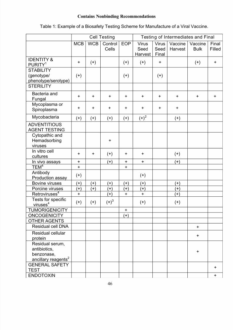
8/11/2019 biologicos produc.pdf
http://slidepdf.com/reader/full/biologicos-producpdf 49/50
8/11/2019 biologicos produc.pdf
http://slidepdf.com/reader/full/biologicos-producpdf 50/50
Contains Nonbinding Recommendations
1 tests may be different for cells versus intermediate and final products2 if applicable3 including ALV test in case of chicken cells4 may be combined with virus induction assays, as applicable+
required stage for testing(+) alternative stage for testing