Embed Size (px)
Citation preview
Cannabinoids production in Cannabis sativa L.:
An in vitro approach
Zur Erlangung des akademischen Grades eines
Dr. rer. nat.
von der Fakultät Bio- und Chemieingenieurwesen
der Technischen Universität Dortmund
genehmigte Dissertation
vorgelegt von
Sayed Hussein Farag Hussein, M.Sc.
aus
Assuit, Ägypten
Tag der mündlichen Prüfung: 26.11.2014
1. Gutachter: Prof. Dr. Oliver Kayser
2. Gutachter: Prof. Dr. Herbert Kolodziej
Dortmund 2014

This thesis is dedicated to my
parents, wife and two children
Abstract
iii
I. Abstract
Cannabis sativa L. (Cannabaceae) is the oldest known medicinal plant. For millennia,
the plant has also been used for fibre and oil production.The most prominent feature
of C. sativa is the psychoactive effect ascribed to its secondary metabolites,
cannabinoids (mainly to tetrahydrocannabinol, THC). However, many other
pharmacological properties of the aforementioned specialized compounds have been
described. Currently, the demand for THC for various medical applications is
substantial, while cultivation and breeding of Cannabis in most countries is strictly
regulated and limited to serving research purposes and meeting therapeutic needs.
Therefore, the hereby proposed and discussed production of THC using in vitro
cultures could be a viable alternative. In the work presented here, in vitro
organogenesis from callus cultures (undifferentiated plant cell masses grown on solid
media) was successfully established, ultimately resulting in regeneration of the
complete C. sativa plant. Further, production of THC as well as other important
cannabinoids was achieved in cell suspension, hairy root and trichome cultures of
Cannabis. The optimal combination of phytohormones, as applied to the B5 growth
medium, fostering the development of meristemoids from callus cultures was: 1-
naphthaleneacetic acid (NAA), 6-benzylaminopurine (BA) and adenine hemisulfate
salt (AS) in respective concentrations of 0.5, 5 and 40 mg/l. Concurrently, the most
favourable augmentation protocols of the B5 medium for the induction and
differentiation of shootlets (small plants with leaves but without roots) were: 0.5 mg/l
of gibberelic acid (GA3) or 0.25 mg/l of thidiazouron (TDZ) and 3 mg/l of GA3 (8.5 ±
1.73 and 7.25 ±1.03 shootlets/callus, respectively). The subsequent root formation of
shootlets was most prominent after supplementation with 1.5 mg/l of indole-3-acetic
acid (IAA). In vitro acclimatized plants growing in Erlenmeyer flasks formed
tetrahydrocannabinolic acid (THCA), cannabigerolic acid (CBGA) and cannabidiolic
acid (CBDA), retrieved at respective concentrations of about 0.33, 0.45 and 157.1
mg/g fresh weight. In contrast, ex vitro acclimatized plants (grown hydroponically for
8 weeks) synthesized THCA, THC, CBGA, cannabigerol (CBG) and CBDA at
corresponding concentrations of 1.54, 28.30, 6.0, 0.125 and 1121.4 mg/g fresh
weight. The obtained results confirmed the generation of pharmacologically important
cannabinoids; however, the biosynthetic abilities of the investigated cell and hairy
root cultures did not provide sufficient levels of the valuable metabolites to warrant
scaling-up of the proposed in vitro production platform.
Zusammenfassung
iv
II. Zusammenfassung
Cannabis sativa L. (Cannabaceae) ist die älteste bekannte medizinisch
genutzte Pflanze. Seit Jahrtausenden wird die Pflanze auch für die Produktion
von Fasern und Öl genutzt. Die bekannteste Eigenschaft von C. sativa ist die
psychoaktive Wirkung, aber auch zahlreiche weitere pharmakologische
Eigenschaften sind beschrieben, die auf die Cannabinoide zurückzuführen
sind, besonders auf Tetrahydrocannabinol (THC). Gegenwärtig gibt es einen
hohen Bedarf an THC für diverse medizinische Anwendungen. Die
Kultivierung und Züchtung von Cannabis ist in den meisten Ländern verboten,
mit Ausnahme für Forschungs- und pharmazeutische Zwecke. Daher könnte
die Produktion von THC mit in vitro-Kulturen eine Alternative sein. In der
vorliegenden Arbeit wird die Regeneration ganzer Pflanzen ausgehend von
Kallus-Kulturen (undifferenziertes Zellwachstum auf festen Medien) und die
Bildung von THC und anderen Cannabinoiden in Zellsupsensionen,
Haarwurzel-Kulturen und Trichomkulturen beschrieben. Die optimale
Kombination zur Stimulierung der Bildung von Vorstufen zur Blattbildung
(Meristemoide) ausgehend von Kalluskulturen wurde auf festem B5-Medium
erzielt, welches mit den Pflanzenhormonen 1-Naphthalensäure (NAA, 0,5
g/Liter Medium)), 6-Benzylaminopurin (BA, 5 mg/Liter) und Adenein-
Hemisulfat (AS, 40 mg/Liter). Das optimale Medium für die Induktion der
Bildung kleiner Pflanzen mit Blättern ohne Wurzeln („Shootlets“) war B5 + 0,5
mg Gibberellinsäure (GA3)/Liter mit 8,5 ± 1,73 Shootlets pro Kallus und B5-
Medium + 0,25 mg Thidiazuron/Liter + 3 mg GA3/Liter (7,25 ± 1,03 Shootlets
pro Kallus). Die beste Wurzelbildung der Shootlets wurde mit 1,5 mg Indol-3-
Essigsäure (IAA) erzielt. Die in vitro in Erlenmeyerkolben angezogenen
Pflanzen bildeten THCA, CBGA und CBDA mit einer Konzentration von 033,
0,45 und 157,1 mg/g Frischgewicht. Werden die Pflanzen außerhalb von
Erlenmeyerkolben in hydroponischer Kultur über acht Wochen angezogen,
werden THCA, THC, CBGA, CBG und CBDA mit Konzentrationen von 1,54,
28,3, 6,0, 0,125 und 1121,4 mg/g Frischgewicht gebildet. Die Untersuchungen
zeigten die Bildung pharmakologisch wichtiger Cannabinoide durch C. sativa,
aber die Biosynthese-Leistung der Zellen ergab keine ausreichenden
Konzentrationen, die eine Maßstabsvergrößerung sinnvoll erscheinen lassen
würde.
Contents
v
III. Contents Page
I. Abstract iii
II. Zusammenfassung iv
III. Table of contents v
1. Introduction 1
1.1. Brief history of Cannabis 3
1.2. Botanical description of Cannabis 3
1.2.1. Macroscopic features 3
1.2.2. Microscopic features 4
1.3. In vivo cultivation and breeding of Cannabis 6
1.3.1. Indoor cultivation 6
1.3.2. Seed selection and germination 7
1.3.3. Selection of mother plants and cloning 7
1.3.4. Vegetative period 8
1.3.5. Flowering period 8
1.4. Secondary metabolites of Cannabis 8
1.4 1. Cannabinoids 8
1.4 1.1. Biosynthetic pathway 9
1.4 1.2. Changes in cannabinoid profile over time 11
1.4 1.3. Harvest and processing 13
1.4.1.3.1. Harvesting and drying 13
1.4.1.3.1. Processing 14
1.4.2. Non-cannabinoid constituents 15
1.4.2.1. Terpenoids 15
1.4.2.2. Flavonoids 15
1.4.2.3. Alkaloids 16
1.4.2.4. Other compounds 16
1.5. Approved medicines and therapeutic potential 16
1.6. In vitro culture studies 16
1.6.1. Nutrients and requirements of growth 18
1.6.1.1. Nutrient media composition 18
1.6.1.2. Inorganic nutrients 18
Contents
vi
1.6.1.3. Macroelements 19
1.6.1.4. Microelements 19
1.6.1.5. Organic nutrients 19
1.6.1.5.1. Carbohydrates 20
1.6.1.5.2. Vitamins 20
1.6.1.5.3. Plant growth regulators (PGRs) 20
1.6.1.5.4. Other organic supplements 21
1.7. Phytohormone-regulated cell cycle control 22
1.8. Establishment of callus and cell suspension cultures 23
1.9. Regeneration and plantlet adaptation 25
1.10. Hairy root cultures 27
1.11. Synthetic seed technology and aspects of cryopreservation 27
1.12. Aims of the study
28
2. Materials and methods 29
2.1. Materials 29
2.1.1. Plant material 29
2.1.2. Solvents and chemicals 29
2.1.3. Plant culture media 30
2.1.4. Equipment 31
2.1.5. Culture vessels 31
2.1.6. Sterilisation 32
2.1.6.1. Culture instruments and glassware 32
2.1.6.2. Tissue culture media and other materials 33
2.1.6.3. Plant material 33
2.1.7. Plant growth chambers 33
2.1.7.1. Standard culture conditions 33
2.2. Methods 34
2.2.1. In vitro micropropagation of C. sativa leaf-derived calli 34
2.2.1.1. Sterilization and explant preparation 34
2.2.1.2. Callogenesis 34
2.2.1.3. Meristemoid initiation 34
2.2.1.4. Shootlet induction and multiplication 34
Contents
vii
2.2.1.5. Rooting of shootlets 35
2.2.1.6. Ex vitro acclimatization 35
2.2.2. Shake flask suspension cultures 36
2.2.2.1. Determination of growth kinetics 36
2.2.3. Hairy root cultures 37
2.2.3.1. Initiation of adventitious root cultures 37
2.2.3.2. Shake flask hairy root cultures 37
2.2.3.3. Determination of growth kinetics 39
2.2.4. Conservation of hairy root cultures 39
2.2.4.1. Preparation of encapsulation matrix 39
2.2.4.2. Formation of beads 39
2.2.4.3. Reestablishment of shake flask cultures 41
2.2.5. Trichome induction 41
2.2.5.1. Effect of phytohormones on trichomes induction 41
2.2.5.2. Trichome analysis 41
2.2.6. Analytical Methods 41
2.2.6.1. Extraction of cannabinoids and sample preparation 42
2.2.6.1.1. Callus, plantlets grown in solid media, and
hydroponic plants
42
2.2.6.1.2. Cell suspension and hairy root cultures 42
2.2.6.2. Fingerprinting of cannabinoids 42
2.2.6.2.1. LC-ESI-MS (callus cultures, in vitro
plantlets and hydroponic plants)
2.2.6.2.1.1. Standard curves
42
43
2.2.6.2.2. LC-ESI-MS/MS ( cell suspension and
hairy root cultures)
43
2.2.6.2.3. HPLC analysis 44
2.2.6.2.3.1. Standard curves 44
2.2.6.2.4. 1H-NMR 45
2.2.6.2.5. MALDI imaging MS 46
2.2.6.2.5.1. Callus handling and sample preparation
46
2.2.6.2.5.1.1. Cryosectioning of callus- trichome tissue
46
Contents
viii
2.2.6.2.5.1.2. Matrix application
46
2.2.6.2.5.1.3. MALDI and imaging
46
2.2.6.2.5.1.4. Further software
47
2.2.6.2.6. Data processing 47
2.2.7. Experimental design and statistical analysis
47
3. Results 48
3.1. In vitro micropropagation of leaf-derived calli 48
3.1.1. Callogenesis 48
3.1.2. Meristemoid initiation 49
3.1.3. Shoot induction and multiplication 49
3.1.4. Rooting of shootlets 53
3.1.5. Ex vitro acclimatization (indoor cultivation) 54
3.1.6. Quantitative determination of cannabinoid content 54
3.2. Shake flask cultures 57
3.2.1. Characterization of cannabinoids in cell suspension cultures 57
3.2.1.1. Growth rates 57
3.2.1.2. Time course of cannabinoid production 57
3.2.1.2.1. Characterization of high yielding
cell cultures
57
3.2.1.2.2. Characterization of low yielding
cell cultures
60
3.2.1.2.3. Cell culture morphology and
aging
60
3.2.1.3. Fingerprinting of cannabinoids 61
3.2.1.3.1. LC-MS/MS analysis 61
3.2.1.3. 2. 1H NMR 64
3.2.2. Shake flask cultures of hairy roots 66
3.2.2.1. Initiation of adventitious hairy roots from
calli
66
3.2.2.2. Optimization hairy root cultures 68
3.2.2.3. Characterization of cannabinoids 71
3.2.2.3.1. Growth rates 71
Contents
ix
3.2.2.3.2. Time course of cannabinoid production
71
3.2.2.3.3. Fingerprinting of cannabinoids:
LC-MS/MS analysis
73
3.2.2.3.3.1. LC-MS/MS analysis 73
3.2.2.4.Conservation of hairy root cultures 74
3.2.2.4.1. Effect of the encapsulation-dehydration
procedure
74
3.2.2.4.2. Post-conservation characteristics of hairy
root cultures
75
3.2.2.4.2.1. Growth profiling: solid medium 75
3.2.2.4.2.2. Growth profiling: shake flask cultures 76
3.2.3. Initiation of trichomes formation 76
3.2.3.1. Morphotypes of trichome 76
3.2.3.2. Profling of cannabinoids 79
3.2.3.2.1. LC-MS analysis 79
3.2.3.2.2. MALDI imaging MS 79
3.2.3.2.2.1. MALDI imaging MS profiling
of cannabinoids trichomes
80
4. Discussion 83
4.1. Establishment of leaf-derived callus cultures 83
4.2. Micropropagation via leaf-derived calli 83
4.2.1. Meristemoid initiation 83
4.2.2. Shootlet induction and multiplication 84
4.2.3. Rooting of shootlets 85
4.2.4. Ex vitro acclimatization (indoor cultivation) 85
4.2.5. Cannabinoids in the acclimatizated plants 86
4.3. Shake flask suspension cultures 86
4.3.1. Characterization of cannabinoids 87
4.3.1.1. Growth rates 87
4.3.1.2. Time course of cannabinoid production in high
yielding cell cultures
87
4.3.2. Fingerprinting of cannabinoids 88
4.3.2.1. LC-MS/MS analysis 88
Contents
x
4.3.2.2. 1H NMR measurements 89
4.4. Hairy root cultures 89
4.4.1. Initiation of adventitious hairy roots from leaf-derived calli 89
4.4.2. Optimization of hairy root cultures on solid media 90
4.4.3. Characterization of cannabinoids in shake flask root cultures 90
4.4.3.1. Growth rates 90
4.4.3.2. Time course of cannabinoid production 90
4.4.4. Fingerprinting of cannabinoids in root suspensions 91
4.5. Conservation of hairy root cultures 92
4.6. Initiation of trichome formation 92
4.6.1. Trichome morphotypes 92
4.6.2. Profling of cannabinoids 93
4.6.2.1. LC-MS/MS analysis 93
4.6.2.2. MALDI imaging MS 93
5. Concluding remarks and perspectives 94
6. References 97
7. Appendix 122
I. List of Abbreviations 123
II. Acknowledgements 126
III. Curriculum vitae 127
IV. List of publications 128
Review of Literature
1
1. Introduction
1.1. Brief history of Cannabis
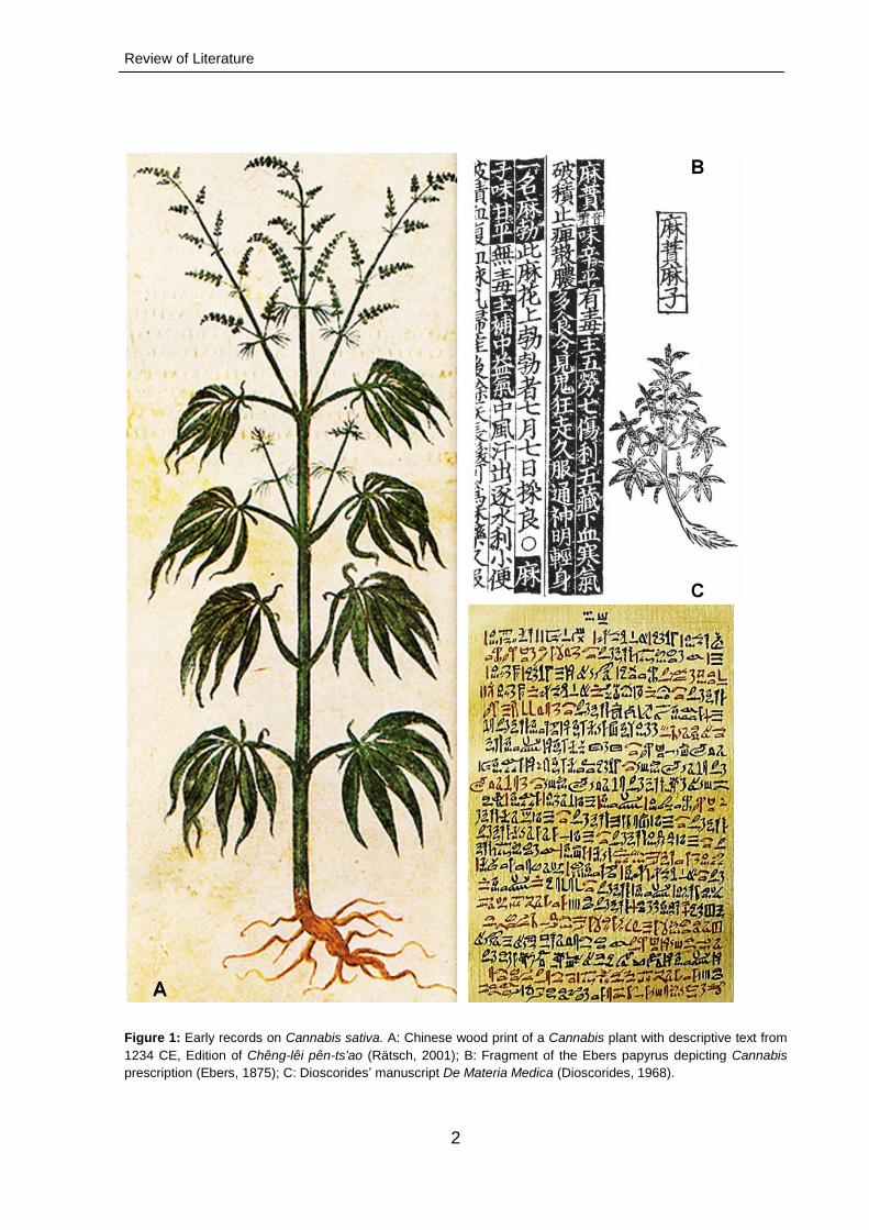
The genus Cannabis (family Cannabaceae) is an annual flowering plant. According
to Li (1974), the first historical reference to Cannabis dates back to 5000 BCE and
places its origins in Central Asia, while its medicinal uses were recorded in stone and
papyrus documents of ancient Egipt circa 1700-1600 BCE (Ebbell, 1937; Russo,
2007; Russo et al., 2008, Figure 1B). Subsequently, the ancient Greeks and Romans
noted the therapeutic value of Cannabis (Figure 1C). In the 2nd century BCE,
Bausanius and Glen documented it in Roman records (Brunner, 1973), while
Diocorides, a Greek physician, in his work De Materia Medica, published in the 1st
century BCE, recommended Cannabis seeds for the treatment of otalgia
(Dioscorides, 1968). By the early 10th century, hashish and its medicinal properties
were widely known to Arabic physicians (Nahas, 1982). During the Industrial
Revolution, marijuana became a popular commodity serving both commercial and
medicinal purposes (Fankhauser, 2002; Waldo, 2006). In the following two decades,
Cannabis was grown extensively in various parts of Europe and the USA (Gaoni and
Mechoulam, 1964; Russo, 2011).
The current systematic classification of Cannabis is as follows (Sytsma et al.,
2002):
Division Magnoliophyta
Class Magnoliopsida
Subclass Hamamelidae
Order Rosales
Family Cannabaceae
Genus Cannabis L.
Species sativa
Review of Literature
2
Figure 1: Early records on Cannabis sativa. A: Chinese wood print of a Cannabis plant with descriptive text from
1234 CE, Edition of Chêng-lêi pên-ts'ao (Rätsch, 2001); B: Fragment of the Ebers papyrus depicting Cannabis
prescription (Ebers, 1875); C: Dioscorides’ manuscript De Materia Medica (Dioscorides, 1968).
Review of Literature
3
1.2. Botanical description of Cannabis
1.2.1. Macroscopic features
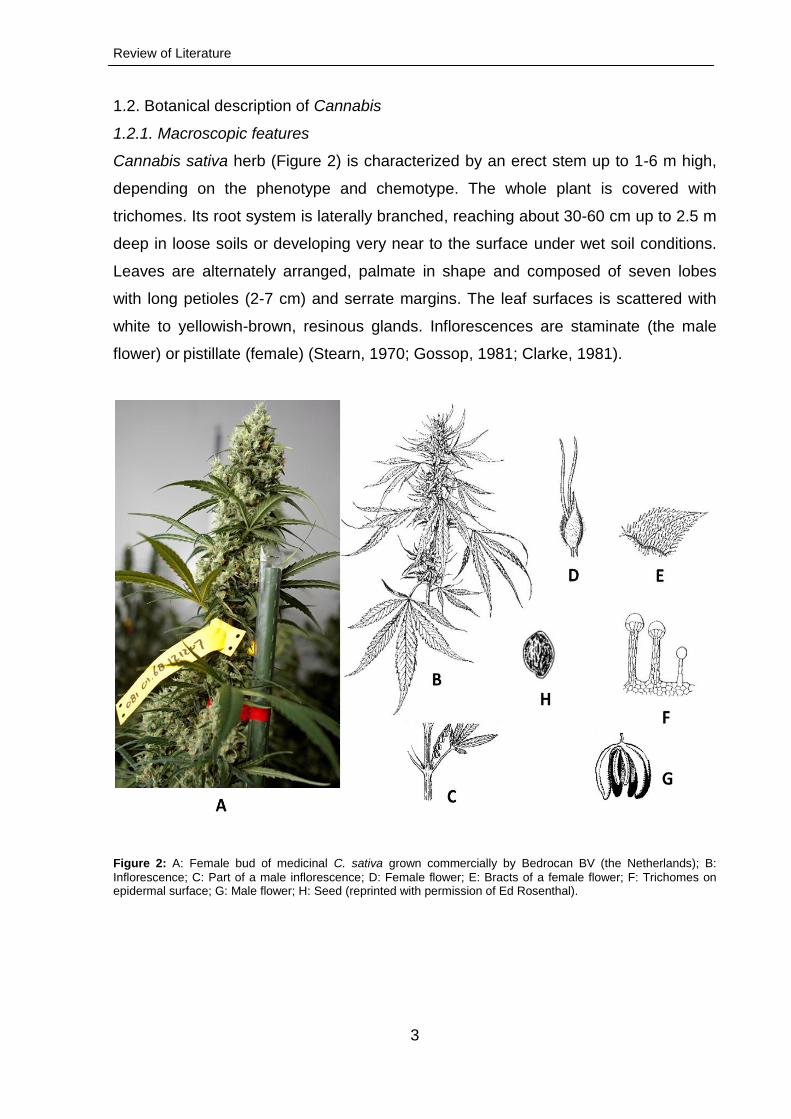
Cannabis sativa herb (Figure 2) is characterized by an erect stem up to 1-6 m high,
depending on the phenotype and chemotype. The whole plant is covered with
trichomes. Its root system is laterally branched, reaching about 30-60 cm up to 2.5 m
deep in loose soils or developing very near to the surface under wet soil conditions.
Leaves are alternately arranged, palmate in shape and composed of seven lobes
with long petioles (2-7 cm) and serrate margins. The leaf surfaces is scattered with
white to yellowish-brown, resinous glands. Inflorescences are staminate (the male
flower) or pistillate (female) (Stearn, 1970; Gossop, 1981; Clarke, 1981).
Figure 2: A: Female bud of medicinal C. sativa grown commercially by Bedrocan BV (the Netherlands); B:
Inflorescence; C: Part of a male inflorescence; D: Female flower; E: Bracts of a female flower; F: Trichomes on epidermal surface; G: Male flower; H: Seed (reprinted with permission of Ed Rosenthal).
Review of Literature
4
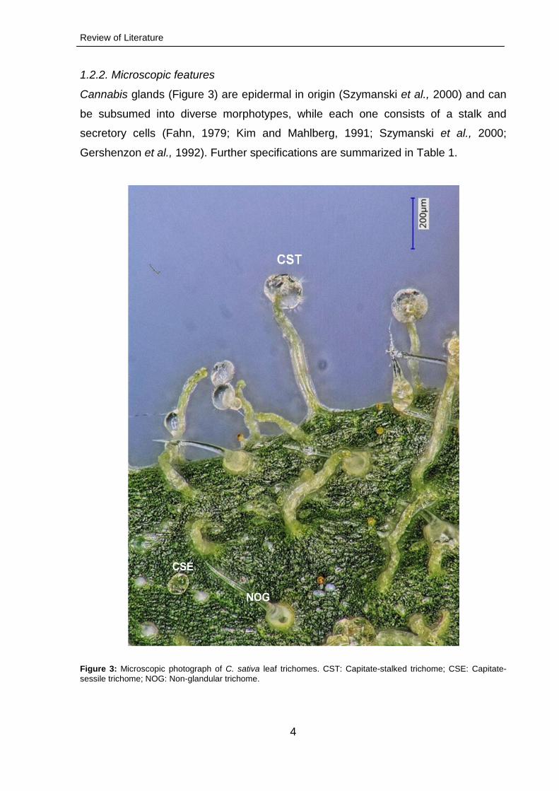
1.2.2. Microscopic features
Cannabis glands (Figure 3) are epidermal in origin (Szymanski et al., 2000) and can
be subsumed into diverse morphotypes, while each one consists of a stalk and
secretory cells (Fahn, 1979; Kim and Mahlberg, 1991; Szymanski et al., 2000;
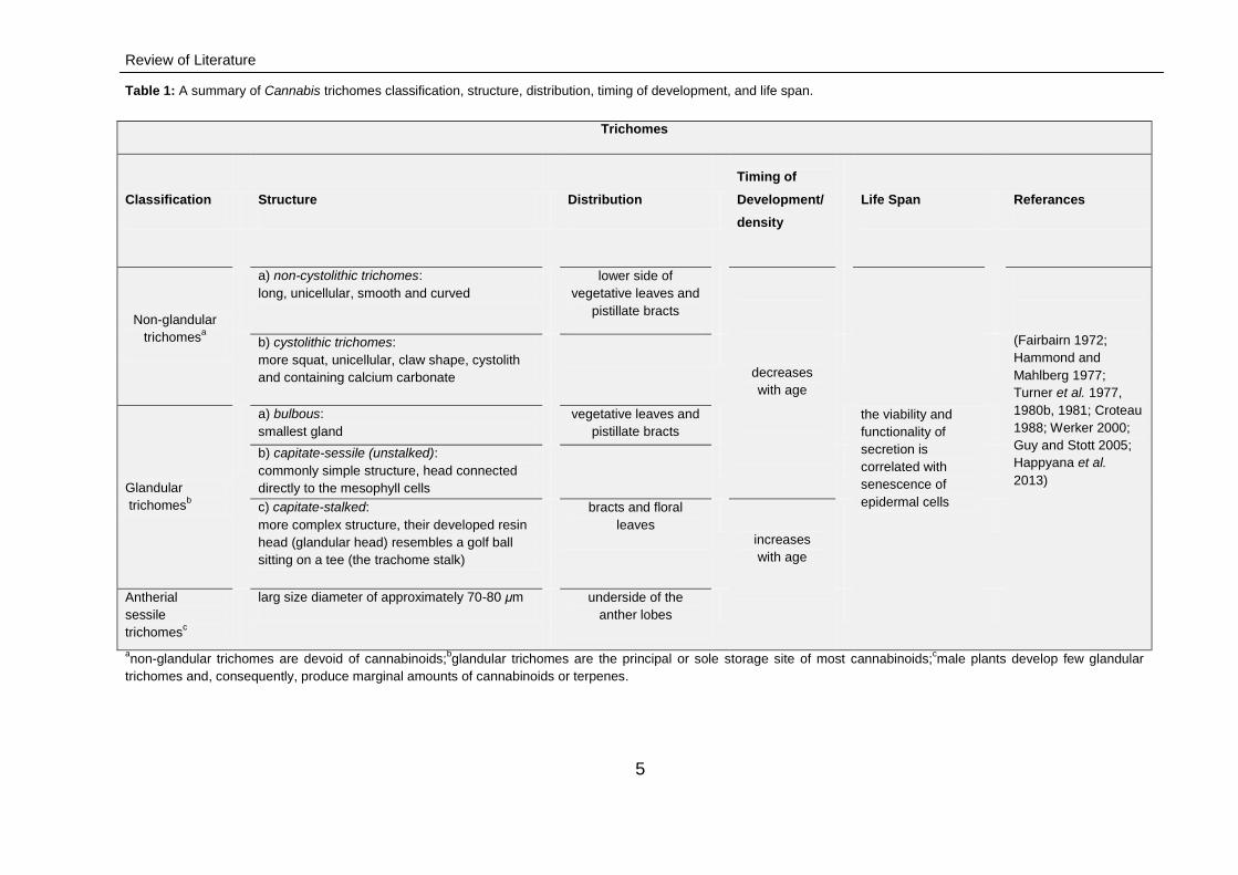
Gershenzon et al., 1992). Further specifications are summarized in Table 1.
Figure 3: Microscopic photograph of C. sativa leaf trichomes. CST: Capitate-stalked trichome; CSE: Capitate-
sessile trichome; NOG: Non-glandular trichome.
Review of Literature
5
Table 1: A summary of Cannabis trichomes classification, structure, distribution, timing of development, and life span.
Trichomes
Classification Structure Distribution
Timing of
Development/
density
Life Span Referances
Non-glandular
trichomesa
a) non-cystolithic trichomes:
long, unicellular, smooth and curved
lower side of
vegetative leaves and
pistillate bracts
decreases
with age
the viability and
functionality of
secretion is
correlated with
senescence of
epidermal cells
(Fairbairn 1972;
Hammond and
Mahlberg 1977;
Turner et al. 1977,
1980b, 1981; Croteau
1988; Werker 2000;
Guy and Stott 2005;
Happyana et al.
2013)
b) cystolithic trichomes:
more squat, unicellular, claw shape, cystolith
and containing calcium carbonate
Glandular
trichomesb
a) bulbous:
smallest gland
vegetative leaves and
pistillate bracts
b) capitate-sessile (unstalked):
commonly simple structure, head connected
directly to the mesophyll cells
c) capitate-stalked:
more complex structure, their developed resin
head (glandular head) resembles a golf ball
sitting on a tee (the trachome stalk)
bracts and floral
leaves
increases
with age
Antherial
sessile
trichomesc
larg size diameter of approximately 70-80 μm
underside of the
anther lobes
anon-glandular trichomes are devoid of cannabinoids;
bglandular trichomes are the principal or sole storage site of most cannabinoids;
cmale plants develop few glandular
trichomes and, consequently, produce marginal amounts of cannabinoids or terpenes.
Review of Literature
6
1.3. In vivo cultivation and breeding of Cannabis
1.3.1. Indoor cultivation
Currently, cultivation and breeding of highly potent varieties of C. sativa is illegal all
over the world. However, select pharmaceutical companies (e.g., Bedrocan BV and
GW Pharmaceuticals Ltd.) have been granted a license to grow the plant under
strictly controlled conditions, for research and medicinal purposes. The main
advantage of indoor cultivation is the ability to control culture conditions facilitating
yield improvement, life cycle regulation, pest control and prevention of self- and
cross-pollination (Rosenthal, 1984; Stamler et al., 1985; Chandra et al., 2010).
Moreover, there are a number of techniques of growing Cannabis hydroponically.
The most important facet in hydroponics is the nutrient solution consisting of all the
essential elements for plant growth and development in appropriate amounts and
proportions. The crucial macronutrients in the solution are three cations (potassium,
calcium and magnesium) and three anions (nitrate, dihydrogen phosphate and
sulphate). Relative proportions of the aforementioned ions should be equilibrated to
avoid ionic imbalances by monitoring the pH within a certain range (5.5-6.5) for
maximum uptake and optimal plant growth (Steiner, 1961; Argo and Fischer, 2002).
On the other hand, many hydroponic growers use special nutrient solution
formulations (“recipes”) based on established plant and crop management conditions
(Le Bot et al., 1998). Oxygenation of either the hydroponic nutrient solution or rooting
medium is another important aspect affecting root function, particularly the rate of
water and nutrient uptake (Porterfield and Musgrave, 1998). Indoor Cannabis crop
cultivation requires artificial light to facilitate and regulate photosynthetic (optimal
plant growth) and photoperiodic processes (controlling flowering and plant shape)
(Coene, 1995). Types of light sources available for that purpose were described by
van Patten (Van Patten, 1992), with a special emphasis placed on high intensity
discharge (HID), metal halide (MH) and high pressure sodium (HPS) lamps (Parker,
1994; Jones, 1997; Zheng et al., 2005).
However, the photosynthetic rate is not only light-dependent but also positively
correlated to carbon dioxide (CO2) concentration surrounding the plant (in ambient
atmosphere, 300-400 ppm) (Jones, 1997; Sicher and Bunce, 1997). Therefore,
delivery of compressed CO2 gas is required for indoor cultivation, increasing plant
size and speed of growth up to 100 % (Jones, 1997). Nowadays, numerous
controlled environment agriculture (CEA) protocols are available for the regulation of

Review of Literature
7
air and root temperature, atmospheric humidity and gas composition, light intensity
and wavelength composition as well as photoperiod duration, water supply and
quality, growth medium composition and nutrient supplementation (Kubota and
Thomson, 2006; Van Os et al., 2008).
1.3.2. Seed selection and germination
High potency Cannabis seeds should be planted in small jiffy pots to standardize
hydroponic indoor cultivation. Germination is initiated in course of three days. Well-
developed seedlings are then transferred to small pots for optimal stimulation of their
vegetative growth. After sufficient development of roots and biomass, the plants are
transferred into a multi-flow hydroponic system equipped with a nutrient reservoir and
an electronic timer to control the flow of nutrients from the reservoir to the plant pots
(Chandra et al., 2010).
1.3.3. Selection of mother plants and cloning
Selection of Cannabis female specimens, exhibiting vitality required to become
mother plants, should be performed under constant vegetative light (HID lamps, 24
h/day) and with the continuous supply of the hydroponic nutrient solution, in order to
foster the generation of the needed propagation stock. To maintain the genotype of
the mother plant throughout subsequent generations, a method relying on the use of
clones is applied (Potter, 2004). Rooted cuttings, termed “clones”, are the result of
asexual or vegetative propagation. These are transferred into a seedling tray
containing a small amount of culture medium and fed with hydroponic solution
nutrients. Initially, all clones are exposed to uniform vegetative light (usually HID
lamps, 24 h/day), 95-100 % relative humidity (rH) and a temperature of 27 °C for one
month. Clones are rooted after 2-3 weeks and, once the roots are of sufficient size,
the plants can be transferred into a larger hydroponic system operating under
modified conditions fostering accelerated growth (Cervantes, 2006; Chandra et al.,
2010).
1.3.4. Vegetative period
At this stage and throughout the growing period, all clones are kept under similar
environmental conditions (light, temperature, rH and CO2 concentration) in a
cultivation room. In course of this research project, full-spectrum light was provided
Review of Literature
8
by means of 1000 W HID lamps; a hot air suction fan was attached to the lambs and
about 3-4 ft distances between plants and bulbs were always maintained to avoid
heating. By adjusting the distance between the plant and the light source,
photosynthetically active radiation (PAR) of about 700 ± 25 µmol m-2s-1 was sustained
at the pot level. Photoperiod (18 h/day) was regulated using an automatic electric
timerto preserve the vegetative stage. Growth room temperature and humidity were
kept nearly constant at ~60 °C and ~60 %, respectively. Plants were supplemented
with a vegetative fertilizer formula for their acclimatization and vegetative
growth.Within this period, the plants reach the height of 50 cm and exhibit a healthy
root system (Potter, 2004; Chandra et al., 2010).
1.3.5. Flowering period
When the desired growth of the plants is attained, they are fortified with a flowering
fertilizer formula and exposed to a 12 h photoperiod to induce flowering. At this
stage, the number of forming inflorescences (especially the female pistillate flowers)
and the cannabinoid content increase gradually. After eight weeks of flowering, the
color of stigmas and trichomes changes into orange/brown and the production of
cannabinoids decreases (Potter, 2004; Chandra et al., 2010).
1.4. Secondary metabolites of Cannabis sativa
The chemistry of C. sativa has received considerable attention, especially due to the
psychoactivity of tetrahydrocannabinol (THC). More than 500 compounds have been
isolated from different Cannabis organs (Zulfiqar et al., 2012), including a wide
variety of secondary metabolites such as polyketides, trepenoids, polyphenolics,
alkaloids, flavonoids, stilbenoids, quinones and terpenophenolics (Formukong et al.,
1989; Appendino et al., 2011).
1.4 1. Cannabinoids
Cannabinoids, a class of terpenophenolic C21 (or C22 for neutral forms) compounds,
have been found, until now, uniquely in C. sativa (Page and Nagel, 2006). Currently,
about 100 meroterpenoids (prenylated polyketides), accumulating mostly in glandular
trichomes and the moss Radula marginata, are known (Appendino et al., 2011).
According to their core structure, cannabinoids are classified into different types:
Review of Literature
9
CBG type, like cannabigerol
CBC type, like cannabichromene
CBD type, like cannabidiol
THC type, like ∆9-tetrahydrocannabinol
∆8-THC type, like cannabicyclol
CBL type, like cannabielsoin
CBE type, like cannabinol and cannabinodiol
CBND and CBT type, like cannabitriol
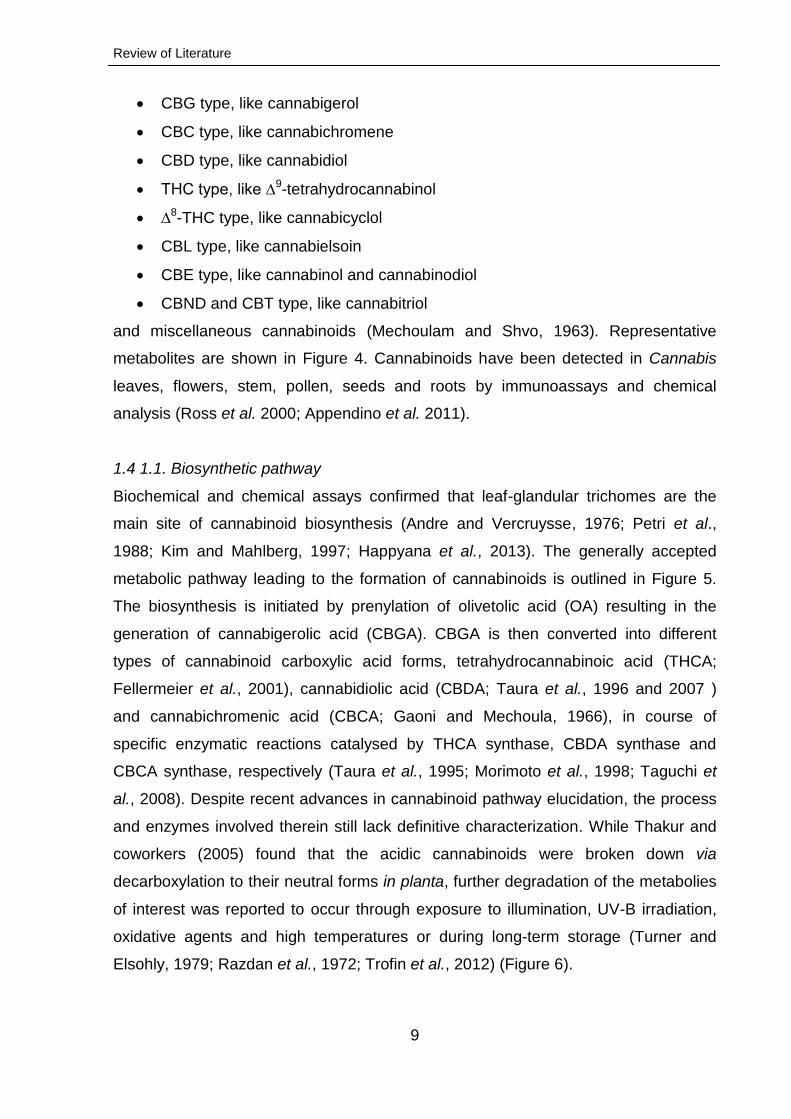
and miscellaneous cannabinoids (Mechoulam and Shvo, 1963). Representative
metabolites are shown in Figure 4. Cannabinoids have been detected in Cannabis
leaves, flowers, stem, pollen, seeds and roots by immunoassays and chemical
analysis (Ross et al. 2000; Appendino et al. 2011).
1.4 1.1. Biosynthetic pathway
Biochemical and chemical assays confirmed that leaf-glandular trichomes are the
main site of cannabinoid biosynthesis (Andre and Vercruysse, 1976; Petri et al.,
1988; Kim and Mahlberg, 1997; Happyana et al., 2013). The generally accepted
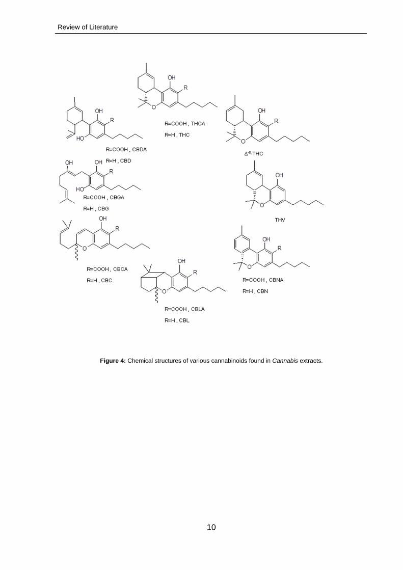
metabolic pathway leading to the formation of cannabinoids is outlined in Figure 5.
The biosynthesis is initiated by prenylation of olivetolic acid (OA) resulting in the
generation of cannabigerolic acid (CBGA). CBGA is then converted into different
types of cannabinoid carboxylic acid forms, tetrahydrocannabinoic acid (THCA;
Fellermeier et al., 2001), cannabidiolic acid (CBDA; Taura et al., 1996 and 2007 )
and cannabichromenic acid (CBCA; Gaoni and Mechoula, 1966), in course of
specific enzymatic reactions catalysed by THCA synthase, CBDA synthase and
CBCA synthase, respectively (Taura et al., 1995; Morimoto et al., 1998; Taguchi et
al., 2008). Despite recent advances in cannabinoid pathway elucidation, the process
and enzymes involved therein still lack definitive characterization. While Thakur and
coworkers (2005) found that the acidic cannabinoids were broken down via
decarboxylation to their neutral forms in planta, further degradation of the metabolies
of interest was reported to occur through exposure to illumination, UV-B irradiation,
oxidative agents and high temperatures or during long-term storage (Turner and
Elsohly, 1979; Razdan et al., 1972; Trofin et al., 2012) (Figure 6).
Review of Literature
10
Figure 4: Chemical structures of various cannabinoids found in Cannabis extracts.
Review of Literature
11
Tetrahydrocannabinolic acid (THCA)
Geranyldiphosphate (GPP)
Cannabigerolic acid (CBGA)
Cannabidiolic acid(CBDA)
Cannabichromenic acid (CBCA)
Olivetolicacid (OA)
OPPCOOH
OH
HO
COOH
OH
HO
COOH
OH
O
COOH
OH
HO
COOH
OH
O
1
2
3
4
5
Figure 5: General pathway for biosynthesis of cannabinoids.1: polyketide synthase (PKS); 2: geranyl
diphosphate:olivetolate geranyltransferase (GOT); 3: THCA synthase; 4: CBGA synthase, 5: CBDA synthase.
1.4 1.2. Changes in cannabinoid profile over time
From sequential harvesting studies on C. sativa chemotypes, it is evident that the
concentration of cannabinoids in the flowers increases as the plant enters the full
flowering phase (Barnicomparini et al., 1984; Vogelmann et al., 1988; Pacifico et al.,
2008; Muntendam et al., 2012). The potency of representatives of the genus
Cannabis is significantly influenced by the pollination (Fetterma et al., 1971; Fairbairn
and Rowan, 1977; Mandolino et al., 2003), phytogeographic region (Hillig and
Mahlberg, 2004), illumnation conditions (Mahlberg and Hemphill, 1983), UV-B
radiation (Zhang and Bjorn, 2009), temepature and humidity, fertilization and the
applied (in- vs. outdoor) breeding method (Latta and Easton, 1975; Staquet et al.,
Review of Literature
12
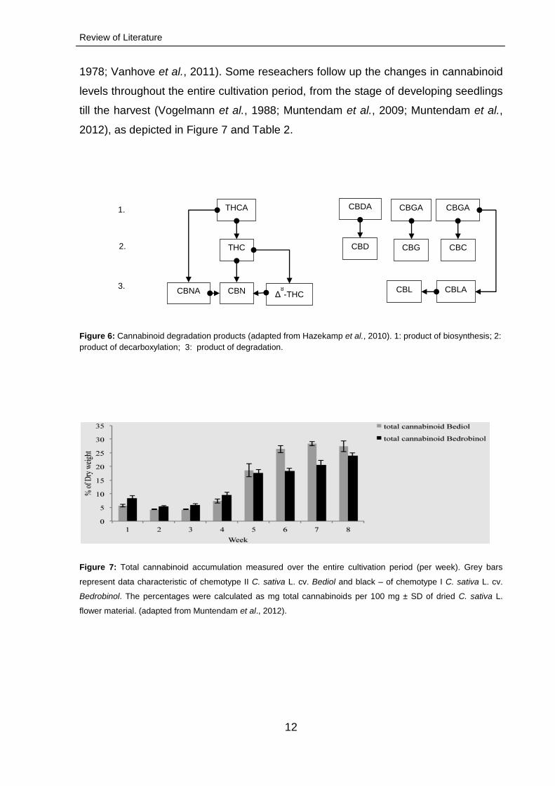
1978; Vanhove et al., 2011). Some reseachers follow up the changes in cannabinoid
levels throughout the entire cultivation period, from the stage of developing seedlings
till the harvest (Vogelmann et al., 1988; Muntendam et al., 2009; Muntendam et al.,
2012), as depicted in Figure 7 and Table 2.
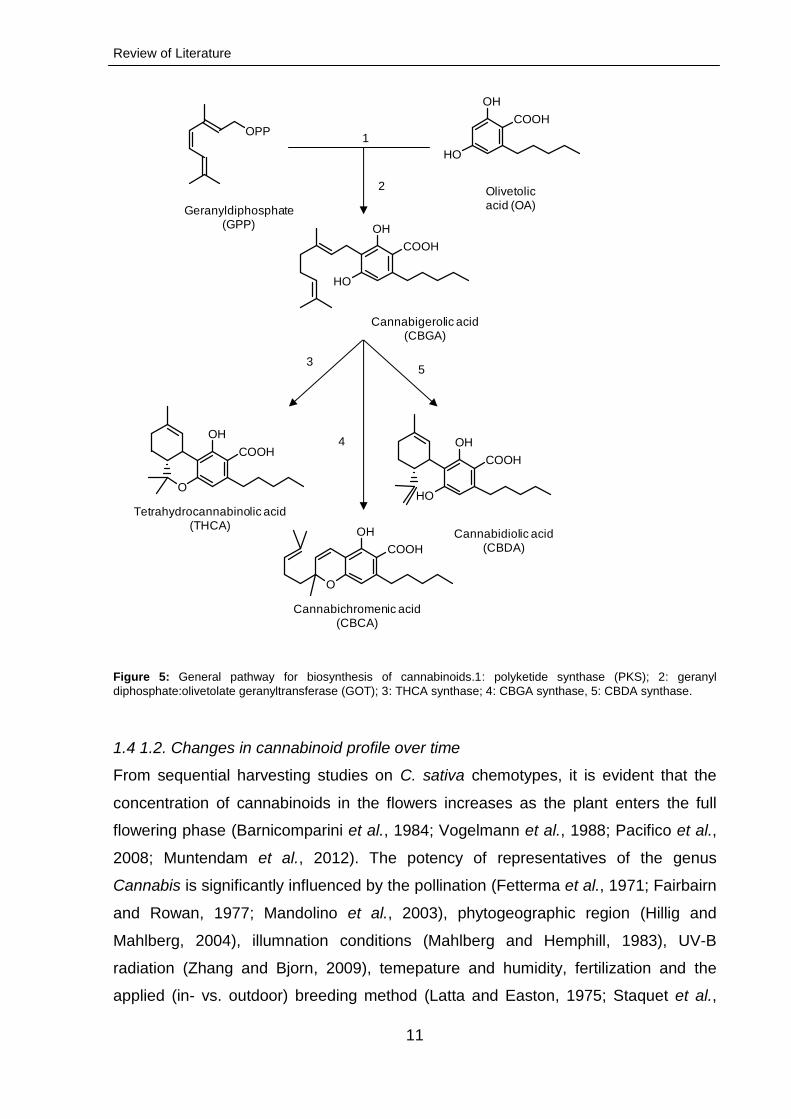
Figure 6: Cannabinoid degradation products (adapted from Hazekamp et al., 2010). 1: product of biosynthesis; 2:
product of decarboxylation; 3: product of degradation.
Figure 7: Total cannabinoid accumulation measured over the entire cultivation period (per week). Grey bars
represent data characteristic of chemotype II C. sativa L. cv. Bediol and black – of chemotype I C. sativa L. cv.
Bedrobinol. The percentages were calculated as mg total cannabinoids per 100 mg ± SD of dried C. sativa L.
flower material. (adapted from Muntendam et al., 2012).
THCA
THC
CBN Δ8-THC CBNA
CBDA CBGA CBGA
CBD CBG CBC
CBLA CBL
1.
3.
2.
Review of Literature
13
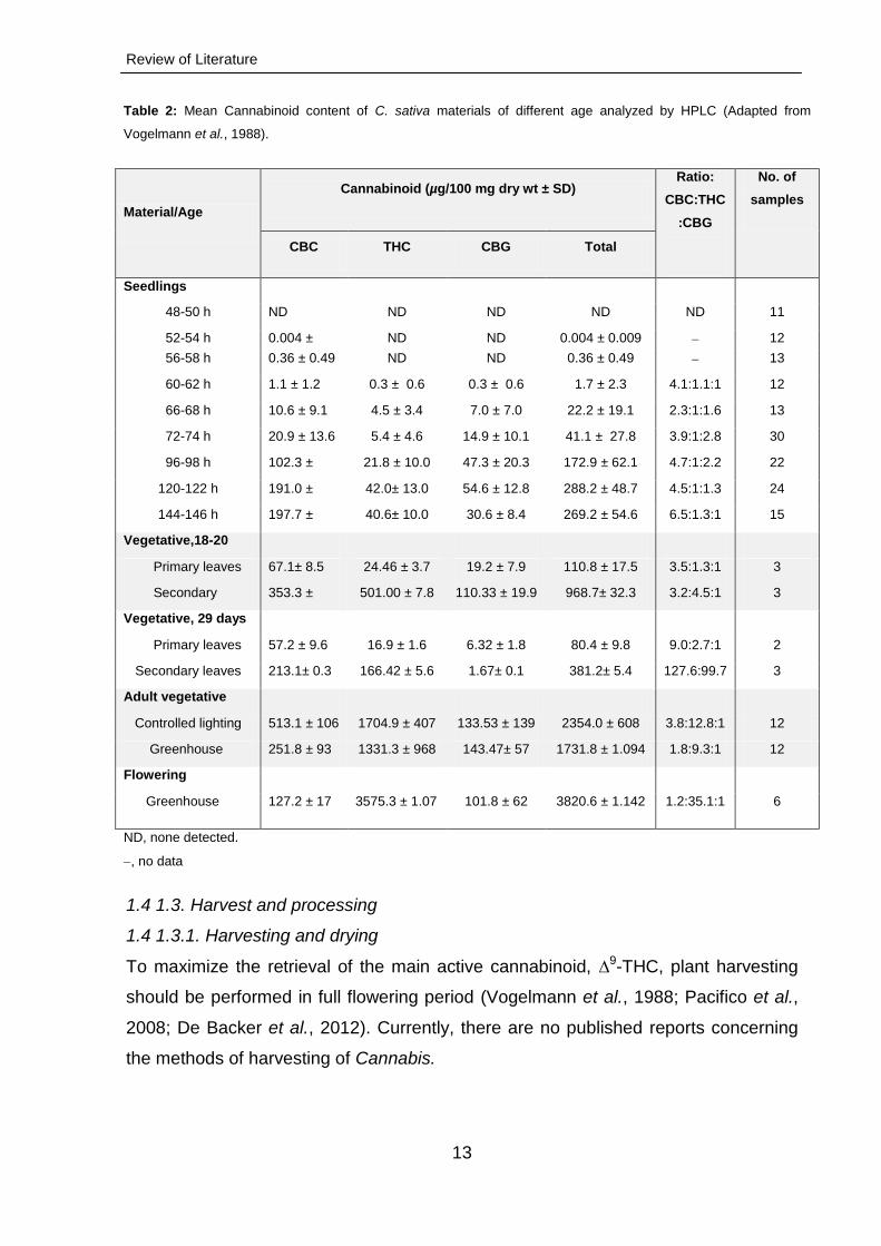
Table 2: Mean Cannabinoid content of C. sativa materials of different age analyzed by HPLC (Adapted from
Vogelmann et al., 1988).
Material/Age
Cannabinoid (µg/100 mg dry wt ± SD) Ratio:
CBC:THC
:CBG
No. of
samples
CBC THC CBG Total
Seedlings
48-50 h ND ND ND ND ND 11
52-54 h 0.004 ±
0.01
ND ND 0.004 ± 0.009 12
56-58 h 0.36 ± 0.49 ND ND 0.36 ± 0.49 13
60-62 h 1.1 ± 1.2 0.3 ± 0.6 0.3 ± 0.6 1.7 ± 2.3 4.1:1.1:1 12
66-68 h 10.6 ± 9.1 4.5 ± 3.4 7.0 ± 7.0 22.2 ± 19.1 2.3:1:1.6 13
72-74 h 20.9 ± 13.6 5.4 ± 4.6 14.9 ± 10.1 41.1 ± 27.8 3.9:1:2.8 30
96-98 h 102.3 ±
37.9
21.8 ± 10.0 47.3 ± 20.3 172.9 ± 62.1 4.7:1:2.2 22
120-122 h 191.0 ±
40.0
42.0± 13.0 54.6 ± 12.8 288.2 ± 48.7 4.5:1:1.3 24
144-146 h 197.7 ±
41.3
40.6± 10.0 30.6 ± 8.4 269.2 ± 54.6 6.5:1.3:1 15
Vegetative,18-20
days
Primary leaves 67.1± 8.5 24.46 ± 3.7 19.2 ± 7.9 110.8 ± 17.5 3.5:1.3:1 3
Secondary
leaves
353.3 ±
12.5
501.00 ± 7.8 110.33 ± 19.9 968.7± 32.3 3.2:4.5:1 3
Vegetative, 29 days
Primary leaves 57.2 ± 9.6 16.9 ± 1.6 6.32 ± 1.8 80.4 ± 9.8 9.0:2.7:1 2
Secondary leaves 213.1± 0.3 166.42 ± 5.6 1.67± 0.1 381.2± 5.4 127.6:99.7
:1
3
Adult vegetative
Controlled lighting 513.1 ± 106 1704.9 ± 407 133.53 ± 139 2354.0 ± 608 3.8:12.8:1 12
Greenhouse 251.8 ± 93 1331.3 ± 968 143.47± 57 1731.8 ± 1.094 1.8:9.3:1 12
Flowering
Greenhouse 127.2 ± 17 3575.3 ± 1.07 101.8 ± 62 3820.6 ± 1.142 1.2:35.1:1 6
ND, none detected.
, no data
1.4 1.3. Harvest and processing
1.4 1.3.1. Harvesting and drying
To maximize the retrieval of the main active cannabinoid, ∆9-THC, plant harvesting
should be performed in full flowering period (Vogelmann et al., 1988; Pacifico et al.,
2008; De Backer et al., 2012). Currently, there are no published reports concerning
the methods of harvesting of Cannabis.
Review of Literature
14
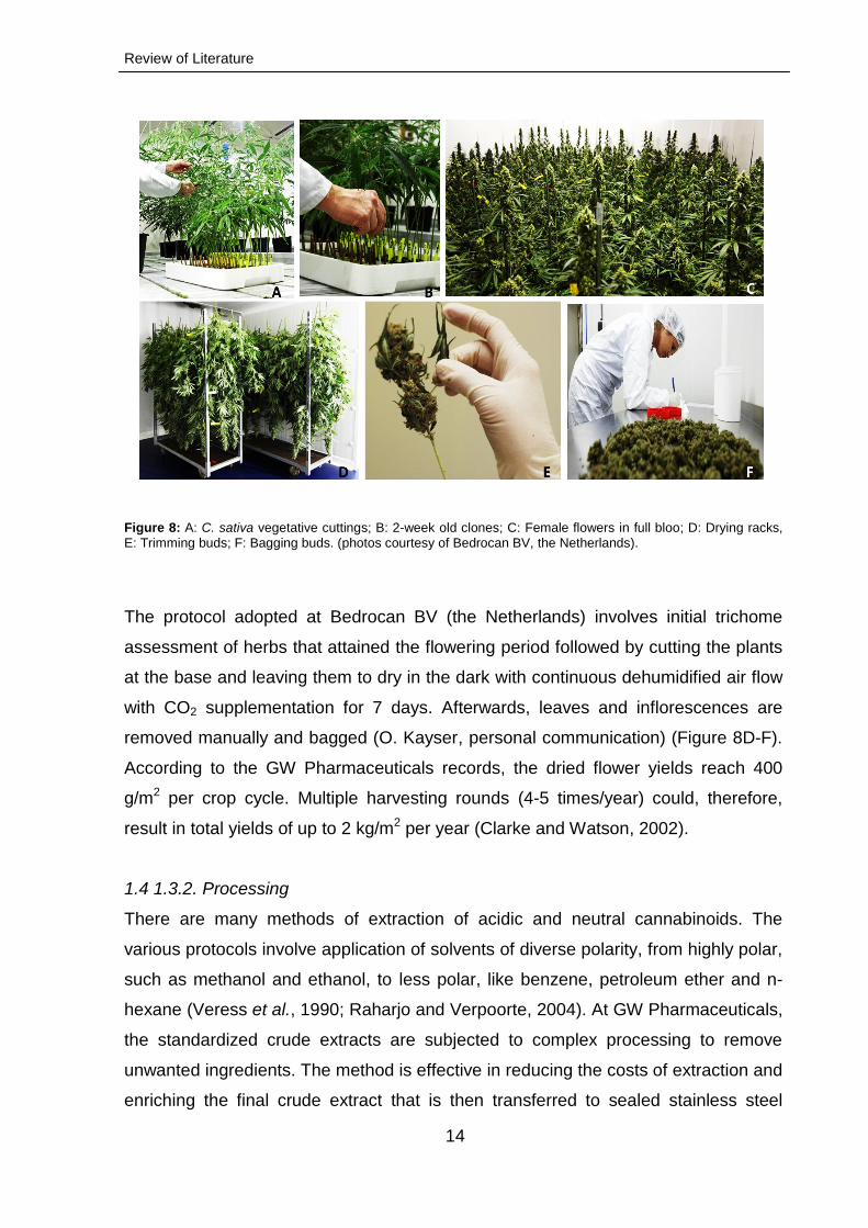
Figure 8: A: C. sativa vegetative cuttings; B: 2-week old clones; C: Female flowers in full bloo; D: Drying racks, E: Trimming buds; F: Bagging buds. (photos courtesy of Bedrocan BV, the Netherlands).
The protocol adopted at Bedrocan BV (the Netherlands) involves initial trichome
assessment of herbs that attained the flowering period followed by cutting the plants
at the base and leaving them to dry in the dark with continuous dehumidified air flow
with CO2 supplementation for 7 days. Afterwards, leaves and inflorescences are
removed manually and bagged (O. Kayser, personal communication) (Figure 8D-F).
According to the GW Pharmaceuticals records, the dried flower yields reach 400
g/m2 per crop cycle. Multiple harvesting rounds (4-5 times/year) could, therefore,
result in total yields of up to 2 kg/m2 per year (Clarke and Watson, 2002).
1.4 1.3.2. Processing
There are many methods of extraction of acidic and neutral cannabinoids. The
various protocols involve application of solvents of diverse polarity, from highly polar,
such as methanol and ethanol, to less polar, like benzene, petroleum ether and n-
hexane (Veress et al., 1990; Raharjo and Verpoorte, 2004). At GW Pharmaceuticals,
the standardized crude extracts are subjected to complex processing to remove
unwanted ingredients. The method is effective in reducing the costs of extraction and
enriching the final crude extract that is then transferred to sealed stainless steel
Review of Literature
15
containers and stored at -20 ± 5 °C to maintain stability and for further use (Guy and
Stott, 2005).
1.4.2. Non-cannabinoid constituents
1.4.2.1. Terpenoids
Plants of the genus Cannabis synthesize a variety of terpenoids. At least 200
structurally different terpenoid compounds have been isolated and characterized
from their flowers (Ross and ElSohly, 1996), roots (Slatkin et al., 1971), leaves
(Hendriks et al., 1975) and trichomes (Kim and Mahlberg, 2003). The most abundant
representatives are β-myrcene, trans-caryophyllene, α-pinene, trans-ocimene and α-
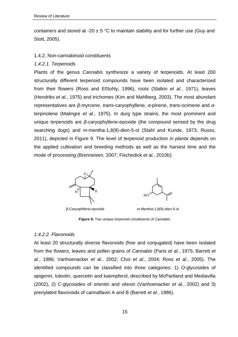
terpinolene (Malingre et al., 1975). In durg type strains, the most prominent and
unique terpenoids are β-caryophyllene-epoxide (the compound sensed by the drug
searching dogs) and m-mentha-1,8(9)-dien-5-ol (Stahl and Kunde, 1973; Russo,
2011), depicted in Figure 9. The level of terpenoid production in planta depends on
the applied cultivation and breeding methods as well as the harvest time and the
mode of processing (Brenneisen, 2007; Fischedick et al., 2010b).
Figure 9: Two unique terpenoid constituents of Cannabis.
1.4.2.2. Flavonoids
At least 20 structurally diverse flavonoids (free and conjugated) have been isolated
from the flowers, leaves and pollen grains of Cannabis (Paris et al., 1975; Barrett et
al., 1986; Vanhoenacker et al., 2002; Choi et al., 2004; Ross et al., 2005). The
identified compounds can be classified into three categories: 1) O-glycosides of
apigenin, luteolin, quercetin and kaempferol, described by McPartland and Mediavilla
(2002), 2) C-glycosides of orientin and vitexin (Vanhoenacker et al., 2002) and 3)
prenylated flavonoids of cannaflavin A and B (Barrett et al., 1986).
H
O
HHO
β-Caryophllene-epoxide m-Mentha-1,8(9)-dien-5-ol
Review of Literature
16
1.4.2.3 Alkaloids
Nitrogenous compounds of C. sativa have been investigated and only a small
number of ten alkaloids have been identified, including some interesting pseudo-
alkaloids and related precursors, such as choline, trigonelline (a pyridine), muscarine
(a protoalkaloid), isoleucine betaine and neurine (Turner et al., 1980a; Ross and
ElSohly, 1995). The aforementioned components have been isolated from Cannabis
leaves, stems, pollen, roots and seeds (ElSohly et al., 1978; Mechoulam, 1988).
1.4.2.4 Other compounds
In addition to the secondary metabolites mentioned above, phenols, steroidal
glycosides, esters and other Cannabis constituents have been reported (ElSohly and
Slade, 2005; Brenneisen, 2007).
1.5. Approved medicines and therapeutic potential
In pharmaceutical industry, cannabinoids (THC and CBD) have become increasingly
important as valuable starting compounds for the development of new drugs.
Sativex® (oral spray: 27 mg/ml Δ9-THC and 25 mg/ml CBD; GW
GW Pharmaceuticals) was approved in Canada, United Kingdom, Germany and
Spain to treat muscle pain and stiffness in multiple sclerosis (MS) and cancer
patients (Whittle and Guy, 2004; Whittle, 2007), while Cannador® (oral capsules:
THC/CBD ratio not stated; Society for Clinical Research, Germany) was reported to
reduce MS-related tremors (Fox et al., 2004; Holdcroft et al., 2006; Rahn and
Hohmann, 2009). Bedrocan®, Bedrobinol® and Bediol® (Bedrocan BV) are dried
flower bud preparations sanctioned for medicinal uses. Moreover, several synthetic
cannabinoid-based drugs have been approved for the alleviation of nausea and
vomiting associated with cancer chemotherapy (Marinol®, Dronabinol, Solvay
Pharmaceuticals and Cesamet®, Nabilone, Valeant Pharmaceuticals International)
(Stott and Guy, 2004; Davis et al., 2007). A number of new cannabinoid-based
products are currently under development and expected to be introduced to the
market in the near future.
1.6 In vitro culture studies
The term "plant tissue culture" is commonly used to describe the maintenance of all
types of plant cells, tissues, organs or whole plants on artificial media, in vitro, under
Review of Literature
17
aseptic conditions (Gamborg and Phillips, 1995). A defined nutrient medium contains
a large number of inorganic salts (macro- and micronutrients), a carbon source, myo-
inositol, glycine and vitamins. Sometimes, growth regulators (auxins, cytokinins,
gibberellins, abscisic acid or ethylene) are added. Other components, such as
organic nitrogen compounds, organic acids and plant extracts may be supplied for
specific purposes. Gelling agents, like agar, gelatin, silica or acrylamide gels,
agarose, alginate or gelrite, are used to obtain a solidified medium. The pH of the
medium should be adjusted to 5.7-5.8 before or after supplementation with the
gelling agent (Gamborg and Phillips, 1995; Robert, 1999).
Plant tissue culture research is a multi-dimensional science with numerous
applications:
– Micropropagation of selected genotypes (Phillips and Hubstenberger, 1995).
– Production of pathogen-free plants by meristematic culturing (Wang and Charles,
1991).
–Germplasm preservation in vitro using cryopreservation techniques (Withers, 1990).
–Vetrification, which is a result of high humidity in the growth-tube, low agar
concentration in the solid medium or high growth in the liquid medium (Sakai and
Engelmann, 2007; Kim et al., 2010).
–Genetic manipulation through: a) somatic hybridization, b) cytoplasmic
hybridization, c) transplantation and uptake of isolated nuclei, chromosomes,
chromosome fragments or organelles (plastids and mitochondria), and d)
transformation (Pierik, 1989).
– Plant regeneration through somatic embryogenesis, directly on an explant or from a
callus (Fransz and Schel, 1994; Choi et al., 1999).
–Organogenesis (the direct or indirect de novo organ formation) (Schwarz and
Beaty,1996).
– Regeneration of haploid, sterile and seedless plants through application of pollen or
microspore cultures (Reed, 1996).
– Isolation of variants, clones and mutant plants with enhanced resistance to biotic
and abiotic stresses (Nabors et al., 1975).
– Overcoming seed dormancy and embryo sterility (Hu and Zanettini, 1995).
– Production of secondary metabolites, such as alkaloids (Verpoorte et al., 1994),
terpenoids (Kummritz et al., 2014), lignans (Elfahmi et al., 2006), etc.
Review of Literature
18
1.6.1. Nutrients and requirements of growth
The requirements of plant tissues grown in vitro are, in general, similar to those of the
intact plants growing in nature (Vasil, 1985b). The success rate of any technology
employing plant cell, tissue or organ cultures depends on several factors. Selection
of nutritional components and growth regulators is a significant one (Street and
Schillito, 1977; Gosal and Kang, 2012), as the isolated cells, tissues and organs lack
the capacity to synthesize their own supply of carbohydrates, most vitamins and
intrinsic growth substances. Accordingly, all the components utilized by plants in
nature must be provided artificially to their in vitro cultures to achieve the desired
results (Murashige and Skoog, 1962; Gamborg et al., 1968a; Schenk and
Hildebrandt, 1972).
1.6.1.1. Nutrient media composition
An ideal nutrient medium consists of inorganic salts, a carbon source, vitamins,
growth regulators and other components serving specific purposes (Hunez-Palenius
and Ochoa-Alejo, 1999). These include organic nitrogen compounds, complex
extracts (casein hydrolysate, yeast extract or coconut milk) and organic acids
(Gamborg, 1986). Murashige and Skoog (MS), Linsmaier and Skoog (LS) or
Gamborg B5 are the most widely used salt compositions, especially in callus
induction or plant regeneration (Sathyanarayana and Verghese, 2007). In general,
the choice of plant tissue culture medium largely depends on the purpose of the
undertaken in vitro cultivation process (Binding, 1986).
1.6.1.2. Inorganic nutrients
The same essential elements that support growth of intact plants are necessary for
the sustained growth and development of in vitro cells and tissues (Ozias-Akins and
Vasil, 1985). The indispensible mineral nutrients have been divided by Clarkson and
Hanson (1980) into two major groups: 1) elements that are convalently bound witin
carbon compounds and are vital constituents of macromolecules, like DNA, RNA and
proteins (nitrogen (N), phosphorus (P),sulfur (S)), and 2) all other elements like
boron (B), chlorine (Cl), manganese (Mn), iron (Fe), zinc (Zn), copper (Cu),
molybdenum (Mo), nickel (Ni), magnesium (Mg), potassium (K) calcium (Ca) that
participate in a variety of often overlapping functions, including control of somatic and
Review of Literature
19
electrochemical gradients, regulation of protein conformation and oxidation-reduction
reactions of metalloproteins.
1.6.1.3. Macroelements
Deficiencies of macroelements N, P, S, K, Mg and Ca manifest more prominently
when cells are cultured in liquid rather than on solid media, since impurities present
in agar (most commonly used gelling agent) are considerable (Heller, 1953).
Nitrogen, generally supplied in the form of NH4+ with NO3
-, stimulates prolific
formation of somatic embryos (as reported for Atropa belladonna, Gamborg et al.,
1968a; Thomas and Street, 1972 and Digitalis lanata, Kuberski et al., 1984).
Phosphorus and sulfur are usually supplied as phosphates and sulfates (Vasil,
1985a). Murashige and Skoog (1962) found that P levels greater than 2 mM were
often inhibitory to the growth of tobacco pith tissues; therefore, they selected 1.25
mM as the near-optimal quantity. This concentration, however, was suboptimal for
Haplopappus gracilis suspension cultures, which showed an increase of 50 % in
growth rate when the amount was doubled (Eriksson, 1965). The reported level of
potassium required in in vitro cell culturing is about 20 mM. Its supplementation in
combination with low N concentrations (about 1 mM) or addition of KNO3 with
NH4NO3 stimulate somatic embryogensis. (Brown et al., 1976).
1.6.1.4. Microelements
Microelements usually included in plant culture media are Fe, Mn, B, Zn, Mo, Cu, I
and Co and have a profound effect on cell and tissue growth in vitro. Of all the
microelements, the deficiency of iron (supplied as Fe-EDTA chelate to avoid
precipitation at high pH) was reported to reduce growth most dramatically, while Zn,
Cu, B, Mn and Mo deficits also resulted in an inhibitory effect (Nabors et al., 1975).
The ideal growth of plant cells and tissues was achieved when the concentration of
micronutrients was reduced to 10 % of the originally proposed levels (Murashige and
Skoog, 1962; Vasil and Hildebrandt, 1966). Two additional microelements, not
proven essential to the whole plant nutrition, are, nevertheless, included in most
tissue culture media; they are Co and I. Cobalt was introduced by Murashige and
Skoog (1962) because of its documented effect on plant metabolism (Salisbury,
1959).
Review of Literature
20
1.6.1.5. Organic nutrients
Three groups of organic nutrients are required by virtually all tissues cultured in vitro,
i.e., carbohydrates, plant growth regulators and vitamins. In addition, numerous
complex natural extracts and liquid endosperm preparations have been included in
culture media.
1.6.1.5.1. Carbohydrates
Carbohydrates are used as carbon sources. The disaccharide, sucrose, at
concentrations of 2-3 %, is the most commonly utilised representative in plant tissue
culture media (Schaeffer, 1981; Neumann et al., 1999). Carbohydrate concentration
may have a pronounced effect on growth and morphogenesis (Negrutiu and Jacobs,
1978; Debnath, 2005). Five other disaccharides (trehalose, maltose, cellobiose,
turanose and gentiobiose) may be utilized, depending on the cultured plant species
(Verma and Dougall, 1977; Blanc et al., 1999).The trisaccharide, raffinose,
tetrasaccharide, stachyose and polysaccharide, starch can be metabolized by some
tissues (Verma and Dougall, 1977). Sucrose, however, is the best source of carbon,
since it is hydrolysed during the heat-sterilization process into more efficiently
utilizable sugars, such as fructose and glucose (Ball, 1953; Wolter and Skoog, 1966).
Notably, in vitro cultures of Helianthus tuberosus tuber proliferated on appropriate
media containing sucrose (Minocha and Halperin, 1974), while embryo formation
from Petunia anther- (Raquin, 1983) and Digitalis lanata suspension cultures
(Kuberski et al., 1984) was reportedly enhanced by of its substitution with maltose.
1.6.1.5.2. Vitamins
Thiamine, pyridoxine, nicotinic acid and calcium pantothanate are considered the
most essential vitamins in in vitro growth systems (Gamborg et al., 1981ab).
1.6.1.5.3. Plant growth regulators (PGRs)
In addition to mineral salts, carbohydrates and vitamins, most tissue cultures require
exogenous supply of plant growth regulators. In general, differentiation of the
cultured tissue depends on the content ratio of auxin to cytokinin (Skoog and Miller,
1957). Cytokinins are 6-substituted purine compounds (Skoog and Armstrong, 1970).
These phytohormones are incorporated into the culture medium mainly for the
stimulation of cell division, differentiation of adventitious shoots from callus and organ
Review of Literature
21
proliferation (Klee, 1994). Commercially available synthetic cytokinins are kinetin
(KIN) and 6-benzylaminopurine (BA), while thoseoccurring naturally in plants are
zeatin and 6-(γ,γ-dimethylallylamino)purine (2iP), and their respective ribosides
(Skoog and Armstrong, 1970).
Auxins, in turn, are included in plant tissue culture media in order to promote cell
growth and division (Perrot-Rechenmann, 2010). Indole-3-acetic acid (IAA) is the
only natural auxin. Its inherent tendency to be decomposed by heat, light and oxygen
(Kefeli and Kalevitch, 2003) precludes its wide application. In contrast, synthetic
auxins, 1-naphthaleneacetic acid (NAA) and 2,4-dichlorophenoxyacetic acid (2,4-D,
especially effective in the induction of somatic embryogenesis; Skoog and Miller,
1957), are stable and commonly used.. Other auxins, such as 2,4,5-
trichlorophenoxyacetic acid (2,4,5-T), 2-methyl-4-chlorophenoxyacetic acid (MCPA),
Dicamba and Picloram (Diazcolo et al., 1972) are also available. Moreover, TDZ has
been used, with considerable success, in the promotion of plant regeneration (Lu,
1993; Jones et al., 2007). Whereas high auxin:cytokinin ratios stimulate the formation
of roots, the opposite phytohormone proportions instigate shoot development. At
intermediate ratio levels, the tissue grows as an undifferentiated callus (Akiyoshi et
al., 1983). There are three more classes of plant growth regulators: gibberelins
(GAs), abscisic acid (ABA, influencing the maturation of somatic embryos; Calic et
al., 2012) and ethylene. While the effects of ABA, GA3 (gibberellic acid) and zeatin on
embryogenesis and organogenesis have been examined by several investigators
(George and Eapen, 1994; Gaspar et al., 1996), only a few studies concerning the
impact of the gaseous ethylene have been carried out (Biddington, 1992; Kumar et
al., 1998).
1.6.1.5.4. Other organic supplements
Addition of amino acids to the culture medium may enhance cell growth and facilitate
plant regeneration (Channarayappa, 2007). L-glutamine, for example, can serve as
the sole source of nitrogen (Saunders et al., 1997). Enzymatically hydrolyzed
proteins, such as N-Z-Amine® Type A (Sigma-Aldrich), which is a casein hydrolyzate,
are effectively used at concentrations of up to 2 g/l (Phillips and Hubstenberger,
1995). Supplementation with malate, citrate, pyruvate and similar organic acids
proved beneficial in protoplast culturing, alleviating salt toxicity salts (Gamborg and
Shyluk, 1970). Moreover, plant extracts, such as coconut milk, can be very effective
Review of Literature
22
in providing an undefined mixture of organic nutrients and growth factors (Letham,
1974).
1.7. Phytohormone-regulated cell cycle control
Plant cell cycle can be divided into four phases: the mitosis stage (M), the phase of
DNA synthesis (S) and two gap periods (G1 and G2) (Inze and De Veylder, 2006;
Kuijt and Schnittger, 2007). For the development to proceed, the timing and rate of
cell division and, consequently, entry into the cell cycle must be precisely controlled.
This is achieved through complex interplay of various kinases, phosphatases and
proteases. The key enzymes that control the transitions between the different stages
of the cell cycle (G1 to S and G2 to M), and the entry of non-dividing cells into it, are
cyclin-dependent protein kinases (CDKs) (Mironov et al., 1999; Umeda et al., 2005).It
has been reported that auxins and cytokinins are implicated in the regulation of the
cell cycle by controlling the activity of CDKs (Zhang et al., 2005). The gene that
encodes the major CDK, cdc2 (cell division cycle 2), is regulated by auxin.
However,its auxin-induced expression results in enzymatically inactive kinase and
high levels of CDK alone are not sufficient to promote cell division. Thus, the process
is arrested at the end of either G1 or G2 (Koens et al., 1995). Concurrently, Zhang et
al. (1996) documented the G2-arrest of cultured cells in the absence of cytokinin,
coinciding with the reduction in CDK activity caused by enhanced phosphorylation of
the tyrosine residues of the enzyme. When such cultures were resupplied with
cytokinin, tyrosine moieties were dephosphorylated, the kinase reactivated, and cell
division resumed. The described functionality of cytokinin provides an insight into the
concerted cytokinin and auxin involvement in cell cycle regulation. Moreover,
cytokinins elevate the expression of the cyclin D3 gene. As D-type cyclins are key
players in the regulation of cell proliferation, the cytokinin-mediated boost in their
level (and thus, the activity of CDKs) is considered the major regulatory function of
the phytohormone (Riou-Khamlichi et al., 1999; Richard et al., 2002).
1.8. Establishment of callus and cell suspension cultures
Callus cultures are clumps of actively dividing undifferentiated cells derived from
plant tissue (often arising from its injury ), usually sustained on gel medium (Pierik,
1987). The nature of the callus tissue, its texture, compactness, friability and
coloration, depends on the genotype and age of the primary explant (Sen et al.,
Review of Literature
23
2014). Callus derived from the original explant can be established and maintained in
an actively growing state through transfer of its fragments to fresh medium at regular
four-week intervals (Remotti and Loffler, 1995). Growth of the callus culture can be
monitored via measurement of its fresh and dry weight or packed cell volume as well
as determination of its growth, cell number or mitotic indices (Dung et al., 1981;
Kittipongpatana et al., 1998; Mustafa et al., 2011). When callus is suspended in liquid
growth medium, its cells disperse producing cell suspension culture characterized by
faster and uniform growth (Mustafa et al., 2011). Many investigators established
callus cultures from explants of different C. sativa organs (roots, hypocotyls,
epicotyls, cotyledons, petioles, leaves and immature flower buds) (Itokawa et al.,
1975; John et al., 1978; Francoise and Vincent, 1981; Fisse et al., 1981; Heitrich and
Binder, 1982; Verzar-Petri et al., 1982; Loh et al., 1983; Braut-Boucher et al., 1985;
Fisse and Andres, 1985). In addition, callus could be derived from seed explants of
numerous hemp varieties, such as Carmagnola, Fibranova, Uniko and Kompolti
(Mandolino and Ranalli, 1999b), Uniko-B, Kompolti Anka and Felina-34 (Feeney and
Punja, 2003), Sileia, Fibriman-24, Novosadska, Juso-15 and Fedrina-74
(Slusarkiewicz-Jarzina et al., 2005), Carmagnola (Pacifico et al., 2008), Beniko,
Silesia and Bialobrzeskie (Wielgus et al., 2008). While numerous publications
describing in vitro studies of many medicinal plants are currently available, scientific
records on cell suspension cultures of Cannabis, established for extraction of
secondary metabolites and analysis of their biotransformation (Figure 10), are
relatively limited. The first relevant report was published by Veliky and Genest in
1972. The authors investigated the accumulation of cannabinoids and phenolic
compounds in cell suspension cultures of C. sativa, concluding that the former were
produced in trace amounts and the latter reached content levels of 0.19 % (as
compared to 2.15 % in the whole plant leaves). In contrast, Itokawa et al. (1975)
examined the constituents of Cannabis callus cultures induced from different
expalnts (roots, hypocotyls, leaves of seedlings and male and female floral axes)
and cultured on MS agar medium supplemented with 0.1-0.01 ppm KIN and 1.0 ppm
2,4-D. While cannabinoids were not detected, other compounds, such as methyl
palmitate, methyl oleate, methyl stearate, 5α-ergostan-3-one, 5α-stigmastan-3-one,
campesterol, stigmasterol, β-sitosterol, 5α-stigmast-22-en-3-one as well as Δ5,24(28)-
unsaturated sterol and fatty acid esters were successfully detected . Subsequently,
the aforementioned group of researchers (Itokawa et al., 1977) investigated
Review of Literature
24
biotransformation of selected natual products in cell cultures geraniol, nerol, trans-
cinnamyl alcohol, isophorol, trans-verbenol and cis-verbenol were transformed into
their corresponding aldehydes. Further on, Hartsel et al. (1983) reported the
biotransformation of CBD to CBE in cell cultures of C. sativa and Saccharum
officinarum grown on 1.5 % agar MS medium containing the vitamins of B5 medium,
with 3 ppm 2,4,5-T as the sole hormone. The suspensions were shaken on a rotary
shaker (100 rpm) at 27 °C, with an eight-hour daily photoperiod. The authors also
found that incubation of the cultures with olivetol resulted in the generation of an
unidentified cannabinoid characterized by a molecular ion of m/z 210 in mass
spectrometry (MS) analysis. Braemer et al. (1987) investigated bioconversion of
flavonoids into their glucosides in suspension cultures of C. sativa. The cells were
grown in B5 medium supplemented with 0.5 mg/l KIN and 1 mg/l 2,4-D on a rotary
shaker (120 rpm), with a 16 h daily photoperiod at 25 °C. They reported that
quercetin was completely transformed into quercetin 3-O-glucoside, quercetin 3-O-
diglucoside, isorhamnetin 3-O-glucoside and isorhamnetin 3-O-diglucoside, while
apigenin was converted to apigenin 7-O-glucoside and 7-O-glucuronide as well as
vitexin. Concurrently, the group studied cannabinoid biotransformations in Cannabis
cultures obtained from calli of leaf explants. The cell suspensions were grown under
the aforementioned conditions; however, they were kept in total darkness.
Subcultures were transferred to fresh medium every three weeks. In effect, CBD was
converted to bound CBE ((R)- and (S)-cannabielsoin) and THC to CBC, as
determined by gas-liquid chromatography (GLC). Unfortunately, quantitative analysis
was not feasible due to insufficient accumulation of the produced compounds
(Braemer and Paris, 1987). More recently, Flores-Sanchez et al. (2009) studied the
influence of biotic and abiotic elicitors on cannabinoid production in C. sativa
cultures. Cell suspensions were obtained from leaf-derived calli and grown in MS
basal medium supplied with B5-specific vitamins, 1mg/l 2,4-D and 1mg/l KIN on an
orbital shaker (110 rpm), under light of 1000-1700 lx intensity and at 25 °C.
Subcultures were transferred to fresh medium every two weeks. The authors
reported that cannabinoid biosynthesis was not stimulated or induced by biotic and
abiotic elicitors. They also noted low levels of THCA synthase gene expression.
Further elicitation studies focused on the influence of jasmonic acid and pectin on
metabolism of two cell lines of C. sativa. The applied nuclear magnetic resonance
Review of Literature
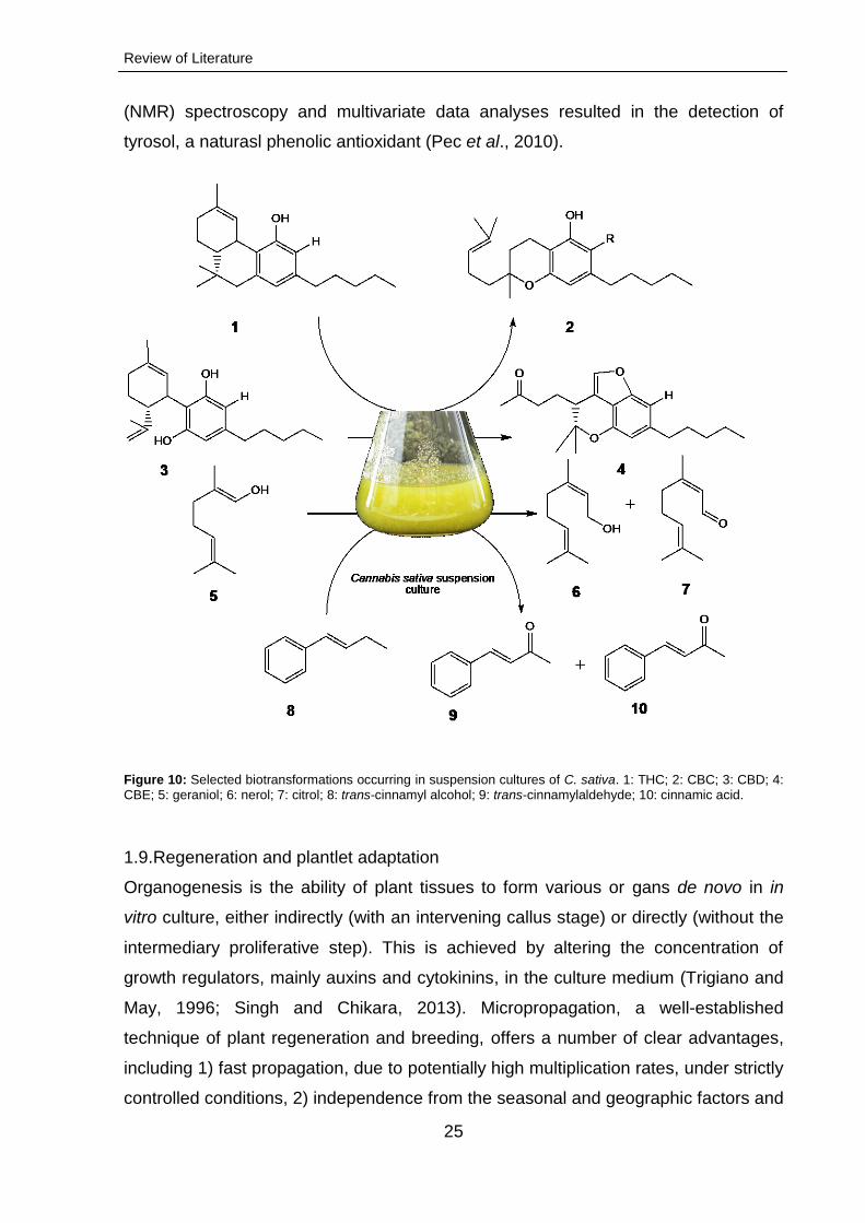
25
(NMR) spectroscopy and multivariate data analyses resulted in the detection of
tyrosol, a naturasl phenolic antioxidant (Pec et al., 2010).
Figure 10: Selected biotransformations occurring in suspension cultures of C. sativa. 1: THC; 2: CBC; 3: CBD; 4: CBE; 5: geraniol; 6: nerol; 7: citrol; 8: trans-cinnamyl alcohol; 9: trans-cinnamylaldehyde; 10: cinnamic acid.
1.9.Regeneration and plantlet adaptation
Organogenesis is the ability of plant tissues to form various or gans de novo in in
vitro culture, either indirectly (with an intervening callus stage) or directly (without the
intermediary proliferative step). This is achieved by altering the concentration of
growth regulators, mainly auxins and cytokinins, in the culture medium (Trigiano and
May, 1996; Singh and Chikara, 2013). Micropropagation, a well-established
technique of plant regeneration and breeding, offers a number of clear advantages,
including 1) fast propagation, due to potentially high multiplication rates, under strictly
controlled conditions, 2) independence from the seasonal and geographic factors and
Review of Literature
26
3) reliable protection from microorganism-borne diseases (Zafar et al., 1992;
Debnath et al., 2006). On the other hand, in vitro propagation of C. sativa through
seeds is possible for most cultivars. This method, however, entails high level of
heterozygosity, which could lead to rapid and dramatic shifts in secondary metabolite
profiles from one generation to the next (Chandra et al., 2010). Many in vitro
propagation protocols (using explants or through organogenesis of callus cultures or
somatic embryogenesis) have been reported for conservation of several medicinal
plants (Sharma et al., 1993; Hosoki et al., 1995; Sudha and Seeni, 1996; Lata et al.,
2002; Bobak et al., 2004; Kanwar and Kumar, 2008).
Despite considerable progress in the field of plant biotechnology, the efficient method
of C. sativa regeneration is still lacking. First reports on de novo organogensis of C.
sativa were published in early 1980s (Fisse et al., 1981). Subsequent studies started
emerging only after nearly two decades; these dealt with the generation of
micropropagation-derived calli from different Cannabis genotypes and explant
sources, including roots (Ranalli and Mandolino, 1999), stems (Mandolino and
Ranalli, 1999a; Wielgus et al., 2008), internodes and axillary buds as well as petioles
(Slusarkiewicz-Jarzina et al., 2005), cotyledons (Wielgus et al., 2008), and young
leaves (Lata et al., 2010). Alternatively, the use of meristematic calli for
micropropagation was investigated (Te-chato and Lim, 1999 and 2000), resulting in
the attainment of genetic stability of elite germplasm. Lata et al. (2010)
demonstrated that in vitro rooting of C. sativa was extremely difficult and the
response to shoot elongation efforts, poor (2-3 cm). The highest proportion of root
differentiation (95 %) was observed by the authors within ten days on half-strength
MS medium supplemented with 2.5 μM indole-3-butyric acid (IBA). Concurrently,
Wang et al. (2009) reported that the proliferated hemp buds were successfully rooted
on MS medium supplemented with 0.1 mg/l IBA and 0.05 mg/l NAA, resulting in 85 %
rate of plantlet rooting. However, to our knowledge, Cannabis regeneration through
direct or indirect somatic embryogenesis has not been reported.
1.10. Hairy root cultures
The possibilities of induction and enhancement of secondary metabolite production in
hairy root systems using phytohormones or through Agrobacterium tumefaciens
transformation have been studied extensively (Brown, 1995; Gelvin 2000; Balvanyos
et al., 2001; Shi et al., 2011).
Review of Literature
27
In early reports on the induction of rhizogenic calli of C. sativa, the hairy root cultures
were established on MS supplemented with different concentrations of NAA (Fisse et
al., 1981). Later on, Feeney and Punja (2003) studied the effect of various PGR
combinations on the Cannabis rhizogenic callus. They found that MS medium, with a
modified, B5-specific, vitamin spectrum and supplemented with different blends of
2,4-D, NAA, IBA, KIN and BA, induced root formation after four weeks in darkness.
The roots were obtained following the infection of the callus cultures with A.
tumefaciens harboring the pNOV3635 plasmid conferring resistance to mannose.
Moreover, Wahby et al. (2013) investigated hairy root induction in several C. sativa
varieties (Futura 77, Delta-llosa, Delta405, CAN0111 and CAN0221) and Nicotiana
tabacum cv. Burley F.13119. The hairy root cultures were established through
infection of seedlings with Agrobacterium strains to investigate the stability of the β-
glucuronidase gene incorported into the genomic DNA of transfomed tissues. About
ten transformants of different C. sativa varities were then screened for desirable traits
in growth. In conclusion, the authors reported that the rolABC transgenic root cultures
were generally characterized by high biosynthetic capacity and biochemical stability.
1.11. Synthetic seed technology and aspects of cryopreservation
The main disadvantage of in vitro cell and tissue cultivation is the necessity of
continuous subculturing which is time-consuming and involves a high risk of microbial
contamination and subsequent loss of original cultures (Grout, 1995; Gangopadhyay
et al., 2011). Synthetic seed technology constitutes a viable alternative, affording
sustainable management and circumventing the aforementioned difficulties. Despite
the progress that has been made in the last decade concerning cryopreservation of
plant material (Ganapathi et al., 2001; Sicurani et al., 2001; Brischia et al., 2002; Hao
and Deng, 2003), there are only few reports on cryoconservation of hairy root
cultures (Lambert et al., 2009). To date, no Cannabis-specific protocols for in vitro
propagation using synthetic seeds or slow freezing of hairy roots are available.

Scope of the thesis
28
1.12.Scope of the thesis
The main goals of this study were 1) to establish an efficient and reliable protocol for
in vitro micropropagation of C. sativa leaf-derived callus using different combinations
of phytohormones and 2) to select the desirable, clonally propagated, plants
according to their cannabinoid profiles and production efficiencies. Moreover, the
growth kinetics and cannabinoid content in shake flask cell suspension and hairy root
cultures were to be analysed and documented. In this context, several questions
regarding relevant technical challenges were addressed.
In order to fulfill the objectives of this thesis, appropriate experiments have been
carried out between 2011 and 2014 under controlled environmental conditions
(temperature, air humidity and light intensity) in growth chambers. To secure high-
cannabinoid genotype of C. sativa, the starting leaf material was obtained from
Bedrocan BV (the Netherlands).
The hereby proposed study addresses the following aspects: 1) In vitro regeneration of leaf-derived C. sativa callus.
i. Formation of meristemiods.
ii. Multiplication of shootlets.
iii. In vitro root formation.
iv. Acclimatization of rooted plantlets to ex vitro conditions in a hydroponic
system followed by stimulation of flowering.
v. Analysis of cannabinoid content in in vitro and ex vitro cultured plants, callus, hairy roots and trichomes.
2) Characterization of cannabinoids in C. sativa cell suspension cultures.
i. Measurement of growth kinetics.
ii. Analysis of cannabinoid content.
iii. Fingerprinting and profiling of cannabinoids. 3) Characterization of cannabinoids in C. sativa hairy root cultures.
i. Measurement of growth kinetics.
ii. Analysis of cannabinoid content.
iii. Fingerprinting and profiling of cannabinoids.
iv. Cryopreservation of hairy root cultures. 4) Induction of trichome formation from callus cultures of C. sativa.
i. Influence of plant growth regulators on trichome differentiation.
ii. MALDI-TOF imaging of cannabinoids.
Material and methods
29
2. Material and methods
2.1 Material
2.1.1. Plant material
Juvenile leaves of high THC yielding medicinal C. sativa L. cv. Bedrobinol (∆9-THC,
approx. 11 % and CBD, approx. < 1 % of plant dry weight) were used for the
preparation of explants. The leaf specimens were obtained from plants grown under
standardized conditions and kindly supplied by Bedrocan BV (Veendam, the
Netherlands).
2.1.2. Solvents and chemicals
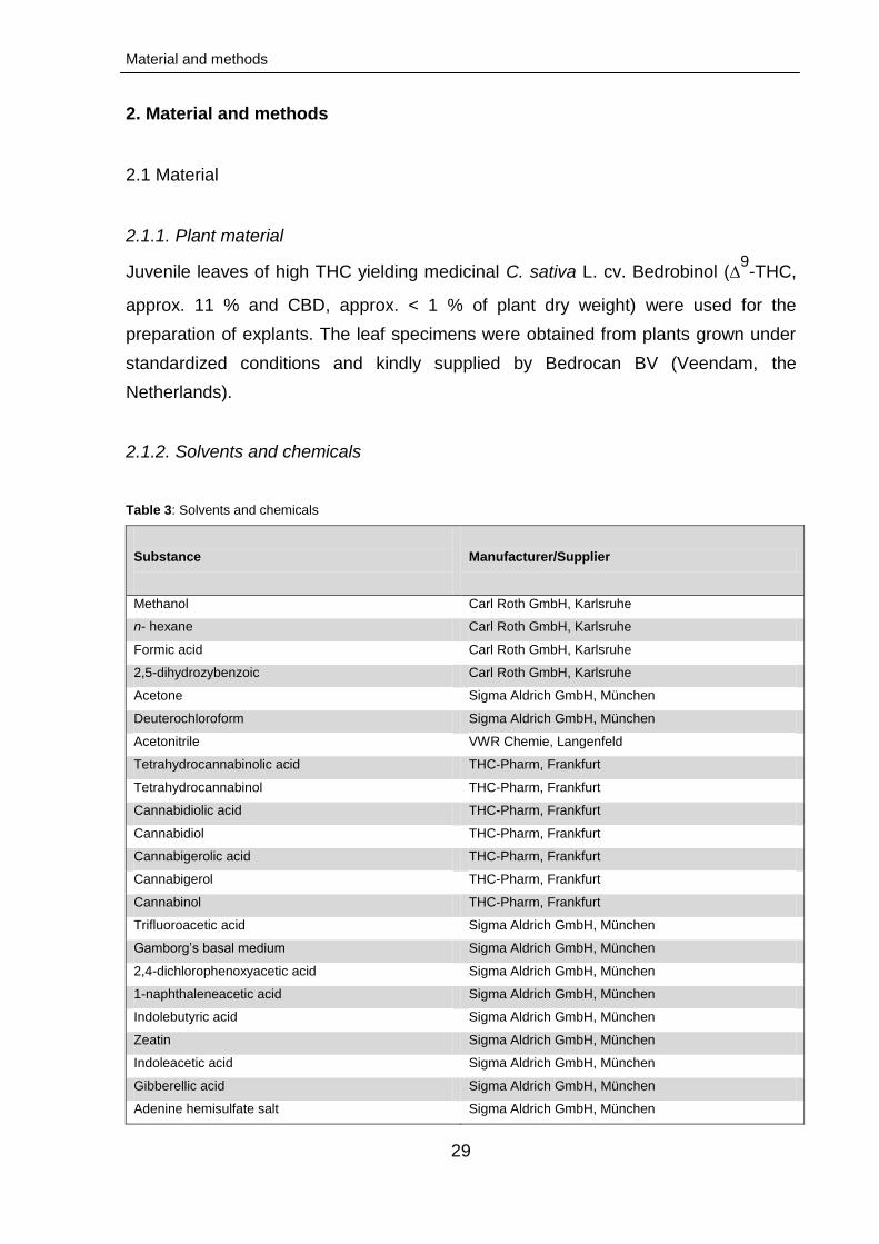
Table 3: Solvents and chemicals
Substance
Manufacturer/Supplier
Methanol Carl Roth GmbH, Karlsruhe
n- hexane Carl Roth GmbH, Karlsruhe
Formic acid Carl Roth GmbH, Karlsruhe
2,5-dihydrozybenzoic Carl Roth GmbH, Karlsruhe
Acetone Sigma Aldrich GmbH, München
Deuterochloroform Sigma Aldrich GmbH, München
Acetonitrile VWR Chemie, Langenfeld
Tetrahydrocannabinolic acid THC-Pharm, Frankfurt
Tetrahydrocannabinol THC-Pharm, Frankfurt
Cannabidiolic acid THC-Pharm, Frankfurt
Cannabidiol THC-Pharm, Frankfurt
Cannabigerolic acid THC-Pharm, Frankfurt
Cannabigerol THC-Pharm, Frankfurt
Cannabinol THC-Pharm, Frankfurt
Trifluoroacetic acid Sigma Aldrich GmbH, München
Gamborg’s basal medium Sigma Aldrich GmbH, München
2,4-dichlorophenoxyacetic acid Sigma Aldrich GmbH, München
1-naphthaleneacetic acid Sigma Aldrich GmbH, München
Indolebutyric acid Sigma Aldrich GmbH, München
Zeatin Sigma Aldrich GmbH, München
Indoleacetic acid Sigma Aldrich GmbH, München
Gibberellic acid Sigma Aldrich GmbH, München
Adenine hemisulfate salt Sigma Aldrich GmbH, München
Material and methods
30
Substance
Manufacturer/Supplier
Benzylaminopurine AppliChem GmbH, Darmstadt
Thiamine-HCl AppliChem GmbH, Darmstadt
Thidiazuron Dr. Ehrenstorfer GmbH, Augsburg
Kinetin Carl Roth GmbH, Karlsruhe
Casein hydrolysate Carl Roth GmbH, Karlsruhe
Myo-Inositol Carl Roth GmbH, Karlsruhe
Sodium Alginate Carl Roth GmbH, Karlsruhe
Sucrose Carl Roth GmbH, Karlsruhe
Sodium sulfate anhydrous Carl Roth GmbH, Karlsruhe
Gelrite Duchefa Biochemie BV, Haarlem
Flora Series General Hydroponics Europe, Fleurance
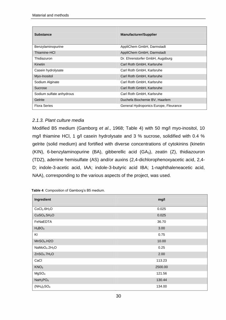
2.1.3. Plant culture media
Modified B5 medium (Gamborg et al., 1968; Table 4) with 50 mg/l myo-inositol, 10
mg/l thiamine HCl, 1 g/l casein hydrolysate and 3 % sucrose, solidified with 0.4 %
gelrite (solid medium) and fortified with diverse concentrations of cytokinins (kinetin
(KIN), 6-benzylaminopurine (BA), gibberellic acid (GA3), zeatin (Z), thidiazouron
(TDZ), adenine hemisulfate (AS) and/or auxins (2,4-dichlorophenoxyacetic acid, 2,4-
D; indole-3-acetic acid, IAA; indole-3-butyric acid IBA; 1-naphthaleneacetic acid,
NAA), corresponding to the various aspects of the project, was used.
Table 4: Composition of Gamborg’s B5 medium.
Ingredient mg/l
CoCl2.6H2O 0.025
CuSO4.5H2O 0.025
FeNaEDTA 36.70
H3BO3 3.00
KI 0.75
MnSO4.H2O 10.00
NaMoO4.2H2O 0.25
ZnSO4.7H2O 2.00
CaCl 113.23
KNO3 2500.00
MgSO4 121.56
NaH2PO4 130.44
(NH4)2SO4 134.00
Material and methods
31
Ingredient mg/l
i-Inositol 100.00
nicotinic acid 1.00
Pyridoxine HCl 1.00
Thiamine HCl 10.00
Sucrose 30000.00
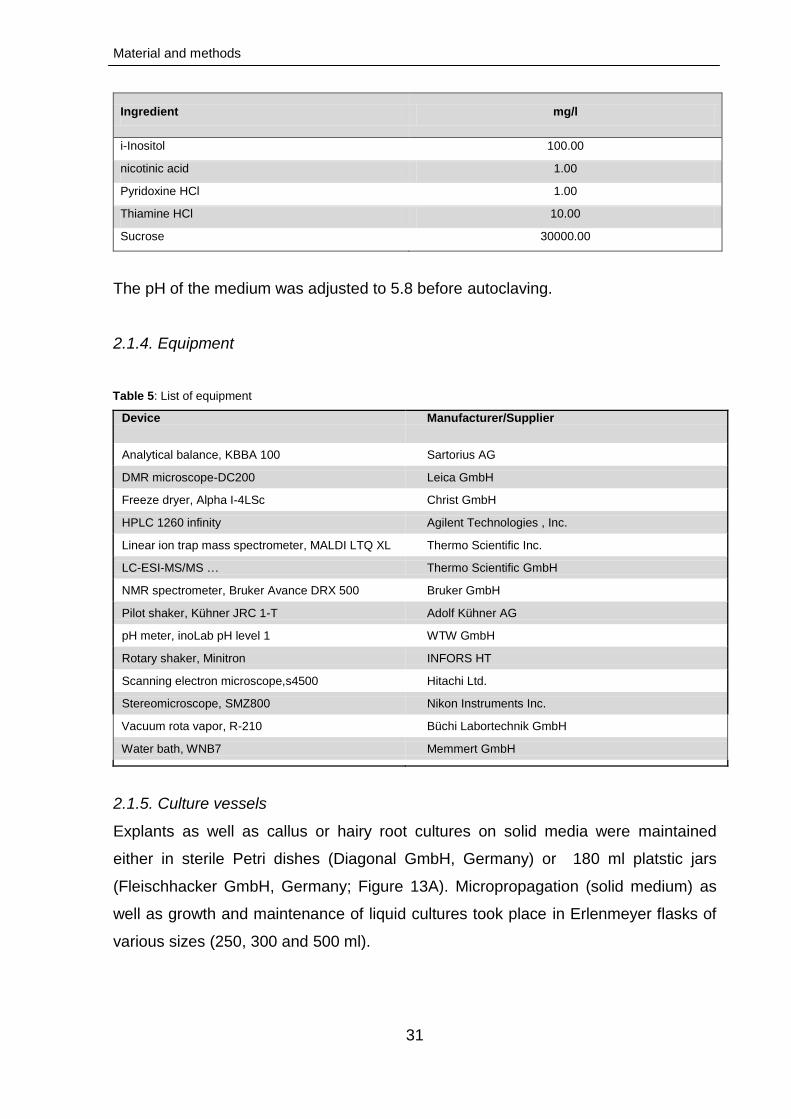
The pH of the medium was adjusted to 5.8 before autoclaving.
2.1.4. Equipment
Table 5: List of equipment
Device
Manufacturer/Supplier
Analytical balance, KBBA 100 Sartorius AG
DMR microscope-DC200 Leica GmbH
Freeze dryer, Alpha I-4LSc Christ GmbH
HPLC 1260 infinity Agilent Technologies , Inc.
Linear ion trap mass spectrometer, MALDI LTQ XL Thermo Scientific Inc.
LC-ESI-MS/MS … Thermo Scientific GmbH
NMR spectrometer, Bruker Avance DRX 500 Bruker GmbH
Pilot shaker, Kühner JRC 1-T Adolf Kühner AG
pH meter, inoLab pH level 1 WTW GmbH
Rotary shaker, Minitron INFORS HT
Scanning electron microscope,s4500 Hitachi Ltd.
Stereomicroscope, SMZ800 Nikon Instruments Inc.
Vacuum rota vapor, R-210 Büchi Labortechnik GmbH
Water bath, WNB7 Memmert GmbH
2.1.5. Culture vessels
Explants as well as callus or hairy root cultures on solid media were maintained
either in sterile Petri dishes (Diagonal GmbH, Germany) or 180 ml platstic jars
(Fleischhacker GmbH, Germany; Figure 13A). Micropropagation (solid medium) as
well as growth and maintenance of liquid cultures took place in Erlenmeyer flasks of
various sizes (250, 300 and 500 ml).
Material and methods
32
2.1.6. Sterilisation
2.1.6.1. Culture instruments and glassware
The aseptic processing of plant cell and tissue cultures was conducted under
a laminar flow hood (decontaminated by wiping all the surfaces with 70 % ethanol
and UV-irradiation for 10 min). Glassware (cylinders, beakers and Erlenmeyer flasks)
and consumables (e.g., paper filters, pipette tips, aluminium foil, etc.) were
autoclaved at 121 °C for 20 min.
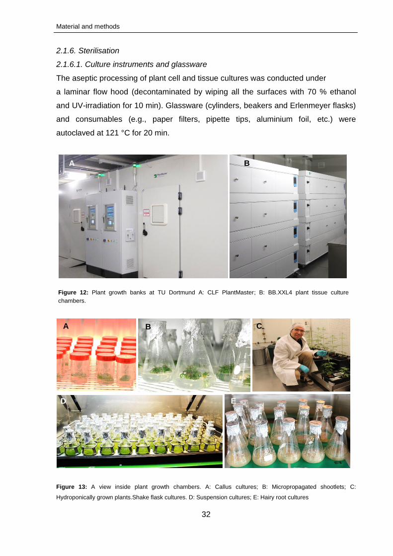
Figure 12: Plant growth banks at TU Dortmund A: CLF PlantMaster; B: BB.XXL4 plant tissue culture
chambers.
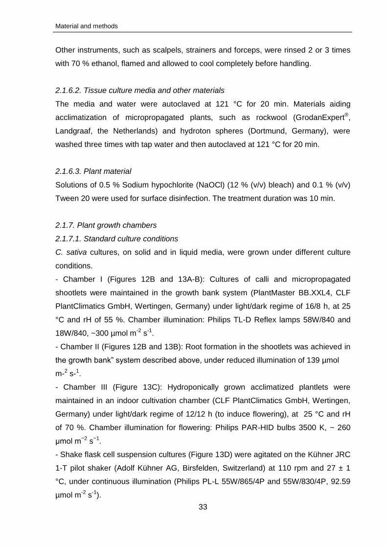
Figure 13: A view inside plant growth chambers. A: Callus cultures; B: Micropropagated shootlets; C:
Hydroponically grown plants.Shake flask cultures. D: Suspension cultures; E: Hairy root cultures
A C B
D E
A B
Material and methods
33
Other instruments, such as scalpels, strainers and forceps, were rinsed 2 or 3 times
with 70 % ethanol, flamed and allowed to cool completely before handling.
2.1.6.2. Tissue culture media and other materials
The media and water were autoclaved at 121 °C for 20 min. Materials aiding
acclimatization of micropropagated plants, such as rockwool (GrodanExpert®,
Landgraaf, the Netherlands) and hydroton spheres (Dortmund, Germany), were
washed three times with tap water and then autoclaved at 121 °C for 20 min.
2.1.6.3. Plant material
Solutions of 0.5 % Sodium hypochlorite (NaOCl) (12 % (v/v) bleach) and 0.1 % (v/v)
Tween 20 were used for surface disinfection. The treatment duration was 10 min.
2.1.7. Plant growth chambers
2.1.7.1. Standard culture conditions
C. sativa cultures, on solid and in liquid media, were grown under different culture
conditions.
- Chamber I (Figures 12B and 13A-B): Cultures of calli and micropropagated
shootlets were maintained in the growth bank system (PlantMaster BB.XXL4, CLF
PlantClimatics GmbH, Wertingen, Germany) under light/dark regime of 16/8 h, at 25
°C and rH of 55 %. Chamber illumination: Philips TL-D Reflex lamps 58W/840 and
18W/840, ~300 µmol m-2 s-1.
- Chamber II (Figures 12B and 13B): Root formation in the shootlets was achieved in
the growth bank” system described above, under reduced illumination of 139 µmol
m-2 s-1.
- Chamber III (Figure 13C): Hydroponically grown acclimatized plantlets were
maintained in an indoor cultivation chamber (CLF PlantClimatics GmbH, Wertingen,
Germany) under light/dark regime of 12/12 h (to induce flowering), at 25 °C and rH
of 70 %. Chamber illumination for flowering: Philips PAR-HID bulbs 3500 K, ~ 260
μmol m−2 s−1.
- Shake flask cell suspension cultures (Figure 13D) were agitated on the Kühner JRC
1-T pilot shaker (Adolf Kühner AG, Birsfelden, Switzerland) at 110 rpm and 27 ± 1
°C, under continuous illumination (Philips PL-L 55W/865/4P and 55W/830/4P, 92.59
µmol m-2 s-1).
Material and methods
34
- Hairy root cultures (Figure 13E) were agitated on a rotary shaker (INFORS HT,
Bottmingen, Switzerland) at 110 rpm and 25 °C, in the dark.
2.2. Methods
2.2.1. In vitro micropropagation of C. sativa leaf-derived calli
2.2.1.1. Sterilization and explant preparation
Juvenile leaves of C. sativa were washed three times in sterile distilled water and
treated as described in section (2.1.6.3.). Subsequently, they were again washed in
sterile distilled water (three times for 5 min) prior to inoculation on the culture
medium.
2.2.1.2. Callogenesis
The sterilized juvenile leaves were cut into several small pieces (explants) of about 2
mm and aseptically cultured on modified B5 medium, fortified with various
concentrations of 2,4-D, NAA, KIN and BA for callus induction. 15 ml of sterile
medium was poured into sterile Petri dishes. Growth, amount and percentage of
callogenesis in each treatment were recorded monthly. The calli were subcultured
every 20 days under the conditions described above.
2.2.1.3. Meristemoid initiation
In order to initiate meristemoid formation from callus cultures, green, healthy and
friable calli were transferred onto solid modified B5 basal medium (as described
above) with phytohormone augmentation. Morphological changes of the calli in each
treatment were recorded monthly. The calli were subcultured in 20-day intervals
under the conditions described above. After 20 days, meristemoid-forming calli were
cut into small pieces and subcultured on the same medium.
2.2.1.4. Shootlet induction and multiplication
Well-grown calli treated for meristemoid formation were used for shootlet induction
after five months of growth, within seven subcultures. Calli of about 2 g and 1-2 cm in
diameter were incubated on modified B5 medium supplemented with various
concentrations and blends of cytokinins (BA, Z, KIN and TDZ) in combination with
GA3. The shoot cultures were observed weekly. After one month, the first shootles
appeared. The evaluated parameters were: average number of shootlets per callus,
Material and methods
35
average shootlet height (cm) and perecnetage of callogensis (g). After one month,
shootlet-forming calli were cut into small pieces (each with about five shootlets) and
subcultured on the same medium.
2.2.1.5. Rooting of shootlets
To obtain whole Cannabis plantlets, single shootlets showing normal development
(about 3 cm in length after three months of growth and subculturing) were separated
and cultivated in 500 ml beakers containing 100 ml solidified B5 medium
supplemented with various concentrations of auxins (0.5, 1.0 and 1.5 mg/l of NAA,
IBA and IAA) for root formation. All shoot cultures were incubated for one week in
darkness at 20 ºC, after which they were transferred back into the light and grown for
four weeks as described in section (2.1.7.1.) The shootlets were observed monthly
for growth and appearerence, shoot height, number of roots per shootlet, root length
per shootlet and percentage of root formation.
2.2.1.6. Ex vitro acclimatization
To obtain whole plants of C. sativa, plantlets were removed from the solid medium
and their root system was rinsed carefully with sterilized water. The plants were then
transferred into flower pots (10×10×10.5 cm) containing sterilized rockwool and
covered with hydroton spheres of about 1 cm diameter. The flower pots were
submerged into the nutrient solution 0.25 ml/l Flora Series® (General Hydroponics
Europe, Fleurance, France). The plants, covered with translucent plastic bags
(31×19.5 cm) to maintain humidity and avoid light stress, were grown for 20 days
under controlled environmental conditions. After 15 days, the plastic bags were
removed daily for 30 min. Gradually, the daily exposure time was increased by 30
min; the bags were removed completely after 20 days. Subsequently, the plantlets
were transferred into an aeroponic system for indoor cultivation. The aeroponic
system culture was performed by placing the plants in bigger pots (15×15×20 cm)
filled with hydroton spheres. The hydroponic solution was circulated regularly to
maintain a sufficient moisture level under the plants (flow rate, 0.5 l/h) and consisted
of tap water supplemented with micro- and macroelements of Flora Series® at pH
6.0. The stock solution and feeding program were applied according to the
manufactuerer’s specifications (GHE, 2014). During different stages of growth, all
Cannabis plants were kept under strictly controlled, indoor environmental conditions.
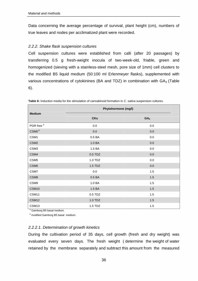
Material and methods
36
Data concerning the average percentage of survival, plant height (cm), numbers of
true leaves and nodes per acclimatized plant were recorded.
2.2.2. Shake flask suspension cultures
Cell suspension cultures were established from calli (after 20 passages) by
transferring 0.5 g fresh-weight inocula of two-week-old, friable, green and
homogenized (sieving with a stainless-steel mesh, pore size of 1mm) cell clusters to
the modified B5 liquid medium (50:100 ml Erlenmeyer flasks), supplemented with
various concentrations of cytokinines (BA and TDZ) in combination with GA3 (Table
6).
Table 6: Induction media for the stimulation of cannabinoid formation in C. sativa suspension cultures.
Medium
Phytohormone (mg/l)
CKs GA3
PGR free a 0.0 0.0
CSM0 b
0.0 0.0
CSM1 0.5 BA 0.0
CSM2 1.0 BA 0.0
CSM3 1.5 BA 0.0
CSM4 0.5 TDZ 0.0
CSM5 1.0 TDZ 0.0
CSM6 1.5 TDZ 0.0
CSM7 0.0 1.5
CSM8 0.5 BA 1.5
CSM9 1.0 BA 1.5
CSM10 1.5 BA 1.5
CSM11 0.5 TDZ 1.5
CSM12 1.0 TDZ 1.5
CSM13 1.5 TDZ 1.5 a Gamborg B5 basal medium.
b modified Gamborg B5 basal medium.
2.2.2.1. Determination of growth kinetics
During the cultivation period of 35 days, cell growth (fresh and dry weight) was
evaluated every seven days. The fresh weight ( determine the weight of water
retained by the membrane separately and subtract this amount from the measured

Material and methods
37
fresh weight ). The cells were then lyophilized for 24 h in a freeze dryer (Alpha I-4LSc
Christ GmbH, Osterode am Harz, Germany), at -55 °C and 0.6 mbar, until constant
dry weight was obtained. Morphological changes in cell suspension cultures of C.
sativa during different time courses were evaluated and documented using the Leica
DMR-DC200 light microscope equipped with Leica DC200 digital camera (Leica
GmbH, Dortmund, Germany).
2.2.3. Hairy root cultures
2.2.3.1. Initiation of adventitious root cultures
Callus cultures (1 g) of C. sativa incubated on modified B5 medium were initiated into
adventitious roots on full-strength B5 medium supplemented with various
concentrations of auxins (1.5, 2.5 and 4 mg/l of NAA, IBA and IAA). The emerging
hairy root cultures were isolated from the callus cultures and subsequently
transferred to the solid B5 medium with 4 mg/l NAA for their further proliferation.
Hairy root cultures were subcultured in 30-day intervals and maintained in the growth
bank system (PlantMaster BB.XXL4; CLF PlantClimatics GmbH, Wertingen,
Germany). The cultivation was accomplished in the dark at 25 °C The initiation
process was evaluated after 30 days.
2.2.3.2. Shake flask hairy root cultures
Once established, the adventitious root cultures were cut transversely and placed on
Petri dishes (2-3 mm explants/treatment) containing half-solid B5 medium with
various concentrations of auxins (0.25, 0.5 and 1.5 mg/l of NAA, IBA and IAA; Table
7 & 8) and cultivated in the dark at 25 °C. Morphology of the hairy roots was
evaluated using a light microscope (Nikon SMZ800) and the images were captured
using a Nikon type 104 projection microscope (Nikon Instruments Inc., Japan). Liquid
cultures were obtained by inoculating root tips into B5 medium (100:250 ml
Erlenmeyer flasks) supplemented with 4 mg/l NAA. The roots were then grown in the
dark at 25 °C on a rotary shaker at about 110 rpm, subcultured every 30 days and
maintained for about three years in the liquid medium.
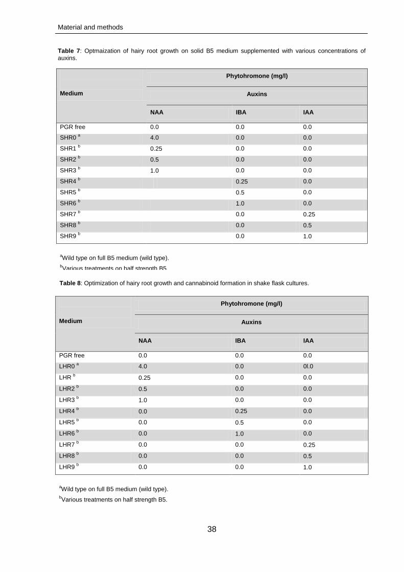
Material and methods
38
Table 7: Optmaization of hairy root growth on solid B5 medium supplemented with various concentrations of
auxins.
Medium
Phytohromone (mg/l)
Auxins
NAA IBA IAA
PGR free 0.0 0.0 0.0
SHR0 a 4.0 0.0 0.0
SHR1 b
0.25 0.0 0.0
SHR2 b 0.5 0.0 0.0
SHR3 b 1.0 0.0 0.0
SHR4 b
0.25 0.0
SHR5 b 0.5 0.0
SHR6 b 1.0 0.0
SHR7 b 0.0 0.25
SHR8 b 0.0 0.5
SHR9 b 0.0 1.0
aWild type on full B5 medium (wild type).
bVarious treatments on half strength B5.
Table 8: Optimization of hairy root growth and cannabinoid formation in shake flask cultures.
Medium
Phytohromone (mg/l)
Auxins
NAA IBA IAA
PGR free 0.0 0.0 0.0
LHR0 a 4.0 0.0 0l.0
LHR b 0.25 0.0 0.0
LHR2 b 0.5 0.0 0.0
LHR3 b 1.0 0.0 0.0
LHR4 b 0.0 0.25 0.0
LHR5 b 0.0 0.5 0.0
LHR6 b 0.0 1.0 0.0
LHR7 b 0.0 0.0 0.25
LHR8 b 0.0 0.0 0.5
LHR9 b 0.0 0.0 1.0
aWild type on full B5 medium (wild type).
bVarious treatments on half strength B5.
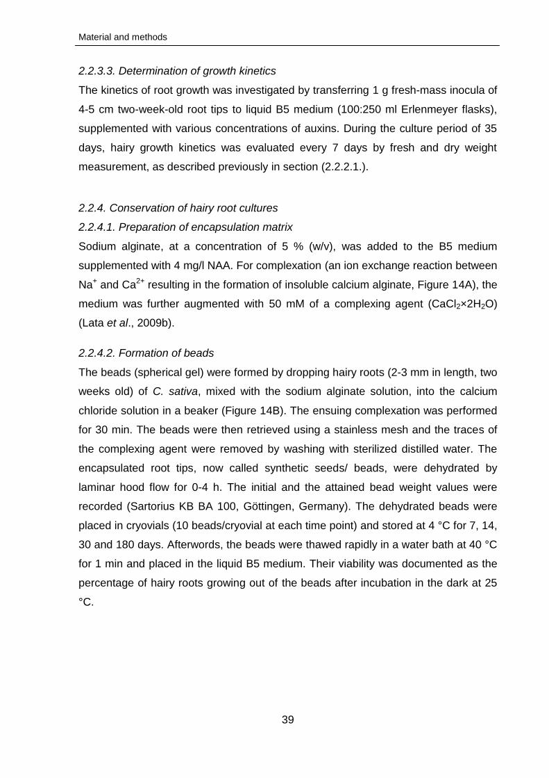
Material and methods
39
2.2.3.3. Determination of growth kinetics
The kinetics of root growth was investigated by transferring 1 g fresh-mass inocula of
4-5 cm two-week-old root tips to liquid B5 medium (100:250 ml Erlenmeyer flasks),
supplemented with various concentrations of auxins. During the culture period of 35
days, hairy growth kinetics was evaluated every 7 days by fresh and dry weight
measurement, as described previously in section (2.2.2.1.).
2.2.4. Conservation of hairy root cultures
2.2.4.1. Preparation of encapsulation matrix
Sodium alginate, at a concentration of 5 % (w/v), was added to the B5 medium
supplemented with 4 mg/l NAA. For complexation (an ion exchange reaction between
Na+ and Ca2+ resulting in the formation of insoluble calcium alginate, Figure 14A), the
medium was further augmented with 50 mM of a complexing agent (CaCl2×2H2O)
(Lata et al., 2009b).
2.2.4.2. Formation of beads
The beads (spherical gel) were formed by dropping hairy roots (2-3 mm in length, two
weeks old) of C. sativa, mixed with the sodium alginate solution, into the calcium
chloride solution in a beaker (Figure 14B). The ensuing complexation was performed
for 30 min. The beads were then retrieved using a stainless mesh and the traces of
the complexing agent were removed by washing with sterilized distilled water. The
encapsulated root tips, now called synthetic seeds/ beads, were dehydrated by
laminar hood flow for 0-4 h. The initial and the attained bead weight values were
recorded (Sartorius KB BA 100, Göttingen, Germany). The dehydrated beads were
placed in cryovials (10 beads/cryovial at each time point) and stored at 4 °C for 7, 14,
30 and 180 days. Afterwords, the beads were thawed rapidly in a water bath at 40 °C
for 1 min and placed in the liquid B5 medium. Their viability was documented as the
percentage of hairy roots growing out of the beads after incubation in the dark at 25
°C.
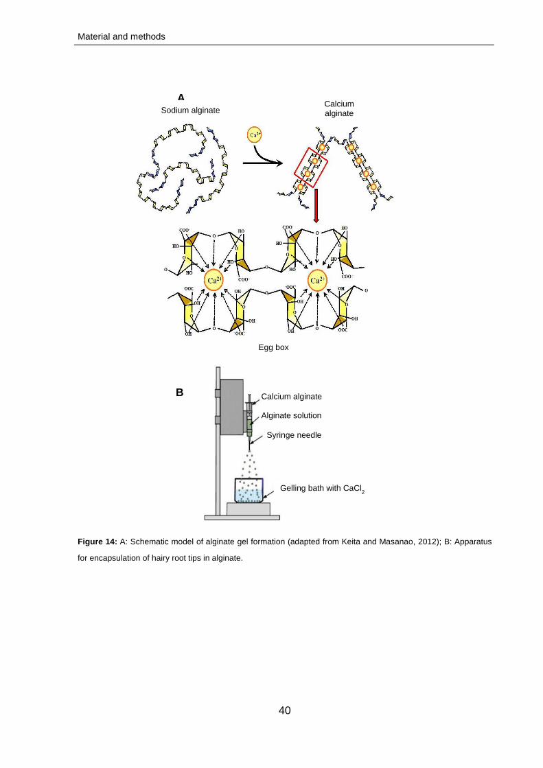
Material and methods
40
Figure 14: A: Schematic model of alginate gel formation (adapted from Keita and Masanao, 2012); B: Apparatus
for encapsulation of hairy root tips in alginate.
B
Sodium alginate Calcium alginate
Egg box
A
Calcium alginate
Alginate solution
Gelling bath with CaCl2
solution
Syringe needle
Material and methods
41
2.2.4.3. Reestablishment of shake flask cultures
The fresh biomass of root growth was investigated by transferring two-week old
bead-developed root samples (1 g) to liquid LHR0 medium (100: 250 ml Erlenmeyer
flask; Table 8; sub-section 2.2.3.2.). After 21 days, the fresh weight was determined.
2.2.5. Induction of trichome formation
2.2.5.1. Effect of phytohormones on trichome induction
In order to initiate trichome formation from C. sativa callus cultures, green, healthy
and friable calli (50 mg, 14 days old) were transferred aseptically onto the solid B5
basal medium supplemented with various concentrations of TDZ and GA3. Trichome
cultures were incubated under the same conditions as those utilized for callus
induction. All experiments were carried out in at least three replicates. After 7, 14 and
21 days of incubation, the morphology and morphotypes of trichomes were evaluated
using a light microscope (Nikon SMZ800) and the images were captured by a digital
camera system (Nikon type-104,Tokyo, Japan).
2.2.5.2. Trichome analysis
Freeze-dried 21-day old callus cultures, grown on B5 supplemented with 1 mg/l TDZ
and 3 mg/l GA3, were used for scanning electron microscopy (SEM) of the induced
trichomes. Square fragments were excised, using a razor blade, from the surface of
the callus (underlying tissue thickness, ~1 mm). The samples were then mounted on
aluminum stubs using two-sided adhesive carbon tape and examined with the Hitachi
s4500 scanning electron microscope (Hitachi, Tokyo, Japan) at an accelerating
voltage of 1 kV. Size and types of trichomes per unit of callus surface area were
determined.
2.2.6. Analytical methods
2.2.6.1. Extraction of cannabinoids and sample preparation
2.2.6.1.1. Callus cultures, plantlets grown in solid media and hydroponic plants
plants grown in vitro (IV) and the flower tops of ex vitro in a growth room (IVH) as well
as control plants (sample fresh weight, 1 g) were ground into a fine powder in the
presence of liquid nitrogen with a mortar and a pestle, then transferred to test tubes.
The powdered samples were extracted by vortexting at room temperature for 1 min
with 3× 5 ml of n-hexane followed by sonication for 5 min. After filtration the
Material and methods
42
extracting solvent, then it was evaporated under vacuum,at 35 ºC and 31 mar
Vacuum rota vapor R-210 (Büchi Labortechnik GmbH, Essen, Germany), and the dry
residue dissolved in 1 ml n-hexane. The obtained hexane crude extracts (HCEs)
were used for cannabinoid quantification by liquid chromatography - electrospray
ionization - mass spectrometry (LC-ESI-MS). Four extract replicates were used.
2.2.6.1.2. Cell suspension and hairy root cultures
One gram of dry cell suspension or hairy root samples were pulverized and
homogenized with methanol (70 %, 3× 10 ml) at 25 ºC for 1 h. The combined extracts
were filtered, solvents evaporated at 35 ºC using a rotavapor R-210 (Büchi
Labortechnik GmbH, Essen, Germany) and the resulting residues dissolved in
chloroform (2× 10 ml). The chloroform extracts were combined, dehydrated over
anhydrous sodium sulfate, then extracting solvent filtered and evaporated in vacuo at
35 ºC to give total cannabinoid mixtures. These were then re-dissolved in 0.5 ml of
methanol and centrifuged at 13000 ×g and 4 °C for 4 min. The clear supernatants
were removed and transferred to 2 ml clean amber vials, deoxygenated with CO2 and
stored at -20 ºC for further qualitative and fingerprinting studies. Calibration curves
were established using CBDA, THCA and THC (THC-Pharm, Frankfurt, Germany) as
standards.
2.2.6.2. Fingerprinting of cannabinoids
2.2.6.2.1. LC-ESI-MS (callus cultures, in vitro plantlets and hydroponic plants)
Cannabinoids in the HCEs were analyzed by LC-ESI-MS. The LTQ-Orbitrap mass
spectrometer (Thermo Scientific GmbH, Schwerte, Germany) was coupled to the
Agilent 1200 HPLC system (Agilent Technologies, Waldbronn, Germany) equipped
with the G1312 pump and the G1367 HIP-ALS autosampler). Nitrogen (N2) was used
as the protective gas (60 arbitrary units) and helium (He) served as the collision gas.
The separations were performed using the Luna C18 column (50×3 mm, 3 µm
particle size; Phenomenex, Aschaffenburg, Germany). The mobile phase consisted
of two solvents: 0.1 % formic acid in water (solvent A) and acetonitrile with 0.1 %
formic acid (solvent B). The compounds were eluted using the following step
gradient: 0-1 min (20 % B), 6-10 min (100 % B) and 10-15 min (20 % B). The flow
rate was 0.5 ml/min and the injection volume was 5 µl. Cannabinoid-corresponding
HPLC peaks were detected at 224 nm and identified through comparison with those
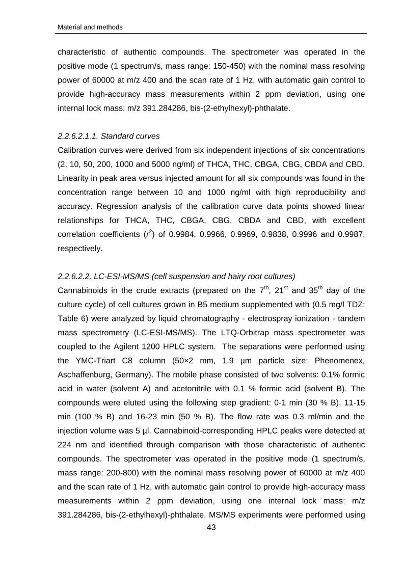
Material and methods
43
characteristic of authentic compounds. The spectrometer was operated in the
positive mode (1 spectrum/s, mass range: 150-450) with the nominal mass resolving
power of 60000 at m/z 400 and the scan rate of 1 Hz, with automatic gain control to
provide high-accuracy mass measurements within 2 ppm deviation, using one
internal lock mass: m/z 391.284286, bis-(2-ethylhexyl)-phthalate.
2.2.6.2.1.1. Standard curves
Calibration curves were derived from six independent injections of six concentrations
(2, 10, 50, 200, 1000 and 5000 ng/ml) of THCA, THC, CBGA, CBG, CBDA and CBD.
Linearity in peak area versus injected amount for all six compounds was found in the
concentration range between 10 and 1000 ng/ml with high reproducibility and
accuracy. Regression analysis of the calibration curve data points showed linear
relationships for THCA, THC, CBGA, CBG, CBDA and CBD, with excellent
correlation coefficients (r2) of 0.9984, 0.9966, 0.9969, 0.9838, 0.9996 and 0.9987,
respectively.
2.2.6.2.2. LC-ESI-MS/MS (cell suspension and hairy root cultures)
Cannabinoids in the crude extracts (prepared on the 7th, 21st and 35th day of the
culture cycle) of cell cultures grown in B5 medium supplemented with (0.5 mg/l TDZ;
Table 6) were analyzed by liquid chromatography - electrospray ionization - tandem
mass spectrometry (LC-ESI-MS/MS). The LTQ-Orbitrap mass spectrometer was
coupled to the Agilent 1200 HPLC system. The separations were performed using
the YMC-Triart C8 column (50×2 mm, 1.9 µm particle size; Phenomenex,
Aschaffenburg, Germany). The mobile phase consisted of two solvents: 0.1% formic
acid in water (solvent A) and acetonitrile with 0.1 % formic acid (solvent B). The
compounds were eluted using the following step gradient: 0-1 min (30 % B), 11-15
min (100 % B) and 16-23 min (50 % B). The flow rate was 0.3 ml/min and the
injection volume was 5 µl. Cannabinoid-corresponding HPLC peaks were detected at
224 nm and identified through comparison with those characteristic of authentic
compounds. The spectrometer was operated in the positive mode (1 spectrum/s,
mass range: 200-800) with the nominal mass resolving power of 60000 at m/z 400
and the scan rate of 1 Hz, with automatic gain control to provide high-accuracy mass
measurements within 2 ppm deviation, using one internal lock mass: m/z
391.284286, bis-(2-ethylhexyl)-phthalate. MS/MS experiments were performed using
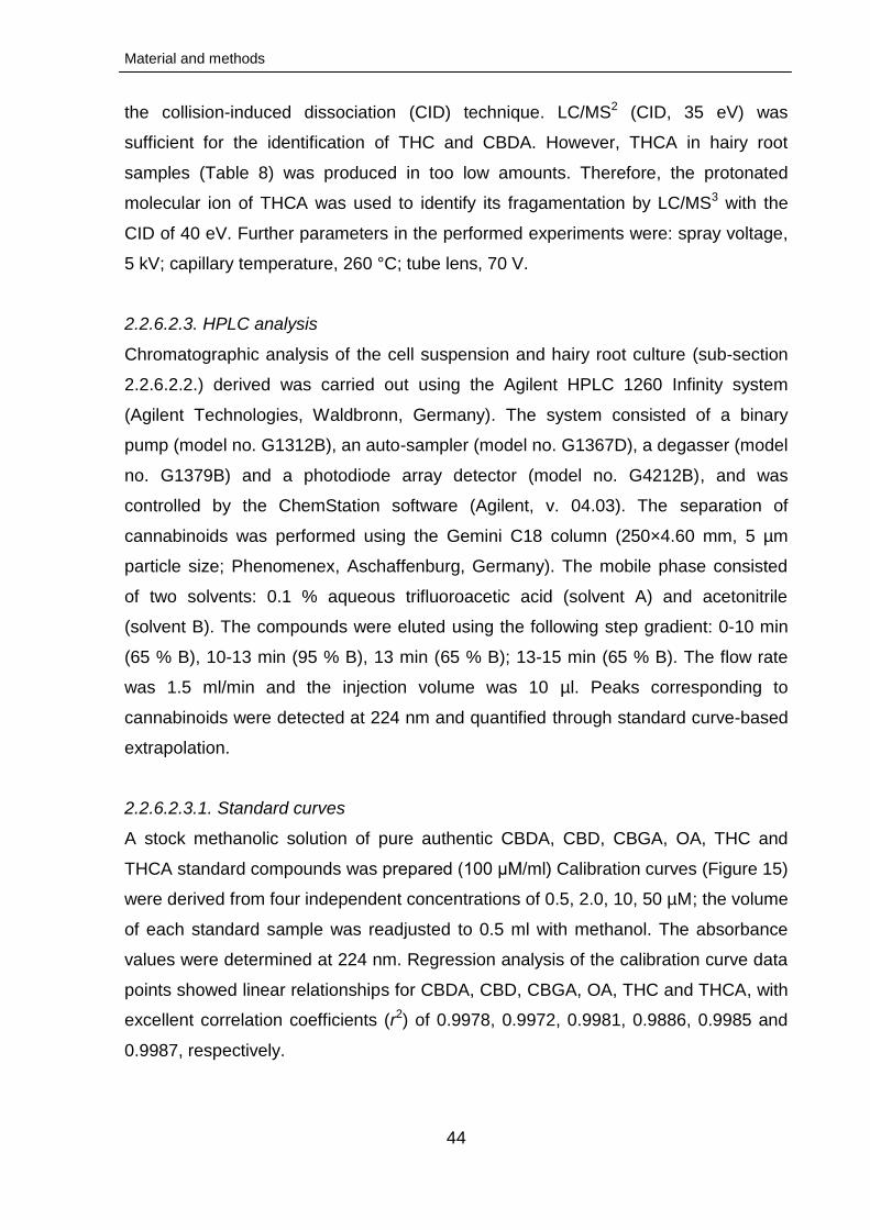
Material and methods
44
the collision-induced dissociation (CID) technique. LC/MS2 (CID, 35 eV) was
sufficient for the identification of THC and CBDA. However, THCA in hairy root
samples (Table 8) was produced in too low amounts. Therefore, the protonated
molecular ion of THCA was used to identify its fragamentation by LC/MS3 with the
CID of 40 eV. Further parameters in the performed experiments were: spray voltage,
5 kV; capillary temperature, 260 °C; tube lens, 70 V.
2.2.6.2.3. HPLC analysis
Chromatographic analysis of the cell suspension and hairy root culture (sub-section
2.2.6.2.2.) derived was carried out using the Agilent HPLC 1260 Infinity system
(Agilent Technologies, Waldbronn, Germany). The system consisted of a binary
pump (model no. G1312B), an auto-sampler (model no. G1367D), a degasser (model
no. G1379B) and a photodiode array detector (model no. G4212B), and was
controlled by the ChemStation software (Agilent, v. 04.03). The separation of
cannabinoids was performed using the Gemini C18 column (250×4.60 mm, 5 µm
particle size; Phenomenex, Aschaffenburg, Germany). The mobile phase consisted
of two solvents: 0.1 % aqueous trifluoroacetic acid (solvent A) and acetonitrile
(solvent B). The compounds were eluted using the following step gradient: 0-10 min
(65 % B), 10-13 min (95 % B), 13 min (65 % B); 13-15 min (65 % B). The flow rate
was 1.5 ml/min and the injection volume was 10 µl. Peaks corresponding to
cannabinoids were detected at 224 nm and quantified through standard curve-based
extrapolation.
2.2.6.2.3.1. Standard curves
A stock methanolic solution of pure authentic CBDA, CBD, CBGA, OA, THC and
THCA standard compounds was prepared (100 μM/ml) Calibration curves (Figure 15)
were derived from four independent concentrations of 0.5, 2.0, 10, 50 µM; the volume
of each standard sample was readjusted to 0.5 ml with methanol. The absorbance
values were determined at 224 nm. Regression analysis of the calibration curve data
points showed linear relationships for CBDA, CBD, CBGA, OA, THC and THCA, with
excellent correlation coefficients (r2) of 0.9978, 0.9972, 0.9981, 0.9886, 0.9985 and
0.9987, respectively.
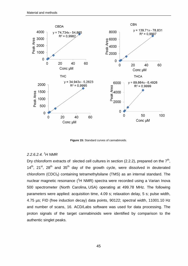
Material and methods
45
Figure 15: Standard curves of cannabinoids.
2.2.6.2.4. 1H NMR
Dry chloroform extracts of slected cell cultures in section (2.2.2), prepared on the 7th,
14th, 21st, 28th and 35th day of the growth cycle, were dissolved in deuterated
chloroform (CDCl3) containing tetramethylsilane (TMS) as an internal standard. The
nuclear magnetic resonance (1H NMR) spectra were recorded using a Varian Inova
500 spectrometer (North Carolina, USA) operating at 499.78 MHz. The following
parameters were applied: acquisition time, 4.09 s; relaxation delay, 5 s; pulse width,
4.75 µs; FID (free induction decay) data points, 90122; spectral width, 11001.10 Hz
and number of scans, 16. ACD/Labs software was used for data processing. The
proton signals of the target cannabinoids were identified by comparison to the
authentic singlet peaks.
Material and methods
46
2.2.6.2.5. MALDI imaging MS
The aim of this study was to use the capabilities of matrix-assisted laser desorption
ionization imaging mass spectromtetry (MALDI imaging MS) to determine the spatial
distribution of cannabinoids in capitate-stalked trichomes induced from C. sativa
callus.
2.2.6.2.5.1. Callus handling and sample preparation
2.2.6.2.5.1.1. Cryosectioning of callus-trichome tissue
Fresh callus cultures (1-3 cm in diameter) were stored at -26 ˚C. Before analysis, the
calli were fixed with a polymeric mixture (polyvinyl alcohol, polyethylene glycol,
potassium formiate, 7: 5: 2) on sample plates for cutting in a cryostate (Microm HM
550, Thermo Scientific Inc., Bremen, Germany). The cutting temperature was
optimized at -15 to -18 ˚C and slice thickness was adjusted to 15-20 µm then kept on
glass slides. Prior to matrix application, a photograph Leica DMR-DC200 (Leica
Camera AG, Solms, Germany) of the plate was taken to use as a reference for
MALDI imaging MS. The glass slides with the samples were transferred and fixed
onto round stainless steel MALDI plates.
2.2.6.2.5.1.2. Matrix application
Matrix application was performed with a sprayer (TransMIT GmbH, Giessen,
Germany) suitable for the generation of matrix layers for high imaging scan resolution
(≤ 10 µm). The applied matrix solution comprized 2,5-dihydroxybenzoic acid (DHB) in
acetone/water 1:1 (v/v) and 0.1 % formic acid. The spray application was performed
with the following parameters: matrix flow rate, 5 µl/min; N2; spray cycle duration, 1×
30 min and sample plate revolution speed, 255 rpm. Afterwards, the sprayed sample
was examined using Leica DMR-DC200 (Leica Camera AG, Solms, Germany) and
the regions of interest were marked to facilitate their location under measurement
conditions.
2.2.6.2.5.1.3. MALDI and imaging
The-ion mobility spectrometry (IMS) experiments were performed with the
atmospheric pressure scanning microprobe matrix-assisted laser
desorption/ionization source, AP-SMALDI (TransMIT GmbH, Giessen, Germany)
coupled with the Q Exactive HRMS (Thermo Scientific Inc., Bremen, Germany). A N2
Material and methods
47
pulsed laser (MNL 100 series, LTB Lasertechnik GmbH, Berlin, and Germany) was
used (60 Hz, 2-5 ns). The laser spot was adjusted to ~6-7 µm. Scan resolution was
adjusted to ~6.25 µm (50 steps; slight oversampling). The scans were taken from a
rectangle raster of ~375×344 µm and performed in full scan positive mode at m/z
150-600 with internal lock mass of DHB, m/z 379.09246 [2M-2H2O+H]+. Further
mass spectrometric parameters (Q-Exactive): mass resolution, 70 at m/z 200; S-lens
level, 65; spray voltage, 4000 V; injection time, 300 ms. Further source parameters
(AP-SMALDI): attenuator temperature, 20 ˚C.
2.2.6.2.5.1.4. Further software
Marathon control, v. 1.60 (LTB Lasertechnik GmbH, Berlin, Germany): laser software
for activation and deactivation of the laser. MCP, v. 3.9.32.15 (TransMIT GmbH,
Giessen, Germany): adjustment of scan raster, resolution and attenuator. IDS
Falcon/Eagle DEMO, v. 4.61.0000.0 (IMS Imaging Development Systems GmbH,
Obersulm, Germany): CCD camera image for orientation on sample plate. Thermo Q-
Exactive Tune Software, v. 2.3 (Thermo Scientific GmbH, Bremen, Germany): mass
spectrometer tune software. Thermo XCalibur, v. 3.0.63: evaluation of MS data files.
Matrix Sprayer Control, v. 1.9.2890 (TransMIT GmbH, Giessen, Germany):
controlling the spray program and parameters.
2.2.6.2.6. Data processing
Selected MALDI ion images (sub-section 2.2.6.2.5.1.3.) were analyised with
software Mirion, v. 2.1.4.411 (TransMIT GmbH, Giessen, Germany).
2.2.7. Experimental design and statistical analysis
All the cultures were examined periodically in replicates. The morphological changes
were recorded monthly on the basis of weekly visual observations. The data were
analyzed and recorded as mean values ± SE (standard error).
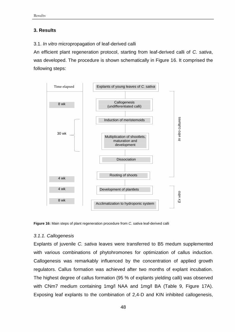
Results
48
3. Results
3.1. In vitro micropropagation of leaf-derived calli
An efficient plant regeneration protocol, starting from leaf-derived calli of C. sativa,
was developed. The procedure is shown schematically in Figure 16. It comprised the
following steps:
Figure 16: Main steps of plant regeneration procedure from C. sativa leaf-derived calli
3.1.1. Callogenesis
Explants of juvenile C. sativa leaves were transferred to B5 medum supplemented
with various combinations of phytohromones for optimization of callus induction.
Callogenesis was remarkably influenced by the concentration of applied growth
regulators. Callus formation was achieved after two months of explant incubation.
The highest degree of callus formation (95 % of explants yielding calli) was observed
with CNm7 medium containing 1mg/l NAA and 1mg/l BA (Table 9, Figure 17A).
Exposing leaf explants to the combination of 2,4-D and KIN inhibited callogenesis,
Explants of young leaves of C. sativa
Callogenesis (undifferentiated calli)
Induction of meristemoids
Multiplication of shootlets, maturation and development
Rooting of shoots
Development of plantlets
Acclimatization to hydroponic system
In v
itro
cu
ltu
res
Ex v
itro
8 wk
30 wk
4 wk
4 wk
8 wk
Dissociation
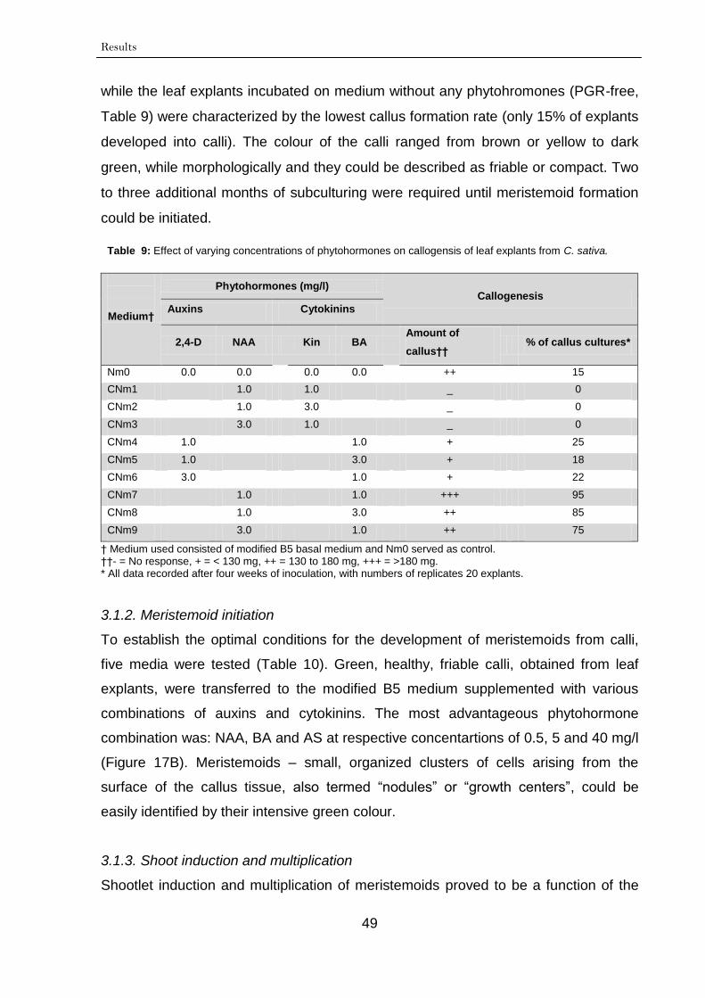
Results
49
while the leaf explants incubated on medium without any phytohromones (PGR-free,
Table 9) were characterized by the lowest callus formation rate (only 15% of explants
developed into calli). The colour of the calli ranged from brown or yellow to dark
green, while morphologically and they could be described as friable or compact. Two
to three additional months of subculturing were required until meristemoid formation
could be initiated.
Table 9: Effect of varying concentrations of phytohormones on callogensis of leaf explants from C. sativa.
Medium†
Phytohormones (mg/l) Callogenesis
Auxins Cytokinins
2,4-D NAA Kin BA Amount of
callus†† % of callus cultures*
Nm0 0.0 0.0 0.0 0.0 ++ 15
CNm1 1.0 1.0 _ 0
CNm2 1.0 3.0 _ 0
CNm3 3.0 1.0 _ 0
CNm4 1.0 1.0 + 25
CNm5 1.0 3.0 + 18
CNm6 3.0 1.0 + 22
CNm7 1.0 1.0 +++ 95
CNm8 1.0 3.0 ++ 85
CNm9 3.0 1.0 ++ 75
† Medium used consisted of modified B5 basal medium and Nm0 served as control. ††- = No response, + = < 130 mg, ++ = 130 to 180 mg, +++ = >180 mg.
* All data recorded after four weeks of inoculation, with numbers of replicates 20 explants.
3.1.2. Meristemoid initiation
To establish the optimal conditions for the development of meristemoids from calli,
five media were tested (Table 10). Green, healthy, friable calli, obtained from leaf
explants, were transferred to the modified B5 medium supplemented with various
combinations of auxins and cytokinins. The most advantageous phytohormone
combination was: NAA, BA and AS at respective concentartions of 0.5, 5 and 40 mg/l
(Figure 17B). Meristemoids – small, organized clusters of cells arising from the
surface of the callus tissue, also termed “nodules” or “growth centers”, could be
easily identified by their intensive green colour.
3.1.3. Shoot induction and multiplication
Shootlet induction and multiplication of meristemoids proved to be a function of the
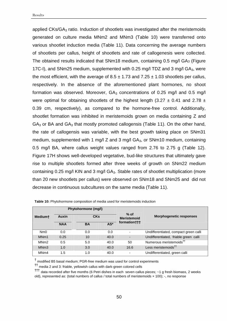
Results
50
applied CKs/GA3 ratio. Induction of shootlets was investigated after the meristemoids
generated on culture media MNm2 and MNm3 (Table 10) were transferred onto
various shootlet induction media (Table 11). Data concerning the average numbers
of shootlets per callus, height of shootlets and rate of callogenesis were collected.
The obtained results indicated that SNm18 medium, containing 0.5 mg/l GA3 (Figure
17C-I), and SNm25 medium, supplemented with 0.25 mg/l TDZ and 3 mg/l GA3, were
the most efficient, with the average of 8.5 ± 1.73 and 7.25 ± 1.03 shootlets per callus,
respectively. In the absence of the aforementioned plant hormones, no shoot
formation was observed. Moreover, GA3 concentrations of 0.25 mg/l and 0.5 mg/l
were optimal for obtaining shootlets of the highest length (3.27 ± 0.41 and 2.78 ±
0.39 cm, respectively), as compared to the hormone-free control. Additionally,
shootlet formation was inhibited in meristemoids grown on media containing Z and
GA3 or BA and GA3 that mostly promoted callogensis (Table 11). On the other hand,
the rate of callogensis was variable, with the best growth taking place on SNm31
medium, supplemented with 1 mg/l Z and 3 mg/l GA3, or SNm10 medium, containing
0.5 mg/l BA, where callus weight values ranged from 2.76 to 2.75 g (Table 12).
Figure 17H shows well-developed vegetative, bud-like structures that ultimately gave
rise to multiple shootlets formed after three weeks of growth on SNm22 medium
containing 0.25 mg/l KIN and 3 mg/l GA3. Stable rates of shootlet multiplication (more
than 20 new shootlets per callus) were observed on SNm18 and SNm25 and did not
decrease in continuous subcultures on the same media (Table 11).
Table 10: Phytohormone composition of media used for meristemoids induction
Medium†
Phytohormone (mg/l)
% of Meristemoid formation†††
Morphogenetic responses Auxin
CKs
NAA BA AS*
Nm0 0.0 0.0 0.0 - Undifferentiated, compact green calli
MNm1 0.25 10 40.0 - Undifferentiated, friable green calli
MNm2 0.5 5.0 40.0 50 Numerous meristemoids††
MNm3 1.0 3.0 40.0 16.6 Less meristemoids††
MNm4 1.5 1.0 40.0 - Undifferentiated, green calli
† modified B5 basal medium; PGR-free medium was used for control experiments
†† media 2 and 3: friable, yellowish callus with dark-green colored cells
††† data recorded after five months (6 Petri dishes in each seven callus pieces; ~1 g fresh biomass, 2 weeks
old), represented as: (total numbers of callus / total numbers of meristemoids × 100); -, no response
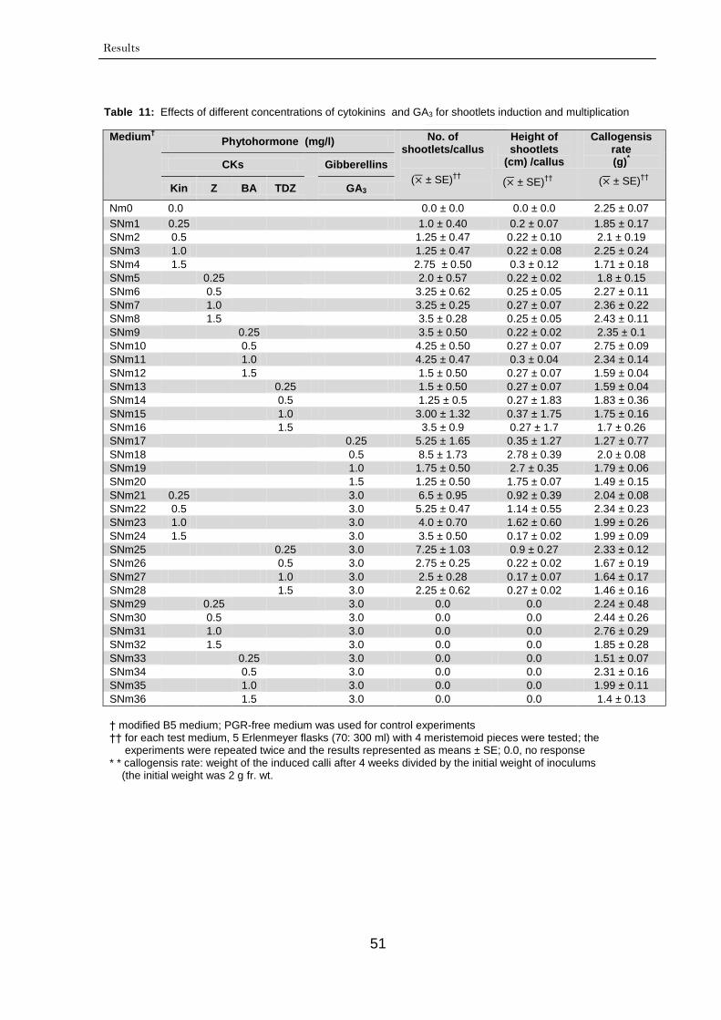
Results
51
Table 11: Effects of different concentrations of cytokinins and GA3 for shootlets induction and multiplication
Medium† Phytohormone (mg/l) No. of
shootlets/callus
Height of shootlets
(cm) /callus
Callogensis rate (g)
* CKs
Gibberellins
Kin Z BA TDZ
GA3
Nm0 0.0 0.0 ± 0.0 0.0 ± 0.0 2.25 ± 0.07
SNm1 0.25
1.0 ± 0.40 0.2 ± 0.07 1.85 ± 0.17
SNm2 0.5
1.25 ± 0.47 0.22 ± 0.10 2.1 ± 0.19
SNm3 1.0
1.25 ± 0.47 0.22 ± 0.08 2.25 ± 0.24
SNm4 1.5
2.75 ± 0.50 0.3 ± 0.12 1.71 ± 0.18
SNm5
0.25
2.0 ± 0.57 0.22 ± 0.02 1.8 ± 0.15
SNm6
0.5
3.25 ± 0.62 0.25 ± 0.05 2.27 ± 0.11
SNm7
1.0
3.25 ± 0.25 0.27 ± 0.07 2.36 ± 0.22
SNm8
1.5
3.5 ± 0.28 0.25 ± 0.05 2.43 ± 0.11
SNm9
0.25
3.5 ± 0.50 0.22 ± 0.02 2.35 ± 0.1
SNm10
0.5
4.25 ± 0.50 0.27 ± 0.07 2.75 ± 0.09
SNm11
1.0
4.25 ± 0.47 0.3 ± 0.04 2.34 ± 0.14
SNm12
1.5
1.5 ± 0.50 0.27 ± 0.07 1.59 ± 0.04
SNm13
0.25 1.5 ± 0.50 0.27 ± 0.07 1.59 ± 0.04
SNm14
0.5 1.25 ± 0.5 0.27 ± 1.83 1.83 ± 0.36
SNm15
1.0 3.00 ± 1.32 0.37 ± 1.75 1.75 ± 0.16
SNm16
1.5 3.5 ± 0.9 0.27 ± 1.7 1.7 ± 0.26
SNm17
0.25 5.25 ± 1.65 0.35 ± 1.27 1.27 ± 0.77
SNm18
0.5 8.5 ± 1.73 2.78 ± 0.39 2.0 ± 0.08
SNm19
1.0 1.75 ± 0.50 2.7 ± 0.35 1.79 ± 0.06
SNm20
1.5 1.25 ± 0.50 1.75 ± 0.07 1.49 ± 0.15
SNm21 0.25
3.0 6.5 ± 0.95 0.92 ± 0.39 2.04 ± 0.08
SNm22 0.5
3.0 5.25 ± 0.47 1.14 ± 0.55 2.34 ± 0.23
SNm23 1.0
3.0 4.0 ± 0.70 1.62 ± 0.60 1.99 ± 0.26
SNm24 1.5
3.0 3.5 ± 0.50 0.17 ± 0.02 1.99 ± 0.09
SNm25 0.25 3.0 7.25 ± 1.03 0.9 ± 0.27 2.33 ± 0.12
SNm26 0.5 3.0 2.75 ± 0.25 0.22 ± 0.02 1.67 ± 0.19
SNm27 1.0 3.0 2.5 ± 0.28 0.17 ± 0.07 1.64 ± 0.17
SNm28 1.5 3.0 2.25 ± 0.62 0.27 ± 0.02 1.46 ± 0.16
SNm29
0.25
3.0 0.0 0.0 2.24 ± 0.48
SNm30
0.5
3.0 0.0 0.0 2.44 ± 0.26
SNm31
1.0
3.0 0.0 0.0 2.76 ± 0.29
SNm32
1.5
3.0 0.0 0.0 1.85 ± 0.28
SNm33
0.25
3.0 0.0 0.0 1.51 ± 0.07
SNm34
0.5
3.0 0.0 0.0 2.31 ± 0.16
SNm35
1.0
3.0 0.0 0.0 1.99 ± 0.11
SNm36
1.5
3.0 0.0 0.0 1.4 ± 0.13
† modified B5 medium; PGR-free medium was used for control experiments †† for each test medium, 5 Erlenmeyer flasks (70: 300 ml) with 4 meristemoid pieces were tested; the experiments were repeated twice and the results represented as means ± SE; 0.0, no response
* * callogensis rate: weight of the induced calli after 4 weeks divided by the initial weight of inoculums (the initial weight was 2 g fr. wt.
( ± SE)††
( ± SE)††
( ± SE)††
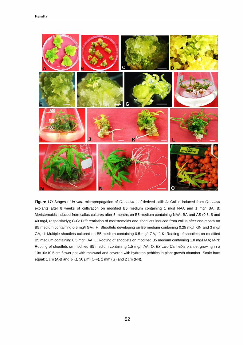
Results
52
Figure 17: Stages of in vitro micropropagation of C. sativa leaf-derived calli: A: Callus induced from C. sativa
explants after 8 weeks of cultivation on modified B5 medium containing 1 mg/l NAA and 1 mg/l BA; B:
Meristemoids induced from callus cultures after 5 months on B5 medium containing NAA, BA and AS (0.5, 5 and
40 mg/l, respectively); C-G: Differentiation of meristemoids and shootlets induced from callus after one month on
B5 medium containing 0.5 mg/l GA3; H: Shootlets developing on B5 medium containing 0.25 mg/l KIN and 3 mg/l
GA3; I: Multiple shootlets cultured on B5 medium containing 0.5 mg/l GA3; J-K: Rooting of shootlets on modified
B5 medium containing 0.5 mg/l IAA; L: Rooting of shootlets on modified B5 medium containing 1.0 mg/l IAA; M-N:
Rooting of shootlets on modified B5 medium containing 1.5 mg/l IAA; O: Ex vitro Cannabis plantlet growing in a
10×10×10.5 cm flower pot with rockwool and covered with hydroton pebbles in plant growth chamber. Scale bars
equal: 1 cm (A-B and J-K), 50 µm (C-F), 1 mm (G) and 2 cm (I-N).
A B C D
E F
G H
I J K
M N
L
O
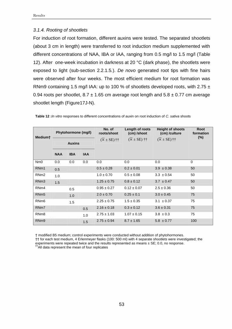
Results
53
3.1.4. Rooting of shootlets
For induction of root formation, different auxins were tested. The separated shootlets
(about 3 cm in length) were transferred to root induction medium supplemented with
different concentrations of NAA, IBA or IAA, ranging from 0.5 mg/l to 1.5 mg/l (Table
12). After one-week incubation in darkness at 20 °C (dark phase), the shootlets were
exposed to light (sub-section 2.2.1.5.). De novo generated root tips with fine hairs
were observed after four weeks. The most efficient medium for root formation was
RNm9 containing 1.5 mg/l IAA: up to 100 % of shootlets developed roots, with 2.75 ±
0.94 roots per shootlet, 8.7 ± 1.65 cm average root length and 5.8 ± 0.77 cm average
shootlet length (Figure17J-N).
Table 12 :In vitro responses to different concentrations of auxin on root induction of C. sativa shoots
Medium†
Phytohormone (mg/l) No. of
roots/shoot
Length of roots (cm) /shoot
Height of shoots (cm) /culture
Root formation
(%)
Auxins
NAA IBA IAA
Nm0 0.0 0.0 0.0 0.0 0.0 0.0 0
RNm1 0.5 0.5 ± 0.28 0.2 ± 0.01 3.9 ± 0.38 50
RNm2 1.0 1.0 ± 0.70 0.5 ± 0.08 3.3 ± 0.54 50
RNm3 1.5 1.25 ± 0.75 0.8 ± 0.12 3.7 ± 0.47 50
RNm4 0.5 0.95 ± 0.27 0.12 ± 0.07 2.5 ± 0.36 50
RNm5 1.0 2.0 ± 0.70 0.25 ± 0.1 3.0 ± 0.45 75
RNm6 1.5 2.25 ± 0.75 1.5 ± 0.35 3.1 ± 0.37 75
RNm7 0.5 2.16 ± 0.18 0.3 ± 0.12 3.6 ± 0.31 75
RNm8 1.0 2.75 ± 1.03 1.07 ± 0.15 3.8 ± 0.3 75
RNm9 1.5 2.75 ± 0.94 8.7 ± 1.65 5.8 ± 0.77 100
† modified B5 medium; control experiments were conducted without addition of phytohormones. †† for each test medium, 4 Erlenmeyer flasks (100: 500 ml) with 4 separate shootlets were investigated; the experiments were repeated twice and the results represented as means ± SE; 0.0, no response. ††
All data represent the mean of four replicates
( ± SE) †† ( ± SE) †† ( ± SE)
††
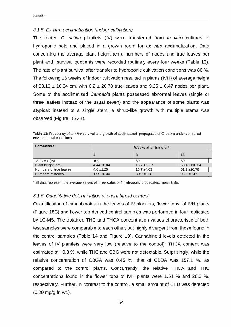
Results
54
3.1.5. Ex vitro acclimatization (indoor cultivation)
The rooted C. sativa plantlets (IV) were transferred from in vitro cultures to
hydroponic pots and placed in a growth room for ex vitro acclimatization. Data
concerning the average plant height (cm), numbers of nodes and true leaves per
plant and survival quotients were recorded routinely every four weeks (Table 13).
The rate of plant survival after transfer to hydroponic cultivation conditions was 80 %.
The following 16 weeks of indoor cultivation resulted in plants (IVH) of average height
of 53.16 ± 16.34 cm, with 6.2 ± 20.78 true leaves and 9.25 ± 0.47 nodes per plant.
Some of the acclimatized Cannabis plants possessed abnormal leaves (single or
three leaflets instead of the usual seven) and the appearance of some plants was
atypical: instead of a single stem, a shrub-like growth with multiple stems was
observed (Figure 18A-B).
Table 13: Frequency of ex vitro survival and growth of acclimatized propagates of C. sativa under controlled
environmental conditions
Parameters Weeks after transfer*
4 8 16
Survival (%) 100 80 80
Plant height (cm) 4.44 ±0.84 16.7 ± 2.67 53.16 ±16.34
Numbers of true leaves 4.6 ±1.25 15,7 ±4,03 61,2 ±20,78
Numbers of nodes 1.99 ±0.30 3.49 ±0.28 9.25 ±0.47
* all data represent the average values of 4 replicates of 4 hydroponic propagates; mean ± SE.
3.1.6. Quantitative determination of cannabinoid content
Quantification of cannabinoids in the leaves of IV plantlets, flower tops of IVH plants
(Figure 18C) and flower top-derived control samples was performed in four replicates
by LC-MS. The obtained THC and THCA concentration values characteristic of both
test samples were comparable to each other, but highly divergent from those found in
the control samples (Table 14 and Figure 19). Cannabinoid levels detected in the
leaves of IV plantlets were very low (relative to the control): THCA content was
estimated at ~0.3 %, while THC and CBG were not detectable. Surprisingly, while the
relative concentration of CBGA was 0.45 %, that of CBDA was 157.1 %, as
compared to the control plants. Concurrently, the relative THCA and THC
concentrations found in the flower tops of IVH plants were 1.54 % and 28.3 %,
respectively. Further, in contrast to the control, a small amount of CBD was detected
(0.29 mg/g fr. wt.).
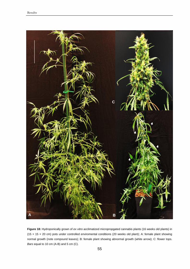
Results
55
Figure 18: Hydroponically grown of ex vitro acclimatized micropropgated cannabis plants (16 weeks old plants) in
(15 × 15 × 20 cm) pots under controlled enviromental conditions (20 weeks old plant); A: female plant showing
normal growth (note compound leaves); B: female plant showing abnormal growth (white arrow); C: flower tops.
Bars equal to 10 cm (A-B) and 5 cm (C).
b
A B
C
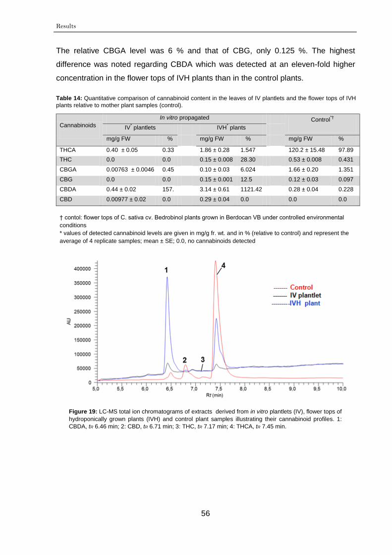
Results
56
The relative CBGA level was 6 % and that of CBG, only 0.125 %. The highest
difference was noted regarding CBDA which was detected at an eleven-fold higher
concentration in the flower tops of IVH plants than in the control plants.
Table 14: Quantitative comparison of cannabinoid content in the leaves of IV plantlets and the flower tops of IVH
plants relative to mother plant samples (control).
Cannabinoids
In vitro propagated Control*†
IV* plantlets IVH
* plants
mg/g FW % mg/g FW % mg/g FW %
THCA 0.40 ± 0.05 0.33 1.86 ± 0.28 1.547 120.2 ± 15.48 97.89
THC 0.0 0.0 0.15 ± 0.008 28.30 0.53 ± 0.008 0.431
CBGA 0.00763 ± 0.0046 0.45
9
0.10 ± 0.03 6.024 1.66 ± 0.20 1.351
CBG 0.0 0.0 0.15 ± 0.001 12.5 0.12 ± 0.03 0.097
CBDA 0.44 ± 0.02 157.
14
3.14 ± 0.61 1121.42 0.28 ± 0.04 0.228
CBD 0.00977 ± 0.02 0.0 0.29 ± 0.04 0.0 0.0 0.0
† contol: flower tops of C. sativa cv. Bedrobinol plants grown in Berdocan VB under controlled environmental
conditions
* values of detected cannabinoid levels are given in mg/g fr. wt. and in % (relative to control) and represent the
average of 4 replicate samples; mean ± SE; 0.0, no cannabinoids detected
Figure 19: LC-MS total ion chromatograms of extracts derived from in vitro plantlets (IV), flower tops of
hydroponically grown plants (IVH) and control plant samples illustrating their cannabinoid profiles. 1:
CBDA, tR 6.46 min; 2: CBD, tR 6.71 min; 3: THC, tR 7.17 min; 4: THCA, tR 7.45 min.
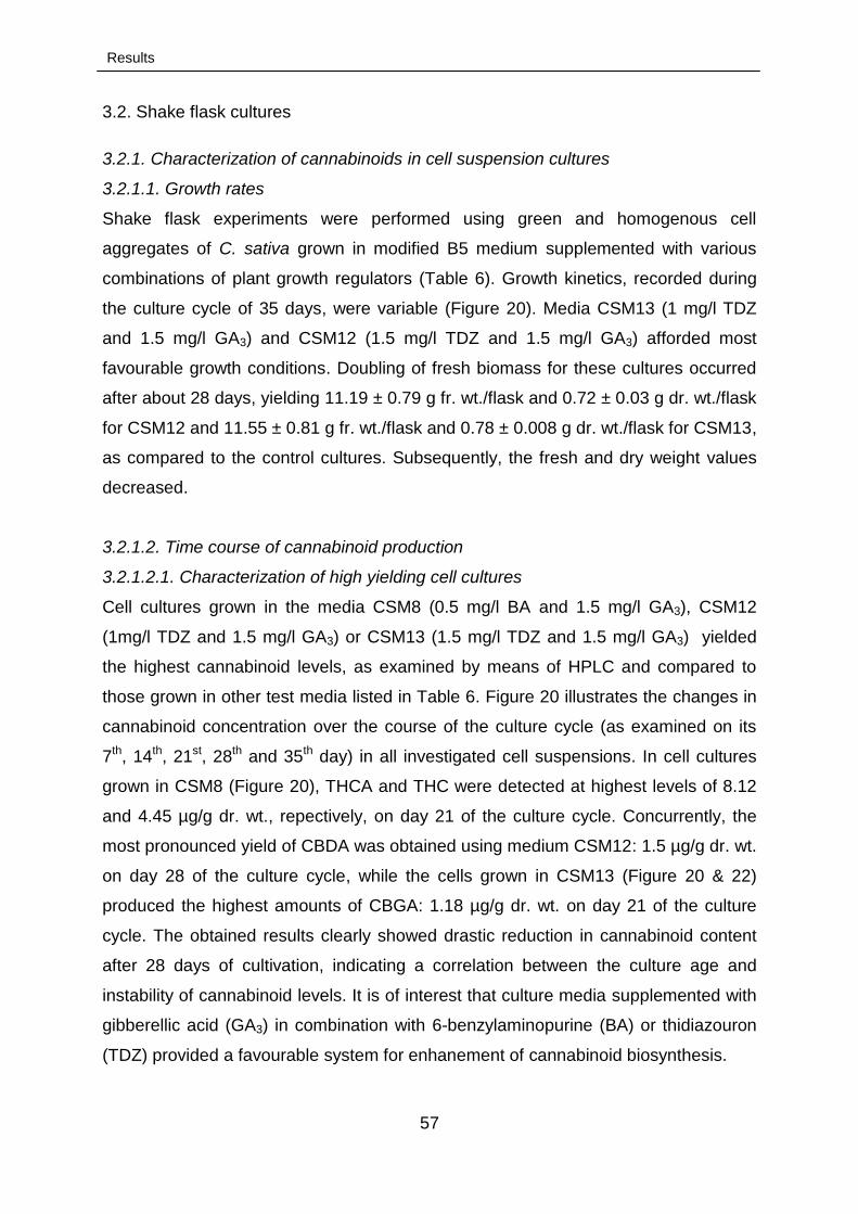
Results
57
3.2. Shake flask cultures
3.2.1. Characterization of cannabinoids in cell suspension cultures
3.2.1.1. Growth rates
Shake flask experiments were performed using green and homogenous cell
aggregates of C. sativa grown in modified B5 medium supplemented with various
combinations of plant growth regulators (Table 6). Growth kinetics, recorded during
the culture cycle of 35 days, were variable (Figure 20). Media CSM13 (1 mg/l TDZ
and 1.5 mg/l GA3) and CSM12 (1.5 mg/l TDZ and 1.5 mg/l GA3) afforded most
favourable growth conditions. Doubling of fresh biomass for these cultures occurred
after about 28 days, yielding 11.19 ± 0.79 g fr. wt./flask and 0.72 ± 0.03 g dr. wt./flask
for CSM12 and 11.55 ± 0.81 g fr. wt./flask and 0.78 ± 0.008 g dr. wt./flask for CSM13,
as compared to the control cultures. Subsequently, the fresh and dry weight values
decreased.
3.2.1.2. Time course of cannabinoid production
3.2.1.2.1. Characterization of high yielding cell cultures
Cell cultures grown in the media CSM8 (0.5 mg/l BA and 1.5 mg/l GA3), CSM12
(1mg/l TDZ and 1.5 mg/l GA3) or CSM13 (1.5 mg/l TDZ and 1.5 mg/l GA3) yielded
the highest cannabinoid levels, as examined by means of HPLC and compared to
those grown in other test media listed in Table 6. Figure 20 illustrates the changes in
cannabinoid concentration over the course of the culture cycle (as examined on its
7th, 14th, 21st, 28th and 35th day) in all investigated cell suspensions. In cell cultures
grown in CSM8 (Figure 20), THCA and THC were detected at highest levels of 8.12
and 4.45 µg/g dr. wt., repectively, on day 21 of the culture cycle. Concurrently, the
most pronounced yield of CBDA was obtained using medium CSM12: 1.5 µg/g dr. wt.
on day 28 of the culture cycle, while the cells grown in CSM13 (Figure 20 & 22)
produced the highest amounts of CBGA: 1.18 µg/g dr. wt. on day 21 of the culture
cycle. The obtained results clearly showed drastic reduction in cannabinoid content
after 28 days of cultivation, indicating a correlation between the culture age and
instability of cannabinoid levels. It is of interest that culture media supplemented with
gibberellic acid (GA3) in combination with 6-benzylaminopurine (BA) or thidiazouron
(TDZ) provided a favourable system for enhanement of cannabinoid biosynthesis.
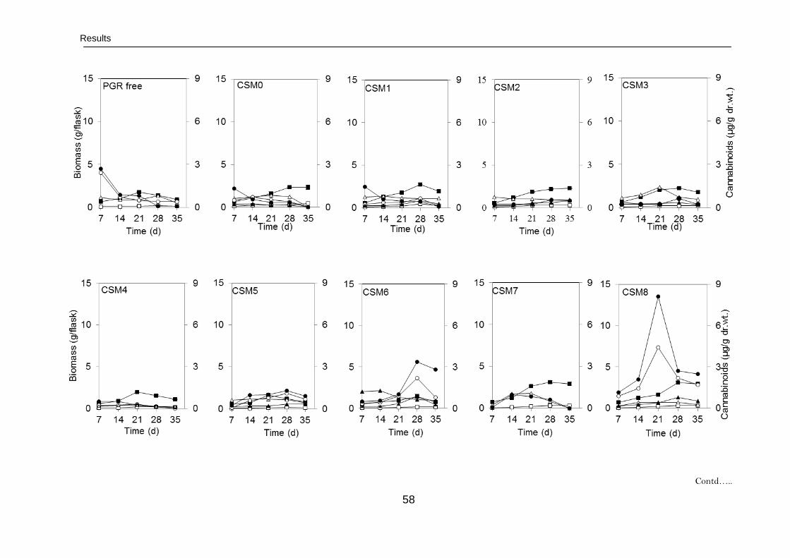
Results
58
Contd…..
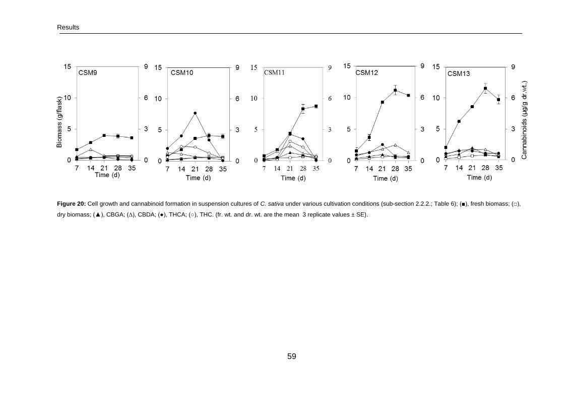
Results
59
Figure 20: Cell growth and cannabinoid formation in suspension cultures of C. sativa under various cultivation conditions (sub-section 2.2.2.; Table 6); (■), fresh biomass; (□),
dry biomass; (▲), CBGA; (∆), CBDA; (●), THCA; (○), THC. (fr. wt. and dr. wt. are the mean 3 replicate values ± SE).
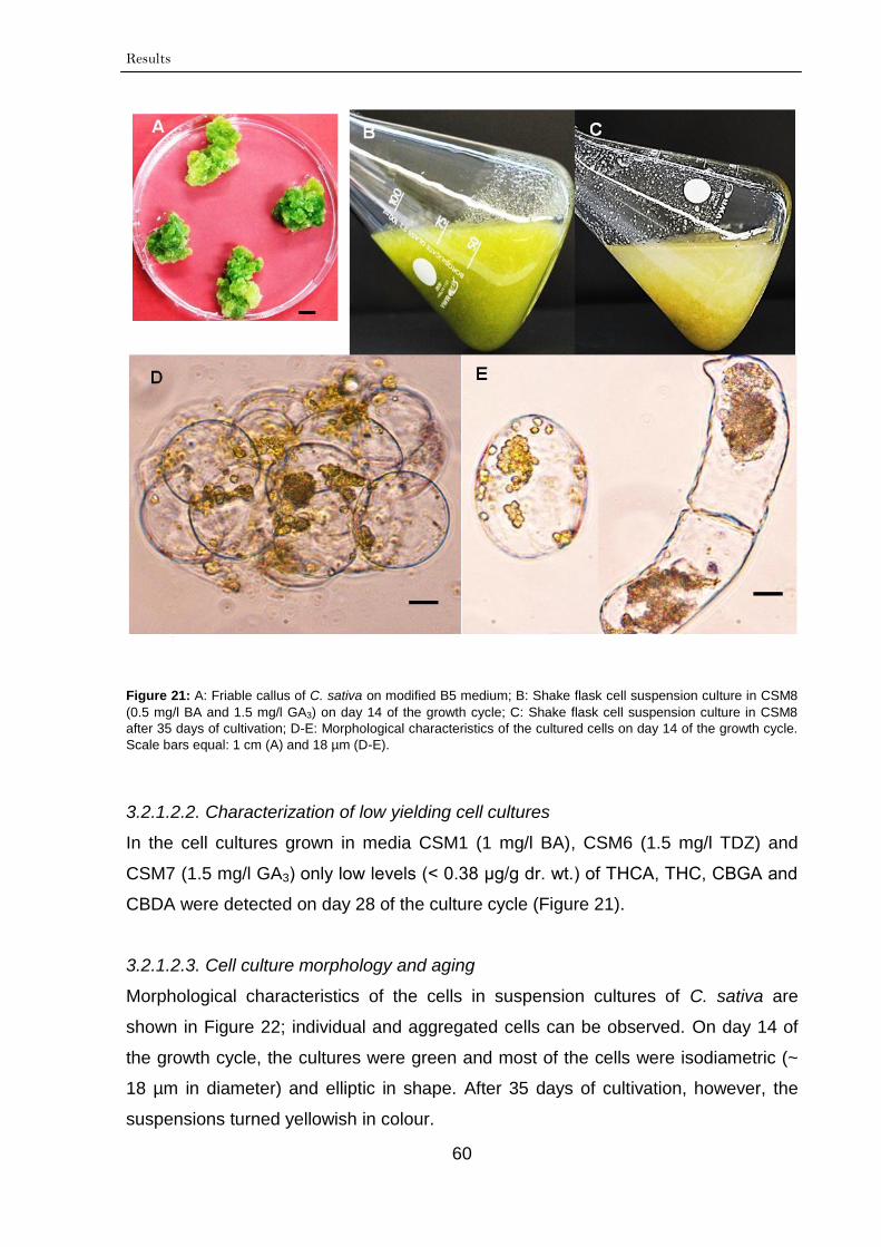
Results
60
Figure 21: A: Friable callus of C. sativa on modified B5 medium; B: Shake flask cell suspension culture in CSM8
(0.5 mg/l BA and 1.5 mg/l GA3) on day 14 of the growth cycle; C: Shake flask cell suspension culture in CSM8
after 35 days of cultivation; D-E: Morphological characteristics of the cultured cells on day 14 of the growth cycle.
Scale bars equal: 1 cm (A) and 18 µm (D-E).
3.2.1.2.2. Characterization of low yielding cell cultures
In the cell cultures grown in media CSM1 (1 mg/l BA), CSM6 (1.5 mg/l TDZ) and
CSM7 (1.5 mg/l GA3) only low levels (˂ 0.38 µg/g dr. wt.) of THCA, THC, CBGA and
CBDA were detected on day 28 of the culture cycle (Figure 21).
3.2.1.2.3. Cell culture morphology and aging
Morphological characteristics of the cells in suspension cultures of C. sativa are
shown in Figure 22; individual and aggregated cells can be observed. On day 14 of
the growth cycle, the cultures were green and most of the cells were isodiametric (~
18 µm in diameter) and elliptic in shape. After 35 days of cultivation, however, the
suspensions turned yellowish in colour.
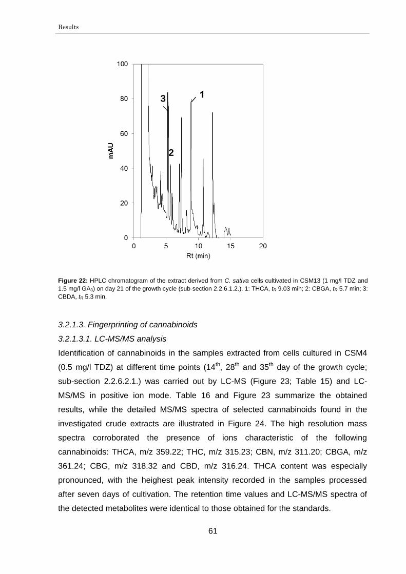
Results
61
Figure 22: HPLC chromatogram of the extract derived from C. sativa cells cultivated in CSM13 (1 mg/l TDZ and
1.5 mg/l GA3) on day 21 of the growth cycle (sub-section 2.2.6.1.2.). 1: THCA, tR 9.03 min; 2: CBGA, tR 5.7 min; 3:
CBDA, tR 5.3 min.
3.2.1.3. Fingerprinting of cannabinoids
3.2.1.3.1. LC-MS/MS analysis
Identification of cannabinoids in the samples extracted from cells cultured in CSM4
(0.5 mg/l TDZ) at different time points (14th, 28th and 35th day of the growth cycle;
sub-section 2.2.6.2.1.) was carried out by LC-MS (Figure 23; Table 15) and LC-
MS/MS in positive ion mode. Table 16 and Figure 23 summarize the obtained
results, while the detailed MS/MS spectra of selected cannabinoids found in the
investigated crude extracts are illustrated in Figure 24. The high resolution mass
spectra corroborated the presence of ions characteristic of the following
cannabinoids: THCA, m/z 359.22; THC, m/z 315.23; CBN, m/z 311.20; CBGA, m/z
361.24; CBG, m/z 318.32 and CBD, m/z 316.24. THCA content was especially
pronounced, with the heighest peak intensity recorded in the samples processed
after seven days of cultivation. The retention time values and LC-MS/MS spectra of
the detected metabolites were identical to those obtained for the standards.
2
3
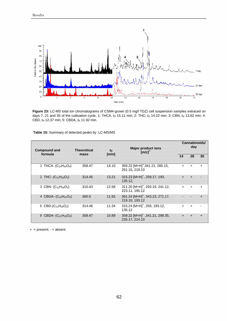
Results
62
Figure 23: LC-MS total ion chromatograms of CSM4-grown (0.5 mg/l TDZ) cell suspension samples extraced on
days 7, 21 and 35 of the cultivation cycle. 1: THCA, tR 15.11 min; 2: THC; tR 14.22 min; 3: CBN, tR 13.62 min; 4:
CBD, tR 12.37 min; 5: CBDA, tR 11.92 min.
+ = present; - = absent
Table 15: Summary of detected peaks by LC-MS/MS
Compound and formula
Theoretical mass
tR [min]
Major product ions [m/z]
+.
Cannabinoids/
day
14 28 35
1 THCA- (C22H30O4)
358.47 14.10 359.22 [M+H]+,341.21, 285.15,
261.15, 219.10 + + +
2 THC- (C21H30O2)
314.45 13.21 315.23 [M+H]+, 259.17, 193,
135.12, + + -
3 CBN- (C21H26O2)
310.43 12.58 311.20 [M+H]+, 293.19, 241.12,
223.11, 195.12 + + +
4 CBGA- (C22H31O4) 360.5 11.55 361.24 [M+H]+, 343.23, 271.17,
219.10, 193.12 - - +
6 CBD-(C21H30O2) 314.46 11.34 315.24 [M+H]+, 259, 193.12,
135.12 + + -
8 CBDA- (C21H30O4) 358.47 10.89 359.22 [M+H]+ ,341.21, 298.35,
235.17, 214.10 + + +
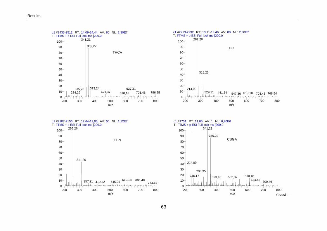
Results
63
Contd…..
c1 #2213-2292 RT: 13,11-13,46 AV: 80 NL: 2,30E7T: FTMS + p ESI Full lock ms [200,00-800,00]
200 300 400 500 600 700 800
m/z
0
10
20
30
40
50
60
70
80
90
100
Re
lative
Ab
un
da
nce
282,28
315,23
214,09329,21 441,34 610,18 768,54703,48547,36
THC
c1 #2433-2512 RT: 14,09-14,44 AV: 80 NL: 2,30E7T: FTMS + p ESI Full lock ms [200,00-800,00]
200 300 400 500 600 700 800
m/z
0
10
20
30
40
50
60
70
80
90
100
Re
lative
Ab
un
da
nce
341,21
359,22
373,24 637,31315,23471,37 796,55284,29 701,46610,18
c1 #2107-2156 RT: 12,64-12,86 AV: 50 NL: 1,12E7T: FTMS + p ESI Full lock ms [200,00-800,00]
200 300 400 500 600 700 800
m/z
0
10
20
30
40
50
60
70
80
90
100
Re
lative
Ab
un
da
nce
256,26
311,20
610,18 696,48357,21 545,35419,32 773,52
CBN
THCA
c1 #1751 RT: 11,05 AV: 1 NL: 6,90E6T: FTMS + p ESI Full lock ms [200,00-800,00]
200 300 400 500 600 700 800
m/z
0
10
20
30
40
50
60
70
80
90
100
Re
lative
Ab
un
da
nce
341,21
359,22
214,09
298,35
235,17 610,18393,18 502,37634,45
700,46
CBGA
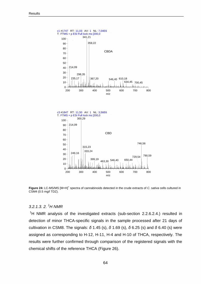
Results
64
Figure 24: LC-MS/MS [M+H]+ spectra of cannabinoids detected in the crude extracts of C. sativa cells cultured in
CSM4 (0.5 mg/l TDZ).
3.2.1.3. 2. 1H NMR
1H NMR analysis of the investigated extracts (sub-section 2.2.6.2.4.) resulted in
detection of minor THCA-specific signals in the sample processed after 21 days of
cultivation in CSM8. The signals: δ 1.45 (s), δ 1.69 (s), δ 6.25 (s) and δ 6.40 (s) were
assigned as corresponding to H-12, H-11, H-4 and H-10 of THCA, respectively. The
results were further confirmed through comparison of the registered signals with the
chemical shifts of the reference THCA (Figure 26).
c3 #1847 RT: 11,50 AV: 1 NL: 3,56E6T: FTMS + p ESI Full lock ms [200,00-800,00]
200 300 400 500 600 700 800
m/z
0
10
20
30
40
50
60
70
80
90
100
Re
lative
Ab
un
da
nce
300,29
214,09
746,56
315,23
333,24249,16
790,59
399,19 650,44546,40
729,54
463,30
CBD
c1 #1747 RT: 11,03 AV: 1 NL: 7,04E6T: FTMS + p ESI Full lock ms [200,00-800,00]
200 300 400 500 600 700 800
m/z
0
10
20
30
40
50
60
70
80
90
100
Re
lative
Ab
un
da
nce
341,21
359,22
214,09
298,35
235,17 367,20 610,18546,40634,45 700,45
CBDA
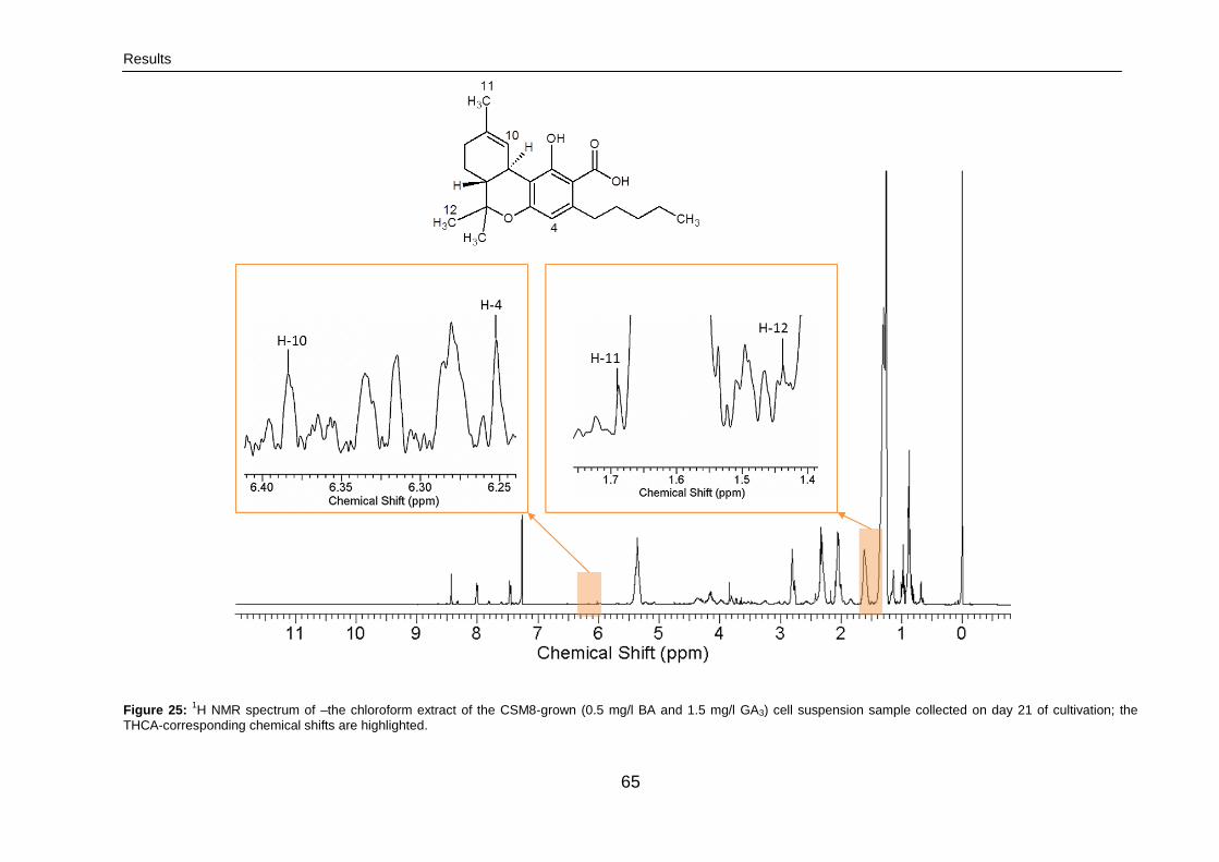
Results
65
Figure 25: 1H NMR spectrum of –the chloroform extract of the CSM8-grown (0.5 mg/l BA and 1.5 mg/l GA3) cell suspension sample collected on day 21 of cultivation; the
THCA-corresponding chemical shifts are highlighted.
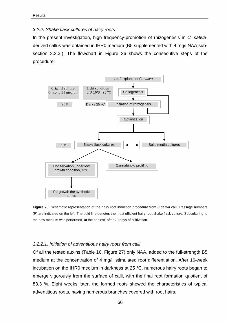
Results
66
3.2.2. Shake flask cultures of hairy roots
In the present investigation, high frequency-promotion of rhizogenesis in C. sativa-
derived callus was obtained in IHR0 medium (B5 supplemented with 4 mg/l NAA;sub-
section 2.2.3.). The flowchart in Figure 26 shows the consecutive steps of the
procedure:
Figure 26: Schematic representation of the hairy root induction procedure from C.sativa calli. Passage numbers
(P) are indicated on the left. The bold line denotes the most efficient hairy root shake flask culture. Subculturing to
the new medium was performed, at the earliest, after 20 days of cultivation.
3.2.2.1. Initiation of adventitious hairy roots from calli
Of all the tested auxins (Table 16, Figure 27) only NAA, added to the full-strength B5
medium at the concentration of 4 mg/l, stimulated root differentiation. After 16-week
incubation on the IHR0 medium in darkness at 25 °C, numerous hairy roots began to
emerge vigorously from the surface of calli, with the final root formation quotient of
83.3 %. Eight weeks later, the formed roots showed the characteristics of typical
adventitious roots, having numerous branches covered with root hairs.
Leaf explants of C. sativa
Callogenesis
Initiation of rhizogensis
Optimization
Solid media cultures
Cannabinoid profiling
Shake flask cultures
L/D 16/8 25 ºC
Dark / 25 ºC
Conservation under low growth condition, 4 ºC
Re-growth the synthetic seeds
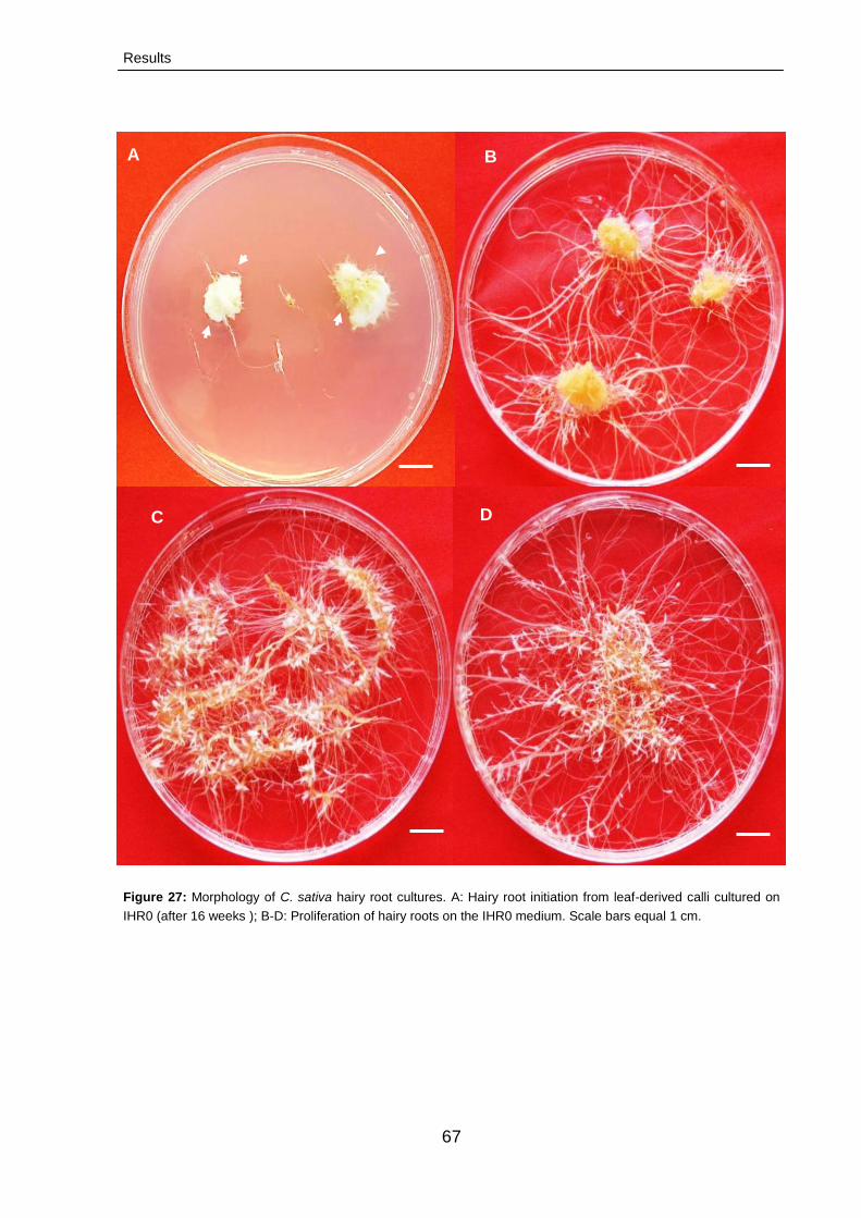
Results
67
Figure 27: Morphology of C. sativa hairy root cultures. A: Hairy root initiation from leaf-derived calli cultured on
IHR0 (after 16 weeks ); B-D: Proliferation of hairy roots on the IHR0 medium. Scale bars equal 1 cm.
A B
C D
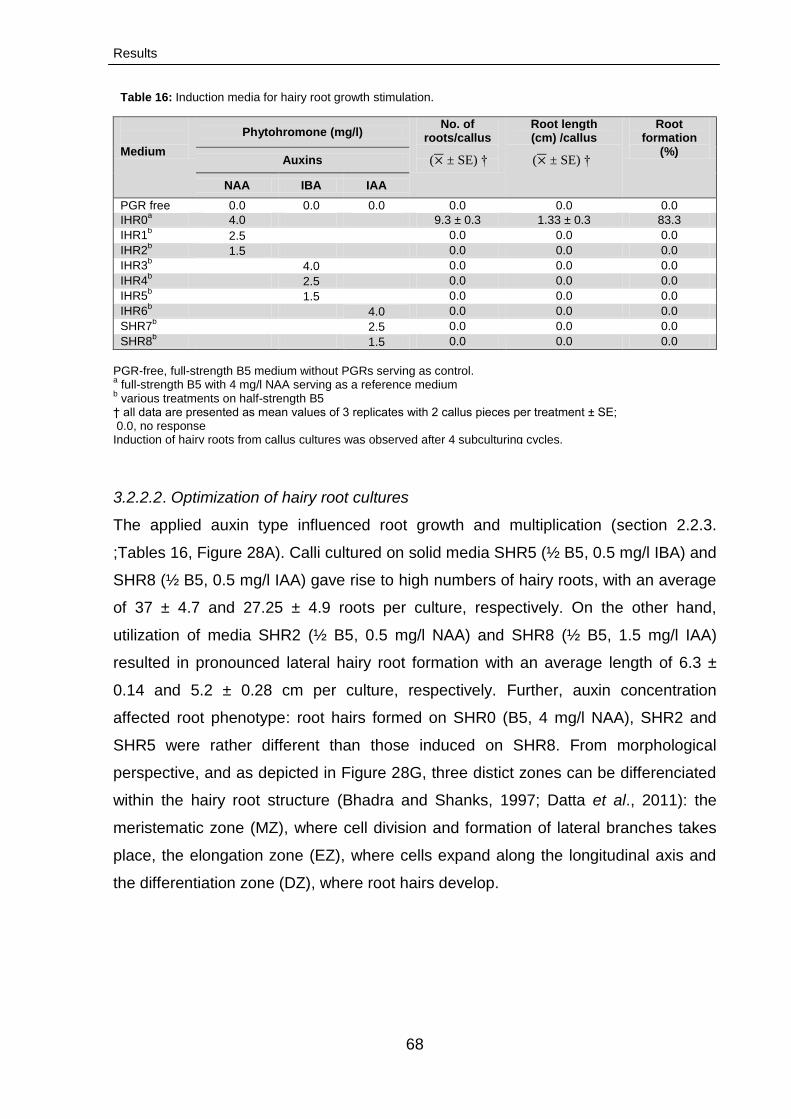
Results
68
Table 16: Induction media for hairy root growth stimulation.
Medium
Phytohromone (mg/l) No. of
roots/callus
Root length (cm) /callus
Root formation
(%) Auxins
NAA IBA IAA
PGR free 0.0 0.0 0.0 0.0 0.0 0.0
IHR0a 4.0 9.3 ± 0.3 1.33 ± 0.3 83.3
IHR1b 2.5 0.0 0.0 0.0
IHR2b 1.5 0.0 0.0 0.0
IHR3b
4.0 0.0 0.0 0.0
IHR4b 2.5 0.0 0.0 0.0
IHR5b 1.5 0.0 0.0 0.0
IHR6b 4.0 0.0 0.0 0.0
SHR7b 2.5 0.0 0.0 0.0
SHR8b 1.5 0.0 0.0 0.0
PGR-free, full-strength B5 medium without PGRs serving as control. a full-strength B5 with 4 mg/l NAA serving as a reference medium
b various treatments on half-strength B5
† all data are presented as mean values of 3 replicates with 2 callus pieces per treatment ± SE; 0.0, no response Induction of hairy roots from callus cultures was observed after 4 subculturing cycles.
3.2.2.2. Optimization of hairy root cultures
The applied auxin type influenced root growth and multiplication (section 2.2.3.
;Tables 16, Figure 28A). Calli cultured on solid media SHR5 (½ B5, 0.5 mg/l IBA) and
SHR8 (½ B5, 0.5 mg/l IAA) gave rise to high numbers of hairy roots, with an average
of 37 ± 4.7 and 27.25 ± 4.9 roots per culture, respectively. On the other hand,
utilization of media SHR2 (½ B5, 0.5 mg/l NAA) and SHR8 (½ B5, 1.5 mg/l IAA)
resulted in pronounced lateral hairy root formation with an average length of 6.3 ±
0.14 and 5.2 ± 0.28 cm per culture, respectively. Further, auxin concentration
affected root phenotype: root hairs formed on SHR0 (B5, 4 mg/l NAA), SHR2 and
SHR5 were rather different than those induced on SHR8. From morphological
perspective, and as depicted in Figure 28G, three distict zones can be differenciated
within the hairy root structure (Bhadra and Shanks, 1997; Datta et al., 2011): the
meristematic zone (MZ), where cell division and formation of lateral branches takes
place, the elongation zone (EZ), where cells expand along the longitudinal axis and
the differentiation zone (DZ), where root hairs develop.
( ± SE) † ( ± SE) †
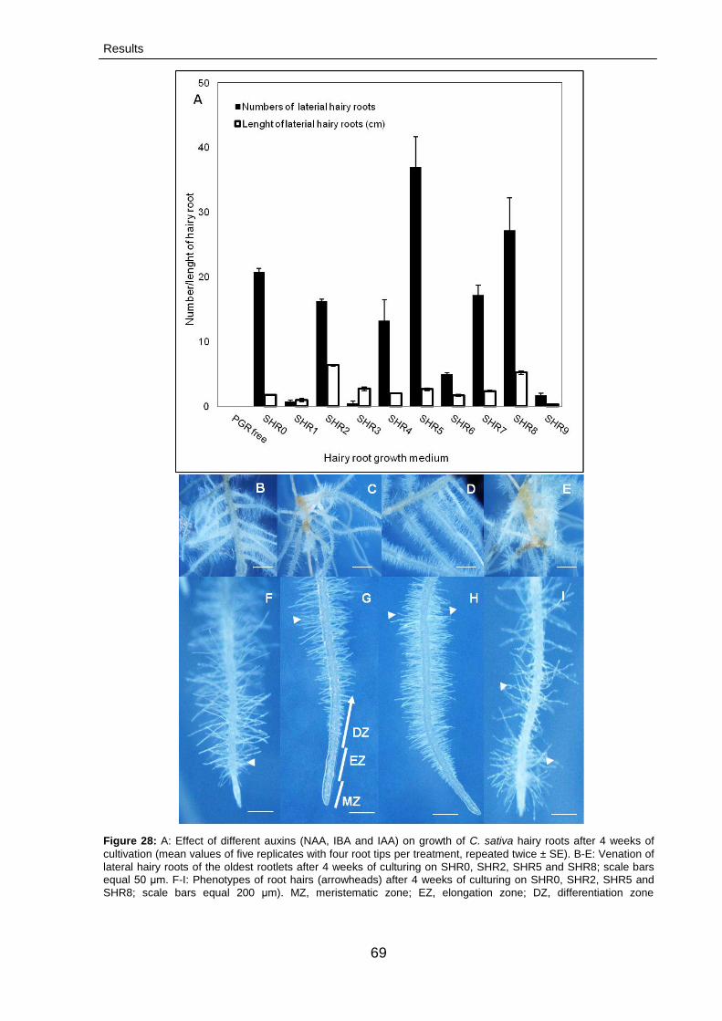
Results
69
Figure 28: A: Effect of different auxins (NAA, IBA and IAA) on growth of C. sativa hairy roots after 4 weeks of
cultivation (mean values of five replicates with four root tips per treatment, repeated twice ± SE). B-E: Venation of
lateral hairy roots of the oldest rootlets after 4 weeks of culturing on SHR0, SHR2, SHR5 and SHR8; scale bars
equal 50 μm. F-I: Phenotypes of root hairs (arrowheads) after 4 weeks of culturing on SHR0, SHR2, SHR5 and
SHR8; scale bars equal 200 μm). MZ, meristematic zone; EZ, elongation zone; DZ, differentiation zone
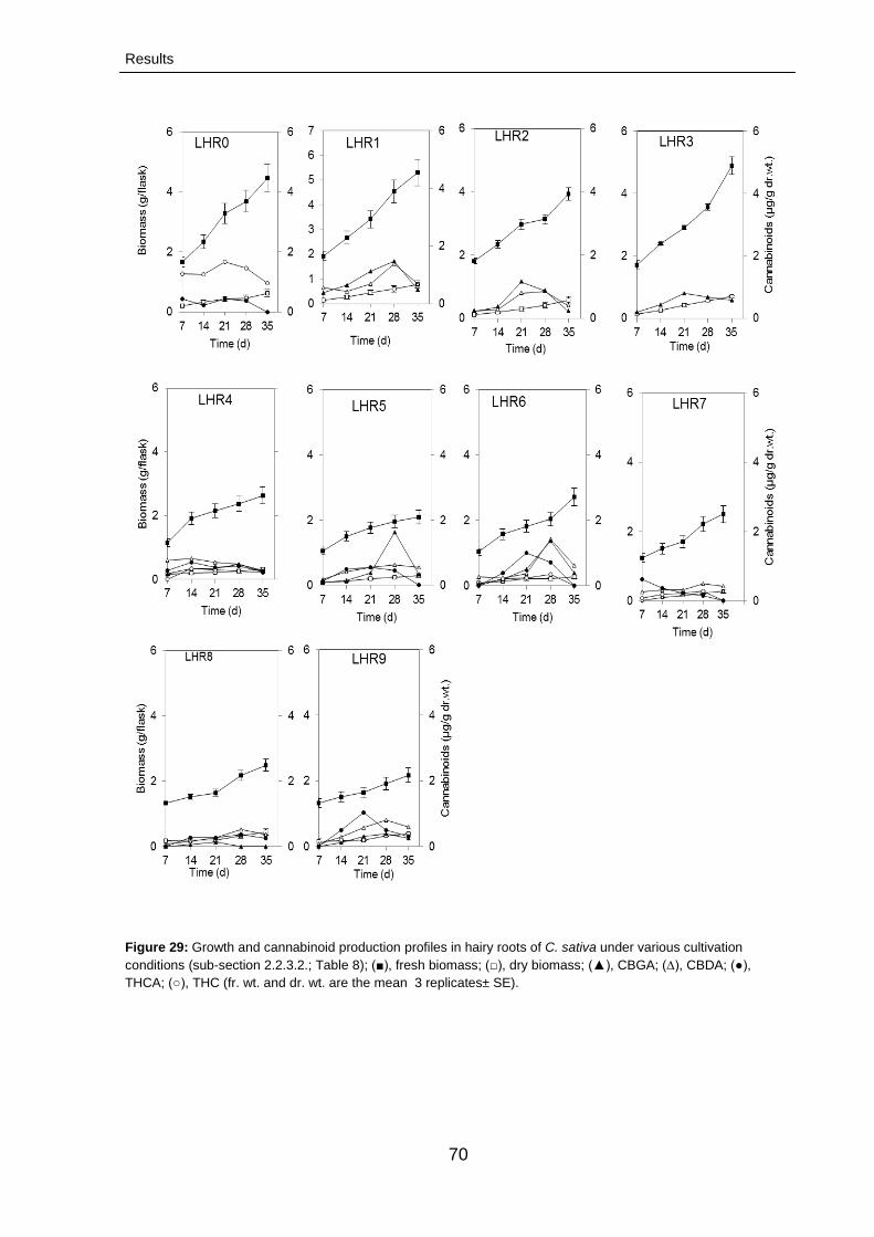
Results
70
Figure 29: Growth and cannabinoid production profiles in hairy roots of C. sativa under various cultivation
conditions (sub-section 2.2.3.2.; Table 8); (■), fresh biomass; (□), dry biomass; (▲), CBGA; (∆), CBDA; (●),
THCA; (○), THC (fr. wt. and dr. wt. are the mean 3 replicates± SE).
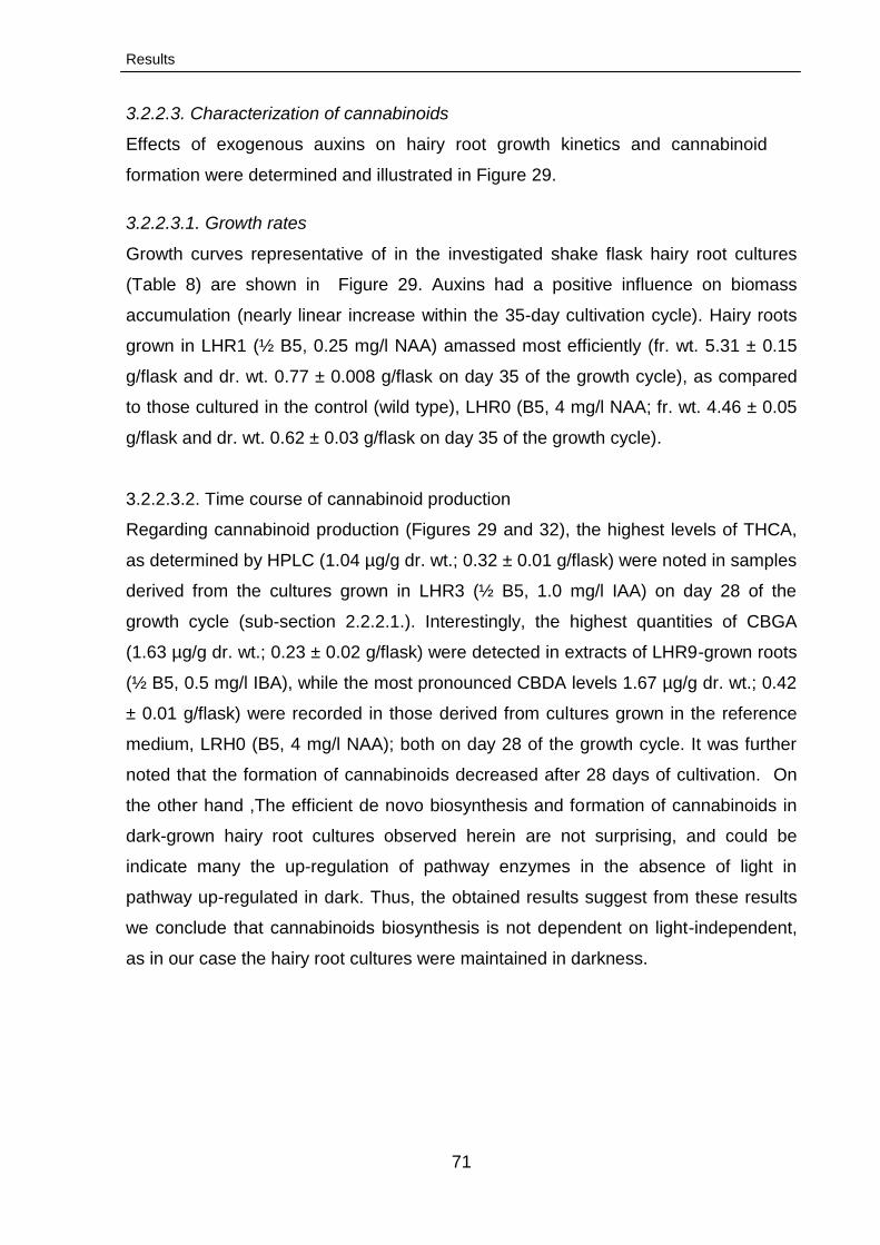
Results
71
3.2.2.3. Characterization of cannabinoids
Effects of exogenous auxins on hairy root growth kinetics and cannabinoid
formation were determined and illustrated in Figure 29.
3.2.2.3.1. Growth rates
Growth curves representative of in the investigated shake flask hairy root cultures
(Table 8) are shown in Figure 29. Auxins had a positive influence on biomass
accumulation (nearly linear increase within the 35-day cultivation cycle). Hairy roots
grown in LHR1 (½ B5, 0.25 mg/l NAA) amassed most efficiently (fr. wt. 5.31 ± 0.15
g/flask and dr. wt. 0.77 ± 0.008 g/flask on day 35 of the growth cycle), as compared
to those cultured in the control (wild type), LHR0 (B5, 4 mg/l NAA; fr. wt. 4.46 ± 0.05
g/flask and dr. wt. 0.62 ± 0.03 g/flask on day 35 of the growth cycle).
3.2.2.3.2. Time course of cannabinoid production
Regarding cannabinoid production (Figures 29 and 32), the highest levels of THCA,
as determined by HPLC (1.04 µg/g dr. wt.; 0.32 ± 0.01 g/flask) were noted in samples
derived from the cultures grown in LHR3 (½ B5, 1.0 mg/l IAA) on day 28 of the
growth cycle (sub-section 2.2.2.1.). Interestingly, the highest quantities of CBGA
(1.63 µg/g dr. wt.; 0.23 ± 0.02 g/flask) were detected in extracts of LHR9-grown roots
(½ B5, 0.5 mg/l IBA), while the most pronounced CBDA levels 1.67 µg/g dr. wt.; 0.42
± 0.01 g/flask) were recorded in those derived from cultures grown in the reference
medium, LRH0 (B5, 4 mg/l NAA); both on day 28 of the growth cycle. It was further
noted that the formation of cannabinoids decreased after 28 days of cultivation. On
the other hand ,The efficient de novo biosynthesis and formation of cannabinoids in
dark-grown hairy root cultures observed herein are not surprising, and could be
indicate many the up-regulation of pathway enzymes in the absence of light in
pathway up-regulated in dark. Thus, the obtained results suggest from these results
we conclude that cannabinoids biosynthesis is not dependent on light-independent,
as in our case the hairy root cultures were maintained in darkness.
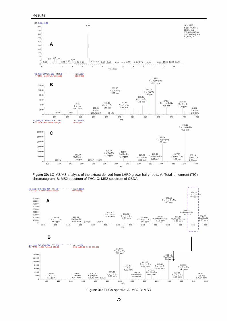
Results
72
Figure 30: LC-MS/MS analysis of the extract derived from LHR0-grown hairy roots. A: Total ion current (TIC)
chromatogram; B: MS2 spectrum of THC; C: MS2 spectrum of CBDA.
Figure 31: THCA spectra. A: MS2;B: MS3.
w1_ms2_CID #291-302 RT: 5,80-6,00 AV: 6 NL: 1,23E4F: FTMS + c ESI Full ms2 315,00@cid40,00 [85,00-320,00]
100 120 140 160 180 200 220 240 260 280 300 320
m/z
0
2000
4000
6000
8000
10000
12000
Inte
nsity
255,21C 14 H 27 O 2 N 2
-2,51 ppm
193,12C 12 H 17 O 2
-2,39 ppm
233,15C 15 H 21 O 2
-1,75 ppm272,17
C 11 H 22 O 3 N 5
0,95 ppm
297,22C 21 H 29 O
-2,00 ppm
207,14C 13 H 19 O 2
-1,86 ppm
245,15C 16 H 21 O 2
-2,48 ppm
181,12C 11 H 17 O 2
-1,96 ppm
135,12C 10 H 15
-1,97 ppm
124,43104,38
167,91C 2 O 9
-265,76 ppm 184,78
315,12C 25 H 15
-1,18 ppm
B
w1_ms2_CID #254-273 RT: 5,06-5,42 AV: 10 NL: 3,08E5F: FTMS + c ESI Full ms2 359,00@cid40,00 [95,00-360,00]
100 120 140 160 180 200 220 240 260 280 300 320 340 360
m/z
0
50000
100000
150000
200000
250000
300000
Inte
nsity
341,17C 19 H 23 O 3 N 3
0,95 ppm
301,14C 16 H 19 O 3 N 3
1,39 ppm
207,10C 12 H 15 O 3
-2,74 ppm
233,08C 13 H 13 O 4
-2,59 ppm
289,14C 15 H 19 O 3 N 3
2,19 ppm
327,21C 18 H 25 O N 5
1,56 ppm
153,09C 9 H 13 O 2
-2,52 ppm
265,26C 17 H 33 N 2
-2,98 ppm190,51117,75
355,19C 25 H 25 O N
-0,89 ppm175,57
RT: 0,00 - 13,99
0 1 2 3 4 5 6 7 8 9 10 11 12 13
Time (min)
0
10
20
30
40
50
60
70
80
90
100
Re
lative
Ab
un
da
nce
4,54
1,26 1,421,10 3,062,78 4,78 5,30 13,356,620,18 5,50 13,032,30 12,398,50 9,753,34 9,513,86 11,8310,517,38 8,02
NL: 3,27E7
TIC F: FTMS + c ESI Full ms2 359,00@cid40,00 [95,00-360,00] MS w1_ms2_CID
A
C
w1_ms2_CID #295-324 RT: 5,90-6,46 AV: 15 NL: 9,19E4F: FTMS + c ESI Full ms2 359,00@cid40,00 [95,00-360,00]
100 120 140 160 180 200 220 240 260 280 300 320 340 360
m/z
0
10000
20000
30000
40000
50000
60000
70000
80000
90000
Inte
nsity
341,17C 19 H 23 O 3 N 3
0,90 ppm301,14
C 16 H 19 O 3 N 3
1,27 ppm
207,10C 12 H 15 O 3
-2,64 ppm
233,08C 13 H 13 O 4
-2,71 ppm
358,35C 25 H 44 N
-2,78 ppm
316,10C 7 H 12 O 5 N 10
-1,81 ppm
153,09C 9 H 13 O 2
-2,65 ppm
288,10C 6 H 12 O 4 N 10
-2,01 ppm
331,12C 7 H 19 O 9 N 6
1,25 ppm
120,10C 5 H 14 O 2 N
-3,63 ppm
262,09C 4 H 10 O 4 N 10
-2,03 ppm190,50170,80
A
w1_ms3_CID #310-322 RT: 6,31-6,56 AV: 7 NL: 1,43E4F: FTMS + c ESI Full ms3 359,00@cid40,00 341,00@cid35,00 [90,00-360,00]
100 120 140 160 180 200 220 240 260 280 300 320 340 360
m/z
0
2000
4000
6000
8000
10000
12000
14000
Inte
nsity
219,10C 13 H 15 O 3
-0,15 ppm
299,16C 19 H 23 O 3
-0,84 ppm
261,15C 16 H 21 O 3
-0,33 ppm
285,15C 18 H 21 O 3
-0,88 ppm
323,16C 21 H 23 O 3
-1,24 ppm
233,12C 14 H 17 O 3
-0,29 ppm 271,13C 17 H 19 O 3
-0,22 ppm
207,10C 12 H 15 O 3
-0,32 ppm
247,13C 15 H 19 O 3
-1,03 ppm
313,18C 20 H 25 O 3
-1,35 ppm
149,06C 9 H 9 O 2
0,32 ppm
107,07C 4 H 11 O 3
-0,11 ppm
176,29C 8 H 24 N 4
501,80 ppm 190,57
352,47C 25 H 52
175,64 ppm
B
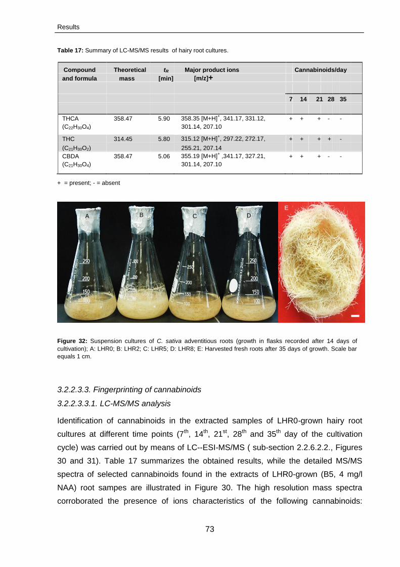
Results
73
Table 17: Summary of LC-MS/MS results of hairy root cultures.
Compound Theoretical tR Major product ions Cannabinoids/day
and formula mass [min] [m/z]+
7 14 21 28 35
THCA 358.47 5.90 358.35 [M+H]
+, 341.17, 331.12, + + + - -
(C22H30O4) 301.14, 207.10
THC 314.45 5.80 315.12 [M+H]+, 297.22, 272.17, + + + + -
(C21H30O2) 255.21, 207.14 CBDA 358.47 5.06 355.19 [M+H]
+ ,341.17, 327.21, + + + - -
(C21H30O4) 301.14, 207.10
+ = present; - = absent
Figure 32: Suspension cultures of C. sativa adventitious roots (growth in flasks recorded after 14 days of
cultivation); A: LHR0; B: LHR2; C: LHR5; D: LHR8; E: Harvested fresh roots after 35 days of growth. Scale bar
equals 1 cm.
3.2.2.3.3. Fingerprinting of cannabinoids
3.2.2.3.3.1. LC-MS/MS analysis
Identification of cannabinoids in the extracted samples of LHR0-grown hairy root
cultures at different time points (7th, 14th, 21st, 28th and 35th day of the cultivation
cycle) was carried out by means of LC--ESI-MS/MS ( sub-section 2.2.6.2.2., Figures
30 and 31). Table 17 summarizes the obtained results, while the detailed MS/MS
spectra of selected cannabinoids found in the extracts of LHR0-grown (B5, 4 mg/l
NAA) root sampes are illustrated in Figure 30. The high resolution mass spectra
corroborated the presence of ions characteristics of the following cannabinoids:
A B C D
E
Results
74
THCA, m/z 358.35; THC, m/z 315.12 and CBDA, m/z 355.19. Highest THCA-, THC-,
and CBDA-specific peak intensity values were recorded after seven days of
cultivation. After 21 days, however, the compounds were not detectable. The
retention time values and LC-MS/MS spectra of the detected compounds were
identical to those obtained for the standards.
The fragmentation pattern of THCA was further confirmed through comparison of
MS2 and MS3 spectra of the standard sample with those of the investigated extract.
While the MS2 spectrum of the THCA ion [M + H]+showed an intense signal at m/z
341, its further analysis by MS3 (Figure 31) revealed a dominant fragmentation
pattern: m/z 323 → m/z 285 → m/z 261 → m/z 233 → m/z 219. The enumerated
product ions were identical to those of the standard.
3.2.2.4. Conservation of hairy root cultures
Hairy roots were conserved through encapsulation-dehydration in the form of
synthetic seeds (beads). In all experiments, the preservation efficiency was
monitored, starting with the observation of the seeds for ruptures caused by the
emerging root tips followed by root elongation (Figure 33). Newly formed lateral roots
were developed for 10 to 12 days after treatment.
3.2.2.4.1. Effect of the encapsulation-dehydration procedure
According to previous reports, the intracellular moisture content of synthetic seeds
should be reduced by laminar airflow desiccation prior to conservation (Lata et al.,
2012). The investigated Cannabis hairy root beads were dehydrated for different time
periods to determine the optimal desiccation time (they were weighed every hour
within the timespan of 0-4 h and the percentage of moisture content before and after
desiccation was determined). The moisture content in the desiccated seeds
decreased rapidly until the 2nd hour of treatment; from then on, the values remained
approximately constant, reaching a minimum after 4 h with 13 %. After 7, 14, 30 and
180 days of storage at 4 °C, the encapsulated hairy roots desiccated for one h
showed 100 % recovery, whereas the non-dessicated beads – about 75 %. On the
other hand, the bead-encapsulated root tips desiccated for up to three-four h lost
their viability (Figure 34).
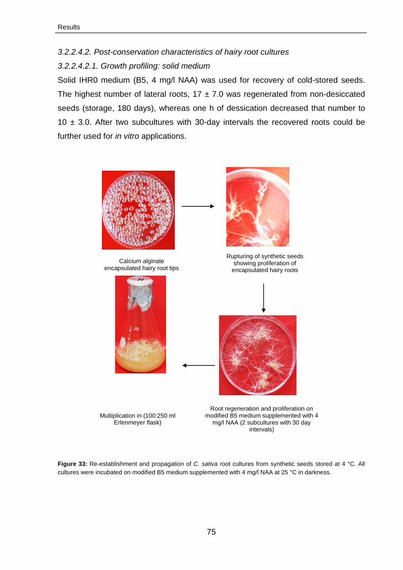
Results
75
3.2.2.4.2. Post-conservation characteristics of hairy root cultures
3.2.2.4.2.1. Growth profiling: solid medium
Solid IHR0 medium (B5, 4 mg/l NAA) was used for recovery of cold-stored seeds.
The highest number of lateral roots, 17 ± 7.0 was regenerated from non-desiccated
seeds (storage, 180 days), whereas one h of dessication decreased that number to
10 ± 3.0. After two subcultures with 30-day intervals the recovered roots could be
further used for in vitro applications.
Figure 33: Re-establishment and propagation of C. sativa root cultures from synthetic seeds stored at 4 °C. All
cultures were incubated on modified B5 medium supplemented with 4 mg/l NAA at 25 °C in darkness.
Root regeneration and proliferation on modified B5 medium supplemented with 4
mg/l NAA (2 subcultures with 30 day intervals)
Multiplication in (100:250 ml Erlenmeyer flask)
Calcium alginate encapsulated hairy root tips
Rupturing of synthetic seeds showing proliferation of
encapsulated hairy roots
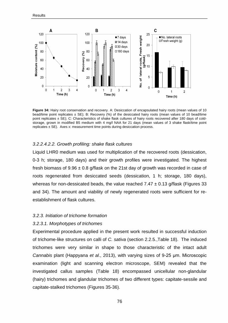
Results
76
Figure 34: Hairy root conservation and recovery. A: Desiccation of encapsulated hairy roots (mean values of 10
bead/time point replicates ± SE); B: Recovery (%) of the desiccated hairy roots (mean values of 10 bead/time
point replicates ± SE); C: Characteristics of shake flask cultures of hairy roots recovered after 180 days of cold-
storage, grown in modified B5 medium with 4 mg/l NAA for 21 days (mean values of 3 shake flask/time point
replicates ± SE). Axes x: measurement time points during desiccation process.
3.2.2.4.2.2. Growth profiling: shake flask cultures
Liquid LHR0 medium was used for multiplication of the recovered roots (dessication,
0-3 h; storage, 180 days) and their growth profiles were investigated. The highest
fresh biomass of 9.96 ± 0.8 g/flask on the 21st day of growth was recorded in case of
roots regenerated from desiccated seeds (dessication, 1 h; storage, 180 days),
whereas for non-dessicated beads, the value reached 7.47 ± 0.13 g/flask (Figures 33
and 34). The amount and viability of newly regenerated roots were sufficient for re-
establishment of flask cultures.
3.2.3. Initiation of trichome formation
3.2.3.1. Morphotypes of trichomes
Experimental procedure applied in the present work resulted in successful induction
of trichome-like structures on calli of C. sativa (section 2.2.5.,Table 18). The induced
trichomes were very similar in shape to those characteristic of the intact adult
Cannabis plant (Happyana et al., 2013), with varying sizes of 9-25 μm. Microscopic
examination (light and scanning electron microscope, SEM) revealed that the
investigated callus samples (Table 18) encompassed unicellular non-glandular
(hairy) trichomes and glandular trichomes of two different types: capitate-sessile and
capitate-stalked trichomes (Figures 35-36).

Results
77
Figure 35: Microscopic images of trichome morphotypes induced in vitro from C. sativa callus. A and B: Capitate-sessile trichomes (B5= 0.5 mg/l TDZ); C: Capitate-stalked
trichomes (B5= 1.0 mg/l TDZ); D-G: Hairy trichomes (B5= 1.0 mg/l TDZ + 3 mg/l GA3). Scale bars equal: 25 μm (A-C and G) and 10 μm (D-F).
A B C
D E F G
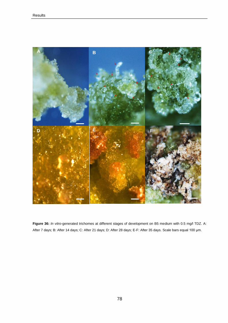
Results
78
Figure 36: In vitro-generated trichomes at different stages of development on B5 medium with 0.5 mg/l TDZ. A:
After 7 days; B: After 14 days; C: After 21 days; D: After 28 days; E-F: After 35 days. Scale bars equal 100 μm.
A B C
F E D F
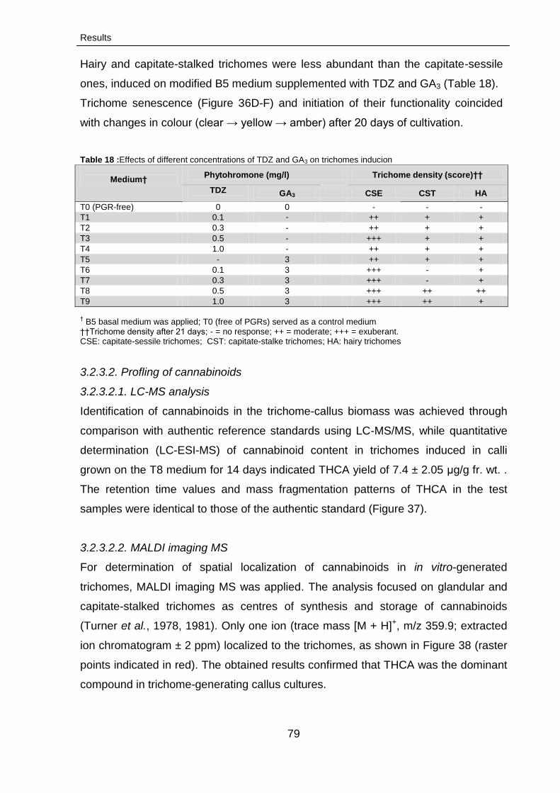
Results
79
3.2.3.2. Profling of cannabinoids
3.2.3.2.1. LC-MS analysis
Identification of cannabinoids in the trichome-callus biomass was achieved through
comparison with authentic reference standards using LC-MS/MS, while quantitative
determination (LC-ESI-MS) of cannabinoid content in trichomes induced in calli
grown on the T8 medium for 14 days indicated THCA yield of 7.4 ± 2.05 μg/g fr. wt. .
The retention time values and mass fragmentation patterns of THCA in the test
samples were identical to those of the authentic standard (Figure 37).
3.2.3.2.2. MALDI imaging MS
For determination of spatial localization of cannabinoids in in vitro-generated
trichomes, MALDI imaging MS was applied. The analysis focused on glandular and
capitate-stalked trichomes as centres of synthesis and storage of cannabinoids
(Turner et al., 1978, 1981). Only one ion (trace mass [M + H]+, m/z 359.9; extracted
ion chromatogram ± 2 ppm) localized to the trichomes, as shown in Figure 38 (raster
points indicated in red). The obtained results confirmed that THCA was the dominant
compound in trichome-generating callus cultures.
Hairy and capitate-stalked trichomes were less abundant than the capitate-sessile
ones, induced on modified B5 medium supplemented with TDZ and GA3 (Table 18).
Trichome senescence (Figure 36D-F) and initiation of their functionality coincided
with changes in colour (clear → yellow → amber) after 20 days of cultivation.
Table 18 :Effects of different concentrations of TDZ and GA3 on trichomes inducion
Medium†
Phytohromone (mg/l) Trichome density (score)††
TDZ
GA3 CSE CST HA
T0 (PGR-free) 0 0 - - -
T1 0.1 -
++ + +
T2 0.3 - ++ + +
T3 0.5 - +++ + +
T4 1.0 - ++ + +
T5 - 3 ++ + +
T6 0.1 3 +++ - +
T7 0.3 3 +++ - +
T8 0.5 3 +++ ++ ++
T9 1.0 3 +++ ++ +
† B5 basal medium was applied; T0 (free of PGRs) served as a control medium
††Trichome density after 21 days; - = no response; ++ = moderate; +++ = exuberant. CSE: capitate-sessile trichomes; CST: capitate-stalke trichomes; HA: hairy trichomes

Results
80
Due to low THCA concentrations and small sizes of the investigated trichomes
(preventing efficient sample preparation), the applied MALDI imagining MS
procedure did not prove reproducible.
3.2.3.2.2.1. MALDI imaging MS profiling of cannabinoids trichomes
Our goal was to determine if MALDI-imagining MS could be accomplished on in vitro
trichomes for determining the spatial localization of cannabinoids trichomes. In this
context, glandular trichome and capitate stalked was used, because of their
capability of synthesis and storage of cannabinoids (Turner et al. 1978, 1981). To
accomplish the imaging on trichome, a 15 μm thick section was prepared using a
cryo-microtome and mounted onto a MALDI plate, the sample was then covered with
a uniform crystalline matrix and subjected to MALDI-imagining MS. Using this
approach, only one ion localized to trichome with trace mass [M + H]+ m/z 359.9
(extracted ion chromatogram with ± 2 ppm) from the indicated raster points (red
region) as shown in Figure 38. The data in clearly provides supported the result of
LC-MS/MS that THCA is the dominant compound in callus cultures. However,
MALDI- imagining MS procedure was not reproducible for detection of THCA due to
its low concentration and fine scale (size) of trichome which prevents obtaining good
slices. In the available literature no comparable data were reported, since trichomes
were not investigated with this technique.
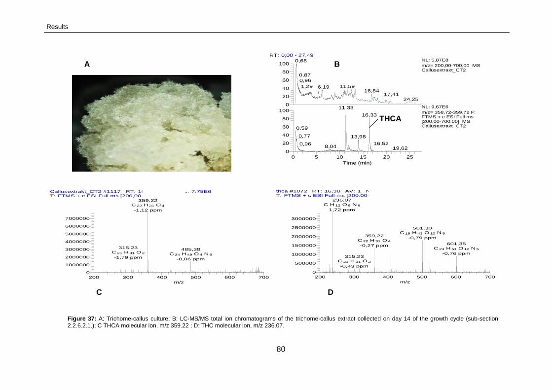
Results
80
Figure 37: A: Trichome-callus culture; B: LC-MS/MS total ion chromatograms of the trichome-callus extract collected on day 14 of the growth cycle (sub-section
2.2.6.2.1.); C THCA molecular ion, m/z 359.22 ; D: THC molecular ion, m/z 236.07.
Callusextrakt_CT2 #1117 RT: 16,33 AV: 1 NL: 7,75E6T: FTMS + c ESI Full ms [200,00-700,00]
200 300 400 500 600 700
m/z
0
1000000
2000000
3000000
4000000
5000000
6000000
7000000
Inte
nsity
359,22C 22 H 31 O 4
-1,12 ppm
315,23C 21 H 31 O 2
-1,79 ppm
485,38C 24 H 49 O 4 N 6
-0,06 ppm
thca #1072 RT: 16,38 AV: 1 NL: 3,36E6T: FTMS + c ESI Full ms [200,00-700,00]
200 300 400 500 600 700
m/z
0
500000
1000000
1500000
2000000
2500000
3000000
Inte
nsity
236,07C H 12 O 8 N 6
1,72 ppm
501,30C 19 H 43 O 10 N 5
-0,79 ppm359,22
C 22 H 31 O 4
-0,27 ppm 601,35C 24 H 51 O 12 N 5
-0,76 ppm315,23
C 21 H 31 O 2
-0,43 ppm
RT: 0,00 - 27,49
0 5 10 15 20 25
Time (min)
0
20
40
60
80
100
0
20
40
60
80
1000,68
0,87
0,96
1,29 11,596,1916,84
17,4124,25
11,33
16,33
0,59
0,77 13,98
16,520,968,04 19,62
NL: 5,87E8
m/z= 200,00-700,00 MS Callusextrakt_CT2
NL: 9,67E6
m/z= 358,72-359,72 F: FTMS + c ESI Full ms [200,00-700,00] MS Callusextrakt_CT2
THCA
A
C
B
D
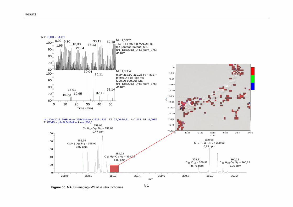
Results
81
nr1_Dez2013_DHB_6um_375x344um #1625-1837 RT: 27,00-30,51 AV: 213 NL: 9,09E2T: FTMS + p MALDI Full lock ms [200,00-800,00]
358,8 359,0 359,2 359,4 359,6 359,8 360,0 360,2
m/z
0
20
40
60
80
100
Re
lativ
e A
bu
nd
an
ce
359,08C 4 H 17 O 14 N 5 = 359,08
-0,47 ppm
359,99C 10 H 6 O 12 N 3 = 359,99
0,25 ppm
359,91C 10 O 15 = 359,92
-45,71 ppm
359,22C 18 H 27 O 2 N 6 = 359,22
1,85 ppm
358,96C 5 H 3 O 15 N 4 = 358,96
3,07 ppm
360,22C 13 H 28 O 4 N 8 = 360,22
-1,06 ppm
RT: 0,00 - 54,81
0 10 20 30 40 50
Time (min)
60
70
80
90
100
60
70
80
90
100
Re
lativ
e A
bu
nd
an
ce
0,92 38,129,30 52,4813,33 37,131,95
21,64
30,0435,11
53,1415,9137,1219,6515,70
NL: 1,33E7
TIC F: FTMS + p MALDI Full ms [200,00-800,00] MS nr1_Dez2013_DHB_6um_375x344um
NL: 1,35E4
m/z= 358,90-359,26 F: FTMS + p MALDI Full lock ms [200,00-800,00] MS nr1_Dez2013_DHB_6um_375x344um
Figure 38. MALDI-imaging- MS of in vitro trichomes
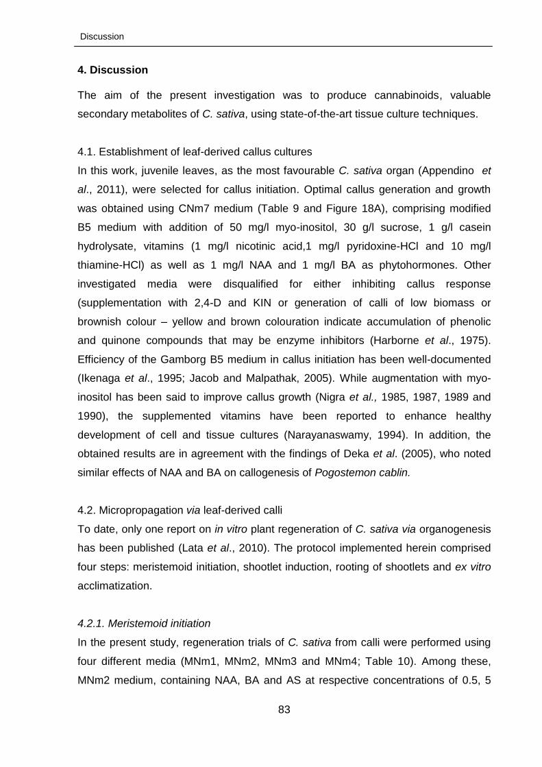
Discussion
83
4. Discussion The aim of the present investigation was to produce cannabinoids, valuable
secondary metabolites of C. sativa, using state-of-the-art tissue culture techniques.
4.1. Establishment of leaf-derived callus cultures
In this work, juvenile leaves, as the most favourable C. sativa organ (Appendino et
al., 2011), were selected for callus initiation. Optimal callus generation and growth
was obtained using CNm7 medium (Table 9 and Figure 18A), comprising modified
B5 medium with addition of 50 mg/l myo-inositol, 30 g/l sucrose, 1 g/l casein
hydrolysate, vitamins (1 mg/l nicotinic acid,1 mg/l pyridoxine-HCl and 10 mg/l
thiamine-HCl) as well as 1 mg/l NAA and 1 mg/l BA as phytohormones. Other
investigated media were disqualified for either inhibiting callus response
(supplementation with 2,4-D and KIN or generation of calli of low biomass or
brownish colour – yellow and brown colouration indicate accumulation of phenolic
and quinone compounds that may be enzyme inhibitors (Harborne et al., 1975).
Efficiency of the Gamborg B5 medium in callus initiation has been well-documented
(Ikenaga et al., 1995; Jacob and Malpathak, 2005). While augmentation with myo-
inositol has been said to improve callus growth (Nigra et al., 1985, 1987, 1989 and
1990), the supplemented vitamins have been reported to enhance healthy
development of cell and tissue cultures (Narayanaswamy, 1994). In addition, the
obtained results are in agreement with the findings of Deka et al. (2005), who noted
similar effects of NAA and BA on callogenesis of Pogostemon cablin.
4.2. Micropropagation via leaf-derived calli
To date, only one report on in vitro plant regeneration of C. sativa via organogenesis
has been published (Lata et al., 2010). The protocol implemented herein comprised
four steps: meristemoid initiation, shootlet induction, rooting of shootlets and ex vitro
acclimatization.
4.2.1. Meristemoid initiation
In the present study, regeneration trials of C. sativa from calli were performed using
four different media (MNm1, MNm2, MNm3 and MNm4; Table 10). Among these,
MNm2 medium, containing NAA, BA and AS at respective concentrations of 0.5, 5
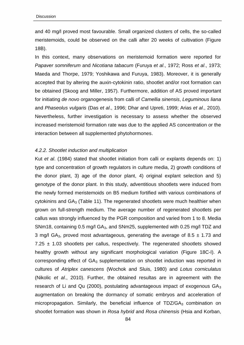
Discussion
84
and 40 mg/l proved most favourable. Small organized clusters of cells, the so-called
meristemoids, could be observed on the calli after 20 weeks of cultivation (Figure
18B).
In this context, many observations on meristemoid formation were reported for
Papaver somniferum and Nicotiana tabacum (Furuya et al., 1972; Ross et al., 1973;
Maeda and Thorpe, 1979; Yoshikawa and Furuya, 1983). Moreover, it is generally
accepted that by altering the auxin-cytokinin ratio, shootlet and/or root formation can
be obtained (Skoog and Miller, 1957). Furthermore, addition of AS proved important
for initiating de novo organogenesis from calli of Camellia sinensis, Leguminous liana
and Phaseolus vulgaris (Das et al., 1996; Dhar and Upreti, 1999; Arias et al., 2010).
Nevertheless, further investigation is necessary to assess whether the observed
increased meristemoid formation rate was due to the applied AS concentration or the
interaction between all supplemented phytohormones.
4.2.2. Shootlet induction and multiplication
Kut et al. (1984) stated that shootlet initiation from calli or explants depends on: 1)
type and concentration of growth regulators in culture media, 2) growth conditions of
the donor plant, 3) age of the donor plant, 4) original explant selection and 5)
genotype of the donor plant. In this study, adventitious shootlets were induced from
the newly formed meristemoids on B5 medium fortified with various combinations of
cytokinins and GA3 (Table 11). The regenerated shootlets were much healthier when
grown on full-strength medium. The average number of regenerated shootlets per
callus was strongly influenced by the PGR composition and varied from 1 to 8. Media
SNm18, containing 0.5 mg/l GA3, and SNm25, supplemented with 0.25 mg/l TDZ and
3 mg/l GA3, proved most advantageous, generating the average of 8.5 ± 1.73 and
7.25 ± 1.03 shootlets per callus, respectively. The regenerated shootlets showed
healthy growth without any significant morphological variation (Figure 18C-I). A
corresponding effect of GA3 supplementation on shootlet induction was reported in
cultures of Atriplex canescens (Wochok and Sluis, 1980) and Lotus corniculatus
(Nikolic et al., 2010). Further, the obtained resultas are in agreement with the
research of Li and Qu (2000), postulating advantageous impact of exogenous GA3
augmentation on breaking the dormancy of somatic embryos and acceleration of
micropropagation. Similarly, the beneficial influence of TDZ/GA3 combination on
shootlet formation was shown in Rosa hybrid and Rosa chinensis (Hsia and Korban,
Discussion
85
1996), while according to other reports, growth of shootlets was initiated from calli
cultured on media supplemented with GA3 and alternative cytokinins, Z or BA (Lata et
al., 2010). In conclusion, it is possible to produce clones of C. sativa and thus
propagate the valuable plant for commercial uses.
4.2.3. Rooting of shootlets
Roots were generated from the bases of shootlets of C. sativa using B5 medium
supplemented with different auxins (NAA, IBA or IAA) at concentrations ranging from
0.5 to 1.5 mg/l (Table 12). In accord with previous reports on stimulation of
rhizogenesis in Malus domestica (Zimmerman, 1984) and Acacia mangium
(Monteuuis and Bon, 2000) through exposure of shootlets to a period of darkness,
Canabis roots were formed on 100 % of the investigated shootlets, reaching the
mean length of 8.7 cm, after one week of cultivation in the dark followed by a four
week culture period with a light/dark cycle of 16/8 h (Figure18J-N). The inducing
effect of IAA on in vitro rooting of shootlets was documented previously in C. sativa,
M. domestica cv. Jork 9 and Adesmia latifolia (Lata et al., 2009 ab; DeKlerk et al.,
1997; Muniz et al., 2013).
4.2.4. Ex vitro acclimatization (indoor cultivation)
The success of any micropropagation protocol depends chiefly on rooting efficiency
and indoor environmental conditions applied in the ex vitro acclimatization procedure
(Zimmerman, 1988). The latter constitutes a critical experimental step, as the in vitro-
grown plantlets are forced to attune their system to autotrophy and, in most cases,
mortality of plantlets proves high under ex vitro conditions (Hazarika, 2003 & 2006).
Wang et al. (2007) reported that ex vitro cultivated Camptotheca acuminata required
high humidity and protection from light stress to survive under greenhouse
conditions. Of the propagated plantlets, 80 % developed into healthy whole plants
after eight weeks of growth. The following 16 weeks of cultivation resulted in
specimens reaching the average height of 53.16 ± 16.34 cm, with 6.2 ± 20.78 true
leaves and 9.25 ± 0.47 nodes per plant (Table 13). In some cases, abnormal leaf
morphology was observed (single or three leaflets instead of the usual seven), as
well as abnormal plant appearancecharacterized by a shrub-like growth with multiple
stems (Figure 19B-C). In this context, Pontaroli and Camadro (2005) suggested the
term “somaclonal variation”. The noted morphological disparities may have been
Discussion
86
brought about by environmental factors, like high concentration of salts in the
hydroponic solution, and possibly other determinants, not yet identified.
4.2.5. Cannabinoids in the acclimatizated plants
Cannabinoid analysis was performed on leaf-extracts of IV plantlets and ex vitro
plants IVH. Mother plant leaves were extracted to provide a control sample.
Investigation of cannabinoids of IV origin revealed that their concentrations varied
within the entire plantlet. While the recorded amounts of THCA and CBGA reached
0.3 %and 0.45 % (relative to the control), respectively, THC and CBG were not
detected. Concurrently, the noted CBDA accumulation was 157 %. Still higher CBDA
content, exceeding that of the control eleven-fold, was recorded in flower tops of IVH
plants (at the flowering stage of their development), eight weeks after transfer to the
hydroponic system (Table 14). The observed changes in cannabinoid concentration
were probably the result of somaclonal variation, reported for many in vitro
propagated plants (Larkin and Scowcroft, 1981; Meins, 1983; Mangolin et al., 1994;
Mohanty et al., 2008). Moreover, while cannabinoids are enzymatically
biosynthesized in the plant as their corresponding carboxylic acid forms (Taura et al.,
2007), they are thermally unstable and can be readily converted, through exposure to
light, heat or as a result of prolonged storage, to their corresponding neutral forms via
decarboxylation (Thakur et al., 2005). Further oxidative degradation or isomerization
of the metabolites of interest was reported to occur as a result of exposure to various
influences, such as those of UV-light or oxidative agents (Turner and Elsohly, 1979;
Razdan et al., 1972; Trofin et al., 2012). Composition of the acclimatizated plant
extracts described in this work was comparable, but not identical, to the data
presented by Lata et al. (2010). The observed disparities may be the result of
different plant growth regulators applied in each step of culture maintenence.
4.3. Shake flask cell suspension cultures
Growth kinetics parameters, such as changes in fresh (fr. wt.) and dry cell weight (dr.
wt.) as well as the corresponding cannabinoid content values were recorded in
course of a 35-day cultivation cycle of the established C. sativa cell suspensions.

Discussion
87
4.3.1. Characterization of cannabinoids
4.3.1.1. Growth rates
Maximum values of fresh and dry cell weight were variable, especially within the
period between the 21st and 28th day of the cultivation period, depending on the type
and concentration of the applied plant growth regulators. Growth curves (Figure 21)
were delineated to determine growth characteristics and of the established cultures.
The obtained and illustrated development patterns comprised three phases:
– The lag phase, noted within the first week of cultivation, during which only little
growth was observed. Such cell behaviour was expected, since their suspension
cultures had to adjust and establish themselves in the new medium.
– The log phase, in which a moderate, then dramatic increase in growth occurred,
throughout the 2nd and 3rd weeks of the culture period. At the early stages of
development, the cell cultures were actively proliferating through cell multiplication
and enlargement. During this period, synthesis of proteins, nucleic acids and
phospholipids, as well as multiplication of organelles and utilization of energy in the
form of ATP occur (Kumar, 1999). The investigated shake flask cell cultures were
green and most of the cells were isodiametric (~ 18 µm) and elliptic in shape (Figure
22).
– After the log phase (4-5 weeks of cultivation), the growth slightly or even sharply
decreased, while the cultures turned yellowish in colour (Figures 21 and 22). De and
Roy (1985) attributed reduction in fresh weight values of biomass to the degradation
of compounds in synthetic processes and/or the production and release of
extracellular material accumulating in the medium. Similar results were considered an
outcome of cell culture aging due to chromosome variation (Deus-Neumann and
Zenk, 1984; Sierra et al., 1992), or cell aging (Roychoudhury et al., 2006) and
supported by free radical theory-based argumentation (Bremner et al., 1997). In
addition, it was reported that the THCA synthase-catalyzed reaction resulted not only
in THCA formation, but also in the release of hydrogen peroxide – an agent inducing
cell death in C. sativa (Sirikantaramas et al., 2004; Sirikantaramas et al., 2005;
Morimoto et al., 2007). Other reports suggested that the loss of productivity was
related to nutrient depletion (Liu and Zhong, 1998; Abreu et al., 2007).
4.3.1.2. Time course of cannabinoid production in high yielding cell cultures
Several studies have shown that optimized application of plant growth regulators
Discussion
88
results in enhancement of secondary metabolite yields (Suthar et al., 1980). Until
now, however, only a few studies concerning cell suspension cultures of C. sativa as
a potential cannabinoid production platform have been published (Pacifico et al.,
2008; Flores-Sanchez et al., 2009; Pec et al., 2010), while no specific reports
regarding the effect of PGRs on the generation of the secondary metabolites of
interest are available. In this investigation, cannabinoid content values were rather
low in cell extracts derived from most of the tested cultures (Figure 21). Highest
concentrations of THCA and THC (8.12 and 4.45 µg/g dr. wt., respectively) were
recorded after 21 days of cultivation in CSM8 medium containing 0.5 mg/l BA and 1.5
mg/l GA3. In addition, CBDA formation reached its maximum level of 1.5 µg/g dr. wt.
in cell extracts derived from cultures grown in CSM12 medium, supplemented with 1
mg/l TDZ and 1.5 mg/l GA3, on day 28 of the culture cycle. Concurrently, CBGA
content reached its peak (1.18 µg/g dr. wt. after 21 days of cultivation) in cultures
maintained in CSM13 medium containing 1.5 mg/l TDZ and 1.5 mg/l GA3. The
obtained results are in agreement with the available literature, reporting on
modulation of secondary metabolite biosynthesis by addition of cytokinins (BA and
TDZ) to the culture media (Nam et al., 2012; Bhargava et al., 2013). Similarly, the
regulatory role of GA3 in mevalonate (MVA) and methyl erythritol phosphate (MEP)
pathways in C. sativa has been reported (Crozier et al., 2000; Mansouri et al., 2009).
4.3.2. Fingerprinting of cannabinoids
4.3.2.1. LC-MS/MS analysis
The liquid chromatography – tandem mass spectrometry analyses were carried out
upon the standard cannabinoid samples and the extracts of cells cultured in CSM4
medium containing 0.5 mg/l TDZ, processed at different time points of the cultivation
cycle (day 14, 28 and 35). The investigation was aimed at higher degree identity
confirmation of the extracted analytes. On the one hand, the LC-peaks were matched
with those of the standard compounds: THCA, tR 14.10 min; THC, tR 13.21 min; CBN,
tR 12.58 min; CBGA, tR 11.55 min; CBD, tR 11.34 min and CBDA, tR 10.89 min.
Further, the mass-to-charge ratio values of their protonated molecular ions [M + H]+
ultimately confirmed detection of cannabinoids in the investigated cell samples:
THCA, m/z 359.22; THC, m/z 315.23; CBN, m/z 311.20; CBGA, m/z 361.24; CBG,
m/z 318.32 and CBD, m/z 316.24 (Table 15, Figures 24 and 25). Presence of the
enumerated cannabinoids in C. sativa cell cultures has not been previously reported
Discussion
89
(Pacifico et al., 2008; Flores-Sanchez et al., 2009; Pec et al., 2010). It is important to
note that THCA, CBN, CBGA and CBDA were detected after 35 days of cultivation,
while the presence of THC and CBD was recorded in the early stages of the growth
cycle (the compounds were undetectable after 28 days of cultivation).
4.3.2.2. 1H NMR measurements
Analysis of cannabinoid content of the investigated cell extracts by 1H NMR
spectroscopy resulted in detection of but minor THCA-specific signals in the sample
processed after 21 days of cultivation in CSM8 medium supplemented with 0.5 mg/l
TDZ and 1.5 mg/l BA (Figure 26). However, no signals corresponding to THCA, or
any other cannabinoids, were detected in the samples extracted on days 7, 14, 28
and 35 of the culture cycle. Neither THCA nor other secondary metabolites of interest
have been previously detected in C. sativa cell cultures (Pacifico et al., 2008; Flores-
Sanchez et al., 2009; Pec et al., 2010).
4.4. Hairy root cultures
Detailed in vitro C. sativa hairy root induction procedure as well as analyses of the
corresponding biomass accumulation and cannabinoid generation, in the presence of
auxins, have not been reported before. The main objective of this study was twofold:
to increase fundamental understanding of the effects of exogenously applied plant
growth regulators on hairy root induction from leaf-derived calli of C. sativa and to
develop an optimized shake flask root culture system for cannabinoid production.
4.4.1. Initiation of adventitious hairy roots from leaf-derived calli
Generally, the presence of auxins, accompanying the carbon source, in the culture
medium has been known to stimulate rhizogensis (Blazich, 1988). The induced roots
of C. sativa displayed similar growth patterns after 16 weeks of cultivation, with 83.3
% root formation when the initiating calli were incubated on full-strength B5 medium
supplemented with 4 mg/l NAA (IHR0), in darkness at 25 °C (Table 16 and Figure
28). The obtained result is in accord with a previous report on rhizohgenesis
stimulating effect of NAA in C. sativa callus cultures (Fisse et al., 1981). In contrast,
all calli tested with various IAA concentrations (1.5, 2.5 and 4 mg/l) did not generate
hairy roots, although the ability of callus cultures in other plant systems to proliferate
hairy roots in response to PGRs, including IAA, was studied and documented
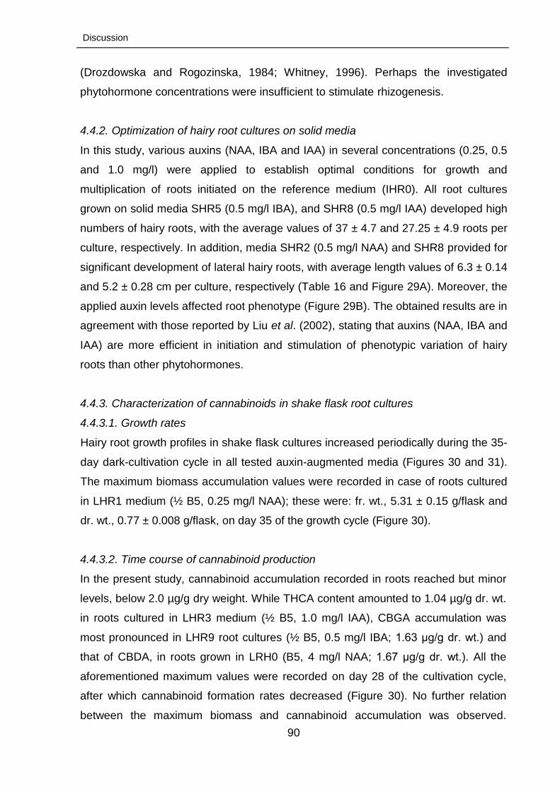
Discussion
90
(Drozdowska and Rogozinska, 1984; Whitney, 1996). Perhaps the investigated
phytohormone concentrations were insufficient to stimulate rhizogenesis.
4.4.2. Optimization of hairy root cultures on solid media
In this study, various auxins (NAA, IBA and IAA) in several concentrations (0.25, 0.5
and 1.0 mg/l) were applied to establish optimal conditions for growth and
multiplication of roots initiated on the reference medium (IHR0). All root cultures
grown on solid media SHR5 (0.5 mg/l IBA), and SHR8 (0.5 mg/l IAA) developed high
numbers of hairy roots, with the average values of 37 ± 4.7 and 27.25 ± 4.9 roots per
culture, respectively. In addition, media SHR2 (0.5 mg/l NAA) and SHR8 provided for
significant development of lateral hairy roots, with average length values of 6.3 ± 0.14
and 5.2 ± 0.28 cm per culture, respectively (Table 16 and Figure 29A). Moreover, the
applied auxin levels affected root phenotype (Figure 29B). The obtained results are in
agreement with those reported by Liu et al. (2002), stating that auxins (NAA, IBA and
IAA) are more efficient in initiation and stimulation of phenotypic variation of hairy
roots than other phytohormones.
4.4.3. Characterization of cannabinoids in shake flask root cultures
4.4.3.1. Growth rates
Hairy root growth profiles in shake flask cultures increased periodically during the 35-
day dark-cultivation cycle in all tested auxin-augmented media (Figures 30 and 31).
The maximum biomass accumulation values were recorded in case of roots cultured
in LHR1 medium (½ B5, 0.25 mg/l NAA); these were: fr. wt., 5.31 ± 0.15 g/flask and
dr. wt., 0.77 ± 0.008 g/flask, on day 35 of the growth cycle (Figure 30).
4.4.3.2. Time course of cannabinoid production
In the present study, cannabinoid accumulation recorded in roots reached but minor
levels, below 2.0 µg/g dry weight. While THCA content amounted to 1.04 µg/g dr. wt.
in roots cultured in LHR3 medium (½ B5, 1.0 mg/l IAA), CBGA accumulation was
most pronounced in LHR9 root cultures (½ B5, 0.5 mg/l IBA; 1.63 μg/g dr. wt.) and
that of CBDA, in roots grown in LRH0 (B5, 4 mg/l NAA; 1.67 μg/g dr. wt.). All the
aforementioned maximum values were recorded on day 28 of the cultivation cycle,
after which cannabinoid formation rates decreased (Figure 30). No further relation
between the maximum biomass and cannabinoid accumulation was observed.
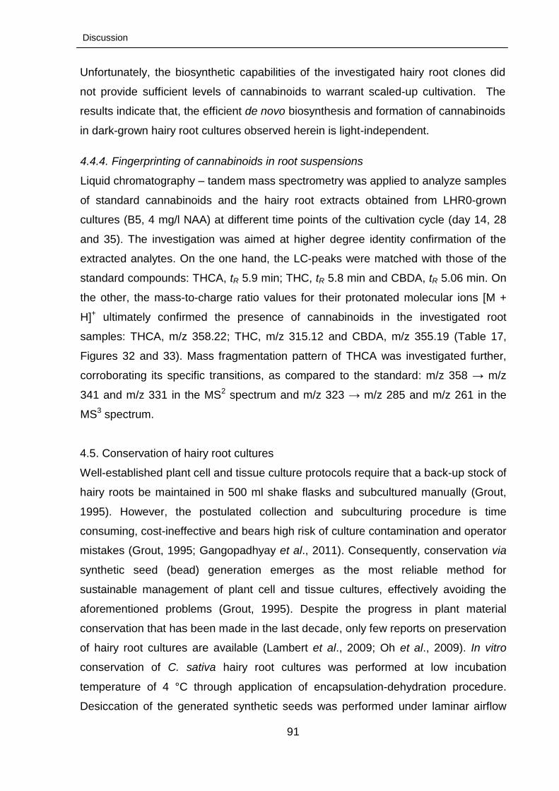
Discussion
91
Unfortunately, the biosynthetic capabilities of the investigated hairy root clones did
not provide sufficient levels of cannabinoids to warrant scaled-up cultivation. The
results indicate that, the efficient de novo biosynthesis and formation of cannabinoids
in dark-grown hairy root cultures observed herein is light-independent.
4.4.4. Fingerprinting of cannabinoids in root suspensions
Liquid chromatography – tandem mass spectrometry was applied to analyze samples
of standard cannabinoids and the hairy root extracts obtained from LHR0-grown
cultures (B5, 4 mg/l NAA) at different time points of the cultivation cycle (day 14, 28
and 35). The investigation was aimed at higher degree identity confirmation of the
extracted analytes. On the one hand, the LC-peaks were matched with those of the
standard compounds: THCA, tR 5.9 min; THC, tR 5.8 min and CBDA, tR 5.06 min. On
the other, the mass-to-charge ratio values for their protonated molecular ions [M +
H]+ ultimately confirmed the presence of cannabinoids in the investigated root
samples: THCA, m/z 358.22; THC, m/z 315.12 and CBDA, m/z 355.19 (Table 17,
Figures 32 and 33). Mass fragmentation pattern of THCA was investigated further,
corroborating its specific transitions, as compared to the standard: m/z 358 → m/z
341 and m/z 331 in the MS2 spectrum and m/z 323 → m/z 285 and m/z 261 in the
MS3 spectrum.
4.5. Conservation of hairy root cultures
Well-established plant cell and tissue culture protocols require that a back-up stock of
hairy roots be maintained in 500 ml shake flasks and subcultured manually (Grout,
1995). However, the postulated collection and subculturing procedure is time
consuming, cost-ineffective and bears high risk of culture contamination and operator
mistakes (Grout, 1995; Gangopadhyay et al., 2011). Consequently, conservation via
synthetic seed (bead) generation emerges as the most reliable method for
sustainable management of plant cell and tissue cultures, effectively avoiding the
aforementioned problems (Grout, 1995). Despite the progress in plant material
conservation that has been made in the last decade, only few reports on preservation
of hairy root cultures are available (Lambert et al., 2009; Oh et al., 2009). In vitro
conservation of C. sativa hairy root cultures was performed at low incubation
temperature of 4 °C through application of encapsulation-dehydration procedure.
Desiccation of the generated synthetic seeds was performed under laminar airflow

Discussion
92
(0-4 h) and was followed by their cold storage (7, 14, 30 and 180 days). Synthetic
seeds dessicated for one hour showed 100 % root recovery, whereas the restoration
rate of non-dehydrated beads reached about 75 % (Figures 34 and 35). On the other
hand, the bead-encapsulated root tips desiccated for up to three to four hours lost
their viability. Morphology of the recovered hairy roots was identical to that
characteristic of the in vitro stock cultures. Fresh biomass accumulation in shake
flask cultures of roots restored from desiccated seeds after 180 days of cold storage
was significantly more pronounced than that recorded for cultures developed from
non-desiccated beads. In general, this preservation technique has been successfully
used in different plant species (Takagi, 2000; Lambert et al., 2009; Lata et al., 2012),
as it does not require dealing with cytotoxic compounds (Surenciski et al., 2012). The
present study is the first report on synthetic seed generation by encapsulation-
dehydration of hairy roots of C. sativa. It further confirms the applicability of the
described technique in in vitro and ex vitro cultivation protocols, as well as
germplasm banks ex situ conservation affording prevention of genetic shifts,
reduction of operational costs and space requirements.
4.6. Initiation of trichome formation
4.6.1. Trichome morphotypes
In C. sativa, glandular trichomes constitute the main site of cannabinoid production
(Appendino et al., 2011; Happyana et al., 2013). In the present study, trials for in vitro
induction of different morphotypes of C. sativa trichomes resulted in generation of
unicellular non-glandular (hairy) and glandular trichomes of two different types,
capitate-sessile and capitate-stalked, especially pronounced on T8-maintained (B5,
0.5 mg/l TDZ and 3 mg/l GA3) leaf-derived calli (Figure 32). SEM imaging was
performed to confirm the initial, light microscopic examination (Figure 38). Hairy and
capitate-stalked trichomes were observed less frequently than capitate-sessile ones
on calli maintained on most of the tested PGR-augmented media (Table 18). Among
the applied phytohormones, TDZ is considered superior to other cytokinins used for
morphogensis stimulation in plant tissue culture protocols, due to its unique mode of
action resulting in accumulation of endogenous auxins and cytokinins (Jones et al.,
2007). Further reports indicated that GA3 was favourable for in vitro promotion of
growth and biomass production (Lata et al., 2009ab). Thus, the hereby recorded in
vitro induction of trichomes does not contradict the hypothesis postulating that all
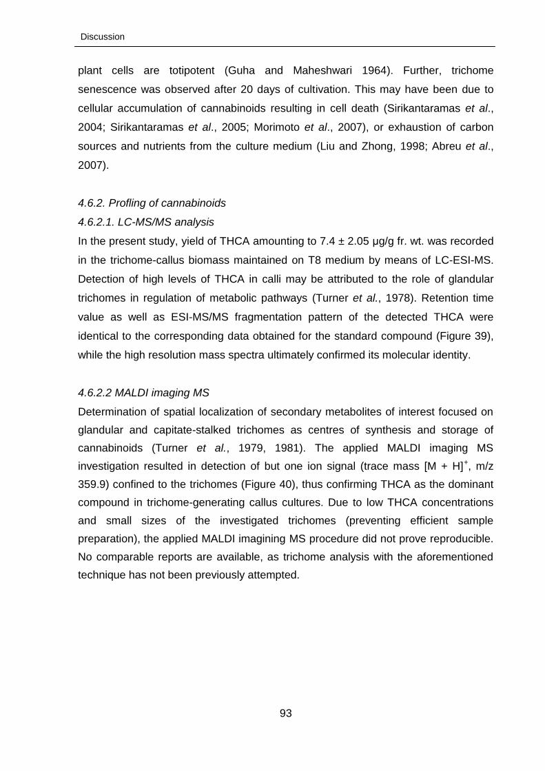
Discussion
93
plant cells are totipotent (Guha and Maheshwari 1964). Further, trichome
senescence was observed after 20 days of cultivation. This may have been due to
cellular accumulation of cannabinoids resulting in cell death (Sirikantaramas et al.,
2004; Sirikantaramas et al., 2005; Morimoto et al., 2007), or exhaustion of carbon
sources and nutrients from the culture medium (Liu and Zhong, 1998; Abreu et al.,
2007).
4.6.2. Profling of cannabinoids
4.6.2.1. LC-MS/MS analysis
In the present study, yield of THCA amounting to 7.4 ± 2.05 μg/g fr. wt. was recorded
in the trichome-callus biomass maintained on T8 medium by means of LC-ESI-MS.
Detection of high levels of THCA in calli may be attributed to the role of glandular
trichomes in regulation of metabolic pathways (Turner et al., 1978). Retention time
value as well as ESI-MS/MS fragmentation pattern of the detected THCA were
identical to the corresponding data obtained for the standard compound (Figure 39),
while the high resolution mass spectra ultimately confirmed its molecular identity.
4.6.2.2 MALDI imaging MS Determination of spatial localization of secondary metabolites of interest focused on
glandular and capitate-stalked trichomes as centres of synthesis and storage of
cannabinoids (Turner et al., 1979, 1981). The applied MALDI imaging MS
investigation resulted in detection of but one ion signal (trace mass [M + H]+, m/z
359.9) confined to the trichomes (Figure 40), thus confirming THCA as the dominant
compound in trichome-generating callus cultures. Due to low THCA concentrations
and small sizes of the investigated trichomes (preventing efficient sample
preparation), the applied MALDI imagining MS procedure did not prove reproducible.
No comparable reports are available, as trichome analysis with the aforementioned
technique has not been previously attempted.
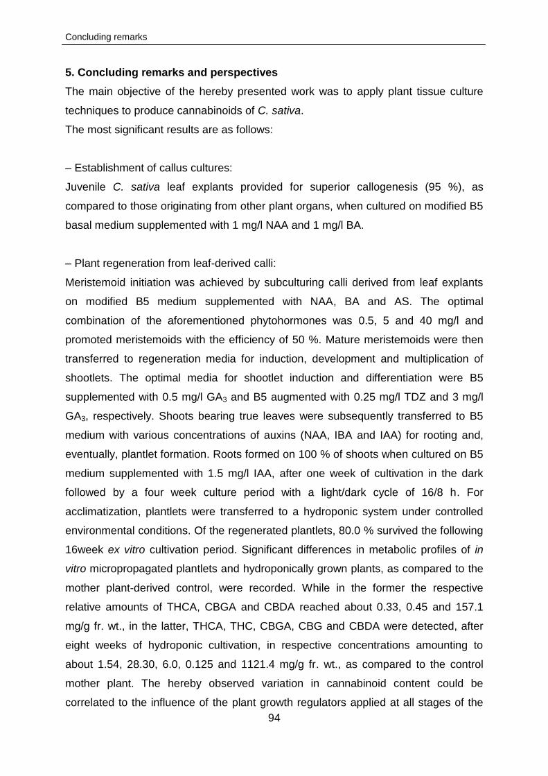
Concluding remarks
94
5. Concluding remarks and perspectives
The main objective of the hereby presented work was to apply plant tissue culture
techniques to produce cannabinoids of C. sativa.
The most significant results are as follows:
– Establishment of callus cultures:
Juvenile C. sativa leaf explants provided for superior callogenesis (95 %), as
compared to those originating from other plant organs, when cultured on modified B5
basal medium supplemented with 1 mg/l NAA and 1 mg/l BA.
– Plant regeneration from leaf-derived calli:
Meristemoid initiation was achieved by subculturing calli derived from leaf explants
on modified B5 medium supplemented with NAA, BA and AS. The optimal
combination of the aforementioned phytohormones was 0.5, 5 and 40 mg/l and
promoted meristemoids with the efficiency of 50 %. Mature meristemoids were then
transferred to regeneration media for induction, development and multiplication of
shootlets. The optimal media for shootlet induction and differentiation were B5
supplemented with 0.5 mg/l GA3 and B5 augmented with 0.25 mg/l TDZ and 3 mg/l
GA3, respectively. Shoots bearing true leaves were subsequently transferred to B5
medium with various concentrations of auxins (NAA, IBA and IAA) for rooting and,
eventually, plantlet formation. Roots formed on 100 % of shoots when cultured on B5
medium supplemented with 1.5 mg/l IAA, after one week of cultivation in the dark
followed by a four week culture period with a light/dark cycle of 16/8 h. For
acclimatization, plantlets were transferred to a hydroponic system under controlled
environmental conditions. Of the regenerated plantlets, 80.0 % survived the following
16week ex vitro cultivation period. Significant differences in metabolic profiles of in
vitro micropropagated plantlets and hydroponically grown plants, as compared to the
mother plant-derived control, were recorded. While in the former the respective
relative amounts of THCA, CBGA and CBDA reached about 0.33, 0.45 and 157.1
mg/g fr. wt., in the latter, THCA, THC, CBGA, CBG and CBDA were detected, after
eight weeks of hydroponic cultivation, in respective concentrations amounting to
about 1.54, 28.30, 6.0, 0.125 and 1121.4 mg/g fr. wt., as compared to the control
mother plant. The hereby observed variation in cannabinoid content could be
correlated to the influence of the plant growth regulators applied at all stages of the
Concluding remarks
95
micropropagation procedure. Further optimization is necessary to reduce the total
time period of plant regeneration.
– Cannabinoids in the established shake flask cell suspension cultures:
Modified B5 medium supplemented with different concentrations of phytohormones
was used to stimulate and optimize cannabinoid production in the established cell
suspensions. Highest concentration values of THCA and THC (8.12 and 4.45 µg/g dr.
wt.) were recorded after 21 days of cultivation in medium containing 0.5 mg/l BA and
1.5 mg/l GA3. Similarly, a 3-week culture period in medium supplemented with 1.5
mg/l TDZ and 1.5 mg/l GA3 resulted in highest CBGA accumulation reaching 1.18
μg/g dr. wt. On the other hand, CBDA formation reached its maximum level of 1.5
µg/g dr. wt. on day 28 of the cultivation cycle in medium augmented with 1 mg/l TDZ
and 1.5 mg/l GA3. Moreover, gradual reduction in cannabinoid accumulation during
the culture cycle was noted; further investigation is needed to fully elucidate this
phenomenon. Improvement of cannabinoid production in cell cultures through elicitor
augmentation and/or precursor feeding warrants additional experimental efforts.
Similarly, manipulation of culture conditions, such as fermenter aeration rate and
agitation speed, should be further studied.
– Cannabinoids in the established hairy root cultures; root culture conservation:
Hairy roots were induced by incubation of aseptically grown callus cultures on solid
B5 medium supplemented with 4 mg/l NAA in darkness at 25 °C. Hairy root growth in
shake flask cultures increased periodically during the 35-day cultivation cycle.
Cannabinoids were produced in minor amoints and remained below 2.0 µg/g dr. wt..
Additionally, encapsulation-dehydration procedure was applied to conserve
adventitious roots of C. sativa. Hairy root tips were successfully encapsulated in
calcium alginate beads and stored at 4 °C for up to 6 months, with maximum viability
of 100 % in case of beads dessicated for one hour and highest biomass
accumulation of 9.96 ± 0.7 g/flask recorded after 21 days of cultivation in B5 medium
supplemented with 4 mg/l NAA. Advantages of the applied encapsulation-
dehydration technique include: high survival rates, rapid recovery, reduced labour,
work-space and time input as well as prevention of mutations and contaminations.
Concluding remarks
96
Further investigations are being conducted to improve 1) cannabinoid production in
root cultures to commercially interesting levels through implementation of various
strategies and 2) viability of post-freeze root cultures.
– Induction of trichomes:
Trichome-like structures were induced from C. sativa calli. B5 medium supplemented
with 0.5 mg/l TDZ and 3 mg/l GA3 proved most favourable. Micromorphology of the
induced trichomes was investigated by means of SEM, which led to their
classification into three categories: unicellular non-glandular (hairy) trichomes and
glandular trichomes of two different types: capitate-sessile and capitate-stalked.
Quantitative determation of cannabinoid content in callus-trichomes (14 days of
incubation on B5 medium with 0.5 mg/l TDZ) by LC-MS pointed to THCA
accumulation (7.4 ± 2.05 μg/g fr. wt.). Spatial distribution of the valuable metabolite in
the in vitro-induced capitate-stalked trichomes was further confirmed by MALDI
imaging MS. However, the procedure was not reproducible due to low THCA
concentrations and fine sacle (small size) of trichomes, preventing efficient sample
preparation.
References
97
5. References
Abreu IN, Andreazza NL, Sawaya ACHF, Eberlin MN, Mazzafera P (2007) Cell
suspension as a tool to study the biosynthesis of pilocarpine in Jaborandi. Plant
Biol 9 (6):793-799.
Akiyoshi DE, Morris RO, Hinz R, Mischke BS, Kosuge T, Garfinkel DJ, Gordon MP,
Nester EW (1983) Cytokinin auxin balance in crown gall tumors is regulated by
specific loci in the T-DNA. P Natl Acad of Sci- -Biol 80 (2):407-411.
Andre C, Vercruysse A (1976) Histochemical study of the stalked glandular hairs of the
female Cannabis plants, using fast blue salt. Planta Med 29 (4):361-366.
Appendino G, Chianese G, Taglialatela-Scafati O (2011) Cannabinoids: Occurrence and
medicinal chemistry. Curr Med Chem18 (7):1085-1099.
Argo WR, Fischer PR (2002) Understanding pH management for container-grown crops.
Meister, Willoughby, Ohio.
Arias AMG, Valverde JM, Fonseca PR, Melara MV (2010) In vitro plant regeneration
system for common bean (Phaseolus vulgaris): effect of n-6-benzylaminopurine
and adenine sulphate. Electron J Biotechn 13 (1):1-8.
Ball E (1953) Hydrolysis of sucrose by autoclaving media, a neglected aspect in the
culture of plant tissues. Bul Torrey Bot Club 80:409-411.
Balvanyos I, Kursinszki L, Szoke E (2001) The effect of plant growth regulators on
biomass formation and lobeline production of Lobelia inflata L. hairy root cultures.
Plant Growth Regul 34 (3):339-345.
Barnicomparini I, Ferri S, Centini F (1984) Cannabinoid level in the leaves as a tool for
the early discrimination of Cannabis chemiovariants. Forensic Sci Int 24 (1):37-
42.
Barrett ML, Scutt AM, Evans FJ (1986) Cannflavin A and cannflavin B, prenylated
flavones from Cannabis sativa L. Experientia 42 (4):452-453.
Bhargava A, Clabaugh I, To JP, Maxwell BB, Chiang YH, Schaller GE, Loraine A, Kieber
JJ (2013) Identification of cytokinin responsive genes using microarray meta-
analysis and RNA-Seq in Arabidopsis. Plant Physiol 162 (1):272-294.
References
98
Biddington NL (1992) The influence of ethylene in plant tissue culture. Plant Growth
Regul 11 (2):173-187.
Binding H (1986) Regeneration from protoplast In: Vasil IK (ed) Cell culture and somatic
cell genetic of plants, vol 3. Academic Press, New York, pp 259-274.
Blanc G, Michaux-Ferriere N, Teisson C, Lardet L, Carron MP (1999) Effects of
carbohydrate addition on the induction of somatic embryogenesis in Hevea
brasiliensis. Plant Cell Tiss Org 59 (2):103-112.
Blazich FA (1988) Chemicals and formulations used to promote adventitious rooting, in:
“Adventitious root formation by cuttings” In: Davis, T.D., B.E. Haissig, N. Sakhla,
(eds.) Advances in Plant Science Series vol 2, p. 312. Dioscorides Press,
Portland.
Bobak M, Samaj J, Pret'ova A, Blehova A, Hlinkova E, Ovecka M, Hlavacka A,
Kutarnova Z (2004) The histological analysis of indirect somatic embryogenesis
on Drosera spathulata Labill. Acta Physiol Plant 26 (3):353-361.
Braemer R, Paris M (1987) Biotransformation of cannabinoids by a cell suspension
culture of Cannabis sativa L. Plant Cell Rep 6 (2):150-152.
Braemer R, Tsoutsias Y, Hurabielle M, Paris M (1987) Biotransformations of quercetin
and apigenin by a cell suspension culture of Cannabis sativa. Planta Medica
(2):225-226.
Braut-Boucher F, Paillard N, Phytotherapie PMe (1985) Detection of volatile products in
tissue cultures of Cannabis sativa L. strains. Plantes Medicinales et
Phytotherapie (1985), 19(1), 22-8.
Bremner DH, Magill WJ, Deighton N, Benson EE (1997) An evaluation of analytical
methods for the detection of secondary oxidation products in dedifferentiated
plant cultures. Phyton Ann Rei Bot A 37 (3):39-44.
Brenneisen R (1983) Psychotropic drugs. I. Cannabis sativa L. (Cannabinaceae). Pharm
Acta Helv 58 (11):314-320.
Brenneisen R (2007) Chemistry and analysis of phytocannabinoids and other Cannabis
constituents. In: Elsohly MA (ed) Marijuana and the cannabinoids. Humana
Press, Totowa, pp 17-49.
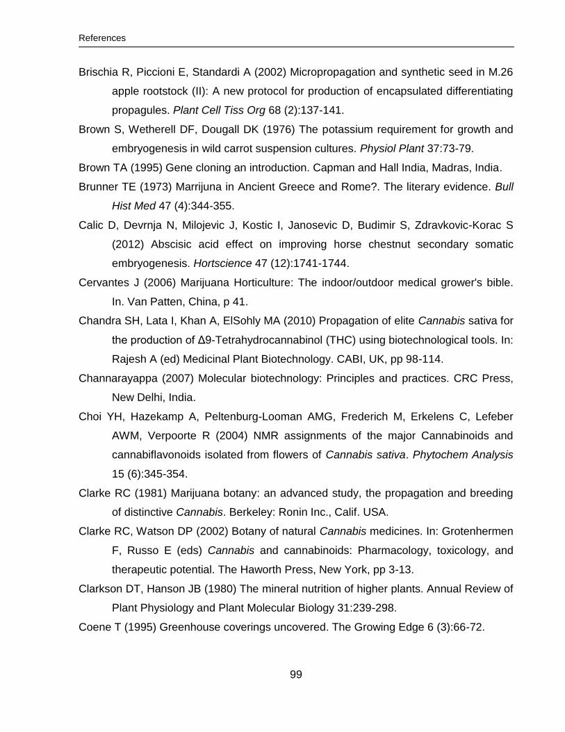
References
99
Brischia R, Piccioni E, Standardi A (2002) Micropropagation and synthetic seed in M.26
apple rootstock (II): A new protocol for production of encapsulated differentiating
propagules. Plant Cell Tiss Org 68 (2):137-141.
Brown S, Wetherell DF, Dougall DK (1976) The potassium requirement for growth and
embryogenesis in wild carrot suspension cultures. Physiol Plant 37:73-79.
Brown TA (1995) Gene cloning an introduction. Capman and Hall India, Madras, India.
Brunner TE (1973) Marrijuna in Ancient Greece and Rome?. The literary evidence. Bull
Hist Med 47 (4):344-355.
Calic D, Devrnja N, Milojevic J, Kostic I, Janosevic D, Budimir S, Zdravkovic-Korac S
(2012) Abscisic acid effect on improving horse chestnut secondary somatic
embryogenesis. Hortscience 47 (12):1741-1744.
Cervantes J (2006) Marijuana Horticulture: The indoor/outdoor medical grower's bible.
In. Van Patten, China, p 41.
Chandra SH, Lata I, Khan A, ElSohly MA (2010) Propagation of elite Cannabis sativa for
the production of Δ9-Tetrahydrocannabinol (THC) using biotechnological tools. In:
Rajesh A (ed) Medicinal Plant Biotechnology. CABI, UK, pp 98-114.
Channarayappa (2007) Molecular biotechnology: Principles and practices. CRC Press,
New Delhi, India.
Choi YH, Hazekamp A, Peltenburg-Looman AMG, Frederich M, Erkelens C, Lefeber
AWM, Verpoorte R (2004) NMR assignments of the major Cannabinoids and
cannabiflavonoids isolated from flowers of Cannabis sativa. Phytochem Analysis
15 (6):345-354.
Clarke RC (1981) Marijuana botany: an advanced study, the propagation and breeding
of distinctive Cannabis. Berkeley: Ronin Inc., Calif. USA.
Clarke RC, Watson DP (2002) Botany of natural Cannabis medicines. In: Grotenhermen
F, Russo E (eds) Cannabis and cannabinoids: Pharmacology, toxicology, and
therapeutic potential. The Haworth Press, New York, pp 3-13.
Clarkson DT, Hanson JB (1980) The mineral nutrition of higher plants. Annual Review of
Plant Physiology and Plant Molecular Biology 31:239-298.
Coene T (1995) Greenhouse coverings uncovered. The Growing Edge 6 (3):66-72.
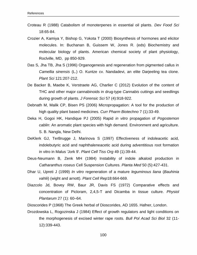
References
100
Croteau R (1988) Catabolism of monoterpenes in essential oil plants. Dev Food Sci
18:65-84.
Crozier A, Kamiya Y, Bishop G, Yokota T (2000) Biosynthesis of hormones and elicitor
molecules. In: Buchanan B, Guissem W, Jones R. (eds) Biochemistry and
molecular biology of plants. American chemical society of plant physiology,
Roclville, MD, pp 850-929.
Das S, Jha TB, Jha S (1996) Organogenesis and regeneration from pigmented callus in
Camellia sinensis (L.) O. Kuntze cv. Nandadevi, an elite Darjeeling tea clone.
Plant Sci 121:207-212.
De Backer B, Maebe K, Verstraete AG, Charlier C (2012) Evolution of the content of
THC and other major cannabinoids in drug-type Cannabis cuttings and seedlings
during growth of plants. J Forensic Sci 57 (4):918-922.
Debnath M, Malik CP, Bisen PS (2006) Micropropagation: A tool for the production of
high quality plant based medicines. Curr Pharm Biotechno 7 (1):33-49.
Deka H, Gogoi HK, Handique PJ (2005) Rapid in vitro propagation of Pogostemon
cablin: An aromatic plant species with high demand. Environment and agriculture.
S. B. Nangla, New Delhi.
DeKlerk GJ, TerBrugge J, Marinova S (1997) Effectiveness of indoleacetic acid,
indolebutyric acid and naphthaleneacetic acid during adventitious root formation
in vitro in Malus 'Jork 9'. Plant Cell Tiss Org 49 (1):39-44.
Deus-Neumann B, Zenk MH (1984) Instability of indole alkaloid production in
Catharanthus roseus Cell Suspension Cultures. Planta Med 50 (5):427-431.
Dhar U, Upreti J (1999) In vitro regeneration of a mature leguminous liana (Bauhinia
vahlii) (wight and arnott). Plant Cell Rep18:664-669.
Diazcolo Jd, Bovey RW, Baur JR, Davis FS (1972) Comparative effects and
concentration of Picloram, 2,4,5-T and Dicamba in tissue culture. Physiol
Plantarum 27 (1): 60–64.
Dioscorides P (1968) The Greek herbal of Dioscorides, AD 1655. Hafner, London.
Drozdowska L, Rogozinska J (1984) Effect of growth regulators and light conditions on
the morphogenesis of excised winter rape roots. Bull Pol Acad Sci Biol 32 (11-
12):339-443.
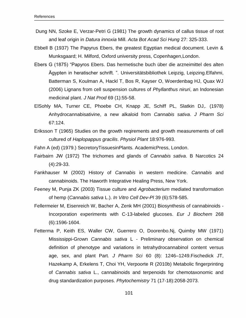
References
101
Dung NN, Szoke E, Verzar-Petri G (1981) The growth dynamics of callus tissue of root
and leaf origin in Datura innoxia Mill. Acta Bot Acad Sci Hung 27: 325-333.
Ebbell B (1937) The Papyrus Ebers, the greatest Egyptian medical document. Levin &
Munksgaard; H. Milford, Oxford university press, Copenhagen,London.
Ebers G (1875) “Papyros Ebers. Das hermetische buch über die arzneimittel des alten
Ägypten in heratischer schrift. ”. Universitätsbibliothek Leipzig, Leipzing.Elfahmi,
Batterman S, Koulman A, Hackl T, Bos R, Kayser O, Woerdenbag HJ, Quax WJ
(2006) Lignans from cell suspension cultures of Phyllanthus niruri, an Indonesian
medicinal plant. J Nat Prod 69 (1):55-58.
ElSohly MA, Turner CE, Phoebe CH, Knapp JE, Schiff PL, Slatkin DJ,. (1978)
Anhydrocannabisativine, a new alkaloid from Cannabis sativa. J Pharm Sci
67:124.
Eriksson T (1965) Studies on the growth reqirements and growth measurements of cell
cultured of Haplopappus gracilis. Physiol Plant 18:976-993.
Fahn A (ed) (1979.) SecretoryTissuesinPlants. AcademicPress, London.
Fairbairn JW (1972) The trichomes and glands of Cannabis sativa. B Narcotics 24
(4):29-33.
Fankhauser M (2002) History of Cannabis in western medicine. Cannabis and
cannabinoids. The Haworth Integrative Healing Press, New York.
Feeney M, Punja ZK (2003) Tissue culture and Agrobacterium mediated transformation
of hemp (Cannabis sativa L.). In Vitro Cell Dev-Pl 39 (6):578-585.
Fellermeier M, Eisenreich W, Bacher A, Zenk MH (2001) Biosynthesis of cannabinoids -
Incorporation experiments with C-13-labeled glucoses. Eur J Biochem 268
(6):1596-1604.
Fetterma P, Keith ES, Waller CW, Guerrero O, Doorenbo.Nj, Quimby MW (1971)
Mississippi-Grown Cannabis sativa L - Preliminary observation on chemical
definition of phenotype and variations in tetrahydrocannabinol content versus
age, sex, and plant Part. J Pharm Sci 60 (8): 1246–1249.Fischedick JT,
Hazekamp A, Erkelens T, Choi YH, Verpoorte R (2010b) Metabolic fingerprinting
of Cannabis sativa L., cannabinoids and terpenoids for chemotaxonomic and
drug standardization purposes. Phytochemistry 71 (17-18):2058-2073.
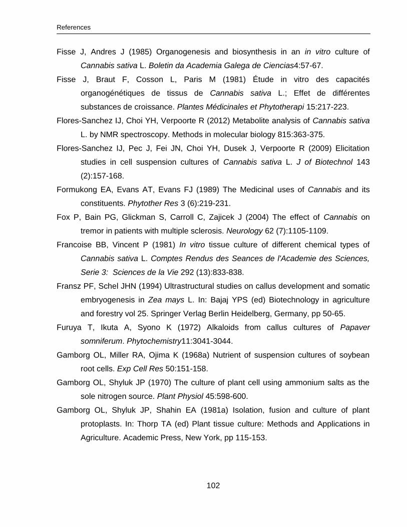
References
102
Fisse J, Andres J (1985) Organogenesis and biosynthesis in an in vitro culture of
Cannabis sativa L. Boletin da Academia Galega de Ciencias4:57-67.
Fisse J, Braut F, Cosson L, Paris M (1981) Étude in vitro des capacités
organogénétiques de tissus de Cannabis sativa L.; Effet de différentes
substances de croissance. Plantes Médicinales et Phytotherapi 15:217-223.
Flores-Sanchez IJ, Choi YH, Verpoorte R (2012) Metabolite analysis of Cannabis sativa
L. by NMR spectroscopy. Methods in molecular biology 815:363-375.
Flores-Sanchez IJ, Pec J, Fei JN, Choi YH, Dusek J, Verpoorte R (2009) Elicitation
studies in cell suspension cultures of Cannabis sativa L. J of Biotechnol 143
(2):157-168.
Formukong EA, Evans AT, Evans FJ (1989) The Medicinal uses of Cannabis and its
constituents. Phytother Res 3 (6):219-231.
Fox P, Bain PG, Glickman S, Carroll C, Zajicek J (2004) The effect of Cannabis on
tremor in patients with multiple sclerosis. Neurology 62 (7):1105-1109.
Francoise BB, Vincent P (1981) In vitro tissue culture of different chemical types of
Cannabis sativa L. Comptes Rendus des Seances de l'Academie des Sciences,
Serie 3: Sciences de la Vie 292 (13):833-838.
Fransz PF, Schel JHN (1994) Ultrastructural studies on callus development and somatic
embryogenesis in Zea mays L. In: Bajaj YPS (ed) Biotechnology in agriculture
and forestry vol 25. Springer Verlag Berlin Heidelberg, Germany, pp 50-65.
Furuya T, Ikuta A, Syono K (1972) Alkaloids from callus cultures of Papaver
somniferum. Phytochemistry11:3041-3044.
Gamborg OL, Miller RA, Ojima K (1968a) Nutrient of suspension cultures of soybean
root cells. Exp Cell Res 50:151-158.
Gamborg OL, Shyluk JP (1970) The culture of plant cell using ammonium salts as the
sole nitrogen source. Plant Physiol 45:598-600.
Gamborg OL, Shyluk JP, Shahin EA (1981a) Isolation, fusion and culture of plant
protoplasts. In: Thorp TA (ed) Plant tissue culture: Methods and Applications in
Agriculture. Academic Press, New York, pp 115-153.
References
103
Gamborg OL, Shyluk JP (1981b) Nutrition, media and characteristics of plant tissue
culture. In: Thorpe T (ed) Plant tissue culture. Methods and applications in
agricultures. Academic Press, New York, pp 21-44.
Gamborg OL (1986) Protoplasts and plant regeneration in culture. In: Demain AL,
Solomon NA (eds) Manual of industrial microbiology and biotechnology. American
Society for Microbiology, Washington, DC, pp 263-273.
Gamborg OL, Phillips GC (1995) Plant cell, tissue and organ culture: Fundamental
methods. Springer-Verlag Berlin Heidelberg, Germany, pp 81-90.
Ganapathi TR, Srinivas L, Suprasanna P, Bapat VA (2001) Regeneration of plants from
alginate-encapsulated somatic embryos of banana cv. Rasthali (Musa spp. AAB
group). In Vitro Cell Dev-Pl 37 (2):178-181.
Gangopadhyay M, Dewanjee S, Chakraborty D, Bhattachary S (2011) Role of
exogenous phytohormones on growth and plumbagin accumulation in Plumbago
indica hairy roots and conservation of elite root clones via synthetic seeds. Ind
Crop Prod 33 (2):445-450.
Gaoni Y, Mechoula.R (1966) Structure of cannabichromene a new active constituent of
Hashish. Chem comm: 20-21.
Gaoni Y, Mechoulam R (1964) Isolation, structure, and partial synthesis of an active
constituent of Hashish. J Am Chem Soc 86 (8):1646-1647.
Gaspar T, Kevers C, Penel C, Greppin H, Reid DM, Thorpe TA (1996) Plant hormones
and plant growth regulators in plant tissue culture. In Vitro Cell Dev-Pl 32 (4):272-
289.
Gelvin, S (2000) Agrobacterium and plant genes involved in T-DNA transfer and
integration. Annu. Rev. Plant Physiol. Plant Mol. Biol. 51:223-256.
Gelvin, S (2003) Agrobacterium-Mediated Plant Transformation: the Biology behind the
“Gene-Jockeying” Tool. Microbiol Mol Biol Rev. 67(1): 16–37.
George L, Eapen S (1994) Organogenesis and embryogenesis from diverse explants in
Pigeonpea (Cajanus cajan L). Plant Cell Rep 13 (7):417-420.
Gershenzon J, Mccaskill D, Rajaonarivony JIM, Mihaliak C, Karp F, Croteau R (1992)
Isolation of secretory cells from plant glandular trichomes and their use in
References
104
biosynthetic studies of monoterpenes and other gland products. Anal Biochem
200 (1):130-138.
Gosal SS, Kang MS (2012) Plant tissue culture and genetic transformation for crop
improvement. In: Narendra T, Sarvajeet SG, Tiburcio AF, Renu T (eds) Improving
crop resistance to abiotic stress. Wiley-VCH, Weinheim, pp 357-387.
Gossop M (1981) The Botany and chemistry of hallucinogens - Schultes, RE, Hofmann,
A. Brit J Addict 76 (3):332-332.
Grout BWW (1995) Introduction to the in vitro preservation of plant cells, tissues and
organs. In: Grout BWW (ed) Genetic preservation of plant cells in vitro. Springer,
Berlin, pp 1-20.
Guha S, Maheshwari SC (1964) In vitro production of embryos from anthers of Datura.
Nature 204: 497.
Guy GW, Stott C (2005) The development of Sativex® - a natural Cannabis-based
medicine. In: Mechoulam R, Bruinrels J (eds) Cannabinoids as therapeutics
milestones in drug therapy. Springer, Berlin, pp 231-263.
Hammond CT, Mahlberg PG (1977) Morphogenesis of capitate glandular hairs of
Cannabis sativa (Cannabaceae). Am J Bot 64 (8):1023-1031.
Hao YJ, Deng XX (2003) Genetically stable regeneration of apple plants from slow
growth. Plant Cell Tiss Org 72 (3):253-260.
Happyana N, Agnolet S, Muntendam R, Van Dam A, Schneider B, Kayser O (2013)
Analysis of cannabinoids in laser-microdissected trichomes of medicinal
Cannabis sativa using LCMS and cryogenic NMR. Phytochemistry 87:51-59.
Hartsel SC, Loh WHT, Robertson LW (1983) Biotransformation of cannabidiol to
Cannabielsoin by suspension cultures of Cannabis sativa and Saccharum
officinarum. Planta Medica 48 (1):17-19.
Hazarika BN (2003) Acclimatisation of tissue-culture plants. Curr sci 85, 1704-1712.
Hazarika BN (2006) Morpho-physiological disorders in in vitro culture of plants, Sci Horti
108, 105-120.
Hazekamp A, Justin TF, D. ML, L. A, R. RL (2010) 3.24 - Chemistry of Cannabis.
Comprehensive. Nat. Prod. II 3:1033-1084.
References
105
Harborne JB, Mabry TJ, Mabry H (eds.) (1975) The flavonoids, part 2, Chapman and
Hall Ltd., Academic Press, Inc., New York, San Francisco.
Heitrich A, Binder M (1982) dentification of (3R, 4R)-delta1(6)- tetrahydrocannabinol as
an isolation artefact of cannabinoid acids formed by callus cultures of Cannabis
sativa L. Experientia 38 (8): 898-899.
Heller R (1953) Recherches sur la nutrition minerale des tissus vegetaux cultives in
vitro.Ann Sci Nat Bot Biol Veg 14:1-223.
Hendriks H, Malingre TM, Batterman S, Bos R (1975) Mono-terpene and sesqui-terpene
hydrocarbons of essential oil of Cannabis sativa. Phytochemistry 14 (3):814-815.
Hillig KW, Mahlberg PG (2004) A chemotaxonomic analysis of cannabinoid variation in
Cannabis (Cannabaceae). Am J Bot 91 (6):966-975.
Holdcroft A, Maze M, Dore C, Tebbs S, Thompson S (2006) A multicenter dose-
escalation study of the analgesic and adverse effects of an oral Cannabis extract
(Cannador) for postoperative pain management. Anesthesiology 104 (5):1040-
1046.
Hosoki T, Mochida M, Sakamoto T, Ohta K (1995) In vitro propagation of white Stokesia
(Stokesia Laevis Greene var alba hort) by Leaf and Root Culture. J Jpn Soc
Hortic Sci 64 (2):375-380.
Hsia CN, Korban SS (1996) Organogenesis and somatic embryogenesis in callus
cultures of Rosa hybrida and Rosa chinensis minima. Plant Cell Tiss Org 44
(1):1-6.
Hu C, Zanettini MH (1995) Embryo culture and embryo Rescue for wide cross hybrids.
In: Gamborg OL, Phillips GC (eds) Plant cell, tissue and organ culture:
fundamental methods, vol 11. Springer, Verlag Berlin Heidelberg, Germany, pp
129 – 141.
Hunez-palenius HG, Ochoa-Alejo N (1999) In vitro mass cultivation of cells and tissues.
In: Paredes-Lopez O (ed) Molecular biotechnology for plant food production.
Technomic, Pennsylvania, p 90.
Inze D, De Veylder L (2006) Cell cycle regulation in plant development. Annu Rev Genet
40:77-105.
References
106
Ikenaga T, Oyama T, Muranaka T (1995) Growth and steroidal saponin production in
hairy root cultures of Solanum aculeatissimum. Plant Cell Rep. 14 (7): 413 – 417.
Itokawa H, Takeya K, Akasu M (1975) Studies on the constituents isolated from the
callus of Cannabis sativa L. Shoyakugaku Zasshi 29 (2):106-112.
Itokawa H, Takeya K, Mihashi S (1977) Biotransformation of cannabinoid precursors and
related alcohols by suspension cultures of callus induced from Cannabis sativa L.
Chem Pharm Bull 25 (8):1941-1946.
Jacob A, Malpathak N (2005) Manipulation of MS and B5 components for enhancement
of growth and solasodine production in hairy root cultures of Solanum khasianum
Clarke. Plant Cell Tiss. Org. Cult. 80: 247 – 257.
John KH, Jocelyn CT, Paul GM (1978) Studies on growth and cannabinoid composition
of callus derived from different strains of Cannabis sativa L. Lloydia 41 (5):453-
462.
Jones JB (ed) (1997) Hydroponics: A practical guide for the soilless grower. St. Lucie
Press, Bocan Raton.
Jones MP, Yi Z, Murch SJ, Saxena PK (2007) Thidiazuron induced regeneration of
Echinacea purpurea L.: micropropagation in solid and liquid culture systems.
Plant Cell Rep 26 (1):13-19.
Kanwar JK, Kumar S (2008) In vitro propagation of Gerbera - A review. Hortic Sci 35
(1):35-44.
Kim ES, Mahlberg PG (1991) Secretory cavity development in glandular trichomes of
Cannabis sativa L (Cannabaceae). Am J Bot 78 (2):220-229.
Kim ES, Mahlberg PG (2003) Secretory vesicle formation in the secretory cavity of
glandular trichomes of Cannabis sativa L. (Cannabaceae). Mol Cells 15 (3):387-
395.
Kim HH, Popova EV, Yi JY, Cho GT, Park SU, Lee SC, Engelmann F (2010)
Cryopreservation of hairy roots of Rubia Akane (Nakai) using a droplet vitrification
procedure. Cryoletters 31 (6):473-484.
Kittipongpatana N, Hock RS, Porter JR (1998) Production of solasodine by hairy root,
callus, and cell suspension cultures of Solanum aviculare Forst. Plant Cell Tiss
Org 52 (3):133-143.
References
107
Klee HJ (1994) Transgenic plants and cytokinin biology. In: Mok WS, Mok MC (eds)
Cytokinins chemistry, activity, and function. CRC Press, Inc., Boca Raton, Florida,
p 289.
Kuberski C, Scheibner H, Steup C, Diettrich B, Luckner M (1984) Embryogenesis and
cardenolide formation in tissue cultures of Digitalis lanata. Phytochemistry 23
(7):1407-1412.
Kubota C, Thomson CA (2006) Controlled environments for production of value-added
food crops with high phytochemical concentrations: Lycopene in tomato as an
example. Hortscience 41 (3):522-525.
Kuijt S, Schnittger A (2007) Plant cell cycle and control, vol 2. Handbook of plant
science. John Wiley & Sons, Ltd, Weinheim, Germany.
Kumar PP, Lakshmanan P, Thorpe TA (1998) Regulation of morphogenesis in plant
tissue culture by ethylene. In Vitro Cell Dev-Pl 34 (2):94-103.
Kummritz S, Haas C, Pavlov AI, Geib D, Ulber R, Bley T, Steingroewer J (2014)
Determination of triterpenic acids and screening for valuable secondary
metabolites in Salvia sp suspension cultures. Nat Prod Commun 9 (1):17-20.
Kut A. S, Bravo J. E, Evans D. A. (1984) Tomato, Chapter 12, In: Ammirato, PV; Evans,
DA; Sharp, WR and Yamada, Y (eds.), Handbock of plant tissue culture, Volume
3, Macmillan Publishing company, New York.
Lambert E, Goossens A, Panis B, Van Labeke MC, Geelen D (2009) Cryopreservation
of hairy root cultures of Maesa lanceolata and Medicago truncatula. Plant Cell
Tiss Org 96 (3):289-296.
Larkin P, Scowcroft W (1981) Somaclonal variation a novel source of variability from cell
cultures for plant improvement. Theoretical and Applied Genetics 60:197-214.
Lata H, Bedir E, Hosick A, Ganzera M, Khan I, Moraes RM (2002) In vitro plant
regeneration from leaf-derived callus of Cimicifuga racemosa. Planta Med 68
(10):912-915.
Lata H, Chandra S, Khan I, ElSohly M (2009a) Thidiazuron induced high-frequency
direct shoot organogenesis of Cannabis sativa L. In Vitro Cell Dev-Pl 45 (1):12-
19.
References
108
Lata H, Chandra S, Khan IA, ElSohly MA (2009b) Propagation through alginate
encapsulation of axillary buds of Cannabis sativa L. an important medicinal plant.
Physiol Mol Biol Plants 15 (1):79-86.
Lata H, Chandra S, Khan IA, ElSohly MA (2010) high frequency plant regeneration from
leaf derived callus of high delta(9)-tetrahydrocannabinol yielding Cannabis sativa
L. Planta Medica 76 (14):1629-1633.
Lata H, Chandra S, Mehmedic Z, Khan IA, ElSohly MA (2012). In vitro germplasm
conservation of high delta (9)-tetrahydrocannabinol yielding elite clones of
Cannabis sativa L. under slow growth conditions. Acta Physiol Plant 34(2):743-
750.
Latta R, Easton B (1975) Seasonal fluctuationsincannabinoid in cannabinoid content of
Kansas marijuana. Econ Bot 29:153-163.
Le Bot J, Adamowicz S, Robin P (1998) Modelling plant nutrition of horticultural crops: a
review. Sci Hortic Amsterdam 74 (1-2):47-82.
Letham DS (1974) Regulators of cell division in plant tissues .20. Cytokinins of coconut
milk. Physiologia Plantarum 32 (1):66-70.
Li HL (1974) Archeological and historical account of Cannabis in China. Econ Bot 28
(4):437-448
Li L, Qu R (2002) In vitro somatic embryogenesis in turf type bermudagrass: roles of
Abscisic Acid and gibberellic acid, and occurrence of secondary somatic
embryogenesis. Plant Breeding 121 (2):155-158.
Liu CF, Zhu JY, Liu ZL, Li L, Pan RC, Jin LH (2002) Exogenous auxin effects on growth
and phenotype of normal and hairy roots of Pueraria lobata (Willd.) Ohwi. Plant
Growth Regul 38 (1):37-43.
Liu S, Zhong JJ (1998) Phosphate effect on production of ginseng saponin and
polysaccharide by cell suspension cultures of Panax ginseng and Panax
quinquefolium. Process Biochem 33 (1):69-74.
Loh WHT, Hartsel SC, Robertson LW (1983) Tissue culture of Cannabis sativa L. and in
vitro biotransformation of phenolics. Zeitschrift fuer Pflanzenphysiologie111
(5):395-400.
Lu CY (1993) The use of thidiazuron in tissue culture. In Vitro Cell Dev-Pl 29P (2):92-96
References
109
Maeda E, Thorpe TA (1979) Shoot histogenesis in tobacco callus cultures. In
Vitro15:415-414-424.
Mahlberg PG, Hemphill JK (1983) Effect of light quality on cannabinoid content of
Cannabis sativa L (Cannabaceae). Bot Gaz 144 (1):43-48.
Malpathak NP, David SB (1992) Stimulation of solasodine production by combining
fungal elicitors and immobilized cell suspension cultures of Solanum surattense
Burm. Biot lett14(10): 965 – 968.
Malingre T, Hendriks H, Batterman S, Bos R, Visser J (1975) The essential oil of
Cannabis sativa. Planta Med 28 (1):56-61.
Mandolino G, Bagatta M, Carboni A, Ranalli P, de Meijer EPM (2003) Qualitative and
quantitative aspects of the inheritance of chemical phenotype in Cannabis. J Ind
Hemp 8:51-72.
Mandolino G, Ranalli P (eds) (1999a) Advances in biotechnological approaches for
hemp breeding and industry. Advances in hemp research. Haworth, New York.
Mandolino G, Ranalli P (1999b) Advances in biotechnological approaches for hemp
breeding and industry. Advances in hemp research. Haworth, New York.
Mangolin CA, Prioli AJ, Machado MFPS (1994) Isozyme patterns, in callus Cultures and
in plants regenerated from calli of Cereus peruvianus (Cactaceae). Biochem
Genet 32 (7-8):237-247.
Mansouri H, Asrar Z, Amarowicz R (2011) The response of terpenoids to exogenous
gibberellic acid in Cannabis sativa L. at vegetative stage. Acta Physiol Plant 33
(4):1085-1091.
McPartland J, Mediavilla V (2002) Noncannabinoid components. In: Grotenhermen F,
Russo E (eds) Cannabis and cannabinoids pharmacology, toxicology, and
therapeutic potential Haworth Press, New York, pp 401-409.
Mechoulam R (1988) Alkaloids in Cannabis sativa L. In: Brossi A (ed) The Alkaloids.
Academic Press, San Diego, pp 77-93.
Meins F (1983) Heritable variation in plant cell culture. Ann Rev Plant Physiol 37:327-
346.
References
110
Minocha SC, Halperin W (1974) Hormones and metabolites which control tracheid
differentiation with or without concomitant effect on growth in cultured tuber tissue
of Helianthus tuberosus L. . Planta Medica116:319-331.
Mironov V, De Veylder L, Van Montagu M, Inze D (1999) Cyclin-dependent kinases and
cell division in plants: The nexus. Plant Cell 11 (4):509-521.
Mohanty S, Panda MK, Subudhi E, Nayak S (2008) Plant regeneration from callus
culture of Curcuma aromatica and in vitro detection of somaclonal variation
through cytophotometric analysis. Biol Plantarum 52 (4):783-786.
Monteuuis O, Bon MC (2000) Influence of auxins and darkness on in vitro rooting of
micropropagated shoots from mature and juvenile Acacia mangium. Plant Cell
Tiss Org 63 (3):173-177.
Morimoto S, Komatsu K, Taura F, Shoyama Y (1998) Purification and characterization of
cannabichromenic acid synthase from Cannabis sativa. Phytochemistry 49
(6):1525-1529.
Morimoto S, Tanaka Y, Sasaki K, Tanaka H, Fukamizu T, Shoyama Y, Taura F (2007)
Identification and characterization of cannabinoids that induce cell death through
mitochondrial permeability transition in Cannabis leaf cells. J Biol Chem 282
(28):20739-20751.
Muniz AW, de Sa EL, Dalagnol GL, Filho JA (2013) Rooting and acclimatization of
micropropagated marubakaido apple rootstock using Adesmia latifolia rhizobia.
Springerplus 2:437.
Muntendam R, Erkelens T, Kayser O (2009) Genetic and metabolic studies of
cannabinoids in standardized medicinal Cannabis sativa. Planta Med 75 (4):417-
417.
Muntendam R, Happyana N, Erkelens T, Bruining F, Kayser O (2012) Time dependent
metabolomics and transcriptional analysis of cannabinoid biosynthesis in
Cannabis sativa var. bedrobinol and bediol grown under standardized condition
and with genetic Homogeneity. Online Int J Med Plant Res 1 (2):31-40.
Murashige T, Skoog F (1962) A revised medium for rapid growth and bioassays with
tobacco tissue cultures. Physiol Plant 15:473-479.
References
111
Mustafa NR, de Winter W, van Iren F, Verpoorte R (2011) Initiation, growth and
cryopreservation of plant cell suspension cultures. Nature Protocols 6 (6):715-
742.
Nabors MW, Daniels A, Nadolny L, Brown C (1975) Sodium-Chloride Tolerant Lines of
Tobacco Cells. Plant Science Letters 4 (3):155-159.
Nahas GG (1982) Hashish in Islam 9th-Century to 18th-Century. B New York Acad Med
58 (9):814-831.
Narayanaswamy S (ed.) (1994) Plant Cell and Tissue Culture, pp. 651, Tate Mc Grew,
Hill Publishing Compamy Limited, New Delhi.
Nam YJ, Tran LS, Kojima M, Sakakibara H, Nishiyama R, Shin R (2012) Regulatory
roles of cytokinins and cytokinin signaling in response to potassium deficiency in
Arabidopsis. PLOS One 7 (10):e47797.
Negrutiu I, Jacobs M (1978) Restoration of the morphogenetic capacity in long term
callus cultures of Arabidopsist haliana. Zeitschrift Fur Pflanzenphysiologie 90
(5):431-441.
Neumann KH, Kumar A, Imani J (1999) Plant cell and tissue culture a tool in
biotechnology: Basics and application. Springer Verlag-Berlin-Heidelberg,
Germany.
Nigra HM, Caso O, Giulietti AM (1985) Produccion de solasodina en callos de Solanum
elaeagnifolium Cav. Resumenes XVI Reunion Nacional de Fisiologia Vegetal. La
Plata, Argentina, pp. 76.
Nigra HM, Caso O, and Giulietti AM (1987) Production of solasodine by calli from
different parts of Solanum elaeagnifolium Cav. Plants. Plant Cell Rep 6: 135 –
137.
Nigra HM, Alvarez MA, Giulietti AM (1989) The influence of auxins, light and cell
differentiation on solasodine production by Solanum elaeagnifolium Cav. Plant
Cell Rep 8: 230 – 233.
Nigra HM, Alvarez MA, Giulietti AM (1989) Effect of carbon and nitrogen sources on
growth and solasodine production in bath suspension cultures of Solanum
elaeagnifolium Cav. Plant Cell Tiss Org 21 (1): 55 – 60.
References
112
Nikolic R, Mitic N, Ninkovic S, Vinterhalter B, Zdravkovic-Korac S, Neskovic M (2010)
Gibberellic acid promotes in vitro regeneration and shoot multiplication in Lotus
corniculatus L. Plant Growth Regul 62 (2):181-188.
Oh SY, Wu CH, Popova E, Hahn EJ, Paek KY (2009) Cryopreservation of Panax
ginseng adventitious roots. J Plant Biol 52 (4):348-354.
Ozias-Akins P, Vasil IK (1985) Nutrition of plant cultures. In: K. VI (ed) Cell culture and
Somatic Cell Genetic of Plants vol 2. Plenum Press, New York, pp 129-147.
Pacifico D, Miselli F, Carboni A, Moschella A, Mandolino G (2008) Time course of
cannabinoid accumulation and chemotype development during the growth of
Cannabis sativa L. Euphytica 160 (2):231-240.
Page JE, Nagel J (2006) Biosynthesis of terpenophenolic metabolites in hop and
Cannabis. J Recent Adv Phytochem 40:179- 210
Paris M, Boucher F, Cosson L (1975) Constituents of Cannabis sativa Pollen. Econ Bot
29 (3):245-253.
Parker D (1994) Lighting for beginners part I: The meaning of light. The Growing Edge 5
(4):53-67.
Pec J, Flores-Sanchez IJ, Choi YH, Verpoorte R (2010) Metabolic analysis of elicited
cell suspension cultures of Cannabis sativa L. by H-1-NMR spectroscopy. Biot
Lett 32 (7):935-941.
Petri G, Oroszlan P, Fridvalszky L (1988) Histochemical detection of hemp trichomes
and their correlation with the THC content. Acta Biol Hung 39 (1):59-73.
Phillips GC, Hubstenberger JE (1995) Basic plant regeneration and plant propagation
procedures. In: Gamborg OL, Phillips GC (eds) Plant Cell, Tissue and Organ
Culture: Fundamental Methods. Springer Verlag Berlin Heidelberg, Germany, pp
45 – 54.
Pierik RLM (1987) In vitro culture of higher plants. Martinus Nijhoff Dordrecht, the
Netherlands.
Pierik RLM (1989) In vitro culture of higher plants. Martinus Nijhoff Dordrecht, Boston,
Lancaster.
References
113
Pontaroli AC, Camadro EL (2005) Somaclonal variation in Asparagus officinalis plants
regenerated by organogenesis from long-term callus cultures. Genet Mol Biol 28
(3):423-430.
Porterfield DM, Musgrave ME (1998) The tropic response of plant root to oxygen:
oxitropism in Pisum sativum L. Planta Med 206:1-6
Potter DJ (2004) Growth and morphology of medicinal Cannabis. In: Guy GW, Whittle
BA, Robson PJ (eds) The medicinal uses of Cannabis and cannabinoids.
Pharmaceutical Press, London, pp 17-54:
Raharjo TJ, Verpoorte R (2004) Methods for the analysis of cannabinoids in biological
materials: a review. Phytochem Analysis 15 (2):79-94.
Rahn EJ, Hohmann AG (2009) Cannabinoids as pharmacotherapies for neuropathic
pain: From the bench to the bedside. Neurotherapeutics 6 (4):713-737.
Ranalli P, Mandolino G (1999) Advances in hemp research In: Ranalli P (ed) Advances
in biotechnological approaches for hemp breeding and industry. Haworth Press,
New York, pp 185-208.
Raquin C (1983) Utilization of different sugars as carbon source for in vitro anther
culture of Petunia. Z Pflanzenphysiol 111:453-457.
Rätsch C (2001) Marijuana medicine: A world tour of the healing and visionary powers of
Cannabis. Healing Arts Press, Rochester, New York.
Razdan RK, Puttick AJ, Zitko BA, Handrick GR (1972) Hashish .6. Conversion of (-)-
delta1(6)-tetrahydrocannabinol to (-)-delta1(7)-tetrahydrocannabinol - stability of
(-)-delta1- and "(-)-delta1(6)-tetrahydrocannabinols. Experientia 28 (2): 121–123.
Reed SM (1996) Haploid cultures. In: Trigiano RN, Gray DJ (eds) Plant tissue culture
concepts and laboratory exercises. CRC Press, Inc., Boca Raton, Florida, USA,
pp 241-246.
Remotti PC, Loffler HJM (1995) Callus induction and plant regeneration from Gladiolus.
Plant Cell Tiss Org 42 (2):171-178.
Richard C, Lescot M, Inze D, De Veylder L (2002) Effect of auxin, cytokinin, and sucrose
on cell cycle gene expression in Arabidopsis thaliana cell suspension cultures.
Plant Cell Tiss Org 69 (2):167-176.
References
114
Riou-Khamlichi C, Huntley R, Jacqmard A, Murray JAH (1999) Cytokinin activation of
Arabidopsis cell division through a D-type cyclin. Science 283 (5407):1541-1544.
Robert DH (ed) (1999) Plant cell culture protocols. Humana Press Inc., Totowa, New
Jersey.
Rosenthal E (ed) (1984) Marijuana growers handbook; indoor/greenhouse. Quick
American publishing company, San Fransisco, California.
Ross SA, ElSohly MA (1995) Constituents of Cannabis sativa L. XXVIII-A review of the
natural constituents: 1980-1994. Zagazig J Pharm Sci 4 (2): 1-10.
Ross SA, ElSohly MA (1996) The volatile oil composition of fresh and air-dried buds of
Cannabis sativa. J Nat Prod 59 (1):49-51.
Ross SA, Mehmedic Z, Murphy TP, Elsohly MA (2000) GC-MS analysis of the total
delta9-THC content of both drug and fiber type Cannabis seeds. J Anal Toxicol
24 (8):715-717.
Ross ΜΚ, Thorpe ΤA, Costerton JW (1973) Ultrastructural aspects of shoot initiation in
tobacco callus cultures. Am J Bot 60 788-795.
Roychoudhury PK, Khaparde SS, Mattiasson B, Kumar A (2006) Synthesis, regulation
and production of urokinase using mammalian cell culture: a comprehensive
review. Biotechnol Adv 24 (5):514-528.
Russo EB (2007) History of Cannabis and its preparations in saga, science, and
sobriquet. Chem Biodivers 4 (8):1614-1648.
Russo EB (2011) Taming THC: potential Cannabis synergy and phytocannabinoid-
terpenoid entourage effects. Br J Pharmacol 163 (7):1344-1364.
Russo EB, Jiang HE, Li X, Sutton A, Carboni A, del Bianco F, Mandolino G, Potter DJ,
Zhao YX, Bera S, Zhang YB, Lu EG, Ferguson DK, Hueber F, Zhao LC, Liu CJ,
Wang YF, Li CS (2008) Phytochemical and genetic analyses of ancient Cannabis
from Central Asia. J Exp Bot 59 (15):4171-4182.
Sakai A, Engelmann F (2007) Vitrification, encapsulation vitrification and droplet
vitrification: a review. Cryo letters 28 (3):151-172.
Salisbury FB (1959) Growth regulators and flowering. II. The cobaltous ion. Plant Physiol
34:598-604.
References
115
Sathyanarayana BN, Verghese DB (2007) Plant tissue culture: practices and new
experimental protocols. I K International Publishing House, New Delhi.
Saunders JW, Tsai CJ, Samper E (1997) Evaluation of alternative nitrogen and carbon
sources for sugarbeet suspension culture platings in development of cell selection
schemes. In Vitro Cell Dev-Pl 33 (1):56-61.
Schaeffer GW (1981) Mutation and cell selection: Increased protein from regenerated
rice tissue cultures. Environ Exp Bot 21:333-345.
Schenk RU, Hildebrandt AC (1972) Medium and techniqes for induction and growth of
monocotyledonous and dicotyledonous plant cell culture. Can J Bot 50:199-204.
Schwarz OJ, Beaty RM (1996) Propagation from non-meristematic tissues
organogenesis. In: Trigiano RN, Gray DJ (eds) Plant Tissue Culture Concepts
and Laboratory Exercises. CRC Press, Inc., Boca Raton, Florida, USA, pp 95 –
105.
Sen MK, Nasrin S, Rahman S, Jamal AH (2014) In vitro callus induction and plantlet
regeneration of Achyranthes aspera L., a high value medicinal plant. Asian Pac J
Trop Biomed 4 (1):40-46.
Sharma N, Chandel KPS, Paul A (1993) In vitro propagation of Gentiana kurroo : An
indigenous threatened plant of medicinal importance. Plant Cell Tiss Org 34
(3):307-309.
Shi HP, Long YY, Sun TS, Eric TPK (2011) Induction of hairy roots and plant
regeneration from the medicinal plant Pogostemon cablin. Plant Cell Tiss Org 107
(2):251-260.
Sicher RC, Bunce JA (1997) Relationship of photosynthetic acclimation to changes of
Rubisco activity in field-grown winter wheat and barley during growth in elevated
carbon dioxide. Photosynth Res 52 (1):27-38.
Sicurani M, Piccioni E, Standardi A (2001) Micropropagation and preparation of
synthetic seed in M.26 apple rootstock I: Attempts towards saving labor in the
production of adventitious shoot tips suitable for encapsulation. Plant Cell Tiss
Org 66 (3):207-216.
References
116
Sierra MI, Vanderheijden R, Vanderleer T, Verpoorte R (1992) Stability of alkaloid
production in cell suspension cultures of Tabernaemontana divaricata during long
term subculture. Plant Cell Tiss Org 28 (1):59-68.
Singh A, Chikara J (2013) Biotechnology micropropagation of Jatropha curcas for large
scale multiplication of quality germplasm. In: Carels N, Sujatha M, Bahadur B
(eds) Jatropha, Challenges for a New Energy Crop: Genetic Improvement and
Biotechnology, vol 2. Springer, New York, pp 503-5011
Sirikantaramas S, Morimoto S, Shoyama Y, Ishikawa Y, Wada Y, Taura F (2004) The
gene controlling marijuana psychoactivity: molecular cloning and heterologous
expression of Delta1-tetrahydrocannabinolic acid synthase from Cannabis sativa
L. J Biol Chem 279 (38):39767-39774.
Sirikantaramas S, Taura F, Tanaka Y, Ishikawa Y, Morimoto S, Shoyama Y (2005)
Tetrahydrocannabinolic acid synthase, the enzyme controlling marijuana
psychoactivity, is secreted into the storage cavity of the glandular trichomes.
Plant Cell Physiol 46
Skoog F, Armstrong DJ (1970) Cytokinins. Annu Rev Plant Physiol Plant 21:359-384.
Skoog F, Miller CO (1957) Chemical regulation of growth and organ formation in plant
tissues cultured in vitro. Symposia Soc Exp Biol 11:118-131.
Slatkin DJ, Doorenbo.Nj, Harris LS, Masoud AN, Quimby MW, Schiff PL (1971)
Chemical constituents of Cannabis sativa L root. J Pharm Sci 60 (12): 1891–
1892.
Slusarkiewicz-Jarzina A, Ponitka A, Kaczmarek Z (2005) Influence of cultivar, explant
source and plant growth regulator on callus induction and plant regeneration of
Cannabis sativa L. Acta Biol Cracov Ser Bot 47 (2):145-151.
Stahl E, Kunde R (1973) Neue Inhaltsstoffe aus dem atherischen 01 von Cannabis
sativa. Tetrahedron Lett 30:2841-2844.
Stamler RT, Fahlman RC, Vigeant H (1985) Illicit traffic and abuse of Cannabis in
Canada. B Narcotics 37 (4):37-49.
Staquet M, Gantt C, Machin D (1978) Effect of a nitrogen analog of
tetrahydrocannabinol on cancer pain. Clin Pharmacol Ther 23 (4):397-401.
References
117
Stearn WT (1970) The Cannabis plant: Botanical characteristics. In: Joyce CRB, Curry
SH (eds) The botany and chemistry of Cannabis. Churchill, London, pp 1-10.
Steiner AA (1961) A universal method for preparing nutrient solutions of certain desired
composition. Plant and Soil 15 (2):134-154.
Stott CG, Guy GW (2004) Cannabinoids for the pharmaceutical industry. Euphytica 140
(1-2):83-93.
Street HE, Schillito RD (1977) Nutrient media for plant organ, tissue and cell culture. In:
Rechcigl M (ed) CRC Handbook in Nutrition and Food vol IV. CRC Press, Boca
Raton, Florida., pp 305-359.
Sudha CG, Seeni S (1996) In vitro propagation of Rauwolfia micrantha, a rare medicinal
plant. Plant Cell Tiss Org 44 (3):243-248.
Surenciski MR, Flachsland EA, Terada G, Mroginski LA, Rey HY (2012)
Cryopreservation of Cyrtopodium hatschbachii Pabst (Orchidaceae) immature
seeds by encapsulation-dehydration. Biocell 36 (1):31-36.
Suthar KC, Ramawat KG, Arya, HC (1980) Effect of growth regulators on diosgenin
production in Balanites aegyptiaca (L.) Dellile, India J Exp Biol18: 1207 – 1208.
Sytsma K, Morawetz J, Pires J, Nepokroeff M, Conti E, Zjhra M, Hall J, Chase M (2002)
Urticalean rosids: Circumscription, rosid ancestry, and phylogenetics based on
rbcL, trnL-F, and ndhF sequences. Am J Bot 89 (9):1531-1546.
Szymanski DB, Lloyd AM, Marks MD (2000) Progress in the molecular genetic analysis
of trichome initiation and morphogenesis in Arabidopsis. Trends Plant Sci 5
(5):214-219.
Taguchi C, Taura F, Tamada T, Shoyama Y, Shoyama Y, Tanaka H, Kuroki R, Morimoto
S (2008) Crystallization and preliminary X-ray diffraction studies of polyketide
synthase-1 (PKS-1) from Cannabis sativa. Acta Crystallogr F 64:217-220.
Takagi H (2000) Recent developments in cryopreservation of shoots apices of tropical
species. In: Engelmann F, Takagi H (eds) Cryopreservation of tropical plant
germplasm. IPGRI, Rome, Italy, pp 178-193.
Taura F, Morimoto S, Shoyama Y (1996) Purification and characterization of
cannabidiolic acid synthase from Cannabis sativa L. Biochemical analysis of a
References
118
novel enzyme that catalyzes the oxidocyclization of cannabigerolic acid to
cannabidiolic acid. J Biol Chem 271 (29):17411-17416.
Taura F, Morimoto S, Shoyama Y, Mechoulam R (1995) First direct evidence for the
mechanism of delta(1)-tetrahydricannabinolic acid biosynthesis. J Am Chem Soc
117 (38):9766-9767.
Taura F, Sirikantaramas S, Shoyama Y, Shoyama Y, Morimoto S (2007)
Phytocannabinoids in Cannabis sativa: Recent studies on biosynthetic enzymes.
Chem Biodivers 4 (8):1649-1663.
Te-chato S, Lim M (1999) Plant regeneration of mangosteen via nodular callus
formation. Plant Cell Tiss Org 59 (2):89-93.
Thakur GA, Duclos RI, Makriyannis A (2005) Natural cannabinoids: Templates for drug
discovery. Life Sci 78 (5):454-466.
Thomas E, Street HE (1972) Factors influencing morphogenesis in excised roots and
suspension cultures of Atropa belladonna. Annals of Botany 36 (145): 239-247.
Trigiano RT, May RA (1996) Direct organogenesis from leaf explants of
Chrysanthemum. In: Trigiano RN, Gray DJ (eds) Plant tissue culture concepts
and laboratory exercises. CRC Press, Inc. , Boca Raton, Florida, USA, pp 105 –
112.
Trofin IG, Dabija G, Vaireanu DI, Filipescu L (2012) The influence of long term storage
conditions on the stability of cannabinoids derived from Cannabis resin. Rev Chim
Bucharest 63 (4):422-427.
Turner CE, Cheng PC, Lewis GS, Russell MH, Sharma GK (1979) Constituents of
Cannabis sativa .15. Botanical and chemical profile of indian variants. Planta Med
37 (3):217-225.
Turner CE, Elsohly MA (1976) Anhydrocannabisativine, a new alkaloid isolated from
Cannabis sativa. Lloydia 39 (6):474-474.
Turner JC, Hemphill JK, Mahlberg PG (1977) Gland distribution and cannabinoid content
in clones of Cannabis sativa L. Am J Bot 64 (6):687-693.
Turner CE, Elsohly MA (1979) Constituents of Cannabis sativa L .16. Possible
decomposition pathway of delta-9-tetrahydrocannabinol to cannabinol. J
Heterocyclic Chem 16 (8):1667-1668.
References
119
Turner CE, Elsohly MA, Boeren EG (1980a) Constituents of Cannabis sativa L .17. A
Review of the natural constituents. J Nat Prod 43 (2):169-234.
Turner JC, Hemphill JK, Mahlberg PG (1980b) Trichomes and cannabinoid content of
developing leaves and bracts of Cannabis sativa L (Cannabaceae). Am J Bot 67
(10):1397-1406.
Turner JC, Hemphill JK, Mahlberg PG (1981) Interrelationships of glandular trichomes
and cannabinoid content .2. Developing vegetative leaves of Cannabis sativa L
(Cannabaceae). B Narcotics 33 (3):63-71.
Umeda M, Shimotohno A, Yamaguchi M (2005) Control of cell division and transcription
by cyclin-dependent kinase-activating kinases in plants. Plant Cell Physiol 46
(9):1437-1442.
Van Os E, Gieling TH, Lieth JH (2008) Technical equipment in soilless production
systems. In: Raviv M, Lieth JH (eds) Soilless culture: Theory and practice.
Elsevier, Amsterdam, pp 157-209.
Van Patten GF (1992) Hydroponics for the rest of us. Growing Edge 3 (3):24––51.
Vanhoenacker G, Van Rompaey P, De Keukeleire D, Sandra P (2002 )
Chemotaxonomic features associated with flavonoids of cannabinoid free
Cannabis (Cannabis sativa subsp sativa L.) in relation to hops (Humulus lupulus
L.). Nat Prod Lett 16 (1):57-63.
Vanhove W, Van Damme P, Meert N (2011) Factors determining yield and quality of
illicit indoor Cannabis (Cannabis spp.) production. Forensic sci int 212 (1-3):158-
163.
Vasil IK (1985b) Somatic embryogenesis and its consequences in the gramineae. In:
Hughes R, Hollaender A (eds) Tissue culture in forestry and agriculture, vol 3.
Plenum Press, New York, pp 31-48.
Vasil IK, Hildebrandt AC (1966) Growth and chlorophyll production in plant callus tissues
grown in vitro. Planta Medica 68:69-82.
Veliky IA, Genest K (1972) Growth and metabolites of Cannabis sativa cell suspension
cultures. Lloydia 35 (4):450-456.
Veress T, Szanto JI, Leisztner L (1990) Determination of cannabinoid acids by high
performance liquid chromatography of their neutral derivatives formed by thermal
References
120
decarboxylation.1. Study of the decarboxylation process in open reactors. J
Chromatogr 520:339-347.
Verma DC, Dougall DK (1977) Influence of carbohydrates on quantitative aspects of
growth and embryo formation in wild carrot suspension cultures. Plant Physiol 59
(1):81-85.
Verpoorte R, Vanderheijden R, Hoge JHC, Tenhoopen HJG (1994) Plant cell
biotechnology for the production of secondary metabolites. Pure Appl Chem 66
(10-11):2307-2310.
Verzar-Petri G, Ladocsy T, Oroszlan P (1982) Differentiation and production of
cannabinoids in tissue cultures of Cannabis sativa L. Acta Botanica Academiae
Scientiarum Hungaricae 28 (1-2):279-290.
Vogelmann AF, Turner JC, Mahlberg PG (1988) Cannabinoid composition in seedlings
compared to adult plants of Cannabis sativa. J Nat Prod 51 (6):1075-1079.
Wahby I, Caba JM, Ligero F (2013) Agrobacterium infection of hemp (Cannabis sativa
L.): establishment of hairy root cultures. J Plant Interact 8 (4):312-320.
Waldo AZ (2006) History of Cannabis as a medicine: a review. Rev Bras Psiquiatr 28
(2):153-157.
Wang PJ, Charles A (1991) Micropropagation through meristem culture. In: Bajaj YPS
(ed) Biotechnology in agriculture and forestry: High tech and micropropagation I,
vol 17. Springer, Verlag Berlin Heidelberg, Germany, pp 32-52.
Wang HM, Zu, YG, Liu H (2007) Efficient rooting and root development after transfer of
regenerated plantlets of Camptotheca acuminata. Eurasian J. For. Res.10, 179-
184.
Wang R, He LS, Xia B, Tong JF, Li N, Peng F (2009) A Micropropagation system for
cloning of Hemp (Cannabis Sativa L.) by shoot tip culture. Pakistan J Bot 41
(2):603-608.
Werker E (2000) Trichome diversity and development. Adv Bot Res 31:1-35.
Whitney PJ (1996) Hormone independent root organ cultures of rye (Secale cereale).
Plant Cell Tiss Org 46 (2):109-115.
Whittle BA (2007) Cannabis as a medicine (benefits and risks associated with
phytomedicines). J Pharm Pharmacol 59:A69-A69.
References
121
Whittle BA, Guy GW (2004) Development of Cannabis based medicines: risk, benefit
and serendipity. In: Guy GW, Robson PJ, Whittle BA (eds) The Medicinal Uses of
Cannabis and Cannabinoids. Pharmaceutical Press, London, pp 427-463.
Wielgus K, Luwanska A, Lassocinski W, Kaczmarek Z (2008) Estimation of Cannabis
sativa L. Tissue Culture Conditions Essential for Callus Induction and Plant
Regeneration. J Nat Fib 5 (3):199-207.
Withers LA (1990) Cryopreservation of plant cells. Methods Mol Biol 6:39-48.
Wochok ZS, Sluis CJ (1980) Gibberellic acid promotes Atrfiplex shoot multiplication and
elongation. Plant Sci Lett 17 (3):363-369.
Wolter KE, Skoog F (1966) Nutritional requirements of Fraxinus callus cultures. Am J
Bot 53:263-269.
Yoshikawa T, Furuya T (1983) Regeneration and in vitro flowering of plants derived from
callus cultures of opium poppy (Papaver somniferum). Experientia 39:1031-1033.
Zafar R, Aeri V, Datta A (1992) Application of plant tissue and cell culture for production
of secondary metabolites. Fitoterapia LXIII: 33-43.
Zhang K, Letham DS, John PCL (1996) Cytokinin controls the cell cycle at mitosis by
stimulating the tyrosine dephosphorylation and activation of p34(cdc2)-like H1
histone kinase. Planta 200 (1):2-12.
Zhang WJ, Bjorn LO (2009) The effect of ultraviolet radiation on the accumulation of
medicinal compounds in plants. Fitoterapia 80 (4):207-218.
Zheng YB, Zhang P, Dixon M (2005) Evaluation of four lamp types for the production of
tomato plants in controlled environments. Hort technology 15 (3):646-652.
Zimmerman RH (1984) Rooting apple cultivars in vitro: Interactions among light,
temperature, phlorglucinol and auxin. Plant Cell Tiss Org 3:301-311.
Zulfiqar F, Ross SA, Slade D, Ahmed SA, Radwan MM, Ali Z, Khan IA, ElSohly MA
(2012) Cannabisol, a novel delta(9)-THC dimer possessing a unique methylene
bridge, isolated from Cannabis sativa. Tetrahedron Lett 53 (28):3560-3562.

Appendix
Appendix I-IV
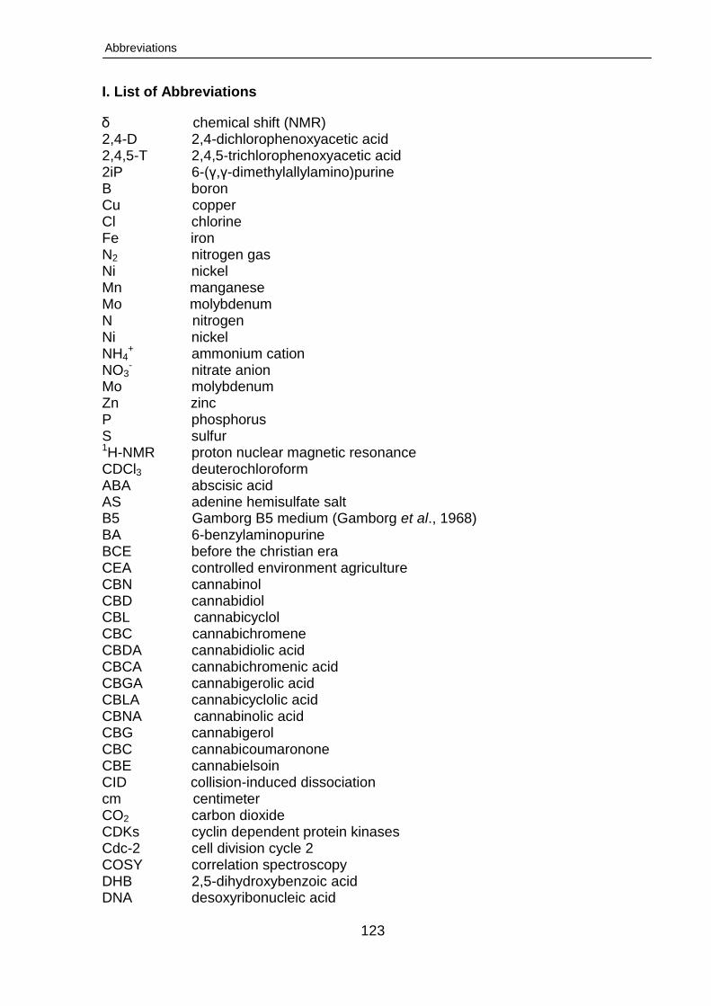
Abbreviations
123
I. List of Abbreviations
δ chemical shift (NMR) 2,4-D 2,4-dichlorophenoxyacetic acid 2,4,5-T 2,4,5-trichlorophenoxyacetic acid 2iP 6-(γ,γ-dimethylallylamino)purine B boron Cu copper Cl chlorine Fe iron N2 nitrogen gas Ni nickel Mn manganese Mo molybdenum N nitrogen Ni nickel NH4
+ ammonium cation NO3
- nitrate anion Mo molybdenum Zn zinc P phosphorus S sulfur 1H-NMR proton nuclear magnetic resonance CDCl3 deuterochloroform ABA abscisic acid AS adenine hemisulfate salt B5 Gamborg B5 medium (Gamborg et al., 1968) BA 6-benzylaminopurine BCE before the christian era CEA controlled environment agriculture CBN cannabinol CBD cannabidiol CBL cannabicyclol CBC cannabichromene CBDA cannabidiolic acid CBCA cannabichromenic acid CBGA cannabigerolic acid CBLA cannabicyclolic acid CBNA cannabinolic acid CBG cannabigerol CBC cannabicoumaronone CBE cannabielsoin CID collision-induced dissociation cm centimeter CO2 carbon dioxide CDKs cyclin dependent protein kinases Cdc-2 cell division cycle 2 COSY correlation spectroscopy DHB 2,5-dihydroxybenzoic acid DNA desoxyribonucleic acid

Abbreviations
124
T-DNA transfer DNA Dicamba 2-methoxy-3,6-dichlorobenzoic acid dr. wt. dry weight ESI electrospray ionization EDTA ethylenediaminetetraacetic acid FID free induction decay fr. wt. fresh weight ft feet GPP geranylpyrophosphate GRIN germplasm resources information network GAs gibberellins GA3 gibberelic acid GLC gas-liquid chromatography HID high intensity discharge HPS high pressure sodium HPLC high performance liquid chromatography HCE hexane crude extract HMBC heteronuclear multiple bond correlation h hour IAA indole-3-acetic acid IBA indole-3-butyric acid IV plantlets plantlets grown in vitro IVH plants ex vitro plants in a growth room IMS ion-mobility spectrometry ISSR inter simple sequence repeat KIN N-furfuryl-7-H-purin-6-amine (kinetin) LS Linsmaier and Skoog medium (Linsmaier and Skoog,1965) LTQ linear ion trap lx lux LC-MS liquid chromatography-mass spectrometry LC-MS/MS liquid chromatography-tandem mass spectrometry L/D light/dark MEP methyl erythritol phosphate MH metal halide MCPA 2-methyl-4-chlorophenoxyacetic acid MS Murashige and Skoog medium (Murashige and Skoog, 1962) MS multiple sclerosis m meter NAA 1-naphthaleneacetic acid OA olivetolic acid PAR photosynthetically active radiation PGRs plant growth regulators Picloram 4-amino-3,5,6-trichloropicolinic acid PCA principal component analysis PLS-DA partial least square-discriminant analysis RAPD random amplified polymorphic DNA RFLP restriction fragment length polymorphism RNA ribonucleic acid rpm revolutions per minute rH relative humidity
Abbreviations
125
ppm parts per million SE standard error SD standard deviation s second SEM scanning electron microscope TOF time of flight TDZ thidiazouron THC tetrahydrocannabinol, Δ-9-tetrahydrocannabinol THCA tetrahydrocannabinolic acid Ti tumour-inducing TMS tetramethylsilane tR retention time UV ultraviolet radiation UV-B ultraviolet radiation (medium wavelength, 315-280 nm) v/v volume to volume vir virulence W watt Z zeatin
Acknowledgements
126
II. Acknowledgements
I am most grateful to my supervisor Prof. Dr. Oliver Kayser, Head of the Technical
Biochemistry Group at the Technical University of Dortmund, for accepting me as a
Ph.D. student and giving me the opportunity to work on a fascinating topic. I also
thank him for his continuous guidance and invaluable advice throughout my scientific
work and for reviewing this manuscript.
I would like to thank the Technical University of Dortmund for financing my work
during the four years of my Ph.D project.
I sincerely thank Dr. Armin Quentmeier for her support in the preparation of my
thesis, for her critical review of this manuscript and helpful comments on my work. I
also express my thanks to Dr. Felix Stehle for valuable tips, new ideas and
stimulating discussions.
This work would not have been possible without the brilliant expertise in mass
spectrometry of Dr. Marc Lamshöft, Dr. Sebastian Zühlke and Selahaddin Sezgin
(Analytical and Environmental Chemistry, Institute of Environmental Research,
Department of Chemistry and Chemical Biology, Technical University of Dortmund),
to whom I am extremely grateful.
For the good atmosphere in the lab, many thanks go to my current and former
colleagues from the TB chair: Torsten, Nizar (a good office colleague), Marcello,
Sara, Evamaria, Parijat, Bastian, Kathleen (always ready to help), Jörg, Fabian,
Bettina and Friederike.
Finally, I would like to thank my parents and my brothers for their love and moral
support over the course of my research away from home. And special thanks to my
wife Rasha for caring, understanding and supporting me during our stay in Germany.
I highly appreciate your skills in tending to our loving son (Omar) and daughter
(Qamar) and managing my schedules very efficiently.
Sayed
Curriculum Vitae
127
III. Curriculum Vitae
Personal information
Name Sayed Hussein Farag Hussein
Date of birth 7th February, 1976
Nationality Egyptian
Family status Married, two children
Education
2010-2014 Doctoral studies and dissertation at the Laboratory of Technical
Biochemistry, Technical University of Dortmund, Germany, under
the supervision of Prof. Dr. Oliver Kayser
2009-2010 Perpetration for the ParOwn Scholarship at TU Dortmund
2005-2008 Master studies in phytochemistry (M. Sc.) at the Department of
Phytochemistry, National Research Centre (NRC), Cairo, Egipt,
under the supervision of Prof. Dr. Medhat M. Seif-El-Nasr
2003-2005 Postgraduate studies, Faculty of Science, Al-Azhar University,
Cairo, Egypt
1999-2003 Bachelor studies in Botany (B. Sc.) Faculty of Science, Al-Azhar
University, Cairo, Egypt
1996-1998 Secondary–school education, Foad Al Awal Institute, Assuit, Egypt
Professional experience
since 2010 Research assistant at the Department of Phytochemistry, National
Research Centre (NRC), Cairo, Egypt
List of Publications
128
IV. List of Publications
Peer Reviewed Articles
1. Sayed Farag, Sebastian Zühlke, Michael Spiteller, Oliver Kayser. In vitro
micropropagation of leaf-derived calli from Cannabis sativa L.: Cannabinoid content
of the regenerated plants, 2014 (submitted to In vitro cellular and developmental
biology – plant).
2. Sayed Farag, Oliver Kayser. Cannabinoids production by hairy root cultures of
Cannabis sativa L. 2014 (submitted to American journal of plant science).
3. Sayed Farag, Nizar Happyana, Marc Lamshöft, Michael Spiteller, Oliver Kayser
“Characterization of cannabinoids biosynthesis in Cannabis sativa L. cell
suspensioncultures”. 2014 (submitted to Plant cell reports).
4. Sayed Farag, Oliver Kayser Encapsulation-dehydration of in vitro hairy root cultures
of Cannabis sativa L.: Towards a practical conservation protocol”.2014 (submitted to
Journal of Horticultural Science).
Book Chapters
1. Book Chapter 10:Sayed Farag, Oliver Kayser (2015) Cultivation and Breeding of
Cannabis sativa L. for Preparation of Standardized Extracts for Medicinal Purposes.
In: Máthé Ákos (ed) Medicinal and Aromatic Plants of the World, Springer (inpress)
2. Book Chapter (II):Sayed Farag, Oliver Kayser (2015).The Cannabis Plant:
Botanical Aspects. In: Victor Preedy (ed) The Comprehensive Handbook of Cannabis
Related Pathology.Elsevier. (inpress).
Poster
Sayed Farag, Marc Lamshöft, Michael Spiteller, Oliver Kayser (2013) LCMS Spectral
Evidence of the Occurrence of Cannabinoid in Cannabis sativa Cell Cultures. Planta
Med 2013; 79 - PM2 DOI: 10.1055/s-0033-1352335. 61st International Congress and
Annual Meeting of the Society for Medicinal Plant and Natural Product Research (GA),
1st – 5th of September, 2013, Münster.