Embed Size (px)
Citation preview
Cardiovascular Diseases:Case PresentationsAngela Sharkey, MD FAAPProfessor of Pediatrics
Disclosures• I have no financial relationships with the manufacturer of any commercial product(s) and/or provider of commercial services discussed in this CME activity.
• I am not on any Speakers Bureau• I do not intend to discuss unapproved uses of commercial products

Objectives• Recognize and manage atrial and ventricular arrhythmias.
• Distinguish cardiac etiologies for chest pain and syncope.
• Develop an appreciation for the impact that childhood obesity has had on the incidence of childhood hypertension and hyperlipidemia

Case #1• 2 week old infant comes to your office for well child check. Benign history, feeding well with good weight gain. On exam, occasional irregular heart beats are noted.
• What is the differential diagnosis?• Important question: what is the heart rate?
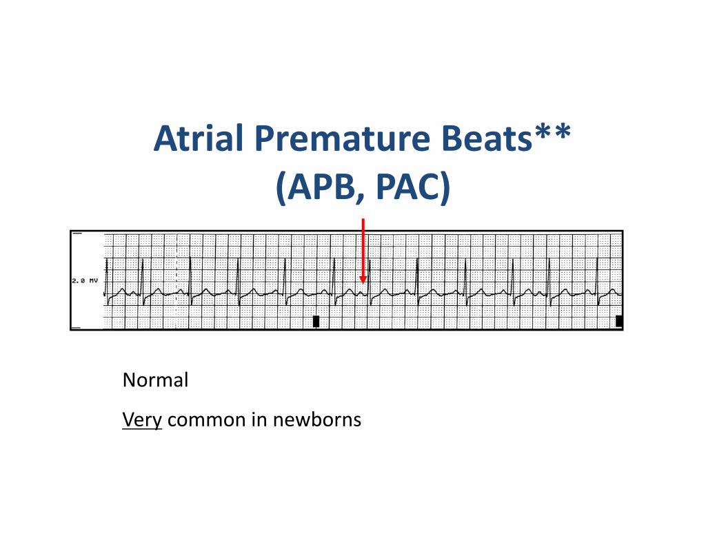
Atrial Premature Beats**(APB, PAC)
Normal
Very common in newborns

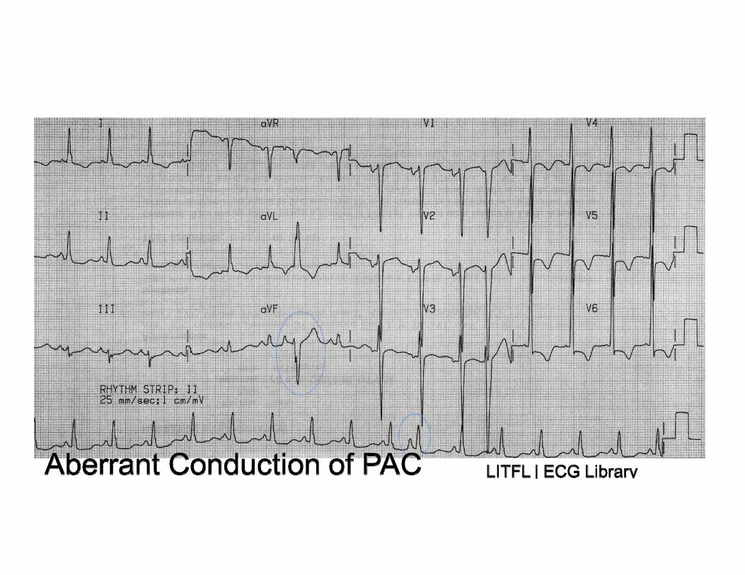
Aberrant Conduction of PAC
Look for some variation of a P wave preceding the beat.
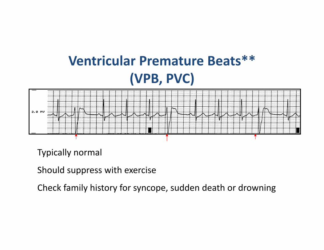
Ventricular Premature Beats**(VPB, PVC)
Typically normal
Should suppress with exercise
Check family history for syncope, sudden death or drowning
Same Baby, but…• On auscultation the heart rate is noted to be slow…..
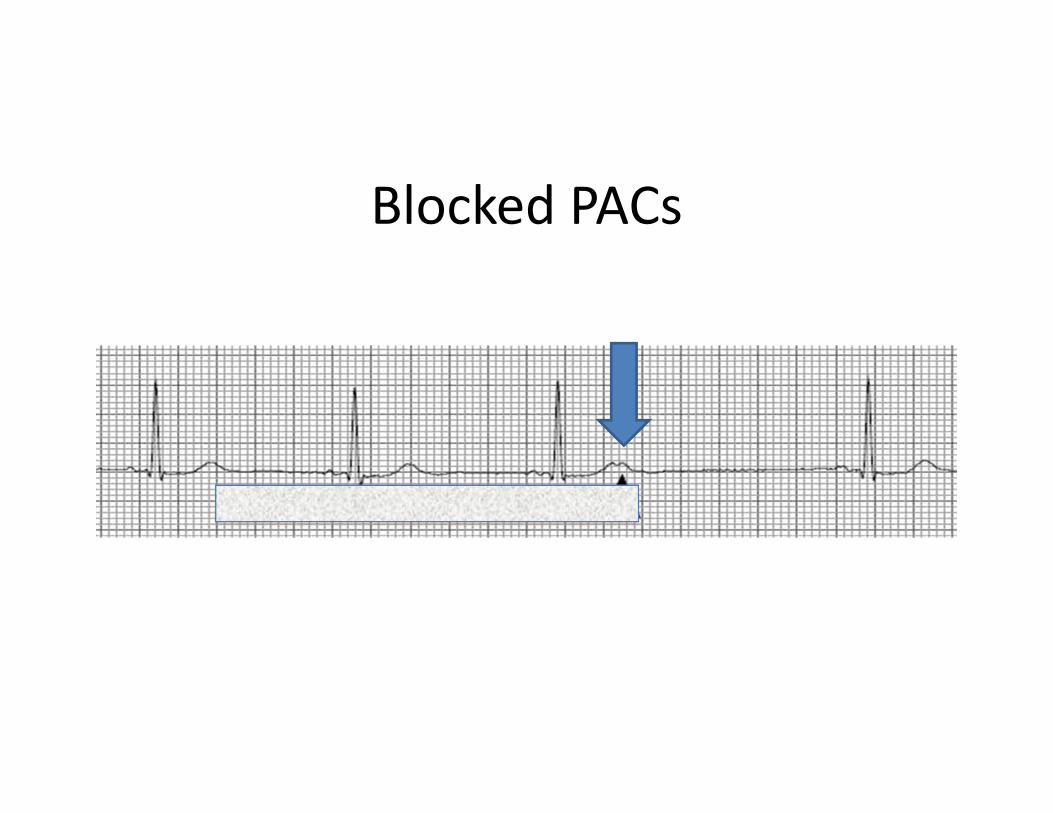
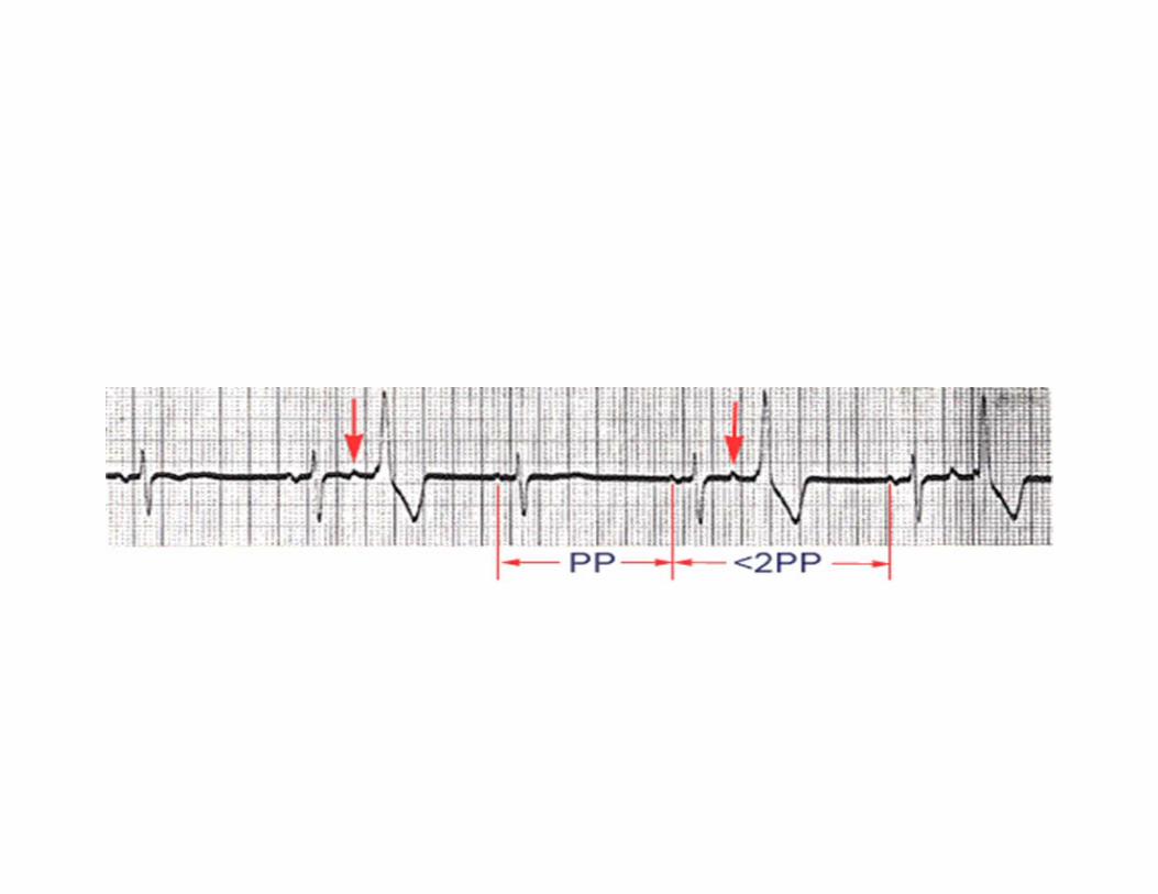
Blocked PACs

What is the Rhythm?
Can be seen in infants of mothers with anti-SSA and antiRo antibodiesCan be associated with structural heart disease
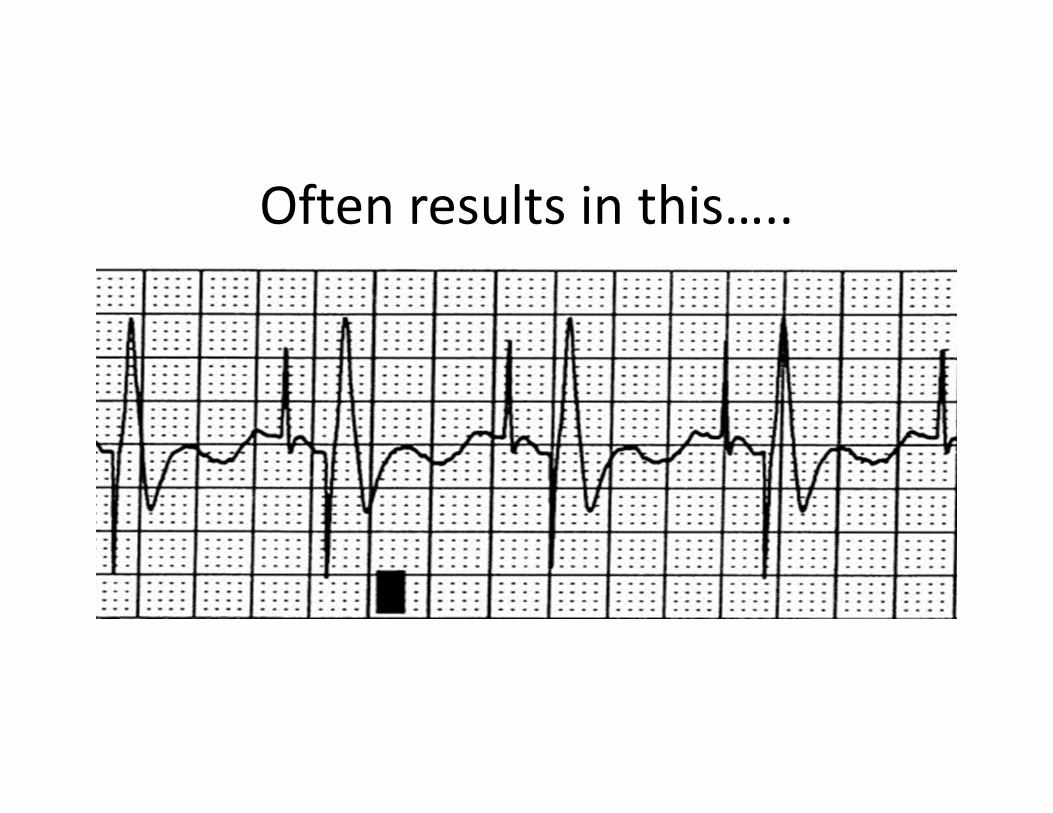
Often results in this…..
Another 2 week old• This time noted to be feeding poorly, fussy, sleeping more. Exam with tachycardia and tachypnea, hepatomegaly, pulse too rapid to count……
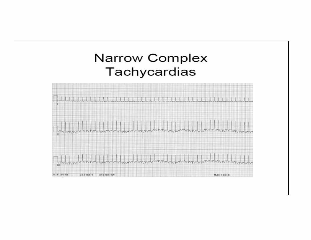
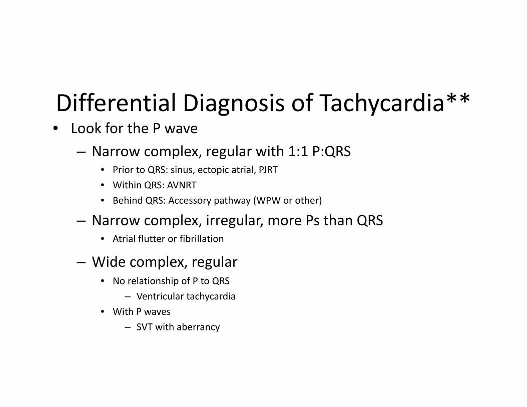
Differential Diagnosis of Tachycardia**• Look for the P wave
– Narrow complex, regular with 1:1 P:QRS• Prior to QRS: sinus, ectopic atrial, PJRT• Within QRS: AVNRT• Behind QRS: Accessory pathway (WPW or other)
– Narrow complex, irregular, more Ps than QRS• Atrial flutter or fibrillation
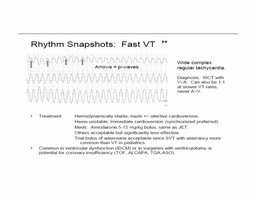
– Wide complex, regular• No relationship of P to QRS
– Ventricular tachycardia• With P waves
– SVT with aberrancy
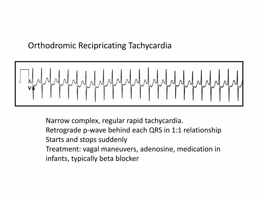
Narrow complex, regular rapid tachycardia.Retrograde p‐wave behind each QRS in 1:1 relationshipStarts and stops suddenlyTreatment: vagal maneuvers, adenosine, medication in infants, typically beta blocker
Orthodromic Recipricating Tachycardia

Adenosine• Intravenous, fast push• Dosing based on patient weight
– First dose: 100mcg/kg (max dose 6mg)– Second dose: 200mcg/kg (max dose 12mg) PALS protocol
• Transiently blocks conduction through AV node. If you are not seeing a pause in ECG tracing, you are not getting the drug to the patient or not enough drug
• Will terminate reentrant SVT or will slow ventricular response in atrial flutter
**
Bibliography for Rhythm
• How to Read Pediatric ECG’s by Myung K. Park
• Pediatric Advanced Life Support Circulation 2010; 122[Suppl3]S876‐S908
Case #2• 14 year old female presents for school physical. Has gained quite a bit of weight since evaluation last year. Not participating in organized sports. BP 150/90, HR 80. No murmur on exam.
• What additional historical information would help?• What other exam features?• What is your differential?
AAP HTN Guidelines• Hypertension is more common in preschoolers who are overweight and obese and who had low activity
• 60 min/day of moderate to vigorous activity
JPeds July 2015 167 pg 92 and 98
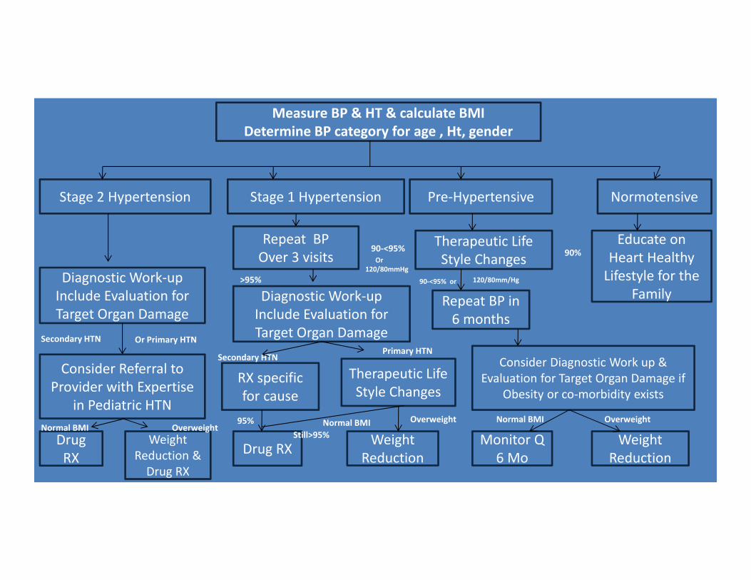
Measure BP & HT & calculate BMIDetermine BP category for age , Ht, gender
Stage 2 Hypertension Stage 1 Hypertension Pre‐Hypertensive Normotensive
Repeat BPOver 3 visits
Therapeutic Life Style Changes
Educate on Heart Healthy Lifestyle for the
FamilyDiagnostic Work‐up Include Evaluation for Target Organ Damage
Diagnostic Work‐up Include Evaluation for Target Organ Damage
Repeat BP in 6 months
Consider Referral to Provider with Expertise
in Pediatric HTN
RX specific for cause
Therapeutic Life Style Changes
Consider Diagnostic Work up & Evaluation for Target Organ Damage if
Obesity or co‐morbidity exists
Drug RX
Weight Reduction & Drug RX
Drug RX Weight Reduction
Monitor Q 6 Mo
Weight Reduction
OverweightNormal BMIOverweightNormal BMIStill>95%
95%OverweightNormal BMI
Secondary HTN Or Primary HTN
Secondary HTNPrimary HTN
>95%
90‐<95%Or
120/80mmHg
90‐<95% or 120/80mm/Hg
90%
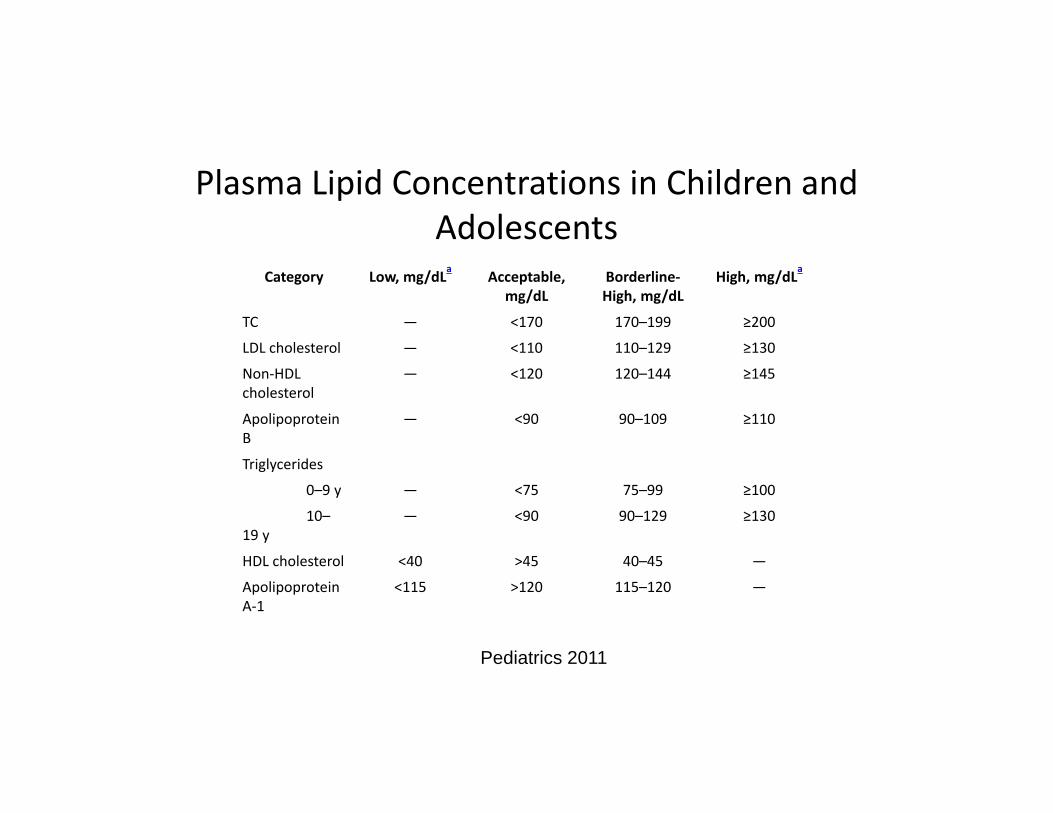
Plasma Lipid Concentrations in Children and Adolescents
Category Low, mg/dLa Acceptable, mg/dL
Borderline‐High, mg/dL
High, mg/dLa
TC — <170 170–199 ≥200
LDL cholesterol — <110 110–129 ≥130
Non‐HDL cholesterol
— <120 120–144 ≥145
Apolipoprotein B
— <90 90–109 ≥110
Triglycerides
0–9 y — <75 75–99 ≥100
10–19 y
— <90 90–129 ≥130
HDL cholesterol <40 >45 40–45 —
Apolipoprotein A‐1
<115 >120 115–120 —
Pediatrics 2011

Bibliography• Pediatrics 2004; 114(2):555‐575, National High Blood Pressure Education Program
(NHBPEP)• Expert Panel on Integrated Guidelines for Cardiovascular Health and Risk
Reduction in Children and Adolescents: Summary Report. Pediatrics 2011;128(6). • http://www.nhlbi.nih.gov/health/prof/heart/hbp/hbp_ped.pdf• http://www.nhlbi.nih.gov/guidelines/hypertension/child_tbl.htm• Jolliffe C, Janssen I. Distribution of lipoproteins by age and gender in adolescents.
Circulation. 2006;114:1056‐1062.
Case #3 ‐ Child with Chest Pain• 6 year old male presents with episodes of chest pain. Parent reports ‘I can see his heart beating through his shirt when he complains about this pain’.
• What additional history is important?• What physical findings?• What testing if any is indicated?
Causes of Chest Pain• Most of pediatric and adolescent chest pain in noncardiac
• 45% of cases of chest pain the etiology will not be determined
• Supratentorial component cannot be overlooked– Referrals increase dramatically when an athlete dies suddenly
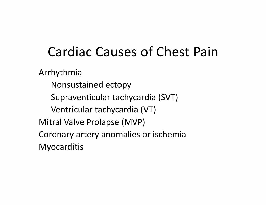
Cardiac Causes of Chest PainArrhythmia
Nonsustained ectopySupraventicular tachycardia (SVT)Ventricular tachycardia (VT)
Mitral Valve Prolapse (MVP)Coronary artery anomalies or ischemiaMyocarditis
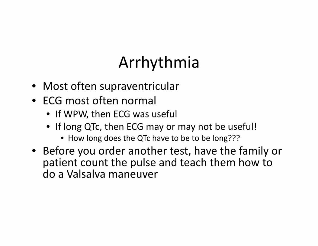
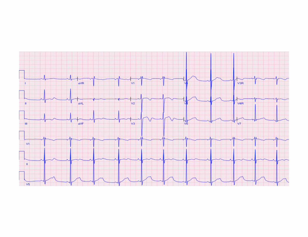
Arrhythmia• Most often supraventricular• ECG most often normal
• If WPW, then ECG was useful• If long QTc, then ECG may or may not be useful!
• How long does the QTc have to be to be long???• Before you order another test, have the family or patient count the pulse and teach them how to do a Valsalva maneuver
Case #4 – Adolescent Chest Pain• 14 year old female presents with episodes of chest pain which is left sided.
• What additional history is important?• What physical findings?• What testing if any is needed?
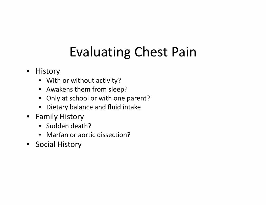
Evaluating Chest Pain• History
• With or without activity?• Awakens them from sleep?• Only at school or with one parent?• Dietary balance and fluid intake
• Family History• Sudden death? • Marfan or aortic dissection?
• Social History

Evaluating Chest Pain ‐ Exam• Vital signs• BMI• Reproducible chest wall tenderness• Presence of murmur• Dysmorphic characteristics
Noncardiac Causes of Chest Pain• Idiopathic
• Some 25‐45% of cases of chest pain
• Musculoskeletal• Trauma• Over exertion• Costochondritis• Precordial catch
Noncardiac Causes Of Chest Pain• Pulmonary
• Asthma• Exercise Induced Bronchospasm• Vocal Cord Dysfunction• Pneumothorax
• Rheumatologic• Pleural or pericardial effusion
• SLE (lupus)• Juvenile Rheumatoid Arthritis
Noncardiac Causes of Chest Pain• GI
• Reflux• Esophagitis secondary to medication
• Minocycline• Nonsteroidal anti‐inflammatory agents
Case #5 – Chest Pain• 14 year old female presents with episodes of chest pain which is left sided. The pain occurs only with activity.
• What additional history is important?• What physical findings?• What testing if any is needed?
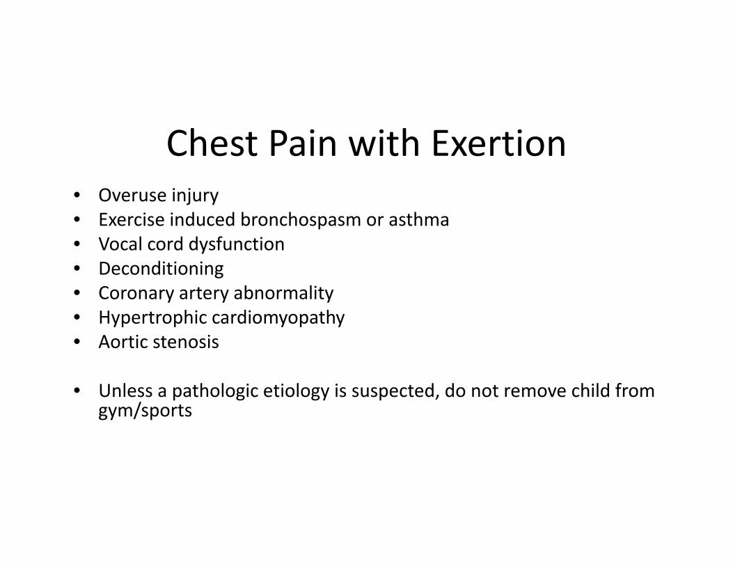
Chest Pain with Exertion• Overuse injury• Exercise induced bronchospasm or asthma• Vocal cord dysfunction• Deconditioning• Coronary artery abnormality• Hypertrophic cardiomyopathy• Aortic stenosis
• Unless a pathologic etiology is suspected, do not remove child from gym/sports

So do you need any testing???• Pain that interferes with sleep, • is precipitated by exercise, or• is associated with
• dizziness, • palpitations, • syncope, or • shortness of breath
• Electrocardiogram and/or chest radiograph

Myocarditis• Inflammation of the myocytes resulting from viral illness• Laboratory testing
• ECG• Troponin• BNP
• Diagnosis by• MRI• Endomyocardial biopsy

Pathogens Known to Cause Myocarditis• Varicella • Parvovirus B‐19 • Adenovirus • Streptococcus • Influenza• Epstein‐Barr virus• Coxsackievirus and echovirus• Chlamydia• HIV
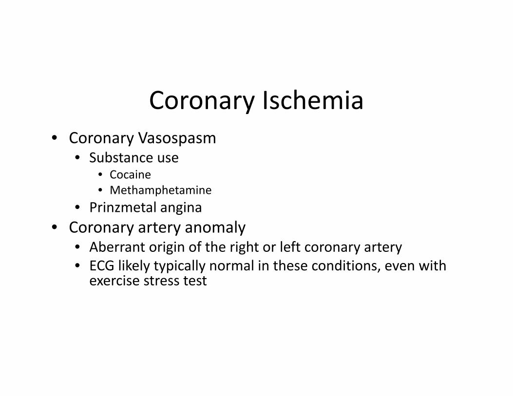
Coronary Ischemia• Coronary Vasospasm
• Substance use• Cocaine• Methamphetamine
• Prinzmetal angina• Coronary artery anomaly
• Aberrant origin of the right or left coronary artery• ECG likely typically normal in these conditions, even with exercise stress test

Case #6 – The Weak and Dizzy• 16 year old female complains of dizziness and ‘curtain like’ visual change when standing up too fast after watching a movie or sitting in class.
• What additional history is important?• What physical findings?• What testing if any is needed?

Evaluation**o History
o Prodrome: Visual changes, ringing in ears, sweating, nausea
o Dietary history – ask what they are drinking!o Family History
o Sudden death, arrhythmia, medications in houseo Physical Exam
o Includes orthostatics and assessment for murmuro +/‐ ECG; beta HCG, CBC

What about the Dizzy?• Orthostasis• Autonomic Instability
• How to treat?• How to monitor hydration?How to find time in the day to drink and use the bathroom!!!!

“It’s all about the letter P”o Dehydration – What color is your urine?
o Assess caffeine intakeo Assess beverage consumptiono Urine coloro Assess use of bathroom at school
o Treatment – Hydration and increase salt intakeo Pretzels, popcorn, baked potato chips, pickles, pepperonio Water bottle – may need a note for school

POTSo Postural Orthostatic Tachycardia Syndrome (POTS)o Chronic day to day symptoms of orthostatic incompetence associated with excessive tachycardia
o Defined by meeting criteria for Orthostatic Incompetence
o In the average adolescent, an increase in heart rate of >35 beats/min or heart rate >120‐130 bpm after two minutes standing
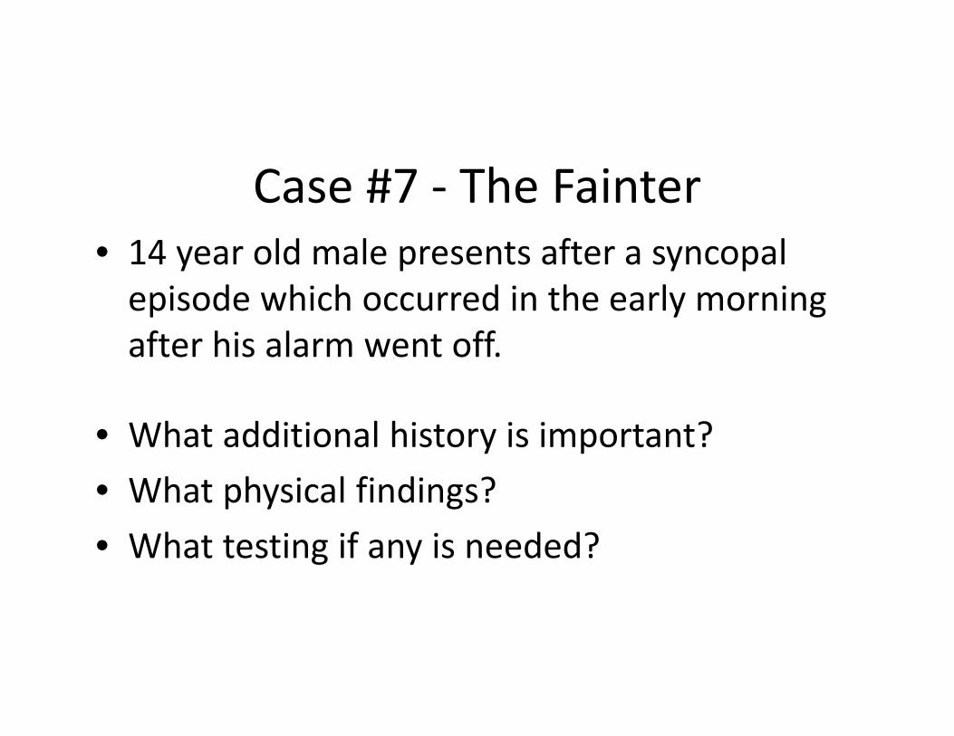
Case #7 ‐ The Fainter• 14 year old male presents after a syncopal episode which occurred in the early morning after his alarm went off.
• What additional history is important?• What physical findings?• What testing if any is needed?

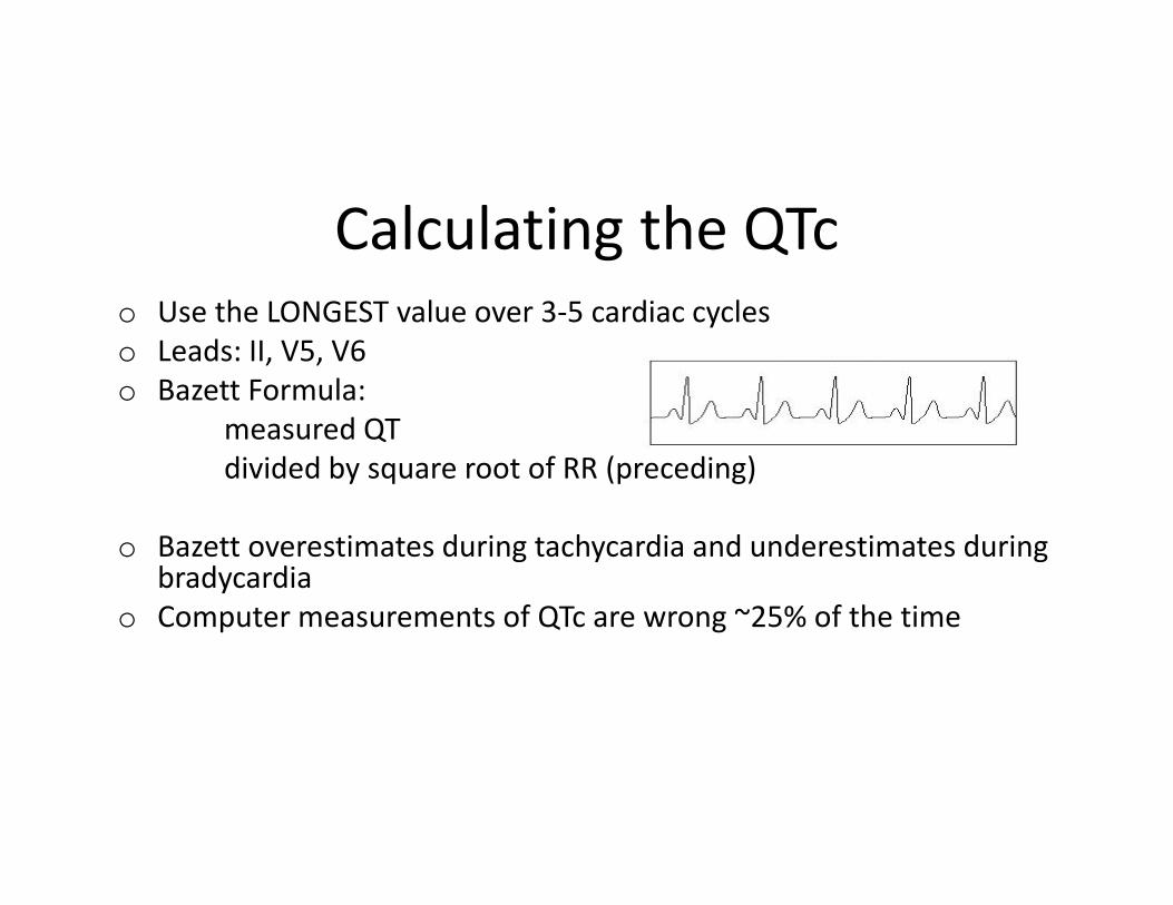
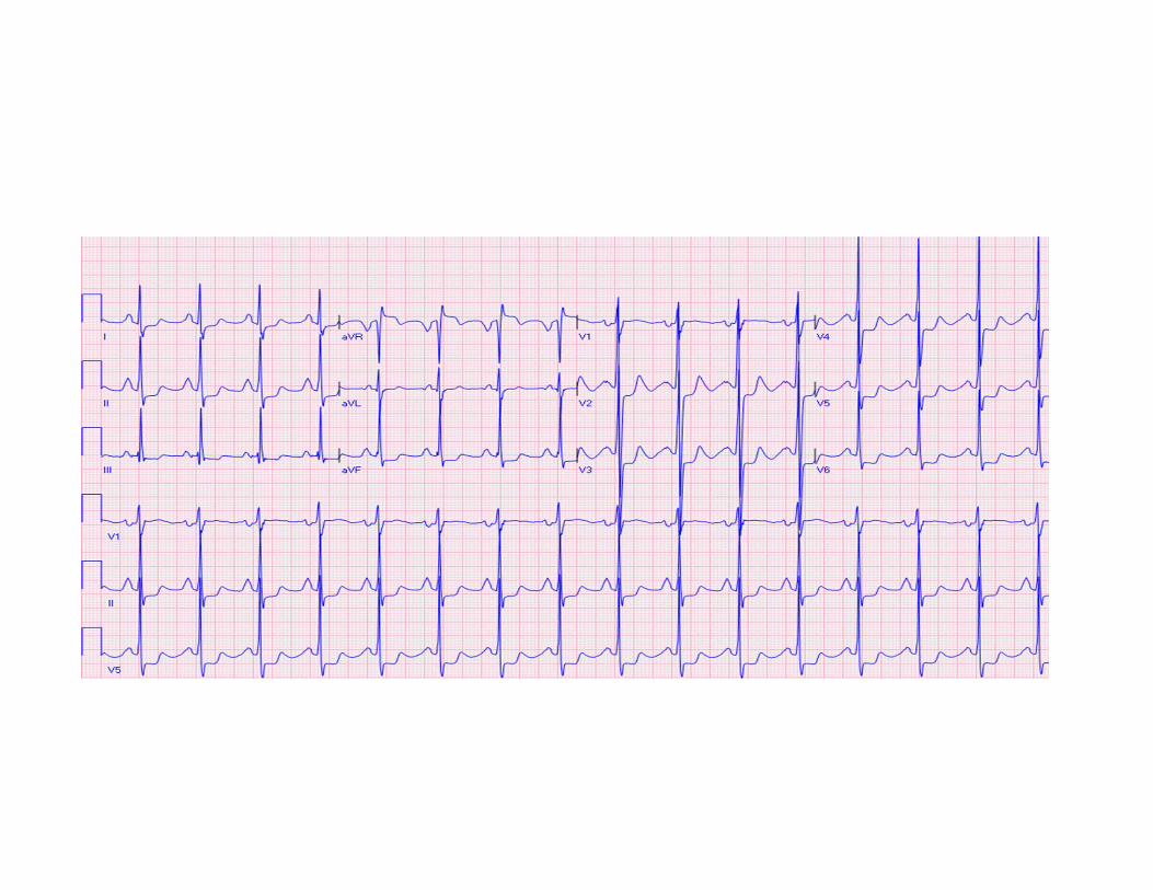
Calculating the QTco Use the LONGEST value over 3‐5 cardiac cycleso Leads: II, V5, V6o Bazett Formula:
measured QTdivided by square root of RR (preceding)
o Bazett overestimates during tachycardia and underestimates during bradycardia
o Computer measurements of QTc are wrong ~25% of the time
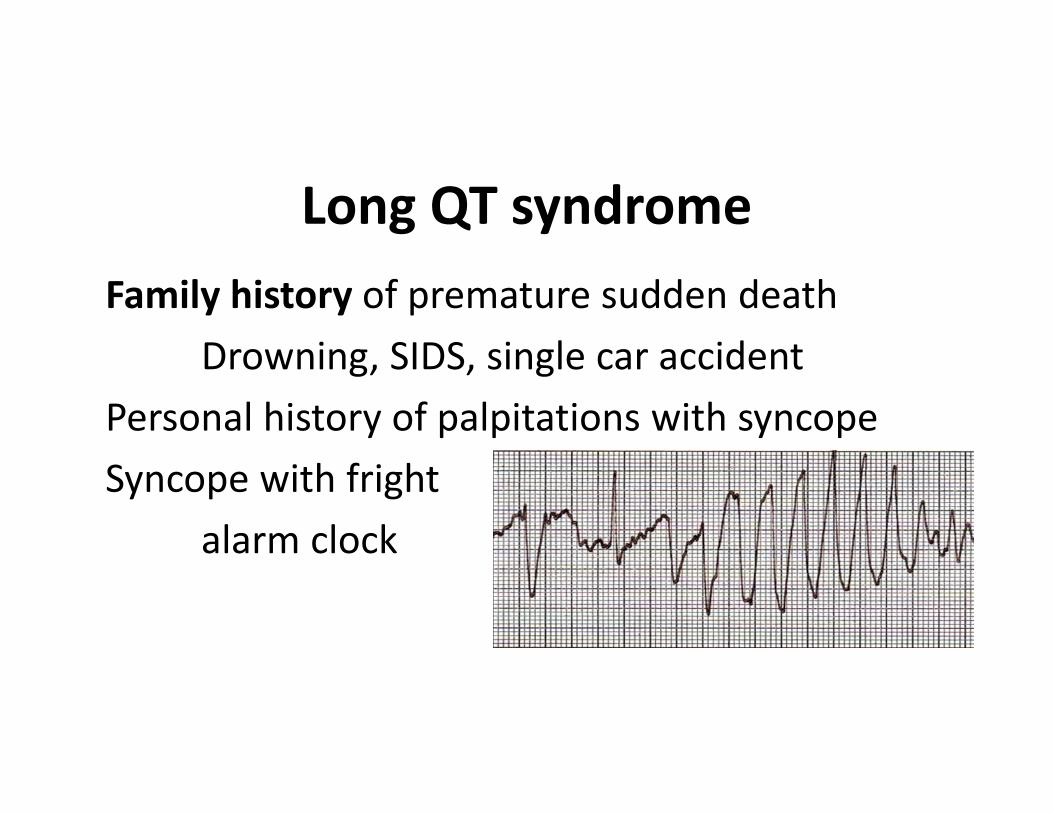
Long QT syndromeFamily history of premature sudden death
Drowning, SIDS, single car accidentPersonal history of palpitations with syncopeSyncope with fright
alarm clock
Neurally‐mediated syncope (Simple faint)
• Peak incidence 15 yrs• Female:Male 2:1• Seen in 15‐25% of all
normal adolescents• Triggered by:
– Prolonged standing– Dehydration, hot days– Intercurrent illness– Disgust, sight of blood
• Micturation syncope– Or in shower, while washing or
combing hair
• May occur post‐exercise, NOT during exercise
• May occur during or after a meal
Classification of Syncopeo Neurally mediated
o Vasovagalo Carotido Situationalo Atypical
o Orthostatic hypotensiono Primary autonomic failureo Secondary autonomic failureo Drug‐inducedo Volume depletion
• The good news, treatment is pretty much the same regardless of the exact diagnosis – volume repletion– Fludrocortisone 0.1mg daily– beta blockers (Atenolol)
o Unless, Cardiovascularo Arrhythmiao Structural cardiac disease
Non‐cardiovascular causes:• Breath‐holding spells• Hypoglycemia• Hyperventilation• Vertigo• Seizures• Basilar artery migraine• Hysterical syncope
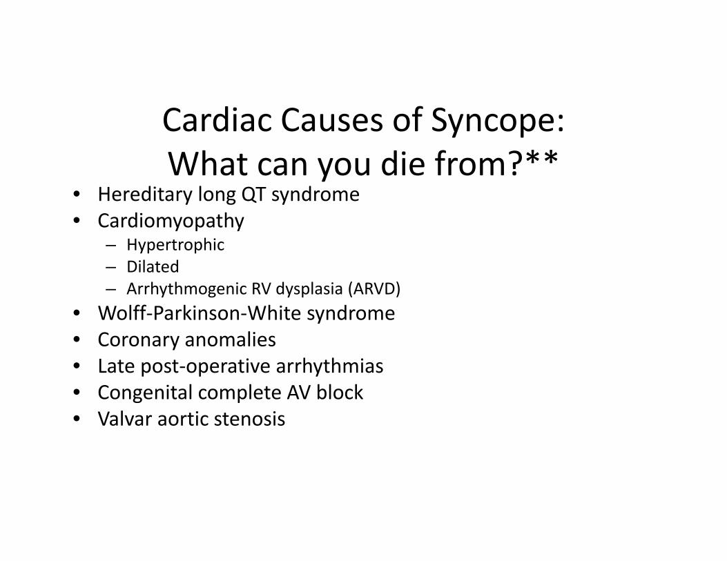
Cardiac Causes of Syncope: What can you die from?**
• Hereditary long QT syndrome• Cardiomyopathy
– Hypertrophic– Dilated– Arrhythmogenic RV dysplasia (ARVD)
• Wolff‐Parkinson‐White syndrome• Coronary anomalies• Late post‐operative arrhythmias• Congenital complete AV block• Valvar aortic stenosis
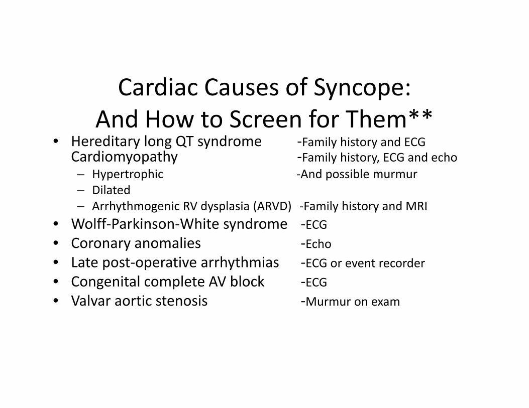
Cardiac Causes of Syncope:And How to Screen for Them**
• Hereditary long QT syndrome ‐Family history and ECG Cardiomyopathy ‐Family history, ECG and echo– Hypertrophic ‐And possible murmur– Dilated– Arrhythmogenic RV dysplasia (ARVD) ‐Family history and MRI
• Wolff‐Parkinson‐White syndrome ‐ECG• Coronary anomalies ‐Echo• Late post‐operative arrhythmias ‐ECG or event recorder• Congenital complete AV block ‐ECG• Valvar aortic stenosis ‐Murmur on exam
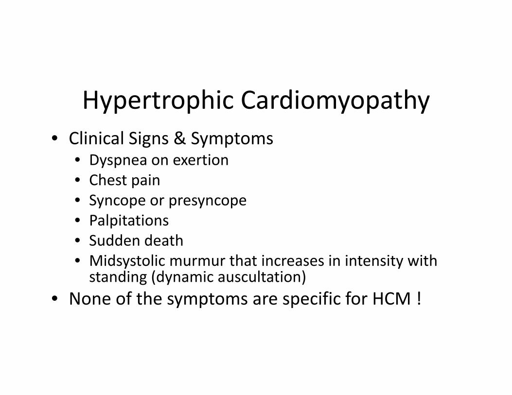
Hypertrophic Cardiomyopathy• Clinical Signs & Symptoms
• Dyspnea on exertion• Chest pain• Syncope or presyncope• Palpitations• Sudden death• Midsystolic murmur that increases in intensity with standing (dynamic auscultation)
• None of the symptoms are specific for HCM !

Hypertrophic Cardiomyopathy• Autosomal dominant, but many affected relatives have much milder disease
• Clear age‐related progression• Repeated screening of family members is indicated, more often in adolescence
• +/‐ Genetic testing
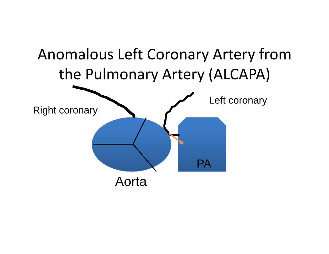
Anomalous Left Coronary Artery from the Pulmonary Artery (ALCAPA)
PAAorta
Right coronaryLeft coronary
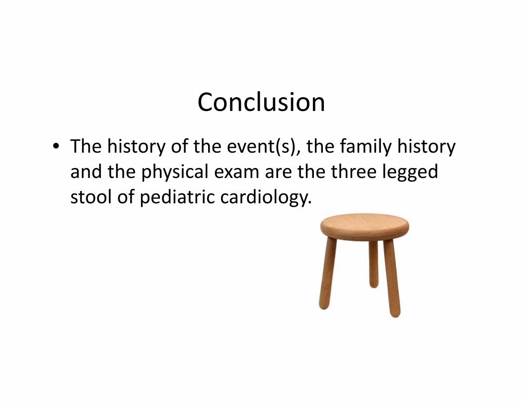
Conclusion• The history of the event(s), the family history and the physical exam are the three legged stool of pediatric cardiology.
Bibliography• ACC/AAP/AHA/ASE/HRS/SCAI/SCCT/SCMR/SOPE 2014 Appropriate Use Criteria for
Initial Transthoracic Echocardiography in Outpatient Pediatric Cardiology: J Am Coll Cardiol. 2014;64(19):2039‐2060.
• Maron BJ, Douglas PS, Graham TP, Nishimura RA, Thompson PD. Task Force 1: preparticipation screening and diagnosis of cardiovascular disease in athletes. Journal of the American College of Cardiology.2005;45:1322–6.
• Guidelines on management (diagnosis and treatment) of syncope – Update 2004 The task force on Syncope, European Society of Cardiology. European Heart Journal DOI: http://dx.doi.org/10.1016/j.ehj.2004.09.004

Potentially Useful Classification:What can you die from?
• Wolff‐Parkinson‐White syndrome• Hereditary long QT syndrome• Cardiomyopathy
• Hypertrophic• Dilated• Arrhythmogenic RV dysplasia (ARVD)
• Coronary anomalies• Valvar aortic stenosis• Aortic dissection

Practice Change• Evaluation of the patient with CHEST PAIN should include• A careful history including family history• A careful physical examination• Testing only in those cases where a specific pathologic diagnosis is suspected
• Restriction from activity is rarely warranted, and usually only in those with chest pain with exertion or with associated signs/symptoms