Embed Size (px)
Citation preview
Int J Clin Exp Pathol 2015;8(11):15333-15337www.ijcep.com /ISSN:1936-2625/IJCEP0016697
Case ReportA case of matrix-producing metaplastic carcinoma of the breast exhibiting similarities to pleomorphic adenoma on fine-needle aspiration cytology
Shogo Tajima1, Kenji Koda1, Yumie Ishii2, Satoshi Hasegawa2, Hidetarou Yokoyama2
Departments of 1Pathology, 2Surgery, Fujieda Municipal General Hospital, Shizuoka, Japan
Received September 23, 2015; Accepted October 26, 2015; Epub November 1, 2015; Published November 15, 2015
Abstract: The distinction between matrix-producing metaplastic carcinoma (MPMC) and pleomorphic adenoma (PA) is sometimes unclear in breast pathology, especially on core needle biopsy. Herein, we presented a 66-year-old woman with MPMC of the breast that looked like PA on fine-needle aspiration cytology (FNAC). On FNAC, the appear-ance of abundant myxoid matrix along with cellular clusters composed of monotonous cellular populations looked like salivary PA, which we were familiar with owing to the frequency in routine pathological practice. Thus, the pos-sibility of breast PA, the counterpart of salivary PA, was considered. However, the tumor location was different from where breast PA frequently occurs, i.e. the retroareolar region. Therefore, we eliminated the possibility of breast PA and avoided the erroneous cytological diagnosis. It is should be kept in mind that MPMC can look like PA on FNAC.
Keywords: Matrix-producing carcinoma, pleomorphic adenoma, breast, fine-needle aspiration cytology
Introduction
In breast pathology, the distinction between matrix-producing metaplastic carcinoma (MP- MC) and pleomorphic adenoma (PA) is some-times unclear especially on core needle biopsy [1]. PA of the breast seemed to be more unfa-miliar than MPMC, and cases of PA have been reported as MPMC on core needle biopsy [2, 3]. The absence of myoepithelial cells are con-firmed at the large part of breast PA, which is different from its well-known salivary counter-part [1]. This absence suggests that PA is close to low-grade breast carcinoma [1]. Conversely, to the best of our knowledge, there has been no documented case of MPMC misinterpreted as PA.
We present a case of MPMC that looked like PA on fine-needle aspiration cytology (FNAC) for cautionary and educational purposes. We could diagnose this case as MPMC on cytologi-cal examination because breast PA frequently occurs in the retroareolar region [1], which is different from the site of the occurrence of this MPMC.
Clinical summary
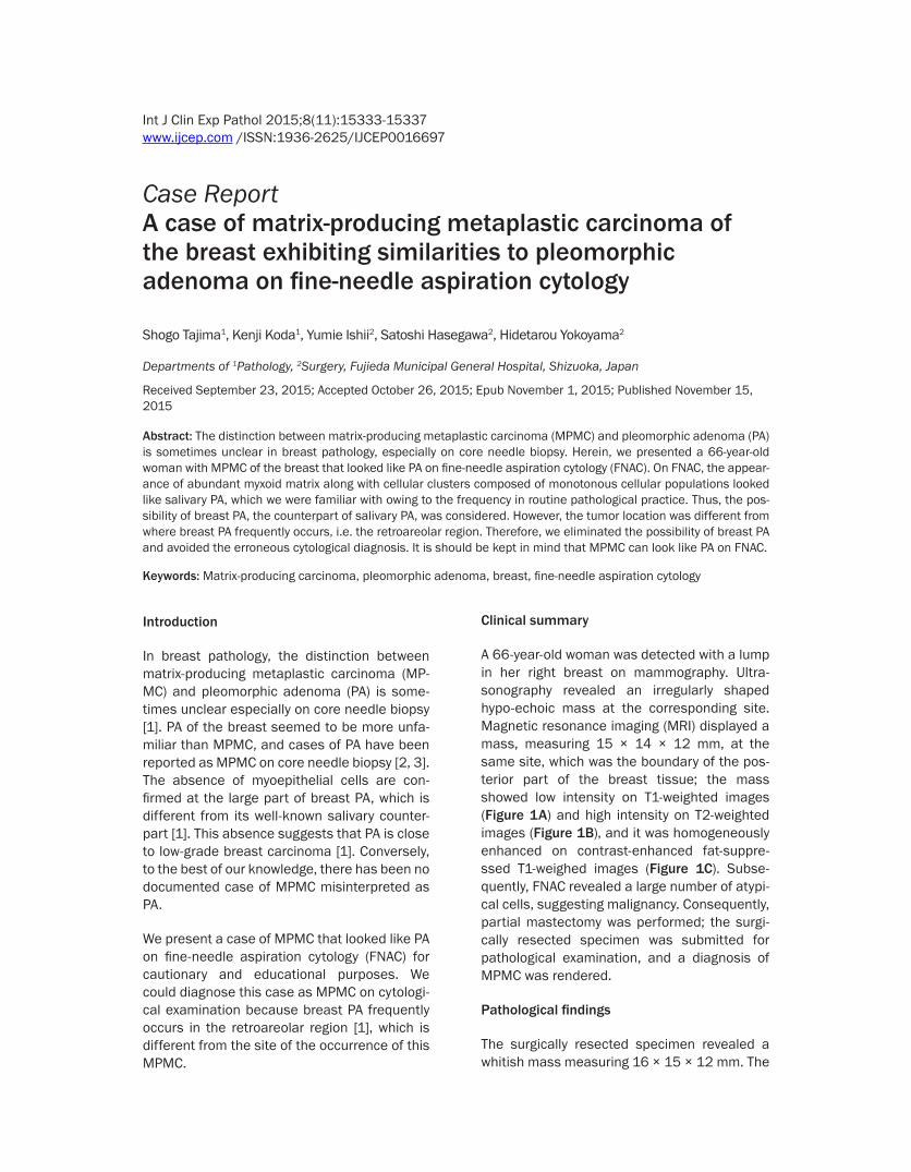
A 66-year-old woman was detected with a lump in her right breast on mammography. Ultra- sonography revealed an irregularly shaped hypo-echoic mass at the corresponding site. Magnetic resonance imaging (MRI) displayed a mass, measuring 15 × 14 × 12 mm, at the same site, which was the boundary of the pos-terior part of the breast tissue; the mass showed low intensity on T1-weighted images (Figure 1A) and high intensity on T2-weighted images (Figure 1B), and it was homogeneously enhanced on contrast-enhanced fat-suppre- ssed T1-weighed images (Figure 1C). Subse- quently, FNAC revealed a large number of atypi-cal cells, suggesting malignancy. Consequently, partial mastectomy was performed; the surgi-cally resected specimen was submitted for pathological examination, and a diagnosis of MPMC was rendered.
Pathological findings
The surgically resected specimen revealed a whitish mass measuring 16 × 15 × 12 mm. The
MPMC of the breast similar to PA on FNAC
15334 Int J Clin Exp Pathol 2015;8(11):15333-15337
Figure 1. Magnetic resonance imaging findings. A. The mass shows low intensity on a T1-weighted image. B. The mass shows high intensity on a T2-weighted image. C. The mass is homogenously enhanced on a contrast-en-hanced fat-suppressed T1-weighed image.
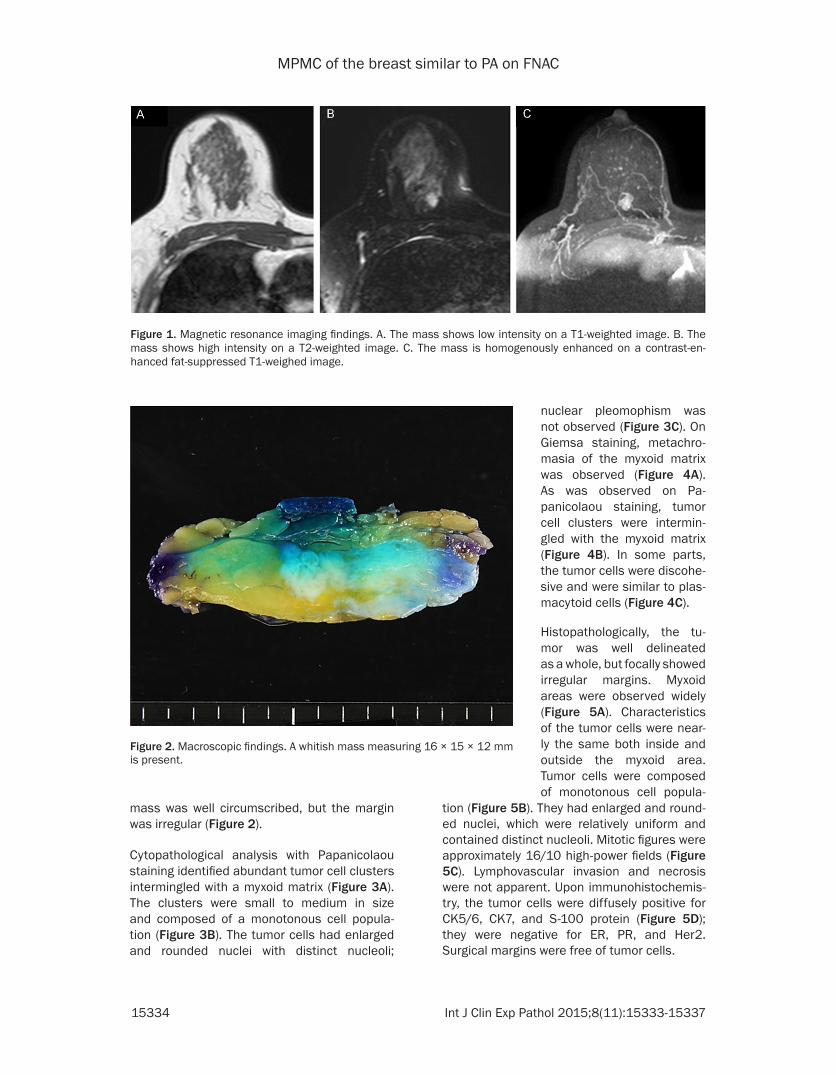
Figure 2. Macroscopic findings. A whitish mass measuring 16 × 15 × 12 mm is present.
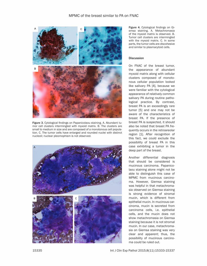
nuclear pleomophism was not observed (Figure 3C). On Giemsa staining, metachro-masia of the myxoid matrix was observed (Figure 4A). As was observed on Pa- panicolaou staining, tumor cell clusters were intermin-gled with the myxoid matrix (Figure 4B). In some parts, the tumor cells were discohe-sive and were similar to plas-macytoid cells (Figure 4C).
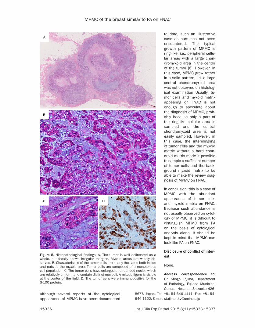
Histopathologically, the tu- mor was well delineated as a whole, but focally showed irregular margins. Myxoid areas were observed widely (Figure 5A). Characteristics of the tumor cells were near- ly the same both inside and outside the myxoid area. Tumor cells were composed of monotonous cell popula-
mass was well circumscribed, but the margin was irregular (Figure 2).
Cytopathological analysis with Papanicolaou staining identified abundant tumor cell clusters intermingled with a myxoid matrix (Figure 3A). The clusters were small to medium in size and composed of a monotonous cell popula- tion (Figure 3B). The tumor cells had enlarged and rounded nuclei with distinct nucleoli;
tion (Figure 5B). They had enlarged and round-ed nuclei, which were relatively uniform and contained distinct nucleoli. Mitotic figures were approximately 16/10 high-power fields (Figure 5C). Lymphovascular invasion and necrosis were not apparent. Upon immunohistochemis-try, the tumor cells were diffusely positive for CK5/6, CK7, and S-100 protein (Figure 5D); they were negative for ER, PR, and Her2. Surgical margins were free of tumor cells.
MPMC of the breast similar to PA on FNAC
15335 Int J Clin Exp Pathol 2015;8(11):15333-15337
Figure 3. Cytological findings on Papanicolaou staining. A. Abundant tu-mor cell clusters intermingled with myxoid matrix. B. The clusters are small to medium in size and are composed of a monotonous cell popula-tion. C. The tumor cells have enlarged and rounded nuclei with distinct nucleoli; nuclear pleomophism is not observed.
Discussion
On FNAC of the breast tumor, the appearance of abundant myxoid matrix along with cellular clusters composed of monoto-nous cellular population looked like salivary PA [4], because we were familiar with the cytological appearance of relatively common salivary PA during routine patho-logical practice. By contrast, breast PA is an exceedingly rare tumor [5] and one may not be aware of the characteristics of breast PA. If the presence of breast PA is suspected, it should also be noted that breast PA fre-quently occurs in the retroareolar region [1]. After recognition of this fact, we could exclude the possibility of breast PA in this case exhibiting a tumor in the deep part of the breast.
Another differential diagnosis that should be considered is mucinous carcinoma. Papanico- laou staining alone might not be able to distinguish this case of MPMC from mucinous carcino-ma. However, Giemsa staining was helpful in that metachroma-sia observed on Giemsa staining is strong evidence of stromal mucin, which is different from epithelial mucin. In mucinous car-cinoma, mucin is secreted from carcinoma cells, i.e. epithelial cells, and the mucin does not show metachromasia on Giemsa staining because it is not stromal mucin. In our case, metachroma-sia on Giemsa staining was very clear and apparent; thus, the possibility of mucinous carcino-ma could be ruled out.
Figure 4. Cytological findings on Gi-emsa staining. A. Metachromasia of the myxoid matrix is observed. B. Tumor cell clusters are intermingled with the myxoid matrix. C. In some parts, the tumor cells are discohesive and similar to plasmacytoid cells.
MPMC of the breast similar to PA on FNAC
15336 Int J Clin Exp Pathol 2015;8(11):15333-15337
Figure 5. Histopathological findings. A. The tumor is well delineated as a whole, but focally shows irregular margins. Myxoid areas are widely ob-served. B. Characteristics of the tumor cells are nearly the same both inside and outside the myxoid area. Tumor cells are composed of a monotonous cell population. C. The tumor cells have enlarged and rounded nuclei, which are relatively uniform and contain distinct nucleoli. A mitotic figure is visible at the center of the field. D. The tumor cells were immunopositive for the S-100 protein.
to date, such an illustrative case as ours has not been encountered. The typical growth pattern of MPMC is ring-like, i.e., peripheral cellu-lar areas with a large chon- dromyxoid area in the center of the tumor [6]. However, in this case, MPMC grew rather in a solid pattern, i.e. a large central chondromyxoid area was not observed on histolog-ical examination Usually, tu- mor cells and myxoid matrix appearing on FNAC is not enough to speculate about the diagnosis of MPMC, prob-ably because only a part of the ring-like cellular area is sampled and the central chondromyxoid area is not easily sampled. However, in this case, the intermingling of tumor cells and the myxoid matrix without a hard chon-droid matrix made it possible to sample a sufficient number of tumor cells and the back-ground myxoid matrix to be able to make the review diag-nosis of MPMC on FNAC.
In conclusion, this is a case of MPMC with the abundant appearance of tumor cells and myxoid matrix on FNAC. Because such abundance is not usually observed on cytol-ogy of MPMC, it is difficult to distinguish MPMC from PA on the basis of cytological analysis alone. It should be kept in mind that MPMC can look like PA on FNAC.
Disclosure of conflict of inter-est
None.
Address correspondence to: Dr. Shogo Tajima, Department of Pathology, Fujieda Municipal General Hospital, Shizuoka 426-
Although several reports of the cytological appearance of MPMC have been documented
8677, Japan. Tel: +81-54-646-1111; Fax: +81-54-646-1122; E-mail: [email protected]
MPMC of the breast similar to PA on FNAC
15337 Int J Clin Exp Pathol 2015;8(11):15333-15337
References
[1] Rakha EA, Badve S, Eusebi V, Reis-Filho JS, Fox SB, Dabbs DJ, Decker T, Hodi Z, Ichihara S, Lee AH, Palacios J, Richardson AL, Vincent-Salo-mon A, Schmitt FC, Tan PH, Tse GM and Ellis IO. Breast lesions of uncertain malignant na-ture and limited metastatic potential: Propos-als to improve their recognition and clinical management. Histopathology 2015; [Epub ahead of print].
[2] Djakovic A, Engel JB, Geisinger E, Honig A, Tsc-hammler A and Dietl J. Pleomorphic adenoma of the breast initially misdiagnosed as meta-plastic carcinoma in preoperative stereotactic biopsy: a case report and review of the litera-ture. Eur J Gynaecol Oncol 2011; 32: 427-430.
[3] Rakha EA, Aleskandarany MA, Samaka RM, Hodi Z, Lee AH and Ellis IO. Pleomorphic ade-noma-like tumour of the breast. Histopatholo-gy 2015; [Epub ahead of print].
[4] Ahn S, Kim Y and Oh YL. Fine needle aspiration cytology of benign salivary gland tumors with myoepithelial cell participation: an institution-al experience of 575 cases. Acta Cytol 2013; 57: 567-574.
[5] Sato K, Ueda Y, Shimasaki M, Ozaki M, Nitta N, Chada K, Ishikawa Y and Katsuda S. Pleomor-phic adenoma (benign mixed tumor) of the breast: a case report and review of the litera-ture. Pathol Res Pract 2005; 201: 333-339.
[6] Shui R, Bi R, Cheng Y, Lu H, Wang J and Yang W. Matrix-producing carcinoma of the breast in the Chinese population: a clinicopathological study of 13 cases. Pathol Int 2011; 61: 415-422.