Embed Size (px)
Citation preview
CH 3. Vital Signs
- 생체 징후 -
GACHON UNIVERSITY Depart. Physical Therapy
Hwi-young Cho, PT, Ph.D
The Pride of Physical Therapy
References
• 책명
– Physical Rehabilitation
– 질환별 물리치료
• 저자
– Susan B. O’Sullivan
• 옮긴이
– 김근조 외
• 출판사
– F.A DAVIS (국외)
– 영문출판사 (국내)
CHAPTER 4.
- Vital Signs-
References
• 책명
– Principle & Techniques of
patient care
– 일상생활동작: 환자관리와 기능훈련
• 저자
– Frank M. Pierson
• 옮긴이
– 장정훈 외
• 출판사
– SAUNDERS (국외)
– E·PUBLIC (국내)
CHAPTER 3.
- Assessment of Vital Signs -
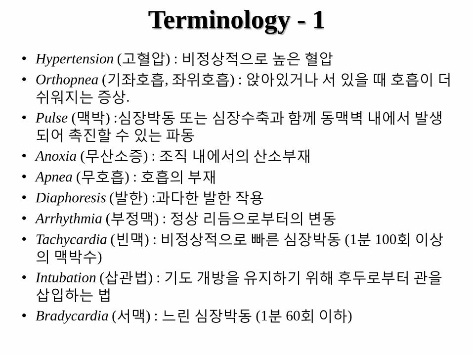
Terminology - 1
• Hypertension (고혈압) : 비정상적으로 높은 혈압
• Orthopnea (기좌호흡, 좌위호흡) : 앉아있거나 서 있을 때 호흡이 더
쉬워지는 증상.
• Pulse (맥박) :심장박동 또는 심장수축과 함께 동맥벽 내에서 발생되어 촉진할 수 있는 파동
• Anoxia (무산소증) : 조직 내에서의 산소부재
• Apnea (무호흡) : 호흡의 부재
• Diaphoresis (발한) :과다한 발한 작용
• Arrhythmia (부정맥) : 정상 리듬으로부터의 변동
• Tachycardia (빈맥) : 비정상적으로 빠른 심장박동 (1분 100회 이상의 맥박수)
• Intubation (삽관법) : 기도 개방을 유지하기 위해 후두로부터 관을
삽입하는 법
• Bradycardia (서맥) : 느린 심장박동 (1분 60회 이하)

Terminology - 2
• Systole (수축기) : 심장이 박동하는 동안 동맥 벽에 압력이 최대로
가해졌을 때의 기간
• Rale (수포음) : 주로 흡식 때 흉부의 청진 시 들리는 비정상적이고
불연속적이며 좋지 않은 심장음. 또한 딱딱소리(crackle)라고도 한다.
• Syncope (실신) : 뇌의 출혈에 의해 야기되는 의식의 일시적인 정지;
기절
• Cardiac output (심박출량) : 각 심장이 수축하는 동안 심장에서부터
박출되어 나오는 혈액의 양
• Fever (열) : 정상 수준 이상의 체온
• Dysrhythmia (율동부정) : 율동의 교란
• Diastole (이완기) : 심장이 박동하는 동안 심벽이나 심동맥에 압력이 최소로 가해지는 기간; 대개 심장 박동의 안정기를 일컫는다.
• Hypotension (저혈압) : 비정상적으로 낮은 혈압
• Ectopic (전위성의) : 비정상적으로 나타나거나 발생된.
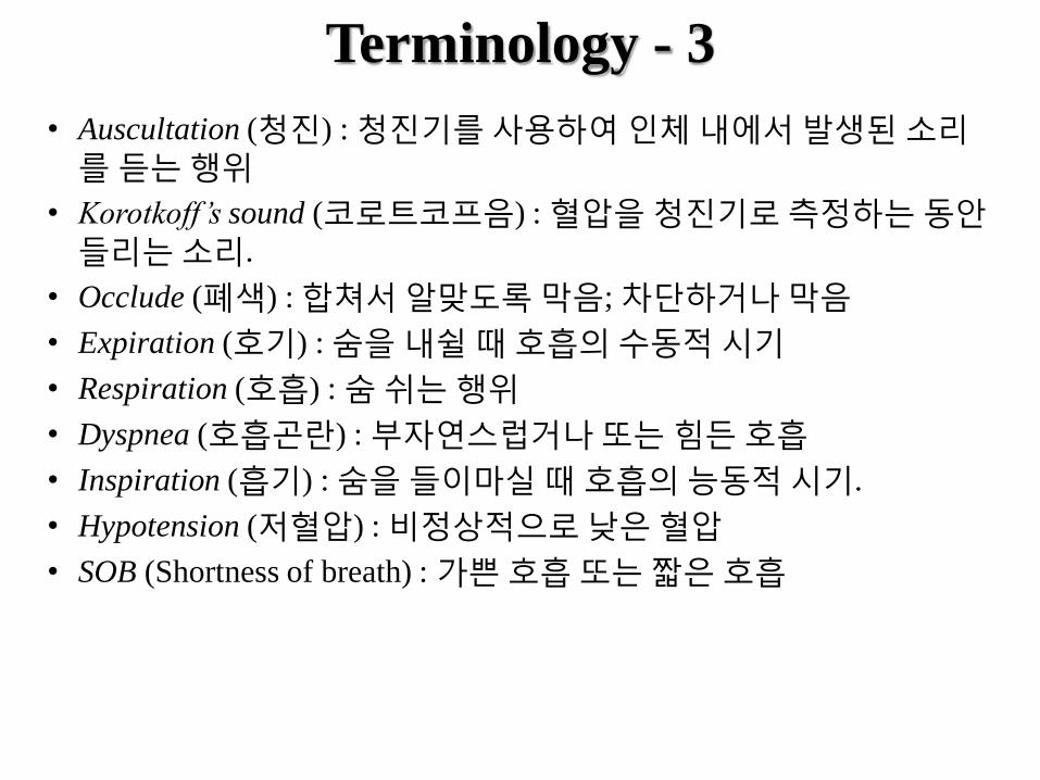
Terminology - 3
• Auscultation (청진) : 청진기를 사용하여 인체 내에서 발생된 소리를 듣는 행위
• Korotkoff’s sound (코로트코프음) : 혈압을 청진기로 측정하는 동안
들리는 소리.
• Occlude (폐색) : 합쳐서 알맞도록 막음; 차단하거나 막음
• Expiration (호기) : 숨을 내쉴 때 호흡의 수동적 시기
• Respiration (호흡) : 숨 쉬는 행위
• Dyspnea (호흡곤란) : 부자연스럽거나 또는 힘든 호흡
• Inspiration (흡기) : 숨을 들이마실 때 호흡의 능동적 시기.
• Hypotension (저혈압) : 비정상적으로 낮은 혈압
• SOB (Shortness of breath) : 가쁜 호흡 또는 짧은 호흡
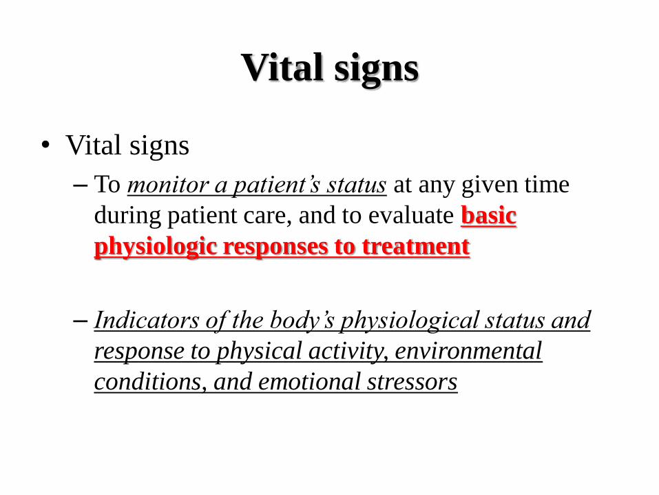
Vital signs
• Vital signs
– To monitor a patient’s status at any given time
during patient care, and to evaluate basic
physiologic responses to treatment
– Indicators of the body’s physiological status and
response to physical activity, environmental
conditions, and emotional stressors
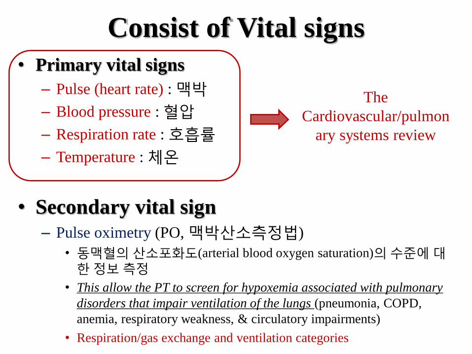
Consist of Vital signs
• Primary vital signs
– Pulse (heart rate) : 맥박
– Blood pressure : 혈압
– Respiration rate : 호흡률
– Temperature : 체온
• Secondary vital sign
– Pulse oximetry (PO, 맥박산소측정법)
• 동맥혈의 산소포화도(arterial blood oxygen saturation)의 수준에 대한 정보 측정
• This allow the PT to screen for hypoxemia associated with pulmonary
disorders that impair ventilation of the lungs (pneumonia, COPD,
anemia, respiratory weakness, & circulatory impairments)
• Respiration/gas exchange and ventilation categories
The
Cardiovascular/pulmon
ary systems review
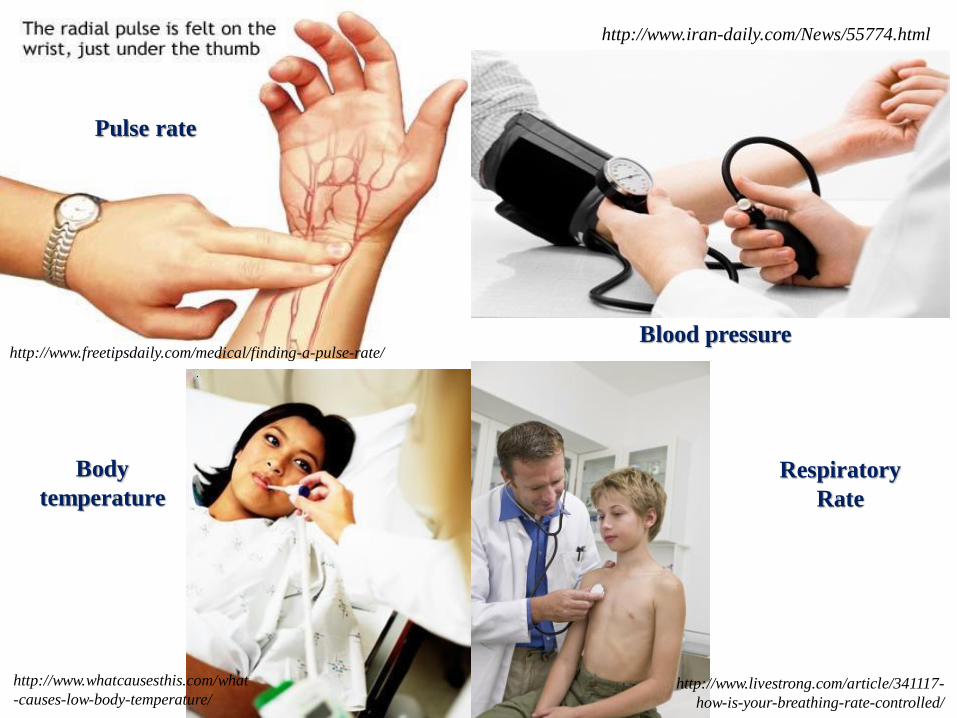
Blood pressure
Pulse rate
http://www.freetipsdaily.com/medical/finding-a-pulse-rate/
http://www.iran-daily.com/News/55774.html
http://www.whatcausesthis.com/what
-causes-low-body-temperature/ http://www.livestrong.com/article/341117-
how-is-your-breathing-rate-controlled/
Body
temperature Respiratory
Rate
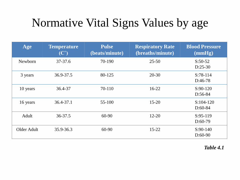
Normative Vital Signs Values by age
Age Temperature
(C˚)
Pulse
(beats/minute)
Respiratory Rate
(breaths/minute)
Blood Pressure
(mmHg)
Newborn 37-37.6 70-190 25-50 S:50-52
D:25-30
3 years 36.9-37.5 80-125 20-30 S:78-114
D:46-78
10 years 36.4-37 70-110 16-22 S:90-120
D:56-84
16 years 36.4-37.1 55-100 15-20 S:104-120
D:60-84
Adult 36-37.5 60-90 12-20 S:95-119
D:60-79
Older Adult 35.9-36.3 60-90 15-22 S:90-140
D:60-90
Table 4.1
Patient observation
• Prior to a formal examination of vital signs, careful systematic observation of the patient can reveal important preliminary data.
- distress or discomfort
- cachexia (악액질) : 만성병으로 인한 건강 악화 상태
- cyanosis (청색증)
- changes in texture and hair growth
- diaphoresis : 인위적인 다량의 발한
- abnormal sitting postures
- use of accessory muscles of breathing
- clubbing
Systemic observation of the patient
• Signs of immediate patient distress or discomfort – Observation of facial expressions
– Use of accessory muscles for breathing
– Irregular breathing pattern
– Frequent positional changes
• Clues about nutritional status – 비만 (Obesity)
– 영양결핍 (Malnutrition)
– 쇠약 (Wasting)
• Signs of Skin color – Central cyanosis (청색증) in mucous membrane (점액막)
– Peripheral cyanosis (secondary to vasoconstriction) in the earlobes, nose, lips, toes

• Changes in texture and hair growth (피부감촉 및 모발성장)
– Diabetes mellitus or atherosclerosis
• Lack hair growth on the legs
• Thickening of the nails of the fingers and toes
• Diaphoresis (발한증)
– Reduced cardiac output
– Myocardiac infarction, hypotension, shock
• Abnormal sitting postures
– Pain or structural abnormalities of the pectoral or vertebral regions
• Interfere with respiratory patterns
• Use of accessory muscles of breathing
– Cardiac or pulmonary impairments
• Peripheral extremities - Edema or clubbing (곤봉모양)
– Clubbing (손가락과 발가락 말단의 bulbous swelling): hypoxia, cyanosis
• congenital heart defects and pulmonary disorders
– Edema
• Right heart failure, venous insufficiency
Body temperature
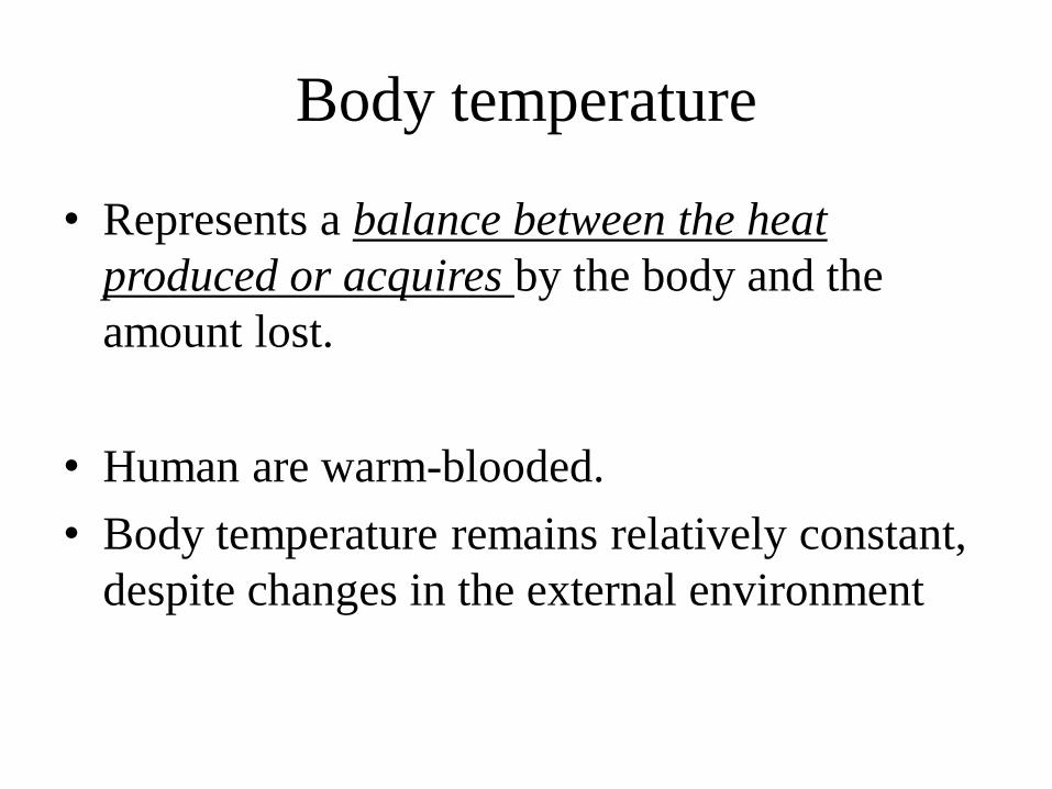
Body temperature
• Represents a balance between the heat
produced or acquires by the body and the
amount lost.
• Human are warm-blooded.
• Body temperature remains relatively constant,
despite changes in the external environment
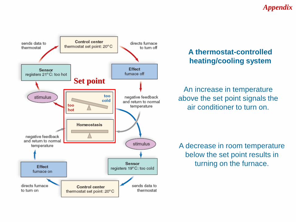
A thermostat-controlled
heating/cooling system
An increase in temperature
above the set point signals the
air conditioner to turn on.
A decrease in room temperature
below the set point results in
turning on the furnace.
Set point
Appendix
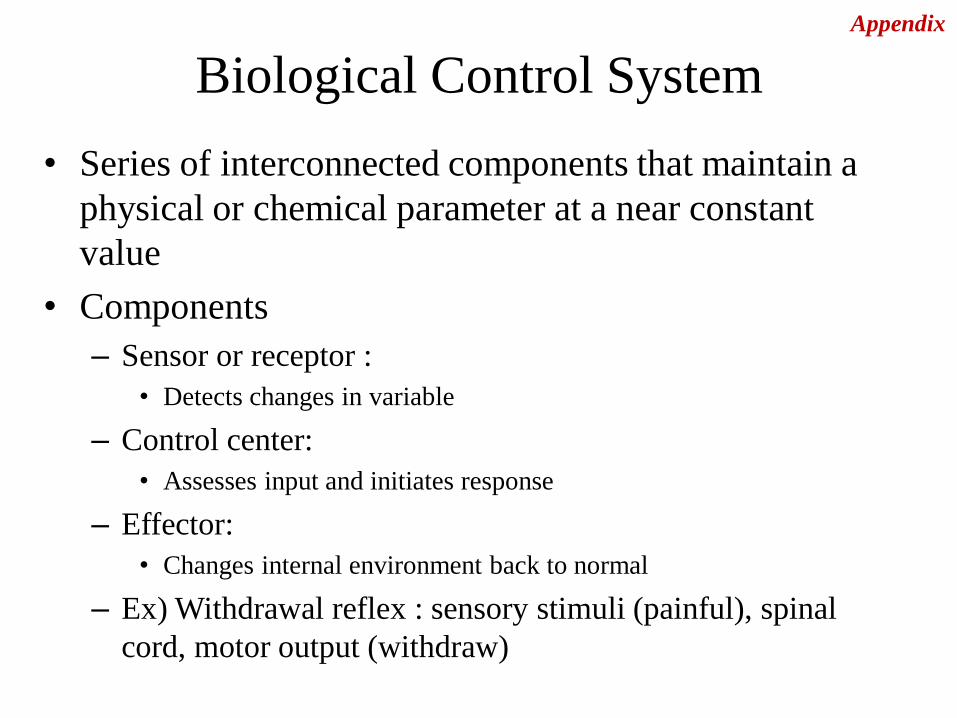
Biological Control System
• Series of interconnected components that maintain a
physical or chemical parameter at a near constant
value
• Components
– Sensor or receptor :
• Detects changes in variable
– Control center:
• Assesses input and initiates response
– Effector:
• Changes internal environment back to normal
– Ex) Withdrawal reflex : sensory stimuli (painful), spinal
cord, motor output (withdraw)
Appendix
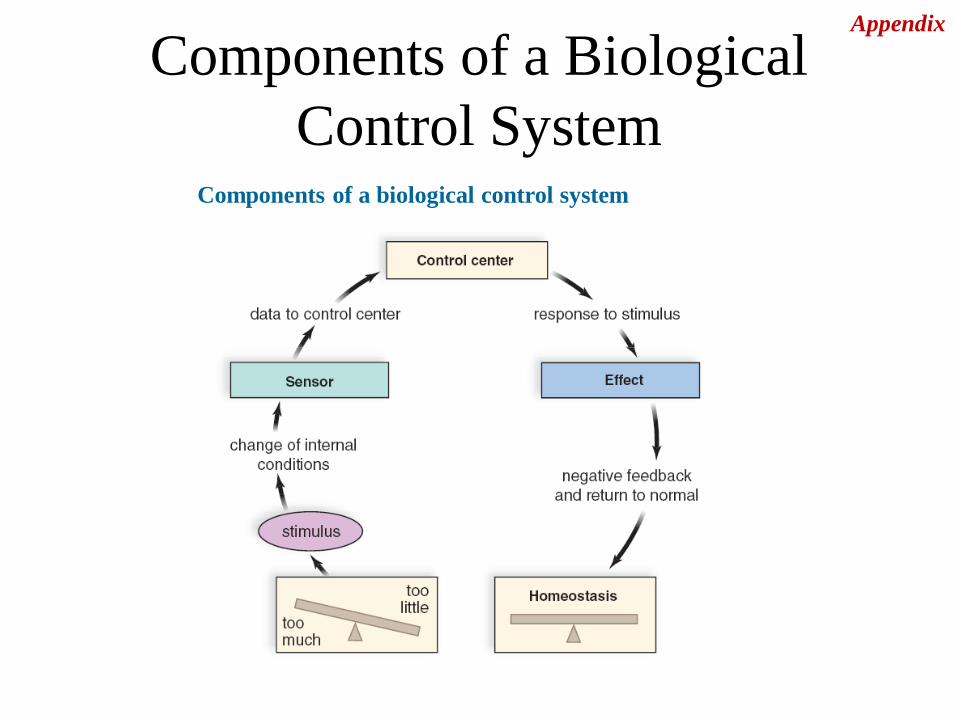
Components of a Biological
Control System Components of a biological control system
Appendix
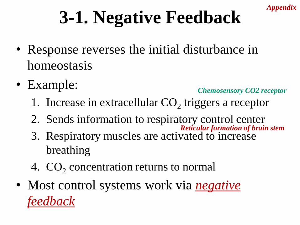
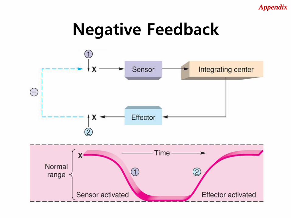
3-1. Negative Feedback
• Response reverses the initial disturbance in
homeostasis
• Example:
1. Increase in extracellular CO2 triggers a receptor
2. Sends information to respiratory control center
3. Respiratory muscles are activated to increase
breathing
4. CO2 concentration returns to normal
• Most control systems work via negative
feedback
Chemosensory CO2 receptor
Reticular formation of brain stem
Appendix

Negative Feedback
Appendix
Negative Feedback
Appendix
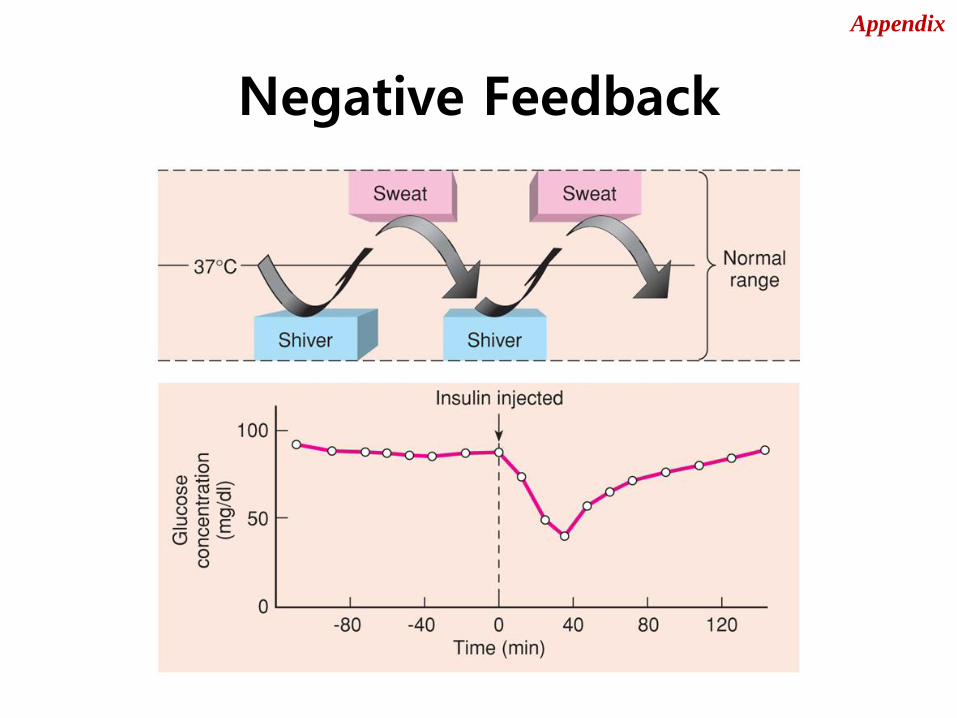
Negative Feedback
Appendix
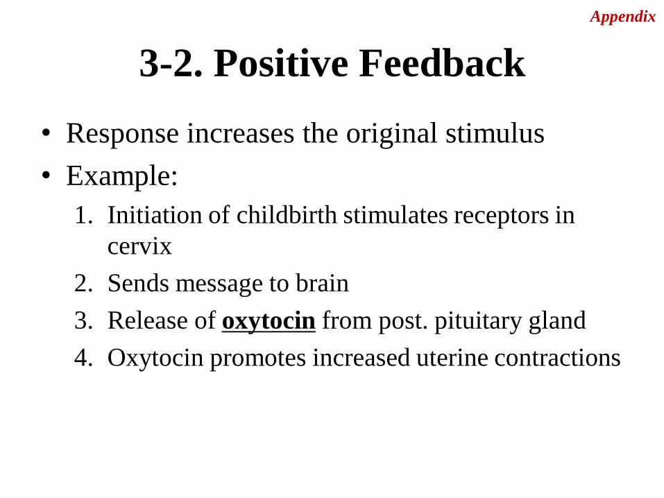
3-2. Positive Feedback
• Response increases the original stimulus
• Example:
1. Initiation of childbirth stimulates receptors in
cervix
2. Sends message to brain
3. Release of oxytocin from post. pituitary gland
4. Oxytocin promotes increased uterine contractions
Appendix
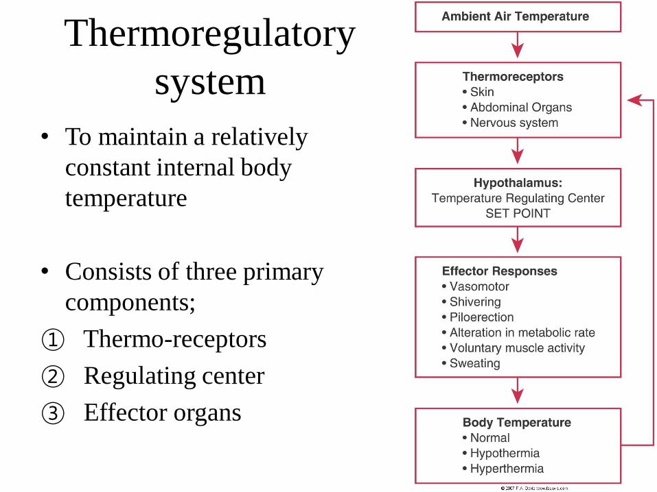
Thermoregulatory
system
• To maintain a relatively
constant internal body
temperature
• Consists of three primary
components;
① Thermo-receptors
② Regulating center
③ Effector organs

Thermoreceptors
• Provide input to the temperature regulating
center located in the hypothalamus
• Peripheral and central thermoreceptors
Peripheral; free nerve ending in the skin,
abdominal organs, and nervous system
Central; hypothalamus

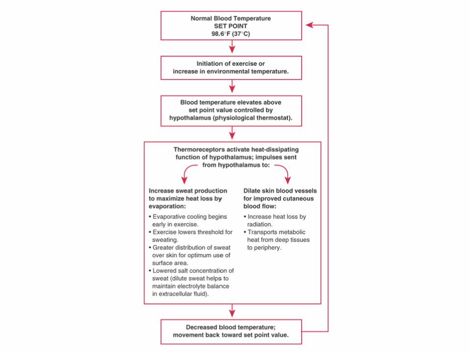
Regulating center
• Located in the hypothalmus
Coordinate the heat production and loss processes
By influencing the effector organs, maintain a
balance
In healthy individual, hypothalamus is set at 98.6
degree F (37℃)
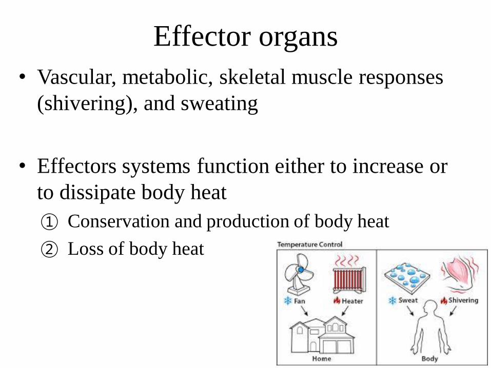
Effector organs
• Vascular, metabolic, skeletal muscle responses
(shivering), and sweating
• Effectors systems function either to increase or
to dissipate body heat
① Conservation and production of body heat
② Loss of body heat
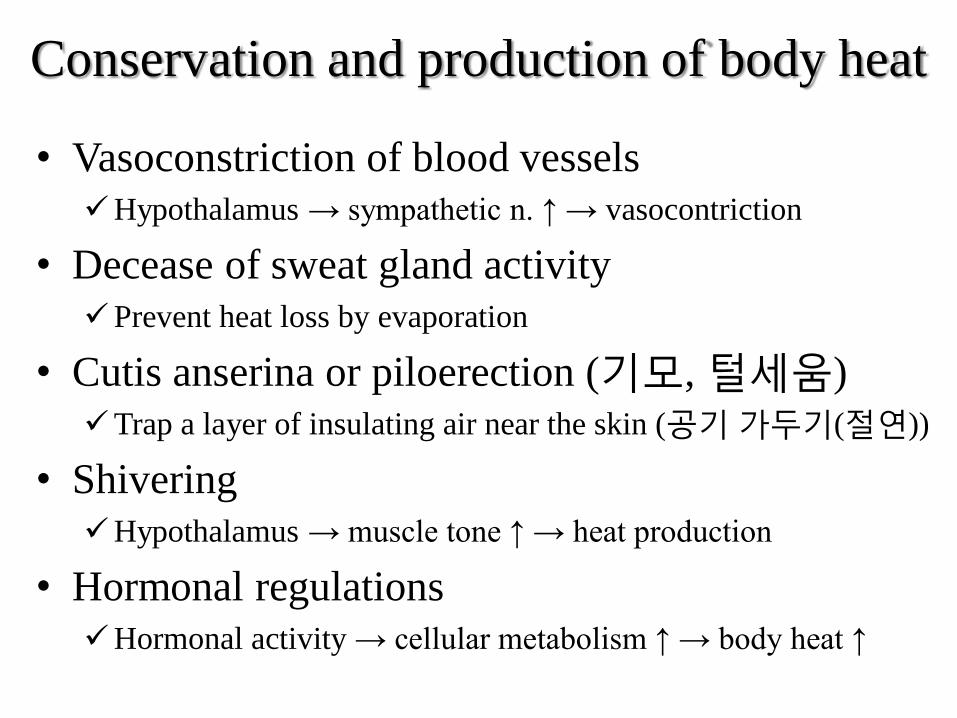
Conservation and production of body heat
• Vasoconstriction of blood vessels
Hypothalamus → sympathetic n. ↑ → vasocontriction
• Decease of sweat gland activity
Prevent heat loss by evaporation
• Cutis anserina or piloerection (기모, 털세움)
Trap a layer of insulating air near the skin (공기 가두기(절연))
• Shivering
Hypothalamus → muscle tone ↑ → heat production
• Hormonal regulations
Hormonal activity → cellular metabolism ↑ → body heat ↑
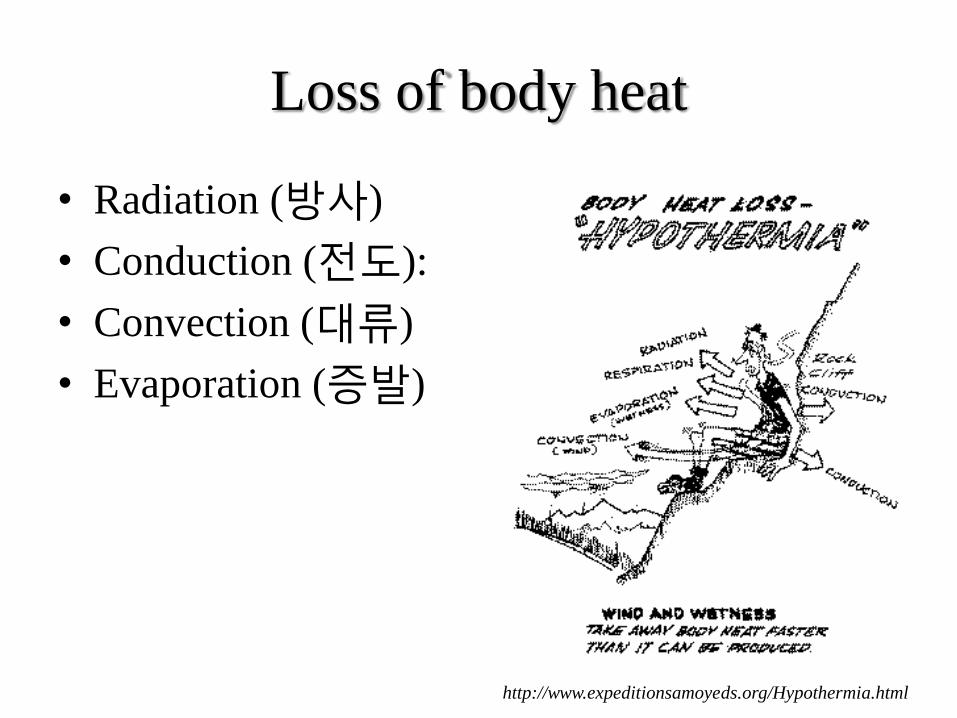
Loss of body heat
• Radiation (방사)
• Conduction (전도):
• Convection (대류)
• Evaporation (증발)
http://www.expeditionsamoyeds.org/Hypothermia.html
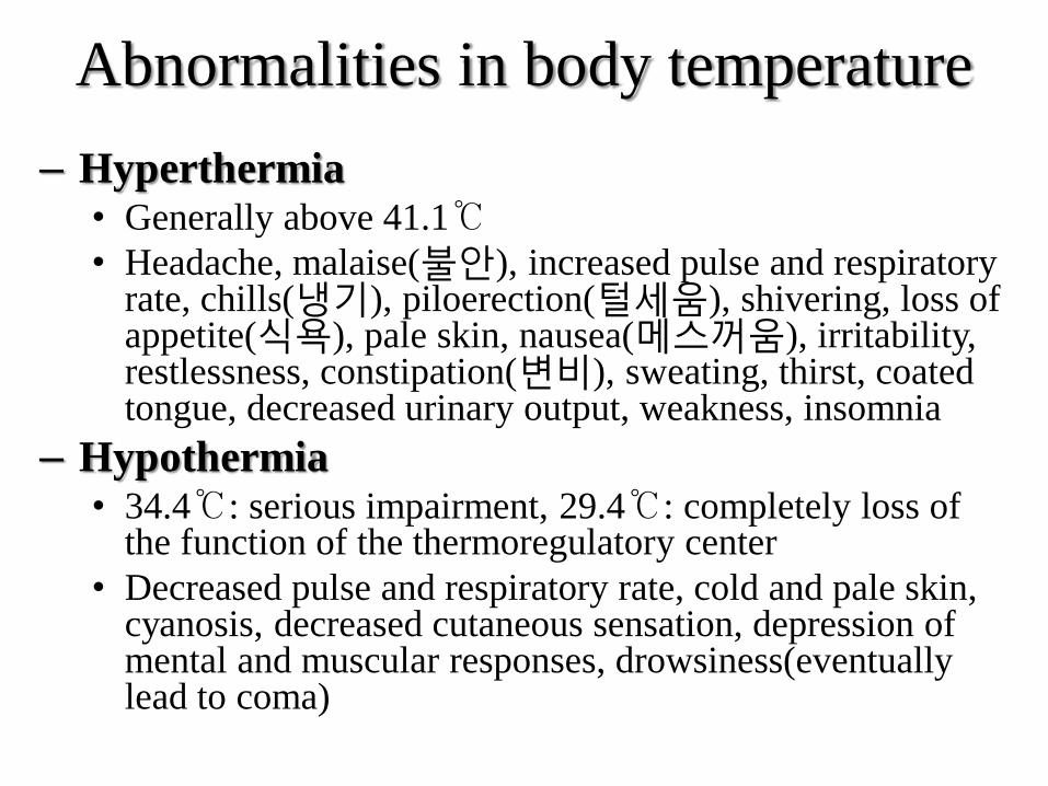
Abnormalities in body temperature
– Hyperthermia • Generally above 41.1℃
• Headache, malaise(불안), increased pulse and respiratory rate, chills(냉기), piloerection(털세움), shivering, loss of appetite(식욕), pale skin, nausea(메스꺼움), irritability, restlessness, constipation(변비), sweating, thirst, coated tongue, decreased urinary output, weakness, insomnia
– Hypothermia • 34.4℃: serious impairment, 29.4℃: completely loss of
the function of the thermoregulatory center
• Decreased pulse and respiratory rate, cold and pale skin, cyanosis, decreased cutaneous sensation, depression of mental and muscular responses, drowsiness(eventually lead to coma)
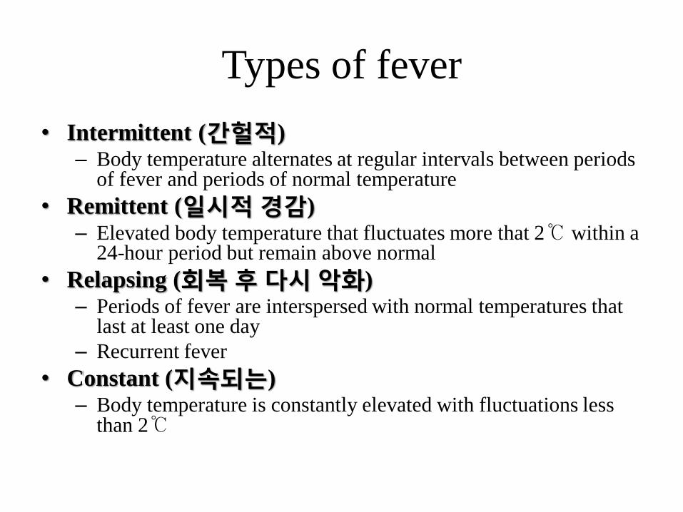
Types of fever
• Intermittent (간헐적) – Body temperature alternates at regular intervals between periods
of fever and periods of normal temperature
• Remittent (일시적 경감) – Elevated body temperature that fluctuates more that 2℃ within a
24-hour period but remain above normal
• Relapsing (회복 후 다시 악화) – Periods of fever are interspersed with normal temperatures that
last at least one day
– Recurrent fever
• Constant (지속되는) – Body temperature is constantly elevated with fluctuations less
than 2℃

Factors influencing body temperature
• Time of Day
– Lowest : 4~6 AM, Highest : 4~8 PM
• Age
• Emotions/Stress
• Exercise
• Menstrual Cycle
– During ovulation : slight elevation(0.3~0.5℃)
• Pregnancy
– Remain elevated by 0.5℃
• External environment
• Measurement site
– Rectal and tympanic (고막) : 0.3~0.5℃ higher than oral
– Axillary : 0.6 ℃ lower than oral
• Ingestion of warm or cold foods
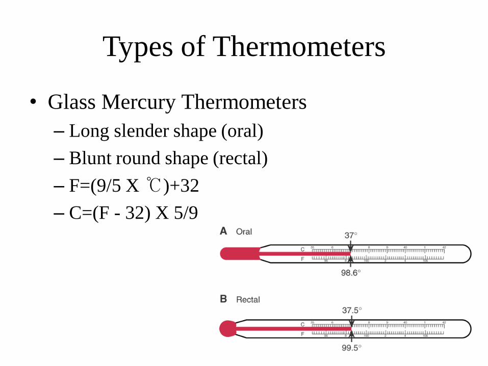
Types of Thermometers
• Glass Mercury Thermometers
– Long slender shape (oral)
– Blunt round shape (rectal)
– F=(9/5 X ℃)+32
– C=(F - 32) X 5/9
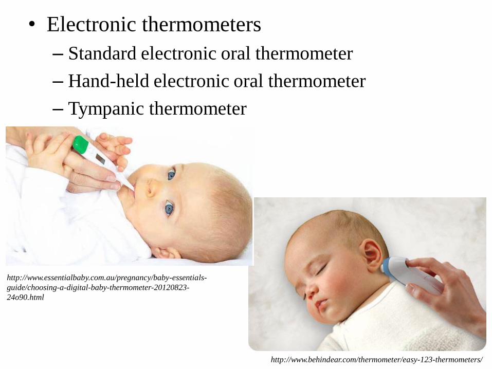
• Electronic thermometers
– Standard electronic oral thermometer
– Hand-held electronic oral thermometer
– Tympanic thermometer
http://www.behindear.com/thermometer/easy-123-thermometers/
http://www.essentialbaby.com.au/pregnancy/baby-essentials-
guide/choosing-a-digital-baby-thermometer-20120823-
24o90.html

• Disposal single-use thermometer
• Temperature-sensitive strips
http://www.4senior.com/thermometer.htm
http://nl.aliexpress.com/store/group/baby-supplies/416785_212269834.html

Procedure for measuring body temperature
• Measuring oral temperature:
Electronic thermometer
Glass mercury thermometer
• Measuring axillary temperature:
Glass mercury thermometer
• Measuring tympanic membrane temperature:
Tympanic thermometer
Pulse
Pulse rate = Heart rate
Pulse
• The wave of blood in the artery created by contraction of the left ventricle during a cardiac cycle
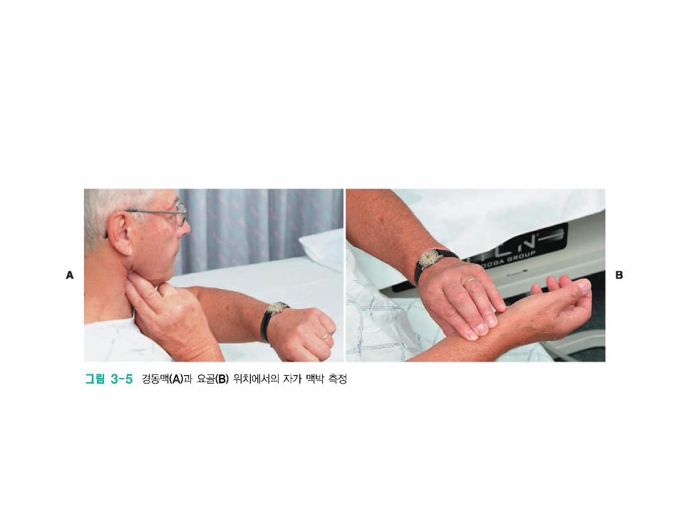
• Peripheral pulses are those located in the periphery of the body that can be felt by palpating an artery over a bony prominence or other firm surface
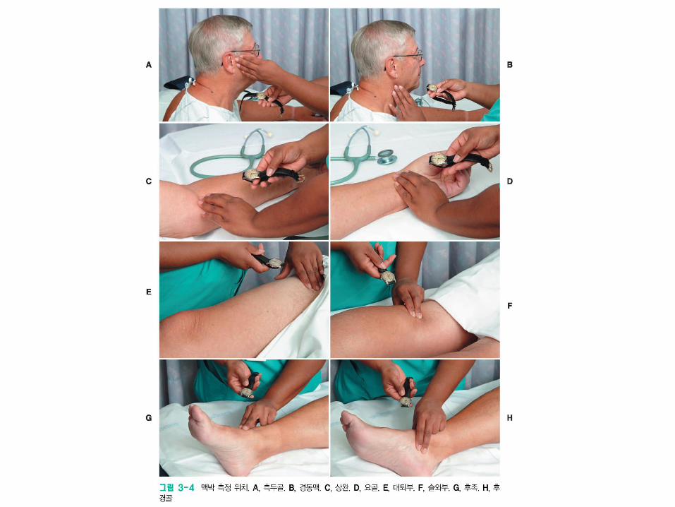
Radial (요골동맥) : palpation
Carotid (경동맥) : palpation
Popliteal (슬와동맥) : palpation
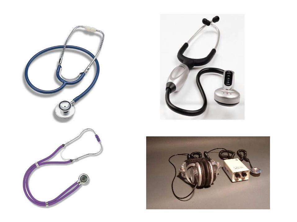
Apical pulse (심첨 백박): using stethoscope (청진기)
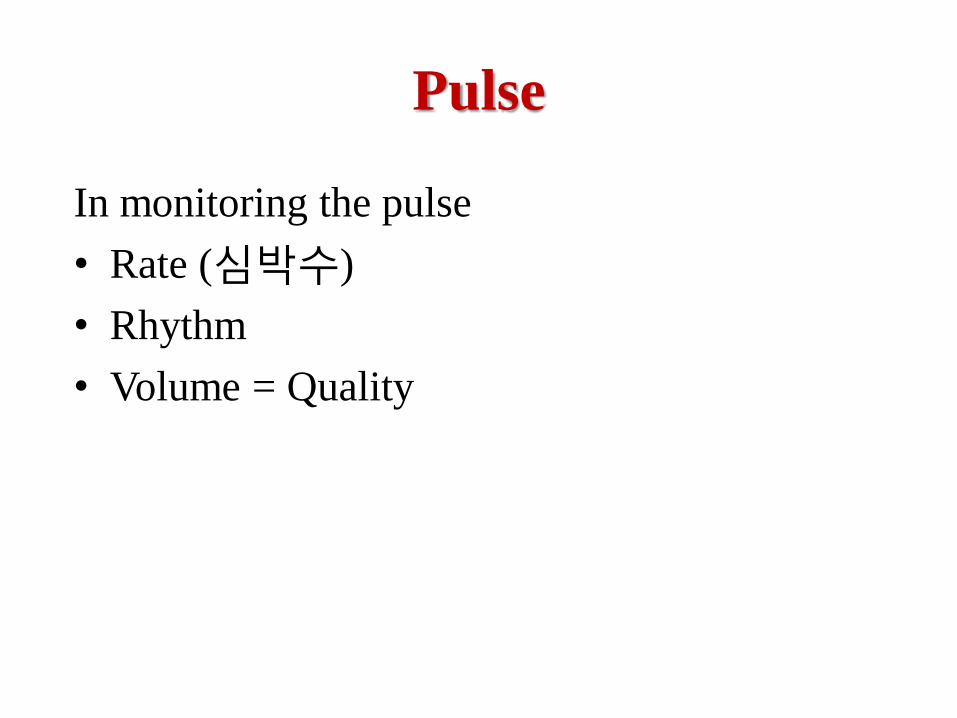
In monitoring the pulse
• Rate (심박수)
• Rhythm
• Volume = Quality
Pulse
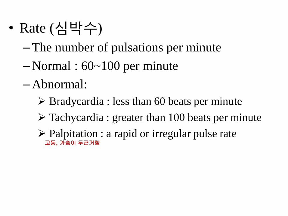
• Rate (심박수)
–The number of pulsations per minute
–Normal : 60~100 per minute
–Abnormal:
Bradycardia : less than 60 beats per minute
Tachycardia : greater than 100 beats per minute
Palpitation : a rapid or irregular pulse rate 고동, 가슴이 두근거림
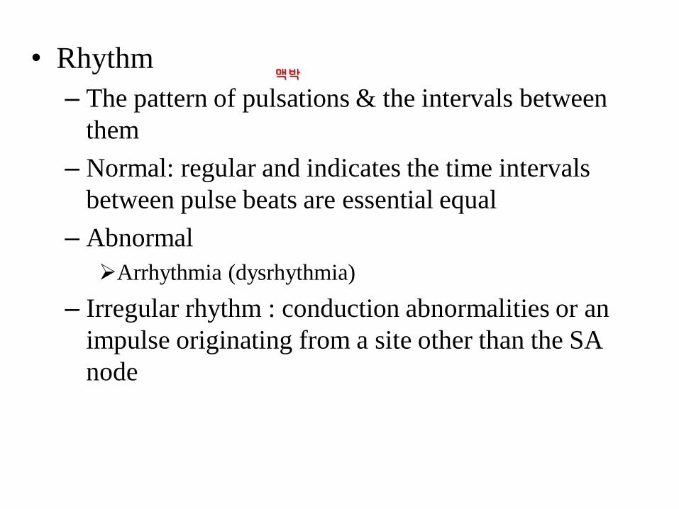
• Rhythm
– The pattern of pulsations & the intervals between
them
– Normal: regular and indicates the time intervals
between pulse beats are essential equal
– Abnormal
Arrhythmia (dysrhythmia)
– Irregular rhythm : conduction abnormalities or an
impulse originating from a site other than the SA
node
맥박
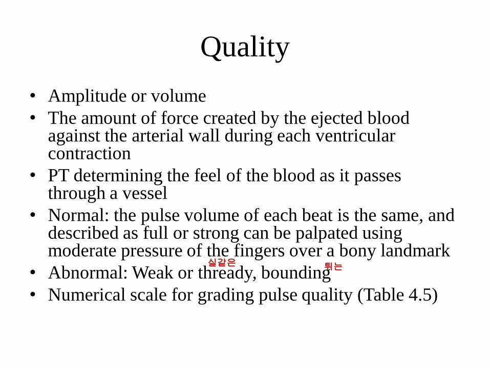
Quality
• Amplitude or volume
• The amount of force created by the ejected blood against the arterial wall during each ventricular contraction
• PT determining the feel of the blood as it passes through a vessel
• Normal: the pulse volume of each beat is the same, and described as full or strong can be palpated using moderate pressure of the fingers over a bony landmark
• Abnormal: Weak or thready, bounding
• Numerical scale for grading pulse quality (Table 4.5)
실같은 튀는
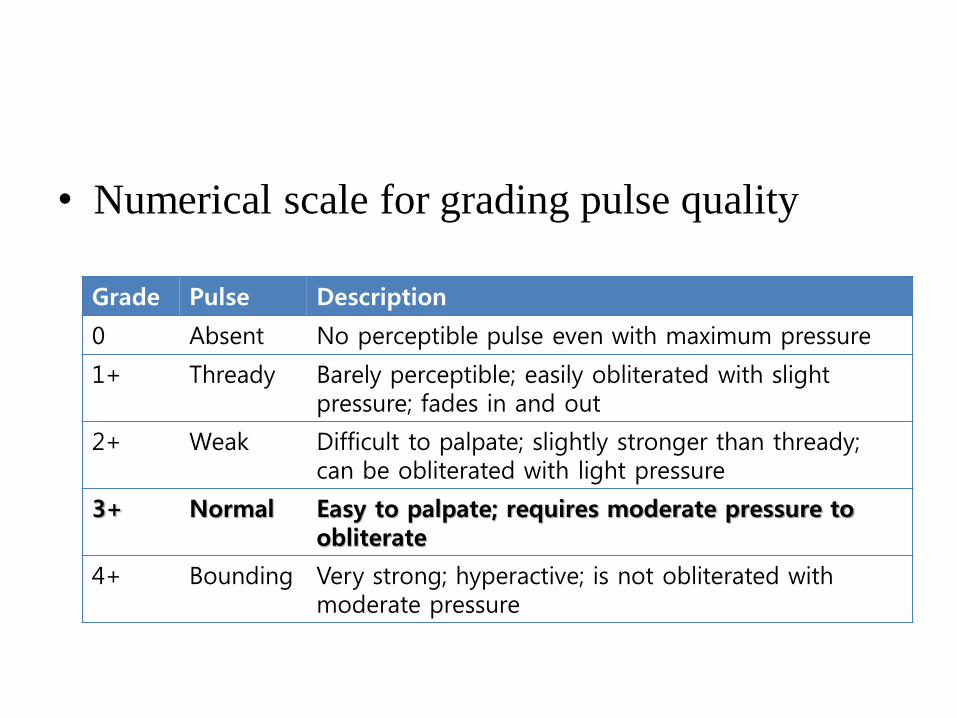
• Numerical scale for grading pulse quality
Grade Pulse Description
0 Absent No perceptible pulse even with maximum pressure
1+ Thready Barely perceptible; easily obliterated with slight pressure; fades in and out
2+ Weak Difficult to palpate; slightly stronger than thready; can be obliterated with light pressure
3+ Normal Easy to palpate; requires moderate pressure to obliterate
4+ Bounding Very strong; hyperactive; is not obliterated with moderate pressure
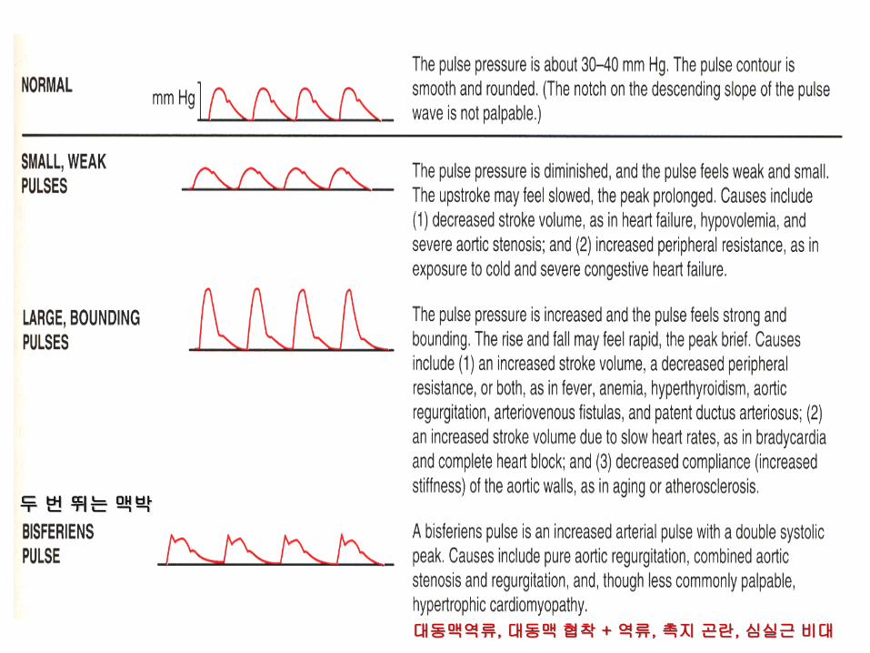
두 번 뛰는 맥박
대동맥역류, 대동맥 협착 + 역류, 촉지 곤란, 심실근 비대
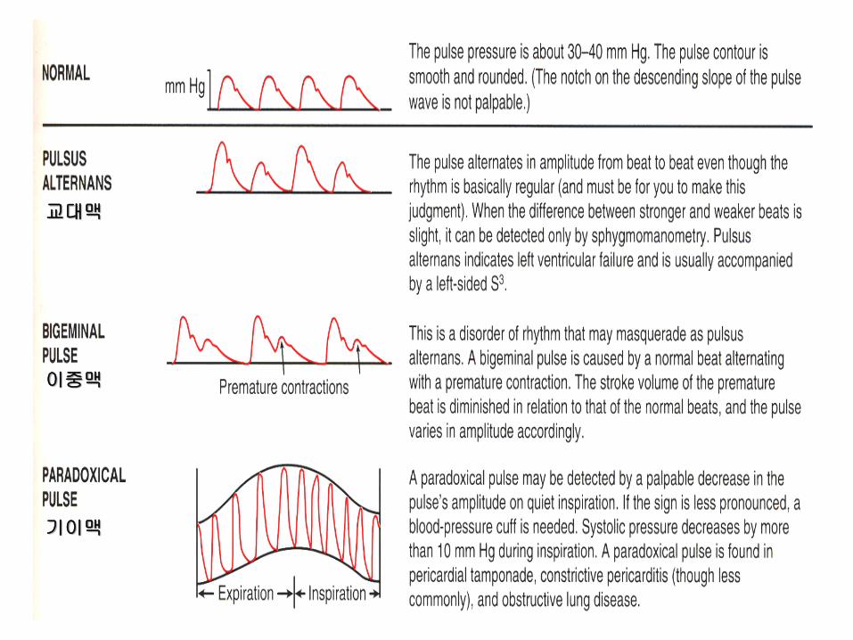
교대맥
이중맥
기이맥
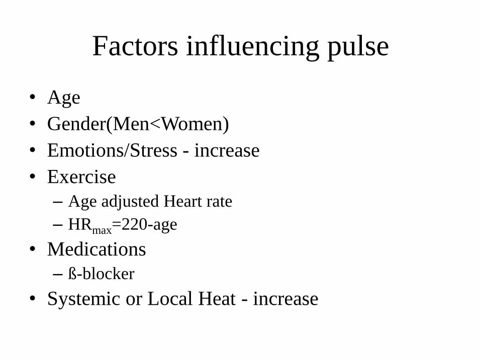
Factors influencing pulse
• Age
• Gender(Men<Women)
• Emotions/Stress - increase
• Exercise
– Age adjusted Heart rate
– HRmax=220-age
• Medications
– ß-blocker
• Systemic or Local Heat - increase
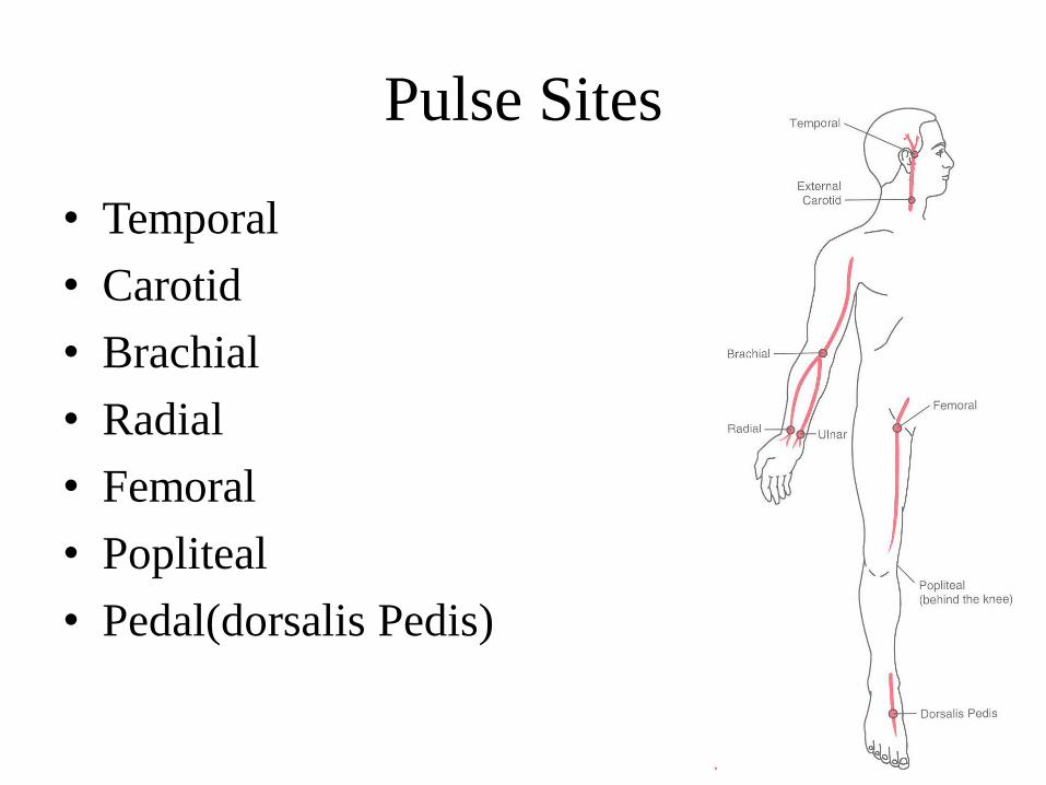
Pulse Sites
• Temporal
• Carotid
• Brachial
• Radial
• Femoral
• Popliteal
• Pedal(dorsalis Pedis)
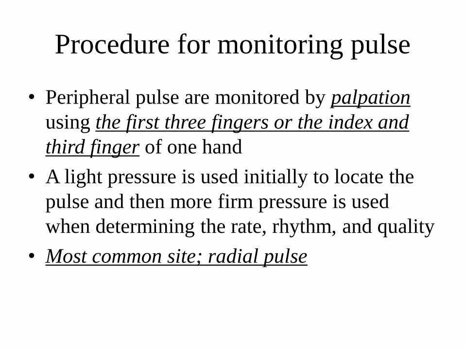
Procedure for monitoring pulse
• Peripheral pulse are monitored by palpation
using the first three fingers or the index and
third finger of one hand
• A light pressure is used initially to locate the
pulse and then more firm pressure is used
when determining the rate, rhythm, and quality
• Most common site; radial pulse

Monitoring pulse
• Measuring radial pulse
• Measuring apical pulse using stethoscope
• Measuring apical-radial pulse
– Two examiners simultaneously measuring the
pulse at two separate locations
• Electronic HR monitoring
• Radial & Apical Pulse
– http://www.youtube.com/watch?v=mPJ_zNa-LQE
Measuring radial pulse
• Assemble equipment
- watch (or wall clock) with a second hand
• Wash hands
• Procedure
- explain procedure to the patient
- Ensure patient understanding, safety, comfort
- the patient’s wrist should be in a neutral position relative to flexion and extension and the forearm supported in pronation
- place the first three fingers squarely and firmly over the radial pulse
Once the strongest pulsation is located, note the position of the second hand on the watch or clock
- wash hands

Electronic Heart Rate Monitoring
• Heart rate Monitors(HRMs)
http://en.wikipedia.org/wiki/Heart_rate_monitor#mediaviewer/File:MF-180.JPG
http://www.dietandfitnessresources.co.uk/scales_monitors/heart_ra
te_monitor_problem_solving.htm
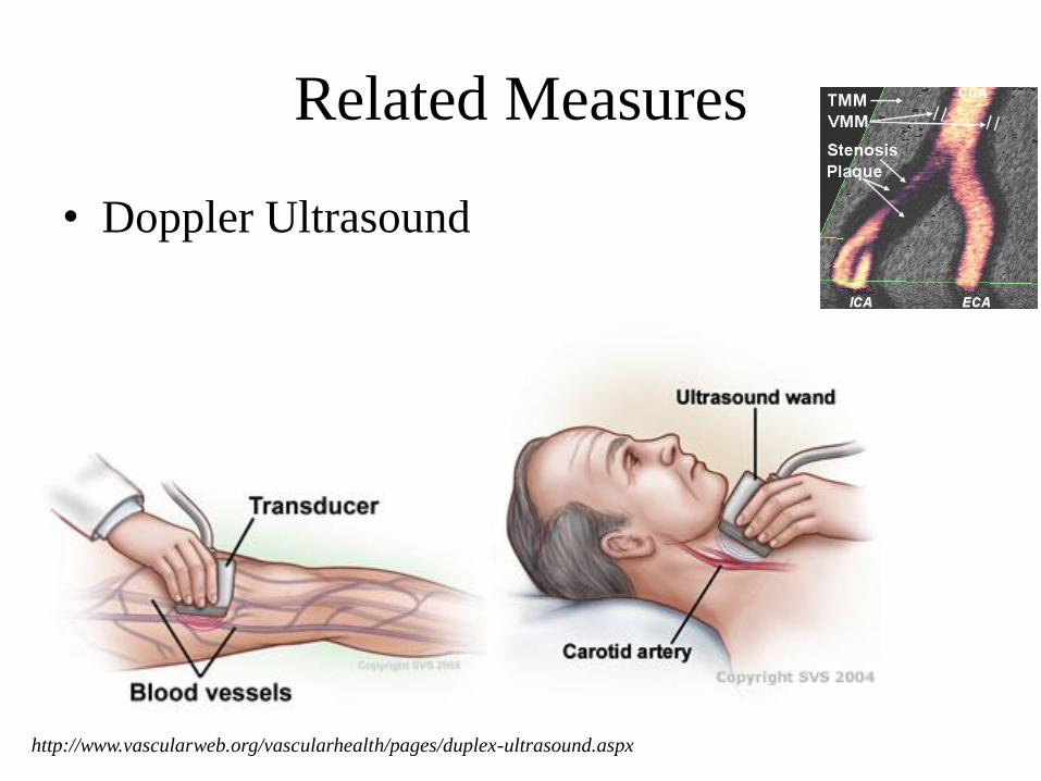
Related Measures
• Doppler Ultrasound
http://www.vascularweb.org/vascularhealth/pages/duplex-ultrasound.aspx
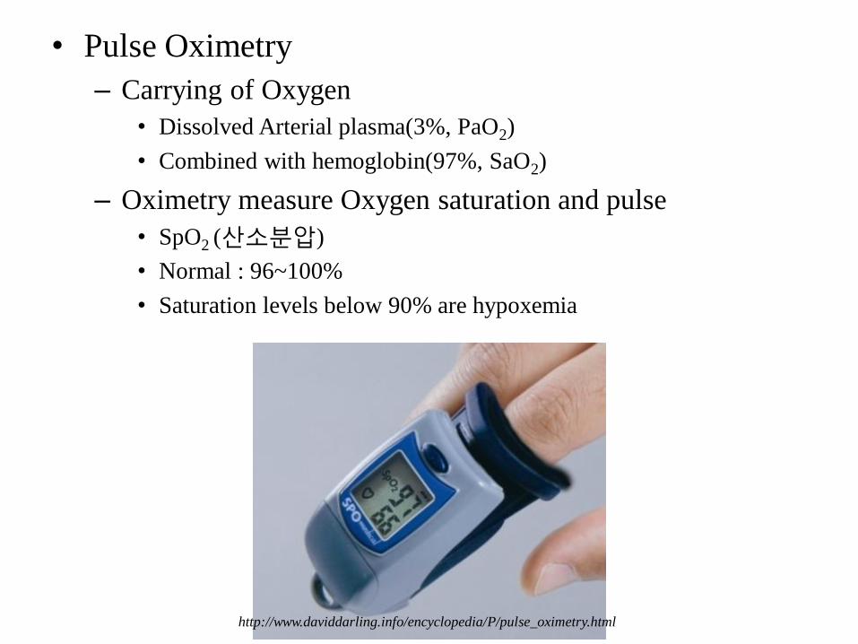
• Pulse Oximetry
– Carrying of Oxygen
• Dissolved Arterial plasma(3%, PaO2)
• Combined with hemoglobin(97%, SaO2)
– Oximetry measure Oxygen saturation and pulse
• SpO2 (산소분압)
• Normal : 96~100%
• Saturation levels below 90% are hypoxemia
http://www.daviddarling.info/encyclopedia/P/pulse_oximetry.html
Respiration

Introduction
• Respiration
1. Pulmonary respiration
Ventilation (환기)
Exchange of O2 and CO2 in the lungs
2. Cellular respiration
O2 utilization and CO2 production by the tissues
Introduction
• Purposes of the respiratory system during
exercise
– Gas exchange between the environment and the
body
– Regulation of acid-base balance during exercise
Maintain the Homeostasis of blood gas (PO2 & PCO2)
1. Function of the Lung
• Main purpose
Gas exchange between the external environment and the body
• Replacing O2
• Removing CO2
• Regulation of acid-base balance
How to do ?
• Ventilation
• Diffusion

1. Function of the Lung
• Ventilation
– Mechanical process of moving air into and out of lungs
• Diffusion
– Random movement of molecules from an area of high concentration to an area of lower concentration
– Fast : ↑ surface area & ↓ diffuse length
– High efficiency
• PO2 & PCO2 in pulmonary vein = PO2 & PCO2 in lung
2. Structure of the Respiratory System
• Organs
1. Nose & nasal cavities
2. Pharynx & larynx
3. Trachea & bronchial tree
4. Lungs
• Alveoli (폐포)
– Site of gas exchange
• Diaphragm
– Major muscle of inspiration
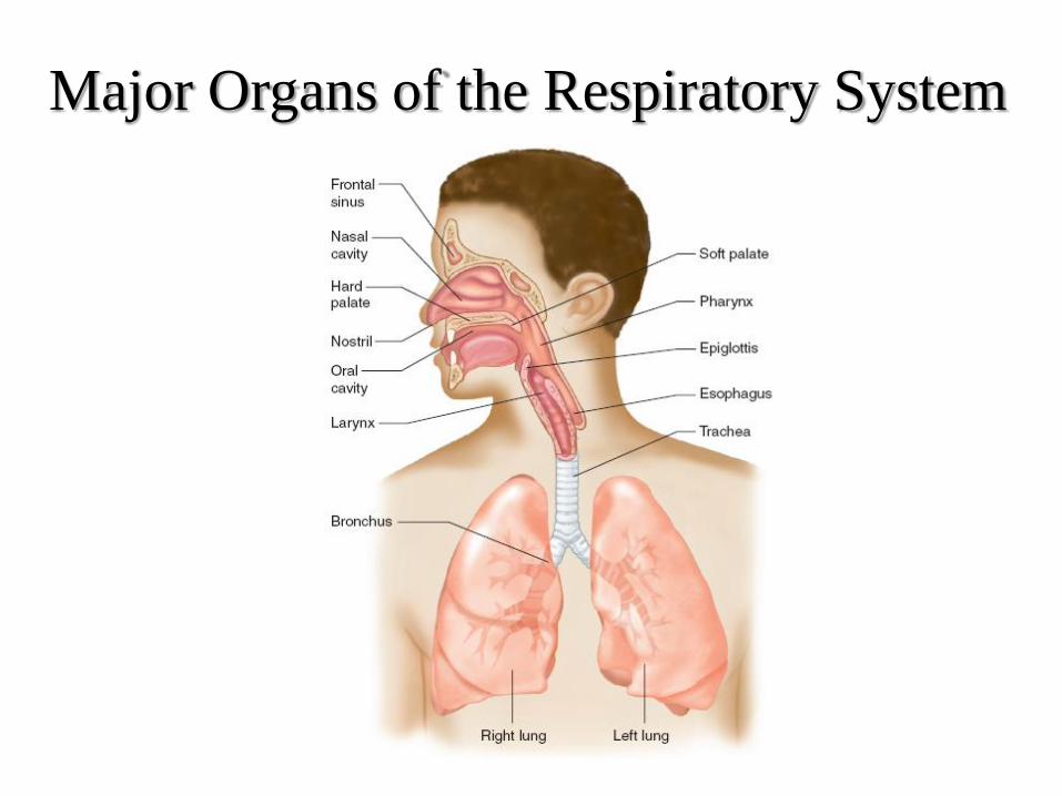
Major Organs of the Respiratory System

2. Structure of the Respiratory System
• Lungs are enclosed by membranes called pleura(흉막)
1. Visceral pleura (inner)
• On outer surface of lung
2. Parietal pleura (outer)
• Lines the thoracic wall
3. Intrapleural space
• Intrapleural pressure is lower than atmospheric
– Prevents collapse of alveoli
Atmosphere (기압; atm)
• Flow direction: ↑→ ↓
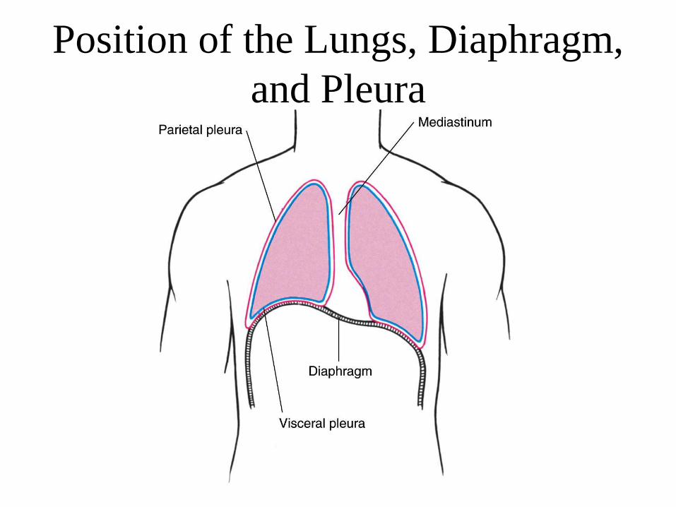
Position of the Lungs, Diaphragm,
and Pleura
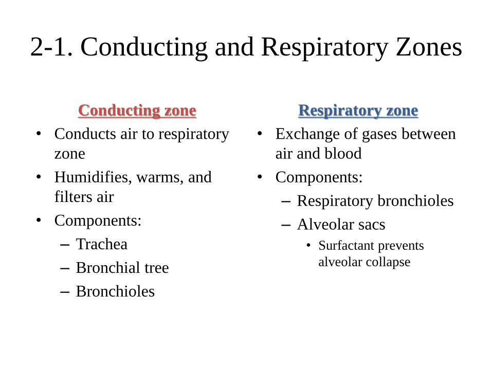
2-1. Conducting and Respiratory Zones
Conducting zone
• Conducts air to respiratory
zone
• Humidifies, warms, and
filters air
• Components:
– Trachea
– Bronchial tree
– Bronchioles
Respiratory zone
• Exchange of gases between
air and blood
• Components:
– Respiratory bronchioles
– Alveolar sacs
• Surfactant prevents
alveolar collapse

Conducting
and
Respiratory
Zones
↑ branches
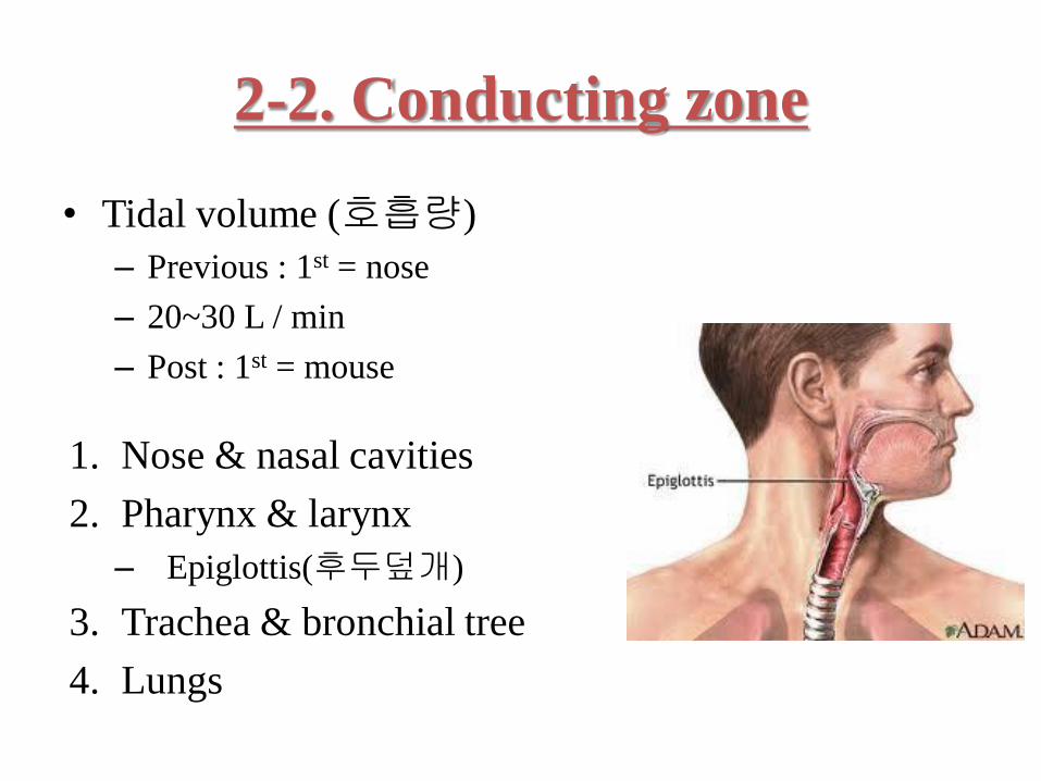
2-2. Conducting zone
• Tidal volume (호흡량)
– Previous : 1st = nose
– 20~30 L / min
– Post : 1st = mouse
1. Nose & nasal cavities
2. Pharynx & larynx
– Epiglottis(후두덮개)
3. Trachea & bronchial tree
4. Lungs
The role of conducting zone in the
respiratory system
• A passageway for air (공기 전도)
• Functions to humidity and filter the air as it
moves toward the respiratory zone of the lung
(습기 첨가 및 여과기능)
– Regardless of the temperature or humidity of the
environment, the air that reaches the lung is
warmed and is saturated with water vapor.
2-2. Conducting zone
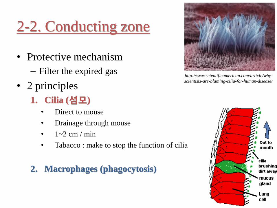
• Protective mechanism
– Filter the expired gas
• 2 principles
1. Cilia (섬모)
• Direct to mouse
• Drainage through mouse
• 1~2 cm / min
• Tabacco : make to stop the function of cilia
2. Macrophages (phagocytosis)
http://www.scientificamerican.com/article/why-
scientists-are-blaming-cilia-for-human-disease/
2-3. Respiratory zone
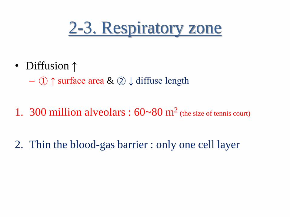
• Diffusion ↑
– ① ↑ surface area & ② ↓ diffuse length
1. 300 million alveolars : 60~80 m2 (the size of tennis court)
2. Thin the blood-gas barrier : only one cell layer
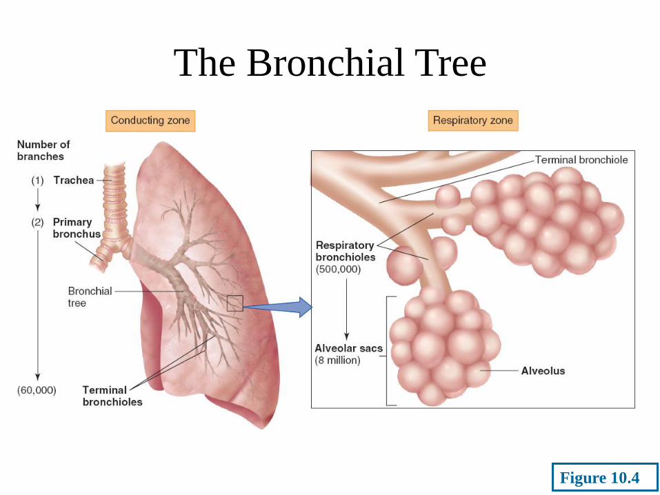
The Bronchial Tree
Figure 10.4
Respiration
• Respiratory rate
– Measurement of breathing rate
• Respiratory cycle
– One inspiration
– Subsequent expiration
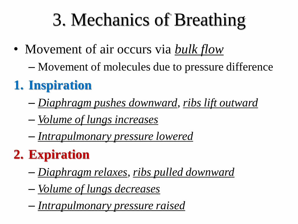
3. Mechanics of Breathing
• Movement of air occurs via bulk flow
– Movement of molecules due to pressure difference
1. Inspiration
– Diaphragm pushes downward, ribs lift outward
– Volume of lungs increases
– Intrapulmonary pressure lowered
2. Expiration
– Diaphragm relaxes, ribs pulled downward
– Volume of lungs decreases
– Intrapulmonary pressure raised
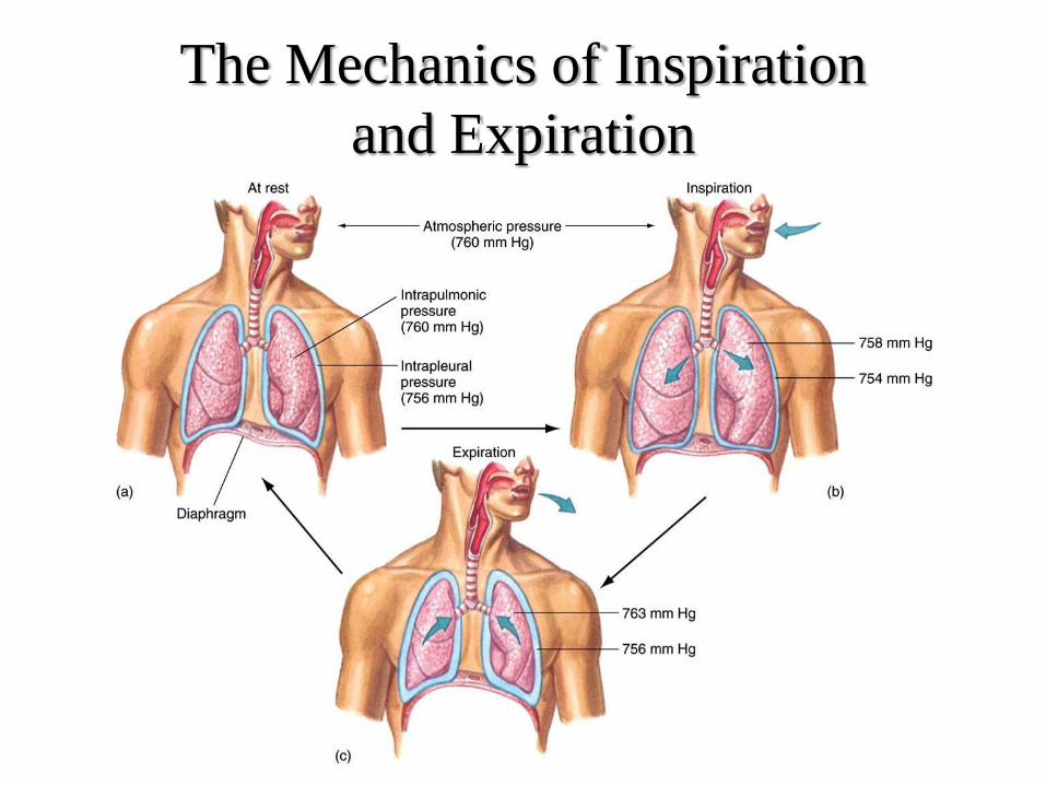
The Mechanics of Inspiration
and Expiration
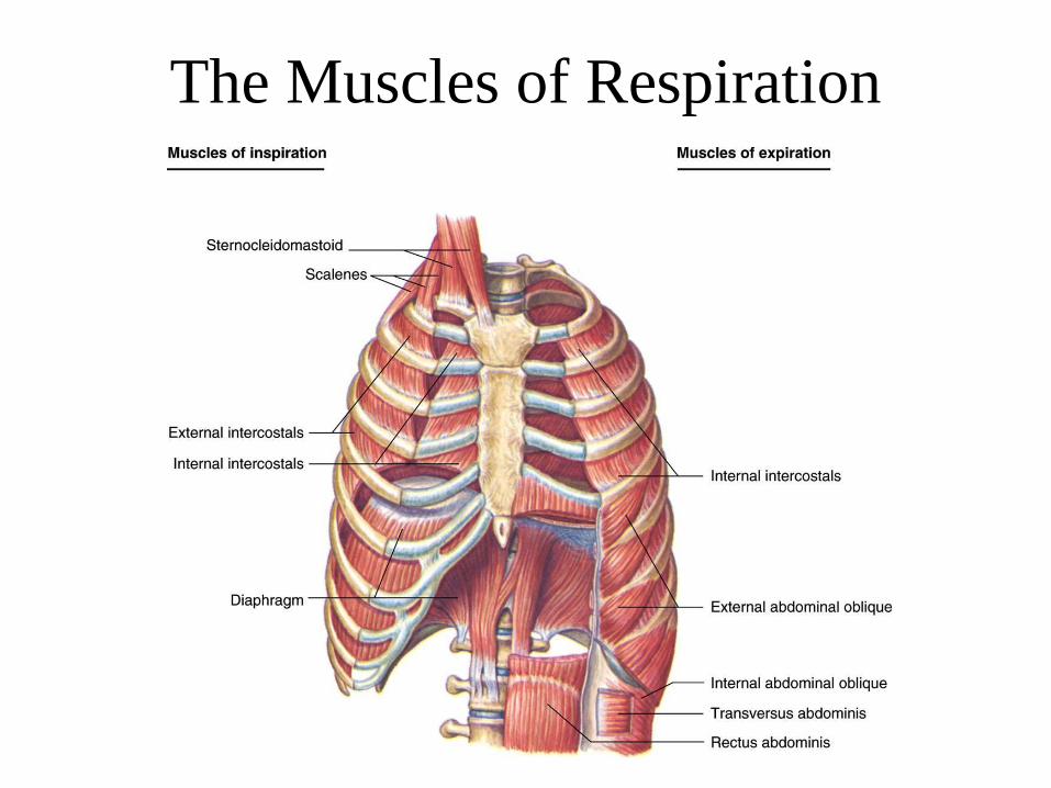
The Muscles of Respiration

3-1. Inspiration
• Diaphragm
– Primary ms
– Phrenic n. : C3-C5
– During normal, quiet breathing : most of work
• Support ms (during Ex.)
– Pec minor
– Intercostalis
– SCM
– Scalenius : TP of C2~7 → 1st & 2nd ribs
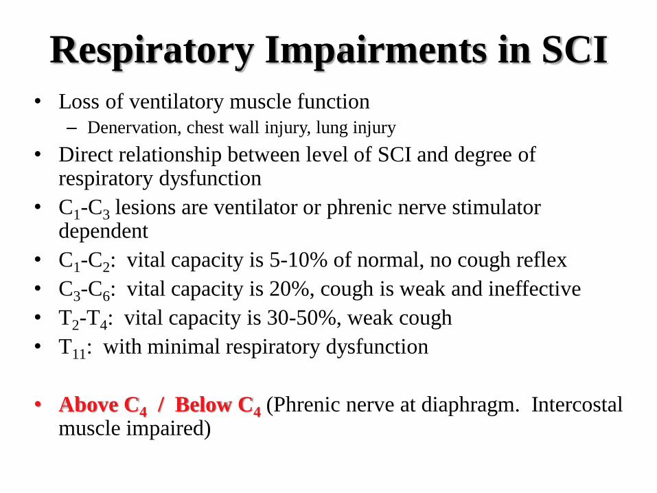
Respiratory Impairments in SCI
• Loss of ventilatory muscle function – Denervation, chest wall injury, lung injury
• Direct relationship between level of SCI and degree of respiratory dysfunction
• C1-C3 lesions are ventilator or phrenic nerve stimulator dependent
• C1-C2: vital capacity is 5-10% of normal, no cough reflex
• C3-C6: vital capacity is 20%, cough is weak and ineffective
• T2-T4: vital capacity is 30-50%, weak cough
• T11: with minimal respiratory dysfunction
• Above C4 / Below C4 (Phrenic nerve at diaphragm. Intercostal muscle impaired)

3-2. Expiration
• At rest
– Passive
– By elasticity of lung & chest wall
• At exercise & work
– Active
– Internal & External Obliqus
– Transverse Abdominis
– Rectus Abdominis
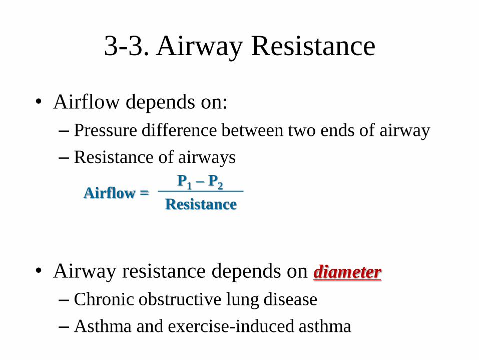
3-3. Airway Resistance
• Airflow depends on:
– Pressure difference between two ends of airway
– Resistance of airways
• Airway resistance depends on diameter
– Chronic obstructive lung disease
– Asthma and exercise-induced asthma
Airflow = P1 – P2
Resistance
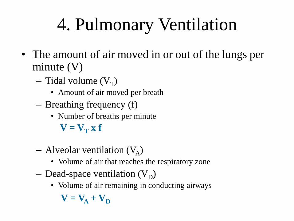
• The amount of air moved in or out of the lungs per minute (V)
– Tidal volume (VT) • Amount of air moved per breath
– Breathing frequency (f) • Number of breaths per minute
– Alveolar ventilation (VA) • Volume of air that reaches the respiratory zone
– Dead-space ventilation (VD) • Volume of air remaining in conducting airways
V = VT x f
V = VA + VD
4. Pulmonary Ventilation
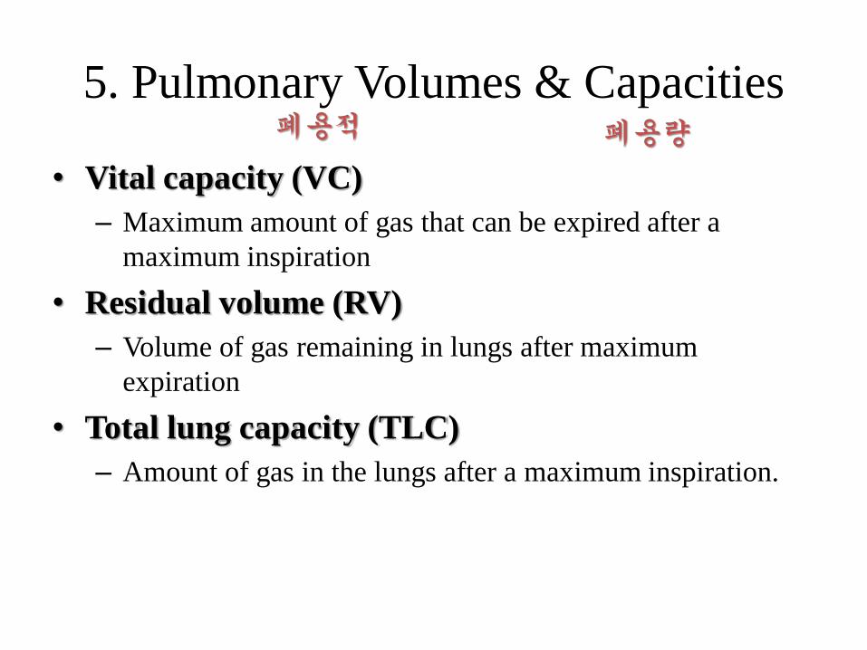
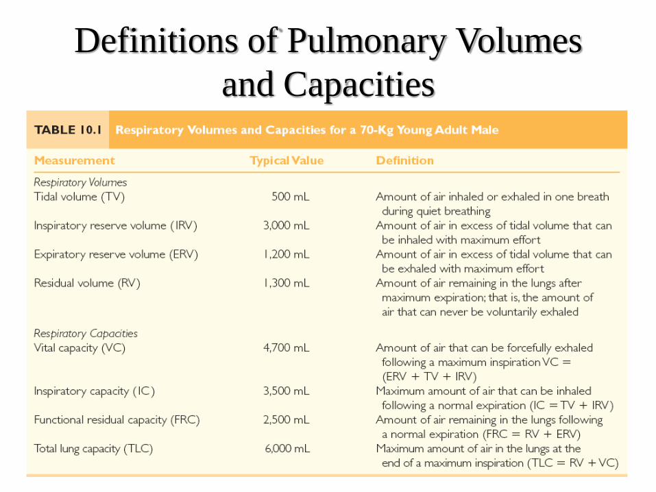
5. Pulmonary Volumes & Capacities
• Vital capacity (VC)
– Maximum amount of gas that can be expired after a
maximum inspiration
• Residual volume (RV)
– Volume of gas remaining in lungs after maximum
expiration
• Total lung capacity (TLC)
– Amount of gas in the lungs after a maximum inspiration.
폐용적 폐용량
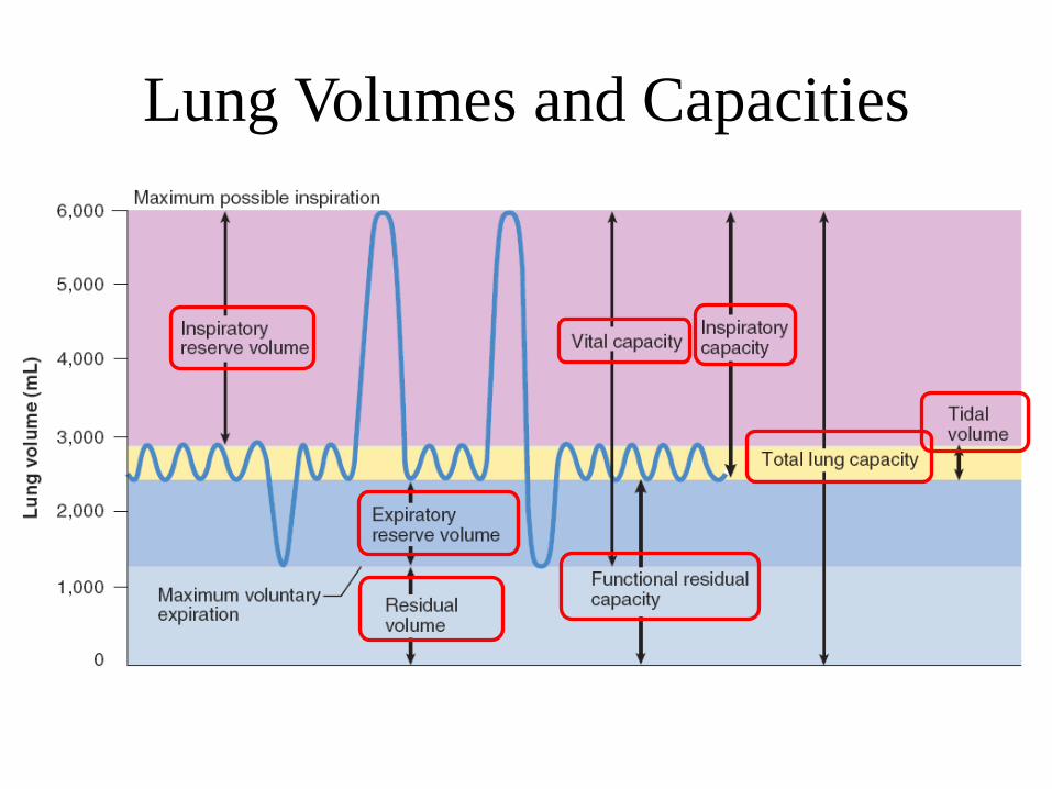
Lung Volumes and Capacities
Definitions of Pulmonary Volumes
and Capacities
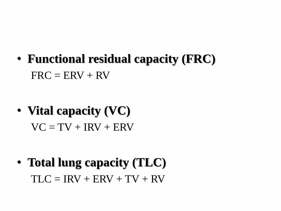
• Functional residual capacity (FRC)
FRC = ERV + RV
• Vital capacity (VC)
VC = TV + IRV + ERV
• Total lung capacity (TLC)
TLC = IRV + ERV + TV + RV
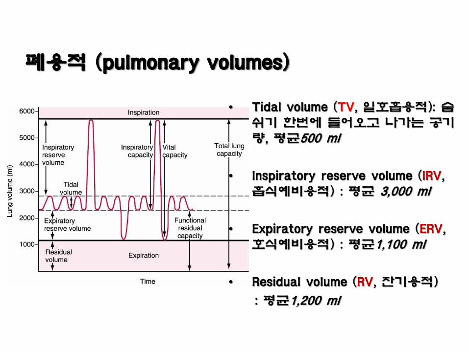
폐용적 (pulmonary volumes)
• Tidal volume (TV, 일호흡용적): 숨쉬기 한번에 들어오고 나가는 공기량, 평균500 ml
• Inspiratory reserve volume (IRV, 흡식예비용적) : 평균 3,000 ml
• Expiratory reserve volume (ERV, 호식예비용적) : 평균1,100 ml
• Residual volume (RV, 잔기용적)
: 평균1,200 ml
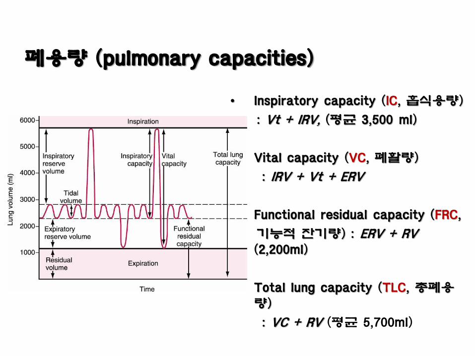
폐용량 (pulmonary capacities)
• Inspiratory capacity (IC, 흡식용량)
: Vt + IRV, (평균 3,500 ml)
• Vital capacity (VC, 폐활량)
: IRV + Vt + ERV
• Functional residual capacity (FRC,
기능적 잔기량) : ERV + RV (2,200ml)
• Total lung capacity (TLC, 총폐용량)
: VC + RV (평균 5,700ml)
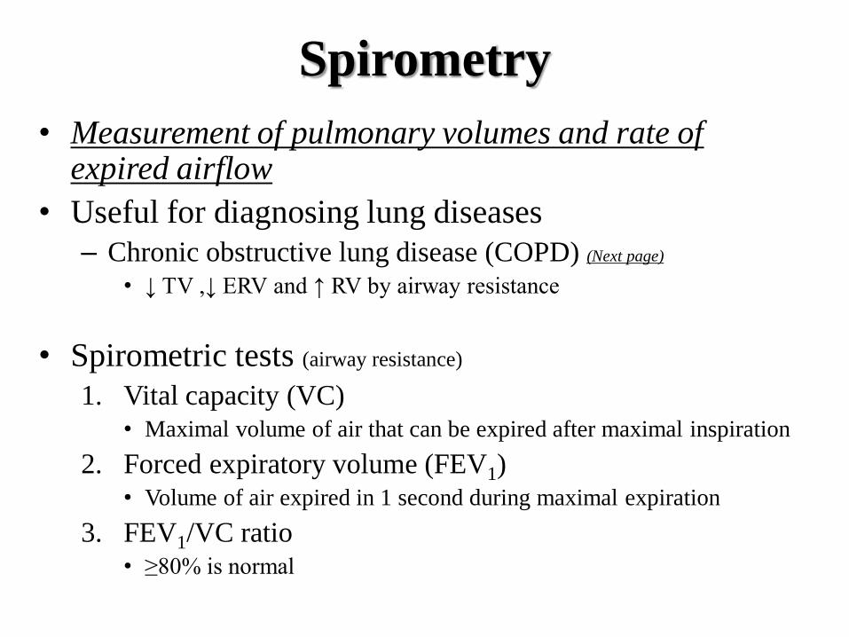
Spirometry
• Measurement of pulmonary volumes and rate of expired airflow
• Useful for diagnosing lung diseases
– Chronic obstructive lung disease (COPD) (Next page)
• ↓ TV ,↓ ERV and ↑ RV by airway resistance
• Spirometric tests (airway resistance)
1. Vital capacity (VC) • Maximal volume of air that can be expired after maximal inspiration
2. Forced expiratory volume (FEV1) • Volume of air expired in 1 second during maximal expiration
3. FEV1/VC ratio • ≥80% is normal

http://thoracic.group.shef.ac.uk/services/services-copd.htm
http://www.medchipsolutions.com/spiroconnect_copd.html
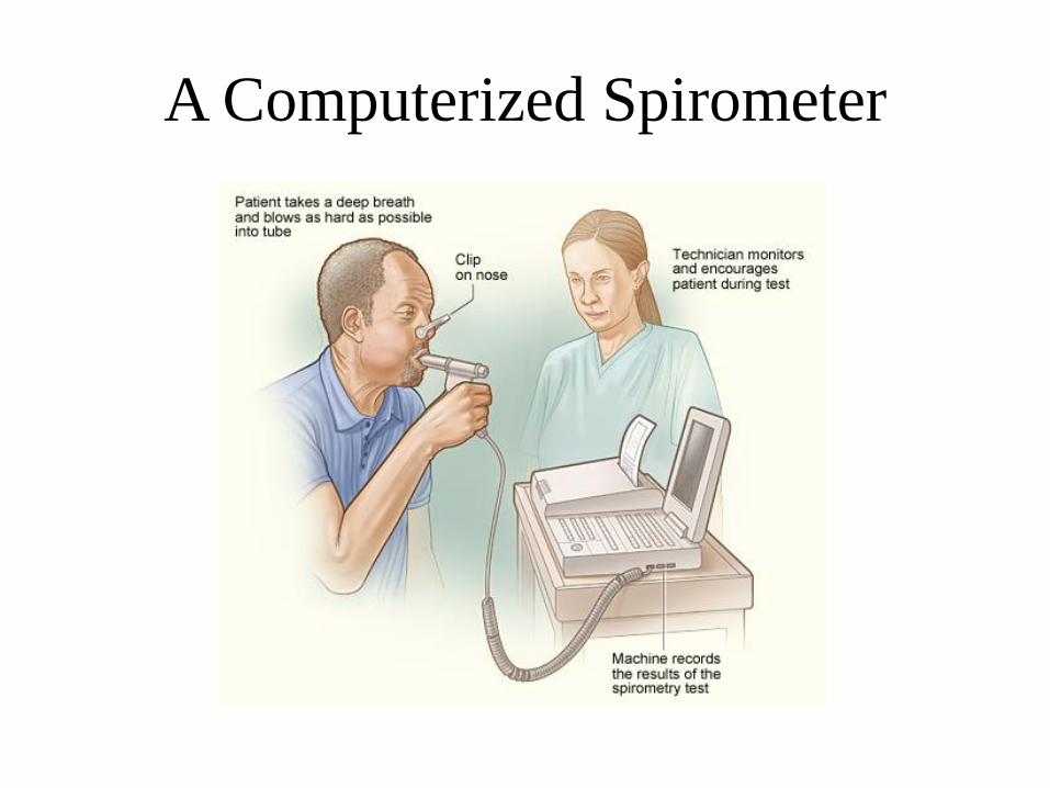
A Computerized Spirometer
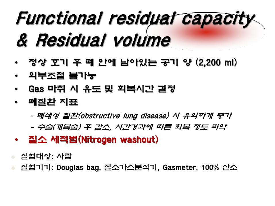
Functional residual capacity & Residual volume • 정상 호기 후 폐 안에 남아있는 공기 양 (2,200 ml)
• 외부조절 불가능
• Gas 마취 시 유도 및 회복시간 결정
• 폐질환 지표
- 폐쇄성 질환(obstructive lung disease) 시 유의하게 증가
- 수술(개복술) 후 감소, 시간경과에 따른 회복 정도 파악
• 질소 세척법(Nitrogen washout)
실험대상: 사람
실험기기: Douglas bag, 질소가스분석기, Gasmeter, 100% 산소
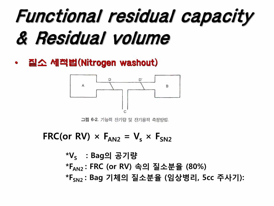
• 질소 세척법(Nitrogen washout)
FRC(or RV) × FAN2 = Vs × FSN2
*VS : Bag의 공기량
*FAN2 : FRC (or RV) 속의 질소분율 (80%)
*FSN2 : Bag 기체의 질소분율 (임상병리, 5cc 주사기):
Functional residual capacity & Residual volume
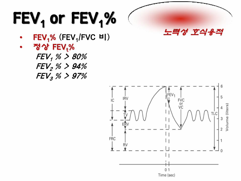
FEV1 or FEV1% • FEV1% (FEV1/FVC 비) • 정상 FEVt% FEV1 % > 80% FEV2 % > 94% FEV3 % > 97%
노력성 호식용적
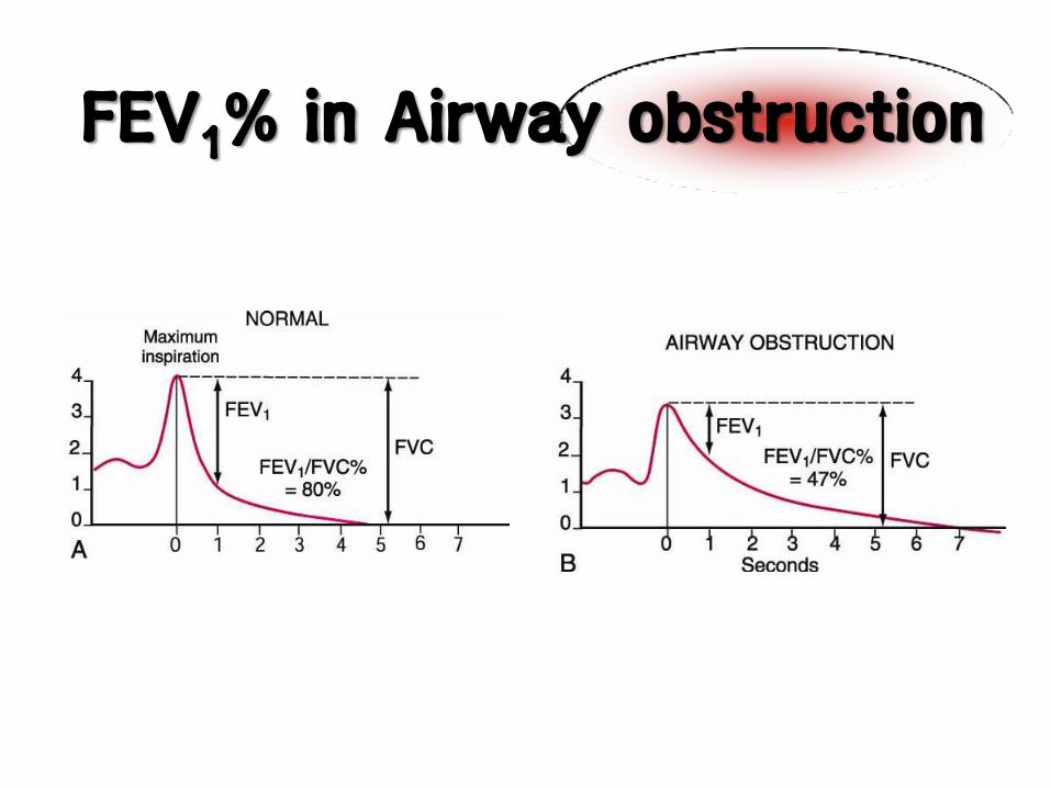
FEV1% in Airway obstruction
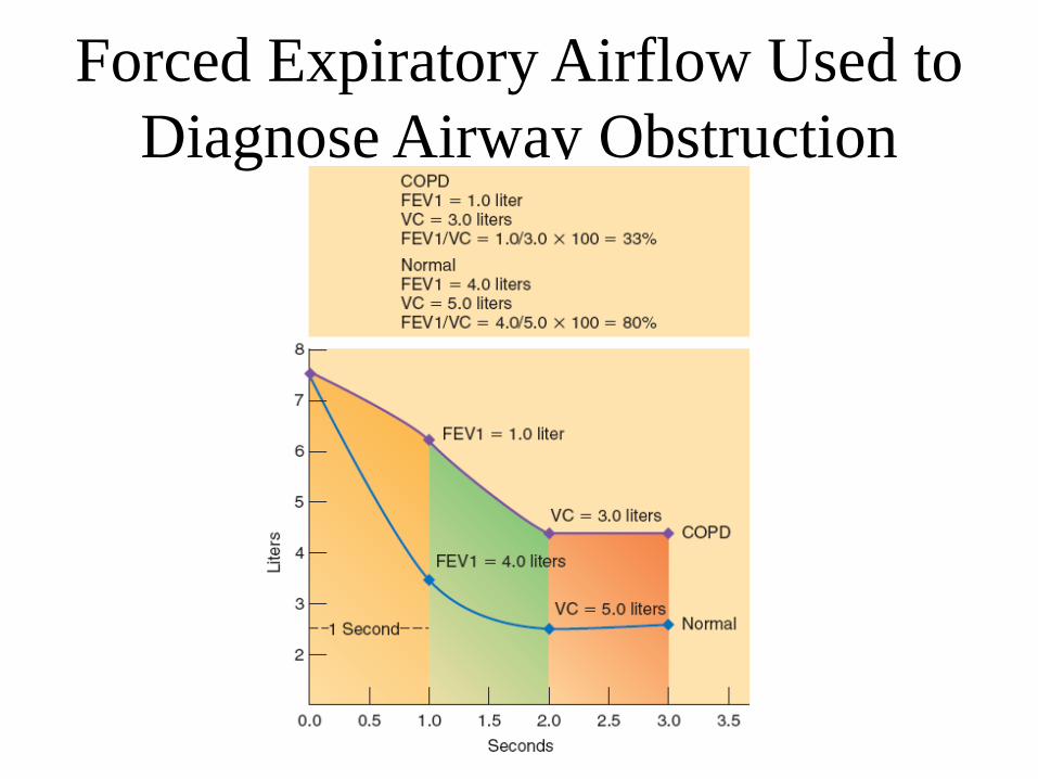
Forced Expiratory Airflow Used to
Diagnose Airway Obstruction

FEV1% 및 FVC%를 이용한 폐질환의 유형 분석
Obstructive Normal or Restrictive
Factors influencing respiration
• Age
• Body size and Stature – Vital capacity
• Men>Women, Adult>adolescent & children
• Tall, Thin>Short, Thick
– With Larger lung capacity there is also a lower RR
• Exercise
• Body position
• Environment
• Emotions/stress - increase
• Pharmacological Agents
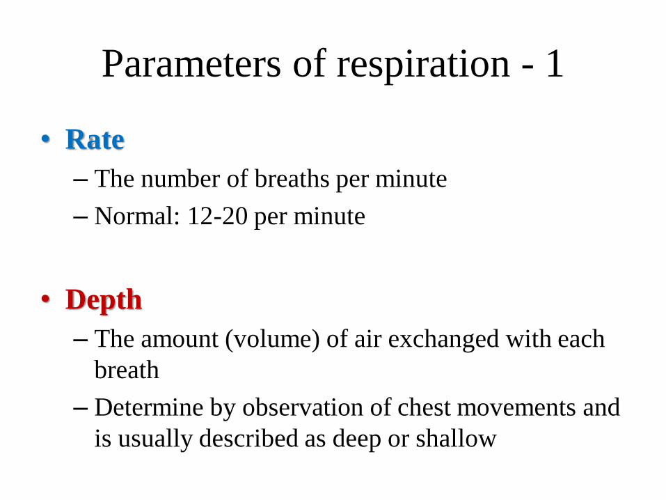
Parameters of respiration - 1
• Rate
– The number of breaths per minute
– Normal: 12-20 per minute
• Depth
– The amount (volume) of air exchanged with each
breath
– Determine by observation of chest movements and
is usually described as deep or shallow
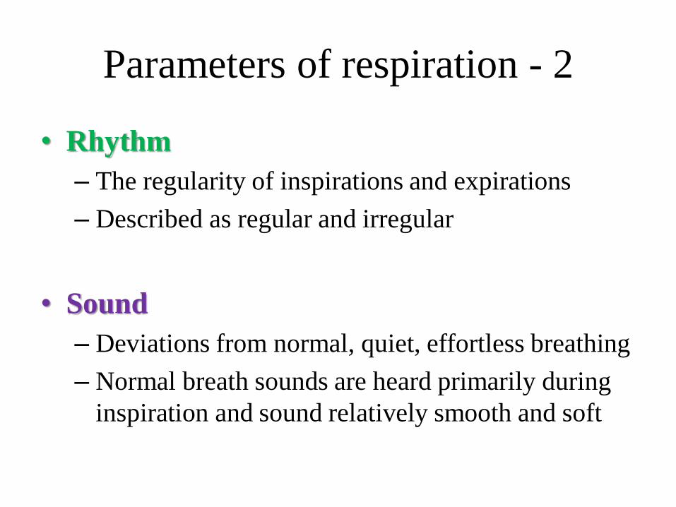
Parameters of respiration - 2
• Rhythm
– The regularity of inspirations and expirations
– Described as regular and irregular
• Sound
– Deviations from normal, quiet, effortless breathing
– Normal breath sounds are heard primarily during
inspiration and sound relatively smooth and soft
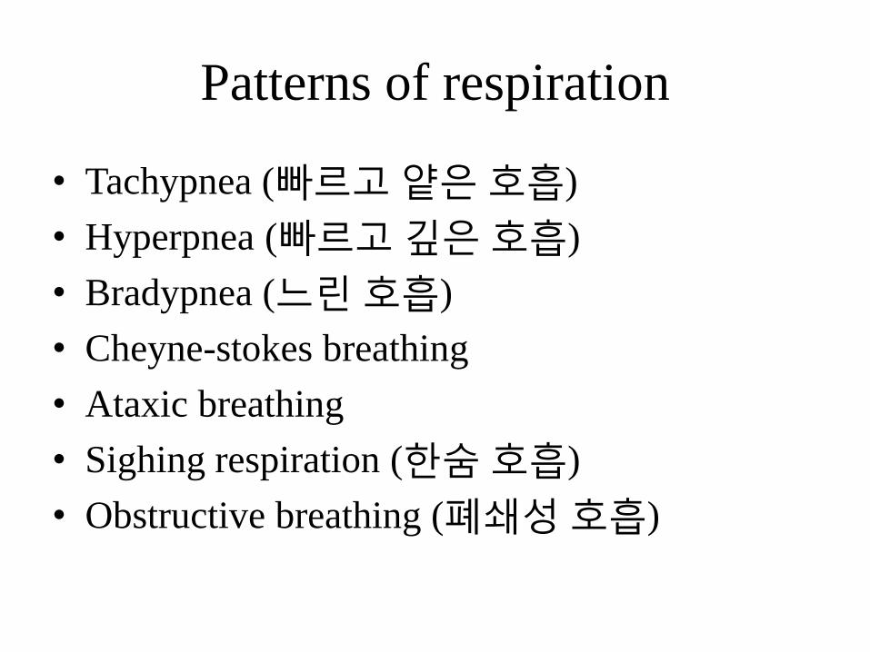
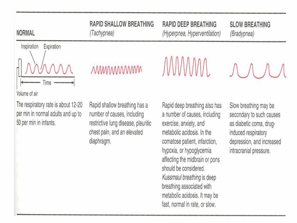
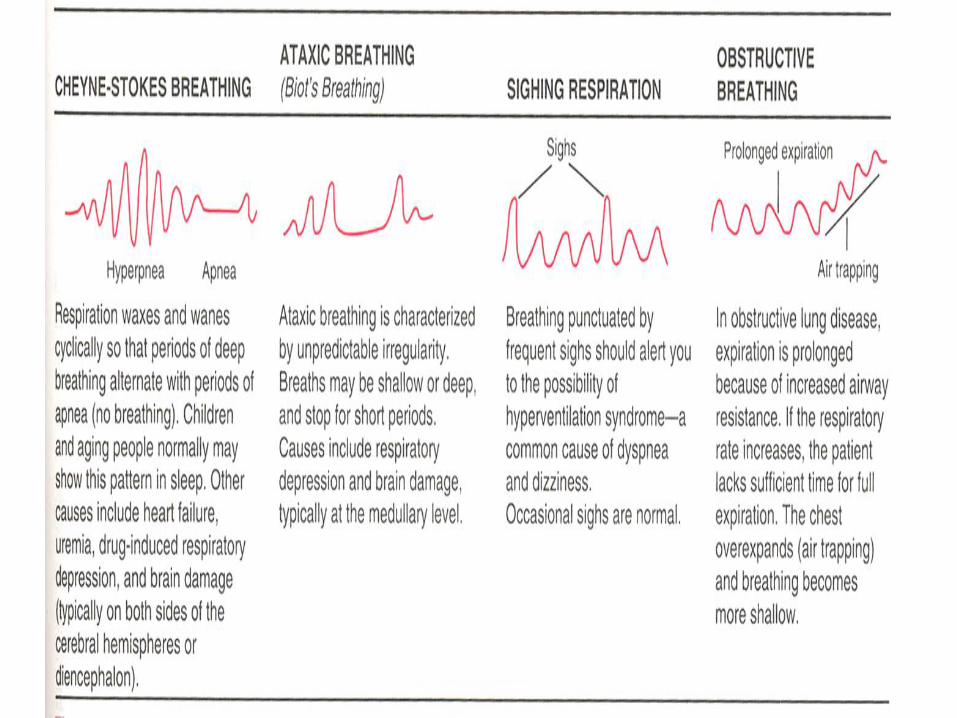
Patterns of respiration
• Tachypnea (빠르고 얕은 호흡)
• Hyperpnea (빠르고 깊은 호흡)
• Bradypnea (느린 호흡)
• Cheyne-stokes breathing
• Ataxic breathing
• Sighing respiration (한숨 호흡)
• Obstructive breathing (폐쇄성 호흡)

Method of measuring respiration
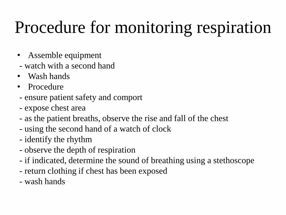
Procedure for monitoring respiration
• Assemble equipment
- watch with a second hand
• Wash hands
• Procedure
- ensure patient safety and comport
- expose chest area
- as the patient breaths, observe the rise and fall of the chest
- using the second hand of a watch of clock
- identify the rhythm
- observe the depth of respiration
- if indicated, determine the sound of breathing using a stethoscope
- return clothing if chest has been exposed
- wash hands


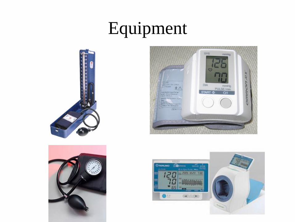
Blood pressure
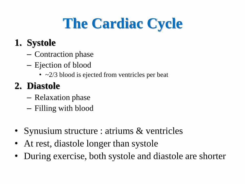
The Cardiac Cycle
1. Systole
– Contraction phase
– Ejection of blood • ~2/3 blood is ejected from ventricles per beat
2. Diastole
– Relaxation phase
– Filling with blood
• Synusium structure : atriums & ventricles
• At rest, diastole longer than systole
• During exercise, both systole and diastole are shorter
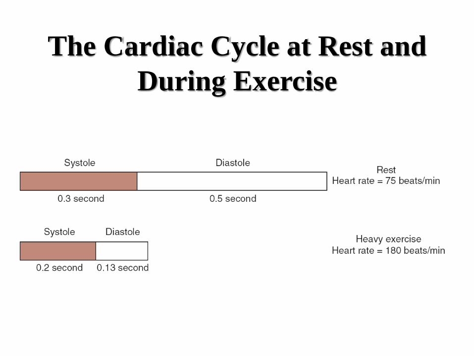
The Cardiac Cycle at Rest and
During Exercise
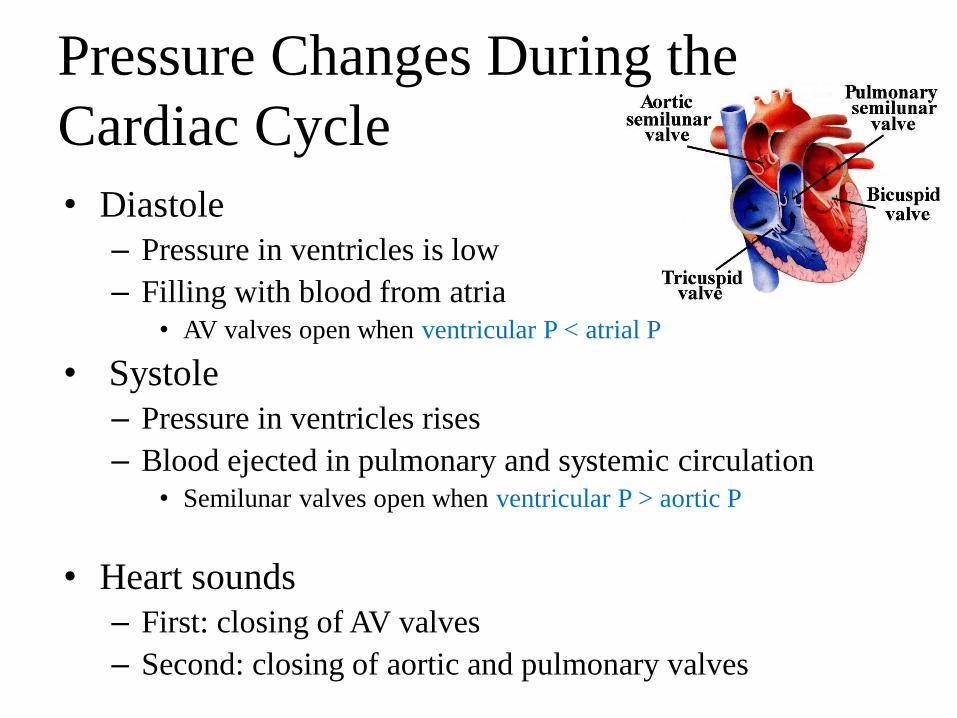
Pressure Changes During the
Cardiac Cycle
• Diastole
– Pressure in ventricles is low
– Filling with blood from atria • AV valves open when ventricular P < atrial P
• Systole
– Pressure in ventricles rises
– Blood ejected in pulmonary and systemic circulation • Semilunar valves open when ventricular P > aortic P
• Heart sounds
– First: closing of AV valves
– Second: closing of aortic and pulmonary valves
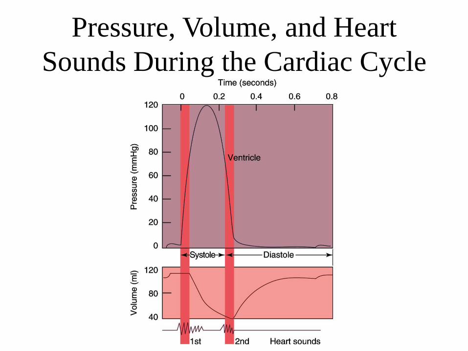
Pressure, Volume, and Heart
Sounds During the Cardiac Cycle
Blood Pressure
• Measure of vascular resistance to blood flow
= The force the blood exerts against a vessel wall
• Two values
– 1st value : systolic pressure (the highest pressure)
– 2nd value : diastolic pressure (the lowest pressure)
• Pulse pressure
– Mathematical difference between the systolic and
diastolic pressure
– 120/80
• Pulse pressure 40mmHg

Arterial Blood Pressure
• Blood pressure (BP)
– The force exerted by blood against the arterial walls
– Determination : how much blood is pumped and the
resistance to blood flow
• BP measurements
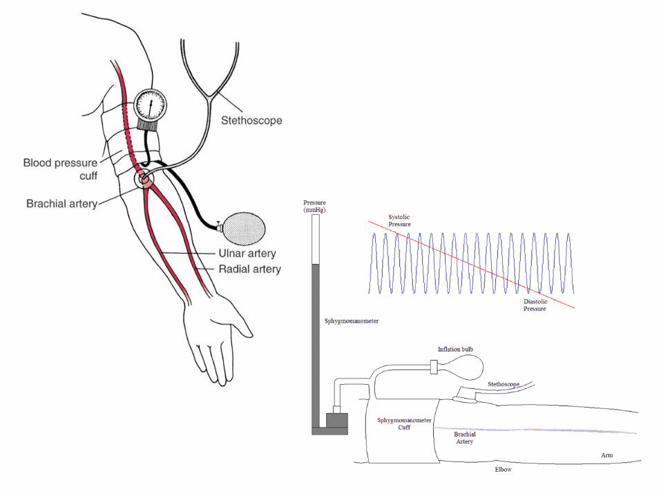
– On Artery
– Sphygmomanometer
MAP = DBP + 0.33(SBP – DBP)
Arterial Blood Pressure
• Expressed as systolic/diastolic
– Normal is 120/80 mmHg (male) / 110/70 mmHg (female)
• Systolic pressure (SBP)
– Pressure generated during ventricular contraction
• Diastolic pressure (DBP)
– Pressure in the arteries during cardiac relaxation
• Pulse pressure
– Difference between systolic and diastolic
• Mean arterial pressure (MAP)
– Average pressure in the arteries
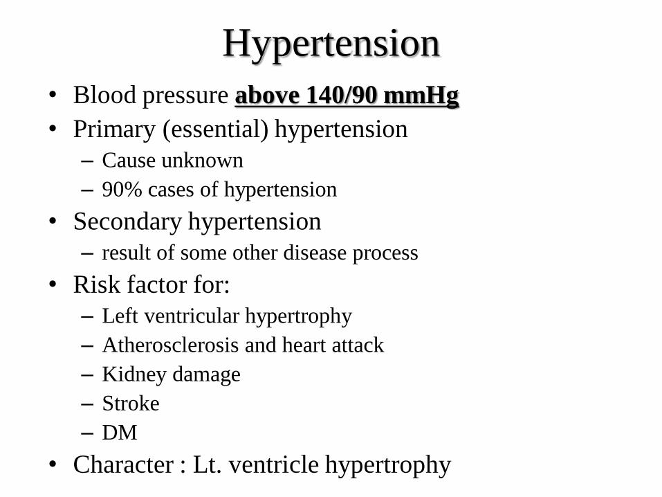
Hypertension
• Blood pressure above 140/90 mmHg
• Primary (essential) hypertension
– Cause unknown
– 90% cases of hypertension
• Secondary hypertension
– result of some other disease process
• Risk factor for:
– Left ventricular hypertrophy
– Atherosclerosis and heart attack
– Kidney damage
– Stroke
– DM
• Character : Lt. ventricle hypertrophy

Ex) Atherosclerosis • Is most common form of
arteriosclerosis
(hardening of arteries)
– Accounts for 50% of
deaths in US
• Localized plaques
(atheromas) reduce flow
in an artery
– And act as sites for
thrombus (blood clots)
13-79

Factors that Influence Arterial
Blood Pressure • Determinants of mean arterial pressure
– Cardiac output
– Total vascular resistance
1. Short-term regulation
– Sympathetic nervous system
– Baroreceptors in aorta and carotid arteries • Increase in BP = increased SNS activity
• Decrease in BP = decreased SNS activity
2. Long-term regulation
– Kidneys • Via control of blood volume
MAP = cardiac output x total vascular resistance

Factors That Influence Arterial
Blood Pressure
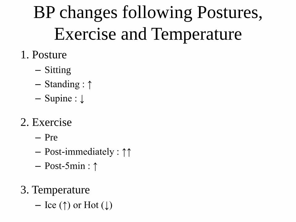
BP changes following Postures,
Exercise and Temperature 1. Posture
– Sitting
– Standing : ↑
– Supine : ↓
2. Exercise
– Pre
– Post-immediately : ↑↑
– Post-5min : ↑
3. Temperature
– Ice (↑) or Hot (↓)
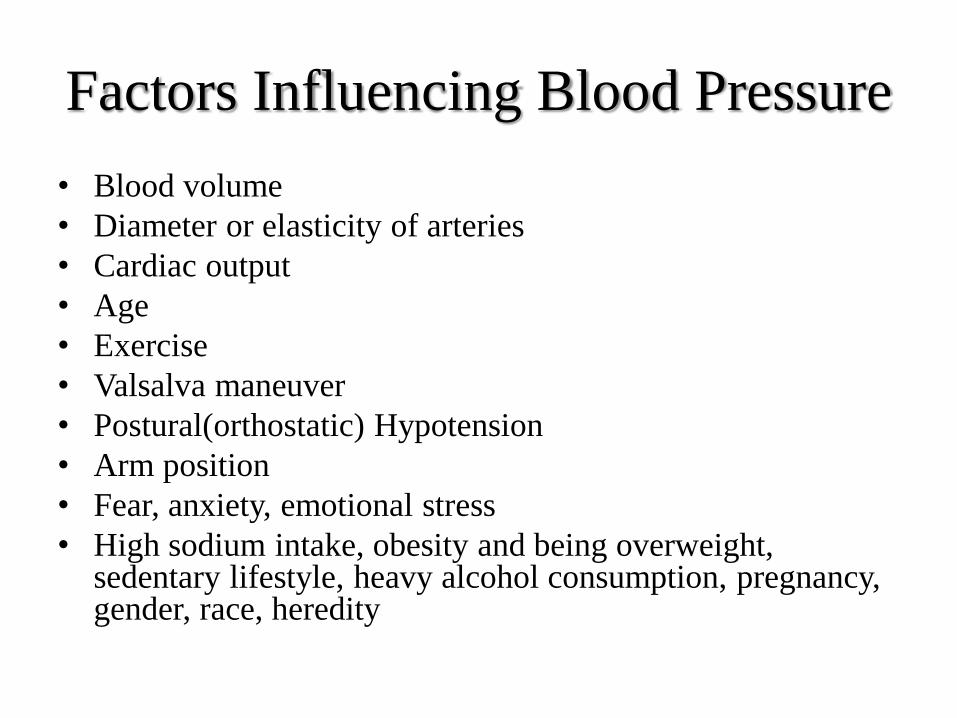
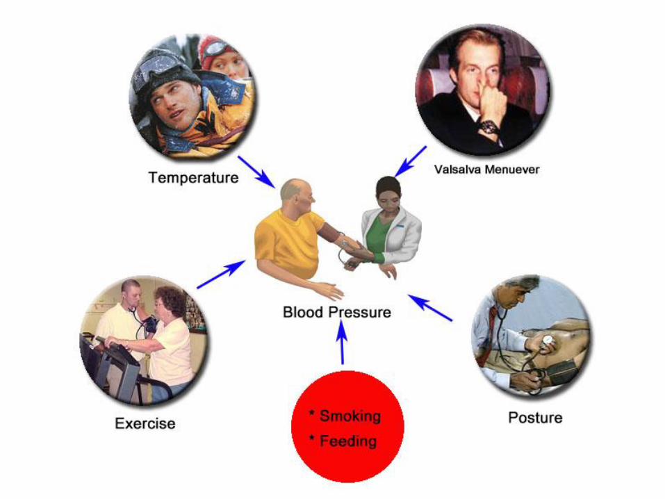
Factors Influencing Blood Pressure
• Blood volume
• Diameter or elasticity of arteries
• Cardiac output
• Age
• Exercise
• Valsalva maneuver
• Postural(orthostatic) Hypotension
• Arm position
• Fear, anxiety, emotional stress
• High sodium intake, obesity and being overweight, sedentary lifestyle, heavy alcohol consumption, pregnancy, gender, race, heredity
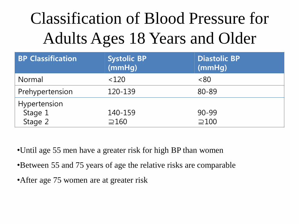
Classification of Blood Pressure for
Adults Ages 18 Years and Older BP Classification Systolic BP
(mmHg) Diastolic BP (mmHg)
Normal <120 <80
Prehypertension 120-139 80-89
Hypertension Stage 1 Stage 2
140-159 ⊇160
90-99 ⊇100
•Until age 55 men have a greater risk for high BP than women
•Between 55 and 75 years of age the relative risks are comparable
•After age 75 women are at greater risk

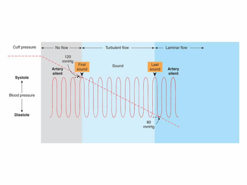
Measurement of
Blood Pressure
9-122
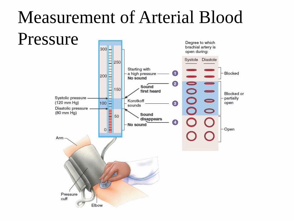
Measurement of Arterial Blood
Pressure
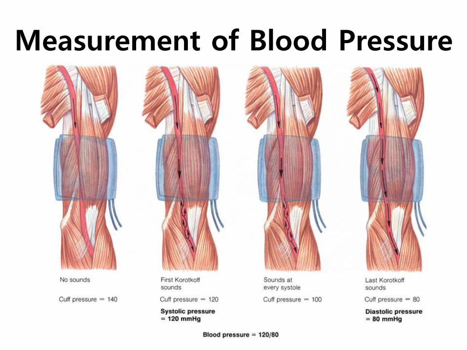
Measurement of Blood Pressure
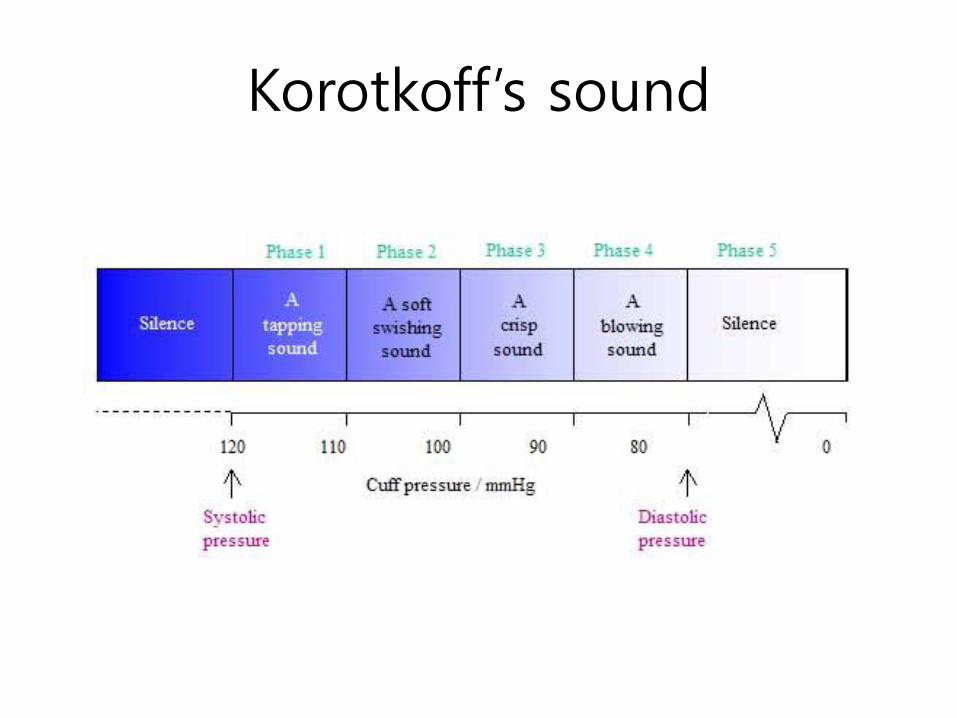
Korotkoff’s sound
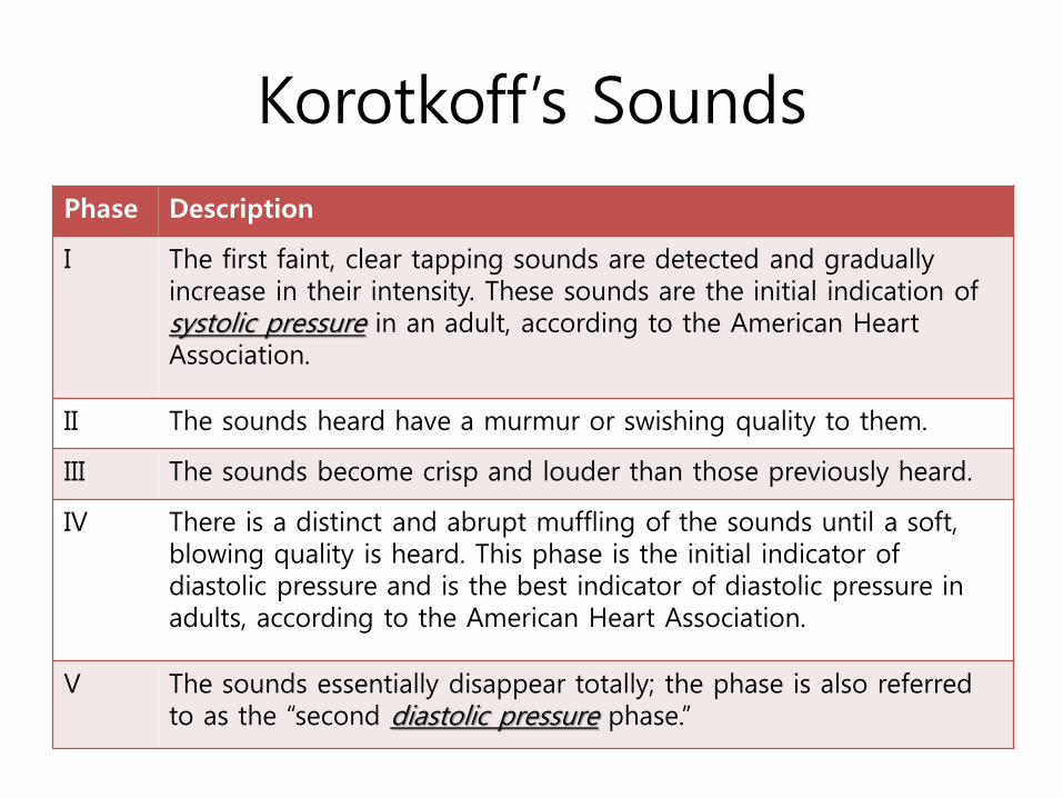
Korotkoff’s Sounds
Phase Description
I The first faint, clear tapping sounds are detected and gradually increase in their intensity. These sounds are the initial indication of systolic pressure in an adult, according to the American Heart Association.
II The sounds heard have a murmur or swishing quality to them.
III The sounds become crisp and louder than those previously heard.
IV There is a distinct and abrupt muffling of the sounds until a soft, blowing quality is heard. This phase is the initial indicator of diastolic pressure and is the best indicator of diastolic pressure in adults, according to the American Heart Association.
V The sounds essentially disappear totally; the phase is also referred to as the “second diastolic pressure phase.”
Electrical Activity of the Heart
• Contraction of the heart depends on electrical
stimulation of the myocardium
• Conduction system
1. Sinoatrial node (SA node)
• Pacemaker, initiates depolarization
2. Atrioventricular node (AV node)
• Passes depolarization to ventricles
• Brief delay to allow for ventricular filling
3. Bundle Branches
• To left and right ventricle
4. Purkinje fibers
• Throughout ventricles
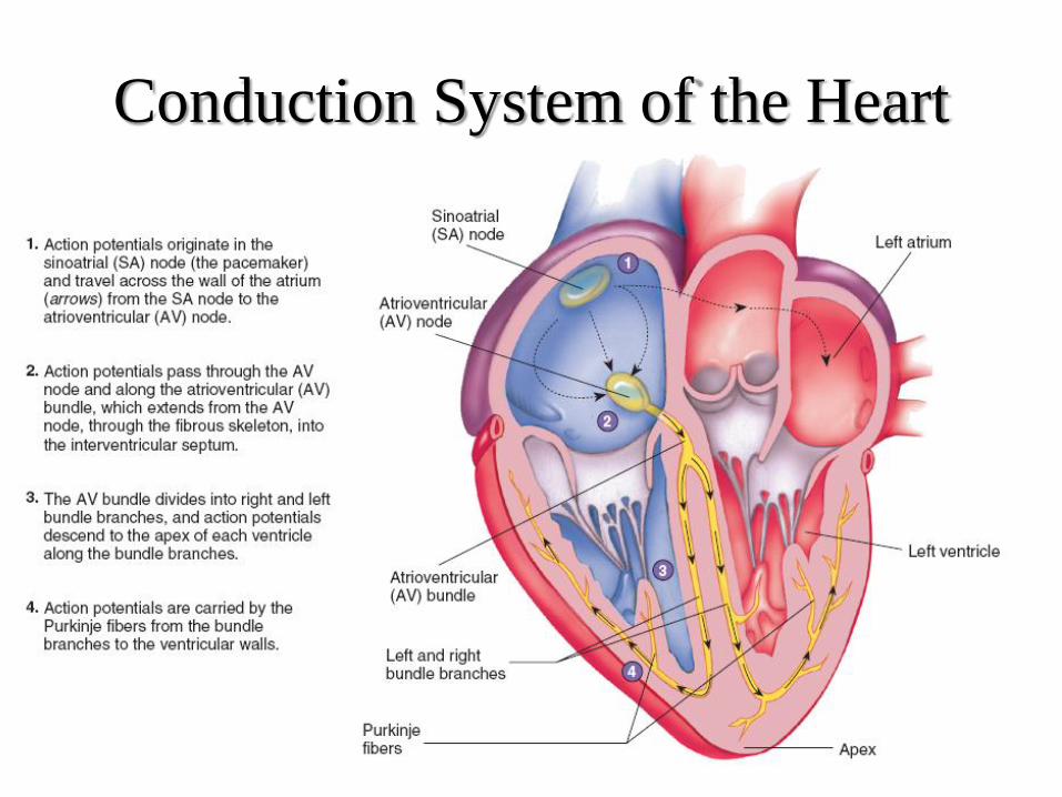
Conduction System of the Heart
Heart muscle characters
• Structural characters
– 2 types : contractile cell & conducting cell
– Contractile cell : atrium & ventricle
– Functional syncytium
• Like one cell
• Functional characters
– Rhythmicity = automatism
– Excitability
– Conductivity
– Contractility
– All or none
– Refractory period : non-fatigue
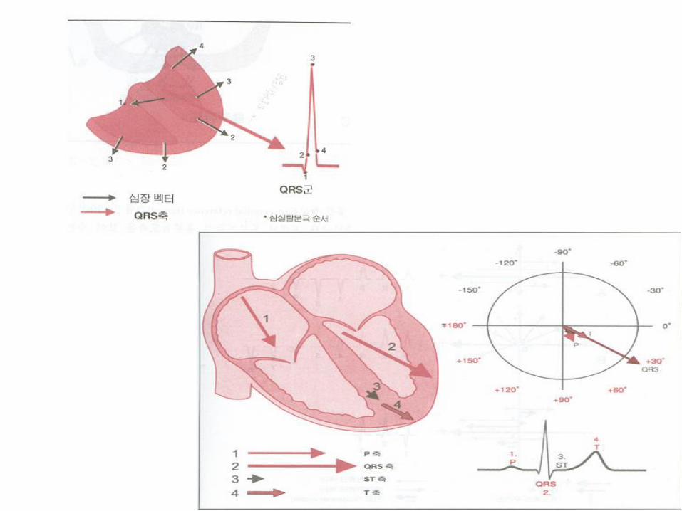
Electrocardiogram (ECG)
• Records the electrical activity of the heart
• P wave
– Atrial depolarization
• QRS complex
– Ventricular depolarization and atrial repolarization
• T wave
– Ventricular repolarization
• ECG abnormalities may indicate coronary heart disease
– ST-segment depression can indicate myocardial ischemia
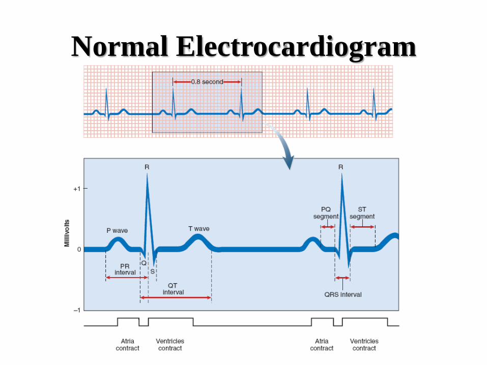
Normal Electrocardiogram
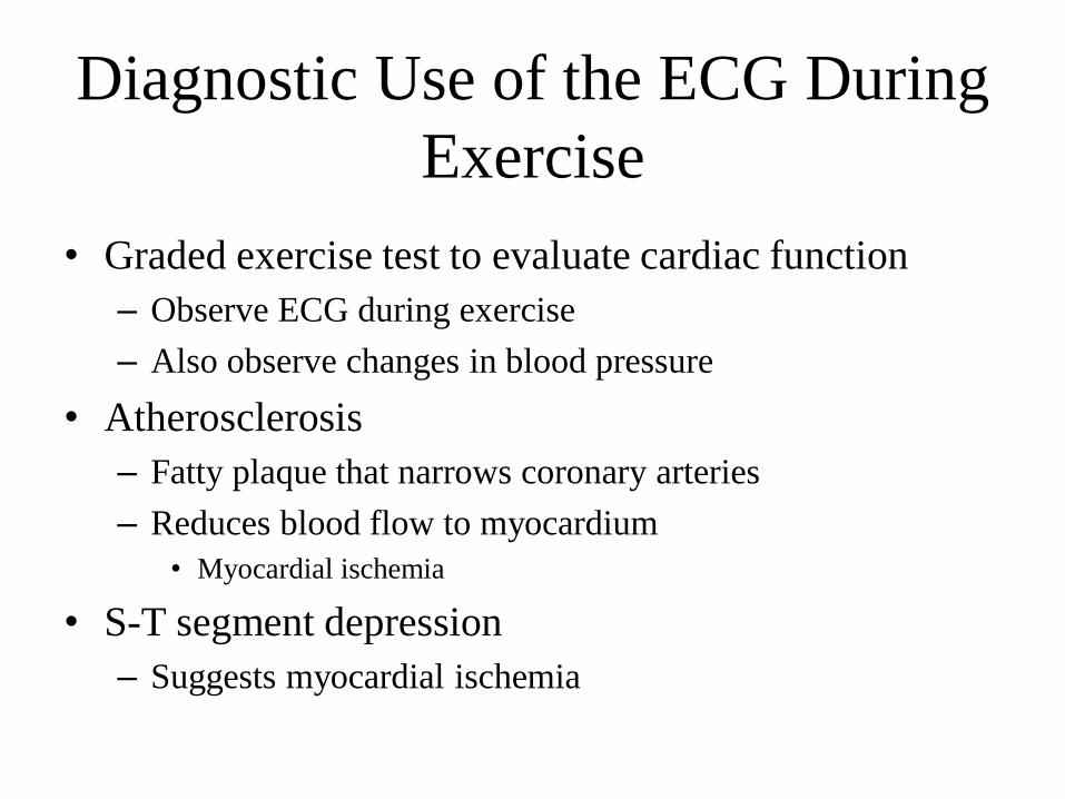
Diagnostic Use of the ECG During
Exercise
• Graded exercise test to evaluate cardiac function
– Observe ECG during exercise
– Also observe changes in blood pressure
• Atherosclerosis
– Fatty plaque that narrows coronary arteries
– Reduces blood flow to myocardium
• Myocardial ischemia
• S-T segment depression
– Suggests myocardial ischemia
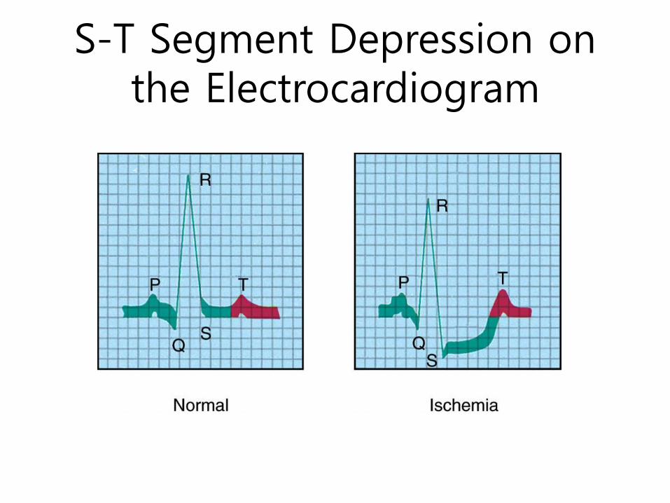
S-T Segment Depression on the Electrocardiogram

Relationship Between Electrical
Events and the ECG

Electrocardiogram (ECG/EKG) • Is a recording of electrical activity of heart
conducted thru ions in body to surface

Types of ECG Recordings
• Bipolar leads record voltage between electrodes placed on wrists and legs (right leg is ground)
• Lead I records between right arm and left arm
• Lead II: right arm and left leg
• Lead III: left arm and left leg
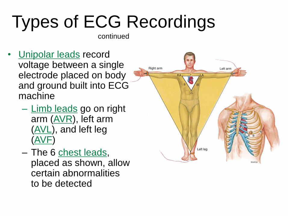
Types of ECG Recordings continued
• Unipolar leads record voltage between a single electrode placed on body and ground built into ECG machine
– Limb leads go on right arm (AVR), left arm (AVL), and left leg (AVF)
– The 6 chest leads, placed as shown, allow certain abnormalities to be detected
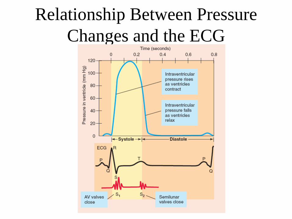
Relationship Between Pressure
Changes and the ECG
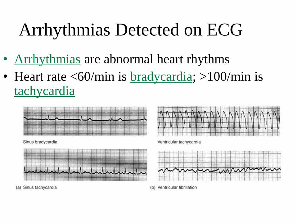
Arrhythmias Detected on ECG
• Arrhythmias are abnormal heart rhythms
• Heart rate <60/min is bradycardia; >100/min is tachycardia
In Summary
The contraction phase of the cardiac cycle is
called systole and the relaxation period is
called diastole.
The pacemaker of the heart is the SA node.
The average blood pressure during a cardiac
cycle is called mean arterial pressure.

In Summary
Blood pressure can be increased by one or all of the
following factors:
a. increase in blood volume
b. increase in heart rate
c. increased blood viscosity
d. increase in stroke volume
e. increased peripheral resistance
A recording of the electrical activity of the heart
during the cardiac cycle is called the
electrocardiogram (ECG).
• Video for measuring ECG
– http://www.youtube.com/watch?v=fOlKUAj1UQo